time will tell: early defibrillation in the … · time will tell: early defibrillation in ... the...
TRANSCRIPT
1
TIME WILL TELL: EARLY DEFIBRILLATION IN THE HOSPITAL
By Judy Boehm, RN, MSN
Part I
An article published January 3rd
in the New England Journal of Medicine caught the attention of
the media.1 Chan et al. reported on “Delayed Time to Defibrillation after In-hospital Cardiac
Arrest”, using data from the National Registry of Cardiopulmonary Resuscitation (NRCPR).
Their conclusions were that 1) delayed defibrillation (longer than 2 minutes after arrest is
reported) is common in hospital arrests, and 2) delayed defibrillation is associated with lower
rates of survival. Are you surprised at these findings? I am not surprised and could reach the
same conclusions based on my experience as a cardiac nurse specialist in a major tertiary care
institution. In this March newsletter I will describe the findings of this study, review the public‟s
response to the study, explore what we know about the science of ventricular fibrillation (VF)
and defibrillation, and give a brief overview of hospital arrests in general from the NRCPR data
– the background information. In next month‟s newsletter I will describe factors that influence
the success of defibrillation and end with ideas on how we can change care and technology to
improve time to defibrillation in the hospital.
Delayed Time to Defibrillation after In-hospital Arrest
The NRCPR is a multicenter registry of in-hospital cardiac arrests that is
sponsored by the American Heart Association (AHA) and managed by
Digital Innovation, Inc. Standardized definitions that are used for data collection arise from the
AHA Scientific Statement “Recommended guidelines for reviewing, reporting, and conducting
research on in-hospital resuscitation: the in-hospital „Utstein style‟.”2 Since the NRCPR began
in 2000, more than 120,000 cardiopulmonary resuscitation events have been entered by over 500
hospitals in the U.S., Canada, Germany, Japan, and Brazil. The NRCPR data has been helpful to
assess processes of care and outcomes during in-hospital cardiac arrest, defined as cessation of
cardiac mechanical activity that is determined by the absence of a palpable central pulse, apnea,
and unresponsiveness. The time to defibrillation is calculated from the reported time of
recognition of the arrest to the reported time of the first attempted defibrillation.
2
Chan‟s report investigated 6789 VF/pulseless ventricular tachycardia (VT) adult cardiac arrests
from January 1, 2000 through July 31, 2005 from 369 acute care hospitals. Data was excluded
for other than the original arrest, patients with implanted cardioverter defibrillators (ICDs), those
receiving intravenous infusions of epinephrine and antiarrhythmics, and those with missing time
data or inconsistent times. Cases were included only from the intensive care units (ICUs) and
inpatient units. Overall, the median time to defibrillation was reported as 1 minute. The time to
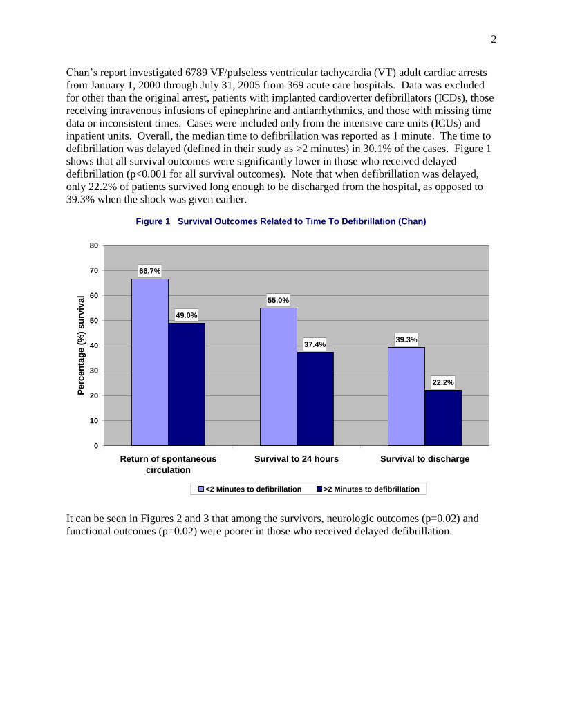
defibrillation was delayed (defined in their study as >2 minutes) in 30.1% of the cases. Figure 1
shows that all survival outcomes were significantly lower in those who received delayed
defibrillation (p<0.001 for all survival outcomes). Note that when defibrillation was delayed,
only 22.2% of patients survived long enough to be discharged from the hospital, as opposed to
39.3% when the shock was given earlier.
Figure 1 Survival Outcomes Related to Time To Defibrillation (Chan)
66.7%
55.0%
39.3%
49.0%
37.4%
22.2%
0
10
20
30
40
50
60
70
80
Return of spontaneous
circulation
Survival to 24 hours Survival to discharge
Pe
rce
nta
ge
(%
) s
urv
iva
l
<2 Minutes to defibrillation >2 Minutes to defibrillation
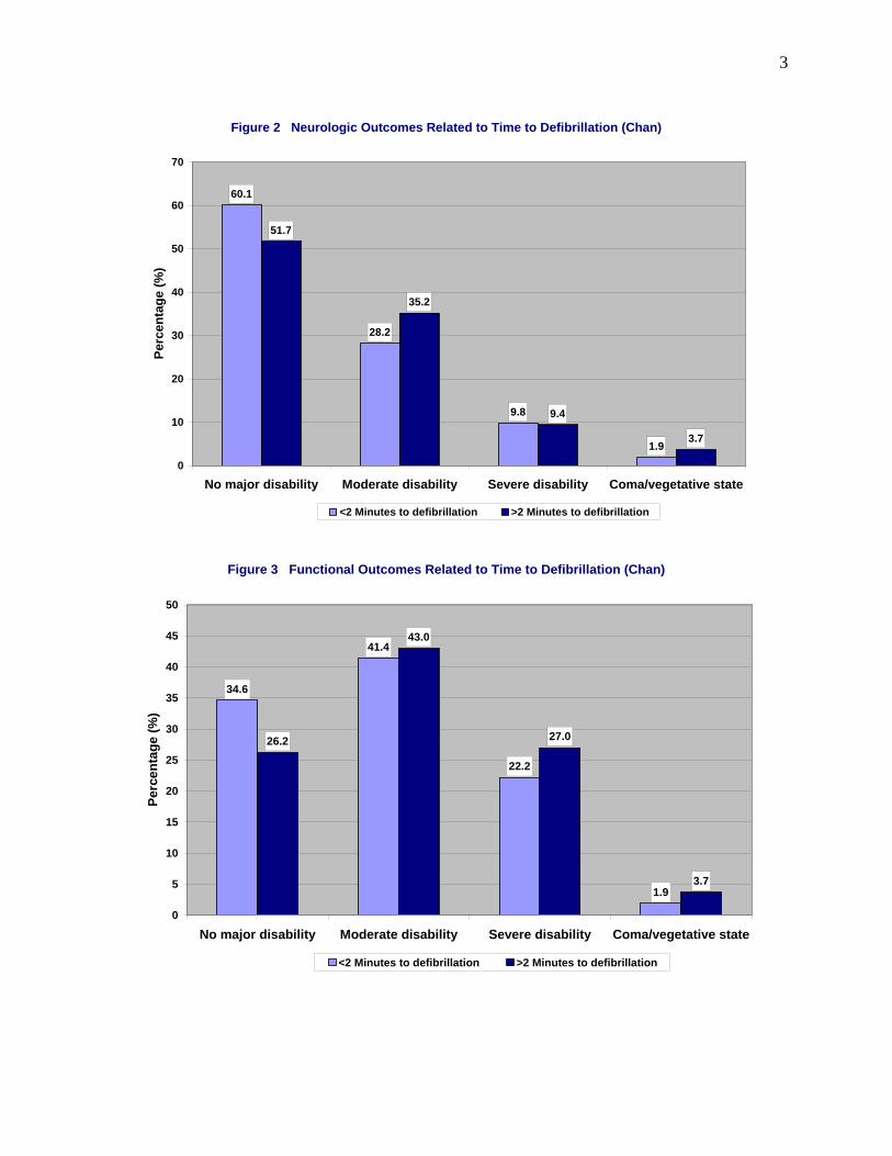
It can be seen in Figures 2 and 3 that among the survivors, neurologic outcomes (p=0.02) and
functional outcomes (p=0.02) were poorer in those who received delayed defibrillation.
3
Figure 2 Neurologic Outcomes Related to Time to Defibrillation (Chan)
60.1
28.2
9.8
1.9
51.7
35.2
9.4
3.7
0
10
20
30
40
50
60
70
No major disability Moderate disability Severe disability Coma/vegetative state
Pe
rce
nta
ge
(%
)
<2 Minutes to defibrillation >2 Minutes to defibrillation
Figure 3 Functional Outcomes Related to Time to Defibrillation (Chan)
41.4
22.2
1.9
26.2
43.0
27.0
3.7
34.6
0
5
10
15
20
25
30
35
40
45
50
No major disability Moderate disability Severe disability Coma/vegetative state
Perc
en
tag
e (
%)
<2 Minutes to defibrillation >2 Minutes to defibrillation
4
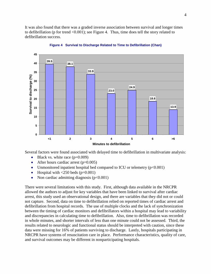
It was also found that there was a graded inverse association between survival and longer times
to defibrillation (p for trend <0.001); see Figure 4. Thus, time does tell the story related to
defibrillation success.
Figure 4 Survival to Discharge Related to Time to Defibrillation (Chan)
39.538.1
33.9
23.0
24.9
18.6
13.9
0
5
10
15
20
25
30
35
40
45
<1 2 3 4 5 6 >6
Minutes to defibrillation
Su
rviv
al
to d
isc
ha
rge
(%
)
Several factors were found associated with delayed time to defibrillation in multivariate analysis:
Black vs. white race (p=0.009)
After hours cardiac arrest (p=0.005)
Unmonitored inpatient hospital bed compared to ICU or telemetry (p<0.001)
Hospital with <250 beds (p=0.001)
Non cardiac admitting diagnosis (p<0.001)
There were several limitations with this study. First, although data available in the NRCPR
allowed the authors to adjust for key variables that have been linked to survival after cardiac
arrest, this study used an observational design, and there are variables that they did not or could
not capture. Second, data on time to defibrillation relied on reported times of cardiac arrest and
defibrillation from hospital records. The use of multiple clocks and the lack of synchronization
between the timing of cardiac monitors and defibrillators within a hospital may lead to variability
and discrepancies in calculating time to defibrillation. Also, time to defibrillation was recorded
in whole minutes, and shorter intervals of less than one minute could not be assessed. Third, the
results related to neurologic and functional status should be interpreted with caution, since these
data were missing for 16% of patients surviving to discharge. Lastly, hospitals participating in
NRCPR have systems of resuscitation care in place. Performance characteristics, quality of care,
and survival outcomes may be different in nonparticipating hospitals.
5
Chan‟s study is unique in that it reports on only patients with VF/VT arrests and excludes those
with ICDs and antiarrhythmics. Also, the study population is the largest ever recorded for a
study of VF in the hospital.
Public Response to This Study
In an editorial that accompanies the above study, Leslie Saxon, MD, points out that physicians
spend a lot of time devising strategies to improve survival for out-of-hospital cardiac arrests, but
efforts should now be refocused on improving outcomes for hospitalized patients.3
Approximately 225,000 out-of-hospital cardiac arrests occur annually in the United States. It is a
little-known fact that at least double that number of cardiac arrests occur in hospitalized patients.
“It is probably fair to say that most patients assume – unfortunately, incorrectly – that a hospital
would be the best place to survive a cardiac arrest,” she writes.
The public have heard that survival from out-of-hospital cardiac arrest has greatly increased
where automated external defibrillators (AEDs) are readily available and used quickly.
Valenzuela reported in 2000 a 53% survival to discharge from the hospital in the setting of U.S.
casinos with security cameras when AEDs were used by security personnel.4 The time interval
from collapse to attachment of the AED in witnessed events was 3.5 + 2.9 minutes, and the
interval from collapse to delivery of the first defibrillation shock was 4.4 + 2.9 minutes -
compared to the interval from collapse to arrival of paramedics at 9.8 + 4.3 minutes. The
survival to hospital discharge rate was 74% for those who received their first defibrillation no
later than 3 minutes after a witnessed collapse and 49% for those who received their first
defibrillation after more than 3 minutes.
American Airlines began equipping its aircraft with AEDs starting in 1997. In the same 2000
issue of New England Journal of Medicine, data was reported on all 200 instances (191 on
aircraft and 9 in the terminal) in which AEDs were used up until July 15, 1999.5 Of those for
whom an AED was used, 99 persons had loss of consciousness and a shock was advised in the
14 patients with VF. The first shock successfully defibrillated the heart in 13 patients, and in one
patient the shock was withheld upon family request. The rate of survival to discharge from the
hospital after shock with the AED was 40%, all with intact neurologic function.
After hearing about these remarkable results with the use of AEDs in the out-of-hospital setting,
the public response was amazement to learn from Chan‟s article that only 34.1% of patients
experiencing cardiac arrest in the hospital survived to discharge. Yes, the hospital cohort has
been admitted with medical problems, but can‟t we do better in a setting where competent
practitioners and emergency equipment are readily available for medical emergencies? How can
1/3 of hospitalized patients with VF/VT arrests have a shock delivered later than 2 minutes? The
New York Times stated that “in the real world, doctors and nurses do not always run fast
enough.”6
Dr. Saxon, speaking on her cell phone to the New York Times correspondent, said
“You‟re better off having your arrest at Nordstrom, where I‟m standing right now, because there
are 15 people around me.”
The Science of Ventricular Fibrillation and Defibrillation
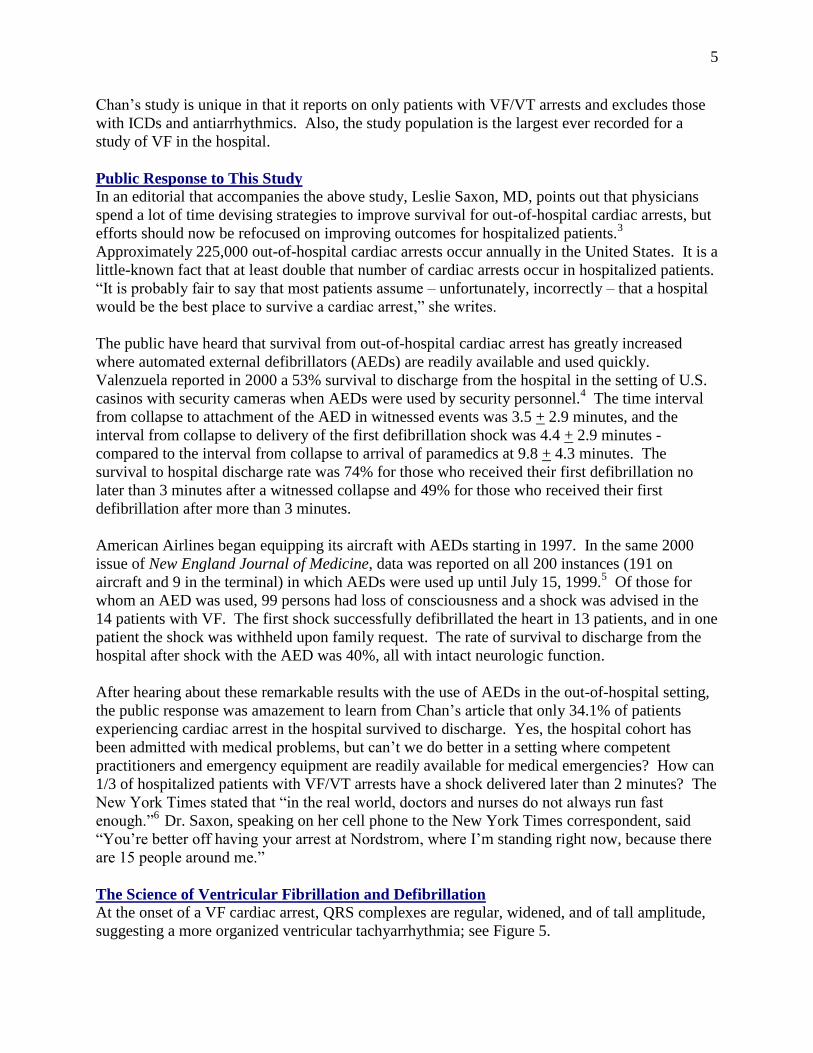
At the onset of a VF cardiac arrest, QRS complexes are regular, widened, and of tall amplitude,
suggesting a more organized ventricular tachyarrhythmia; see Figure 5.
6
Figure 5 Ventricular Tachycardia
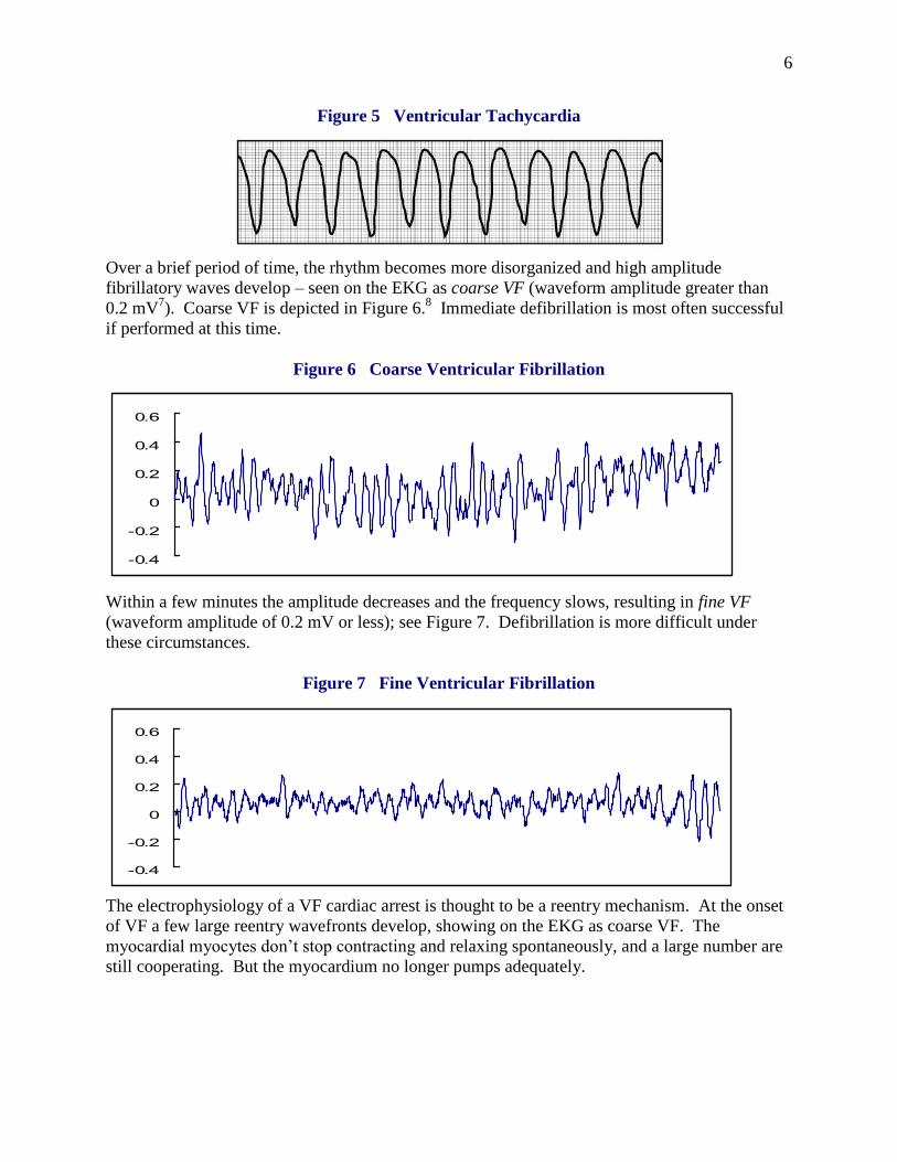
Over a brief period of time, the rhythm becomes more disorganized and high amplitude
fibrillatory waves develop – seen on the EKG as coarse VF (waveform amplitude greater than
0.2 mV7). Coarse VF is depicted in Figure 6.
8 Immediate defibrillation is most often successful
if performed at this time.
Figure 6 Coarse Ventricular Fibrillation
Within a few minutes the amplitude decreases and the frequency slows, resulting in fine VF
(waveform amplitude of 0.2 mV or less); see Figure 7. Defibrillation is more difficult under
these circumstances.
Figure 7 Fine Ventricular Fibrillation
The electrophysiology of a VF cardiac arrest is thought to be a reentry mechanism. At the onset
of VF a few large reentry wavefronts develop, showing on the EKG as coarse VF. The
myocardial myocytes don‟t stop contracting and relaxing spontaneously, and a large number are
still cooperating. But the myocardium no longer pumps adequately.
-0.4
-0.2
0
0.2
0.4
0.6
-0.4
-0.2
0
0.2
0.4
0.6
7
Over time more frequent and smaller wavefronts develop and the myocytes contract
independently. There is no coordinated mechanical activity of the ventricles and thus no
effective ventricular contraction. This shows on the EKG as fine VF.
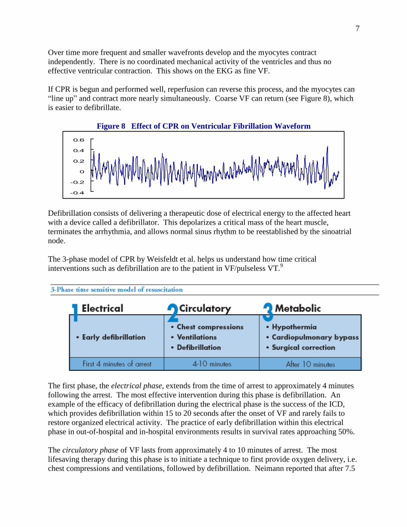
If CPR is begun and performed well, reperfusion can reverse this process, and the myocytes can
“line up” and contract more nearly simultaneously. Coarse VF can return (see Figure 8), which
is easier to defibrillate.
Figure 8 Effect of CPR on Ventricular Fibrillation Waveform
Defibrillation consists of delivering a therapeutic dose of electrical energy to the affected heart
with a device called a defibrillator. This depolarizes a critical mass of the heart muscle,
terminates the arrhythmia, and allows normal sinus rhythm to be reestablished by the sinoatrial
node.
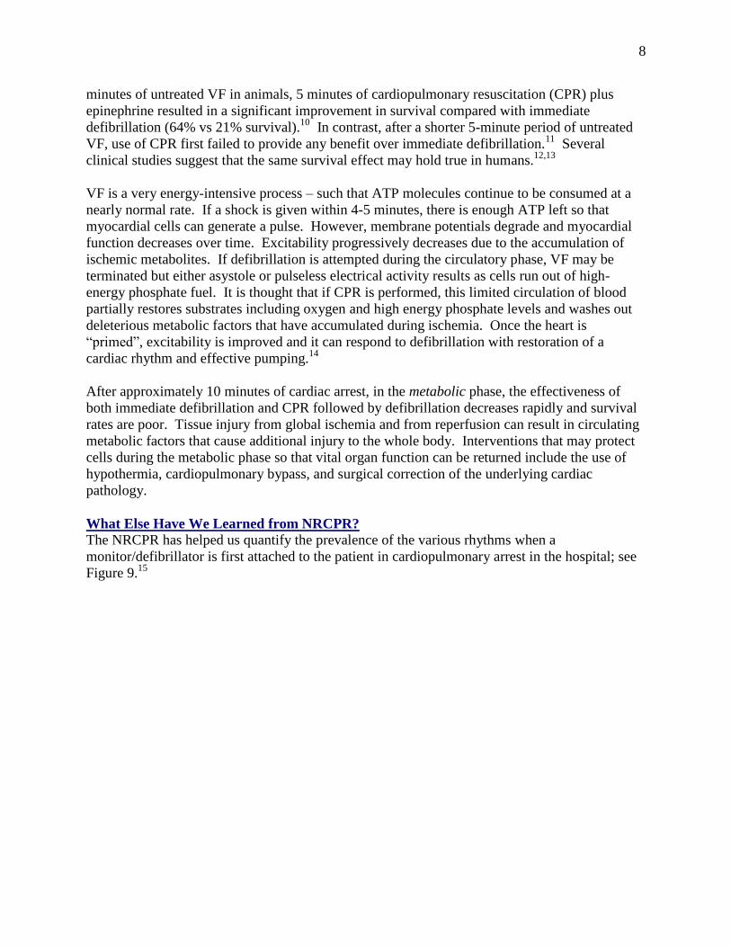
The 3-phase model of CPR by Weisfeldt et al. helps us understand how time critical
interventions such as defibrillation are to the patient in VF/pulseless VT.9
The first phase, the electrical phase, extends from the time of arrest to approximately 4 minutes
following the arrest. The most effective intervention during this phase is defibrillation. An
example of the efficacy of defibrillation during the electrical phase is the success of the ICD,
which provides defibrillation within 15 to 20 seconds after the onset of VF and rarely fails to
restore organized electrical activity. The practice of early defibrillation within this electrical
phase in out-of-hospital and in-hospital environments results in survival rates approaching 50%.
The circulatory phase of VF lasts from approximately 4 to 10 minutes of arrest. The most
lifesaving therapy during this phase is to initiate a technique to first provide oxygen delivery, i.e.
chest compressions and ventilations, followed by defibrillation. Neimann reported that after 7.5
-0.4
-0.2
0
0.2
0.4
0.6
8
minutes of untreated VF in animals, 5 minutes of cardiopulmonary resuscitation (CPR) plus
epinephrine resulted in a significant improvement in survival compared with immediate
defibrillation (64% vs 21% survival).10
In contrast, after a shorter 5-minute period of untreated
VF, use of CPR first failed to provide any benefit over immediate defibrillation.11
Several
clinical studies suggest that the same survival effect may hold true in humans.12,13
VF is a very energy-intensive process – such that ATP molecules continue to be consumed at a
nearly normal rate. If a shock is given within 4-5 minutes, there is enough ATP left so that
myocardial cells can generate a pulse. However, membrane potentials degrade and myocardial
function decreases over time. Excitability progressively decreases due to the accumulation of
ischemic metabolites. If defibrillation is attempted during the circulatory phase, VF may be
terminated but either asystole or pulseless electrical activity results as cells run out of high-
energy phosphate fuel. It is thought that if CPR is performed, this limited circulation of blood
partially restores substrates including oxygen and high energy phosphate levels and washes out
deleterious metabolic factors that have accumulated during ischemia. Once the heart is
“primed”, excitability is improved and it can respond to defibrillation with restoration of a
cardiac rhythm and effective pumping.14
After approximately 10 minutes of cardiac arrest, in the metabolic phase, the effectiveness of
both immediate defibrillation and CPR followed by defibrillation decreases rapidly and survival
rates are poor. Tissue injury from global ischemia and from reperfusion can result in circulating
metabolic factors that cause additional injury to the whole body. Interventions that may protect
cells during the metabolic phase so that vital organ function can be returned include the use of
hypothermia, cardiopulmonary bypass, and surgical correction of the underlying cardiac
pathology.
What Else Have We Learned from NRCPR?
The NRCPR has helped us quantify the prevalence of the various rhythms when a
monitor/defibrillator is first attached to the patient in cardiopulmonary arrest in the hospital; see
Figure 9.15
9
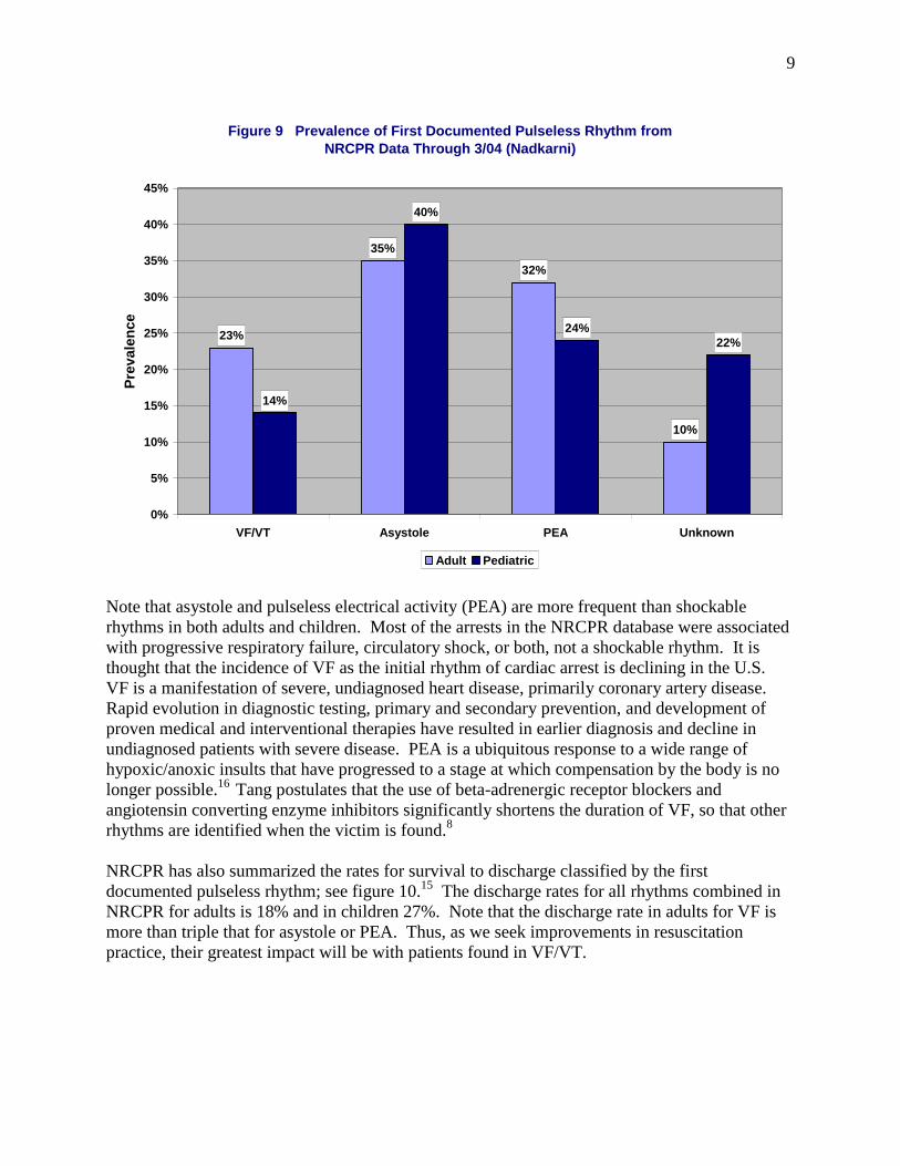
Figure 9 Prevalence of First Documented Pulseless Rhythm from
NRCPR Data Through 3/04 (Nadkarni)
23%
35%
32%
10%
14%
40%
24%
22%
0%
5%
10%
15%
20%
25%
30%
35%
40%
45%
VF/VT Asystole PEA Unknown
Pre
va
len
ce
Adult Pediatric
Note that asystole and pulseless electrical activity (PEA) are more frequent than shockable
rhythms in both adults and children. Most of the arrests in the NRCPR database were associated
with progressive respiratory failure, circulatory shock, or both, not a shockable rhythm. It is
thought that the incidence of VF as the initial rhythm of cardiac arrest is declining in the U.S.
VF is a manifestation of severe, undiagnosed heart disease, primarily coronary artery disease.
Rapid evolution in diagnostic testing, primary and secondary prevention, and development of
proven medical and interventional therapies have resulted in earlier diagnosis and decline in
undiagnosed patients with severe disease. PEA is a ubiquitous response to a wide range of
hypoxic/anoxic insults that have progressed to a stage at which compensation by the body is no
longer possible.16
Tang postulates that the use of beta-adrenergic receptor blockers and
angiotensin converting enzyme inhibitors significantly shortens the duration of VF, so that other
rhythms are identified when the victim is found.8
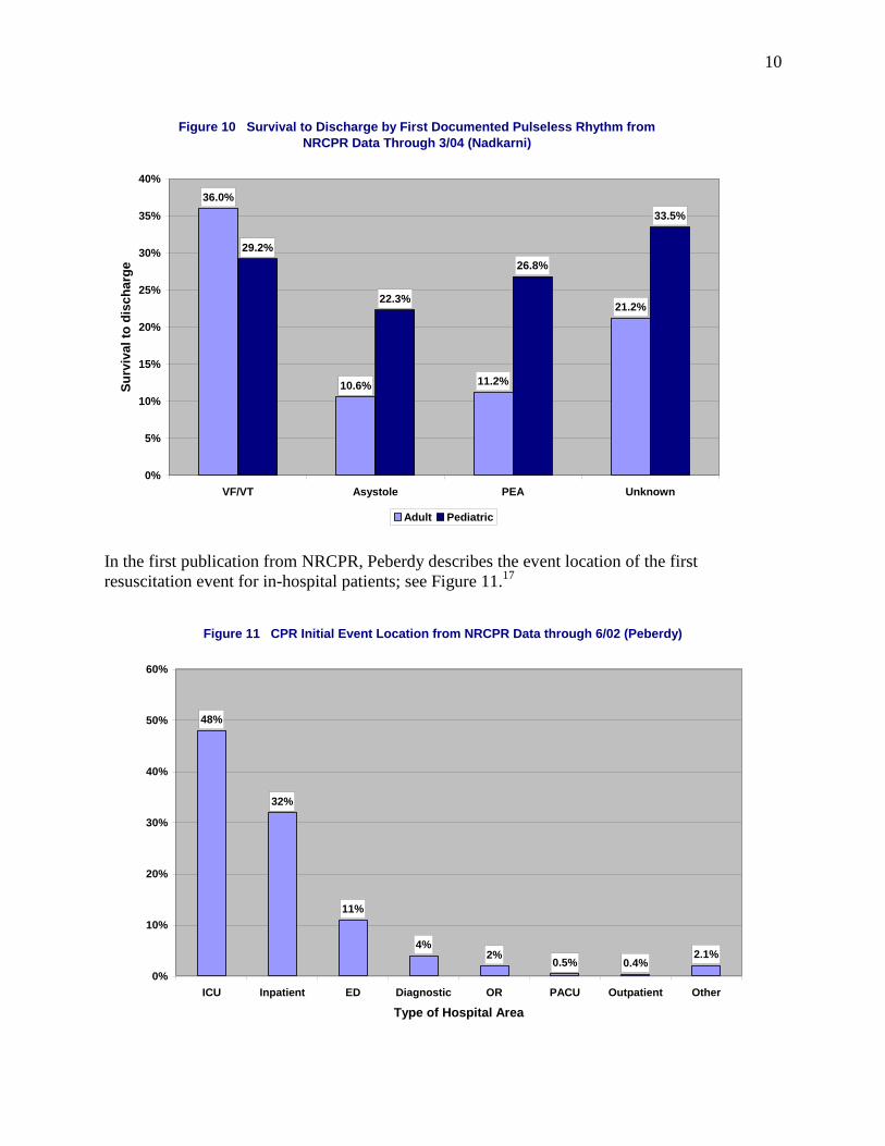
NRCPR has also summarized the rates for survival to discharge classified by the first
documented pulseless rhythm; see figure 10.15
The discharge rates for all rhythms combined in
NRCPR for adults is 18% and in children 27%. Note that the discharge rate in adults for VF is
more than triple that for asystole or PEA. Thus, as we seek improvements in resuscitation
practice, their greatest impact will be with patients found in VF/VT.
10
Figure 10 Survival to Discharge by First Documented Pulseless Rhythm from
NRCPR Data Through 3/04 (Nadkarni)
36.0%
10.6% 11.2%
21.2%
29.2%
22.3%
26.8%
33.5%
0%
5%
10%
15%
20%
25%
30%
35%
40%
VF/VT Asystole PEA Unknown
Su
rviv
al
to d
isch
arg
e
Adult Pediatric
In the first publication from NRCPR, Peberdy describes the event location of the first
resuscitation event for in-hospital patients; see Figure 11.17
Figure 11 CPR Initial Event Location from NRCPR Data through 6/02 (Peberdy)
48%
32%
11%
4%2%
0.5% 0.4%2.1%
0%
10%
20%
30%
40%
50%
60%
ICU Inpatient ED Diagnostic OR PACU Outpatient Other
Type of Hospital Area
11
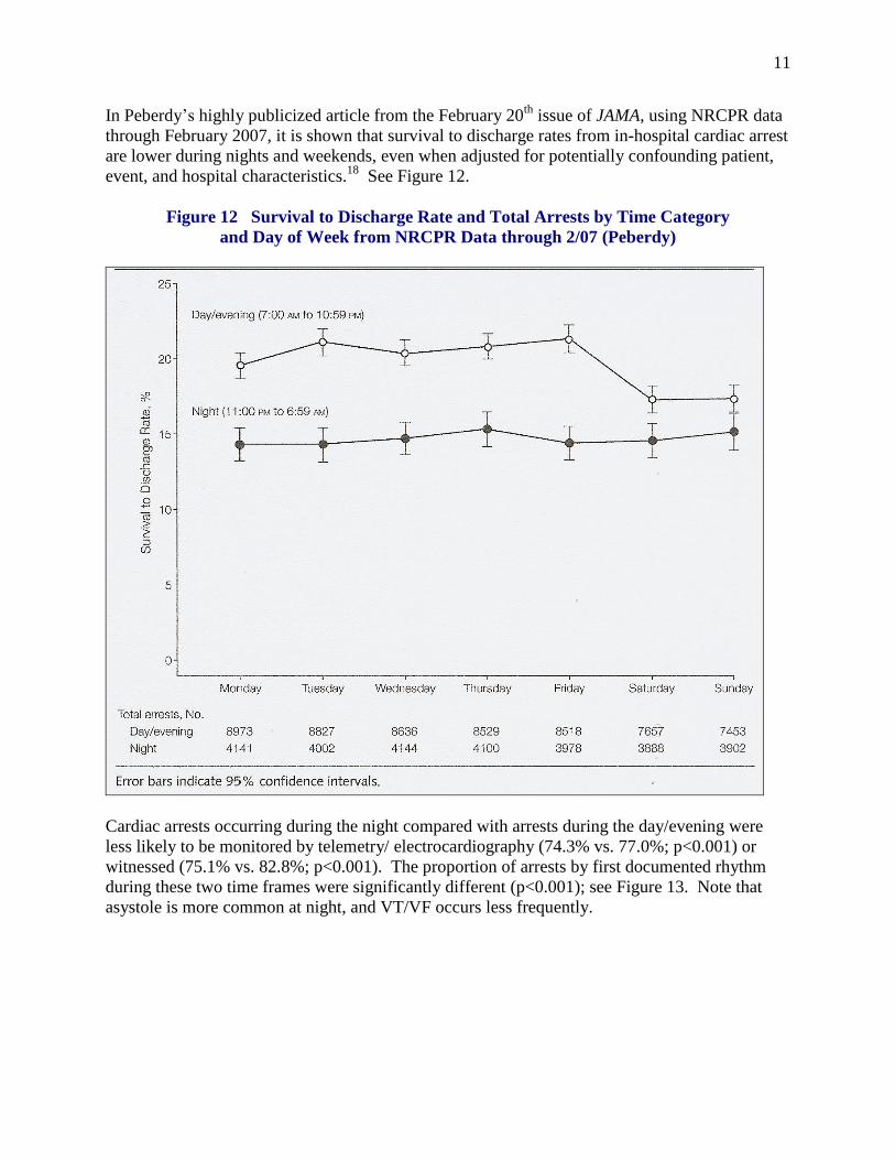
In Peberdy‟s highly publicized article from the February 20th
issue of JAMA, using NRCPR data
through February 2007, it is shown that survival to discharge rates from in-hospital cardiac arrest
are lower during nights and weekends, even when adjusted for potentially confounding patient,
event, and hospital characteristics.18
See Figure 12.
Figure 12 Survival to Discharge Rate and Total Arrests by Time Category
and Day of Week from NRCPR Data through 2/07 (Peberdy)
Cardiac arrests occurring during the night compared with arrests during the day/evening were
less likely to be monitored by telemetry/ electrocardiography (74.3% vs. 77.0%; p<0.001) or
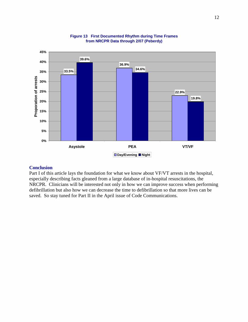
witnessed (75.1% vs. 82.8%; p<0.001). The proportion of arrests by first documented rhythm
during these two time frames were significantly different (p<0.001); see Figure 13. Note that
asystole is more common at night, and VT/VF occurs less frequently.
12
Figure 13 First Documented Rhythm during Time Frames
from NRCPR Data through 2/07 (Peberdy)
33.5%
36.9%
22.9%
19.8%
34.6%
39.6%
0%
5%
10%
15%
20%
25%
30%
35%
40%
45%
Asystole PEA VT/VF
Pro
po
rati
on
of
arr
es
ts
Day/Evening Night
Conclusion
Part I of this article lays the foundation for what we know about VF/VT arrests in the hospital,
especially describing facts gleaned from a large database of in-hospital resuscitations, the
NRCPR. Clinicians will be interested not only in how we can improve success when performing
defibrillation but also how we can decrease the time to defibrillation so that more lives can be
saved. So stay tuned for Part II in the April issue of Code Communications.
13
References 1 Chan, P.S. et al. Delayed time to defibrillation after in-hospital cardiac arrest. New England Journal of
Medicine 2008;358:9-17.
2 Cummins, R.O. et al. In-hospital resuscitation. Recommended guidelines for reviewing, reporting, and
conducting research on in-hospital resuscitation: the in-hospital „Utstein style‟. Circulation
1997;95:2211-2239.
3 Saxon, L.A. Survival after tachyarrhythmic arrest – What are we waiting for? New England Journal of
Medicine 2008;358:77-79.
4 Valenzuela, T.D. et al. Outcomes of rapid defibrillation by security officers after cardiac arrest in casinos.
New England Journal of Medicine 2000;343:1206-1209.
5 Page, R.L. et al. Use of automated external defibrillators by a U.S. airline. New England Journal of
Medicine 2000;343:1210-1216.
6 New York Times. Hospitals slow in heart cases, research finds. From nytimes.com on January 3, 2008.
7 Weaver, W.D. et al. Amplitude of ventricular fibrillation waveform and outcome after cardiac arrest.
Annals of Internal Medicine 1985;102:53-55.
8 Tang, W. Go with the flow: Blood flow and its importance in improving resuscitation. Presented at 8th
Scientific Congress of the European Resuscitation Council, Stavanger, Norway on May 11, 2006.
9 Weisfeldt, M.L., & Becker, L.B. Resuscitation after cardiac arrests. A 3-phase time-sensitive model.
JAMA 2002;288:3035-3038.
10 Neimann, J. T. et al. Treatment of prolonged ventricular fibrillation. Circulation 1992;85:281-287.
11 Niemann, J. T. et al. Immediate countershock versus cardiopulmonary resuscitation before countershock in
a 5-minute swine model of ventricular fibrillation arrest. Annals of Emergency Medicine 2000;36:533-
536.
12 Cobb, L. A. et al. Influence of cardiopulmonary resuscitation prior to defibrillation in patients with out-of-
hospital ventricular fibrillation. JAMA 1999;281:1182-1188.
13 Wik, L. et al. Three minutes of basic cardiopulmonary resuscitation of pre-hospital ventricular fibrillation
patients before defibrillation increases the number of patients who survive to hospital discharge, and one
year survival. Circulation 2002;106(suppl II):A1823.
14 Abella, B.S. We do good CPR – right? Presented at 8th
Scientific Congress of the European Resuscitation
Council, Stavanger, Norway on May 11, 2006.
15 Nadkarni, V. M. et al. First documented rhythm and clinical outcome from in-hospital cardiac arrest
among children and adults. JAMA 2006;295:50-57.
16 Parish, D.C., Chandra, K.M. D., & Dane, F.C. Success changes the problem: Why ventricular fibrillation
is declining, why pulseless electrical activity is emerging, and what to do about it. Resuscitation
2003;58:31-35.
17 Peberdy, M. A. et al. Cardiopulmonary resuscitation of adults in the hospital: A report of 14720 cardiac
arrests from the National Registry of Cardiopulmonary Resuscitation. Resuscitation 2003;58:297-308.
18 Peberdy, M.A. et al. Survival from in-hospital cardiac arrest during nights and weekends. JAMA
2008;299:785-792.