time ribosomal translocation in real - diva...
TRANSCRIPT

ACTAUNIVERSITATIS
UPSALIENSISUPPSALA
2019
Digital Comprehensive Summaries of Uppsala Dissertationsfrom the Faculty of Science and Technology 1877
Ribosomal translocation in realtime
Method development to Applications
CHANG-IL KIM
ISSN 1651-6214ISBN 978-91-513-0808-1urn:nbn:se:uu:diva-396197

Dissertation presented at Uppsala University to be publicly examined in B7:101a, BiomedicalCentrum (BMC), Husargatan 3, Uppsala, Friday, 29 November 2019 at 13:15 for the degreeof Doctor of Philosophy. The examination will be conducted in English. Faculty examiner:Professor Martin Ott (Department of Biochemistry and Biophysics, Stockholm University).
AbstractKim, C.-i. 2019. Ribosomal translocation in real time. Method development to Applications.Digital Comprehensive Summaries of Uppsala Dissertations from the Faculty of Science andTechnology 1877. 52 pp. Uppsala: Uppsala University. ISBN 978-91-513-0808-1.
Translational elongation is the process in which the ribosome adds one amino acid at a timeto the nascent peptide chain. As the ribosome elongates the peptide chain by 14 - 20 aminoacids per second and performs hundreds of such cycles per protein, ‘elongation’ is one ofthe most crucial steps in translation. During elongation, the ribosome must move preciselyby one codon along the mRNA after peptidyl transfer. This step is known as ‘ribosomaltranslocation’, which is catalyzed by the elongation factor G (EF-G). Despite extensive research,the exact sequence of events in translocation is still unclear. Thus, development of an in vitroassay, which would allow precise kinetic measurement of the steps involved in ribosomaltranslocation, is highly important. In 2003, a research group led by Simpson Joseph designed afluorescence-based assay to monitor translocation in stopped-flow using a short mRNA labeledwith fluorescent-dye pyrene at the 3’ end. Although optimized for the highest fluorescencechange, this assay showed significantly slower translocation than what has been measured byconventional translocation assays. Moreover, when performed with a popular peptidyl tRNAanalog, it produced complex multiphasic kinetics. Therefore, this assay was not only limitedfor deriving physiologically relevant rates, but also the analysis of the rates from the kineticdata was challenging. In this thesis, we have optimized the fluorescent-mRNA based stopped-flow translocation assay, by carefully calibrating it with the functional tripeptide formationassay performed in quench-flow. First, we identified the most suitable mRNA length that isoptimal for both the fluorescence signal and the rate of translocation. Further, by systematicallyvarying temperature, Mg2+, EF-G and tRNA analog concentration, we have derived the idealcondition for obtaining near monophasic kinetics in stopped-flow, which allows determinationof the translocation rate in an unambiguous manner. This optimized assay has further beentested in different contexts involving translocation, which include (i) characterization of theGTPase deficient EF-G mutants, (ii) studying the effect of non-hydrolyzable GTP analogs, (iii)evaluating the effect of extension of the C-terminal tail of the ribosomal protein S13 in thedecoding center, and (iv) understanding the mechanism of action of a novel aminoglycosideantibiotic ‘arbekacin’ in translation. These studies altogether provide deep understanding forhow different factors can modulate ribosomal translocation.
Keywords: ribosome, EF-G, pyrene mRNA, aminoglycoside, ribosomal protein, GTPhydrolysis
Chang-il Kim, Department of Cell and Molecular Biology, Box 596, Uppsala University,SE-75124 Uppsala, Sweden.
© Chang-il Kim 2019
ISSN 1651-6214ISBN 978-91-513-0808-1urn:nbn:se:uu:diva-396197 (http://urn.kb.se/resolve?urn=urn:nbn:se:uu:diva-396197)


List of Papers
This thesis is based on the following papers, which are referred to in the text by their Roman numerals.
I. Kim, C., Holm, M., Sanyal, S., (2019) Optimization of a fluores-cent-mRNA based real-time assay for precise kinetic measure-ments of ribosomal translocation. (Submitted Manuscript)
II. Kim, C., Majumdar, S., Mandava, C.S., Sanyal, S., (2019) Char-acterization of two conserved Arg mutations in elongation factor G: Implications in ribosomal translocation. (Manuscript)
III. Kim, C., Koripella R., Ge, X., Holm, M., Sanyal, S., (2019) An extended C-terminal tail of the ribosomal protein S13 modulates the speed of ribosomal translocation. (Submitted Manuscript)
IV. Parajuli, N., Kim, C., Sanyal, S., (2019) Molecular mechanism of inhibition of bacterial protein synthesis by a novel aminoglyco-side arbekacin. (Manuscript)

Contents
Introduction ................................................................................................... 11
Bacterial Ribosome .................................................................................. 11
Ribosomal translation ............................................................................... 12
Initiation............................................................................................... 13
Elongation ............................................................................................ 14
Termination and Recycling.................................................................. 17
Ribosome as a target of Antibiotics ......................................................... 19
Elongation factor G .................................................................................. 21
In vitro kinetic tools for studying translational elongation ...................... 22
Aim of the thesis ...................................................................................... 25
The present work........................................................................................... 26
Optimization of pyrene-labeled mRNA based translocation assay .......... 26
Optimization of pyrene-mRNA for functional kinetic studies in quench-flow ......................................................................................... 27
Translocation with pre-T complex containing NAc-Phe-tRNAPhe ...... 30
Optimization of the reaction conditions for better data analysis ......... 32
Application of optimized translocation assay .......................................... 35
Non-hydrolyzable GTP analogs in translocation ................................. 35
Possible importance of tRNA body in elongation ............................... 36
Investigation of EF-G mutants............................................................. 38
Role of the extended C-terminal tail of the ribosomal protein S13 in protein synthesis .................................................................................. 39
Antibiotic Arbekacin ........................................................................... 44
Conclusions and future perspective .............................................................. 46
Sammanfattning på Svenska ......................................................................... 47

Acknowledgements ....................................................................................... 49
Reference ...................................................................................................... 51

Abbreviations
DNA Deoxyribonucleic acid
RNA Ribonucleic acid
rRNA Ribosomal RNA
mRNA Messenger RNA
tRNA Transfer RNA
aa-tRNA Amino-acyl tRNA
r-protein Ribosomal protein
Mda Mega Dalton
kDa Kilo Dalton
Å Ångström
bps base pairs
nt nucleotide
PTC Peptidyl-Transferase Centre
SD Shine-Dalgarno sequence
IC Initiation Complex
Pre-T Pre translocation complex
Post-T Post translocation complex
A-site Amino-acyl site
P-site Peptidyl site
E-site Exit site
IF Initiation Factor
EF Elongation Factor
RF Release Factor
RRF Ribosome recycling Factor
CTD C terminal domain
NTD N terminal domain
GTP Guanosine triphosphate
GDP Guanosine diphosphate
ATP Adenosine triphosphate
GDPNP Guanylyl-5’-imidodiphosphate
GDPCP β,γ-Methyleneguanosine 5’-triphosphate
GTP-γ-S Guanosine 5’-[γ-thio] triphosphate
Pi Inorganic phosphate
FRET Fluorescence Resonance Energy Transfer
fMet Formyl Methionine
Met Methionine
Leu Leucine
Phe Phenylalanine
cryo-EM cryogenic Electron Microscopy
KI Inhibitory constant
KM Michaelis-Menten constant
KD Dissociation constant
HPLC High performance liquid chromatography
TLC Thin layer Chromatography

11
Introduction
Bacterial Ribosome All the living cells, by the definition of “life”, metabolize, grow, regulate and reproduce. All these “life” activities are progressed and controlled by the ge-netic information encoded in the sequence of deoxyribonucleic acid (DNA). The information in DNA should be transcribed into messenger RNA (mRNA), followed by translation into functional proteins, which catalyze the metabo-lism inside the cells, regulate the cell signaling and ligand binding or provide the cellular structure for the cells to “live”. The whole process of transferring information of DNA into proteins is known as the ‘central dogma’ of molec-ular biology (Crick, 1970).
Ribosomes are large macromolecular machines that translate the genetic in-formation encoded in mRNAs into the functional proteins. In both prokaryotes and eukaryotes, ribosomes are composed of two unequal subunits, which fur-ther comprise ribosomal RNAs (rRNA) and ribosomal proteins (r-proteins). Based on the sedimentation coefficient (S), the bacterial ribosome is called the 70S ribosome, while the two subunits are called 50S (large), 30S (small) sub-unit respectively (Figure 1). The 50S subunit is composed of 23S rRNA (2900 nucleotides), 5S rRNA (120 nucleotides) and 31 proteins; the 30S subunit is composed of 16S rRNA (1540 nucleotides) and 21 proteins. Both subunits of the ribosome contain three tRNA (transfer RNA) binding sites, called the A, P, and E sites. The A site and the P site are the binding sites of the aminoacyl tRNA and peptidyl tRNA, respectively. The E site stands for ‘exit site’, from which the deacylated tRNAs exit the ribosome (Figure 1).
The ribosome contains two important functional sites; decoding center (DC) in the 30S subunit and peptidyl transfer center (PTC) in the 50S subunit (Fig-ure 1). While DC maintains the fidelity of translation by accurate selection of the cognate tRNAs from the large pool of competing non-cognate and near-cognate tRNAs (Schmeing, Voorhees et al., 2009, Wilson & Nierhaus, 2003), PTC catalyzes efficient peptide bond formation, during which the polypeptide

12
is transferred from the P-site tRNA to the amino acid on the A-site tRNA. In Escherichia coli (E. coli), the PTC is comprised of three conserved bases U2585, A2451 and C2063 of 23S rRNA (E. coli numbering) while the DC is comprised of A1492, A1493 and G530 of 16S rRNA (Schmeing et al., 2009, Wilson & Nierhaus, 2003).
Figure 1. Structure of the bacterial ribosome (70S) and its large (50S) and small (30S) subunit depicting three tRNA binding sites (A, P, and E site) and the functional centers DC (decoding center) and PTC (peptidyl transferase center). The 23S rRNA is in silver, the r-proteins of the large subunit in purple, the 16S rRNA in blue-gray and the small subunit proteins are in green. The three tRNAs bound to the A, P and Esites are in red, the mRNA is magenta. The illustra-tions are based on cryo-EM structure of E. coli ribosome, adopted from PDB:5IQR.
Ribosomal translation Ribosomal translation involves four major steps; (i) initiation – during which the initiator tRNA binds to the P-site of the mRNA programmed 30S subunit and the ribosomal subunits assemble, (ii) elongation – during which an ami-noacyl tRNA binds to the mRNA at the A site, then peptide bond forms re-sulting in transfer of the peptide chain from the P-site tRNA to the A-site tRNA, and finally the peptidyl tRNA translocates to the P site along with the

13
mRNA thereby allowing a new round of elongation until a stop codon reaches the A site, (iii) termination – during which the nascent peptide chain is hydro-lyzed from the peptidyl tRNA and released from the ribosome and (iv) recy-cling – during which the ribosomal subunits dissociate and prepare for the next round of initiation. In all four steps, several protein factors, including GTP hydrolyzing factors or translational GTPases, cooperate with the ribosome to facilitate accurate and efficient translation (Figure 1). The major translation factors are initiation factors 1, 2 and 3 (IF1, IF2 and IF3), elongation factors -Tu, -Ts, -G and -P (EF-Tu, EF-Ts, EF-G and EF-P), release factors 1, 2 and 3 (RF1, RF2 and RF3) and ribosome recycling factor (RRF). Efficient and ac-curate translation depends on action of these translational factors in definite sequence, which is detailed below.
Initiation In bacteria, initiation of translation starts by binding of the mRNA to the 30S subunit. It is often facilitated by a specific sequence in the mRNA upstream the start codon (AUG), called the Shine-Dalgarno sequence (SD) (Shine & Dalgarno, 1974, Yusupov, Yusupova et al., 2001). The SD sequence interacts with a complimentary sequence at the 3’-end of 16S rRNA, which ensures the positioning of the mRNA in such a way that the start codon (usually AUG or GUG) is correctly placed in the P site. Correct positioning of the mRNA is important for efficient and accurate initiation; it allows binding of the initiator tRNA to the P site. The efficiency and accuracy of initiation often depend on concerted action of the three initiation factors. IF3 binds to the E site of the free 30S subunit in order to prevent premature and nonfunctional association of 50S with it. Similarly, IF1 occupies the A site of 30S subunit and prevents binding of the incoming aminoacyl-tRNA (aa-tRNA) as well as the initiator tRNA to the A site. IF2 is a GTPase factor. It binds to the 30S subunit as IF2•GTP and ensures stable binding of the initiator tRNA in the P site.
A special initiator tRNA is involved in prokaryotic translational initiation. This tRNA is charged with N-formylated methionine and is called as fMet-tRNAfMet. IF2 recognizes the fMet and enables specific binding of fMet-tRNAfMet to the P site (Antoun, Pavlov et al., 2006). Other features like 3G:C base pair motif in the anticodon stem loop of tRNAfMet confer discrimination against elongator tRNAMet (Varshney, Lee et al., 1993). Positioning of fMet-tRNAfMet with the help of IF2 on the P site (Roy, Liu et al., 2018) and the

14
correct formation of base pairing between the anticodon and the start codon completes the formation of 30S pre-initiation complex. The last step of initia-tion is to form 70S initiation complex (70S IC). This is achieved by association of the 50S subunit with the 30S pre-initiation complex. Here, IF2 constructs a docking site for the 50S by interacting with the L7/L12 stalk. GTP hydrolysis by IF2 is not needed for subunit association. However, GTP hydrolysis re-duces affinity of IF2 to the ribosome and fMet-tRNAfMet, and IF2•GDP disso-ciates. (Andreeva, Belardinelli et al., 2018, Mandava, Peisker et al., 2012). Although the exact sequence of the events is not fully established, the other initiation factors IF1 and IF3 also leave the complex. The finally formed 70S initiation complex containing fMet-tRNAfMet in the P site and empty A site enters into the elongation step of protein synthesis. The steps are summarized in Figure 2.
Elongation The ‘elongation’ step takes the longest time of all in overall synthesis of a protein, as this step repeats for every amino acid in the peptide chain until the ribosome reaches a stop codon (Figure 2). Elongation is divided into two sub steps; (i) decoding and peptidyl transfer, and (ii) coupled mRNA-tRNA trans-location. Two major translational GTPase factors, EF-Tu and EF-G, mediate these two steps. The GTPase factors alternate between the GTP bound active state and GDP bound inactive state. While EF-Ts is a guanosine nucleotide exchange factor (GEF) for EF-Tu, no protein factor has been identified as GEF for EF-G. It has been proposed that 70S ribosome might play a role as GEF for EF-G (Wieden, Gromadski et al., 2002, Zavialov, Hauryliuk et al., 2005a).
EF-Tu•GTP brings aa-tRNAs to the empty A site of the ribosome, where cog-nate tRNAs are accepted and near- and non-cognate tRNAs are rejected by the ribosome in the process called “initial selection” (Fislage, Zhang et al., 2018, Ieong, Uzun et al., 2016). During the delivery of aa-tRNA, tRNA anticodon stem loop interacts with the mRNA codon in the A site by complementary base pairing, while the other end of tRNA remains bound to EF-Tu to prevent peptide bond formation before correct decoding of the mRNA codon (Valle, Zavialov et al., 2003a). The DC of the ribosome has three universally con-served bases of 16S rRNA, A1492, A1493 and G530 (E. coli numbering), known as the ‘monitoring bases’. These bases flip-out upon binding of the aa-

15
tRNA and form hydrogen bonds with the minor groove formed by correct co-don-anticodon base pairing (Ogle, Brodersen et al., 2001). The adjacent ade-nines 1492 and 1493 interact with the first and second positions of the codon-anticodon pair, while G530 interacts with the second and third positions (Fislage et al., 2018, Loveland, Demo et al., 2017). Recent cryogenic electro-microscopy (cryo-EM) based structural studies demonstrate that the hydrogen bond network involving these three monitoring bases is the key to the correct aa-tRNA selection. When the cognate tRNA forms stable complementary base pairing with the A-site codon on mRNA, GTP hydrolysis is triggered in EF-Tu, which is followed by its release from the ribosome (Ogle, Carter et al., 2003, Rodnina, Gromadski et al., 2005). After EF-Tu•GDP release, the aa-tRNA moves and rotates about 70 Å so that its 3’-end can reach the PTC. This process is called “tRNA accommodation”. In some occasions near- or non-cognate tRNA can bypass initial selection. However, there is a high chance that these tRNAs fail in the accommodation step and are rejected. This addi-tional mode of accuracy, which allows rejection of the incorrect tRNAs even after GTP hydrolysis by EF-Tu is called “proof reading”.
After the accommodation of the aa-tRNA to the A site peptide bond is formed between the carboxyl group (-COOH) of the polypeptide at the P site and the amino group (-NH2) of the amino acid in the A site. The peptidyl transfer is a function of the 23S rRNA in the PTC, which makes the ribosome a ribozyme. After peptidyl transfer, the ribosome holds peptidyl tRNA in the A site and a deacylated tRNA in the P site. At this point, the ribosome is ready for translo-cation and the ribosomal complex is called a pre-translocation (pre-T) com-plex.
For the next step of peptide elongation, pre-T complex should translocate by one codon so that the tRNAs at the P site and A site move to the E and P site respectively. The pre-T complex spontaneously fluctuate between so called “hybrid” and “classical” state, primarily suggested by chemical foot printing assay (Jamiolkowski, Chen et al., 2017, Kim, Puglisi et al., 2007, Moazed & Noller, 1989) and then confirmed by single molecule studies (Fei, Bronson et al., 2009, Fei, Kosuri et al., 2008, Flis, Holm et al., 2018, Ning, Fei et al., 2014, Wasserman, Alejo et al., 2016). In the “classical” state, the tRNAs re-main in the A/A and P/P state with respect to both the 30S and the 50S subu-nits. However, in the “hybrid” state, 3’-end of the tRNAs bound to the A-site and P site of the 30S, move to the P site and E site in the 50S, respectively.

16
This is accompanied by the counter clockwise rotation of the ribosomal subu-nits with respect to each other by about 10°, which is known as “ribosome ratcheting” (Agirrezabala, Liao et al., 2012, Frank & Agrawal, 2000, Frank, Gao et al., 2007).
Figure 2. Overview of bacterial translation in four steps. Several translation factors cooperate along the four major steps, namely initiation, elongation, termination and recycling.
Current structural and functional studies suggest that “ribosome ratcheting” is the first step of ribosomal translocation, which ensures stable binding of EF-G•GTP on the ribosome. Translocation cannot happen if the ribosome ratch-eting was blocked (Horan & Noller, 2007). However, completion of translo-cation requires back ratcheting of the ribosomal subunits after GTP hydrolysis on EF-G (Chen, Tsai et al., 2012, Ermolenko & Noller, 2011, Frank et al., 2007, Ling & Ermolenko, 2016). Apart from overall subunit ratcheting, the

17
ribosome goes through extensive conformational changes during transloca-tion. One of the big rearrangements is the swiveling of the 30S head domain by about 20⁰ with respect to the rest of the 30S, which swivels back synchro-nously or after the mRNA-tRNA translocation (Guo & Noller, 2012, Ratje, Loerke et al., 2010, Wasserman et al., 2016). L1 stalk of the 50S subunits also moves in and out of the E site while interacting with the deacylated tRNA (Fei et al., 2008). Thus, ribosomal translocation requires orchestration of several complex events involving conformational dynamics of the ribosome, tRNAs and EF-G.
Efficient translocation requires EF-G, even though the ribosome can translo-cate at rather low rate in factor independent way (Cukras & Green, 2005, Gavrilova & Spirin, 1971, Pestka, 1968). EF-G is a GTPase protein and binds to the pre-T ribosome in GTP form. EF-G prefers binding to the ribosome at the hybrid state (Blanchard, Kim et al., 2004). Alternatively, it pushes the equilibrium of the classical/hybrid state to the hybrid state upon binding to the ribosome (Ermolenko, Majumdar et al., 2007, Fei et al., 2008, Kim et al., 2007, Munro, Altman et al., 2010a, Sharma, Adio et al., 2016, Spiegel, Ermolenko et al., 2007, Valle, Zavialov et al., 2003b). Prompt GTP hydrolysis by EF-G stabilizes the ratcheted conformation and enables EF-G•GDP to move close to A site in the 30S (Spiegel et al., 2007). As a result the A-site tRNA move to the P site together with the mRNA and the P-site tRNA also moves to the E site. The mRNA-tRNA movement is the heart of translocation and depends on GTP hydrolysis by EF-G. However, this is independent of release of inorganic pyrophosphate after GTP hydrolysis (Zavialov, Mora et al., 2002). As the mRNA-tRNAs translocate, ribosome return to the classical state by back-ratcheting. This movement is loosely coupled with back swivel-ing of the 30S head, moving in of the L1-stalk and release of the E-site tRNA (Cornish, Ermolenko et al., 2009, Ermolenko & Noller, 2011, Guo & Noller, 2012, Wasserman et al., 2016). In the back ratcheted state of the ribosome, EF-G•GDP loses its affinity to the ribosome and dissociates leaving empty A site for the next elongation of the peptide chain.
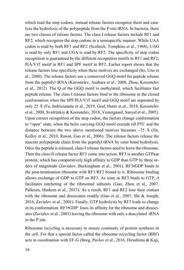
Termination and Recycling Peptide elongation by ribosome continues until it encounters one of the three stop codons UAA, UAG and UGA in the A site. There are no tRNAs available,
18
which read the stop codons, instead release factors recognize them and cata-lyze the hydrolysis of the polypeptide from the P-site tRNA. In bacteria, there are two classes of release factors. The class-I release factors include RF1 and RF2, which recognize the stop codons in a semispecific manner. While UAA codon is read by both RF1 and RF2 (Scolnick, Tompkins et al., 1968), UAG is read by only RF1 and UGA is read by RF2. The specificity of stop codon recognition is guaranteed by the different recognition motifs in RF1 and RF2; P(A/V)T motif in RF1 and SPF motif in RF2. Earlier report shows that the release factors lose specificity when these motives are exchanged (Ito, Uno et al., 2000). The release factors use a conserved GGQ motif for peptide release from the peptidyl tRNA (Korostelev, Asahara et al., 2008, Zhou, Korostelev et al., 2012). The Q of the GGQ motif is methylated, which facilitates fast peptide release. The class-I release factors bind to the ribosome in the closed conformation when the SPF/P(A/V)T motif and GGQ motif are separated by only 25 Å (Fu, Indrisiunaite et al., 2019, Graf, Huter et al., 2018, Korostelev et al., 2008, Svidritskiy & Korostelev, 2018, Vestergaard, Sanyal et al., 2005). Upon correct recognition of the stop codon, the factors change conformation to “open” state, when the helix carrying GGQ motif extends till PTC and the distance between the two above mentioned motives becomes ~75 Å (Jin, Kelley et al., 2010, Rawat, Gao et al., 2006). The release factors release the nascent polypeptide chain from the peptidyl tRNA by ester bond hydrolysis. Once the peptide is released, class-I release factors need to leave the ribosome. Then the class-II release factor RF3 come into action. RF3 is another GTPase protein, which has comparatively high affinity to GDP than GTP by three or-ders of magnitude (Zavialov, Buckingham et al., 2001). RF3•GDP binds to the post-termination ribosome with RF1/RF2 bound to it. Ribosome binding allows exchange of GDP to GTP on RF3. As soon as RF3 binds to GTP, it facilitates ratcheting of the ribosomal subunits (Gao, Zhou et al., 2007, Pallesen, Hashem et al., 2013). As a result, RF1 and RF2 lose their contact with the ribosome and dissociates readily (Gao et al., 2007, Shi & Joseph, 2016, Zavialov et al., 2001). Finally, GTP hydrolysis by RF3 leads to change in its conformation. RF3•GDP loses its affinity for the ribosome and dissoci-ates (Zavialov et al., 2001) leaving the ribosome with only a deacylated tRNA in the P site.
Ribosome recycling is necessary to ensure continuity of protein synthesis in the cell. For that a special factor called the ribosome recycling factor (RRF) acts in coordination with EF-G (Borg, Pavlov et al., 2016, Hirashima & Kaji,

19
1973, Zavialov, Hauryliuk et al., 2005b). RRF resembles an inverted tRNA in its 3D structure. RRF binds to the A site of the ribosome carrying a deacylated tRNA in the P site. Then EF-G•GTP binds to it and the ribosome ratchets causing conformational change on the ribosome similar to that in elongation. Next EF-G hydrolyzes GTP and the ribosome back ratchets. As a result of these conformational dynamics, the ribosomal subunits split and IF3 binds to the 30S subunit to ensure subunit separation. The ribosomal subunits are then ready for next round of translation initiation.
Ribosome as a target of Antibiotics Antibiotics are the natural or semi-synthetic compounds which kill or inhibit the growth of bacteria. Since the discovery of penicillin, antibiotics have be-come the integral part of human lives for their widespread use in modern med-icine. Most of the antibiotics inhibit the growth of susceptible bacteria by af-fecting their cell wall, nucleic acids and protein synthesis. Early penicillins, cephalosporins, monobactams and glycopeptide antibiotics essentially target the bacterial cell wall. Inhibitors for nucleic acids synthesis include sulfona-mides and trimethoprim (on folate synthesis), quinolones (on DNA gyrase) and rifampin (on RNA polymerase). Likewise, tetracyclines, aminoglyco-sides, macrolides and chloramphenicol inhibit the protein synthesis.
Ribosome is as an attractive antibiotic target because it catalyzes the process of protein synthesis in bacteria. Many clinically useful antibiotics interfere with bacterial protein synthesis by binding at highly conserved functional cen-ters of the ribosome. This binding either stabilizes the ribosomal conformation or inhibit the binding of translational factors leading to defective translation. Several modes of action of antibiotics on ribosome has been inferred from structural and functional analysis. Since elongation step is most repetitive and ribosome stays long time in this phase, most of the translation inhibitors target this step (Wilson, 2014).
Aminoglycosides (e.g. Streptomycin) and tuberactinomycin (e. g. viomycin) drugs bind with high affinity to the A site of the 30S subunit, particularly to the conserved helix 44 (h44) of 16S rRNA. The binding of the aminoglyco-sides causes the monitoring bases A1493 and A1492 to flip out to the “locked” position, which in turn leads to erroneous selection of the tRNAs (miscoding).

20
Some aminoglycosides can also block ribosomal translocation or directly in-hibit initiation (Davis, 1987, Kotra, Haddad et al., 2000, Lin, Zhou et al., 2018, Wilson, 2014), mRNA-tRNA translocation (Feldman, Terry et al., 2010, Lin et al., 2018, Tsai, Uemura et al., 2013). Likewise, tetracyclines bind to helix 34 of decoding center and inhibit the binding of aminoacyl-tRNAs. Chloram-phenicol, Oxazolidinones (e.g. linezolid), and lincosamides (e.g. Clindamy-cin) bind to peptidyl transfer center where they prevent binding of amino acid side chain of aminoacyl tRNAs (Lin et al., 2018). Macrolides (e.g. erythro-mycin) and streptogramins bind to the nascent peptide exit tunnel (NPET) and constrict the diameter of tunnel leading to obstruction in peptide elongation (Kannan, Kanabar et al., 2014, Polikanov, Aleksashin et al., 2018). Apart from this, only few antibiotics (for e.g. kirromycin and fusidic acid) bind directly to the translation factors thereby interfering with their function (Borg, Holm et al., 2015, Koripella, Chen et al., 2012, Parmeggiani, Krab et al., 2006).
Emergence of resistance determinants against these translation inhibitors is of main concern. The most common mean of resistance is through development of drug inactivating enzymes or target site modification by bacteria (D'Costa, King et al., 2011). Since most of the bacteria contain multiple copies of the rRNA operon, aminoglycoside resistance by target site mutation is uncom-mon. However, enzymatic drug modification or 16S rRNA modification (e.g. methylation) are common mechanisms of aminoglycoside resistance. To curb these problems, there have been several attempts to design semisynthetic de-rivative of prototype drug with enhanced stability and activity. Arbekacin sul-fate (Abk), a rationally designed semisynthetic aminoglycoside was discov-ered as a derivative of dibekacin (Kondo, 1994) (Figure 3). Previous studies show it has enhanced antibiotic activity and it is less susceptible to the amino-glycoside modifying enzymes (Nakamura, Yamaguchi et al., 2015, Watanabe, Yanagihara et al., 2012) thereby maintaining potency against multidrug-re-sistant (MDR) pathogens.
Figure 3. Chemical structure of Arbekacin

21
Elongation factor G EF-G is a bacterial elongation factor associated with the TRAFAC (named after translation factor) class of the P-loop GTPases (Bourne, Sanders et al., 1991, Leipe, Wolf et al., 2002). In E. coli, EF-G contains 704 amino acids forming five distinct domains namely G (I), II, III, IV and V (Figure 4A). Domain I resembles a canonical G domain conserved across diverse GTPases. It is responsible for binding and hydrolyzing GTP to GDP. The G domain is a prototypical Rossmann fold, comprising five highly conserved motifs G1 (or P-loop), G2 (Switch-I), G3 (Switch-II), G4 and G5 (Figure 4B).
The conserved histidine (H91 in E. coli EF-G) after DXXG motif (G4) is known to play a crucial role in GTPase activation (Li, Liu et al., 2015) and GTP hydrolysis, its mutation impairs GTP hydrolysis and subsequent Pi re-lease (Cunha, Belardinelli et al., 2013, Holtkamp, Cunha et al., 2014, Koripella, Holm et al., 2015). In the available crystal structures of EF-G in free-state, this crucial histidine is flipped away from the guanine nucleotide bound at the active site (Hansson, Singh et al., 2005, Laurberg, Kristensen et al., 2000). The intrinsic GTPase activity of EF-G is feeble like Elongation Factor Tu (EF-Tu). In EF-Tu, it is proposed that a “hydrophobic gate” con-sisting of residues from P-loop and Switch I, occlude the catalytic histidine from acquiring a catalytically competent state (Voorhees et. al., 2010). A sim-ilar scenario could also explain the poor intrinsic GTPase activity of EF-G, where isoleucines (18th and 60th) act as the hydrophobic gate.
A comparison of the high resolution structures of 70S pre-translocation and post-translocation complexes reveal the different orientations of the catalytic histidine prior and post GTP hydrolysis (Gao, Selmer et al., 2009, Lin, Gagnon et al., 2015, Mace, Giudice et al., 2018, Zhou, Lancaster et al., 2013). While it remains oriented towards the γ-phosphate before GTP hydrolysis, it flips away from the active site after GTP hydrolysis, which in turn facilitates Pi release (Brilot, Korostelev et al., 2013, Chen, Feng et al., 2013, Gao et al., 2009, Koripella et al., 2015, Zhou et al., 2013).
Upon binding of EF-G•GTP to the ribosome, domain IV together with domain III and V, moves 25 to 30⁰ relative to the G domain and domain II. This move-ment allows positioning of the tip of the domain IV near the A-site codon in the mRNA during translocation (Chen et al., 2013, Gao et al., 2009, Zhou et al., 2013). On the ribosome, Domain II and V of EF-G interact with 30S (ri-
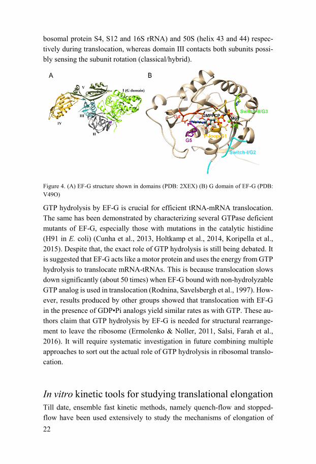
22
bosomal protein S4, S12 and 16S rRNA) and 50S (helix 43 and 44) respec-tively during translocation, whereas domain III contacts both subunits possi-bly sensing the subunit rotation (classical/hybrid).
Figure 4. (A) EF-G structure shown in domains (PDB: 2XEX) (B) G domain of EF-G (PDB: V49O)
GTP hydrolysis by EF-G is crucial for efficient tRNA-mRNA translocation. The same has been demonstrated by characterizing several GTPase deficient mutants of EF-G, especially those with mutations in the catalytic histidine (H91 in E. coli) (Cunha et al., 2013, Holtkamp et al., 2014, Koripella et al., 2015). Despite that, the exact role of GTP hydrolysis is still being debated. It is suggested that EF-G acts like a motor protein and uses the energy from GTP hydrolysis to translocate mRNA-tRNAs. This is because translocation slows down significantly (about 50 times) when EF-G bound with non-hydrolyzable GTP analog is used in translocation (Rodnina, Savelsbergh et al., 1997). How-ever, results produced by other groups showed that translocation with EF-G in the presence of GDP•Pi analogs yield similar rates as with GTP. These au-thors claim that GTP hydrolysis by EF-G is needed for structural rearrange-ment to leave the ribosome (Ermolenko & Noller, 2011, Salsi, Farah et al., 2016). It will require systematic investigation in future combining multiple approaches to sort out the actual role of GTP hydrolysis in ribosomal translo-cation.
In vitro kinetic tools for studying translational elongation Till date, ensemble fast kinetic methods, namely quench-flow and stopped-flow have been used extensively to study the mechanisms of elongation of

23
protein synthesis. These studies mostly use reconstituted bacterial translation system composed of individually purified 70S ribosome, all major transloca-tion factors, all tRNA synthetases, tRNA-bulk and mRNA, in highly opti-mized, near-physiological buffer condition. The reconstituted translation sys-tems are optimized carefully for in vivo like rate and accuracy (Holm, Borg et al., 2016, Jelenc & Kurland, 1979, Johansson, Zhang et al., 2012).
In a quench-flow, equal volumes of two reactants are mixed rapidly in the reaction loop and incubated at constant temperature until the reaction is quenched by the quencher (normally formic acid is used to precipitate the ri-bosome) (Figure 5A). The reaction time can be set as short as few milliseconds (dead time of the quench-flow is normally 1-2 ms), and the products from the quenched reaction are analyzed either in high performance liquid chromatog-raphy (HPLC) or thin-layer chromatography (TLC) with radioactivity detec-tion. Most commonly, radioactively labeled amino acids are used for peptide bond formation experiments, where the di- / tri- / longer peptides can be sep-arated in the reversed phase chromatography in HPLC and detected with radi-ography (Holm et al., 2016). For GTP hydrolysis experiments radioactive GTP is used. After the reaction, the GTP and GDP are separated in TLC or with ion exchange chromatography and quantified by using the radioactivity of the respective peaks/bands. The mean time for the translocation including all the steps starting from EF-G binding, GTP-hydrolysis, couple mRNA-tRNA translocation and Pi release and EF-G release can be measured precisely (Figure 6).
Figure 5. Schematic description of quench-flow (A) and stopped-flow (B)
In a stopped-flow, the mechanism of the detection is often light scattering and fluorescence (Figure 5B). Due to difference in particle size, the 70S ribosome
24
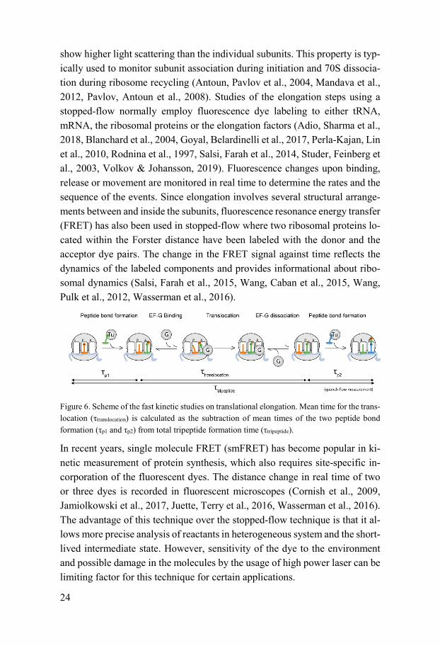
show higher light scattering than the individual subunits. This property is typ-ically used to monitor subunit association during initiation and 70S dissocia-tion during ribosome recycling (Antoun, Pavlov et al., 2004, Mandava et al., 2012, Pavlov, Antoun et al., 2008). Studies of the elongation steps using a stopped-flow normally employ fluorescence dye labeling to either tRNA, mRNA, the ribosomal proteins or the elongation factors (Adio, Sharma et al., 2018, Blanchard et al., 2004, Goyal, Belardinelli et al., 2017, Perla-Kajan, Lin et al., 2010, Rodnina et al., 1997, Salsi, Farah et al., 2014, Studer, Feinberg et al., 2003, Volkov & Johansson, 2019). Fluorescence changes upon binding, release or movement are monitored in real time to determine the rates and the sequence of the events. Since elongation involves several structural arrange-ments between and inside the subunits, fluorescence resonance energy transfer (FRET) has also been used in stopped-flow where two ribosomal proteins lo-cated within the Forster distance have been labeled with the donor and the acceptor dye pairs. The change in the FRET signal against time reflects the dynamics of the labeled components and provides informational about ribo-somal dynamics (Salsi, Farah et al., 2015, Wang, Caban et al., 2015, Wang, Pulk et al., 2012, Wasserman et al., 2016).
Figure 6. Scheme of the fast kinetic studies on translational elongation. Mean time for the trans-location (τtranslocation) is calculated as the subtraction of mean times of the two peptide bond formation (τp1 and τp2) from total tripeptide formation time (τtripeptide).
In recent years, single molecule FRET (smFRET) has become popular in ki-netic measurement of protein synthesis, which also requires site-specific in-corporation of the fluorescent dyes. The distance change in real time of two or three dyes is recorded in fluorescent microscopes (Cornish et al., 2009, Jamiolkowski et al., 2017, Juette, Terry et al., 2016, Wasserman et al., 2016). The advantage of this technique over the stopped-flow technique is that it al-lows more precise analysis of reactants in heterogeneous system and the short-lived intermediate state. However, sensitivity of the dye to the environment and possible damage in the molecules by the usage of high power laser can be limiting factor for this technique for certain applications.

25
Aim of the thesis This thesis aims to optimize a fluorescent-mRNA based real time transloca-tion assay. Further aims are to characterize effects of different factors, which affect ribosomal translocation, using this assay. These factors include EF-G mutations, r-protein modification, different tRNAs and antibiotics.
- The length and the position of the pyrene on the mRNA have been optimized by careful calibration with the functional translocation as-say by tripeptide synthesis in a quench-flow. (Paper I)
- The external factors such as temperature, Mg2+ concentration in the buffer, concentrations of EF-G, NAc-Phe-tRNAPhe have been opti-mized for better signal and reliable data analysis. (Paper I)
- The effects of EF-G mutations, GTP hydrolysis and nucleotide bind-ing on the ribosomal translocation are analyzed using optimized trans-location assay. (Paper I & II)
- The role of the extended tail of the ribosomal protein S13 from Ther-mus thermophilus has been investigated in relation with ribosomal translocation and recycling. (Paper III)
- The mode of action of the novel aminoglycoside antibiotic arbekacin on translocation. (Paper IV)
26
The present work
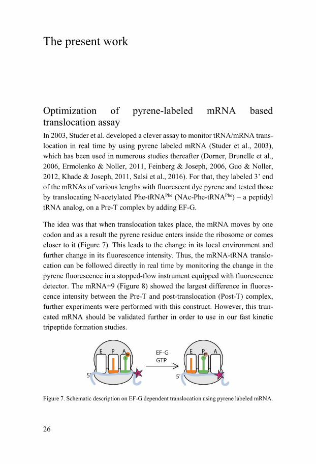
Optimization of pyrene-labeled mRNA based translocation assay In 2003, Studer et al. developed a clever assay to monitor tRNA/mRNA trans-location in real time by using pyrene labeled mRNA (Studer et al., 2003), which has been used in numerous studies thereafter (Dorner, Brunelle et al., 2006, Ermolenko & Noller, 2011, Feinberg & Joseph, 2006, Guo & Noller, 2012, Khade & Joseph, 2011, Salsi et al., 2016). For that, they labeled 3’ end of the mRNAs of various lengths with fluorescent dye pyrene and tested those by translocating N-acetylated Phe-tRNAPhe (NAc-Phe-tRNAPhe) – a peptidyl tRNA analog, on a Pre-T complex by adding EF-G.
The idea was that when translocation takes place, the mRNA moves by one codon and as a result the pyrene residue enters inside the ribosome or comes closer to it (Figure 7). This leads to the change in its local environment and further change in its fluorescence intensity. Thus, the mRNA-tRNA translo-cation can be followed directly in real time by monitoring the change in the pyrene fluorescence in a stopped-flow instrument equipped with fluorescence detector. The mRNA+9 (Figure 8) showed the largest difference in fluores-cence intensity between the Pre-T and post-translocation (Post-T) complex, further experiments were performed with this construct. However, this trun-cated mRNA should be validated further in order to use in our fast kinetic tripeptide formation studies.
Figure 7. Schematic description on EF-G dependent translocation using pyrene labeled mRNA.

27
Optimization of pyrene-mRNA for functional kinetic studies in quench-flow In this work, we have tested the pyrene labeled mRNAs of different lengths +9 to +12, and unlabeled +9 and +10 (Figure 8) in quench-flow based tripep-tide formation assay in parallel to the stopped flow based translocation assay (Figure 9A). The mRNAs, encode for tripeptide Met-Phe-Leu, were designed and named based on the Studer et al, 2003. The process includes two peptide bond formation steps and one translocation step driven by EF-G. The di- and tripeptide formation experiments were conducted in quench-flow, where an elongation factor mix containing the respective ternary complexes (TC) was rapidly mixed with the 70S IC containing mRNA and fMet-tRNAfMet in the P site. By fitting the kinetic data as described in the in Paper I, the mean times of the first peptide bond formation (τp1), second peptide bond formation (τp2) and tripeptide formation (τtripeptide) were determined. The mean time of a full translocation reaction (τtranslocation) was calculated as [τtripeptide – (τp1+ τp2)]. In parallel, the kinetics of translocation starting from the 70S IC was followed in stopped-flow, where the fluorescence change of the 3’ pyrene labeled mRNAs was monitored in real time. The mean time τfluor, indicated total time of all events starting from the 70S IC up to and including mRNA movement. The mean time for mRNA movement, τmRNA, was determined by subtracting τp1 from τfluor.
Figure 8. Sequence of different length of mRNA constructs. ℗ represents the pyrene dye at the 3’-end of mRNAs. Shine-Dalgarno (SD) sequence is in grey font color and mRNAs code for tripeptide fMet-Phe-Leu.
Using quench-flow, we have followed the kinetics of formation of fMet-Phe-Leu formation with six mRNAs (labeled & unlabeled). Two mixes, mix A containing 70S initiation complex (1 µM) with fMet-tRNAfMet in the P site and mix B containing ternary complexes of Phe and Leu (4 µM each), and EF-G (10 µM), were mixed rapidly for tripeptide synthesis. The reaction mixture was quenched at given time points, and treated for further analysis in Reversed Phase-High Performance Liquid Chromatography (RP-HPLC) (C18 column)

28
with on-line radiation detection (Holm et al., 2016) (Figure 9B). The propor-tion of fMet-Phe-Leu was fitted into three consecutive step model to deter-mine τtripeptide (Borg & Ehrenberg, 2015, Holm et al., 2016). Similarly, the mean time for the first peptide bond formation (τp1) and the second peptide bond formation (τp2) were measured as described (Borg et al., 2015).
The mean time value of the first peptide bond formation (τp1) was identical for all six mRNAs, accounted for about 31 ms, indicating that neither the mRNA length or 3’-modification of the mRNAs did not affect this step (Table 1A). However, the mean time of fMet-Phe-Leu tripeptide formation (τtripeptide) and the mean time of formation of the second peptide bond (τp2) varied for the mRNAs. The tripeptide formation was noticeably slower for mRNA+9 and mRNA+9 nodye (315 ± 15 ms and 368 ± 20 ms respectively) than other mRNAs (261 ± 15 ms, 244 ± 12 ms, 255 ± 18 ms and 217 ± 27 ms respectively for mRNA+10 nodye, mRNA+10, mRNA+11 and mRNA+12) (Figure 9B and Table 1A). Similarly, mRNA+9 and mRNA+9 nodye were slower in the second peptide bond formation (Table 1A). The mean time for translocation (τtranslocation), which contains multi- sub-steps between the two peptidyl transfer was determined by subtracting τp1 and τp2 from τtripeptide. τtranslocation for all other mRNAs was faster than for mRNA+9 by about 50 ms, indicating the defects of the short mRNA for translocation. However, these defects are improved by the addition of just one additional base at the 3’- end of the mRNA in mRNA+10, which is as fast as the longer mRNAs.
The same 70S IC with pyrene labeled mRNA were deployed in stopped-flow to determine mRNA-tRNA translocation by following fluorescence upon EF-G addition. Same mixes A and B were rapidly mixed after incubating at 37 ˚C for 15 min. The fluorescence traces showed an initial small increase followed by a predominant monophasic decrease (Figure 9C). The traces were analyzed as described in Materials and Methods in paper I, and the mean time for mRNA movement (τmRNA) was determined. It should be noted that τmRNA is supposedly shorter than τtranslocation as the mRNA-fluorescence based assay is insensitive about EF-G release and ribosomal rearrangement prior to next EF-Tu TC binding (Figure 9A). τmRNA, measured by following pyrene fluores-cence, did not vary between mRNAs, indicating the steps for the defects of mRNA+9 are post mRNA movement (Figure 9D and Table 1A). In relation to the fluorescence intensity data in (Studer et al., 2003), the amplitude of flu-orescence change varied largely. Compared the amplitude for mRNA+9, mRNA+11 changed the fluorescence by only about half, whereas mRNA+12

29
produced mere changes in the fluorescence intensity thus reliable data could not be achieved from fitting (Figure 9C).
Figure 9. (A) Schematic representation of the peptide elongation cycle on the ribosome starting from the 70S IC. (B) Kinetics of the tripeptide formation in quench-flow starting from the 70S IC. (C) Fluorescence trace of pyrene upon EF-G addition to the 70S initiation complex. (D) Mean time values of each step for tripeptide formation in quench-flow and fluorescence meas-urement in stopped-flow. The values of three-colour stacked bar accounts for the mean time of tripeptide formation (τtripeptide), and purple bar accounts for τtrans, acquired from stopped-flow measurement
30
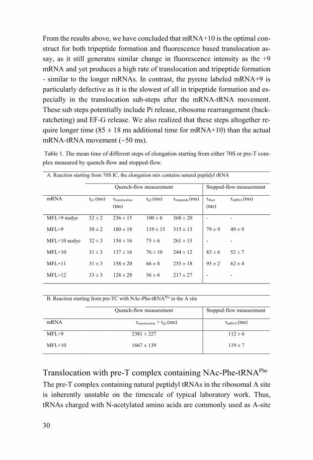
From the results above, we have concluded that mRNA+10 is the optimal con-struct for both tripeptide formation and fluorescence based translocation as-say, as it still generates similar change in fluorescence intensity as the +9 mRNA and yet produces a high rate of translocation and tripeptide formation - similar to the longer mRNAs. In contrast, the pyrene labeled mRNA+9 is particularly defective as it is the slowest of all in tripeptide formation and es-pecially in the translocation sub-steps after the mRNA-tRNA movement. These sub steps potentially include Pi release, ribosome rearrangement (back-ratcheting) and EF-G release. We also realized that these steps altogether re-quire longer time (85 ± 18 ms additional time for mRNA+10) than the actual mRNA-tRNA movement (~50 ms).
Table 1. The mean time of different steps of elongation starting from either 70S or pre-T com-plex measured by quench-flow and stopped-flow.
A. Reaction starting from 70S IC, the elongation mix contains natural peptidyl tRNA
Quench-flow measurement Stopped-flow measurement
mRNA τp1 (ms) τtranslocation
(ms) τp2 (ms) τtripeptide (ms) τfluor
(ms) τmRNA (ms)
MFL+9 nodye 32 ± 2 236 ± 15 100 ± 6 368 ± 20 - -
MFL+9 30 ± 2 180 ± 18 119 ± 15 315 ± 13 79 ± 9 49 ± 9
MFL+10 nodye 32 ± 3 154 ± 16 75 ± 6 261 ± 15 - -
MFL+10 31 ± 3 137 ± 16 76 ± 10 244 ± 12 83 ± 6 52 ± 7
MFL+11 31 ± 3 158 ± 20 66 ± 8 255 ± 18 93 ± 2 62 ± 4
MFL+12 33 ± 3 128 ± 28 56 ± 6 217 ± 27 - -
B. Reaction starting from pre-TC with NAc-Phe-tRNAPhe in the A site
Quench-flow measurement Stopped-flow measurement
mRNA τtranslocation + τp2 (ms) τmRNA (ms)
MFL+9 2381 ± 227 112 ± 6
MFL+10 1667 ± 139 119 ± 7
Translocation with pre-T complex containing NAc-Phe-tRNAPhe
The pre-T complex containing natural peptidyl tRNAs in the ribosomal A site is inherently unstable on the timescale of typical laboratory work. Thus, tRNAs charged with N-acetylated amino acids are commonly used as A-site

31
peptidyl tRNA analogs to mimic the pre-T complex. The binding of such an-alogs is a factor–independent equilibrium process and the substrates can there-fore be supplied in large excesses (Dorner et al., 2006, Ermolenko & Noller, 2011, Feinberg & Joseph, 2006, Feldman et al., 2010, Guo & Noller, 2012, Juette et al., 2016, Khade & Joseph, 2011, Salsi et al., 2016, Studer et al., 2003). We designed a set of experiments to compare translocation of a pre-T complex formed by pre-equilibration with a commonly used peptidyl tRNA analog NAc-Phe-tRNAPhe, to a pre-T complex containing natural fMet-Phe-tRNAPhe, in continuous progression (without pre-equilibration), starting from the 70S IC.
Four pyrene-labeled mRNAs were deployed in stopped-flow measurement for mRNA-tRNA translocation with pre-T complex instead of 70S IC. The trans-location displayed a biphasic fluorescence decay similar to previous reports (Ermolenko & Noller, 2011, Feinberg & Joseph, 2006, Guo & Noller, 2012, Khade & Joseph, 2011, Salsi et al., 2016), and absent of the initial increase presented in the reaction when 70S IC was a starting complex of translocation reaction. The amplitude also varied similarly to the reaction with 70S IC, mRNA+11 and +12 achieved little fluorescence change during translocation, furthermore unreliable for analysis (Figure 10A). The fast phase of the fluo-rescence trace with mRNA+9 and +10 accounted for 80 to 90 % of the total amplitude, which rate was used to determine the mean time of mRNA trans-location (τmRNA). mRNA +10 translocated at a similar rate as +9, but faster in the elongation cycle (peptide bond formation and translocation) measured in quench-flow, validating that mRNA +10 is the optimal mRNA for functional assays (Table 1B). Compared to the rate of the translocation starting from IC, pre-T complex delivered slower translocation by about half rate (~50 ms and ~120 ms for 70S IC and pre-T respectively) despite the two reaction had iden-tical buffer condition and components (Table 1B and Figure 10B). This may be due to the inability of NAc-Phe-tRNAPhe for mimicking the peptidyl-tRNA, as the ribosome sense the peptidyl chain of the A-site tRNA before transloca-tion (Frank & Agrawal, 2000).

32
Figure 10. (A) Translocation starting from pre-T complex with different mRNA constructs. (B) Michaelis Menten parameters for two translocation reactions
Optimization of the reaction conditions for better data analysis One bottle neck of the earlier protocol of the pyrene-mRNA based transloca-tion assay was that it always produced bi-phasic kinetics with a major slow phase of unknown origin. This made mean time estimation very challenging. We have here optimized the conditions for the translocation starting from the pre-T complex so that a near monophasic fluorescence change can be ob-tained. Concentrations of NAc-Phe-tRNAPhe, EF-G and Mg2+, and the temper-ature are the factors, which influence fluorescence change in the translocation reaction. By titrating NAc-Phe-tRNAPhe in the reaction, we have found that the affinity of the NAc-Phe-tRNAPhe to the 70S pre-T complex is low at physio-logical Mg2+ concentration and 37 ⁰C. When it was titrated in the mix of the 70S ribosome with mRNA, deacylated tRNAfMet in the P site and empty A site, the rates of fast and slow phase as well as the proportion of the amplitudes did not differ much. The total amplitude did rather vary, the higher concentra-tion of NAc-Phe-tRNAPhe yielded higher amplitude. We have determined the KD value (2.7 ± 0.2 µM) of NAc-Phe-tRNAPhe binding to the A site of the ribosome, by titrating NAc-Phe-tRNAPhe in the EF-G mix. The proportion of the properly formed 70S pre-T complex calculated from the KD value actually matched with the amplitude data above, validating the fact that NAc-Phe-tRNAPhe has poor affinity to the A site of the 70S ribosome.
By titrating EF-G, we have found that high concentration of EF-G in the trans-location reaction is important for pushing the proportion of the fast phase as well as the rate of the translocation. Same trend was observed with higher temperature and lower Mg2+ concentration (Table 2). The specific cause of
33
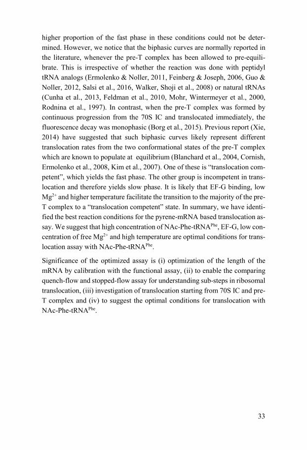
higher proportion of the fast phase in these conditions could not be deter-mined. However, we notice that the biphasic curves are normally reported in the literature, whenever the pre-T complex has been allowed to pre-equili-brate. This is irrespective of whether the reaction was done with peptidyl tRNA analogs (Ermolenko & Noller, 2011, Feinberg & Joseph, 2006, Guo & Noller, 2012, Salsi et al., 2016, Walker, Shoji et al., 2008) or natural tRNAs (Cunha et al., 2013, Feldman et al., 2010, Mohr, Wintermeyer et al., 2000, Rodnina et al., 1997). In contrast, when the pre-T complex was formed by continuous progression from the 70S IC and translocated immediately, the fluorescence decay was monophasic (Borg et al., 2015). Previous report (Xie, 2014) have suggested that such biphasic curves likely represent different translocation rates from the two conformational states of the pre-T complex which are known to populate at equilibrium (Blanchard et al., 2004, Cornish, Ermolenko et al., 2008, Kim et al., 2007). One of these is “translocation com-petent”, which yields the fast phase. The other group is incompetent in trans-location and therefore yields slow phase. It is likely that EF-G binding, low Mg2+ and higher temperature facilitate the transition to the majority of the pre-T complex to a “translocation competent” state. In summary, we have identi-fied the best reaction conditions for the pyrene-mRNA based translocation as-say. We suggest that high concentration of NAc-Phe-tRNAPhe, EF-G, low con-centration of free Mg2+ and high temperature are optimal conditions for trans-location assay with NAc-Phe-tRNAPhe.
Significance of the optimized assay is (i) optimization of the length of the mRNA by calibration with the functional assay, (ii) to enable the comparing quench-flow and stopped-flow assay for understanding sub-steps in ribosomal translocation, (iii) investigation of translocation starting from 70S IC and pre-T complex and (iv) to suggest the optimal conditions for translocation with NAc-Phe-tRNAPhe.

34
Table 2. Effects of varying EF-G and Mg2+ concentration and temperature on mRNA-tRNA movement during ribosomal translocation with NAc-Phe-tRNAPhe
k1 (s-1) k2 (s-1) A1/(A1+A2) τmRNA (ms)
A. EF-G (μM)
0.5 2.8 ± 0.5 0.6 ± 0.05 0.49 ± 0.03 357 ± 67
1.25 4.4 ± 0.1 0.92 ± 0.01 0.76 ± 0.06 227 ± 5
2.5 6 ± 0.2 0.72 ± 0.1 0.84 ± 0.04 167 ± 6
5 8.4 ± 0.5 0.66 ± 0.2 0.88 ± 0.06 119 ± 7
10 10.3 ± 0.3 0.4 ± 0.07 0.91 ± 0.02 97 ± 3
B. Temperature (⁰C)
37 8.4 ± 0.5 0.66 ± 0.2 0.88 ± 0.06 119 ± 7
30 3.2 ± 0.1 0.56 ± 0.02 0.74 ± 0.01 313 ± 10
25 1.25 ± 0.06 0.30 ± 0.01 0.62 ± 0.01 800 ± 38
20 0.35 ± 0.07 0.09 ± 0.02 0.47 ± 0.02 2857 ± 571
C. Extra Mg2+ (mM)
1 5.8 ± 1.3 0.71 ± 0.01 0.66 ± 0.01 172 ± 39
2 3.2 ± 0.2 0.43 ± 0.03 0.62 ± 0.03 313 ± 20
3 1.9 ± 0.2 0.28 ± 0.11 0.52 ± 0.02 526 ± 55
5 0.8 ± 0.3 0.17 ± 0.04 0.48 ± 0.05 1250 ± 469
10 0.19 ± 0.01 0.04 ± 0.01 0.49 ± 0.01 5263 ± 277

35
Application of optimized translocation assay The optimized translocation assay based on the pyrene-labeled mRNA has been employed for the investigation of the effect of different factors on the mRNA-tRNA translocation studies. These are non-hydrolyzable GTP analogs (Paper I), tRNA variable loop (this thesis), EF-G mutants (Paper I and II), ribosomal protein S13 (Paper III) and antibiotic Arbekacin (Paper IV).
Non-hydrolyzable GTP analogs in translocation Translational G-proteins play an important role in the initiation, peptidyl transfer, ribosomal translocation, peptide release, and ribosome recycling. In most cases they facilitate the translation when they are bound to GTP. The GDP or GDPNP (non hydrolyzable analog of GTP) bound form cannot effi-ciently assist the ribosomal translation, indicating the necessity of GTP bind-ing and GTP hydrolysis for their function. Like all other translational GTPases, EF-G undergoes conformational change in the GTP and GDP bound forms (Chen et al., 2013, Lai, Ghaemi et al., 2017, Lin et al., 2015, Munro, Wasserman et al., 2010b). When GTP is replaced with non-hydrolyzable GTP analogs, the protein cannot conduct GTP hydrolysis and hence stays in the “on” state for much longer time. Thus, these GTP analogs are normally used to investigate the role of GTP hydrolysis by EF-G in translocation. It has also been used in many ribosomal translocation studies (Ermolenko & Noller, 2011, Flis et al., 2018, Rodnina et al., 1997, Salsi et al., 2016). Previous studies using same non-hydrolyzable GTP analog, one result showed significant de-fects by more than one order of magnitude supporting the role of GTP hydrol-ysis as promoting ribosomal translocation (Rodnina et al., 1997). The other data showed comparable rates of translocation with the analog to the EF-G•GTP or less than two times defects. This result led to the conclusions stating that of GTP hydrolysis is necessary for EF-G release from the 70S ribosome (Ermolenko & Noller, 2011, Rodnina et al., 1997, Salsi et al., 2016). Such data discrepancy has not been investigated properly, different buffer and reaction condition made it difficult to compare.
Three non-hydrolyzable GTP analogs (GDPNP, GDPCP and GTP-γ-S) were added in the optimized translocation assay with a Pre-T complex formed with NAc-Phe-tRNAPhe. This strategy was essential to avoid another GTPase trans-lation factor, EF-Tu, which would otherwise be required to deliver the aa-tRNAs. With non-hydrolyzable GTP analogs, EF-G translocated almost 50

36
time slower than with GTP, in accordance with earlier report (Rodnina et al., 1997). EF-G with GTP-γ-S was the least defective among the three analogs tested, producing 2-4 times faster translocation than with GDPNP and GDPCP, due to the higher rate of GTP hydrolysis compared to the other two analogs (Kuhle & Ficner, 2014). Surprisingly, GDP showed same degree of defect as GDPNP (at a EF-G concentration of 5 µM), which was apparently unexplainable.
In order to investigate the scenario further, we titrated EF-G in translocation reaction together with GTP, GDPNP and GDP. The rates were plotted against EF-G concentration in the Michaelis Menten plots. For the reactions with GTP the kcat and KM were 11.9 ± 0.9 s-1 and 2.2 ± 0.4 µM. The reaction was highly inefficient with GDPNP. The KM increased by about five fold, but the kcat de-creased about 20 times. Interestingly, EF-G with GDP produced higher kcat (3.4 ± 2.4 s-1) than with GDPNP. It is ~four times slower than the kcat with GTP. But, the reaction could not be saturated even at 20 µM concentration (unlike GTP or GDPNP), and the KM was as high as 70 µM, ~35 fold higher than with GTP (Figure 11). We speculate that EF-G•GDP is defective in the formation of “translocation competent” ribosome complex, showing high KM
value. However, our results with GDP and GDPNP suggest that translocation can occur, albeit inefficiently, even in the absence of GTP hydrolysis. Being said that the possibility that the cause of defect with GDP and GDPNP is dif-ferent, cannot be nullified. Thus, further investigations will be required for further clarification.
Figure 11. Michaelis Menten plots of the translocation rate by titration of EF-G with either GTP (A), GDPNP (B), or GDP (C)
Possible importance of tRNA body in elongation A tRNA is an adapter molecule, pivotal for translation of mRNA to proteins. It acts as a physical link between mRNA codons and the amino acid sequence

37
in the peptide through its unique structure. The tRNAs have L-shaped three dimensional structure which locates the anticodon loop and the acceptor stem to the DC in 30S subunit and the PTC in 50S subunit, respectively. Each tRNA has its unique anticodon and attaches specific amino acid at the acceptor stem. This specificity is conferred by the corresponding aminoacyl tRNA synthetase which endows accuracy and fidelity to the translation. During translation by ribosome, aminoacylated tRNA in the form of ternary complex with EF-Tu•GTP enters the A site in the ribosome, move (translocate) after peptidyl transfer from A to P site and again P to E site before finally exiting the ribo-some. Several studies have investigated the interaction of tRNA with either rRNA or ribosomal proteins (Abdurashidova, Tsvetkova et al., 1991, Bocchetta, Xiong et al., 2001, Demeshkina, Jenner et al., 2010, Shetty, Shah et al., 2017), but little is known about their role in translocation, especially in relation with the length of the variable loop of the tRNAs.
Table 3. Translocation and peptide formation with different tRNAs in the A site.
A. Translocation rate measurement by pyrene-mRNA mRNA Codon
tRNA kcat
(s-1)
KM
(µM)
kcat/KM
(µM-1s-1)
Codon
mismatch
Nucleotides in variable loop
UUC Phe 25.5 ± 0.63 1.5 ± 0.07 17 ± 0.84 - 0
UUU Phe 27.5 ± 0.88 2.5 ± 0.18 11 ± 0.86 yes 0
GCA Ala 29.5 ± 0.5 1.6 ± 0.1 18.4 ± 1.2 - 0
UAC Tyr 43.7 ± 2 2.1 ± 0.13 21.4 ± 1.62 - 7
CUG Leu1 11.4 ± 0.73 1.9 ± 0.17 6 ± 0.64 - 10
AGC Ser 33.1 ± 2 2 ± 0.19 16.6 ± 1.86 - 17
B. Peptide bond formation codon tRNA kcat
(s-1)
KM
(µM)
kcat/KM
(µM-1s-1)
tRNA abundancy
Nucleotides in variable loop
UUC Phe 92.1 ± 9.8 0.91 ± 0.17 101.2 ± 21.8 1.6 0
UUU Phe 100 ± 13.6 2.84 ± 0.62 35.2 ± 9.1 1.6 0
GCA Ala 81.8 ± 9.4 2.47 ± 0.46 33.1 ± 7.2 5.04 0
UAC Tyr 36.7 ± 3.4 1.48 ± 0.28 24.8 ± 5.2 3.14 7
CUG Leu1 38.3 ± 4.4 4.24 ± 0.81 9 ± 2 6.94 10
AGC Ser 30.6 ± 1.8 1.26 ± 0.16 24.3 ± 3.4 2.01 17
38
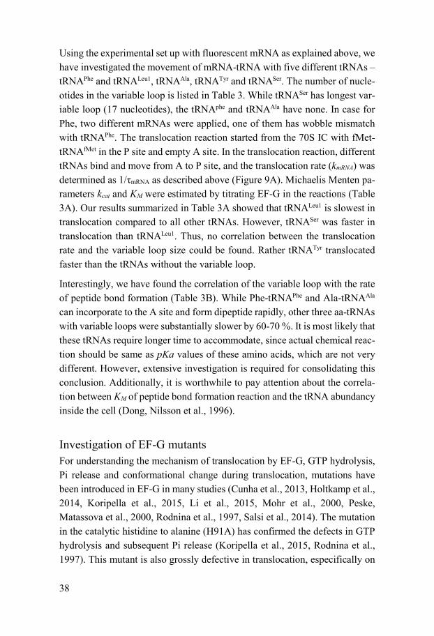
Using the experimental set up with fluorescent mRNA as explained above, we have investigated the movement of mRNA-tRNA with five different tRNAs – tRNAPhe and tRNALeu1, tRNAAla, tRNATyr and tRNASer. The number of nucle-otides in the variable loop is listed in Table 3. While tRNASer has longest var-iable loop (17 nucleotides), the tRNAphe and tRNAAla have none. In case for Phe, two different mRNAs were applied, one of them has wobble mismatch with tRNAPhe. The translocation reaction started from the 70S IC with fMet-tRNAfMet in the P site and empty A site. In the translocation reaction, different tRNAs bind and move from A to P site, and the translocation rate (kmRNA) was determined as 1/τmRNA as described above (Figure 9A). Michaelis Menten pa-rameters kcat and KM were estimated by titrating EF-G in the reactions (Table 3A). Our results summarized in Table 3A showed that tRNALeu1 is slowest in translocation compared to all other tRNAs. However, tRNASer was faster in translocation than tRNALeu1. Thus, no correlation between the translocation rate and the variable loop size could be found. Rather tRNATyr translocated faster than the tRNAs without the variable loop.
Interestingly, we have found the correlation of the variable loop with the rate of peptide bond formation (Table 3B). While Phe-tRNAPhe and Ala-tRNAAla can incorporate to the A site and form dipeptide rapidly, other three aa-tRNAs with variable loops were substantially slower by 60-70 %. It is most likely that these tRNAs require longer time to accommodate, since actual chemical reac-tion should be same as pKa values of these amino acids, which are not very different. However, extensive investigation is required for consolidating this conclusion. Additionally, it is worthwhile to pay attention about the correla-tion between KM of peptide bond formation reaction and the tRNA abundancy inside the cell (Dong, Nilsson et al., 1996).
Investigation of EF-G mutants For understanding the mechanism of translocation by EF-G, GTP hydrolysis, Pi release and conformational change during translocation, mutations have been introduced in EF-G in many studies (Cunha et al., 2013, Holtkamp et al., 2014, Koripella et al., 2015, Li et al., 2015, Mohr et al., 2000, Peske, Matassova et al., 2000, Rodnina et al., 1997, Salsi et al., 2014). The mutation in the catalytic histidine to alanine (H91A) has confirmed the defects in GTP hydrolysis and subsequent Pi release (Koripella et al., 2015, Rodnina et al., 1997). This mutant is also grossly defective in translocation, especifically on

39
30S (Cunha et al., 2013), in accordance with the translocation data with non-hydrolyzable GTP analogs. We have further deployed H91 mutants – H91E and H91Q in the translocation assay starting from the pre-T complex with NAc-Phe-tRNAPhe. The defects were more extensive than GTP analogs by about 2 to 5 times. Furthermore, H91E revealed rather long initial increase phase compared to H91Q mutant, which is also not visible in the translocation reaction starting from 70S IC.
Various mutation was further introduced in the G domain of EF-G, in the po-sition of R28 (R28A), R58 (R58A) and two threonine in the 23rd and 24th po-sition (T23-24A) which associates the function of guanosine nucleotide bind-ing and GTP hydrolysis in the vicinity of P loop and switch I. Mutation of the two threonine (T23-24A) rendered EF-G completely deficient in guanosine nucleotide binding, presenting apo state of EF-G. T23-24A mutant further an-nihilates Pi release and translocation. When R28A and R58A mutants were titrated in translocation assay starting from the 70S IC, kcat could not be esti-mated as the rate did not saturate at highest possible concentrations (20 µM). At the concentration of 10 µM, R28A and R58A mutants translocated by 15 to 30 fold slower than the wild type for R28A and R58A respectively. How-ever, surprisingly, R28A was completely impaired in Pi release similarly to the T23-24A mutant. In addition, R28A mutant had similar affinity towards GDP compared to the wild type EF-G, while KD for R58A was one order of magnitude smaller indicating either tight binding or faster exchange of nucle-otides.
The inconsistency in the Pi release kinetics for two arginine mutants suggests that Pi release is not coupled with translocation. It can occur before or after the translocation, not implying with previous findings (Peske et al., 2000). However, our results indicate that the binding of guanosine nucleotide is ab-solutely vital for EF-G function similar to other G-proteins.
Role of the extended C-terminal tail of the ribosomal protein S13 in protein synthesis Ribosomal protein S13 is a 118 amino acids long protein in E. coli, located in the head part of the 30S subunit. Crystal structures of the bacterial ribosome show several interactions of the S13 protein at the subunit-interface; it plays an important role in forming intersubunit bridges between the subunits. The bridge 1a is formed between helix 38 of 23S rRNA and the 93rd amino acid

40
of S13, while bridge 1b is formed by the N-terminus of S13 and the large subunit protein bL5. Furthermore, Its C-terminal tail makes contact with the anticodon stem loop of the P-site tRNA (Yusupov et al., 2001), thereby po-tentially affecting the speed and accuracy of translocation. Previous studies have demonstrated important role of S13 protein in bacterial fitness and trans-lation. It was shown that the chromosomal deletion of S13 protein either in-duces cell death or creates critical fitness defects in E. coli (Cukras & Green, 2005, Shoji, Dambacher et al., 2011). Moreover, small deletion at the C-ter-minal tail causes noticeable fitness defects in E. coli, possibly resulting from reduced affinity towards the P-site tRNA to the ribosome (Cukras & Green, 2005, Hoang, Fredrick et al., 2004).
The length of the C-terminal tail of S13 varies in different bacteria. While most bacteria have a short C-terminal tail like in E. coli, some bacteria have a longer tail. One such example is Thermus thermophilus, which has seven extra amino acids in the tail compared to E. coli. The sequence of S13 protein from E. coli and T. thermophilus have high similarities, the C-terminal domain con-tains 20-30 identical amino acids. The C-terminus tail of S13 contains RKGPRK sequence followed by different extensions; four amino acids in E. coli and eleven in T. thermophilus, ending with K in both. When compared for their structure on the ribosome, the longer tail of T. thermophilus S13 clearly extends between the P-site and the A-site tRNA (Figure 12) suggesting its potential role in hindering the movement of the tRNAs during transloca-tion. The occurrence of multiple positively charged amino acids in the ex-tended tail of S13 further strengthens the likelihood that it will interact more with the tRNAs, thereby influencing their movement, particularly during translocation.
To understand the implication of the C-terminal extension of the S13 protein in translocation, we have created several variations of the S13 tail in the chro-mosome of the E. coli JE28 strain (Ederth, Mandava et al., 2009) (Figure 13A). The parental strain JE28 carries tetra his-tagged ribosomes, where the nucleotides coding hexa-histidine tags are fused at the chromosomal locus of the rplK gene coding for the ribosomal protein L7/12. The his-tags allow easy pull-down of the intact ribosomes by affinity chromatography. For modifica-tion of the S13 tail, red-recombineering was done at the rpsM locus, where ampicillin resistance (ampR) cassette was used as a reporter gene. The S13 modified strains are named with the suffix CIK28. To ensure that the reporter

41
gene does not affect bacterial growth, only ampR cassette was also introduced in the same locus. This strain is called CIK28.
Figure 12. Superposition of T. thermophilus S13 (blue) on the structure of E. coli S13 (pink). Gray cartoons indicate three tRNAs bound to the A-, P- and the E-site. The structures are adopted from PDB 4K0L and PDB 3E1C. The ribosome components other than S13 have been removed to demonstrate the location of the extended tail of the S13 protein.
To investigate the impact of the length of S13 tail on bacterial growth and tRNA translocation, either four terminal amino acids of E. coli S13 were de-leted (CIK28d), or the C-terminal tail was extended partially or fully with the sequence similar to that of T. thermophilus, resulting into CIK28b and CIK28c strains, respectively. Alternatively, the E. coli S13 tail was extended by three and seven alanines resulting into CIK28a3 and CIK28a7 strains, respectively. In order to investigate the importance of the positive charge in the S13 tail, last two lysines of the E. coli S13 tail were altered into either uncharged ala-nine (CIK28aa) or negatively charged glutamic acid (CIK28ee). Also, all pos-itively charged amino acids of the T. thermophilus-like tail in CIK28c were exchanged with alanine resulting into the CIK28ca strain. The strains and the modifications are illustrated in Figure 13A.
The 70S ribosomes from E. coli S13 mutants were purified by affinity chro-matography, and further dissociated into the subunits following the existing protocol (Ederth et al., 2009). As S13 protein is essential for interactions be-tween the subunits, we have measured subunits association and 70S dissocia-tion kinetics with the 30S subunits from the CIK28 strains. The alterations in the S13 tail did not change the rate or extent of subunit association or 70S

42
dissociation. Likewise, the rate of peptide bond formation (meantime ~18 msec) was also not changed. However, some of these modifications signifi-cantly affected bacterial fitness and tRNA translocation.
Figure 13. S13 tail modification in bacteria E. coli and their implications in the bacterial growth and translocation. (A) The list of the engineered E. coli strains with S13 C-terminal tail modi-fications. (B) Parallel bar diagram illustrating generation time of the engineered CIK28 strains measured in LB at 37 °C. (C) Pyrene-mRNA based translocation with the ribosomes from the CIK28 strains as indicated. (D) The rate of translocation estimated by fitting the fluorescence curves (C) with double exponential function.
The generation time of CIK28 was around 26 min similar to the host strain JE28 (23.6 ± 0.7 min). Thus, incorporation of the amp cassette did not influ-ence the growth rate. However, deletion of the last four amino acids of E. coli S13 (CIK28d) or their replacement with eleven amino acids as in T. thermoph-ilus (CIK28c) or with alanines (CIK28ca and CIK28a7) showed significant growth defect with generation time prolonged by 9 – 10 minutes (~35 min). The CIK28ee strain with double negative charge introduced in the tail replac-ing two positive charges, showed the largest growth defect (46.5 ± 1.9 min). The strain CIK28a3 also showed some defect. However, all other strains with intermediate extension (CIK28b) or with alanines replacing the terminal ly-sines (CIK28aa) did not show alterations of the growth rate. The growth rates are shown as bar diagram in Figure 13B.
43
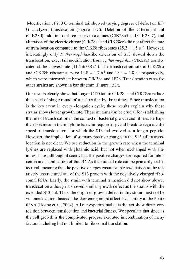
Modification of S13 C-terminal tail showed varying degrees of defect on EF-G catalyzed translocation (Figure 13C). Deletion of the C-terminal tail (CIK28d), addition of three or seven alanines (CIK28a3 and CIK28a7), and alteration of the electric charge (CIK28aa and CIK28ee) did not affect the rate of translocation compared to the CIK28 ribosomes (25.2 ± 1.5 s-1). However, interestingly only T. thermophilus-like extension of S13 slowed down the translocation, exact tail modification from T. thermophilus (CIK28c) translo-cated at the slowest rate (11.4 ± 0.8 s-1). The translocation rate of CIK28ca and CIK28b ribosomes were 14.8 ± 1.7 s-1 and 18.4 ± 1.8 s-1 respectively, which were intermediate between CIK28c and JE28. Translocation rates for other strains are shown in bar diagram (Figure 13D).
Our results clearly show that longer CTD tail in CIK28c and CIK28ca reduce the speed of single round of translocation by three times. Since translocation is the key event in every elongation cycle, these results explain why these strains show slower growth rate. These mutants can be crucial for establishing the role of translocation in the context of bacterial growth and fitness. Perhaps the ribosomes in thermophilic bacteria require a special break to regulate the speed of translocation, for which the S13 tail evolved as a longer peptide. However, the implication of so many positive charges in the S13 tail in trans-location is not clear. We see reduction in the growth rate when the terminal lysines are replaced with glutamic acid, but not when exchanged with ala-nines. Thus, although it seems that the positive charges are required for inter-action and stabilization of the tRNAs their actual role can be primarily archi-tectural, meaning that the positive charges ensure stable association of the rel-atively unstructured tail of the S13 protein with the negatively charged ribo-somal RNA. Lastly, the strain with terminal truncation did not show slower translocation although it showed similar growth defect as the strains with the extended S13 tail. Thus, the origin of growth defect in this strain must not be via translocation. Instead, the shortening might affect the stability of the P-site tRNA (Hoang et al., 2004). All our experimental data did not show direct cor-relation between translocation and bacterial fitness. We speculate that since as the cell growth is the complicated process executed in combination of many factors including but not limited to ribosomal translation.

44
Antibiotic Arbekacin on translocation Antibiotic arbekacin is a semisynthetic aminoglycoside, belongs to the kana-mycin family, and originally synthesized from dibekacin in 1970s. It was clin-ically introduced in Japan in 1990s, and rather successful in the effort to over-come the antibiotic resistance. The modes of action of arbekacin in inhibiting protein synthesis in bacteria has not fully understood, however it was antici-pated in comparison with other similar aminoglycosides. It is believed that arbekacin binds to the common aminoglycoside binding site in the 30S, and affects the accuracy of elongation as well as translocation (Feldman et al., 2010, Romanowska, Reuter et al., 2013, Tsai et al., 2013).
In this study we have investigated the effects of arbekacin on translocation by adding it in the optimized fluorescent-mRNA based translocation assay. As expected from the results with other aminoglycoside and tuberactinomycins, arbekacin showed strong inhibition of translocation. By fitting the transloca-tion data, the fraction of the ribosome inhibited with arbekacin was estimated and plotted against arbekacin concentration. The inhibition constant was de-termined from the midpoint of the transition. When ribosomes were pre-incu-bated with arbekacin, the KI was as low as about 0.4 µM. However, it in-creased to 2 ± 0.3 µM (KIC) when added in the EF-G mix. It means that pre-incubation of ribosome with arbekacin is certainly beneficial for the drug ac-tion. Further, from the rate of the slow phases the dwelling time of the drug could be estimated. Unlike viomycin (Holm et al., 2016), The dwelling time of arbekacin decreased with increasing EF-G concentration (Figure 14). More-over, at a given EF-G concentration the dwelling time of the drug did not change with increasing arbekacin concentration (from 0.25 µM to 20 µM). It means that (i) arbekacin competes with EF-G for ribosome binding, but (ii) it cannot bind back to the ribosome once EF-G binds to it. This results clearly indicates that for effective inhibition arbekacin must bind to the ribosome prior to EF-G binding and it cannot bind back once it gets detached by com-petitive binding of EF-G. We have also followed the role of arbekacin in in-ducing error in translation and inhibiting ribosome recycling. These results are included in paper IV.

45
Figure 14. Effects of Arbekacin on translocation. (A) The translocation mean time at varying concentrations of EF-G and arbekacin. (B) Translocation rates in inhibition by arbekacin are plotted over EF-G concentration.
From the translocation data, the arbekacin mode of action on translocation can be explained as follows (Figure 15). kp is the rate constant for uninhibited 70S to bind to EF-G after formation of peptide bond, which was estimated as 50 s-
1 (from the rate of dipeptide ML formation in the experiments and fast binding of EF-G to the 70S pre-T complex). From the inhibition constant when ar-bekacin was incubated with the 70S (KIC = 2 ± 0.3 µM) and kp, k1 was deter-mined as kp / KIC = 25 ± 4 µM-1 s-1. As KI (0.4 µM) is the ratio of k1 and k-1, k-
1 (10 s-1) can be estimated. kmRNA was estimated as kcat of translocation (11.4 ± 0.7 s-1, Table 3A) while kg (0.007 µM-1 s-1) is EF-G concentration dependent step and determined from the slop value of the translocation plot over arbeka-cin concentration (Figure 14B). It is suggested that arbekacin is a tight binder and EF-G strives to push out arbekacin for successful translocation. Arbekacin binds to the 70S and stabilizes the “flipped off” conformation of the three monitoring bases at the DC, which is a translocation incompetent confor-mation (Feldman et al., 2010, Munro et al., 2010b). Moreover, it has less af-finity to the ratcheted conformation of 70S stabilized by EF-G.
Figure 15. Kinetic model for arbekacin inhibition on translocation. 70S* represents the post-T complex. Abk = arbekacin
46
Conclusions and future perspective
This thesis mainly aimed at the optimization of the pyrene-labeled mRNA based translocation assay for high florescence signal and high in vivo-like rate both in quench-flow and stopped-flow measurement. The optimized condi-tions we suggest are mRNA+10, translocation starting from IC and higher concentrations of EF-G, NAc-Phe-tRNAPhe, low concentration of free Mg2+ in the solution and higher temperature (preferably 37 ⁰C) in the translocation starting from pre-T complex with NAc-Phe-tRNAPhe. We have also suggested the proper analysis of biphasic curve of translocation by increasing the ampli-tude proportion of the fast phase. In the subsequent studies applying the opti-mized the translocation assay, we have confirmed the importance of GTP hy-drolysis, nucleotide binding to the EF-G using non-hydrolysable GTP analogs and EF-G mutants. Furthermore, the C-terminal tail of the ribosomal protein S13 from Thermus thermophilus was revealed to affect the translocation in a manner of length and charge interaction. The mode of action of novel antibi-otic arbekacin was explained kinetically.
The future steps from this thesis is to truly determine the role of GTP hydrol-ysis not only in connection with the mRNA-tRNA movement but also struc-tural rearrangements in EF-G and the ribosome such as subunit ratcheting and back-ratcheting, and swiveling action of 30S head in reference to the body. The sequence of the action is also important for better understanding of the EF-G directed ribosomal translocation. The optimized real time translocation assay opened an opportunity for the investigation of the mechanism of antibi-otics targeting translocation and other factors affecting translocation such as ribosomal proteins and modification of RNAs.
47
Sammanfattning på Svenska
Ribosomen är ett makromolekylärt maskineri vars struktur består utav tre ribosomala RNA:n och tiotal ribosomala proteiner. Dess proteinskapande funktion är livsavgörande för celler. Detta, då proteiner är viktiga molekyler i kroppen; nästan alla enzymer som katalyserar biokemiska reaktioner i kroppen är proteiner samt att de även bygger upp vävnader, muskler och signalreceptorer. Proteiner består vanligtvis utav hundratals aminosyror, och ribosomen använder dessa som byggstenar utifrån informationen den hämtar från den genetiska blåkopian mRNA.
Processen inom proteinsyntesen där ribosomen förlänger proteinet genom att addera aminosyror en efter en kallas för ”elongering” och den repeteras lika många gånger som antalet aminosyror i proteinet. I elongeringscykeln finns ett steg, translokering, där ribosomen rör sig längs mRNA:t. Ribosomtranslokeringen assisteras av elongeringsfaktor G (EF-G). EF-G behöver binda till och hydrolysera GTP för att kunna utföra translokeringen. Det är viktigt att förstå hur ribosomen rör sig ett kodon (tre nukleotider kodar för en aminosyra) korrekt, hur snabbt det sker, samt hur EF-G hjälper ribosomtranslokeringen. En populär metod har varit att använda fluorescerande färger, fästa till mRNA, och sedan följa signalen för att avgöra hur snabbt mRNA:t rör sig i referens till ribosomen. Även om fluorescerande färger är en väldigt kraftfull och smart sätt att studera ribosomfunktionen, krävs det dock att den kalibreras noggrant med hjälp av andra metoder. Vi har använt tripeptidsyntesanalys, vilken har utvecklats för snabba kinetiska studier av elongeringen med radioaktiva aminosyror som detekteringsmetod istället för fluorescens.
Genom kalibering av fluorescerande mRNA med tripeptidsyntesanalys, har vi optimiserat den korrekta positionen av färgen så att den kan utnyttjas tillförlitligt i fortsatta studier. Vi har också utvecklat de optimala förhållandena för pålitliga mätningar inom translokeringen, som temperatur, koncentrationen av tRNA och EF-G samt magnesiumjonkoncentrationen i
48
bufferten. Genom att använda detta optimiserade protokoll har vi undersökt mutationer i EF-G som påverkar hydrolysering av GTP och translokering.
Bakterieribosomen är en vanlig måltavla för de antibiotika som används inom medicin. Arbekacin är en utav dem och används för att behandla multiresistenta cellinjer. Vi har använt den optimiserade fluorescensmetoden för att utröna Arbekacins molekylära mekanism inom translokeringen. Ribosomala protein spelar också en roll i translokeringen och ett exempel är proteinet S13. Strukturen på proteinets C-terminal i bakterien Thermus thermophilus ger indikation på att den hindrar translokering av tRNA som är kopplat till mRNA. Denna C-terminal introducerades i S13 hos modellorganismen Escherichia coli genom mutation och dess effekt på translokeringen kunde förklaras.

49
Acknowledgements
I wish to express my deepest gratitude to my supervisor Suparna Sanyal for accepting me in her group and for the great supervision. The past years are great experiences for me, we have gone through thick and thin together. I have gained more knowledge as well as become a better person, and I can never imagine this without your intelligence and kind-hearted support.
Thank you, Måns Ehrenberg and my co-supervisor Magnus Johansson, for your kind support, it will never be forgotten.
I would also like to thank my former and current colleagues in Sanyal Lab for all the instructions for the instrument, helpful discussions, kind support and all the funs we had together. Mikael, you were the first one that taught me about kinetics and helped me with the projects and you still give me valuable opinions about the research I am doing. You are an intelligent, very under-standing person, and very good friend together with Volance. Thank you, Liang and Narayan for the crucial help you have provided during those years, especially for this thesis. Soneya, I will still thank you for your harsh criticism which made me feel useless during the hard times. Your kind-hearted and val-uable supports are greatly appreciated. I am thankful for Shreya who always supported in the lab as well as in the gym. Ravi, Petar and Rasa, thank you for being the truthful friends and for lovely drinks and fun we had together instead of serious scientific discussion. Ray, thank you for all the components you have purified for the projects and the moral supports you have given to me. Thank you, Josefine for the help in Swedish, and all other people in the molecular biology program. I wish to show my gratitude to people in and out-side the department, head of the department, administrative staff, and other fellow scientists, who have been friendly all these years.
I am extremely thankful to Chandu, for all his selfless help in work and life whenever I needed. You have welcomed me in the office as well as in your family. Thanks to you and your family, Geeta, Nandu and Aashritha, I have survived the last four years.

50
To my friends, Sethu, Srinivas and Sudarsan for the lunches we had together even though I moved to a better table later. Thank you, Yanyu and Kehuan, for being such happy friends in the gang for the last year and all the activities we had together. To Staffan, Liangwen, Jinfeng, Zaiqiu, Ming, and Yu for wonderful times in Gotland. To all the Chinese friends from whom I have learnt a word or two of Chinese together with happiness, Honglian, Tianyi, Miao, Chen Lei, Wangshu, Zhirong, Lu Lu, Zhang Lu and others. Last but not least, the longest and closest friend Kwangchol Mun, who have been un-derstanding and encouraging, thank you for the meals you provided.
I am indebted to Ulrica, international office at UU and Erasmus Mundus pro-gram, for supporting me for this wonderful opportunity, financially and men-tally.
To my parents, thank you for all the love you have poured on me even during the toughest time of yours. It is the most valuable feeling for me, that has nurtured me this far.
51
Reference
Abdurashidova GG, Tsvetkova EA, Budowsky EI (1991) Direct tRNA-protein interactions in ribosomal complexes. Nucleic Acids Res 19: 1909-15
Adio S, Sharma H, Senyushkina T, Karki P, Maracci C, Wohlgemuth I, Holtkamp W, Peske F, Rodnina MV (2018) Dynamics of ribosomes and release factors during translation termination in E. coli. Elife 7
Agirrezabala X, Liao HY, Schreiner E, Fu J, Ortiz-Meoz RF, Schulten K, Green R, Frank J (2012) Structural characterization of mRNA-tRNA translocation intermediates. Proc Natl Acad Sci U S A 109: 6094-9
Andreeva I, Belardinelli R, Rodnina MV (2018) Translation initiation in bacterial polysomes through ribosome loading on a standby site on a highly translated mRNA. Proc Natl Acad Sci U S A 115: 4411-4416
Antoun A, Pavlov MY, Lovmar M, Ehrenberg M (2006) How initiation factors maximize the accuracy of tRNA selection in initiation of bacterial protein synthesis. Molecular Cell 23: 183-193
Antoun A, Pavlov MY, Tenson T, Ehrenberg MM (2004) Ribosome formation from subunits studied by stopped-flow and Rayleigh light scattering. Biol Proced Online 6: 35-54
Blanchard SC, Kim HD, Gonzalez RL, Jr., Puglisi JD, Chu S (2004) tRNA dynamics on the ribosome during translation. Proc Natl Acad Sci U S A 101: 12893-8
Bocchetta M, Xiong L, Shah S, Mankin AS (2001) Interactions between 23S rRNA and tRNA in the ribosomal E site. RNA 7: 54-63
Borg A, Ehrenberg M (2015) Determinants of the rate of mRNA translocation in bacterial protein synthesis. J Mol Biol 427: 1835-47
Borg A, Holm M, Shiroyama I, Hauryliuk V, Pavlov M, Sanyal S, Ehrenberg M (2015) Fusidic acid targets elongation factor G in several stages of translocation on the bacterial ribosome. J Biol Chem 290: 3440-54
Borg A, Pavlov M, Ehrenberg M (2016) Mechanism of fusidic acid inhibition of RRF- and EF-G-dependent splitting of the bacterial post-termination ribosome. Nucleic Acids Res 44: 3264-75
Bourne HR, Sanders DA, McCormick F (1991) The GTPase superfamily: conserved structure and molecular mechanism. Nature 349: 117-27

52
Brilot AF, Korostelev AA, Ermolenko DN, Grigorieff N (2013) Structure of the ribosome with elongation factor G trapped in the pretranslocation state. Proc Natl Acad Sci U S A 110: 20994-9
Chen J, Tsai A, O'Leary SE, Petrov A, Puglisi JD (2012) Unraveling the dynamics of ribosome translocation. Curr Opin Struct Biol 22: 804-14
Chen Y, Feng S, Kumar V, Ero R, Gao YG (2013) Structure of EF-G-ribosome complex in a pretranslocation state. Nat Struct Mol Biol 20: 1077-+
Cornish PV, Ermolenko DN, Noller HF, Ha T (2008) Spontaneous intersubunit rotation in single ribosomes. Mol Cell 30: 578-88
Cornish PV, Ermolenko DN, Staple DW, Hoang L, Hickerson RP, Noller HF, Ha T (2009) Following movement of the L1 stalk between three functional states in single ribosomes. Proc Natl Acad Sci U S A 106: 2571-6
Crick F (1970) Central dogma of molecular biology. Nature 227: 561-3 Cukras AR, Green R (2005) Multiple effects of S13 in modulating the strength
of intersubunit interactions in the ribosome during translation. J Mol Biol 349: 47-59
Cunha CE, Belardinelli R, Peske F, Holtkamp W, Wintermeyer W, Rodnina MV (2013) Dual use of GTP hydrolysis by elongation factor G on the ribosome. Translation (Austin) 1: e24315
D'Costa VM, King CE, Kalan L, Morar M, Sung WWL, Schwarz C, Froese D, Zazula G, Calmels F, Debruyne R, Golding GB, Poinar HN, Wright GD (2011) Antibiotic resistance is ancient. Nature 477: 457-461
Davis BD (1987) Mechanism of bactericidal action of aminoglycosides. Microbiol Rev 51: 341-50
Demeshkina N, Jenner L, Yusupova G, Yusupov M (2010) Interactions of the ribosome with mRNA and tRNA. Curr Opin Struct Biol 20: 325-32
Dong H, Nilsson L, Kurland CG (1996) Co-variation of tRNA abundance and codon usage in Escherichia coli at different growth rates. J Mol Biol 260: 649-63
Dorner S, Brunelle JL, Sharma D, Green R (2006) The hybrid state of tRNA binding is an authentic translation elongation intermediate. Nat Struct Mol Biol 13: 234-41
Ederth J, Mandava CS, Dasgupta S, Sanyal S (2009) A single-step method for purification of active His-tagged ribosomes from a genetically engineered Escherichia coli. Nucleic Acids Res 37: e15
Ermolenko DN, Majumdar ZK, Hickerson RP, Spiegel PC, Clegg RM, Noller HF (2007) Observation of intersubunit movement of the ribosome in solution using FRET. J Mol Biol 370: 530-40
Ermolenko DN, Noller HF (2011) mRNA translocation occurs during the second step of ribosomal intersubunit rotation. Nat Struct Mol Biol 18: 457-62

53
Fei J, Bronson JE, Hofman JM, Srinivas RL, Wiggins CH, Gonzalez RL, Jr. (2009) Allosteric collaboration between elongation factor G and the ribosomal L1 stalk directs tRNA movements during translation. Proc Natl Acad Sci U S A 106: 15702-7
Fei J, Kosuri P, MacDougall DD, Gonzalez RL, Jr. (2008) Coupling of ribosomal L1 stalk and tRNA dynamics during translation elongation. Mol Cell 30: 348-59
Feinberg JS, Joseph S (2006) Ribose 2'-hydroxyl groups in the 5' strand of the acceptor arm of P-site tRNA are not essential for EF-G catalyzed translocation. RNA 12: 580-8
Feldman MB, Terry DS, Altman RB, Blanchard SC (2010) Aminoglycoside activity observed on single pre-translocation ribosome complexes. Nat Chem Biol 6: 244
Fislage M, Zhang JJ, Brown ZP, Mandava CS, Sanyal S, Ehrenberg M, Frank J (2018) Cryo-EM shows stages of initial codon selection on the ribosome by aa-tRNA in ternary complex with GTP and the GTPase-deficient EF-Tu(H84A). Nucleic Acids Research 46: 5861-5874
Flis J, Holm M, Rundlet EJ, Loerke J, Hilal T, Dabrowski M, Burger J, Mielke T, Blanchard SC, Spahn CMT, Budkevich TV (2018) tRNA Translocation by the Eukaryotic 80S Ribosome and the Impact of GTP Hydrolysis. Cell Rep 25: 2676-2688 e7
Frank J, Agrawal RK (2000) A ratchet-like inter-subunit reorganization of the ribosome during translocation. Nature 406: 318-22
Frank J, Gao H, Sengupta J, Gao N, Taylor DJ (2007) The process of mRNA-tRNA translocation. Proc Natl Acad Sci U S A 104: 19671-8
Fu Z, Indrisiunaite G, Kaledhonkar S, Shah B, Sun M, Chen B, Grassucci RA, Ehrenberg M, Frank J (2019) The structural basis for release-factor activation during translation termination revealed by time-resolved cryogenic electron microscopy. Nat Commun 10: 2579
Gao H, Zhou Z, Rawat U, Huang C, Bouakaz L, Wang C, Cheng Z, Liu Y, Zavialov A, Gursky R, Sanyal S, Ehrenberg M, Frank J, Song H (2007) RF3 induces ribosomal conformational changes responsible for dissociation of class I release factors. Cell 129: 929-41
Gao YG, Selmer M, Dunham CM, Weixlbaumer A, Kelley AC, Ramakrishnan V (2009) The structure of the ribosome with elongation factor G trapped in the posttranslocational state. Science 326: 694-9
Gavrilova LP, Spirin AS (1971) Stimulation of "non-enzymic" translocation in ribosomes by p-chloromercuribenzoate. FEBS Lett 17: 324-326
Goyal A, Belardinelli R, Rodnina MV (2017) Non-canonical Binding Site for Bacterial Initiation Factor 3 on the Large Ribosomal Subunit. Cell Rep 20: 3113-3122
54
Graf M, Huter P, Maracci C, Peterek M, Rodnina MV, Wilson DN (2018) Visualization of translation termination intermediates trapped by the Apidaecin 137 peptide during RF3-mediated recycling of RF1. Nat Commun 9: 3053
Guo Z, Noller HF (2012) Rotation of the head of the 30S ribosomal subunit during mRNA translocation. Proc Natl Acad Sci U S A 109: 20391-4
Hansson S, Singh R, Gudkov AT, Liljas A, Logan DT (2005) Crystal structure of a mutant elongation factor G trapped with a GTP analogue. FEBS Lett 579: 4492-7
Hirashima A, Kaji A (1973) Role of elongation factor G and a protein factor on the release of ribosomes from messenger ribonucleic acid. J Biol Chem 248: 7580-7
Hoang L, Fredrick K, Noller HF (2004) Creating ribosomes with an all-RNA 30S subunit P site. Proc Natl Acad Sci U S A 101: 12439-43
Holm M, Borg A, Ehrenberg M, Sanyal S (2016) Molecular mechanism of viomycin inhibition of peptide elongation in bacteria. Proc Natl Acad Sci U S A 113: 978-83
Holtkamp W, Cunha CE, Peske F, Konevega AL, Wintermeyer W, Rodnina MV (2014) GTP hydrolysis by EF-G synchronizes tRNA movement on small and large ribosomal subunits. EMBO J 33: 1073-85
Horan LH, Noller HF (2007) Intersubunit movement is required for ribosomal translocation. P Natl Acad Sci USA 104: 4881-4885
Ieong KW, Uzun U, Selmer M, Ehrenberg M (2016) Two proofreading steps amplify the accuracy of genetic code translation. P Natl Acad Sci USA 113: 13744-13749
Ito K, Uno M, Nakamura Y (2000) A tripeptide 'anticodon' deciphers stop codons in messenger RNA. Nature 403: 680-4
Jamiolkowski RM, Chen C, Cooperman BS, Goldman YE (2017) tRNA Fluctuations Observed on Stalled Ribosomes Are Suppressed during Ongoing Protein Synthesis. Biophys J 113: 2326-2335
Jelenc PC, Kurland CG (1979) Nucleoside triphosphate regeneration decreases the frequency of translation errors. Proc Natl Acad Sci U S A 76: 3174-8
Jin H, Kelley AC, Loakes D, Ramakrishnan V (2010) Structure of the 70S ribosome bound to release factor 2 and a substrate analog provides insights into catalysis of peptide release. P Natl Acad Sci USA 107: 8593-8598
Johansson M, Zhang JJ, Ehrenberg M (2012) Genetic code translation displays a linear trade-off between efficiency and accuracy of tRNA selection. P Natl Acad Sci USA 109: 131-136
55
Juette MF, Terry DS, Wasserman MR, Altman RB, Zhou Z, Zhao H, Blanchard SC (2016) Single-molecule imaging of non-equilibrium molecular ensembles on the millisecond timescale. Nat Methods 13: 341-4
Kannan K, Kanabar P, Schryer D, Florin T, Oh E, Bahroos N, Tenson T, Weissman JS, Mankin AS (2014) The general mode of translation inhibition by macrolide antibiotics. Proc Natl Acad Sci U S A 111: 15958-63
Khade PK, Joseph S (2011) Messenger RNA interactions in the decoding center control the rate of translocation. Nat Struct Mol Biol 18: 1300-1302
Kim HD, Puglisi JD, Chu S (2007) Fluctuations of transfer RNAs between classical and hybrid states. Biophys J 93: 3575-82
Kondo S (1994) Development of arbekacin and synthesis of new derivatives stable to enzymatic modifications by methicillin-resistant Staphylococcus aureus. The Japanese Journal of Antibiotics 47: 561-74
Koripella RK, Chen Y, Peisker K, Koh CS, Selmer M, Sanyal S (2012) Mechanism of elongation factor-G-mediated fusidic acid resistance and fitness compensation in Staphylococcus aureus. J Biol Chem 287: 30257-67
Koripella RK, Holm M, Dourado D, Mandava CS, Flores S, Sanyal S (2015) A conserved histidine in switch-II of EF-G moderates release of inorganic phosphate. Sci Rep 5: 12970
Korostelev A, Asahara H, Lancaster L, Laurberg M, Hirschi A, Zhu J, Trakhanov S, Scott WG, Noller HF (2008) Crystal structure of a translation termination complex formed with release factor RF2. Proc Natl Acad Sci U S A 105: 19684-9
Kotra LP, Haddad J, Mobashery S (2000) Aminoglycosides: perspectives on mechanisms of action and resistance and strategies to counter resistance. Antimicrob Agents Chemother 44: 3249-56
Kuhle B, Ficner R (2014) A monovalent cation acts as structural and catalytic cofactor in translational GTPases. EMBO J 33: 2547-63
Lai J, Ghaemi Z, Luthey-Schulten Z (2017) The Conformational Change in Elongation Factor Tu Involves Separation of Its Domains. Biochemistry 56: 5972-5979
Laurberg M, Kristensen O, Martemyanov K, Gudkov AT, Nagaev I, Hughes D, Liljas A (2000) Structure of a mutant EF-G reveals domain III and possibly the fusidic acid binding site. J Mol Biol 303: 593-603
Leipe DD, Wolf YI, Koonin EV, Aravind L (2002) Classification and evolution of P-loop GTPases and related ATPases. J Mol Biol 317: 41-72
Li W, Liu Z, Koripella RK, Langlois R, Sanyal S, Frank J (2015) Activation of GTP hydrolysis in mRNA-tRNA translocation by elongation factor G. Sci Adv 1
56
Lin J, Gagnon MG, Bulkley D, Steitz TA (2015) Conformational changes of elongation factor G on the ribosome during tRNA translocation. Cell 160: 219-27
Lin J, Zhou D, Steitz TA, Polikanov YS, Gagnon MG (2018) Ribosome-Targeting Antibiotics: Modes of Action, Mechanisms of Resistance, and Implications for Drug Design. Annu Rev Biochem 87: 451-478
Ling C, Ermolenko DN (2016) Structural insights into ribosome translocation. Wires Rna 7: 620-636
Loveland AB, Demo G, Grigorieff N, Korostelev AA (2017) Ensemble cryo-EM elucidates the mechanism of translation fidelity. Nature 546: 113-+
Mace K, Giudice E, Chat S, Gillet R (2018) The structure of an elongation factor G-ribosome complex captured in the absence of inhibitors. Nucleic Acids Res 46: 3211-3217
Mandava CS, Peisker K, Ederth J, Kumar R, Ge X, Szaflarski W, Sanyal S (2012) Bacterial ribosome requires multiple L12 dimers for efficient initiation and elongation of protein synthesis involving IF2 and EF-G. Nucleic Acids Res 40: 2054-64
Moazed D, Noller HF (1989) Intermediate states in the movement of transfer RNA in the ribosome. Nature 342: 142-8
Mohr D, Wintermeyer W, Rodnina MV (2000) Arginines 29 and 59 of elongation factor G are important for GTP hydrolysis or translocation on the ribosome. EMBO J 19: 3458-64
Munro JB, Altman RB, Tung CS, Cate JH, Sanbonmatsu KY, Blanchard SC (2010a) Spontaneous formation of the unlocked state of the ribosome is a multistep process. Proc Natl Acad Sci U S A 107: 709-14
Munro JB, Wasserman MR, Altman RB, Wang L, Blanchard SC (2010b) Correlated conformational events in EF-G and the ribosome regulate translocation. Nat Struct Mol Biol 17: 1470-7
Nakamura I, Yamaguchi T, Tsukimori A, Sato A, Fukushima S, Matsumoto T (2015) New options of antibiotic combination therapy for multidrug-resistant Pseudomonas aeruginosa. Eur J Clin Microbiol 34: 83-87
Ning W, Fei J, Gonzalez RL, Jr. (2014) The ribosome uses cooperative conformational changes to maximize and regulate the efficiency of translation. Proc Natl Acad Sci U S A 111: 12073-8
Ogle JM, Brodersen DE, Clemons WM, Jr., Tarry MJ, Carter AP, Ramakrishnan V (2001) Recognition of cognate transfer RNA by the 30S ribosomal subunit. Science 292: 897-902
Ogle JM, Carter AP, Ramakrishnan V (2003) Insights into the decoding mechanism from recent ribosome structures. Trends Biochem Sci 28: 259-266
57
Pallesen J, Hashem Y, Korkmaz G, Koripella RK, Huang C, Ehrenberg M, Sanyal S, Frank J (2013) Cryo-EM visualization of the ribosome in termination complex with apo-RF3 and RF1. Elife 2: e00411
Parmeggiani A, Krab IM, Watanabe T, Nielsen RC, Dahlberg C, Nyborg J, Nissen P (2006) Enacyloxin IIa pinpoints a binding pocket of elongation factor Tu for development of novel antibiotics. J Biol Chem 281: 2893-900
Pavlov MY, Antoun A, Lovmar M, Ehrenberg M (2008) Complementary roles of initiation factor 1 and ribosome recycling factor in 70S ribosome splitting. EMBO J 27: 1706-17
Perla-Kajan J, Lin X, Cooperman BS, Goldman E, Jakubowski H, Knudsen CR, Mandecki W (2010) Properties of Escherichia coli EF-Tu mutants designed for fluorescence resonance energy transfer from tRNA molecules. Protein Eng Des Sel 23: 129-36
Peske F, Matassova NB, Savelsbergh A, Rodnina MV, Wintermeyer W (2000) Conformationally restricted elongation factor G retains GTPase activity but is inactive in translocation on the ribosome. Mol Cell 6: 501-5
Pestka S (1968) Studies on the formation of trensfer ribonucleic acid-ribosome complexes. V. On the function of a soluble transfer factor in protein synthesis. Proc Natl Acad Sci U S A 61: 726-33
Polikanov YS, Aleksashin NA, Beckert B, Wilson DN (2018) The Mechanisms of Action of Ribosome-Targeting Peptide Antibiotics. Front Mol Biosci 5: 48
Ratje AH, Loerke J, Mikolajka A, Brunner M, Hildebrand PW, Starosta AL, Donhofer A, Connell SR, Fucini P, Mielke T, Whitford PC, Onuchic JN, Yu Y, Sanbonmatsu KY, Hartmann RK, Penczek PA, Wilson DN, Spahn CM (2010) Head swivel on the ribosome facilitates translocation by means of intra-subunit tRNA hybrid sites. Nature 468: 713-6
Rawat U, Gao H, Zavialov A, Gursky R, Ehrenberg M, Frank J (2006) Interactions of the release factor RF1 with the ribosome as revealed by cryo-EM. J Mol Biol 357: 1144-53
Rodnina MV, Gromadski KB, Kothe U, Wieden HJ (2005) Recognition and selection of tRNA in translation. FEBS Lett 579: 938-42
Rodnina MV, Savelsbergh A, Katunin VI, Wintermeyer W (1997) Hydrolysis of GTP by elongation factor G drives tRNA movement on the ribosome. Nature 385: 37-41
Romanowska J, Reuter N, Trylska J (2013) Comparing aminoglycoside binding sites in bacterial ribosomal RNA and aminoglycoside modifying enzymes. Proteins 81: 63-80
Roy B, Liu Q, Shoji S, Fredrick K (2018) IF2 and unique features of initiator tRNA(fMet) help establish the translational reading frame. RNA Biol 15: 604-613
58
Salsi E, Farah E, Dann J, Ermolenko DN (2014) Following movement of domain IV of elongation factor G during ribosomal translocation. Proc Natl Acad Sci U S A 111: 15060-5
Salsi E, Farah E, Ermolenko DN (2016) EF-G Activation by Phosphate Analogs. J Mol Biol 428: 2248-58
Salsi E, Farah E, Netter Z, Dann J, Ermolenko DN (2015) Movement of elongation factor G between compact and extended conformations. J Mol Biol 427: 454-67
Schmeing TM, Voorhees RM, Kelley AC, Gao YG, Murphy FVt, Weir JR, Ramakrishnan V (2009) The crystal structure of the ribosome bound to EF-Tu and aminoacyl-tRNA. Science 326: 688-694
Scolnick E, Tompkins R, Caskey T, Nirenberg M (1968) Release factors differing in specificity for terminator codons. Proc Natl Acad Sci U S A 61: 768-74
Sharma H, Adio S, Senyushkina T, Belardinelli R, Peske F, Rodnina MV (2016) Kinetics of Spontaneous and EF-G-Accelerated Rotation of Ribosomal Subunits. Cell Rep 16: 2187-96
Shetty S, Shah RA, Chembazhi UV, Sah S, Varshney U (2017) Two highly conserved features of bacterial initiator tRNAs license them to pass through distinct checkpoints in translation initiation. Nucleic Acids Res 45: 2040-2050
Shi X, Joseph S (2016) Mechanism of Translation Termination: RF1 Dissociation Follows Dissociation of RF3 from the Ribosome. Biochemistry 55: 6344-6354
Shine J, Dalgarno L (1974) Identical 3'-terminal octanucleotide sequence in 18S ribosomal ribonucleic acid from different eukaryotes. A proposed role for this sequence in the recognition of terminator codons. Biochem J 141: 609-15
Shoji S, Dambacher CM, Shajani Z, Williamson JR, Schultz PG (2011) Systematic chromosomal deletion of bacterial ribosomal protein genes. J Mol Biol 413: 751-61
Spiegel PC, Ermolenko DN, Noller HF (2007) Elongation factor G stabilizes the hybrid-state conformation of the 70S ribosome. Rna 13: 1473-1482
Studer SM, Feinberg JS, Joseph S (2003) Rapid kinetic analysis of EF-G-dependent mRNA translocation in the ribosome. J Mol Biol 327: 369-81
Svidritskiy E, Korostelev AA (2018) Conformational Control of Translation Termination on the 70S Ribosome. Structure 26: 821-828 e3
Tsai A, Uemura S, Johansson M, Puglisi EV, Marshall RA, Aitken CE, Korlach J, Ehrenberg M, Puglisi JD (2013) The impact of aminoglycosides on the dynamics of translation elongation. Cell Rep 3: 497-508
Valle M, Zavialov A, Li W, Stagg SM, Sengupta J, Nielsen RC, Nissen P, Harvey SC, Ehrenberg M, Frank J (2003a) Incorporation of aminoacyl-tRNA into
59
the ribosome as seen by cryo-electron microscopy. Nat Struct Biol 10: 899-906
Valle M, Zavialov A, Sengupta J, Rawat U, Ehrenberg M, Frank J (2003b) Locking and unlocking of ribosomal motions. Cell 114: 123-34
Varshney U, Lee CP, RajBhandary UL (1993) From elongator tRNA to initiator tRNA. Proc Natl Acad Sci U S A 90: 2305-9
Vestergaard B, Sanyal S, Roessle M, Mora L, Buckingham RH, Kastrup JS, Gajhede M, Svergun DI, Ehrenberg M (2005) The SAXS solution structure of RF1 differs from its crystal structure and is similar to its ribosome bound cryo-EM structure. Mol Cell 20: 929-38
Volkov IL, Johansson M (2019) Single-Molecule Tracking Approaches to Protein Synthesis Kinetics in Living Cells. Biochemistry 58: 7-14
Walker SE, Shoji S, Pan D, Cooperman BS, Fredrick K (2008) Role of hybrid tRNA-binding states in ribosomal translocation. Proc Natl Acad Sci U S A 105: 9192-7
Wang J, Caban K, Gonzalez RL, Jr. (2015) Ribosomal initiation complex-driven changes in the stability and dynamics of initiation factor 2 regulate the fidelity of translation initiation. J Mol Biol 427: 1819-34
Wang L, Pulk A, Wasserman MR, Feldman MB, Altman RB, Cate JH, Blanchard SC (2012) Allosteric control of the ribosome by small-molecule antibiotics. Nat Struct Mol Biol 19: 957-63
Wasserman MR, Alejo JL, Altman RB, Blanchard SC (2016) Multiperspective smFRET reveals rate-determining late intermediates of ribosomal translocation. Nat Struct Mol Biol 23: 333-41
Watanabe A, Yanagihara K, Matsumoto T, Kohno S, Aoki N, Oguri T, Sato J, Muratani T, Yagisawa M, Ogasawara K, Koashi N, Kozuki T, Komoto A, Takahashi Y, Tsuji T, Terada M, Nakanishi K, Hattori R, Hirako Y, Maruo A et al. (2012) Nationwide surveillance of bacterial respiratory pathogens conducted by the Surveillance Committee of Japanese Society of Chemotherapy, Japanese Association for Infectious Diseases, and Japanese Society for Clinical Microbiology in 2009: general view of the pathogens' antibacterial susceptibility. J Infect Chemother 18: 609-620
Wieden HJ, Gromadski K, Rodnin D, Rodnina MV (2002) Mechanism of elongation factor (EF)-Ts-catalyzed nucleotide exchange in EF-Tu. Contribution of contacts at the guanine base. J Biol Chem 277: 6032-6
Wilson DN (2014) Ribosome-targeting antibiotics and mechanisms of bacterial resistance. Nat Rev Microbiol 12: 35-48
Wilson DN, Nierhaus KH (2003) The ribosome through the looking glass. Angew Chem Int Ed Engl 42: 3464-86
Xie P (2014) An explanation of biphasic characters of mRNA translocation in the ribosome. Biosystems 118: 1-7
60
Yusupov MM, Yusupova GZ, Baucom A, Lieberman K, Earnest TN, Cate JH, Noller HF (2001) Crystal structure of the ribosome at 5.5 A resolution. Science 292: 883-96
Zavialov AV, Buckingham RH, Ehrenberg M (2001) A posttermination ribosomal complex is the guanine nucleotide exchange factor for peptide release factor RF3. Cell 107: 115-24
Zavialov AV, Hauryliuk VV, Ehrenberg M (2005a) Guanine-nucleotide exchange on ribosome-bound elongation factor G initiates the translocation of tRNAs. J Biol 4: 9
Zavialov AV, Hauryliuk VV, Ehrenberg M (2005b) Splitting of the posttermination ribosome into subunits by the concerted action of RRF and EF-G. Molecular Cell 18: 675-686
Zavialov AV, Mora L, Buckingham RH, Ehrenberg M (2002) Release of peptide promoted by the GGQ motif of class 1 release factors regulates the GTPase activity of RF3. Mol Cell 10: 789-98
Zhou J, Korostelev A, Lancaster L, Noller HF (2012) Crystal structures of 70S ribosomes bound to release factors RF1, RF2 and RF3. Curr Opin Struct Biol 22: 733-42
Zhou J, Lancaster L, Donohue JP, Noller HF (2013) Crystal Structures of EF-G-Ribosome Complexes Trapped in Intermediate States of Translocation. Science 340: 1543-+

61

Acta Universitatis UpsaliensisDigital Comprehensive Summaries of Uppsala Dissertationsfrom the Faculty of Science and Technology 1877
Editor: The Dean of the Faculty of Science and Technology
A doctoral dissertation from the Faculty of Science andTechnology, Uppsala University, is usually a summary of anumber of papers. A few copies of the complete dissertationare kept at major Swedish research libraries, while thesummary alone is distributed internationally throughthe series Digital Comprehensive Summaries of UppsalaDissertations from the Faculty of Science and Technology.(Prior to January, 2005, the series was published under thetitle “Comprehensive Summaries of Uppsala Dissertationsfrom the Faculty of Science and Technology”.)
Distribution: publications.uu.seurn:nbn:se:uu:diva-396197
ACTAUNIVERSITATIS
UPSALIENSISUPPSALA
2019