tibia, fibula, ankle, and foot •articulates the distal portion of the knee joint with the ankle...
TRANSCRIPT

Tibia, Fibula, Ankle, and Foot
PSK 4U
Mr. S. Kelly
North Grenville DHS
Tibia
• Articulates the distal portion of the knee joint with the ankle joint
• Aka the “shin”, found medial and anterior to the smaller fibula
• Gender difference: in males, the direction is more vertical, more oblique in females to compensate for a more oblique shape of the femur
Tibia Structures
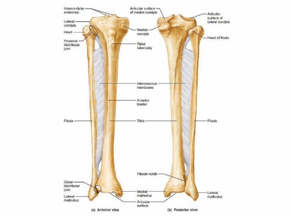
• Medial and lateral condyles: distal borders of the knee joint
• Interosseus membrane: connects tibia and fibula along the body or shaft of the bones
• Fibular notch: where the fibula articulates (distally)
• Medial malleolus: medial ankle protrusion
• Intercondylar eminence: splits intercondylar area into anterior and posterior (anterior area is ACL attachment site
Tibial Tuberosity
• Attachment site for the patellar ligament
• Terminal point of the “lever” (quads, suprapatellar ligament, patella, patellar ligament) that extends the knee AND prevents knee from collapsing on foot-strikes
• Fractured rarely, but most commonly in adolescents (avulsion fractures)
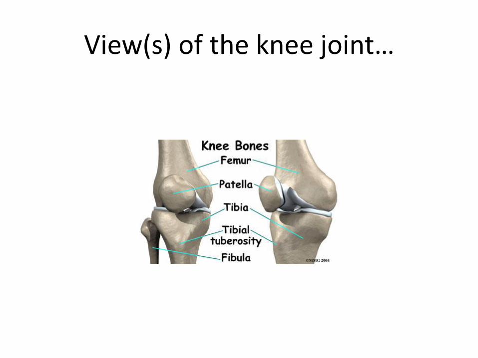
View(s) of the knee joint…
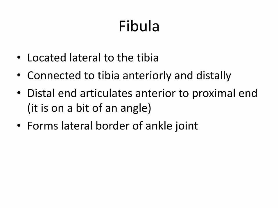
Fibula
• Located lateral to the tibia
• Connected to tibia anteriorly and distally
• Distal end articulates anterior to proximal end (it is on a bit of an angle)
• Forms lateral border of ankle joint
Fibula Structures
• Head: articulates below the tibia
• Lateral malleolus: lateral ankle protrusion
The Ankle
• Distal ends of tibia and fibula articulate with the talus
• The tibial articulation with the talus bears more weight than the fibular articulation (size difference)
• Numerous ligaments allow for the wide range of motion at this joint while bearing a relatively large weight
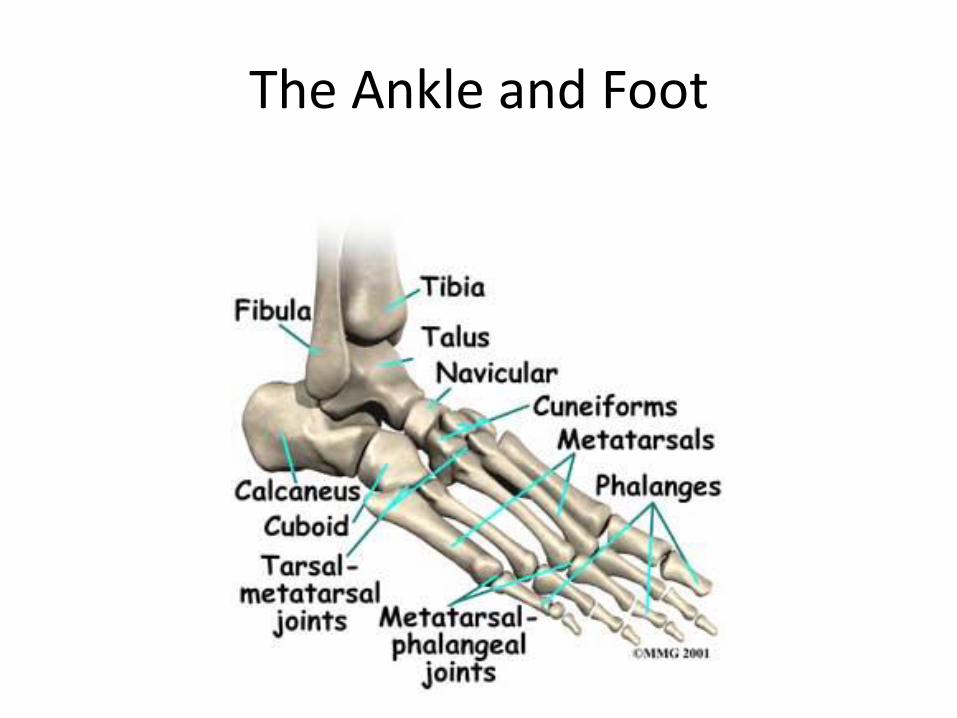
The Ankle and Foot
Talus
• Second-largest tarsal bone
• Large portion of the bone is covered in articulating cartilage
• Blood supply is “retrograde” meaning that the blood enters at the distal end
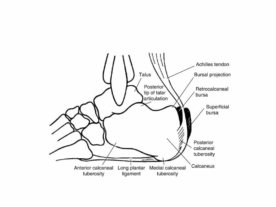
Calcaneus
• Heel bone
• Largest bone of the foot
• Calcaneal Tuberosity: insertion point for the Achilles tendon, origin for tendons (muscles) of the foot
Tarsal Bones
• Be able to locate and identify all the tarsal bones as follows:
• Cuneiform(s): medial, intermediate, lateral
• Navicular
• Cuboid
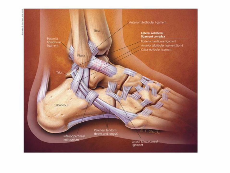
Lateral Ankle Ligaments
• Anterior Talofibular
• Posterior Talofibular
• Calcaneofibular
• Posterior Tibiofibular
• Anterior Tibiofibular
• These are, with lateral talocalcaneal, the most common ligaments involved in inversion sprains
Medial Ankle Ligaments
• The deltoid ligament, shaped like the Greek letter “delta”, also like a triangle
• From posterior to anterior:
• Posterior tibiotalar
• Tibiocalcaneal
• Anterior tibiotalar
• Tibionavicular
• Most commonly involved in painful eversion sprains

Metatarsals
• Numbered 1-5 from medial to lateral
• Similar to the metacarpals in the hand
• PHALANGES: similar to those of hand
• Be familiar with the joint names!!