thyroid autoimmunity and female gender
TRANSCRIPT

L. Chiovato, P. Lapi, E. Fiore, et al.
43. Ashitaka Y., Tojo S. Human placental thyrotropins. In: Grudzinskas J.G., Teisner B., Seppala M., (Eds.), Pregnancy proteins, biology, chemistry and clinical applications. Academic Press, New York, 1982, p. 357.
44. Hennen G., Pierce J.G., Freychet P. Human chorionic thyrotropin: Further characterization and study of its secretion during pregnancy. J. Clin. Endocrinol. Metab. 29:581,1969.
45. Harada A., Hershman J.M. Extraction of human chorionic thyrotropin (hCT) from term placentas: Failure to recover thyrotropic activity. J. Clin. Endocrinol. Metab. 47: 681,1978.
46. Mann K., Schneider N., Hoermann R. Thyrotropic activity of isoelectric variants of human chorionic gonadotropin from trophoblastic tumors. Endocrinology 118: 1558, 1986.
47. Yazaki K., Yazaki C., Wakabayashi K., Igarashi M. Isoelectric heterogeneity of human chorionic gonadotropin: presence of choriocarcinoma specific components. Am. J. Obstet. Gynecol. 138: 189,1980.
48. Kobata A. Structures, function, and transformational changes of the sugar chains of glycohormones. J. Cell. Biochem. 37: 79, 1988.
49. Ballabio M., Poshychinda M., Ekins R.P. Pregnancy-induced changes in thyroid function: role of human chorionic gonadotropin as putative regulator of maternal thyroid. J. Clin. Endocrinol. Metab. 73: 824, 1991.
50. Pekonen F., Weintraub BD. Interaction of crude and pure chorionic gonadotropin with the thyrotropin receptor. J. Clin. Endocrinol. Metab. 50: 280, 1980.
51. Amir S.M., Sullivan R.C., Ingbar S.H. The effect of desialylation on the in vitro interaction of human chorionic gonadotropin with human thyroid plasma membranes. Endocrinology 109: 1203, 1981.
Thyroid autoimmunity and female gender1
L. Chiovato, P. Lapi, E. Fiore, M. Tonacchera, and A. Pinchera Istituto di Endocrinologia, Universita di Pisa, Tirrenia, Pisa, Italy 1This paper was supported by grants from the National Research Council (CNR, Rome, Italy). Target Project: Biotechnology and
384
Bioinstrumentation. Grant 91. 01219.PF70. and Target Project Prevention and Control of Disease Factors (FATMA).
Key-words. Sexual dimorphism, thyroid autoimmunity, postpartum thyroid dysfunction, pregnancy, neonatal thyroid diseases.
Correspondence: Dr. Luca Chiovato, Istituto di Endocrinologia,Unlverslta di Pisa, Viale del Tirreno 64, 56018 Tirrenia, Pisa. Italy.
ABSTRACT. Sexual dimorphism exists in regard to the immune response between women and men, and it accounts for the greater prevalence of thyroid autOimmunity in women. Similarly to the human situation a sex-related susceptibility to autoimmune thyroiditis is evident in animal models. A direct influence of genes on sex chromosomes (X or Y) on the immune response has been postulated in some models of autoimmune thyroiditis in rats. On the other hand sex hormones have been implicated to explain the majority of sex differences in the autoimmune response against the thyroid. A state of immune suppression during pregnancy influences the clinical course of autoimmune thyroid diseases, in that a typical amelioration during pregnancy is accompanied by aggravation following delivery. This immmunologic rebound phenomenon may also underly the post partum thyroid dysfunction in otherwise healthy women with a genetic predisposition to autoimmune thyroid disease. Thyroid autoimmunity also interferes with the female reproductive function. Hypothyroidism and less frequently hyperthyroidism due to thyroid autoimmune disorders may produce menstrual dysfunction, anovulation and eventually infertility. Maternal hyper-or hypothyroidism can affect the outcome of pregnancy, producing a higher incidence of miscarriages, maternal complications, and congenital malformations. Untreated maternal hypothyroidism produced by Hashimoto's disease during pregnancy can impair the neurological development of the fetus due to a reduced availability of maternal thyroxine during early gestation. More specifically, fetal and/or neonatal hypo- or hyperthyroidism produced by the transplacental passage of maternal thyroid autoantibodies can impair growth and neuropsychological development of affected children.
SEXUAL DIMORPHISM IN AUTOIMMUNE THYROID DISEASES
Sexual dimorphism exists in regard to the immune response between women and men (1-4). It has long been recognised that females have better humoral and cell-mediated immunity than their male counterparts. Females have higher immunoglobu-

lin levels (particularly IgM) in serum, and higher levels of IgA in the lung; they produce higher levels of antibody to a variety of antigens (particularly to T cell independent antigens); they resist better than males to infections and reject allografts more rapidly (1-4). Moreover, females show a relative resistance to immune tolerance, have better in vitro response to mitogens, have reduced incidence of certain tumours, and may even show regression of some tumours (1-4). This higher immune responsiveness of women is also reflected by their increased susceptibility to most autoimmune diseases, including non organ specific and organ specific autoimmune diseases (2). In the latter group there is a striking female preponderance of autoimmune thyroid diseases. For example the female-to-male susceptibility ratio ranges between 25 and 4 to 1 in goitrous Hashimoto's thyroiditis, is 6 to 1 in atrophic thyroiditis and 4 to 1 in Graves' disease, but if severe ophthalmopathy is present, the female to male ratio is 9 to 1 (2). Furthermore, surveys of normal populations revealed that there is a higher prevalence of autoantibodies to thyroid antigens in women·than in men of the same age. For example, in the Whickham study, microsomal antibodies detected by immunofluorescence were demonstrated in 7% of the whole study group, but the female to male ratio was 4 to 1 at any age studied (5). Similarly to the human situation, a sex-related susceptibility to autoimmune diseases is also evident in animal models, which include autoimmune thyroiditis, systemic lupus erythematosus, rheumatoid arthritis, hemolytic anemia and autoimmune thrombocitopenia (2, 6). In particular, a female predominance of spontaneous autoimmune thyroiditis is seen in Buffalo rats and was initially observed in obese strain chickens (2, 6). A female predominance is also observed in experimental autoimmune thyroiditis produced in rats and mice. In strains of rats which are susceptible to the induction of thyroiditis by immunization with crude homologous thyroglobulin, females were found to have significantly more severe thyroid lesions and higher antibody titers to thyroglobulin than males (2, 6). Similarly, chronic autoimmune thyroiditis, resembling Hashimoto's thyroiditis, which is induced in PVG/c rats by early thymectomy and sublethal irradiation, occurs more frequently and with greater severity in females than in males (2, 6). Therefore, apart from few exceptions, there is a strong evidence demonstrating a preferential susceptibility of females to autoimmune thyroid diseases in both humans and animals. The mechanisms underlying this differential re-
385
Thyroid autoimmunity and sex
sponse are still imperfectly understood. In this respect the main possibilities are a direct influence of genes on sex chromosomes (X or Y) on the immune response, or a pathogenetic role of sex hormones. Evidence for a direct influence of genes on sex chromosomes derives mainly from animal models. In BXSB mice, autoimmune lupus syndrome occurs more frequently in males than in females due to the accelerating effect of Y chromosome (2, 6). Moreover, in some models of experimental autoimmune thyroiditis in rats, the sex-related autoimmune response to rat thyroglobulin is found to be associated with the X chromosome (2, 6). On the other hand, sex hormones have been implicated to explain the majority of sex differences in immune responses. Indirect clinical evidence in favour of this hypothesis is summarized in Table 1. Direct evidence for a role of sex hormones in the pathogenesis of autoimmune thyroid diseases has been provided by studies in animal models of autoimmune thyroiditis. Male rats which are resistant to chronic autoimmune thyroiditis induced by thymectomy and irradiation can be rendered susceptible by orchidectomy (2). Testosterone replacement in orchidectomized rats abrogated the development of thyroiditis (2, 6). Testosterone was also found beneficial in the treatment of already established autoimmune thyroiditis (7). However, in orchidectomized rats, estrogens at high dose were found to reduce the expression of autoimmune thyroiditis, while the repeated administration of progesterone appeared
Table 1 - Presumptive clinical evidence supporting an influence of sex hormones in the pathogenesis of autoimmune diseases.
Abnormal estrogen metabolism toward more feminizing 16-hydroxylated metabolites leading to hyperestrogenism in patients with systemic lupus erythematosus
Reduction of serum androgens in male patients with systemic lupus erythematosus and rheumatoid arthritis
Higher incidence of systemic lupus erythematosus in Klinefelter's syndrome
Hormonal changes during pregnancy modulate several autoimmune diseases such as systemic lupus erythematosus and rheumatoid arthritis, and autoimmune thyroid diseases
Higher incidence of autoimmune thyroiditis in women with early menarche and/or late menopause
Oral contraceptives exacerbate systemic lupus erythematosus, and may trigger clinical disease in predisposed individuals
In alcohol-induced chirrosis the development of anti-nuclear and anti-smooth muscle Ab is inversely related with testosterone levels in male patients
'Data from references: 1-4.
L. Chiovato, P. Lapi, E Fiore, et a/.
to augment the levels of autoimmunity (2). By contrast, in a strain of good responder mice, prepubertal estradiol administration to either sham operated or castrated males increased the production of antibodies to injected thyroglobulin (8). Finally, in a spontaneous model of autoimmune thyroiditis, the obese strain chicken, administration of testosterone after hatching prevented the development of thyroiditis, as measured by lymphocytic infiltration and thyroglobulin antibody levels (9, 10). In summary: the depletion of gonadal male hormones by orchidectomy increases the autoimmune potential, while the administration of testosterone can abrogate or ameliorate experimental autoimmune thyroiditis; estrogens can accelerate autoantibody production, however estrogens can also suppress the autoimmune process, the degree of suppression depending on the dose (usually high) and the experimental design. Although the influence of sex hormones on the immune system has long been recognized, the precise mechanisms underlying the effect of these steroids are still poorly understood. The complexity and variability of actions of sex hormones are summarized in Tables 2 and 3, in which the effects of gonadectomy and of the administration of androgens or estrogens on the immune system are reported. Because of different experimental designs, approaches and conditions employed by several investigators it is difficult to arrive at an embracing consensus on the mechanism of action of sex hormones. However, it is relevant to the present discussion that, contrary to androgens, estrogens decrease suppressor cell activity, while increase phagocitic cell activity and MHC class II antigens expression on macrophages. Moreover, both in vivo and in vitro, estrogens have been shown to increase the production of antibodies to thymic independent antigens and to autoantigens. The effect on the thymus is also important. While neonatal administration of estrogens leads to involution of the thymus, in adult animals the enhancement of humoral immunity by estrogens requires the thymus, and thymosin 5 exerts a permissive role on this action of estrogens (11). The effect of sex steroids on the production of cytokines may explain some gender-specific differences in autoimmunity. In mice, IL-1 production by macrophages is increased by estrogens, but not by testosterone (12). In vitro exposure of splenic lymphocytes to estrogens at supraphysiological doses decreases the capacity to produce IL-2 (13). Moreover, lymphocytes from female mice infected with encephalomyocarditis virus produce more IFNgamma than lymphocytes from immunized males (14). The latter phenomenon may be explained by experiments showing that 17 beta-estradiol
386
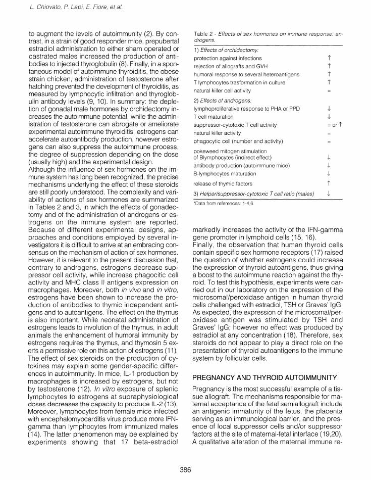
Table 2 - Effects of sex hormones on immune response,' androgens.
1) Effects of orchidectomy:
protection against infections t rejection of allografts and GVH t humoral response to several heteroantigens t T lymphocytes trasformation in culture t natural killer cell activity
2) Effects of androgens:
Iymphoproliferative response to PHA or PPD J-T cell maturation J-suppressor-cytotoxic T cell activity = or t natural killer activity
phagocytic cell (number and activity)
pokeweed mitogen stimulation of Blymphocytes (indirect effect) J-antibody production (autoimmune mice) J-B-Iymphocytes maturation J-
release of thymic factors t 3) Helper/suppressor-cytotoxic T cell ratio (males) J-'Data from references: 1-4.6.
markedly increases the activity of the IFN-gamma gene promoter in lymphoid cells (15, 16). Finally, the observation that human thyroid cells contain specific sex hormone receptors (17) raised the question of whether estrogens could increase the expression of thyroid autoantigens, thus giving a boost to the autoimmune reaction against the thyroid. To test this hypothesis, experiments were carried out in our laboratory on the expression of the microsomal/peroxidase antigen in human thyroid cells challenged with estradiol, TSH or Graves' IgG. As expected, the expression of the microsomal/peroxidase antigen was stimulated by TSH and Graves' IgG; however no effect was produced by estradiol at any concentration (18). Therefore, sex steroids do not appear to playa direct role on the presentation of thyroid autoantigens to the immune system by follicular cells.
PREGNANCY AND THYROID AUTOIMMUNITY
Pregnancy is the most successful example of a tissue allograft. The mechanisms responsible for maternal acceptance of the fetal semiallograft include an antigenic immaturity of the fetus, the placenta serving as an immunological barrier, and the presence of local suppressor cells and/or suppressor factors at the site of maternal-fetal interface (19,20). A qualitative alteration of the maternal immune re-
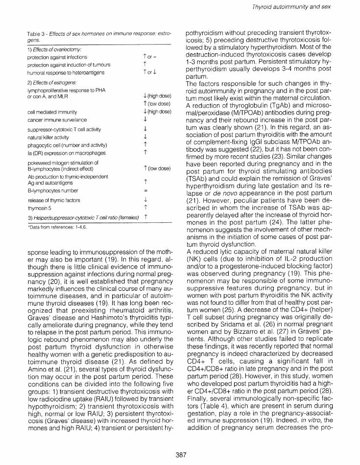
Table 3 - Effects of sex hormones on immune response: estrogens.
1) Effects of ovariectomy:
protection against infections
protection against induction of tumours
humoral response to heteroantigens
2) Effects of estrogens:
Iymphoproliferative response to PHA or con A, and MLR
cell mediated immunity
cancer immune surveilance
suppressor-cytotoxic T cell activity
natural killer activity
phagocytic cell (number and activity)
la (DR) expression on macrophages
pokeweed mitogen stimulation of B-Iymphocytes (indirect effect)
Ab production to thymic-independent Ag and autoantigens
B-Iymphocytes number
release of thymic factors
thymosin 5
l' or =
l' l' od
,J. (high dose)
l' (low dose)
,J. (high dose) ,J.
,J. ,J.
l' l'
l' (low dose)
l'
3) Helper/suppressor-cytotoxic T cell ratio (females) l' 'Data from references: 1-4,6.
sponse leading to immunosuppression of the mother may also be important (19), In this regard, although there is little clinical evidence of immunosuppression against infections during normal pregnancy (20), it is well established that pregnancy markedly influences the clinical course of many autoimmune diseases, and in particular of autoimmune thyroid diseases (19). It has long been recognized that preexisting rheumatoid arthritis, Graves' disease and Hashimoto's thyroiditis typically ameliorate during pregnancy, while they tend to relapse in the post partum period. This immunologic rebound phenomenon may also underly the post partum thyroid dysfunction in otherwise healthy women with a genetic predisposition to autoimmune thyroid disease (21). As defined by Amino et al. (21), several types of thyroid dysfunction may occur in the post partum period, These conditions can be divided into the following five groups: 1) transient destructive thyrotoxicosis with low radioiodine uptake (RAIU) followed by transient hypothyroidism; 2) transient thyrotoxicosis with high, normal or low RAIU; 3) persistent thyrotoxicosis (Graves' disease) with increased thyroid hormones and high RAIU; 4) transient or persistent hy-
387
Thyroid autoimmunity and sex
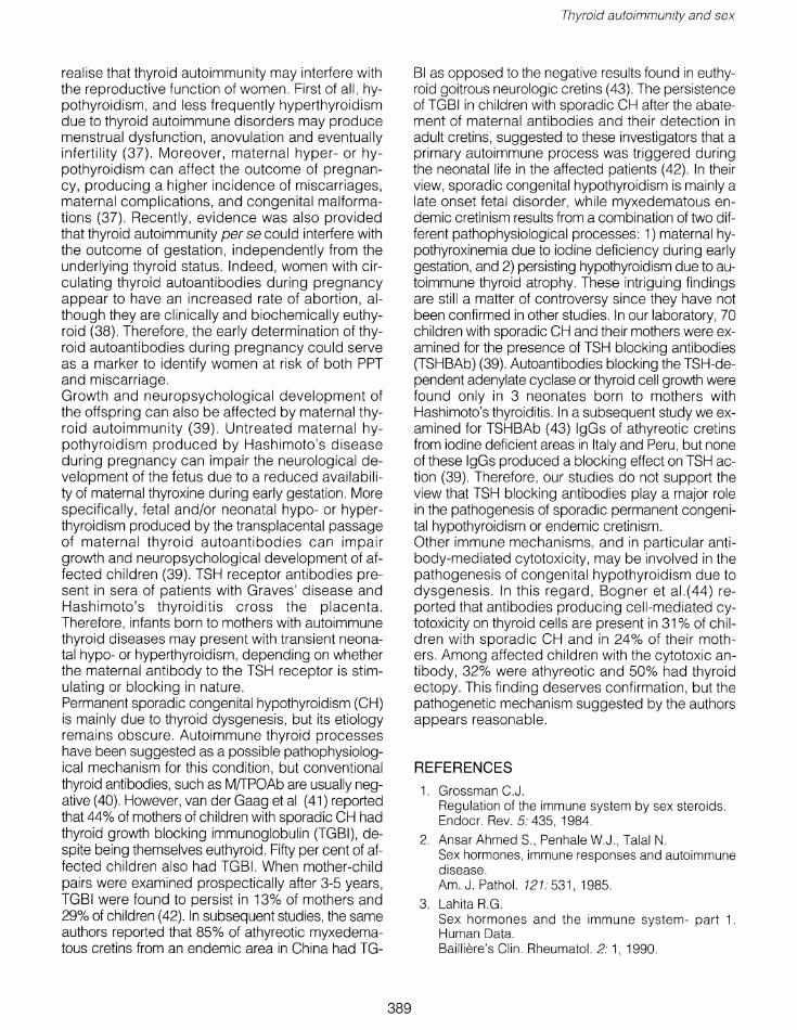
pothyroidism without preceding transient thyrotoxicosis; 5) preceding destructive thyrotoxicosis followed by a stimulatory hyperthyroidism. Most of the destruction-induced thyrotoxicosis cases develop 1-3 months post partum, Persistent stimulatory hyperthyroidism usually develops 3-4 months post partum. The factors responsible for such changes in thyroid autoimmunity in pregnancy and in the post partum most likely exist within the maternal circulation . A reduction of thyroglobulin (TgAb) and microsomal/peroxidase (M(TPOAb) antibodies during pregnancy and their rebound increase in the post partum was clearly shown (21). In this regard, an association of post partum thyroiditis with the amount of complement-fixing IgGI subclass M(TPOAb antibody was suggested (22), but it has not been confirmed by more recent studies (23), Similar changes have been reported during pregnancy and in the post partum for thyroid stimulating antibodies (TSAb) and could explain the remission of Graves' hyperthyroidism during late gestation and its relapse or de novo appearance in the post partum (21). However, peculiar patients have been described in whom the increase of TSAb was appearently delayed after the increase of thyroid hormones in the post partum (24). The latter phenomenon suggests the involvement of other mechanisms in the initiation of some cases of post partum thyroid dysfunction, A reduced lytic capacity of maternal natural killer (NK) cells (due to inhibition of IL-2 production and/or to a progesterone-induced blocking factor) was observed during pregnancy (19). This phenomenon may be responsible of some immunosuppressive features during pregnancy, but in women with post partum thyroiditis the NK activity was not found to differ from that of healthy post partum women (25). A decrease of the CD4+ (helper) T cell subset during pregnancy was originally described by Sridama et al. (26) in normal pregnant women and by Bizzarro et al. (27) in Graves' patients. Although other studies failed to replicate these findings, it was recently reported that normal pregnancy is indeed characterized by decreased CD4+ T cells, causing a significant fall in CD4+/CD8+ ratio in late pregnancy and in the post partum period (28), However, in this study, women who developed post partum thyroiditis had a higher CD4+/CD8+ ratio in the post partum period (28). Finally, several immunologically non-specific factors (Table 4), which are present in serum during gestation, playa role in the pregnancy-associated immune suppression (19). Indeed, in vitro, the addition of pregnancy serum decreases the pro-
L. Chiovato, P Lapi, E. Fiore, et al.
duction of IL-1 in mixed lymphocyte reactions , reduces T cell blastogenesis induced by PHA, diminishes IL-2 production by T lymphocytes, and impairs complement-mediated cell lysis (19) . Circulating inhibitory factors may be produced by the mother, the fetus or the placenta. Among them, cortisol in physiological concentrations, and to a lower extent progesterone, are capable of reproducing some suppressive effects of pregnancy serum (19). Of the other factors , pregnancy associated plasma protein A (PAPP-A) may also contribute to the suppressive effect (19). With regard to autoimmune thyroid disorders, Scott et al. (29) stud ied the behaviour of alpha 2-pregnancy associated glycoprotein (alpha 2-PAG) in patients with Graves ' disease and Hashimoto's thyroiditis. They found that patients undergoing remission of their disease during pregnancy had significantly higher serum levels of alpha 2-PAG than those with active disease.
Changes of Graves ' disease and Hashimoto 's thyroiditis during pregnancy and in the post partum
Whatever the underlying mechanisms, it has been shown that among female patients with Graves' disease nearly 50% may experience a spontaneous restoration of euthyroidism during late pregnancy while more than 70% of them may develop thyrotoxicosis in the post partum (21) . Similarly, in Hashimoto's thyroiditis , serum levels of TSH may decrease significantly in late pregnancy, while FT4 has a tendency to spontaneously increase . However, in the post partum period significant thyroid dysfunction characterized by destructive thyrotoxicosis followed by hyperthyroidism or hypothyroidism often occurs (21) .
Post partum thyroiditis (PPT)
The incidence of post partum thyroid dysfunction in the general population has been reported to range from 1.9% and 16.7%, depending on the study (30,31). However, when strict epidemiological criteria (32) are applied and patients with Graves' disease are excluded, the range of incidence of PPT, as observed in Japan, Sweden and Denmark, appears to be narrow, ranging from 3.7% and 5.9% . PPT presents as hypothyroidism alone in 2.2%, transient thyrotoxicosis followed by hypothyroidism in 1.0% and thyrotoxicosis alone in 1.7%. Documented Graves' disease occurs with an incidence of 0.2%. Thyrotoxicosis appears to be more frequent in Japan than in Sweden (32) and such a variation could be attributed to differences in genetic profiles or iodine intake, which is high in
388
Table 4 - Immunologically non-specific factors involved in pregnancy-associated immune suppression.
Steroid hormones
Cortisol
Estrogens
Progesterone
Peptide hormones
Human chorionic gonadotropin
Human placental lactogen
Other proteins
Pregnancy associated plasma proteins
Pregnancy associated u 2-glycoprotein
Placental proteins
u-fetoprotein
Progesterone-induced blocking factor
Japan and normal-low in Sweden . Indeed , evidence was provided suggesting that iodine may aggravate rather than ameliorate PPT. This is because iodine supplementation in women predisposed to the development of PPT was found to be associated with greater hormone changes in both the thyrotoxic and the hypothyroid phase (33) . Although the direct involvement of M(TPOAb in the pathogenesis of PPT is still debated, most studies indicate a strong association of M(TPOAb with PPT (31). This observation suggests that M/TPOAb could be used as a marker to predict the occurrence of PPT in an unselected population of pregnant women. However, the predictive value of M(TPOAb determination varies with time. The specificity of the test is always higher than 90%, but the sensitivity progressively increases from 45% at delivery to more than 80% at 5-7 months post partum (34) . Since the titers of M(TPOAb decrease during pregnancy, if a screening for M(TPOAb should be done, it must be performed early in gestation and not at delivery. Women who develop PPT often relapse after subsequent pregnancies. Long term follow-up of patients with PPT was reported from Japan (35) , Sweden (31) and Wales (36). The incidence of permanent hypothyroidism, either subclinical or clinical, was remarkably similar ranging from 22% to 29%, independently from genetic background and iodine intake.
THYROID AUTOIMMUNITY AND FEMALE REPRODUCTIVE FUNCTION
When looking to the problem of thyroid autoimmunity and gender from another point of view, we must
realise that thyroid autoimmunity may interfere with the reproductive function of women. First of all, hypothyroidism, and less frequently hyperthyroidism due to thyroid autoimmune disorders may produce menstrual dysfunction, anovulation and eventually infertility (37). Moreover, maternal hyper- or hypothyroidism can affect the outcome of pregnancy, producing a higher incidence of miscarriages, maternal complications, and congenital malformations (37). Recently, evidence was also provided that thyroid autoimmunity per se could interfere with the outcome of gestation, independently from the underlying thyroid status. Indeed, women with circulating thyroid autoantibodies during pregnancy appear to have an increased rate of abortion, although they are clinically and biochemically euthyroid (38). Therefore, the early determination of thyroid autoantibodies during pregnancy could serve as a marker to identify women at risk of both PPT and miscarriage. Growth and neuropsychological development of the offspring can also be affected by maternal thyroid autoimmunity (39). Untreated maternal hypothyroidism produced by Hashimoto's disease during pregnancy can impair the neurological development of the fetus due to a reduced availability of maternal thyroxine during early gestation. More specifically, fetal and/or neonatal hypo- or hyperthyroidism produced by the transplacental passage of maternal thyroid autoantibodies can impair growth and neuropsychological development of affected children (39). TSH receptor antibodies present in sera of patients with Graves' disease and Hashimoto's thyroiditis cross the placenta. Therefore, infants born to mothers with autoimmune thyroid diseases may present with transient neonatal hypo- or hyperthyroidism, depending on whether the maternal antibody to the TSH receptor is stimulating or blocking in nature. Permanent sporadic congenital hypothyroidism (CH) is mainly due to thyroid dysgenesis, but its etiology remains obscure. Autoimmune thyroid processes have been suggested as a possible pathophysiological mechanism for this condition, but conventional thyroid antibodies, such as M/TPOAb are usually negative (40). However, van der Gaag et al (41) reported that 44% of mothers of children with sporadic CH had thyroid growth blocking immunoglobulin (TGBI), despite being themselves euthyroid. Fifty per cent of affected children also had TGBI. When mother-child pairs were examined prospectically after 3-5 years, TGBI were found to persist in 13% of mothers and 29% of children (42). In subsequent studies, the same authors reported that 85% of athyreotic myxedematous cretins from an endemic area in China had TG-
389
Thyroid autoimmunity and sex
BI as opposed to the negative results found in euthyroid goitrous neurologic cretins (43). The persistence of TGBI in children with sporadic CH after the abatement of maternal antibodies and their detection in adult cretins, suggested to these investigators that a primary autoimmune process was triggered during the neonatal life in the affected patients (42). In their view, sporadic congenital hypothyroidism is mainly a late onset fetal disorder, while myxedematous endemic cretinism results from a combination of two different pathophysiological processes: 1) maternal hypothyroxinemia due to iodine deficiency during early gestation, and 2) persisting hypothyroidism due to autoimmune thyroid atrophy. These intriguing findings are still a matter of controversy since they have not been confirmed in other studies. In our laboratory, 70 children with sporadic CH and their mothers were examined for the presence of TSH blocking antibodies (TSHBAb) (39). Autoantibodies blocking the TSH-dependent adenylate cyclase or thyroid cell growth were found only in 3 neonates born to mothers with Hashimoto's thyroiditis. In a subsequent study we examined for TSHBAb (43) IgGs of athyreotic cretins from iodine deficient areas in Italy and Peru, but none of these IgGs produced a blocking effect on TSH action (39). Therefore, our studies do not support the view that TSH blocking antibodies playa major role in the pathogenesis of sporadic permanent congenital hypothyroidism or endemic cretinism. Other immune mechanisms, and in particular antibody-mediated cytotoxicity, may be involved in the pathogenesis of congenital hypothyroidism due to dysgenesis. In this regard, Bogner et al.(44) reported that antibodies producing cell-mediated cytotoxicity on thyroid cells are present in 31 % of children with sporadic CH and in 24% of their mothers. Among affected children with the cytotoxic antibody, 32% were athyreotic and 50% had thyroid ectopy. This finding deserves confirmation, but the pathogenetic mechanism suggested by the authors appears reasonable.
REFERENCES 1. Grossman C.J.
Regulation of the immune system by sex steroids. Endocr. Rev. 5: 435, 1984.
2. Ansar Ahmed S., Penhale w.J., Talal N. Sex hormones, immune responses and autoimmune disease. Am. J. Pathol. 121:531,1985.
3. Lahita R.G. Sex hormones and the immune system- part 1. Human Data. Bailliere's Clin. Rheumatol. 2: 1, 1990.

L. Chiovato, P. Lapi, E Fiore, et aJ.
4. Lahita R.G. Sex hormones and the immune system. In: Talal N. (Ed.), Molecular autoimmunity. Academic Press, New York, 1991, p.385.
5. Tunbridge W.M.G., Evered D., Hall R., Appleton D., Brewis M., Clark F., Grimley Evans J., Young E., Bird T., Smith PA The spectrum of thyroid disease in a community: the Whickham Survey. Clin. Endocrinoi. 7: 481, 1977.
6. Ansar Ahmed S., Talal N. Sex hormones and the immune system- part 2. Animal data. Bailliere's Clin. Rheumatoi. 2: 13, 1990.
7. Ansar Ahmed S., Young P.R., Penhale W.J. Beneficial effect of testosterone in the treatment of chronic autoimmune thyroiditis in rats. J. Immunol. 136: 143,1986.
8. Okayasu I., Kong Y.M., Rose N.R. Effect of castration and sex hormones on experimental autoimmune thyroiditis. Clin. Immunoi. Immunopathol. 20: 240, 1981.
9. Gause W.C., Marsh J.A. Effect of testosterone treatment for varying periods on autoimmune development and on specific infiltrating leukocyte populations in the thyroid gland of obese strain chickens. Clin. Immunoi. Immunopathoi. 39: 464, 1986.
10. Fassler R., Dietrich H., Kromer G., Bock G., Brezinschek H-P., Wick G. The role of testosterone in spontaneous autoimmune thyroiditis of obese strain (OS) chickens. Autoimmunity 1: 97, 1988.
11. Erbach G.T., Bahr J.M. Enhancement of in vivo humoral immunity by estrogen: permissive effect of a thymic factor. Endocrinology 128: 1352, 1991.
12. Flynn A Expression of la and the production of interleukin-I by peritoneal exudate macrophages activated in vivo by steroids. Life Sci. 38: 2455, 1986.
13. Henriksen 0., Frey J.R. Control of the expression of interleukin-2 activity. Celi. Immunoi. 73: 106, 1982.
14. McFarland H.I., Bigley N.J. Sex-dependent, early cytokine production by NK-like spleen cells following infection with the 0 variant of encephalomyocarditis virus (EMCV-D). Virallmmunol. 2: 205,1989.
15. Sarvetnick N., Fox H.S. Interferon-gamma and the sexual dimorphism of autoimmunity. Mol. Bioi. Med. 7: 323, 1990.
16. Fox H.S., Bond B.L., Parlow T.G. Estrogen regulates the IFN-y promoter. J. Immunoi. 146: 4362, 1991.
390
17. Money R.S., Muss W, Thelmo W.L., Boeckl 0., Pimpl W, Kaindl H., Sungler P., Kirwin J, Waclawicek H, Jaffe B.M., Pertshuk L.P. Immunocytochemical localization of estrogen and progesterone receptors in human thyroid. Surgery 106: 975, 1989.
18. Chiovato L., Vitti P., Cucchi P., Mammoli C., Carayon P., Pinchera A The expression of the microsomal antigen in human thyroid cells is thyrotropin-dependent. Clin. Exp. Immunoi. 76: 47, 1989.
19. Pope R. M. Immunoregulatory mechanism present in the maternal circulation during pregnancy. Bailliere's Clin. Rheumatol. 2: 33, 1990.
20. Feinberg B.B., Gonik B. General precepts of the immunology of pregnancy. Clin. Obstet. Gynecol. 34: 3, 1991.
21. Amino N. Postpartum thyroid disease. In: Bercu B.B., Shulman 0.1. (Eds.), Advances in perinatal thyroidology. Plenum Press, New York, 1991, p.167.
22. Jansson R., Thompson P.M., Clark F., McLachlan S.M. Association between thyroid microsomal antibodies of subclass IgG-1 and hypothyroidism in autoimmune postpartum thyroiditis. Clin. Exp. Immunoi. 63: 80, 1986.
23. Weetman A.P., Fung H.Y.M., Richards C.J., McGregor M. IgG subclass distribution and relative functional affinity of thyroid microsomal antibodies in postpartum thyroiditis. Eur. J. Clin. Inv. 20: 133, 1990.
24. Tamaki H., Amino N., Aozasa M., Mori M., Tanizawa 0., Miyai K. Serial changes in thyroid-stimulating antibody and thyrotropin binding inhibitor immunoglobulin at the time of postpartum occurrence of thyrotoxicosis in Graves' disease. J. Clin. Endocrinoi. Metab. 65: 324,1987.
25. Hayslip C.C., Baker J.R., Wartofsky L., Klein TA, Opsahl M.S., Burman KD. Natural killer cell activity and serum autoantibodies in women with postpartum thyroiditis. J. Clin. Endocrinoi. Metab. 66: 1089, 1988.
26. Sridama V., Pacini F., Yang S-L., Moawad A., Reilly M., De Groot L.J. Decreased levels of helper T cells. A possible cause of immunodeficiency in pregnancy. N. Engl. J. Med. 307: 352, 1982.
27. Bizzarro A, Fontana A, De Bellis A, Daponte A, Iacono G., Peluso G. T-Iymphocyte subsets in pregnant women with Graves' disease. Acta Endocrinoi. 114: 218,1987.
28. Stagnaro-Green A., Roman S.H., Cobin R.H., EI-
Harazy E, Wallenstein S., Davies T.F. A prospective study of lymphocyte-initiated immunosuppression in normal pregnancy: evidence of a T-cell etiology for postpartum thyroid dysfunction. J. Clin. Endocrinol. Metab. 74: 645, 1992.
29. Scott R.M., How J., Gerrie L.M., Khir AS.M., Bewsher p.o., Horne C.HW., Thomson AW. Serum levels of pregnancy (a2-glycoprotein (a2-PAG) during pregnancy in autoimmune thyroid disease: relationship to disease activity. Clin. Exp. Immunol. 59: 564,1985.
30. Roti E., Emerson C. Clinical review 29 - Postpartum thyroiditis. J. Clin. Endocrinol. Metab. 74: 3, 1992.
31. Jansson R., Dahlberg PA, Karlsson FA Postpartum thyroiditis. Balliere's Clin. Endocrinol. Metabol. 2: 619, 1988.
32. Gerstein H.C. How common is postpartum thyroiditis? A methodologic overview of the literature. Arch. Intern. Med. 50: 1397,1990.
33. Kampe 0., Jansson R., Karlsson FA Effects of L-thyroxine and iodide on the development of autoimmune postpartum thyroiditis. J. Clin. Endocrinol. Metab. 70: 1014, 1990.
34. Vargas M.T., Briones-Urbina R., Gladman D., Papsin F.R., Walfish P.G. Antithyroid microsomal autoantibodies and HLA-DR5 are associated with postpartum thyroid dysfunction: evidence supporting an autoimmune pathogenesis. J. Clin. Endocrinol. Metab. 67: 327, 1988.
35. Tachi J., Amino N., Tamaki H., Aozasa M., Iwatani Y., Miyai K. Long term follow-up and HLA association in patients with postpartum hypothyroidism. J. Clin. Endocrinol. Metab. 66: 480, 1988.
36. Othman S., Phillips D.IW., Parkes AB., Richards J.C., Harris B., Fung H., Darke C., John R., Hall R., Lazarus J.H. Long-term follow-up of postpartum thyroiditis. Clin. Endocrinol. 32: 559, 1990.
37. Becks G.P., Burrow G.N. Thyroid disease in pregnancy. Med. Clin. North Am. 75: 121, 1991.
38. Glinoer D., Fernandez Soto M., Bourdoux P., Lejeune B., Delange F., Lemone M., Kinthaert J., Robin C., Grun J-P., De Nayer P. Pregnancy in patients with mild thyroid abnormalities: maternal and neonatal repercussions. J. Clin. Endocrinol. Metab. 73:421,1991.
39. Chiovato L., Tonacchera M., Lapi P., Fiore E., Vitti P., Pinchera A Thyroid autoimmunity and neuropsychological development. Acta Med. Aust. 19: 91, 1992.
40. Chiovato L., Giusti F.L., Santini F., Bassi P., Vitti P., Marcocci C., Tonacchera M., Ciampi M., Fenzi G.F.,
391
Thyroid autoimmunity and sex
Pinchera A Incidence and functional effect of thyroid autoantibodies in newborns recalled for suspected congenital hypothyroidism during a screening program. In: Delange F., Fisher D.A., Glinoer D. (Eds.), Research in congenital hypothyroidism. Plenum Press, New York, 1989, p. 141.
41. Van Der Gaag RD., Drexhage HA, Dussault J.H. Role of maternal immunoglobulins blocking TSH-induced thyroid growth in sporadic form of congenital hypothyroidism. Lancet 1: 246, 1985.
42. Boyages S.C, Lens JW., van der Gaag R.D., Maberly G.F., Eastman C.J., Drexhage HA Sporadic and endemic congenital hypothyroidism: evidence for autosensitization. In: Delange F., Fisher DA, Glinoer D.(Eds.), Research in Congenital Hypothyroidism. Plenum Press, New York, 1989, p. 123.
43. Boyages S.C., Halpern J.P., Maberly G.F., Creswell J.E., Chen J., ZhenHua W., van der Gaag R.D., Drexhage HA Endemic cretinism: possible role for thyroid autoimmunity. Lancet 2: 529, 1989.
44. Bogner U., Gruters A, Sigle B., Helge H., Schleusener H. Cytotoxic antibodies in congenital hypothyroidism. J. Clin. Endocrinol. Metab. 68: 671, 1989.
Treatment of hyper- and hypothyroidsm in pregnancy J. H. Lazarus Department of Medicine, University of Wales College of Medicine, Cardiff, U.K. Key-words: Hyperthyroidism, hypothyroidism, pregnancy, antithyroid drugs. thyroxine. obstetric complications. neonatal thyroid function. hyperemesis gravidarum.
Correspondence: Dr. J.H. Lazarus, Senior Lecturer in Medicine, Dept. Medicine, University of Wales College of Medicine. Heath Park, Cardiff CF4 4XN Wales, U.K.
ABSTRACT. In healthy subjects there are changes in thyroid function during pregnancy consequent on the increased synthesis of TBG and the thyroid stimulating effect of hCG. Serum thyroid hormones are elevated in the first trimester but fall during the latter half of pregnancy. Iodine deficiency may accentuate these changes. Hyperemesis gravidarum