three-dimensional reconstruction of neurovascular network
TRANSCRIPT

Received: Jun 20, 2019 Accepted: Aug 1, 2019 Published online Aug 27, 2019Correspondence to: Ji-Kan Ryu https://orcid.org/0000-0003-2125-0212 National Research Center for Sexual Medicine and Department of Urology, Inha University School of Medicine, 27 Inhang-ro, Jung-gu, Incheon 22332, Korea. Tel: +82-32-890-3505, Fax: +82-32-890-3099, E-mail: [email protected] to: Jun-Kyu Suh https://orcid.org/0000-0002-1812-9449 National Research Center for Sexual Medicine and Department of Urology, Inha University School of Medicine, 27 Inhang-ro, Jung-gu, Incheon 22332, Korea.Tel: +82-32-890-3441, Fax: +82-32-890-3097, E-mail: [email protected]*These authors contributed equally to this study as co-first authors.
Copyright © 2019 Korean Society for Sexual Medicine and Andrology
Three-Dimensional Reconstruction of Neurovascular Network in Whole Mount Preparations and Thick-Cut Transverse Sections of Mouse Urinary Bladder
Nhat Minh Nguyen1,* , Kang-Moon Song1,* , Min-Ji Choi1 , Kalyan Ghatak1 , Anita Limanjaya1 , Mi-Hye Kwon1 , Doo Yong Chung1 , Jiyeon Ock1 , Guo Nan Yin1 , Chang-Shin Park2 , Jun-Kyu Suh1 , Ji-Kan Ryu1
1National Research Center for Sexual Medicine and Department of Urology, 2Department of Pharmacology and Medicinal Toxicology Research Center, Inha University School of Medicine, Incheon, Korea
Purpose: Proper functional and structural integrity of nervous and vascular system in urinary bladder plays an important role in normal bladder function and the disruption of these structures is known to be related to lower urinary tract symptoms. Here, we present an immunohistochemical staining method that delineates neurovascular structures in the mouse urinary bladder by using immunohistochemical staining with three-dimensional reconstruction.Materials and Methods: The urinary bladder was harvested from 8-week-old C57BL/6 male mouse. Lamina propria and de-trusor muscle layer were dissected for whole mount staining, and thick-cut (60-μm) sections were prepared for full-thickness bladder staining. Immunofluorescent staining of bladder tissue was performed with antibodies against CD31 (an endothelial cell marker), smooth muscle α-actin (a smooth muscle cell marker), NG2 (a pericyte marker), and βIII-tubulin (a neuronal marker). We reconstructed three-dimensional images of bladder neurovascular system from stacks of two-dimensional im-ages.Results: Three-dimensional images obtained from thick-cut sections clearly provided good anatomic information about neu-rovascular structures in the three layers of bladder, such as urothelium, lamina propria, and detrusor muscle layer. Whole mount images of lamina propria and detrusor muscle layer also clearly delineated spatial relationship between nervous and vascular systems. The microvessel density was higher in the lamina propria than in the detrusor muscle layer. Nerve fibers were evenly innervated into the lamina propria and detrusor muscle.Conclusions: This study provides comprehensive insight into three-dimensional neurovascular structures of mouse urinary bladder. Our technique may constitute a standard tool to evaluate pathologic changes in a variety of urinary bladder diseases.
Keywords: Blood vessels; Mouse; Nervous system; Three-dimensional imaging; Urinary bladder
This is an Open Access article distributed under the terms of the Creative Commons Attribution Non-Commercial License (http://creativecommons.org/licenses/by-nc/4.0) which permits unrestricted non-commercial use, distribution, and reproduction in any medium, provided the original work is properly cited.
Original Article
pISSN: 2287-4208 / eISSN: 2287-4690World J Mens Health Published online Aug 27, 2019https://doi.org/10.5534/wjmh.190089
Male voiding dysfunctions

https://doi.org/10.5534/wjmh.190089
2 www.wjmh.org
INTRODUCTION
The urinary bladder is richly supplied by nerves and blood vessels [1-4]. Proper functional and structural integrity of nervous and vascular system in urinary bladder plays an important role in normal bladder function; the storage and emptying of urine [5-7]. Al-though the underlying mechanisms of bladder dys-function and associated lower urinary tract symptoms (LUTS) have not yet been understood clearly, the im-pairments of microvascular structures and peripheral nervous system are regarded to be responsible for the development of these conditions [7]. In human studies, neurovascular injury caused by aging, diabetes, hyper-tension, or hyperlipidemia are known to be involved in the progression of LUTS [8,9].
Recent studies provide an evidence that crosstalk between neuronal and endothelial cells is crucial to maintain normal function of nervous and vascular systems [10,11]. While neuronal cells promote angiogen-esis by secreting angiogenic factors, such as vascular endothelial growth factor, endothelial cells can also regulate development of nervous system by releasing neurotrophic factors [11,12]. It was reported that the expression of endothelin-1 and interleukin-6, which are known as biomarkers for endothelial dysfunction, are upregulated in patients with bladder outlet obstruction [13]. In addition, the derangements in microvascular structures and increase in the number of apoptotic en-dothelial cells have been well characterized in patients with bladder pain syndrome [14]. Microvascular dys-function from chronic bladder ischemia and repeated ischemia/reperfusion during a micturition cycle ulti-mately leads to denervation of the urinary bladder [7,8].
The visualization of changes in tissue structures and cellular distribution is critical to understand the pro-gression of many diseases. Immunohistochemistry has been used widely as a standard tool to evaluate struc-tural changes in a variety of urinary bladder diseases. However, the most of studies characterized the urinary bladder at two-dimensional (2D) level, which provides limited anatomical information. Especially, 2D imaging from thin-cut tissue section is not appropriate to com-prehensive analysis of the association between nervous and vascular systems due to limited sample depths. For decades, several methods have been developed to visu-alize tissue structure at three-dimensional (3D) level [15]; for example, reconstructing images from serially
stained thin-cut tissue sections, and immunostaining of whole mount preparations or thick-cut tissue sections. The disadvantages of serially sectioned samples include the requirement of large number of sections, high-cost of antibodies, and time-consuming. In contrast, limita-tions of whole mount samples or thick-cut tissue sec-tions are difficulties in its technique and limited pen-etration of antibodies.
Although previous studies demonstrated urinary bladder innervation or suburothelial microvascular structure in human or rodents at 3D level [16-19], ana-tomic information about neurovascular network in whole layers of urinary bladder are still lacking. In the present study, therefore, we present an immunohisto-chemical staining method that delineates 3D neuro-vascular systems of mouse urinary bladder at a high resolution. To visualize neural and vascular network in a mouse urinary bladder, full-thickness frozen section of urinary bladder, and whole mount preparations of lamina propria and detrusor muscle layer were used.
MATERIALS AND METHODS
1. AnimalsEight-week-old C57BL/6 male mice weighing 20 to 25
g were purchased from Orient Bio (Seongnam, Korea). All experiments protocols were approved by the Insti-tutional Animal Care and Use Subcommittee of Inha University (IACUC No. 180727-580).
2. Tissue preparation
1) Preparation of transverse full-thickness frozen section of urinary bladder
The mouse urinary bladders were harvested and im-mersed in 4% paraformaldehyde in phosphate buffer saline (PBS) for 24 hours at 4oC. After washing several times with PBS, samples were embedded in optimal cutting temperature compound medium. The samples were kept inside of the cryostat at -20oC for 20 minutes and thick-cut (60 μm) sections were prepared for full-thickness bladder staining.
2) Preparation of lamina propria and detrusor muscle layer for whole mount staining
The mouse urinary bladders were harvested, and the urothelium and lamina propria were carefully dissect-ed from underlying detrusor muscle with fine micro-

Nhat Minh Nguyen, et al: Three-Dimensional Images of Mouse Urinary Bladder
3www.wjmh.org
forceps. Urothelial layer was then removed by gentle swabbing with a Q-tip. All procedures were done with the aid of a dissecting microscope (Zeiss, Göttingen, Germany). The remaining whole lamina propria and detrusor muscle layer were flattened and fixed in 4% paraformaldehyde in PBS for 24 hours at 4oC.
3. ImmunohistochemistryAfter washing several times with PBS, samples were
then immersed in 3% bovine serum albumin (BSA) for 1 hour at room temperature to minimize nonspecific binding of antibodies. Samples were incubated with antibodies to CD31 (an endothelial cell marker; Milli-pore, Temecula, CA, USA; 1:50; Cat #MAB1398Z), fluo-rescein isothiocyanate-conjugated antibody to smooth muscle α-actin (a smooth muscle cell marker; Sigma-Aldrich, St. Louis, MO, USA; 1:200; Cat #F3777), smooth muscle α-actin (a smooth muscle cell marker; Abcam, Cambridge, UK; 1:200; Cat #ab5694), NG2 chondroitin sulfate proteoglycan (a pericyte cell marker; Millipore; 1:50; Cat #ab5320), and βIII-tubulin (a neuronal marker; Abcam; 1:100; Cat #ab107216) for 48 hours at 4oC. Af-ter washing several times with PBS, the samples were incubated with Donkey Anti-Mouse IgG H&L Alexa Fluor® 405 (Abcam; 1:200; Cat #ab175658), rhodamine AffiniPure Goat Anti-Armenian Hamster IgG (H+L) (Jackson ImmunoResearch, West Grove, PA, USA; 1:50; Cat #127-025-160), rhodamine Affinipure Donkey Anti-Chicken IgY (H+L) (JacksonImmuno Research; 1:50; Cat #703-025-155). Primary and secondary antibodies were diluted in PBS containing 0.1% triton X-100 and 3% BSA (Sigma-Aldrich). Samples were mounted in a solution containing 4,6-diamidino-2-phenylindole (DAPI; Vector Laboratories Inc., Burlingame, CA, USA) for nuclei staining.
4. Imaging and three-dimensional reconstruction
To visualize microvascular and neural structures in the mouse urinary bladder, images were obtained from full-thickness bladder sections or whole mount prepa-rations of lamina propria and detrusor muscle layer under confocal laser scanning microscopy (FV1000; Olympus, Tokyo, Japan). Confocal images were cap-tured with multi-lasers (405 nm, 48 nm, and 595 nm). For 3D reconstruction, Z-stack imaging was performed from top to the bottom of tissue samples with serial images obtained from 1.0 µm-depth. Projection of Z-
stack images and 3D reconstruction were built up by Olympus Fluoview software or ImageJ software (Na-tional Institutes of Health IMAGE J 1.34; http://rsbweb.nih.gov/ij/).
5. Ethics statementAll experiments were conducted in accordance with
laboratory animal and institutional animal care guide-lines. Protocols used in this study were approved by the Institutional Animal Care and Use Subcommittee of Inha University (IACUC No. 180727-580).
RESULTS
1. Three-dimensional reconstruction of neurovascular structures in mouse urinary bladder
High-resolution 3D images of neurovascular system in mouse urinary bladder were presented in Fig. 1 and Fig. 2. The image for full-thickness transverse section of urinary bladder was reconstructed with stacks of eight 2D images, which allow visualization of entire neurovascular system (Fig. 1A). The urothelium and lamina propria form bladder mucosa and have numer-ous folds that disappear when the bladder is filled (Fig. 1A). The reconstructed 3D images clearly dem-onstrate the distribution of CD31-positive endothelial cells, α-actin-positive smooth muscle cells, and βIII-tubulin-positive nerve fibers, and their relationship in urothelium, lamina propria, and detrusor muscle layer (projection depth: 25 μm) (Fig. 1B, 1C).
Z projection was performed from a series of 2D im-ages, which permits the 2D dataset to be visualized as a 3D image (Fig. 2A, 2B). The three dimensional images clearly demonstrate lamina propria and detru-sor muscle layer. CD31-positive suburothelial capillary plexus and mucosal plexus were well recognized in the lamina propria (Fig. 2B, 2C). The microvessel density was higher in the lamina propria than in the detrusor muscle layer (Fig. 2B). The reconstructed 3D images clearly delineate typical wavy appearance of nerve fibers that evenly innervate to lamina propria and detrusor muscle (Fig. 2B-2D). CD31-positive endothelial cells were covered with NG2-positive pericytes, espe-cially in the suburothelial capillary plexus underneath urothelium (Fig. 2E).
In contrast, immunofluorescent double staining of bladder tissue with antibodies to CD31 and smooth mus-

https://doi.org/10.5534/wjmh.190089
4 www.wjmh.org
cle α-actin and subsequent 3D reconstruction (projec-tion depth: 25 μm) revealed that α-actin-positive smooth muscle cells are mainly expressed in perpendicular blood vessels of suburothelial capillary plexus, and in blood vessels of mucosal plexus and detrusor muscle (Fig. 3).
2. Neurovascular structures of lamina propria in a whole mount preparation
High-resolution 3D reconstruction of neurovascular system in lamina propria of urinary bladder was per-formed by using whole mount immunostaining (Fig.
Fig. 1. The principle of three-dimensional (3D) reconstruction of neurovascular structures in mouse urinary bladder. High-resolution 3D image was reconstructed from stacks of confocal two-dimen sional (2D) images, which permit to render the volume and visualize microvascular struc-ture and nerve innervation. (A) Transverse section of whole urinary bladder. The image was merged from stacks of eight 2D images by ImageJ software. (B, C) High-magnification images of transverse section of urinary bladder. (B) Twenty-five confocal images were obtained from the thick-cut transverse section of urinary bladder. The step size was kept at 1.0 μm constantly. (C) 3D reconstruction of uri-nary bladder from stacks of twenty-five 2D images. CD31 (red): an endothelial cell marker; smooth muscle α-actin (blue): a smooth muscle cell marker; βIII-tubulin (green): a neuronal marker.
Fig. 2. Three-dimensional reconstruction of neurovascular structures in mouse urinary bladder. (A) Twenty-five confocal im ages were taken from different focal planes (60-µm tissue section). (B) The entire sample volume were rendered and visualized by generating Z-stack im age. DML: detrusor muscle layer, LP: lamina propria. High-magnification Z-stack im-age of bladder neurovascular system: urothelium and lamina propria (C), and detrusor muscle layer (D). CD31 (red): an endothelial cell marker; smooth muscle α-actin (blue): a smooth muscle cell marker; βIII-tubulin (green): a neuronal marker. (E) High-magnification Z stack image of vascular system in lamina pro-pria. Please see abundant microvascular structures in mucosal plexus (MP) and suburothelial capillary plexus (SCP). CD31-positive endothelial cells were covered by NG2-positive pericytes, especially in the suburothelial capillary plexus underneath urothelium (arrows). CD31 (red): an endo-thelial cell marker; NG2 (green): pericyte marker.

Nhat Minh Nguyen, et al: Three-Dimensional Images of Mouse Urinary Bladder
5www.wjmh.org
4A). The complexity of vascular structures and nerve fibers, and their relationship were clearly delineated by Z projection (Fig. 4A) and 3D reconstruction (Fig. 4B-4E). The 3D images demonstrate abundant distribution of vascular structures and nerve fibers in the lamina propria. The relatively large blood vessels were covered
with α-actin-positive smooth muscle cells. Moreover, nerve fibers also run very close to the large-caliber blood vessels (Fig. 4A).
3. Neurovascular structures of detrusor muscle layer in a whole mount preparation
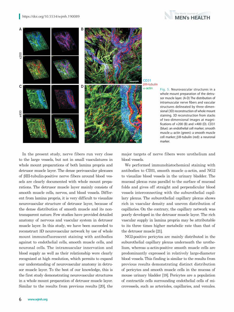
We also successfully demonstrated neurovascular structures in detrusor muscle layer by using whole mount immunostaining. Similar to the findings in the lamina propria, the large-diameter blood vessels are covered with α-actin-positive smooth muscle cells and run parallel to the nerve fibers (Fig. 5).
DISCUSSION
The wall of urinary bladder consists of transitional epithelial lining supported by lamina propria and mus-cularis [3]. Although conventional 2D imaging from thin section of urinary bladder is often useful to visu-alize some cell types, the spatial distribution of certain anatomic structures, such as nervous and vascular sys-tems, cannot be readily appreciated in two-dimensions. We successfully visualized neurovascular structures in whole mount preparations of lamina propria and detrusor muscle as well as full-thickness thick-cut bladder sections by increasing incubation time with primary antibodies up to 48 hours. It was reported that incubation time in primary antibodies should be extended up to 7 days for complete staining in rectal whole mounts [19].
Fig. 3. Three-dimensional (3D) reconstruction of vascular structures in mouse urinary bladder. 3D reconstruction from stacks of 25 two-dimensional images at magnifications of ×100 (A) and ×200 (B, C). α-actin-positive smooth muscle cells are mainly expressed in main arteries of suburothelial plexus, mucosal plexus, and detrusor muscle layer (arrows). CD31 (red): an endothelial cell marker; smooth muscle α-actin (green): a smooth muscle cell marker.
Fig. 4. Neurovascular structures in a whole mount preparation of the lamina propria. (A) The complexity of vascular struc tures and nervous innervation, and their relationship delineated by three-dimensional (3D) reconstruction of whole mount staining. (B-E) Reconstruction of a 3D image from stacks of two-dimen sional images in selected area (white square). CD31 (blue): an endothelial cell marker; smooth muscle α-actin (green): a smooth muscle cell marker; βIII-tubulin (red): a neuronal marker.
https://doi.org/10.5534/wjmh.190089
6 www.wjmh.org
In the present study, nerve fibers run very close to the large vessels, but not in small vasculatures in whole mount preparations of both lamina propria and detrusor muscle layer. The dense perivascular plexuses of βIII-tubulin-positive nerve fibers around blood ves-sels are clearly documented with whole mount prepa-rations. The detrusor muscle layer mainly consists of smooth muscle cells, nerves, and blood vessels. Differ-ent from lamina propria, it is very difficult to visualize neurovascular structure of detrusor layer, because of the dense distribution of smooth muscle and its non-transparent nature. Few studies have provided detailed anatomy of nervous and vascular system in detrusor muscle layer. In this study, we have been succeeded to reconstruct 3D neurovascular network by use of whole mount immunofluorescent staining with antibodies against to endothelial cells, smooth muscle cells, and neuronal cells. The intramuscular innervation and blood supply as well as their relationship were clearly recognized at high resolution, which permits to expand our understanding of neurovascular anatomy in detru-sor muscle layer. To the best of our knowledge, this is the first study demonstrating neurovascular structures in a whole mount preparation of detrusor muscle layer. Similar to the results from previous results [20], the
major targets of nerve fibers were urothelium and blood vessels.
We performed immunohistochemical staining with antibodies to CD31, smooth muscle α-actin, and NG2 to visualize blood vessels in the urinary bladder. The mucosal plexus runs parallel to the surface of mucosal folds and gives off straight and perpendicular blood vessels interconnecting with the suburothelial capil-lary plexus. The suburothelial capillary plexus shows rich in vascular density and uneven distribution of capillaries. On the contrary, the capillary network was poorly developed in the detrusor muscle layer. The rich vascular supply in lamina propria may be attributable to its three times higher metabolic rate than that of the detrusor muscle [21].
NG2-positive pericytes are mainly distributed in the suburothelial capillary plexus underneath the urothe-lium, whereas α-actin-positive smooth muscle cells are predominantly expressed in relatively large-diameter blood vessels. This finding is similar to the results from previous results demonstrating distinct distribution of pericytes and smooth muscle cells in the mucosa of mouse urinary bladder [18]. Pericytes are a population of contractile cells surrounding endothelial cells of mi-crovessels, such as arterioles, capillaries, and venules.
Fig. 5. Neurovascular structures in a whole mount preparation of the detru-sor muscle layer. (A-D) The distribution of intra muscular nerve fibers and vascular struc tures delineated by three-dimen-sional (3D) reconstruction of whole mount staining. 3D reconstruction from stacks of two-dimensional images at mag ni-fications of ×200 (B) and ×400 (D). CD31 (blue): an endothelial cell marker; smooth muscle α-actin (green): a smooth muscle cell marker; βIII-tubulin (red): a neuro nal marker.

Nhat Minh Nguyen, et al: Three-Dimensional Images of Mouse Urinary Bladder
7www.wjmh.org
The functional roles of pericytes include the regulation of vascular contractility, permeability, and develop-ment [22]. We recently demonstrated the dropout of pericytes and subsequent increase in vascular perme-ability in the corpus cavernosum of diabetic mice [23,24]. Therefore, further studies are required to determine the differential distribution of pericytes and their functional role in a variety of bladder disorders.
It was also demonstrated the difference in cell mor-phology between venules and arterioles; α-actin-positive smooth muscle cells are distributed circumferentially in arterioles, whereas venules has stellate-shaped smooth muscle cells [18]. Our technique also clearly demonstrated distinct morphologic features between venules and arterioles at a high-magnification image (data not shown).
Immunohistochemically stained whole mounts and thick-cut sections of mouse urinary bladder have pro-vided more valuable information for defining the in-nervation and the distribution of blood vessels than in sections of conventional thickness. By using this technique, we could delineate the entire urinary blad-der innervation and vascular supply. Especially, whole mount preparations of lamina propria and detrusor muscle layer have nicely demonstrated the running course of the nerve fibers as well as their association with vascular structures. With advent of genetically modified mice, information on the fine details of neu-rovascular structures of the mouse urinary bladder will give us an additional value. Further studies are needed to determine the differential distribution of specific nerve fibers, such as sympathetic, parasympa-thetic, and somatic nerves.
CONCLUSIONS
The visualization of neurovascular structures is crucial to expand our understanding on the patho-physiology of urinary bladder disorders in a variety of conditions. We have developed an effective, rapid, and highly reproducible imaging technique to visualize neurovascular systems in mouse urinary bladder. Our 3D imaging technique that utilizes immunofluorescent staining of full-thickness frozen section of urinary bladder or whole mount preparations of lamina pro-pria and detrusor muscle layers clearly demonstrates association between nervous and vascular systems. Our technique may constitute a standard tool to evaluate
pathologic changes in a variety of urinary bladder dis-eases.
ACKNOWLEDGEMENTS
This work was supported by the National Re-search Foundation of Korea (NRF) grant (Ji-Kan Ryu [2019R1A2C2002414]) and by a Medical Research Cen-ter Grant (Ji-Kan Ryu, 2014R1A5A2009392) funded by the Korean government (Ministry of Science, ICT and Future Planning).
Conflicts of Interest
The authors have nothing to disclose.
Author Contribution
Conceptualization: NMN, CSP, JKS, JKR. Data curation: NMN, KMS, MJC, KG, AL. Formal analysis: MHK, DYC, JO, GNY. Software: MJC, MHK. Funding acquisition: JKR. Writ-ing–original draft: NMN, KMS, JKR. Writing–review & editing: JKS, JKR
Data Sharing Statements
The data required to reproduce these findings cannot be shared at this time as the data also forms part of an ongoing study.
REFERENCES
1. Hossler FE, Monson FC. Microvasculature of the rabbit uri-nary bladder. Anat Rec 1995;243:438-48.
2. Miodoński AJ, Litwin JA. Microvascular architecture of the human urinary bladder wall: a corrosion casting study. Anat Rec 1999;254:375-81.
3. Hossler FE, Lametschwandtner A, Kao R, Finsterbusch F. Mi-crovascular architecture of mouse urinary bladder described with vascular corrosion casting, light microscopy, SEM, and TEM. Microsc Microanal 2013;19:1428-35.
4. Ek A, Alm P, Andersson KE, Persson CG. Adrenergic and cholinergic nerves of the human urethra and urinary bladder. A histochemical study. Acta Physiol Scand 1977;99:345-52.
5. Sakakibara R, Takahashi O, Nishimura H, Tateno F, Kishi M, Tsuyusaki Y, et al. The relationship between bladder, periarte-rial and somatic neuropathy in diabetes. Intern Med 2018;57: 2165-8.

https://doi.org/10.5534/wjmh.190089
8 www.wjmh.org
6. Andersson KE. The many faces of impaired bladder empty-ing. Curr Opin Urol 2014;24:363-9.
7. Yamaguchi O, Nomiya M, Andersson KE. Functional con-sequences of chronic bladder ischemia. Neurourol Urodyn 2014;33:54-8.
8. Ponholzer A, Temml C, Wehrberger C, Marszalek M, Mader-sbacher S. The association between vascular risk factors and lower urinary tract symptoms in both sexes. Eur Urol 2006; 50:581-6.
9. Traish AM, Johansen V. Impact of testosterone deficiency and testosterone therapy on lower urinary tract symptoms in men with metabolic syndrome. World J Mens Health 2018;36:199-222.
10. Quaegebeur A, Lange C, Carmeliet P. The neurovascular link in health and disease: molecular mechanisms and therapeutic implications. Neuron 2011;71:406-24.
11. Li Q, Ford MC, Lavik EB, Madri JA. Modeling the neurovas-cular niche: VEGF- and BDNF-mediated cross-talk between neural stem cells and endothelial cells: an in vitro study. J Neurosci Res 2006;84:1656-68.
12. Grasman JM, Kaplan DL. Human endothelial cells secrete neurotropic factors to direct axonal growth of peripheral nerves. Sci Rep 2017;7:4092.
13. Anastasiadis A, Dimitriadis G, Gkagkalidis K, Leung S, Vakalopoulos I, de la Rosette J. Relation between bladder out-let obstruction and systemic vascular endothelial dysfunction in the male. A preliminary study. Int J Adv Res 2017;5:1053-60.
14. Yamada T, Nishimura M, Mita H. Increased number of apop-totic endothelial cells in bladder of interstitial cystitis patients. World J Urol 2007;25:407-13.
15. Wang Y, Xu R, Luo G, Wu J. Three-dimensional reconstruc-
tion of light microscopy image sections: present and future. Front Med 2015;9:30-45.
16. Purves JT, Spruill L, Rovner E, Borisko E, McCants A, Mugo E, et al. A three dimensional nerve map of human bladder trigone. Neurourol Urodyn 2017;36:1015-9.
17. Spradling K, Khoyilar C, Abedi G, Okhunov Z, Wikenheiser J, Yoon R, et al. Redefining the autonomic nerve distribution of the bladder using 3-dimensional image reconstruction. J Urol 2015;194:1661-7.
18. Mitsui R, Hashitani H. Immunohistochemical characteristics of suburothelial microvasculature in the mouse bladder. His-tochem Cell Biol 2013;140:189-200.
19. Llewellyn-Smith IJ, Gnanamanickam GJ. Immunoperoxidase detection of neuronal antigens in full-thickness whole mount preparations of hollow organs and thick sections of central nervous tissue. J Neurosci Methods 2011;196:1-11.
20. Gabella G, Davis C. Distribution of afferent axons in the blad-der of rats. J Neurocytol 1998;27:141-55.
21. Hypolite JA, Longhurst PA, Gong C, Briscoe J, Wein AJ, Levin RM. Metabolic studies on rabbit bladder smooth muscle and mucosa. Mol Cell Biochem 1993;125:35-42.
22. Armulik A, Abramsson A, Betsholtz C. Endothelial/pericyte interactions. Circ Res 2005;97:512-23.
23. Yin GN, Ock J, Choi MJ, Song KM, Ghatak K, Nguyen NM, et al. A simple and nonenzymatic method to isolate human corpus cavernosum endothelial cells and pericytes for the study of erectile dysfunction. World J Mens Health 2019. doi: 10.5534/wjmh.180091 [Epub].
24. Yin GN, Das ND, Choi MJ, Song KM, Kwon MH, Ock J, et al. The pericyte as a cellular regulator of penile erection and a novel therapeutic target for erectile dysfunction. Sci Rep 2015;5:10891.