thoracic trauma powerpoint · • in the broadest sense, injuries to the chest fall into two ......
TRANSCRIPT
5/2/12
1
Thoracic Trauma
Sheryl M. Sahr, MD Iowa Society for Respiratory Care
2012 Lung Conference April 20, 2012
Basic Facts
• Trauma to the thorax is the second most common injury found in trauma paIents – 764,000 admissions in 2004 alone – It’s esImated that 10% of trauma paIents have at least one rib fracture
– Even more (15%) may have pulmonary contusions – These injuries rarely occur in isolaIon – 94% of paIents with rib fractures had other injuries
Basic Facts
• Thoracic trauma causes up to 25% of deaths in trauma vicIms – This is second only to traumaIc brain injury in mortality
– The risk of death aWer rib fractures increases with: • Age greater than 65 • More than 3 rib fractures • Presence of comorbidiIes like heart disease or diabetes • Development of pneumonia while on the venIlator
Basic Facts
• The most frequent mechanism of injury is motor vehicle crash (MVC) – Up to 80%
• Falls are a distant second in many studies – About 20% • As a populaIon ages, falls become more common and MVC becomes less common.
• Assault with blunt object and sports injuries are infrequent causes of thoracic injury
Basic Facts • Thoracic trauma commonly results in bony injury – Rib fractures – Sternal fracture – Clavicle and scapular fracture
• The underlying organs and structures are also at risk for damage – Blunt cardiac injury and myocardial dysfuncIon – Pulmonary contusion – Pulmonary laceraIon – Tracheobronchial injury – Pneumothorax and hemothorax
Mechanisms of Injury
• In the broadest sense, injuries to the chest fall into two categories: – Direct impact (steering wheel into chest, fall from height)
– DeceleraIon (steering wheel into chest, fall from height) • No, this isn’t a typo…
5/2/12
2
More on Mechanisms • Direct impact involves compressive forces across the bony structures of the chest. – These forces are usually directed antero‐posteriorly (head‐on collision) or laterally (T‐bone accident) • Compression results in deformaIon of the somewhat elasIc ribcage
• Compression of a rib beyond 20% deformity results in a fracture – Compression beyond 40% is usually required for flail chest
• Overpressure to the lungs can occur at impact (especially if the breath is held in Valsalva by reflex)
• Compression forces are transmided across the chest wall to the structures beneath
More on Mechanisms • Direct impact injuries: – Rib fractures – first the ribs bend, then they break
• Younger paIents withstand more force – Pulmonary contusion
• Force transmided through the chest wall – Pulmonary laceraIon or pneumothorax
• LaceraIon by broken ribs • Overpressure from impact during Valsalva
– Sternal fracture, clavicular fracture, scapular fracture • These fractures are associated with pulmonary contusions – not necessarily because the lung is being struck, but because of the force required to fracture these bones
More on Mechanisms • DeceleraIon refers to the problem of the organs of the
chest slowing (at the Ime of impact) at varying speeds – The sternum stops first (on the steering wheel) – The heart stops milliseconds later (on the sternum) – The lungs stop against the deforming ribcage – Stretching occurs to both the heart and the lungs, since they are tethered centrally (to each other, to the posterior chest wall, and to the great vessels) • Stretching occurs at the organ level (tearing of the trachea or bronchi, or of the aorta)
• It also occurs in the Issues at every tether point – each bifurcaIon from the bronchus to the alveolus
• At impact, the ribs are moving toward the deceleraIng lungs; some areas of lung are dragged across the ribs harder than others because of the impact. This causes localized severe shearing
More on Mechanisms • DeceleraIon / Shear injuries – Pulmonary laceraIon
• DeceleraIon against adhesions or at bronchial bifurcaIons • Also against deforming ribs
– Pulmonary contusion • Shear at the capillary level
– Tracheobronchial disrupIon • Shear at the macro level • Rarely seen in the hospital because it’s so immediately lethal, both in loss of airway and because of the immense forces required – Think about the snapping towel
Rib Fractures • Rib fractures have varying implicaIons based on their
locaIon – Ribs 1‐3 (the top of the rib cage, behind the clavicles) are fixed in place • Fractures of these ribs, as with scapular fractures, are markers for high‐impact force to the chest wall
– Ribs 4‐8 • These ribs move with respiraIon • Flail chest usually involves these ribs
– Ribs 8‐11 • Markers for intra‐abdominal injury when fractured • Right sided rib fractures carry a 20‐55% risk of concomitant liver injury • LeW sided rib fractures have a 22‐28% risk of associated splenic injury
Rib Fractures
• Clinical consideraIons in the ED – Posterior rib fractures are difficult to appreciate on CXR – especially on paIents strapped to a backboard • On the other hand, they’re oWen hard to see in upright PA/Lateral films…
– If a paIent receives isolated rib X‐rays, a formal CXR showing both lung fields is a good idea as well.
– Rib fractures should be suspected in paIents with exquisite tenderness to palpaIon of the chest wall • Bony crepitus may also be noted • AddiIonal clues: seatbelt sign, bruising, significant splinIng
5/2/12
3
Rib Fractures • Rib fractures cause pulmonary dysfuncIon directly and indirectly – Associated with pulmonary contusion – Pain‐related hypovenIlaIon
• The elderly are parIcularly suscepIble, which is why many major studies demonstrate mortality up to 35 or 40% in elderly paIents with mulIple rib fractures (3+)
– Progressive atelectasis and hypoxia • PaIent may require aggressive pulmonary toilet with modaliIes that provide posiIve pressure
– Increased work of breathing • The end result of pain‐related hypovenIlaIon and atelectasis; also related to the decreased lung compliance in contused lung
Flail Chest • Defined most commonly as three adjacent ribs each broken in two places
• About 10% of paIents who have blunt chest trauma will have a flail segment
• Ribs become bridle with age. Young paIents with flail chests have absorbed much more force than elderly flail paIents – The likelihood of underlying severe pulmonary contusion is higher in younger paIents
– Rib fractures in children (without an extraordinary mechanism) should prompt evaluaIon for abuse • The corollary is that pulmonary contusion should be anIcipated in children even in the absence of rib fractures
Flail Chest
• Clinical ConsideraIons in the ED – PaIents can have their flail segment diagnosed in many ways • InspecIon: the chest wall contour is “weird” on one side, when looking up from the foot of the bed
• PalpaIon: “mushy” or crackly chest wall • AuscultaIon: crepitus, either from broken ribs or from soW Issue air bubbles (sub‐cu emphysema)
• May be seen on CXR, if the flail is anterior and lateral • Most reliably diagnosed on CT
Flail Chest • Flail chest causes pulmonary dysfuncIon because: – The primary cause of hypoxia in flail chest is due to the underlying pulmonary contusion
– Causes of hypovenIlaIon in flail chest vary: • “PendelluW” – increases dead space venIlaIon (air moving between areas of differing compliance rather than between the lungs and the pharynx)
• There is local loss of the elasIc recoil (directed outward) of the chest wall to counteract the natural elasIc recoil (directed inward) of the lung; air will not rush into these areas on acIve inspiraIon – Work of breathing is increased, both due to the contusion and to the fact
that the same inspiratory acIon brings less Idal volume in (since the flail segment is not inflaIng)
– Stasis of secreIons in the flail segment
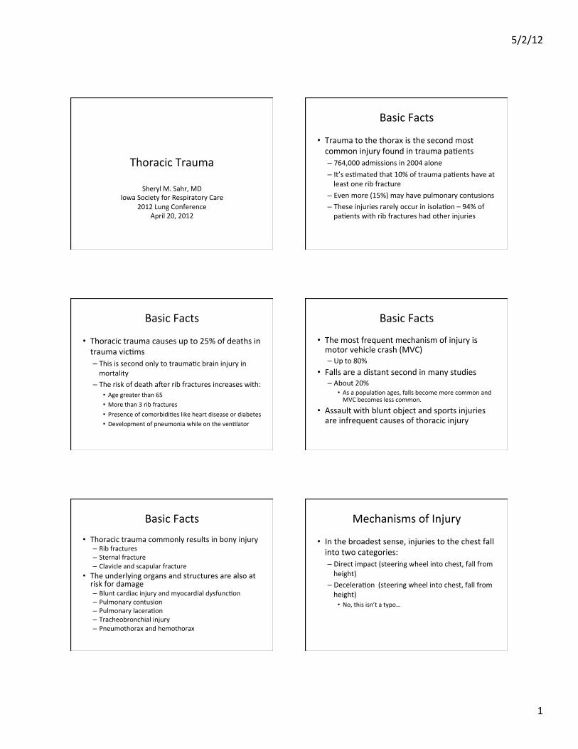
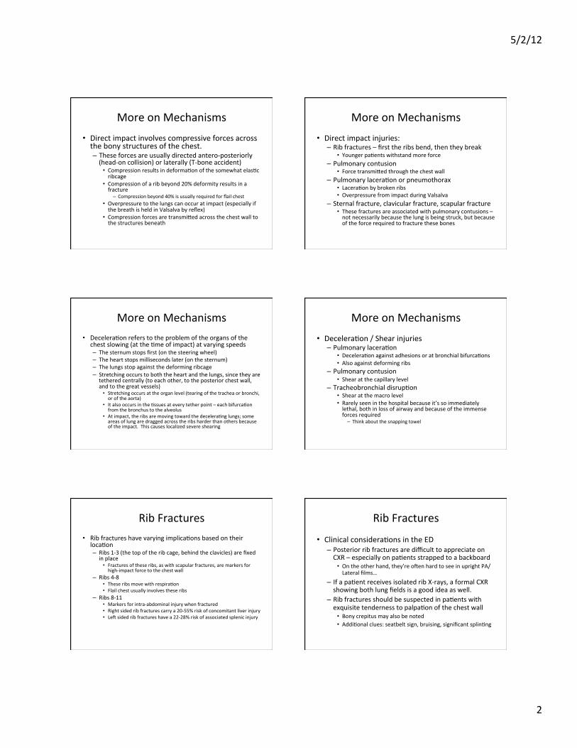
Pulmonary Contusion
• Appear on CT or CXR as infiltrates that cross lung fissures and lobes (so are not infiltrates related to aspiraIon)
• On CT, do not show the classic dependent padern that aspiraIon does
• Blood leaves the vessels and enters the parenchyma and the alveoli of the injured lung (“bruised lung”)
• This can be due to direct impact or to shear injury
Pulmonary Contusion • Clinical ConsideraIons in the ED – The paIent who is unusually hypoxic (70’s to mid 80’s, as a ballpark) should be closely evaluated for contusion
– Beware the paIent who is “crazy” and will not keep their pulse oximeter on… • SomeImes the paIent is high or badly concussed. SomeImes the paIent is deeply hypoxic.
– Severe contusions will be visible on CXR even in the ED – but not all are. A “clear” CXR is not reassuring if the paIent is sIll hypoxic.
– PaIents with severe hypoxia may require intubaIon in the ED. • Even with intubaIon, hypoxia may persist: use of a PEEP valve or adding PEEP to the venIlator oWen helps. (More on this later)
5/2/12
4
Savetamal and Livingston, Current Therapy of Trauma Savetamal and Livingston, Current Therapy of Trauma
Pulmonary Contusion • Blood in the alveoli and the lung Issues causes pulmonary dysfuncIon in several ways – Compliance in the injured lung decreases
• PaIent works harder to inhale the same Vt • In venIlated paIents, higher inspiratory pressures are needed to move the same volume – PaIents are “hard to bag”
– ShunIng increases (V/Q mismatch; blood travels through regions of lung that are injured and poorly venIlated
– Plasma proteins inacIvate lung surfactant, causing alveolar collapse and shear stress on neighboring alveoli • The picture is depressingly similar to ALI or ARDS
Pneumothorax
• 10% or so across all trauma paIents, but more like 30‐40% of blunt trauma paIents have PTX
• Causes – PenetraIng trauma (stabs, gunshot wounds) – LaceraIon by broken rib (direct impact) – LaceraIon at previous scars (shear) – Ruptured bleb (overpressure) – Simple overpressure (impact while performing Valsalva)
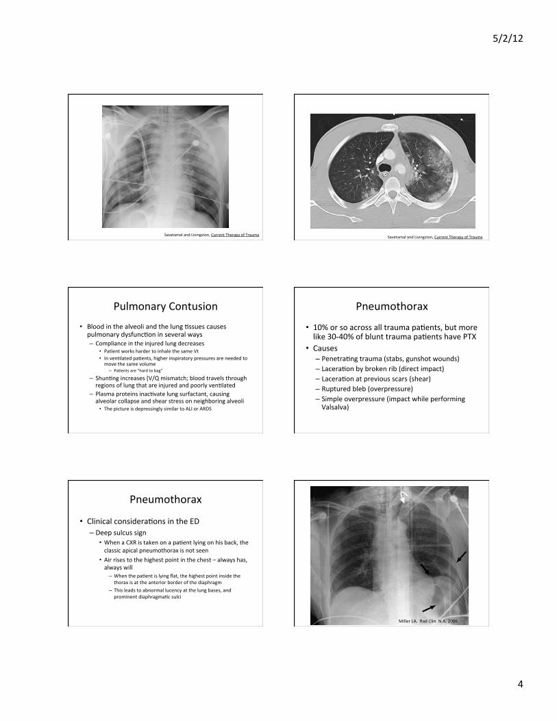
Pneumothorax
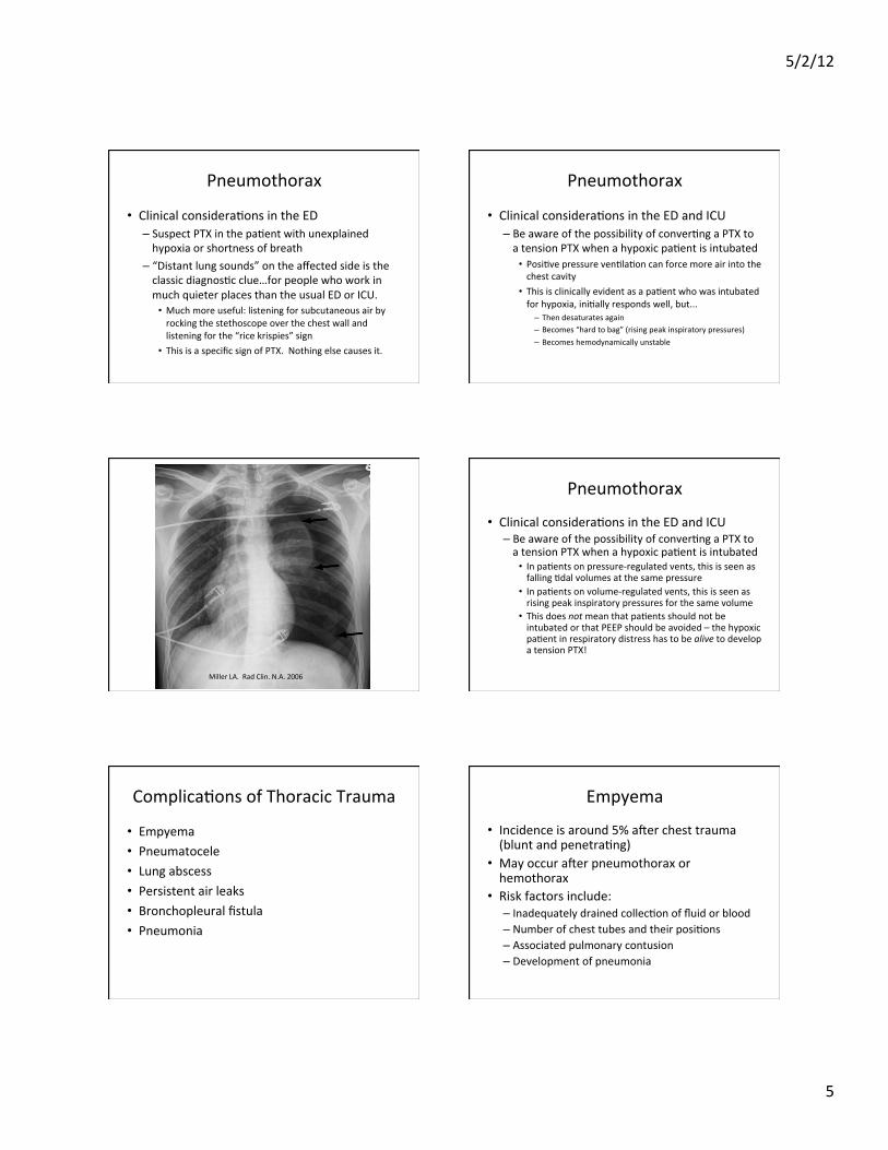
• Clinical consideraIons in the ED – Deep sulcus sign
• When a CXR is taken on a paIent lying on his back, the classic apical pneumothorax is not seen
• Air rises to the highest point in the chest – always has, always will – When the paIent is lying flat, the highest point inside the thorax is at the anterior border of the diaphragm
– This leads to abnormal lucency at the lung bases, and prominent diaphragmaIc sulci
Miller LA. Rad Clin. N.A. 2006
5/2/12
5
Pneumothorax
• Clinical consideraIons in the ED – Suspect PTX in the paIent with unexplained hypoxia or shortness of breath
– “Distant lung sounds” on the affected side is the classic diagnosIc clue…for people who work in much quieter places than the usual ED or ICU. • Much more useful: listening for subcutaneous air by rocking the stethoscope over the chest wall and listening for the “rice krispies” sign • This is a specific sign of PTX. Nothing else causes it.
Pneumothorax
• Clinical consideraIons in the ED and ICU – Be aware of the possibility of converIng a PTX to a tension PTX when a hypoxic paIent is intubated • PosiIve pressure venIlaIon can force more air into the chest cavity
• This is clinically evident as a paIent who was intubated for hypoxia, iniIally responds well, but... – Then desaturates again – Becomes “hard to bag” (rising peak inspiratory pressures) – Becomes hemodynamically unstable
Miller LA. Rad Clin. N.A. 2006
Pneumothorax
• Clinical consideraIons in the ED and ICU – Be aware of the possibility of converIng a PTX to a tension PTX when a hypoxic paIent is intubated • In paIents on pressure‐regulated vents, this is seen as falling Idal volumes at the same pressure
• In paIents on volume‐regulated vents, this is seen as rising peak inspiratory pressures for the same volume
• This does not mean that paIents should not be intubated or that PEEP should be avoided – the hypoxic paIent in respiratory distress has to be alive to develop a tension PTX!
ComplicaIons of Thoracic Trauma
• Empyema • Pneumatocele • Lung abscess • Persistent air leaks • Bronchopleural fistula • Pneumonia
Empyema
• Incidence is around 5% aWer chest trauma (blunt and penetraIng)
• May occur aWer pneumothorax or hemothorax
• Risk factors include: – Inadequately drained collecIon of fluid or blood – Number of chest tubes and their posiIons – Associated pulmonary contusion – Development of pneumonia
5/2/12
6
Empyema • Usually presents as an unexplained fever or WBC, days to weeks
aWer the injury – This may be associated with pleuriIc pain, increased work of
breathing, and potenIally new vent‐dependent respiratory failure • Diagnosed with CT (CXR may not be useful, depending on the size
of the empyema) • Treatment (from least to most invasive):
– Chest tube drainage – InsIlling a clot‐buster (like alteplase) via the chest tube – Video‐assisted thoracoscopy (VATS) – Thoracotomy with decorIcaIon (pulling the pus layer off the lung – …the longer it takes to diagnose the empyema, the more invasive the
treatment will have to be.
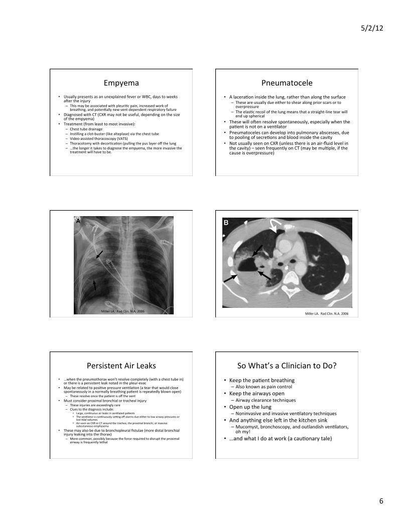
Pneumatocele • A laceraIon inside the lung, rather than along the surface
– These are usually due either to shear along prior scars or to overpressure
– The elasIc recoil of the lung means that a straight‐line tear will end up spherical
• These will oWen resolve spontaneously, especially when the paIent is not on a venIlator
• Pneumatoceles can develop into pulmonary abscesses, due to pooling of secreIons and blood inside the cavity
• Not usually seen on CXR (unless there is an air‐fluid level in the cavity) – seen frequently on CT (may be mulIple, if the cause is overpressure)
Miller LA. Rad Clin. N.A. 2006 Miller LA. Rad Clin. N.A. 2006
Persistent Air Leaks • …when the pneumothorax won’t resolve completely (with a chest tube in)
or there is a persistent leak noted in the pleur‐evac • May be related to posiIve pressure venIlaIon (a tear that would close
spontaneously in a normally breathing paIent is repeatedly blown open) – These resolve once the paIent is off the vent
• Must consider proximal bronchial or tracheal injury – These injuries are exceedingly rare – Clues to the diagnosis include:
• Large, conInuous air leaks in venIlated paIents • The venIlator is conInuously serng off alarms due either to low airway pressures or
lost Idal volumes • Air seen on CXR or CT around the trachea, the proximal bronchi, or massive
subcutaneous emphysema • These may also be due to bronchopleural fistulae (more distal bronchial
injury leaking into the thorax) – More common, possibly because the force required to disrupt the proximal
airway is frequently lethal
So What’s a Clinician to Do? • Keep the paIent breathing – Also known as pain control
• Keep the airways open – Airway clearance techniques
• Open up the lung – Noninvasive and invasive venIlatory techniques
• And anything else leW in the kitchen sink – Mucomyst, bronchoscopy, and outlandish venIlators, oh my!
• …and what I do at work (a cauIonary tale)
5/2/12
7
Pain Control
• Local and regional pain control: rib blocks – Local anesthesia is injected between the ribs, one rib above and below the fracture(s) • Pro: a temporary method of controlling pain • Con: a temporary method of controlling pain
– Risk of pneumothorax is real – Can’t use on higher rib fractures safely
– A newer take on this is the paravertebral catheter adached to a long‐acIng infusion system • Must be placed under sterile condiIons • Risk of infecIon, pneumothorax
Pain Control • Epidural anesthesia – a mainstay at many faciliIes for
treatment of thoracic trauma – Usually include both opioids (fentanyl) and local anestheIc (bupivicaine)
– Pro: can improve the work of breathing by decreasing splinIng • The relief that the epidural provides can be dramaIc
– Con: many contraindicaIons • PaIents on anIcoagulaIon – even prophylacIcally • Thoracic vertebral fractures • PaIents who are not ready to come off the vent
– Con: recent meta‐analyses have not proven benefit in mortality, vent days, or hospital length of stay
– Con: may actually increase morbidity and mortality in elderly paIents (who must “move or die”)
Pain Control • Whole–body pain control – Opioid infusions (e.g. fentanyl in intubated paIents) – PaIent‐controlled anesthesia (PCA)
• Dilaudid, morphine, fentanyl – Long‐acIng oral opioids
• Oxy‐conIn, methadone (may be beder in the elderly) – AdjuncIve NSAIDs
• Toradol…a wonder drug that is unfortunately not for everyone – Bleeding risk in trauma paIents – Risk of kidney failure (parIcularly in the elderly, diabeIc, hypertensive populaIon…which is most of the state)
Airway Clearance Techniques • Why is a trauma surgeon talking about airway clearance?
– The last Ime I had a cysIc fibrosis paIent was…predy much never – Emphysematous and bronchiIc paIents are common – Trauma itself usually includes an aspiraIon event
• Abdominal overpressure “on impact” • Loss of consciousness • Facial fractures bleed profusely • Many paIents are intoxicated at the Ime of trauma
– Blood in the airways should be cleared • Percussive therapy – CPT or vest percussion
– Not popular in the rib fracture populaIon – Not advised in unstable vertebral fractures or early closed head injury – Can be useful in paIents who are unable or unwilling to cough independently
• Fluder valve – Provides repeatable cough sImulaIon for the nonintubated paIent
Airway Clearance Techniques • Bronchoscopy and lavage
– PaIent must be intubated. This is usually a rescue therapy (blood casts in the airways)
• High frequency oscillaIon with nebulized treatments – Provides both posiIve end‐expiratory pressure and moistening of secreIons for ease of expectoraIon
• Mucomyst (N‐acetylcysteine) insIllaIon – The taste is not for the faint of heart…so usually used on intubated paIents
– InsIlled and not nebulized (can clog and block the air filter on the expiratory circuit, leading to overpressure injury to the paIent)
– Use for limited Ime period (can itself sImulate bronchial secreIons, aWer about two days)
VenIlatory Techniques • Non‐invasive techniques – the goal is to avoid intubaIon – PosiIve pressure venIlaIon (CPAP or bi‐level PAP)
• Recruit alveoli, which indirectly can decrease the work of breathing
• Direct reducIon in work of breathing • “Internal” or “PneumaIc” splinIng of flail chest
– This is likely the recruitment of contused lung under the flail segment
– High flow nasal cannula • EffecIvely provides the equivalent of PEEP • Cannula is beder tolerated than CPAP masks, parIcularly in paIents with facial fractures
5/2/12
8
VenIlatory Techniques • Thoracic trauma results in both primary and secondary damage to lung Issue – Primary damage:
• contusion (blood in the airways and parenchyma) • laceraIon • pneumothorax
– Secondary damage: this should sound oddly familiar… • loss of surfactant and inducIon of inflammaIon • alveolar collapse • increased right‐to‐leW shunt • decreased compliance • increased work of breathing • progressive hypoxemia and venIlatory failure
VenIlatory Techniques
• Considering the secondary damage that flail chest, pulmonary laceraIon and contusion all can cause – it isn’t surprising that the majority of thoracic trauma paIents will respond to the same therapies – And considering the litany of secondary pulmonary injury listed…it isn’t surprising that techniques for ALI and ARDS frequently have applicaIon in trauma. • Low Idal volumes based on paIents’ ideal weights • Stepwise increase in PEEP in hypoxic paIents (and minimum of 5 cm H2O in all paIents)
VenIlatory Techniques • RaIonale for PEEP – End‐expiratory pressure helps to recruit alveoli
• For vented paIents, this is reflected as increased compliance and lower inspiratory pressures over Ime (at the same Idal volume in volume‐control modes) – The same Idal volume is going into more lung volume
– Recruited alveoli are spared repeIIve open/close cycles that cause shear and inflammaIon at the cellular level • TheoreIcally, this may prevent extension of secondary injury – with the hope of avoiding ARDS enIrely
– As perfused (but collapsed) alveoli are opened for gas exchange, right‐to‐leW shunt decreases
VenIlatory Techniques • SituaIons where more PEEP is not helpful
– Persistent air leaks aWer pneumothorax • These may persist unIl the paIent is extubated and performing normal negaIve‐pressure venIlaIon
– Conversion of simple PTX to tension PTX • This is noted as a cauIon, not as a rule; the treatment team must be vigilant for signs of PTX, and place chest tubes to prevent conversion to tension PTX.
– Major tracheobronchial injury • May require pressure‐control venIlaIon with extremely low Idal volumes (and permissive hypercapnia)
• May require alternaIve venIlaIon modes – Air‐trapping in COPD paIents
• Some COPD paIents simply will not tolerate low Idal volume / higher frequency venIlaIon because they require very long expiratory Imes.
The VenIlatory Strategies of DesperaIon
• …when PEEP is not enough • Airway pressure release venIlaIon
– This is considered a variant of extreme reverse I:E venIlaIon (the inspiratory phase can last up to 5 or 6 seconds, with the expiratory release of pressure lasIng less than one second)
– Can permit independent breathing effort in paIents, which means less sedaIon needed • Should be used very cauIously in paIents who are not breathing
spontaneously • High frequency oscillatory venIlaIon
– Reduces the shear associated with repeIIve open/close cycles at the alveolus
– Can be useful in paIents with persistent or massive air leaks (or bronchopleural fistula)
– Has been used in paIents requiring independent lung venIlaIon (with a convenIonal venIlator for the uninjured lung)
The VenIlatory Strategies of DesperaIon
• …When PEEP is not enough. – Severe bilateral pulmonary contusion – ALI from fat embolus – Severe single pulmonary contusion – Massive air leak from chest wall destrucIon
• Single lung venIlaIon – Can be used in massive air leak or in single‐sided pulmonary contusion
• Put the good lung down…to pull blood to the venIlated lung by gravity • Independent lung venIlaIon
– Used, for example, if one lung has a massive air leak, and the other a severe contusion • Can use a double lumen tube • Some physicians have placed two ETT via tracheostomy • Remember that 55% of the usual Vt should go to the right lung

5/2/12
9
What Do We See at Methodist?
• 43% of our paIents have “fall” as their mechanism of injury – Fall from height, fall from ladder, fall from bed..
• This reflects the older and rural populaIons in central Iowa • In our paIents over 65 with mulIple rib fractures, nearly two‐thirds have fallen down
• 30% are involved in MVC – Motorcycles are included…ATVs are not
• 8% suffer penetraIng injury – Stabs, gunshot wounds, the occasional road sign…
This Week at Methodist
• Two pediatric pulmonary contusions – Neither child has rib fractures
• One child is intubated, the other is not • Two adult pulmonary contusions – One with rib fractures, one without
• Three adults with mulIple rib fractures • Two of these paIents are older than 75 • None of them have yet to require intubaIon
– Two flail chests • One flail chest paIent also fractured the sternum
What Happens…
• In the ED? – A supine CXR is taken on more than 90% of paIents within the first 10 minutes of evaluaIon
– The chest wall is exposed, examined, and auscultated
– PaIents with PTX large enough to be obvious on a supine CXR have chest tubes placed within the first 30 minutes of evaluaIon (barring more serious injuries)
– The paIent that almost got away…
What Happens
• To intubated paIents in the ED? – PaIents who arrive intubated have their airways re‐checked for tube movement during transfer • StarIng Idal volumes range between 400 and 600 in adults • StarIng rates range between 16 to 20 • I start asking for a PEEP valve to be placed in‐line with the bag‐valve mask…starIng between 5 and 7, occasionally 10
– PaIents who require intubaIon receive ETT from surgical residents, anesthesiologists, or helicopter personnel • I start asking for a PEEP valve to be placed in‐line with the bag‐valve mask
What Happens • To paIents with mulIple rib fractures, pulmonary contusion, or flail chest in the ED? – PaIents in acute hypoxic or respiratory distress are intubated
– The remainder…enter rib fracture boot camp in the ICU • Aggressive mobilizaIon, when possible, with PT • ConInuous iv and/or oral pain control • Frequent airway clearance and recruitment therapies
– High‐frequency posiIve pressure nebs up to 6 Imes daily – Occasionally, use of CPAP as an intermident recruitment tool – If scheduled recruitment therapies do not maintain venIlaIon and
oxygenaIon, the paIent is placed on conInuous posiIve end‐expiratory pressure (CPAP, bi‐level PAP, high‐flow nasal cannula)
The Obligatory Clean‐Up Slide
• QuesIons, comments are welcome…so long as they are wildly appreciaIve.
• Speaking of wild appreciaIon – the Trauma team could not do what we do without the assistance and guidance of the Respiratory Therapy team. – ParIcular thanks to Julie Jackson, who invited me to speak here today
– And thank you for your Ime and adenIon!
5/2/12
10
Bibliography • Savetamal, A. and Livingston, D.H. Thoracic Wall Injuries, Chapter 34 in Current Therapy of
Trauma, ed. Livingston • Hauser, C.J. and Livingston, D.H. Pulmonary Contusion and Flail Chest, Chapter 36 in Current
Therapy of Trauma, ed. Livingston • Miller, L.A. 2006. Chest wall, lung , and pleural space trauma. Radiologic Clinics of North America
44:213‐224. • Rico, F.R., et al. 2007. Mechanical venIlaIon strategies in massive chest trauma. CriIcal Care
Clinics 23:299‐315. • Perford, B.L., et al. 2007. The management of flail chest. Thoracic Surgery Clinics 17:25‐33. • Carrier, F.M., et al. 2009. Effect of epidural anesthesia in paIents with traumaIc rib fractures: a
systemaIc review and meta‐analysis of randomized controlled trials. Canadian Journal of Anesthesiology 56:230‐242.
• Kiraly, L. and Schreiber, M. 2010. Management of the crushed chest. CriIcal Care Medicine 38(9 Suppl): S469‐477.
• Oikonomou, A. and Prassopoulos, P. 2011. CT imaging of chest trauma. Insights Imaging 2:281‐295.
• Ghadiali, S.N. and Huang, Y. 2011. Role of airway recruitment and derecruitment in lung injury. CriIcal Reviews in Biomedical Engineering 39(4):297‐318.
• Badle, C.E., et al. 2012. Risk factors that predict mortality in paIents with blunt chest wall trauma: a systemaIc review and meta‐analysis. Injury 43:8‐17.