thoracic spine anatomy table 7.1 concept...
TRANSCRIPT
Cook, Orthopedic Manual Therapy: An Evidence-Based Approach, 2/E © 2012 by Pearson Education, Inc., Upper Saddle River, NJ
THORACIC SPINE ANATOMY
The thoracic spine is unique from the cervical and lumbar spine because of the size and
extent of the region and the articulations with the rib cage. The articulation with the rib cage leads to
regional variations in movement patterns and function (1). The upper thoracic spine mimics the
movement and to some extent, the anatomy of the cervical spine and the lower thoracic vertebra
mimics the lumbar spine.
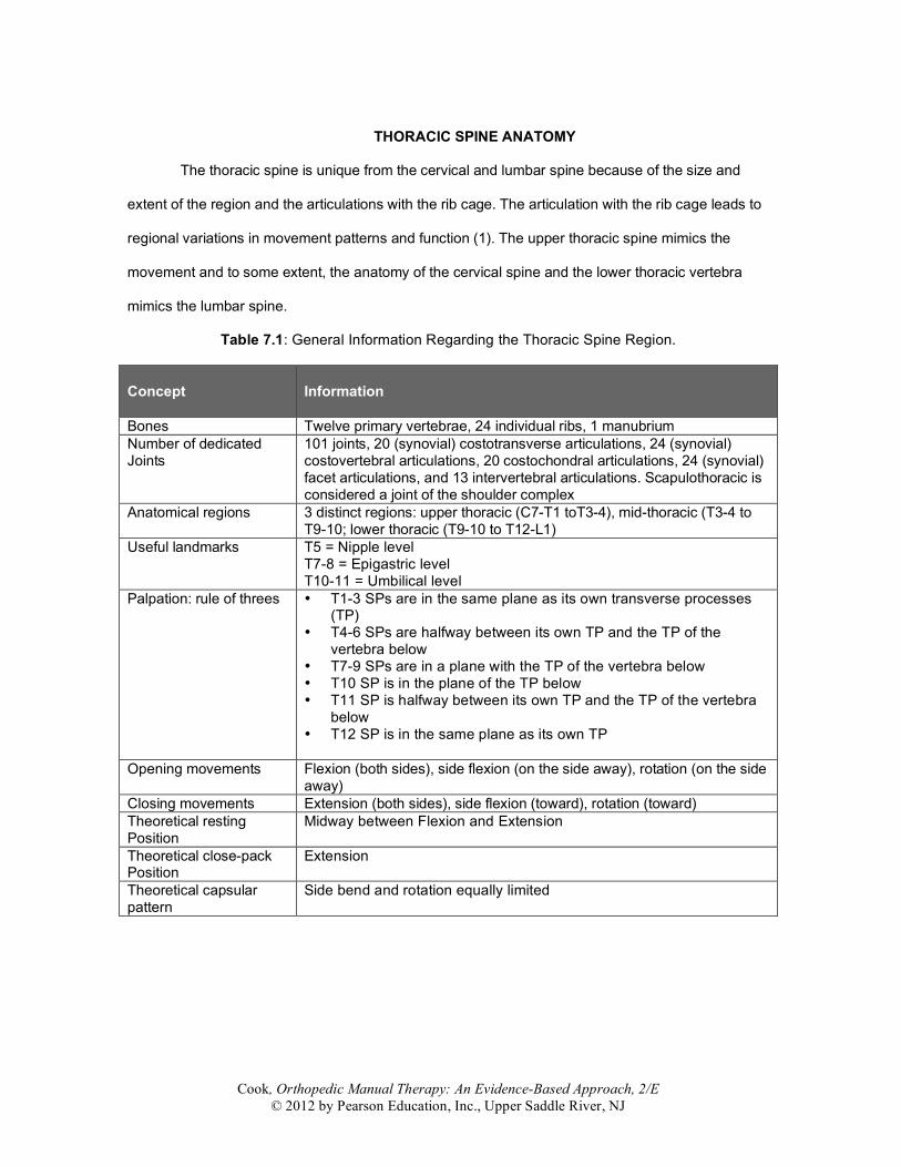
Table 7.1: General Information Regarding the Thoracic Spine Region. Concept
Information
Bones Twelve primary vertebrae, 24 individual ribs, 1 manubrium Number of dedicated Joints
101 joints, 20 (synovial) costotransverse articulations, 24 (synovial) costovertebral articulations, 20 costochondral articulations, 24 (synovial) facet articulations, and 13 intervertebral articulations. Scapulothoracic is considered a joint of the shoulder complex
Anatomical regions 3 distinct regions: upper thoracic (C7-T1 toT3-4), mid-thoracic (T3-4 to T9-10; lower thoracic (T9-10 to T12-L1)
Useful landmarks T5 = Nipple level T7-8 = Epigastric level T10-11 = Umbilical level
Palpation: rule of threes • T1-3 SPs are in the same plane as its own transverse processes (TP)
• T4-6 SPs are halfway between its own TP and the TP of the vertebra below
• T7-9 SPs are in a plane with the TP of the vertebra below • T10 SP is in the plane of the TP below • T11 SP is halfway between its own TP and the TP of the vertebra
below • T12 SP is in the same plane as its own TP
Opening movements Flexion (both sides), side flexion (on the side away), rotation (on the side away)
Closing movements Extension (both sides), side flexion (toward), rotation (toward) Theoretical resting Position
Midway between Flexion and Extension
Theoretical close-pack Position
Extension
Theoretical capsular pattern
Side bend and rotation equally limited
Cook, Orthopedic Manual Therapy: An Evidence-Based Approach, 2/E © 2012 by Pearson Education, Inc., Upper Saddle River, NJ
Osseous Structures
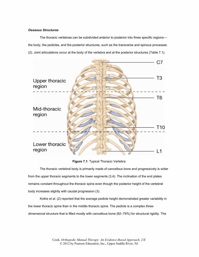
The thoracic vertebrae can be subdivided anterior to posterior into three specific regions—
the body, the pedicles, and the posterior structures, such as the transverse and spinous processes
(2). Joint articulations occur at the body of the vertebra and at the posterior structures (Table 7.1).
Figure 7.1: Typical Thoracic Vertebra
The thoracic vertebral body is primarily made of cancellous bone and progressively is wider
from the upper thoracic segments to the lower segments (3,4). The inclination of the end plates
remains constant throughout the thoracic spine even though the posterior height of the vertebral
body increases slightly with caudal progression (3).
Kothe et al. (2) reported that the average pedicle height demonstrated greater variability in
the lower thoracic spine than in the middle thoracic spine. The pedicle is a complex three-
dimensional structure that is filled mostly with cancellous bone (62–79%) for structural rigidity. The
Cook, Orthopedic Manual Therapy: An Evidence-Based Approach, 2/E © 2012 by Pearson Education, Inc., Upper Saddle River, NJ
outer cortical shell showed different thickness throughout its perimeter and variations in trabeculae at
disparate levels.
The posterior structures include the transverse processes and the articulations, the facets,
and the spinous processes. The spinous processes angle inferiorly and progressively from the upper
thoracic spine to the mid- to lower thoracic spine. The transverse processes angle posteriorly and
provide the contact points for the facets (5).
The ribs are long, thin bones that are commonly fractured during trauma to the thoracic
region (6) and connect to the thoracic spine anteriorly and posteriorly. Each rib has a convex head
that articulates with the concave facets of the vertebral body (costovertebral joint) and transverse
processes of the thoracic spine (costotransverse joint). The anterior connection is called the costo-
sternal attachment and identifies the two separate articulations of the sternum to the costal cartilage
and the costal cartilage to the rib. The anterior articulation is a flattened, concave depression.
The Intervertebral Disc
The intervertebral disc plays a major role in movement control of the thoracic spine, a much
more significant role than the posterior structures (7). With respect to height, the disc in the thoracic
spine demonstrates less height in ratio to the vertebral body than the cervical and lumbar spines (8).
Additionally, the thoracic disc has a relatively small nucleus pulposus (9).
It is expected that the compliance of the thoracic disc is lost much earlier than the cervical or
lumbar disc (13). Disc space narrowing is common from the third decade of life and disc
degeneration, osteophytes, and subsequent degenerative changes are frequent findings in the mid-
thoracic segment (16).
With respect to intradiscal pressures, Polga et al. (11) found that the positions of standing
upright with 10-kg weights in each arm display the highest pressure versus other positions such as
prone lying, sidelying, sitting with and without flexion, and other variations of standing including
twisting.
Cook, Orthopedic Manual Therapy: An Evidence-Based Approach, 2/E © 2012 by Pearson Education, Inc., Upper Saddle River, NJ
Joints
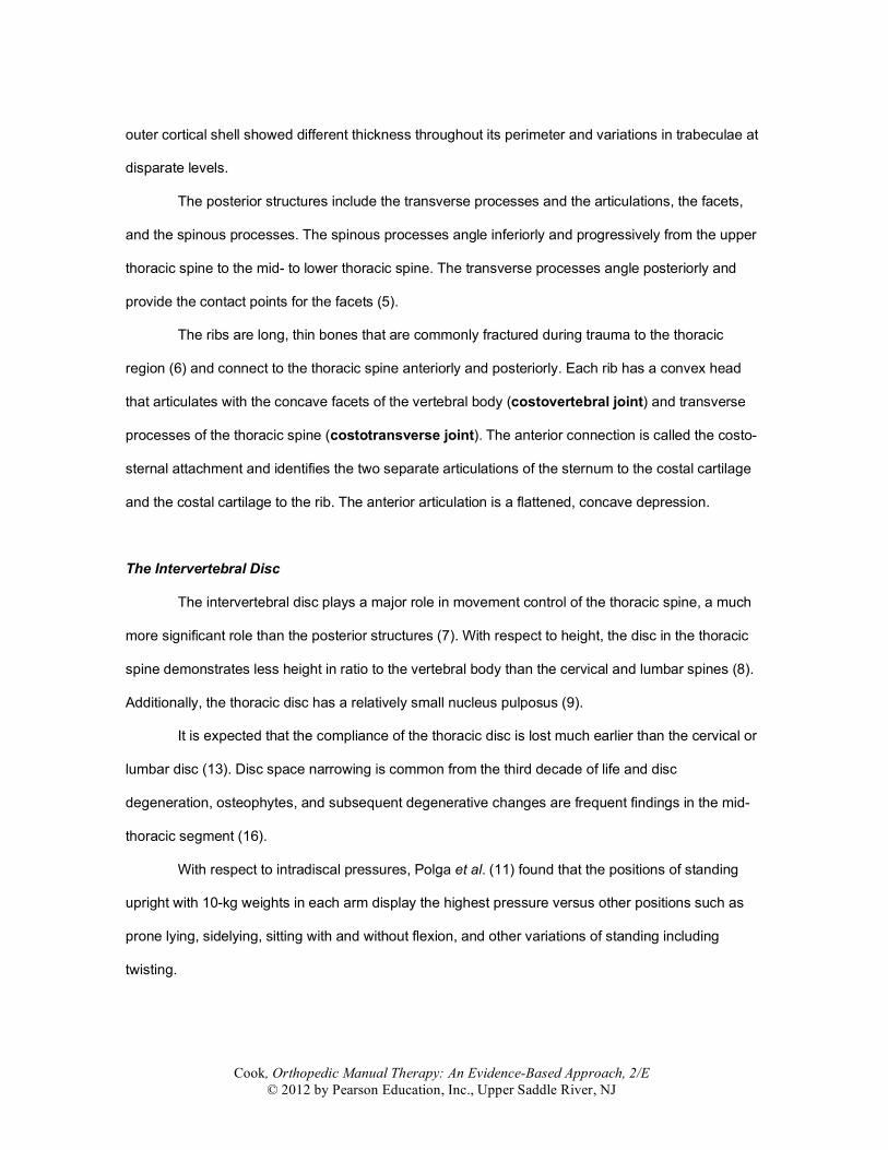
Table 7.2: Joints of the Thoracic Spine. Joint
Information
Intervertebral disc Not frequently studied. High incidence of asymptomatic herniated discs and can be a pain generator
Zygapophyseal (facet) joints
Synovial, planar, diarthrodial joints. All thoracic joints are in the frontal plane and vary between 0° and 30° from vertical. This allows significant movement in all 3 planes
Costovertebral
A synovial joint that allows rolling and gliding. The heads of ribs 2–9 (and occasionally the 10th) articulate with 2 vertebral bodies and the disc
Costotransverse
A synovial joint and is shaped differently according to the thoracic level. In the upper thorax, there is a concave/convex articulation between the convex costal surface and concave TP surface. The costotransverse joints gradually flatten and are more planar in the lower thorax. This change in joint shape allows for more rotation and torsional movement above rib 7 and more planar gliding movement below that level During inspiration, the upper chest wall rises (flexes) in the sagittal plane, whereas the lower ribs widen (abduct) in the frontal plane. During spinal flexion, the rib rotates anterior (posterior elements move superiorly and anterior elements move inferiorly)—an internal torsional movement. During spinal extension, the rib rotates posteriorly (posterior elements move inferiorly and anterior elements move superiorly)
There are variations in the zygapophyseal joints (facets) throughout the length of the
thoracic spine. In general, the superior facets face anteriorly but are not completely aligned in the
frontal plane (12). This angulation is reduced as the thoracic spine descends, culminating at T12. At
T12, the facets face both an inferior and superior a similar orientation of the lumbar spine (12).
Cook, Orthopedic Manual Therapy: An Evidence-Based Approach, 2/E © 2012 by Pearson Education, Inc., Upper Saddle River, NJ
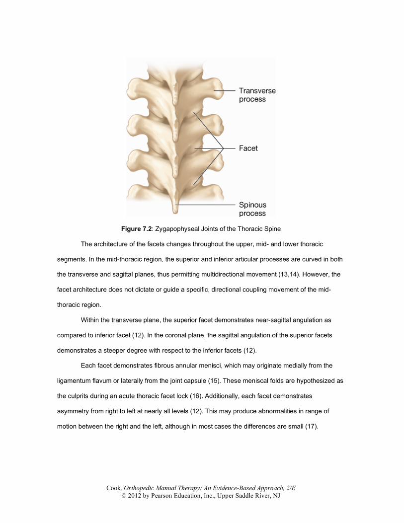
Figure 7.2: Zygapophyseal Joints of the Thoracic Spine
The architecture of the facets changes throughout the upper, mid- and lower thoracic
segments. In the mid-thoracic region, the superior and inferior articular processes are curved in both
the transverse and sagittal planes, thus permitting multidirectional movement (13,14). However, the
facet architecture does not dictate or guide a specific, directional coupling movement of the mid-
thoracic region.
Within the transverse plane, the superior facet demonstrates near-sagittal angulation as
compared to inferior facet (12). In the coronal plane, the sagittal angulation of the superior facets
demonstrates a steeper degree with respect to the inferior facets (12).
Each facet demonstrates fibrous annular menisci, which may originate medially from the
ligamentum flavum or laterally from the joint capsule (15). These meniscal folds are hypothesized as
the culprits during an acute thoracic facet lock (16). Additionally, each facet demonstrates
asymmetry from right to left at nearly all levels (12). This may produce abnormalities in range of
motion between the right and the left, although in most cases the differences are small (17).
Cook, Orthopedic Manual Therapy: An Evidence-Based Approach, 2/E © 2012 by Pearson Education, Inc., Upper Saddle River, NJ
Rib Cage Joints
There are three primary joints associated with the rib cage—the costovertebral joints, the
costotransverse joints, and the costosternal joints. The costovertebral joint is formed by a convex rib
head with two adjacent vertebral bodies, superiorly and inferiorly. The concave inferior costal demi-
facet of the superior vertebral body and the concave superior costal construction of the inferior
vertebral body provide a synovial attachment to the rib head. The rib head articulates with the lateral
aspect of the intervertebral disc in addition to the two separate vertebral connections. The joint has
two synovial cavities separated by an intra-articular ligament (13). This joint also houses meniscoids
that may be involved during acute costovertebral pain (18).
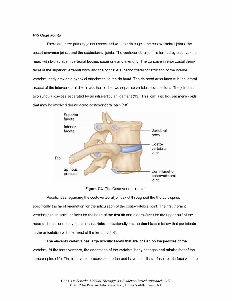
Figure 7.3: The Costovertebral Joint
Peculiarities regarding the costovertebral joint exist throughout the thoracic spine,
specifically the facet orientation for the articulation of the costovertebral joint. The first thoracic
vertebra has an articular facet for the head of the first rib and a demi-facet for the upper half of the
head of the second rib, yet the ninth vertebra occasionally has no demi-facets below that participate
in the articulation with the head of the tenth rib (14).
The eleventh vertebra has large articular facets that are located on the pedicles of the
vertebra. At the tenth vertebra, the orientation of the vertebral body changes and mimics that of the
lumbar spine (19). The transverse processes shorten and have no articular facet to interface with the
Cook, Orthopedic Manual Therapy: An Evidence-Based Approach, 2/E © 2012 by Pearson Education, Inc., Upper Saddle River, NJ
rib. The twelfth vertebra is similar to the eleventh except that the facet orientation is further sagittal,
thus mimicking the biomechanical inclination of the lumbar spine (19).
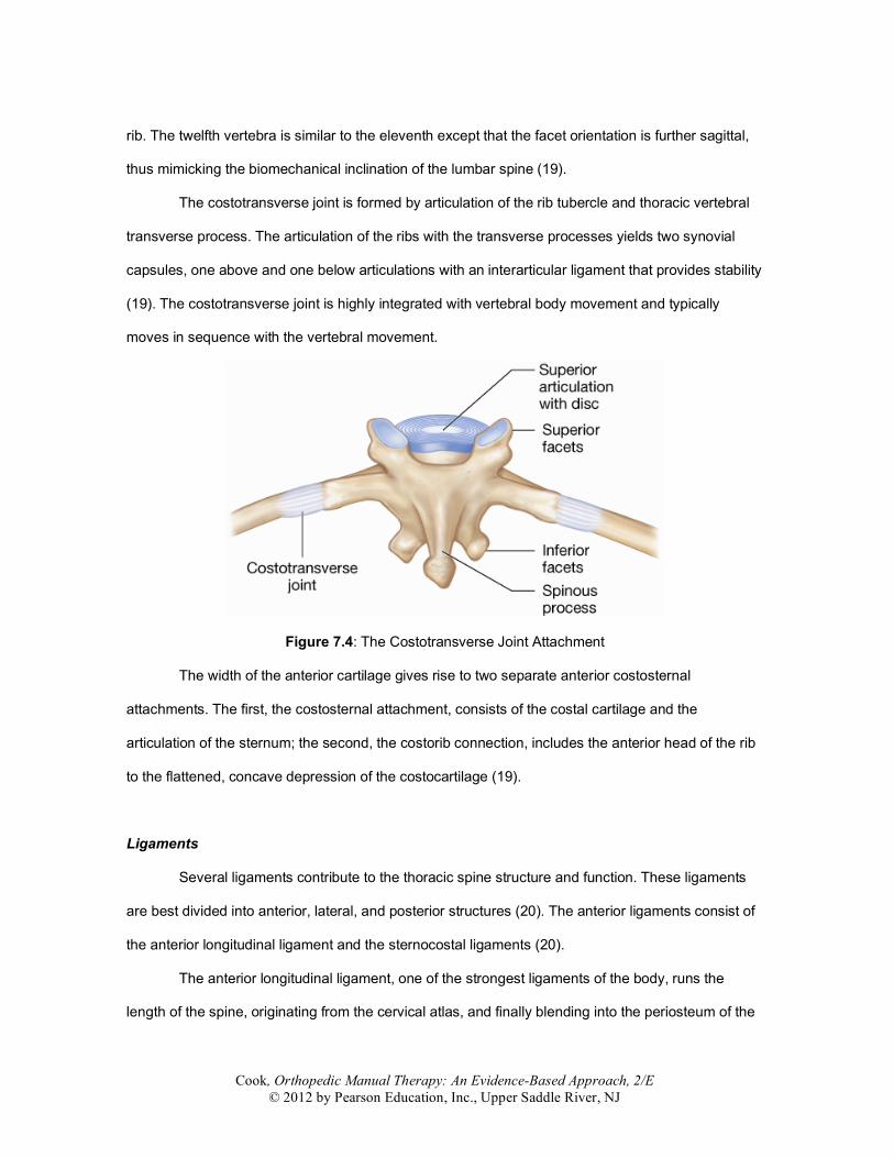
The costotransverse joint is formed by articulation of the rib tubercle and thoracic vertebral
transverse process. The articulation of the ribs with the transverse processes yields two synovial
capsules, one above and one below articulations with an interarticular ligament that provides stability
(19). The costotransverse joint is highly integrated with vertebral body movement and typically
moves in sequence with the vertebral movement.
Figure 7.4: The Costotransverse Joint Attachment
The width of the anterior cartilage gives rise to two separate anterior costosternal
attachments. The first, the costosternal attachment, consists of the costal cartilage and the
articulation of the sternum; the second, the costorib connection, includes the anterior head of the rib
to the flattened, concave depression of the costocartilage (19).
Ligaments
Several ligaments contribute to the thoracic spine structure and function. These ligaments
are best divided into anterior, lateral, and posterior structures (20). The anterior ligaments consist of
the anterior longitudinal ligament and the sternocostal ligaments (20).
The anterior longitudinal ligament, one of the strongest ligaments of the body, runs the
length of the spine, originating from the cervical atlas, and finally blending into the periosteum of the
Cook, Orthopedic Manual Therapy: An Evidence-Based Approach, 2/E © 2012 by Pearson Education, Inc., Upper Saddle River, NJ
sacrum. This ligament lies on the posterior of the body, while the sternocostal ligaments comprise
the anterior aspect of the body.
The sternocostal ligament is a broad, membranous band that originates from the anterior
and posterior aspect of the sternal cartilage of the upper ribs and inserts to the posterior surfaces of
the sternum. The interarticular sternocostal ligament attaches one rib to another with the
fibrocartilage and the connection to the manubrium.
The lateral costotransverse ligaments are subdivided into three ligaments—the superior
costotransverse ligament, the middle costotransverse ligament, and the lateral costotransverse
ligament (19). The superior costotransverse ligament originates on the border of the transverse
process and inserts on the upper border of the neck and angle of the rib. The middle
costotransverse ligament originates between the neck of the rib and inserts on the transverse
process at the same level. The lateral costotransverse ligament originates from the lateral aspect of
the transverse process and inserts on the adjacent rib. The intertransverse ligament is only well
formed within the lumbar region and occasionally demonstrates attachments in the lower thoracic
spine. The ligaments prevent excessive movements of side flexion and rotation (11).
The posterior ligaments include the posterior longitudinal ligament, the ligamentum flavum,
the interspinous ligament, and the supraspinous ligament. The posterior longitudinal ligament
descends along the posterior surfaces of the thoracic vertebra and discs to the insertion point in the
sacrum (20,21). The ligamentum flavum ligaments are paired (right and left) ligaments that insert
upward into the anterior lower third of the vertebral lamina at the level above and originate on the
posterior upper third of the lamina below. The elastic, yellow-colored ligament forms the posterior
wall of the vertebral canal and assists in preventing the synovial capsule and menisci from
entrapment into the facet joints (19–21). The interspinous ligament runs from each spinous process
and controls the movements of flexion. The radiate ligament connects the anterior portion of each rib
with the bodies of the two adjacent vertebrae and the intervertebral fibrocartilage between the
vertebrae. Lastly, the supraspinous ligament is composed of a bundle of fibrous tissue that courses
over the tips of each spinous process inserting in the lumbar region at L3-4 (19,21).
Cook, Orthopedic Manual Therapy: An Evidence-Based Approach, 2/E © 2012 by Pearson Education, Inc., Upper Saddle River, NJ
The Nervous System
Within the thoracic spine, the sympathetic nervous system plays an important part in pain,
auto regulation, and perception. There are 12 sympathetic ganglia within the thoracic region. The
thoracolumbar sympathetic fibers arise from the dorsolateral region of the anterior column of the
gray matter of the spinal cord and pass with the anterior roots of all the thoracic and the upper two or
three lumbar spinal nerves (19). Some of the fibers connect with the sympathetic trunk, enter the
white rami, and eventually progress to the prevertebral plexuses.
The sympathetic nervous system is responsible for dilation of the bronchi and pupils as well
as other organ-specific responses. Documented evidence supports the benefit of modulation of pain
and remarkably has a nonlocalized effect. Stimulation of the cervical and thoracic spine has
demonstrated upper extremity changes in pain response (pressure-pain) and a measurable
sympathoexcitatory effect (22–24) that can lead to pain reduction.
Muscles
The paraspinal muscles of the thoracic spine are responsible for both trunk and upper
extremity movements and are a common source of injury and pain. Several studies (25–27) have
found that back extensor strength was negatively correlated with degree of kyphosis, suggesting that
strengthening the thoracic spine should reduce the angle of kyphosis. Additionally, strengthening of
the thoracic spinal musculature also improves scapular dynamics and positively alters the
scapulohumeral rhythm (26).
Summary
• The osseous structures of the thoracic spine include the vertebral body and corresponding
right and left ribs. • The thoracic vertebrae can be subdivided anterior to posterior into three specific regions—
the body, the pedicles, and the posterior structures such as the transverse and spinous processes.
• The thoracic intervertebral disc has less height in ratio to the vertebral body as compared to the cervical and lumbar spine.
Cook, Orthopedic Manual Therapy: An Evidence-Based Approach, 2/E © 2012 by Pearson Education, Inc., Upper Saddle River, NJ
• The ribs attach in three joints (costovertebral, costotransverse and costosternal) and are long, thin bones with four primary planes.
• The facets of the spine face anteriorly and within the transverse plane; the superior facet demonstrates near-sagittal angulation as compared to the inferior facet.
• The ligaments of the thoracic spine help reduce spine displacement along with the stability provided by the rib cage.
• A notable component of the nervous system in the thoracic spine is the contribution of the sympathetic nervous system. This system may affect visceral structures as well as joints in the periphery.
• The muscles of the thoracic spine contribute not only to thoracic stability but also to stability of the shoulder girdle.
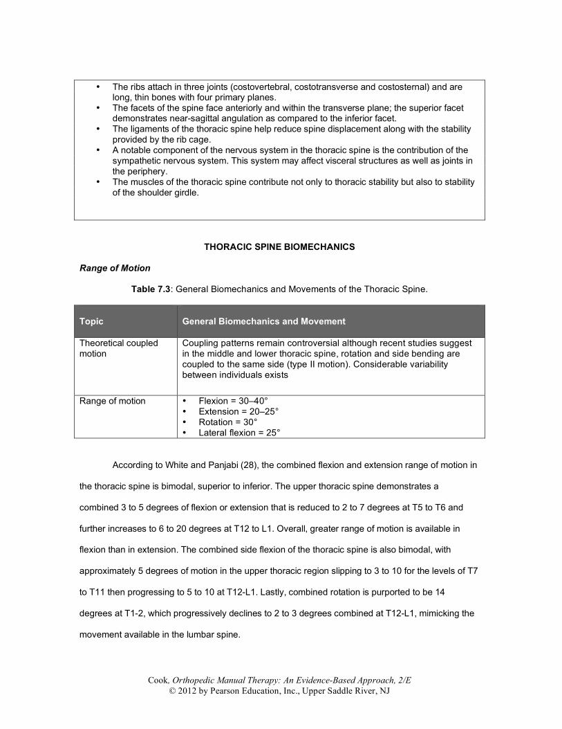
THORACIC SPINE BIOMECHANICS
Range of Motion
Table 7.3: General Biomechanics and Movements of the Thoracic Spine. Topic
General Biomechanics and Movement
Theoretical coupled motion
Coupling patterns remain controversial although recent studies suggest in the middle and lower thoracic spine, rotation and side bending are coupled to the same side (type II motion). Considerable variability between individuals exists
Range of motion • Flexion = 30–40° • Extension = 20–25° • Rotation = 30° • Lateral flexion = 25°
According to White and Panjabi (28), the combined flexion and extension range of motion in
the thoracic spine is bimodal, superior to inferior. The upper thoracic spine demonstrates a
combined 3 to 5 degrees of flexion or extension that is reduced to 2 to 7 degrees at T5 to T6 and
further increases to 6 to 20 degrees at T12 to L1. Overall, greater range of motion is available in
flexion than in extension. The combined side flexion of the thoracic spine is also bimodal, with
approximately 5 degrees of motion in the upper thoracic region slipping to 3 to 10 for the levels of T7
to T11 then progressing to 5 to 10 at T12-L1. Lastly, combined rotation is purported to be 14
degrees at T1-2, which progressively declines to 2 to 3 degrees combined at T12-L1, mimicking the
movement available in the lumbar spine.
Cook, Orthopedic Manual Therapy: An Evidence-Based Approach, 2/E © 2012 by Pearson Education, Inc., Upper Saddle River, NJ
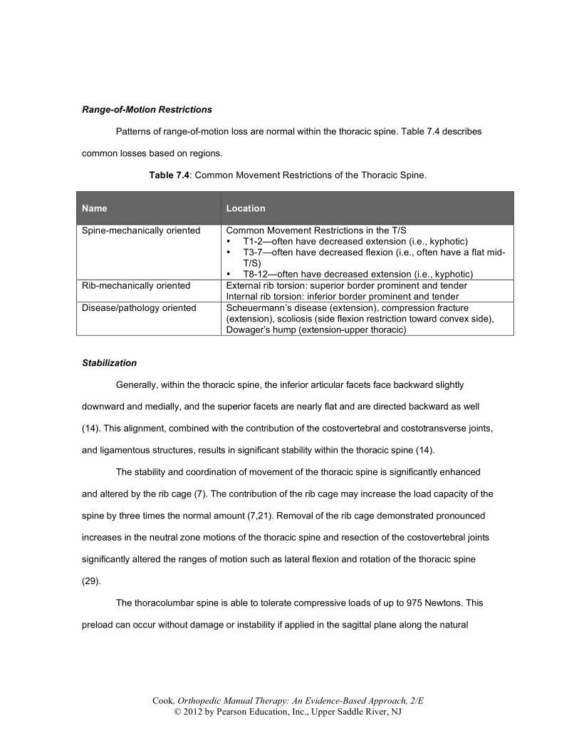
Range-of-Motion Restrictions
Patterns of range-of-motion loss are normal within the thoracic spine. Table 7.4 describes
common losses based on regions.
Table 7.4: Common Movement Restrictions of the Thoracic Spine. Name
Location
Spine-mechanically oriented Common Movement Restrictions in the T/S • T1-2—often have decreased extension (i.e., kyphotic) • T3-7—often have decreased flexion (i.e., often have a flat mid-
T/S) • T8-12—often have decreased extension (i.e., kyphotic)
Rib-mechanically oriented External rib torsion: superior border prominent and tender Internal rib torsion: inferior border prominent and tender
Disease/pathology oriented Scheuermann’s disease (extension), compression fracture (extension), scoliosis (side flexion restriction toward convex side), Dowager’s hump (extension-upper thoracic)
Stabilization
Generally, within the thoracic spine, the inferior articular facets face backward slightly
downward and medially, and the superior facets are nearly flat and are directed backward as well
(14). This alignment, combined with the contribution of the costovertebral and costotransverse joints,
and ligamentous structures, results in significant stability within the thoracic spine (14).
The stability and coordination of movement of the thoracic spine is significantly enhanced
and altered by the rib cage (7). The contribution of the rib cage may increase the load capacity of the
spine by three times the normal amount (7,21). Removal of the rib cage demonstrated pronounced
increases in the neutral zone motions of the thoracic spine and resection of the costovertebral joints
significantly altered the ranges of motion such as lateral flexion and rotation of the thoracic spine
(29).
The thoracolumbar spine is able to tolerate compressive loads of up to 975 Newtons. This
preload can occur without damage or instability if applied in the sagittal plane along the natural
Cook, Orthopedic Manual Therapy: An Evidence-Based Approach, 2/E © 2012 by Pearson Education, Inc., Upper Saddle River, NJ
curvature of the spine through estimated centers of rotation (30). The spine was found to be least
flexible and demonstrated less range of motion during axial compression (31).
Coupled Movement
Recently, Sizer and colleagues (31) performed a systematic literature review of coupling
pattern in the thoracic spine. In summary, their study indicates that coupling movements of the
thoracic spine are inconsistent whether the initial movement involves side flexion or rotation. Under
no circumstances are thoracic spine coupling movements predictable. During conditions such as
scoliosis, it is normal to see the spinous processes reflect toward the convexity of the rotation, or
side flexion and rotation that occurs to the same side. This pattern is highly consistent in the upper
thoracic spine and predominates in the mid- and lower thoracic spine but does demonstrate
variability (31).
Summary
• Generally, thoracic spine range of motion is similar but bimodal. The range of motion
declines near the mid-thoracic region but increases caudal and cephalic to the mid-thoracic segments.
• The thoracic spine is very stable, receiving stability contributions from the ligaments, the joints of the rib cage, and the rib cage structure.
• Coupling of the lumbar spine is location dependent. Generally, the upper thoracic spine couples with the lower cervical spine consistently. The mid-thoracic region demonstrates variable coupling, and the lower thoracic region tends to couple with the upper lumbar spine that is also variable.
ONLINE REFERENCES 1. Willems JM, Jull G, Ng J. An in-vivo study of the primary and coupled rotations of the thoracic
spine. Clin Biomech. 1996;11:311–316. 2. Kothe R, O’Holleran JD, Liu W, Panjabi MM. Internal architecture of the thoracic pedicle. An
anatomic study. Spine. 1996;21(3):264–270. 3. Panjabi MM, Takata K, Goel V, et al. Thoracic human vertebrae. Quantitative three-dimensional
anatomy. Spine. 1991;16(8):888–901. 4. Darwish HH, Ibrahim AF. Three muscles in the upper costovertebral region: description and
clinical anatomy. Clin Anat. 2009;22:352–357. 5. Stone J, Lichtor T, Banerjee S. Intradural thoracic disc herniation. Spine. 1994;19:1281–1284. 6. Karmakar MK, Chui PT, Joynt GM, Ho AM. Thoracic paravertebral block for management of
pain associated with multiple fractured ribs in patients with concomitant lumbar spinal trauma. Reg Anesth Pain Med. 2001;26(2):169–173.
Cook, Orthopedic Manual Therapy: An Evidence-Based Approach, 2/E © 2012 by Pearson Education, Inc., Upper Saddle River, NJ
7. Edmondston SJ, Singer KP. Thoracic spine: anatomical and biomechanical considerations for manual therapy. Man Ther. 1997;2(3):132–143.
8. Kapandji I. The Physiology of the joints. Vol. 3. The trunk and the vertebral column. 2nd ed. London; Churchill Livingstone: 1978.
9. Galante JO. Tensile properties of the human lumbar annulus fibrosis. Acta Orthop Scand. 1967;Suppl 100:1–91.
10. Horton SJ. Acute locked thoracic spine: treatment with a modified SNAG. Man Ther. 2002;7:103–107.
11. Polga DJ, Beaubien BP, Kallemeier PM, et al. Measurement of in vivo intradiscal pressure in healthy thoracic intervertebral discs. Spine. 2004;29(12):1320–1324.
12. Masharawi Y, Rothschild B, Dar G, et al. Facet orientation in the thoracolumbar spine. Spine. 2004;29:1755–1763.
13. Lee D. Rotational instability of the mid thoracic spine: assessment and management. Man Ther. 1996;1:234–241.
14. White AA, Panjabi MM. Clinical biomechanics of the spine. 1st ed. Philadelphia; JB Lippincott: 1975.
15. Bogduk N, Engle R. The menisci of the lumbar zygopophyseal joints. A review of their anatomy and clinical significance. Spine. 1984;9:454–460.
16. Bogduk N, Jull G. (Abstract). The theoretical pathology of acute locked back: A basis for manipulative therapy. Man Med. 1990;1:78–82.
17. Boszczyk B, Boszczyk A, Putz R, Buttner A, Benjamin M, Milz S. An immunohistochemical study of the dorsal capsule of the lumbar and thoracic facet joints. Spine. 2001;26(15):E338–343.
18. Erwin WM, Jackson PC, Homonko D. Innervation of the human intercostals joint: Implications for clinical back pain syndromes. J Manip Physiol Ther. 2000;23:395–403.
19. Williams P, Bannister L. In: Berry M, Collins P. Dyson M, Dussek J, Ferguson M (eds.) Gray's Anatomy, 38th ed. Churchill Livingstone, Edinburgh: 1995.
20. Panjabi MM, Hausfeld J, White A. A biomechanical study of the ligamentous stability of the thoracic spine in man. Acta Orthop Scand. 1981;52:315–326.
21. Andriacchi T, Shultz A, Belytschko T, Galante J. A model for studies of mechanical interactions between the human spine and rib cage. J Biomech. 1974;7:497–507.
22. Cleland J, Selleck B, Stowell T, et al. Short-term effect of thoracic manipulation on lower trapezius muscle strength. J Man Manip Ther. 2004;12(2):82–90.
23. Vicenzino B, Paungmali A, Buratowski S, Wright A. Specific manipulative therapy treatment for chronic lateral epicondylalgia produces uniquely characteristic hypoalgesia. Man Ther. 2001;6:205–212.
24. Simon R, Vicenzino B, Wright A. The influence of an anteroposterior accessory glide of the glenohumeral joint on measures of peripheral sympathetic nervous system function in the upper limb. Man Ther. 1997;2(1):18–23.
25. Sinaki M, Itoi E, Rogers JW, Bergstralh EJ, Wahner HW. Correlation of back extensor strength with thoracic kyphosis and lumbar lordosis in estrogen-deficient women. Am J Phys Med Rehabil. 1996;75(5):370–374.
26. Wang CH, McClure P, Pratt NE, Nobilini R. Stretching and strengthening exercises: their effect on three-dimensional scapular kinematics. Arch Phys Med Rehabil. 1999;80(8):923–929.
27. Itoi E, Sinaki M. Effect of back-strengthening exercise on posture in healthy women 49 to 65 years of age. Mayo Clin Proc. 1994;;69(11):1054–1059.
28. White AA, Panjabi MM. Clinical biomechanics of the spine. 2nd ed. Philadelphia; JB Lippincott: 1990.
29. Oda I, Abumi K, Cunningham B, Kaneda K, McAfee PC. An in vitro human cadaveric study investigating the biomechanical properties of the thoracic spine. Spine. 2002;27(3):E64–70.
30. Tawackoli W, Marco R, Liebschner MA. The effect of compressive axial preload on the flexibility of the thoracolumbar spine. Spine. 2004;29(9):988–993.
Cook, Orthopedic Manual Therapy: An Evidence-Based Approach, 2/E © 2012 by Pearson Education, Inc., Upper Saddle River, NJ
31. Sizer PS Jr, Brismée JM, Cook C. Coupling behavior of the thoracic spine: a systematic review of the literature. J Manipulative Physiol Ther. 2007;30(5):390–399.