thoracic organ doses and cancer risk from low pitch...
TRANSCRIPT
Research ArticleThoracic Organ Doses and Cancer Risk from Low Pitch Helical4-Dimensional Computed Tomography Scans
Chengwen Yang,1,2 Ransheng Liu,1,2 Xin Ming ,1,3 Ningbo Liu,2
Yong Guan,2 and Yuanming Feng 1,2
1Department of Biomedical Engineering, Tianjin University, Tianjin, China2Department of Radiation Oncology, Tianjin Medical University Cancer Institute & Hospital, Tianjin, China3School of Biomedical Engineering, Tianjin Medical University, Tianjin, China
Correspondence should be addressed to Xin Ming; [email protected] and Yuanming Feng; y m [email protected]
Received 31 May 2018; Revised 18 August 2018; Accepted 4 September 2018; Published 24 September 2018
Academic Editor: Yujiang Fang
Copyright © 2018 ChengwenYang et al.This is an open access article distributed under the Creative Commons Attribution License,which permits unrestricted use, distribution, and reproduction in any medium, provided the original work is properly cited.
Purpose. To investigate the dose depositions to organs at risk (OARs) and associated cancer risk in cancer patients scanned with4-dimensional computed tomography (4DCT) as compared with conventional 3DCT. Methods and Materials. The radiotherapytreatment planning CT image and structure sets of 102 patients were converted to CT phantoms. The effective diameters of thosepatients were computed. Thoracic scan protocols in 4DCT and 3DCT were simulated and verified with a validated Monte Carlocode.The doses toOARs (heart, lungs, esophagus, trachea, spinal cord, and skin) were calculated and their correlations with patienteffective diameter were investigated. The associated cancer risk was calculated using the published models in BEIR VII reports.Results. The average of mean dose to thoracic organs was in the range of 7.82-11.84 cGy per 4DCT scan and 0.64-0.85 cGy per3DCT scan. The average dose delivered per 4DCT scan was 12.8-fold higher than that of 3DCT scan. The organ dose was linearlydecreased as the function of patients’ effective diameter. The ranges of intercept and slope of the linear function were 17.17-30.95and -0.0278–0.0576 among patients’ 4DCT scans, and 1.63-2.43 and -0.003–0.0045 among patients’ 3DCT scans. Relative risk ofcancer increased (with a ratio of 15.68:1) resulting from 4DCT scans as compared to 3DCT scans. Conclusions. As compared to3DCT, 4DCT scans deliver more organ doses, especially for pediatric patients. Substantial increase in lung cancer risk is associatedwith higher radiation dose from 4DCT and smaller patients’ size as well as younger age.
1. Introduction
Four-dimensional computed tomography (4DCT) which isan advanced technique to acquire a sequence of 3DCT withrespect to respiration signal which could be used to monitorthe lesion motion in patients has been widely utilized in theradiation therapy as well as diagnostic arena [1]. 4DCT datacould be applied to contour moving target, such as the clinicaltarget volume to determine the internal target volume andobserve the intrafractional motion of organs and lesions inthe thoracic and abdominal regions across the treatment.To overlap the motion of tissues due to respiration, 4DCTallows a highly oversampled CT data acquisition, resulting ina rapid increase in radiation dose to the organs at risk (OARs).Effective organ doses delivered from a 4DCT scan have beenmeasured and estimated by a few research groups [2–5]. Yet,
to our knowledge, no data of patient-specific imaging dosefrom 4DCT protocols have been directly reported. MonteCarlo simulation has been regarded as the golden standardto compute the patient-specific imaging dose in CT andcone beam CT scans [6–10]. In our study, we quantifiedand compared radiation dose to OARs in 4DCT scans withconventional 3DCT scans using Monte Carlo simulationand investigated the imaging dose as function of patientsize. The estimated relative risk of cancer incidence wasalso calculated with the National Research Council BiologicEffects of Ionizing Radiation (BEIR) VII report.
2. Methods and Materials
2.1. Patients Characteristics. With the Institutional EthnicsCommittee approval (CRTOG1601) and patient consent,
HindawiBioMed Research InternationalVolume 2018, Article ID 8927290, 6 pageshttps://doi.org/10.1155/2018/8927290

2 BioMed Research International
radiotherapy treatment planning (RTP) CT images, organcontours, age, and gender of 102 cancer patients (51 malesand 51 females) treated from the year of 2007 to 2017 inour institution were used in this retrospective study. Theaverage age of patients at diagnosis was 65 (range, 6-93).The volumes of OARs were segmented using the Pinnacle3RTP system (Philips Healthcare, Best, Netherlands). Patienteffective diameterwas computed at the nipple level frombodycontours using DICOMan software [11], which ranged from184.50mm to 465.10mm.
2.2. Data Acquisition. A commercially available 16-slice Bril-liance Big Bore CT scanner (Philips Medical System) inour clinic was used. Patients were set up in supine positionand immobilized with Body Pro-Lok immobilization device(CIVCOMedical Solutions, Coralville, IA, USA) during dataacquisition. For each patient, thoracic 4D helical and 3D axialscanswere acquiredwith the collimation of 16× 0.75 mm, 16×1.5mm, and 8× 3mm. Varian real-time positionmanagementsystem v1.7.5 (RPM, Varian Medical Systems, Palo Alto, CA,USA) was used in the 4DCT acquisition. The scan protocolwas set as 120 kV and 100 mAs. The pitch of helical mode is0.059 and rotation time is 0.44s.
The volumes of OARs within the primary beam weresegmented using the Pinnacle3 RTP by one experiencedradiation oncologist and confirmed by another experiencedradiation oncologist. The following OARs were defined:heart, bilateral lungs, spinal cord, trachea, and esophagus.Averaged intensity projection (AveIP) was used in OARdefinition in the 4DCT.With the aid ofDICOMan, the imagesand structures of the patients were converted into EGS4CT phantoms based on scanner-specific Hounsfield units todensity conversion.
2.3. Monte Carlo Simulation. The Monte Carlo method wasused to simulate the kV X-ray beam of 3DCT and 4DCTwith the thoracic protocols. The source model of PhilipsBrilliance Big Bore 16-slice CT scanner was calibrated andvalidated through measurement in our previous work [12].The proposed source model consists of an extended circularsource located at the X-ray target level, with its characteristicsdefined by the energy spectrum, source distribution, andfluence distribution. An in-house C++ code was developedto performan automatic beamcommissioning to generate thesource model based on a set of measurement data, includingcentral axis PDD distribution in water, the dose profilesalong lateral and longitudinal directions at isocenter level,and in-air beam output measured at isocenter through aseries of cylindrical cones. The detailed derivations betweenthe measurement data and source models were described inthe previous work [12]. An EGS4/BEAM Monte Carlo code,MCSIM, was employed to reconstruct the photon beamsfrom the generated source model and to calculate the dosedistributions in water and two CTDI phantoms [13–15]. InMonte Carlo simulations, the energy cutoff for electrons(ECUT) and photons (PCUT) and the energy threshold for𝛿-ray production (AE) and bremsstrahlung production (AP)were set as ECUT = AE = 521 keV and PCUT = AP = 10 keV,
respectively. The number of histories was 500,000 and thecalculation timewas 2-4 h for eachMonte Carlo simulation inorder to achieve a statistical uncertainty (1𝜎) of less than 2%.The benchmark results of EGS4/MCSIM have been reportedpreviously [16].
In the axial scan mode of 3DCT, a series of 12 coplanarfields around the gantry rotation axis with an interval of 30∘were simulated to mimic the axial mode of CT acquisition.After each 360∘ gantry rotation, the phantom isocentermoved by a certain distance equivalent to the table move-ment. In the helical scan mode of 4DCT, the pitch value wasconsidered in computing the incremental table movement(i.e., isocenter movement) between two consecutive gantryfields.
To convert Monte Carlo simulation into absolute dose,absorbed doses were firstmeasured at the isocenter of a CTDIphantom (16 cm in diameter) following the AAPM TG-61protocol with a calibrated EXRADINA12 ionization chamber(Standard Imaging, Middleton, WI, USA) for the thoracicand abdominal scan protocols in 3DCT and 4DCT.TheCTDIphantom was also scanned and the images were importedinto treatment planning system and converted into digitalphantom for Monte Carlo simulation with MCSIM. MonteCarlo simulation was then performed to a chamber volumeinside the phantom with the same beam setups using theproposed sourcemodel.The ratios ofMonte Carlo simulationvalues and measured absorbed doses of the same phantomyielded conversion factors, which were used in the absolutedose calculations in the patient’s anatomy.
2.4. Organ Dose and Risk Estimation. We calculated organdoses for 102 patients from the 3DCT and cine 4DCT byusing Monte Carlo simulation. Doses delivered to the OARswere calculated as the mean doses of all voxels within thedefined structures. Then the doses from 3DCT and 4DCTwere fitted against patient effective diameter to investigate therelationship between the patient size and imaging dose fromCT scanner.
One set of patient data was used for estimating dosedifference calculated with different phantoms generated frommaximum intensity projection (MIP) and AveIP. The OARswere contoured and confirmed by the same radiation oncol-ogists following the same criterion on both MIP and AveIP.
In this study, the function of estimated relative risk (ERR)in BEIR VII models was used in calculation of the cancer riskin female lung cancer andmale lung cancer [17].TheERRwasdefined in BEIR VII report as follows:
ERR (e, 𝑎) = 𝛽𝑠 ×D × exp (𝛾𝑒∗) × ( 𝑎60)𝜂
(1)
where 𝑒 is patient’s age at exposure in years, a is attained age(years), D is the radiation dose (Sv), 𝑒∗is(𝑒 − 30)/10for𝑒 <30and zero for e ≥ 30, and 𝛽𝑠 is the gender- and site-specificparameter (95% confidence interval) as shown in Table 12-2of BEIR VII report as 0.32 (0.15, 0.70) for male and 1.40 (0.94,2.1) for female. 𝛾 and 𝜂 equal -0.30 and -1.4 for lung cancer,respectively.

BioMed Research International 3
5
4.5
4
3.5
3
2.5
2
1.5
1
0.5
0.25
(cGy)
(a)
50
45
40
35
30
25
20
15
10
5
2.5
(cGy)
(b)
Figure 1: Dose distribution of a pediatric patient delivered by one scan of (a) 3DCT (120kV, 100mAs) and (b) 4DCT (120kV, 100mAs).
Table 1: Differences of estimated dose between AveIP- and MIP-based simulations.
AveIP MIPVolume (cc) Dose (cGy) Volume (cc) Dose (cGy) Ratio
Heart 524.28 7.02 558.03 7.87 1.12Lungs 1682.19 7.38 1680.46 8.25 1.12Spinal Cord 9.16 6.67 8.81 7.65 1.15AveIP = averaged intensity projection; MIP = maximum intensity projection.
3. Results
3.1. Organ Dose Distribution for 3DCT and 4DCT. To depictthe dose distribution in patients from3DCTand 4DCT scans,a pediatric patient was exemplified with different isodoselines in Figure 1, which shows that dose gradient distributionsin the 3DCT and 4DCT were inhomogeneous. The hot spotconcentrated in the region of the head and neck.
In the 102 patients, the average of mean dose to heart,bilateral lungs, spinal cord, esophagus, trachea, and skinwas 0.8 (±0.25), 0.71(±0.24), 0.74 (±0.21), 0.79 (±0.24), 0.85(±0.34), and 0.64 (±0.17) cGy in one3DCT scan, while that inone 4DCT scan was 10.3 (±3.03), 9.46 (±2.44), 9.72 (±2.54),10.37 (±3.13), 11.84 (±3.22), and 7.82 (±1.58) cGy. The meandose delivered to whole body per 4DCT scan was 12.8-foldhigher as compared with that of per 3DCT scan.
The differences of estimated dose between AveIP- andMIP-based simulations are shown in Table 1. The ratios ofthese two type simulations were 1.12-1.15.
3.2. Correlation of Organ Doses and Patient Size. For 3DCTand 4DCT scans, the mean doses deposited to the variousorgans decreased with the increasing patients’ effective diam-eters precalculated as shown inFigure 2.Thedose from4DCTscan was much higher than from 3DCT scan for patients’heart (a), lungs (b), esophagus (c), trachea (d), spinal cord (e),and skin (f). The linear correlation between organ dose and
patient size was derived and the following function of organdose was obtained.
𝑂𝑟𝑔𝑎𝑛 𝐷𝑜𝑠𝑒 = 𝐷 + 𝑎 × 𝐸𝑓𝑓𝑒𝑐𝑡𝑖V𝑒 𝐷𝑖𝑎𝑚𝑒𝑡𝑒𝑟 (2)
The parameters (D, a) and the coefficient of determinationR2 were (2.43, -0.0045, 0.70) and (30.95, -0.0574, 0.67) fortrachea, (2.04, -0.0039, 0.84) and (25.90, -0.0491, 0.83) forlungs, (1.91, -0.0035, 0.61) and (24.24, -0.0435, 0.60) forspinal cord, (2.21, -0.0042, 0.67) and (29.17, -0.0559, 0.67) foresophagus, (2.27, -0.0044, 0.73) and (29.43, -0.0576, 0.71) forheart, and (1.63, -0.003, 0.69) and (17.17, -0.0278, 0.79) for skinper 3DCT and 4DCT scan, respectively.
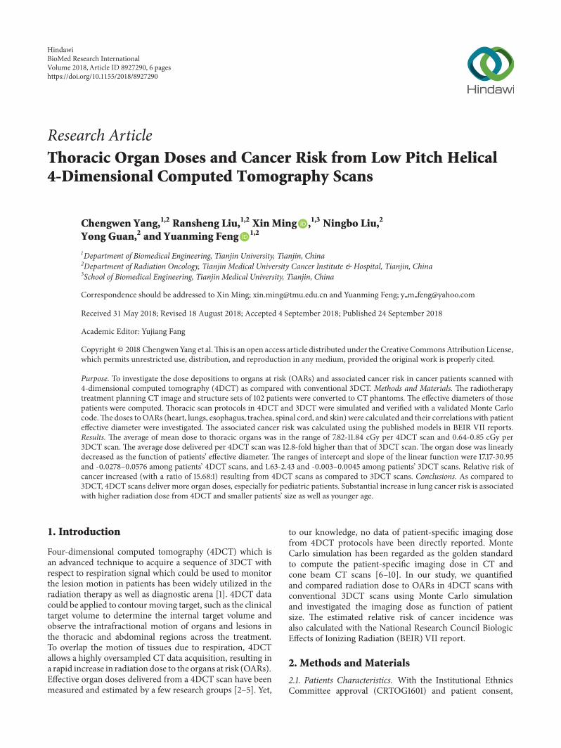
3.3. Estimated Risk from CTScans. Theestimated relative riskthat incorporates the magnitude of radiation exposure, sex,and patient age at the time of exposure in BEIR VII reportwas computed. Sex-specific estimated relative risk of lungcancer was shown in Figure 3. The relative risk for 4DCTscan decreased generally with increasing patients’ effectivediameter, similarly as for 3DCT scan. Yet the relative risk for4DCT scan was much higher than that for 3DCT scan with aratio of 15.68:1.
4. Discussion
4DCT has been used widely in radiation oncology forRT planning which helps reduce the chances of having a

4 BioMed Research International
200 250 300 350 400 450 500
Org
an D
ose (
cGy)
0
5
10
15
20
25
3DCT4DCT
Effective Diameter (mm)
(a) Heart
200 250 300 350 400 450 500
Org
an D
ose(
cGy)
0
5
10
15
20
25
3DCT4DCT
Effective Diameter (mm)
(b) Lungs
200 250 300 350 400 450 500
Org
an D
ose(
cGy)
0
5
10
15
20
25
3DCT4DCT
Effective Diameter (mm)
(c) Esophagus
200 250 300 350 400 450 500
Org
an D
ose(
cGy)
0
5
10
15
20
25
3DCT4DCT
Effective Diameter (mm)
(d) Trachea
Effective Diameter (mm)200 250 300 350 400 450 500
Org
an D
ose(
cGy)
0
5
10
15
20
25
3DCT4DCT
(e) Spinal cord
Effective Diameter (mm)200 250 300 350 400 450 500
Org
an D
ose(
cGy)
0
5
10
15
20
25
3DCT4DCT
(f) Skin
Figure 2: The mean doses to (a) heart, (b) lungs, (c) esophagus, (d) trachea, (e) spinal cord, and (f) skin decreased monotonically withincreasing patients’ effective diameter for 3DCT and 4DCT scans.
BioMed Research International 5
Effective Diameter (mm)250 300 350 400 450
ERR
of M
ale L
ung
Can
cer
0
10
20
30
40
3DCT4DCT
Effective Diameter (mm)250 300 350 400 450
ERR
of F
emal
e Lun
g C
ance
r
0
10
20
30
40
3DCT4DCT
Figure 3: Estimated relative risks for (a) male and (b) female lung cancer from 3DCT and 4DCT scans. The upper and lower bars indicatethe 95% confidence intervals (CI).
geographic miss and increases the chances of local control.4DCT-scan protocols are designed to produce a highlyoversampled CT data set with assistance of a low pitch scan tocover the patients’ respiratory motion, which would involvea steep increase in radiation dose. This work quantifies thedose distribution and demonstrates the relationship betweenthe patient size and organ dose.
With the parameter settings, the thoracic organ dosesfor 4DCT were much higher than that for 3DCT (7.82-11.84cGy versus 0.64-0.85 cGy after normalization to 100mAs). Inhelicalmode, 4DCT-scanprotocols attempt to account for thepatient’s respiratory motion by utilizing a low pitch scan (e.g.,pitch = 0.1) to produce a highly oversampled CT data set [2].Dose increase resulting from the highly oversampled scan isinversely proportional to the table pitch. On a per mAs basis,utilization of a pitch of 0.1 could lead to an approximately10-fold dose increase relative to a pitch of 1.0. Similarly foroversampled data acquisition, effective organ dose measuredwith cine mode in 4DCT scan was four times higher thanthose with conventional 3DCT [3]. In the risk model forlung cancer, 4DCT scan could involve more relative risk thanconventional CT (with a ratio of 15.68:1). Considering theconclusion by Darby et al. [18], the patients who depositedmore heart dose have relatively higher risk of ischemic heartdisease.
Other studies on 4DCT reported that the radiation dosedepends intensively on the setting of different protocols.Effective doses measured in adult anthropomorphic phan-toms were 6.14 cGy for lung (3) in cine mode, 5.72 cGyfor lung, and 5.05 cGy for esophagus (4) in helical modewith the pitch of 0.125, 5.34 cGy for lung, and 8.73 foresophagus (5) in helical mode with the pitch of 0.516. Thescan setups were 120 kV and 100-120mA. All these effectivedoses were measured in anthropomorphic phantoms (54-74 kg) by metal oxide semiconductor field effect transistor(MOSFET) dosimeters or thermoluminescence dosimeters(TLD). Although these effective doses did not reflect the
accurate dose distributions in the real patients’ body, it wasconcluded that lower pitch could involve the more dosedelivered to the OARs in helical 4DCT. For dose estimation,DeMarco et al. [2] conducted the Monte Carlo simulation ona GSF voxel phantom and concluded that average lung dosewas a function of tube potential. Their reported lung dose is15 cGy at 100 mAs and double pitch, slightly higher than ourresults (4.15-14.10 cGy) in this study.
The converted phantoms from patients CTs in this studycould not be used to mimic the dynamic respiratory motion.The differences of estimated dose resulting from AveIP andMIP were in consistent with the one from a research of MV-beam treatment [19] and the MIP-based simulation wouldoverestimate the dose delivered. The AveIP-based simulationwas recommended in 4DCT Monte Carlo simulation for thecomprehensive consideration of respiratory motion.
The helical 4DCT with low pitch could result in 10%speed-up in scanning but 92% dose efficiency and thebroadening of slice sensitivity profile from 1.25 to 2.3mm onthe 16-slice system [20]. Compared with cine 4DCT, helical4DCT leads to doses of approximately 1.5-fold higher (43.5mSv/28.8 mSv) [4]. Cautions should be taken when a helical4DCT is to be used and imaging dose is a concern.
It should be noted that dose from 4DCT or 3DCT scansis much less than the treatment dose delivered in radiationtherapy to lung tumor (generally in the range of 45-70Gy)and the normal tissue dose constrains in conventionallyfractionated radiotherapy (mean dose of less than 20-26Gy)[16]. Nevertheless, accumulated dose to normal tissues from4DCT scans should still be considered to minimize thepotential relative risk.
5. Conclusion
This study with data of 102 patients demonstrated a stronginverse correlation between patient effective diameter andmean organ dose to the thoracic organs. The average dose

6 BioMed Research International
delivered per 4DCT scan was 12.8-fold higher than thatper 3DCT scan. Substantial increase in lung cancer riskis associated with higher radiation dose from 4DCT andsmaller patients’ size.
Data Availability
The data used to support the findings of this study areincluded within the article.
Conflicts of Interest
There are no conflicts of interest associated with this publica-tion.
Acknowledgments
Theauthors would like to thank Dr. JunDeng (Department ofTherapeutic Radiology, Yale University School of Medicine,USA) for his help in the Monte Carlo simulation. This workwas supported by The Science & Technology DevelopmentFund of Tianjin Education Commission for Higher Educa-tion [2017KJ231]
References
[1] Y. Kwong, A. O. Mel, G. Wheeler, and J. M. Troupis, “Four-dimensional computed tomography (4DCT): A review of thecurrent status and applications,” Journal of Medical Imaging andRadiation Oncology, vol. 59, no. 5, pp. 545–554, 2015.
[2] J. J. DeMarco, M. F. McNitt-Gray, C. H. Cagnon, E. Angel, N.Agazaryan, and M. Zankl, “Evaluation of patient dose usinga virtual CT scanner: Applications to 4DCT simulation andkilovoltage cone-beam imaging,” Journal of Physics: ConferenceSeries, vol. 102, no. 1, 2008.
[3] S. Mori, S. Ko, T. Ishii, and K. Nishizawa, “Effective Dosesin Four-Dimensional Computed Tomography for Lung Radio-therapy Planning,”Medical Dosimetry, vol. 34, no. 1, pp. 87–90,2009.
[4] Y. Matsuzaki, K. Fujii, M. Kumagai, I. Tsuruoka, and S. Mori,“Effective and organ doses using helical 4DCT for thoracic andabdominal therapies,” Journal of Radiation Research, vol. 54, no.5, pp. 962–970, 2013.
[5] J. K. Hoang, R. E. Reiman, G. B. Nguyen et al., “Lifetimeattributable risk of cancer from radiation exposure duringparathyroid imaging: comparison of 4D CT and parathyroidscintigraphy,” American Journal of Roentgenology, vol. 204, no.5, pp. W579–W585, 2015.
[6] J. Gu, B. Bednarz, P. F. Caracappa, and X. G. Xu, “Thedevelopment, validation and application of a multi-detectorCT (MDCT) scanner model for assessing organ doses to thepregnant patient and the fetus using Monte Carlo simulations,”Physics in Medicine and Biology, vol. 54, no. 9, pp. 2699–2717,2009.
[7] X. Li, E. Samei, W. P. Segars et al., “Patient-specific radiationdose and cancer risk estimation in CT: Part II. Application topatients,”Medical Physics, vol. 38, no. 1, pp. 408–419, 2011.
[8] C. Lee, K. P. Kim, D. J. Long, and W. E. Bolch, “Organ dosesfor reference pediatric and adolescent patients undergoing
computed tomography estimated by Monte Carlo simulation,”Medical Physics, vol. 39, no. 4, pp. 2129–2146, 2012.
[9] J. C. L. Chow,M. K. K. Leung,M. K. Islam, B. D. Norrlinger, andD. A. Jaffray, “Evaluation of the effect of patient dose from conebeam computed tomography on prostate IMRT using MonteCarlo simulation,” Medical Physics, vol. 35, no. 1, pp. 52–60,2008.
[10] J. C. L. Chow, “Cone-beam CT dosimetry for the positionalvariation in isocenter: A Monte Carlo study,” Medical Physics,vol. 36, no. 8, pp. 3512–3520, 2009.
[11] DICOMan,VersionTX. Little Rock, AR:University ofArkansasfor Medical Sciences; 2014.
[12] X. Ming, Y. Feng, R. Liu et al., “A measurement-based gener-alized source model for Monte Carlo dose simulations of CTscans,” Physics in Medicine and Biology, vol. 62, no. 5, pp. 1759–1776, 2017.
[13] W. Nelson, H. Hirayama, and D. W. Rogers, The EGS4codesystem, SLAC-265, SLAC National Accelerator Laboratory,Stanford, CA, USA, 1985.
[14] D. W. O. Rogers, B. A. Faddegon, G. X. Ding, C.-M. Ma, J.We, and T. R. Mackie, “BEAM: a Monte Carlo code to simulateradiotherapy treatment units,”Medical Physics, vol. 22, no. 5, pp.503–524, 1995.
[15] C. M. Ma, J. S. Li, T. Pawlicki et al., “A Monte Carlo dosecalculation tool for radiotherapy treatment planning,” Physicsin Medicine and Biology, vol. 47, pp. 1671–1689, 2002.
[16] J. S. Li, T. Pawlicki, J. Deng, S. B. Jiang, E. Mok, and C.-M. Ma, “Validation of a Monte Carlo dose calculation toolfor radiotherapy treatment planning,” Physics in Medicine andBiology, vol. 45, no. 10, pp. 2969–2985, 2000.
[17] NRC, Health Risks from Exposure to Low Levels of IonizaingRadiation-BEIR VII, National Research Council, Washington,DC, USA, 2006.
[18] S. C. Darby, M. Ewertz, P. McGale et al., “Risk of ischemic heartdisease in women after radiotherapy for breast cancer,”TheNewEngland Journal of Medicine, vol. 368, no. 11, pp. 987–998, 2013.
[19] Y. Tian, Z. Wang, H. Ge et al., “Dosimetric comparisonof treatment plans based on free breathing, maximum, andaverage intensity projection CTs for lung cancer SBRT,”MedicalPhysics, vol. 39, no. 5, pp. 2754–2760, 2012.
[20] T. Pan, “Comparison of helical and cine acquisitions for 4D-CTimaging with multislice CT,”Medical Physics, vol. 32, no. 2, pp.627–634, 2005.

Stem Cells International
Hindawiwww.hindawi.com Volume 2018
Hindawiwww.hindawi.com Volume 2018
MEDIATORSINFLAMMATION
of
EndocrinologyInternational Journal of
Hindawiwww.hindawi.com Volume 2018
Hindawiwww.hindawi.com Volume 2018
Disease Markers
Hindawiwww.hindawi.com Volume 2018
BioMed Research International
OncologyJournal of
Hindawiwww.hindawi.com Volume 2013
Hindawiwww.hindawi.com Volume 2018
Oxidative Medicine and Cellular Longevity
Hindawiwww.hindawi.com Volume 2018
PPAR Research
Hindawi Publishing Corporation http://www.hindawi.com Volume 2013Hindawiwww.hindawi.com
The Scientific World Journal
Volume 2018
Immunology ResearchHindawiwww.hindawi.com Volume 2018
Journal of
ObesityJournal of
Hindawiwww.hindawi.com Volume 2018
Hindawiwww.hindawi.com Volume 2018
Computational and Mathematical Methods in Medicine
Hindawiwww.hindawi.com Volume 2018
Behavioural Neurology
OphthalmologyJournal of
Hindawiwww.hindawi.com Volume 2018
Diabetes ResearchJournal of
Hindawiwww.hindawi.com Volume 2018
Hindawiwww.hindawi.com Volume 2018
Research and TreatmentAIDS
Hindawiwww.hindawi.com Volume 2018
Gastroenterology Research and Practice
Hindawiwww.hindawi.com Volume 2018
Parkinson’s Disease
Evidence-Based Complementary andAlternative Medicine
Volume 2018Hindawiwww.hindawi.com
Submit your manuscripts atwww.hindawi.com