thoracentesis calderon part 2 (1) - ceconsultants,...
TRANSCRIPT

© 2011 Exempla Healthcare
Thoracentesis
Aaron J. Calderon, MD, FACP, SFHM Associate Program Director, Internal Medicine Residency, Exempla Saint Joseph Hospital
Founder, GME Simulation/Procedural Skills Lab, Exempla Saint Joseph Hospital Associate Clinical Professor of Medicine, University of Colorado Denver
© 2011 Exempla Healthcare
Disclosures
• I have no financial or other disclosures related to this activity
2
© 2011 Exempla Healthcare

© 2011 Exempla Healthcare
Indications
• Unexplained • Parapneumonic • Therapeutic
© 2011 Exempla Healthcare
Relative Contraindications
• PT and/or PTT ≥ 2x normal • Platelet count ≤ 25,000 – 50,000 • Creatinine ≥ 6.0 • Unstable patient • Infection over insertion site • Mechanical Ventilation • If doesn’t meet Light’s criteria for landmark
© 2011 Exempla Healthcare
© 2011 Exempla Healthcare
© 2011 Exempla Healthcare
Procedure
• Time-out • 1-2 interspaces below where dull • Above the rib • Do not go below rib 9 • Use Chlorhexidine • Vacuum vs. Push & Pull • Exhale/Valsalva during removal
© 2011 Exempla Healthcare
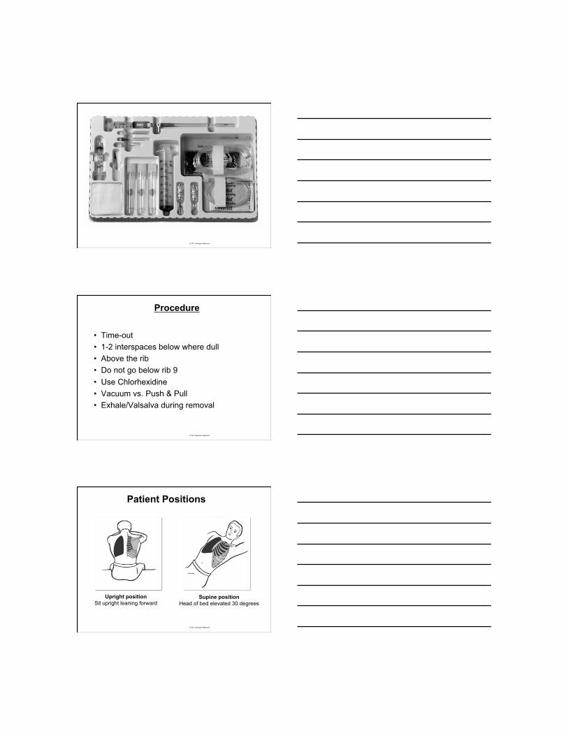
Upright position Sit upright leaning forward
Supine position Head of bed elevated 30 degrees
Patient Positions

© 2011 Exempla Healthcare
© 2011 Exempla Healthcare
Ultrasound Use
• No blinded RCTs • Associated with lower complications • Only “real-time” ultrasound • Consider for:
• Vented patients • Severe COPD or malignant effusions • Coagulopathy • Tiny or loculated • All patients
© 2011 Exempla Healthcare
2010 British Thoracic Society Guidelines
• Thoracic ultrasound guidance is strongly recommended for all pleural procedures for pleural fluid. (B)
• The marking of a site using thoracic ultrasound for subsequent remote aspiration or chest drain insertion is not recommended except for large pleural effusions. (C)

© 2011 Exempla Healthcare
Residency Review Committee for Pulmonary Medicine
• RRC now mandates as of 7/1/2012 • All pulmonary fellows are required to be
trained in the use of ultrasound to perform thoracentesis
© 2011 Exempla Healthcare
Thoracentesis Procedure
• Transducer is placed either sagittal or transverse on the patient’s back
• Locate and mark deepest fluid pocket
• Prep and drape patient and transducer per standard sterile technique
© 2011 Exempla Healthcare
Ultrasound Anatomy
The pleural space lies between the visceral and parietal pleura
liver
diaphragm
pleural fluid

© 2011 Exempla Healthcare
Thoracentesis Procedure
lung liver
diaphragm
pleural fluid
© 2011 Exempla Healthcare
Thoracentesis Procedure
© 2011 Exempla Healthcare

© 2011 Exempla Healthcare 19
© 2011 Exempla Healthcare 20
© 2011 Exempla Healthcare 21

© 2011 Exempla Healthcare 22
© 2011 Exempla Healthcare 23
© 2011 Exempla Healthcare 24

© 2011 Exempla Healthcare 25
© 2011 Exempla Healthcare
© 2011 Exempla Healthcare

© 2011 Exempla Healthcare
Complications
• Pneumothorax • Re-expansion pulmonary edema • Bleeding • Infection • Vagal reaction
© 2011 Exempla Healthcare
Initial chest x ray.
McRoberts R et al. Emerg Med J 2005;22:597-598
Copyright © BMJ Publishing Group Ltd and the College of Emergency Medicine. All rights reserved.
© 2011 Exempla Healthcare

© 2011 Exempla Healthcare
Complication Rate
© 2011 Exempla Healthcare
Tension Pneumothorax
• Chest pain, hypotension, tachycardia • Emergent needle decompression • Use no smaller than 3.25 inch 14 gauge
needle • Smaller needle size associated with high
failure rate • 2nd intercostal space;mid-clavicular line
32
© 2011 Exempla Healthcare
Follow-up CXR
• Debatable • No good evidence to support if procedure
went smoothly and low-risk patient • I wouldn’t recommend routine CXR

© 2011 Exempla Healthcare
2010 British Thoracic Society Guidelines
• Follow Up CXR
- A chest x-ray after a simple pleural aspiration is not required unless air is withdrawn, the procedure is difficult, multiple attempts are required or the patient becomes symptomatic. (C)
© 2011 Exempla Healthcare
CONCLUSIONS: “BEDSIDE ULTRASOUND PERFORMED BY CLINICIANS HAD A HIGHER SENSITIVITY AND SIMILAR SPECIFICITY COMPARED TO CXR FOR THE DIAGNOSIS OF PNEUMOTHORAX, BUT THE ACCURACY OF ULTRASOUND DEPENDED ON THE SKILL OF THE OPERATORS.”
CHEST 2011; 140(4):859-866
35
© 2011 Exempla Healthcare 36

© 2011 Exempla Healthcare
© 2011 Exempla Healthcare
Reexpansion Pulmonary Edema
• Rare complication • Dyspnea, tachypnea, cough, fever, tachycardia • Unilateral pulmonary edema in lung that rapidly
reexpands • Typically occurs in lung that has been collapsed for ≥ 3
days • Usually occurs in first few hours of reexpansion; almost all
by 24hrs • May last 2-5 days
38
© 2011 Exempla Healthcare
Reexpansion Pulmonary Edema Treatment • Supportive care(O2, pain meds) • Diuretics not recommended Prevention • Limit fluid removal to 1-1.5 liter • If measuring pleural pressure keep above -20 cm H20 • Cough or chest tightness correlate
39

© 2011 Exempla Healthcare
© 2011 Exempla Healthcare
What to Order with the Fluid?
• Cell count, gram stain, and culture • LDH and protein • pH • Cytology • Glucose • Serum LDH and protein
41
© 2011 Exempla Healthcare
Light’s Criteria Exudate vs Transudate
Only need one of the following to meet criteria for an exudative pleural effusion: • Pleural LDH/Serum LDH > .6 • Pleural LDH > 2/3 ULN of serum LDH • Pleural protein/Serum protein >.5
42

© 2011 Exempla Healthcare
Light’s Criteria CHF on Diuretics
• Transudates misclassified as exudates 20-25% of the time
• Options to better classify these patients: – Serum albumin/pleural albumin of >1.2 g/dl – Serum protein/pleural protein >3.1 g/dl – Pleural NT-pro-BNP levels >1300 (best test if
available) Light RW. Use of pleural fluid N-terminal-pro-brain natriuretic peptide and brain
natriuretic peptide in diagnosing pleural effusion due to congestive heart failure. Chest. Sep 2009;136(3):656-8.
43
© 2011 Exempla Healthcare
Exudative vs Transudative Based on Pleural Fluid Alone
• Correlates fairly well with Light’s criteria • The presence of any of the following indicates a exudative
effusion: - Pleural LDH >.45 the ULN of serum LDH - Pleural Cholesterol > 45 mg/dl - Pleural Protein > 2.9 g/dl
Heffner JE, Brown LK, Barbieri CA. Diagnostic value of tests that discriminate between exudative and transudative pleural effusions. Primary Study Investigators. Chest. Apr 1997;111(4):970-80
44