thly si - uscap · mo!\thly si.ides · november, 1956 ... pedunculated. subserosal nodule 1.5 em....
TRANSCRIPT
J358
PROTOCOL
MO!\ THLY SI.IDES
· November, 1956
TUMOR ·TISSUE REGISTRY
LOS ANGELES COUNTY HOSPITAL
PLEASE NOTE: 'lhe slides do not cal:'ry the Case Numbers on the right hand side as has been the custo1D. They are numerically numbered in type, i=edia.te-ly fo;l.lowing the Accession number, i.e .• , Ac0 • No •. 8069 {fl.
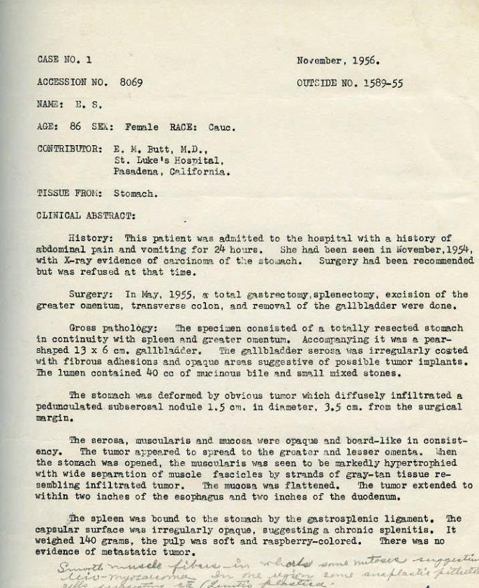
CASE NO, 1
ACCESSION NO. 8069
AGE: 86 SEX: Female RACE: Cauc,
CONTRIBUI'OR: E. ~~ . Butt, ~I.D., St. Luke' s Hospital, Pa.sadena, Calii'ornia .
TISSUE FRQI.f: Stomach.
CLINICAL .~STRACT:
No~ember, 1956.
OUTSIDE NO. 1589-55
History: This pati.ent wa s admitted t o t.,;e hospita l 1·ri th a history of aibdominal pain and vomithg for 24 hours. She had been seen in November ,195lf., With X...ray evidence. of carcinoma of t :1e ntoUlll.ch. Surge ry had been recommended but was r efused at that t ime.
Surgery: In ~y. 1955, ~total ~strectomy, splenectomy , exc1s1on of the gr,eater omentum, t r ansverse colon, a nd remova l of the gallbladder were done.
Gross p-:\thology: 'Ille specimen cons i"sted or a to telly resected stomach in continuity with spleen a nd great er omentum. Accompanying it \'las e. pearshaped 1.3 x 6 em, gallbh<ider. The galll;>ladder serosa \tas irregul.arly ccS)ted with fibrous adhe.sions a nd, opaque area s suggesti v e of possibl e t umor implants, The lumen cont ained 40 cc of ~1cinous bile a nd s~ll mixed stones.
The stomach was deformed by obvious t umor ~thich diffusely infiltrated a pedunculated. subserosal nodule 1.5 em. in di.!!.meter, .3.5 em. from the surgical 1!Brgin.
The serosa, muscularis a nd mucosa H·ere ope.qua a nd board-like in consistency. The tumor a ppeared to spree.d to the greater and lesse r omenta.. <men the ston:a.ch was opened, t l;!e muscularis was s~en to be mrkedlY hypertrophied ~tith wi de separation of muscle fasc i cles by str'l.nds of gl,'aY-tan tissue re-sembling infiltrated tumor . The muoosa was flattened. '!he tumo r extended to within two inches of the esoplw.gus a nd t1~o inches of the duodenum.
'ihe spleen 1~a.s bound t o t he stomach by th e gas t rosplenic ligament . '!he capsllll"ar surface W'I.S i rregularly o:Pa.que, suggesting a chronic splenitis. I t weighed llf.o grams, the pulp was soft and raspberry-colored, There was no ev.idence of metastatic tumor ,
~ ...... ~ -1-U~-:..., ~t)-'Y k '"'"~·<. ·..--~ .JJ,.. ........ ,; (J r- tl e.. - /: 7 1
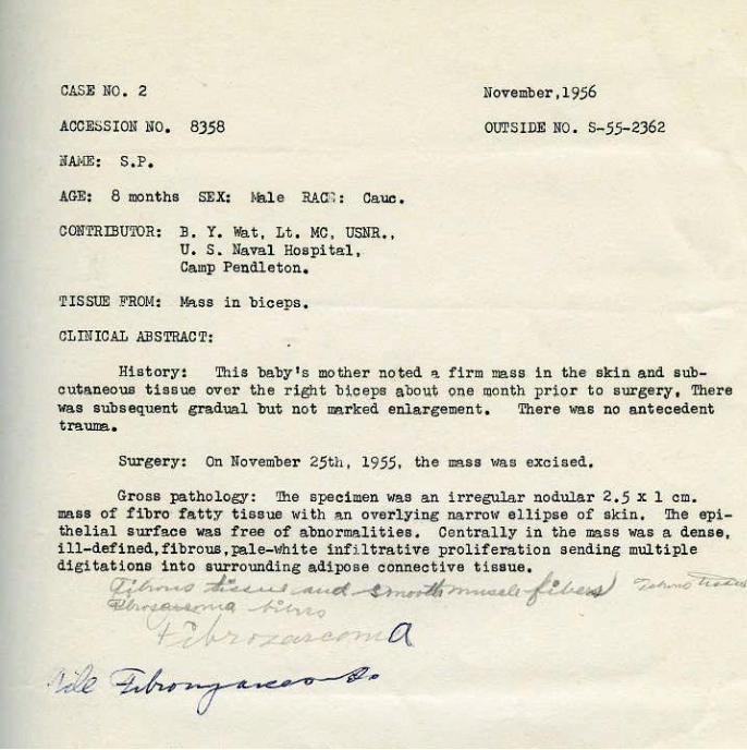
CASE NO. 2
ACCESSION NO, 8358
iiAl·IE: S,P,
AGE: 8 months SEX: Male RP.a•~ : Cauc,
CO}!TRIBU'IDR: ] • Y. Wa t, Lt, MC., USNll:., U. S. Naval Hospital, Camp Pendleton.
TISSUE ~ROM: ~~ss in biceps,
CLINICAL ABSTRACT:
November ,1956
OUTSIDE NO. S-55-2362
History: This baby's mother nq·ted '9: firm mess in the skin and sub-cutaneous tissue OVer the r~gh t b~ceps a'Qov.t QI),~ !DO J;l~h pr~Qr to s~g.ery t There wa~ subsequent gradual but not ·~rked enlargement, There was no .antecedent tmumg,,
Surgery: On lfovember 25th , 19.55. the mass 11as excised..
Gross pathology: The specimen was an irregular nodular 2.5 x 1 em. mass of f'ibro fatty tissue with a.n overl ying. narro11 ellipse of skin, 'lhe epith.elial s.urfe.ce was free of abnormalities. CentrallY in t he mas s was a dense, ill-defined, fibrous, pale-l'lhite i nfi.ltrative proliferation sending multiple digitations into surrounding adipose connective tissue, ;
~..,._, <7 ;:;£~ <-• • • ~ ~$~-/~~ ~A.~·o"' .t. ,..,.,<I
~ • .-U'l~"'> ··4 - -<.._o-. -~ / '~v -
I~,..~ ~-- a. .,. ·,2, 1'7' / . -~t.--o<:.......-1"---~
·Ui:2 ~~"
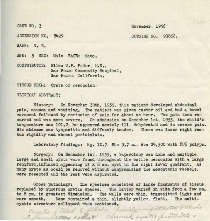
CASE NO. J
ACCESSION NO, ~27
NAME : G. B.
AGE: 5 SEX: i.Jale RACE: Cauo ,
CONTRIBUTOR: Ellen li ,P, Feder, !>!,D . , San Pedro Coro~unity Hospital, San Pedro, California .
TISSUE FROM: Cysts of mesocolon.
CL!UICAL .ABSTRA:;T:
November, 1956
OUTS IDE NO. 9)852.
Histor~ : On November J Oth, 1955. ~~is patient developed abdo~inal pain, nausea and vomiting, The p!\t1ent wa s etven castor oil a-'ld had a bowel movement followed by remission of pain for about a.n hour. 'ille pain then r ecurred and was more severe. On admi ssion on December lst, 1955, the child ' s temperature was 101. 2, he appeared acutely ill. dehyd..-ated and in severe pain. His abdomen vas tymFa.nitis and diffusely tender. There was lower r ight rectus rigidity and absent peristalsis ,
laboratory findings : Hg , 10. ?, Rbc J , ? m., ltbc 24,500 With 60j\l polyps,
Sur~ecy : On December 1st, 1955, a l:l,pe,rotomy 1-1as done and multiple large and small cysts wer e found throughout the entire mesocolon with a. large r eniform,inflamed appearing 11 x 6 em, cyst in the right lo>Ier quadrant . As nany o)'sts as could be removed without compromising the mesenteric vessels, were resected and the rest were aspirat ed,
Grose pathology: The specimen consis t ed of large fragments of tissue, replaced by numerous cystic spaces. 'lhe !Jlttor varied in sil:e from a f ew em. to 8 em. i n greatest dimension , 'lhe wal ls were thin, trans~itted light and were smooth, Some contained a thin, slightly yellot·l, fl ui.d . The multi-cystio structure collapsed when sectioned,

CASE NO, 4
ACCESSION NO , 8428
NAJ.!FJ: V. G,
AGE : 17 SEX: Female· RAaE: Cauc ,
CONTRIBUTOR: Ellen ~I.P. Feder, M.D., San Fedro Community Hospital, San Pedro, California.
TISSUE FROM: Uterine curettement.
CLIN'ICAL AllSTRACT:
November, 1956
OUTSIDE NO . 54-1852
History: This unmarried girl so~~t ~edical attention because of severe menorrhagia t~ith passage of clots and tissue on December Jrd, 1954. She had had irregular menses and menorrhagia for two years; often every two weeks. A year previously, she had been told she ha.d a. tumor . Her last men-strual ~riod had been Npvember 20th, 19.54. She denied intercourse.. On abdominal examination, a firm 15 em, tumor ti'l.S palpable in the lower abdomen. On pelvic examination, the hymen was intact but not tight, the cervix ~1a.s ~ndely dilated and filled with a large blood clot .
Ie.bora·tory findings : Hg, 5, 53, ~!be, 14, 500 t·lith 9o% segs,
Surgery: On Dece·mber ~rd, 19.54, an epesiotomy and curettage were done
Surgical findings: At surgery, the uterus was noted to be the size of an 18 week gestation and the cervix ~las almost completely dilated. 'Ihe hand could be easily introduced into the fundus and man=l curetta~ was done for fear of instru:nental perforation.
Gross pathology: The tissue removed was hemorrhagic and necrotic, totaling about 200 cc·, in volume . The largest piece measured 4 x 5 em.
Follo11-up: The pat i ent was admitted to Harbor General Hospital early i n 1955. Repeat curettage showed much less endomet rial tissue, but it was still abundant with a less exa.ggerateQ. but similar microscopic pattern. Hormone· assays showed no abnormalities ,
~~~, ~~n.d:$dd ~;'7fdf<,~~ ~. #~~~v~

CASE NO . S
ACCJ<.:SSION NO. 8843
NAI•IE : E. T,
AGE: 56 SEX: Hale RACE: Cauc,
CONTRill1J.!'OR: Dorothy Tatter, ~!,D ,, LACH, Los Angeles, California.
TISSUE FROM: Kidney.
CLINICAL ABSTRACT:
N'ovember, 1956
OUTSIDE NO. L.'!CR-e.utopsy No . 55578.
History; This patient 1"as admitted to the Los Angeles County Hospital on A'ugust 6th, 1956, ltith a history of sudden onset of severe subXiphoid pain due to a dissecting thoracic an~·sm. He e:>..'])ired shortly after admission. He had had no symptoms referrable to his kidneys . Autopsy was performed on August 8th, 1956.
Autopsy findings: '!he patient was f ound to have cardiac tamponade secondary to rupture of his aneurysm,
An incidental finliing was a firm, \1hitish-yellow tumor measuring 3 to 4 em, in di~J!.Illeter at the lo~1er pole' of the left kidney , :Il1e tumor did not extend to the J:iilum of the kidneY and appeared to involve only the cortex, The capsules stripped with ease from the surfaces of both organs, '!he left kidney >~eighed 300 grams, the right 250 grams. 'lh!3 right kidney was not grossly re~rkable , '!he pelvices and ureters were not dilated. '!he uri-nary bladder ~las not remarkable.
I ~'l..r~~ u.-."-"( 1..-4<A-J-<- ~f.-... .-f f. -.<.~ ~ (./ ~
.A: -u ~": ,.._ t{L--." r-rZ-« I ~~~~~

CASE NO, 6
ACCESS ION NO, 8465
NA/.IE: P,H,
AGE: 12 SEX: !<!ale RACE: Ca.uc ,
CONTRIBUTOR: L. L. Frost, M.D., Alta Vista Hospita l, Pasadena, Ca lifornia.
TISSut FROH: M\l.SS, hard and soft p?.la te,
CLINICAL ABSTRACT:
November, 1956.
OUTSIDE NO . S1709-55
Histor<': Tc·IO months pri or t o a dmissi on this patient noted sudden on-set of a growth on ~he hard a.nd soft :;nl a t e , , 1'/i til in 48 hours, it had obtained its present s i ze of approximately 5 em.. '1\~.o ~,o~eeks prior to admiss.ion, a 'biopsy had been pe rformed l·lhich >~as unsat i sfactory for diagno$is.
Surgery: On December 27th, 1955. the lesion Was dissected f rom the hard and soft pal ate ,
Surgical findi ngs : Th e lesion W'J.S apparently co!1!pletely excised, It extended to, 'but not into the perios teum of the har d pala te,
Gross pathology: 'L'l1e specimen c onsi-sted of mult.iple irregular frag-lnents of soft, fri able, gra y-white Jtnd pale pink tissue., the largest 1,6 em. in greatest dimens ion, 'l'he external surfaces ~1ere fine ly nod1Jl.ated, so·me of the nodules a ])pea ring externally to be cystic, Cut sections revealed smooth, glistening , gray-1ifhite .and· opd escent surfa c e s . _ ~,.._/
~~ r~.v-~ cy~~?.>./ .. ·• -~ (].~ ~ " ,Lbo:JA./ t+lb~ ~ ~
/p~...Q(.)
UJ . 7~--. .,d~
CASE NO. 7
ACCl!:SSI ON NO . 8423
NM-IE: H. H.
AGl!: 70 SEX: Female RACE: dauo .
CONTRIBUTOR:
'r!SSUE FROH1
ji:llen li ,P. Feder, M.D., San Pedro Communjty Hospital, San Pedro, Cali-fornia .
Tumor, thigh.
CLINICAL ABSTRACT:
NOVE!mber, 1?56
OUTSIDE NO. None give_n.
History: In 191~6. this patient haC. had an int·ramuspular tumor resected from the inner, upper aspect of the righ-t thigh. 'lhe· size was unknown, 1he microscopic report was mrAofibrosarco~. narly in December, 1'55. she. noted for- the first time a mss unde'r the scar. lfu.en seen by her physician in January, 1956, t.he mass "'as the size of an "orange", adherant t o the skin, but freely movable over the deep str uctures .
surgery: Oa J'\!luary 11th., 1956, the mss 11ith ·an ellipse of skin and surrounding subcutaneous fa.t, 1~a.e resected.
Gross pathology: jhe tumor appeared sharply circumscribed, measured 6.5 x 5 x 5 em. and was composed of firm, homogeneous, slightly translucent white tissue. Mucoid mterial exuded from the cut surface . 'Ihe tumor ~las adherent to the deeper lAyer of the dermis and at the base 1~as not covered by fat, but appeared enucleated. 'l'he overlyill€ skin ellpse measured 12 x 5,5 em, and presented a central 6 em. surgical soar.
CASE ~TO . 8
ACCESSIOll NO. 840.5
f!.\JoiF. : R • W.
AGE: 42 Sl':X: Female BACE: Cauc .
COl!'l'RIBU'IDR:
TISSUJ1: FROI~:
D. A. DeSanto, M. D. , Mercy Hospital, San Diego, California.
~ass in breast.
CLINICAL .ABSTRACT:
l!ovember, 19.56.
011l'SIDI. HO. ,56-460.
History: This patient was first seen in October, 19.5.5, with an upper respiratory infection and ~ pai nless swell i ng in the right breast . ~e patient stated that tas mass had been presen t for only th.ree days. The right br east revealed a~ enlarge~ bluish, stony har d lesion with the nipple deeply retracted. Surgery was delayed until Jantl.'\ry 27th, 19.56, because of her re-fusal to submit to surgery.
Laboratory findings: :\-rays of chest, lumbar spine, pelvis and slrull revealed no metastatic lesions .
Surgery: A right radical mastectomy and skin graft 1~ere done on January 27th, 19.56.
Gross pathology: The specimen consisted of a radically resected right breast, 'lhe surface showed a skin ellipse measuring 30 x 2.5 e m. The center of the akin ellipse was bulged outward by a lar ge centrally located tumor which ID88.Bured 1.5 em. in dia.meter and showed nUID8rous bluish injected veins and orange de peau. The nipple was retracted by a triangular scar and th er e was necrosis of the epithelium at the a pex of this scar.
The transverse sections of the tumor exposed a lobulated and lobated glistening grayish-tan surface showing areas of necrosis and hemorrhage. 'lhe entire breaet tissue was replaced by this tUJJlOr which measured 1.5 x 1.5 x 9 cl!l, It penetrated the fa~ei& of the pectoralis major but no strands radiated into the muscle itself. The tumor 11as fairly well circumscribed, but not encapsu,.. lated. Serial sections disclosed a colorful cut surface with yellowish are<!.~> of necrosis. red areas of hemorrhage within fleshy grayish- pink and rubbery stroma.
A total of 20 ~ nodes were identified measuring 0 . 2 to 0. 3 em. ~tey were yellowish- pink, soft and not grosely involved. Two other nodes appeared to contain tumor .
I .,s., ....... <::r ... """' ...,._.::-~ •• Pe"" (-t.-v-~ ~.,.._.._, .._..... ;;r_ ~~;,-w~ .... '-U ~J....L.._.._J:;:;;'.. """' J,t._r-o ~ .. -- \...

COIITRI!U'IOR:
ACCESSION NO. OUTSIDE NO.
NANE: D. C.
J . H. Cremin, H. D. , 679 South Westlake Avenue, Los Angeles, California.
8367 E-1391-.5.5
CASE NO . 9 November, ~19.56 .
AGE: 7 yrs . SEX: Fe"lale RACE: Ca.uc.
TISSU£ FR0:4:
HISTORY:
PHYSICAL FUIDHTGS :
LAllORATORY FINDINGS:
SURGERY:
SURGICAL FINDINGS :
GROSS PATHOIDG Y:
FOLLOW..,.UP:
Tumor of the liver.
'fuis child ha.d been well until December 31st, 19.54, at which time she developed pain in t he right hypochondrium. Sle was adr.titted to the hospital on March 28th , 195.5,1·1:i th a history of eight pounds weight gain since the onset of pain ,
'lhere was a protuberance of the upper abdomen. The liver extended three fingerbreadths belo1~ the right costal margin, had a smooth edge and was non- tender . The upper abdomen showed a prominent venous pattern.
An X-ray of the chest and abdomen revealed a large upper abdomi~l mass and an area of homogeneous density above the diaphragm, suggestive of pulmonary me tastases . ~1ere waa distortion of the calyces of the right kidney.
Cephalin flocculation : negative; t otal protein ,5 . 7 grams ~ with 4.4 grams '1> of albumin and 1 . 3 grams 'f, of globulin; 'Illymol turbidity 1 . 2 Shank and Hoagland units.
Expl oratory celiotomy was perforoed on ~.arch 31st, 19.5.5. ltitb removal of the tumor of the liver.
'!he liver 1·ras tremendously enlar ged, extending thro\l&hout the upper abdomen . Deep within the substance of the liver, expanding it, there was a huge , fairly well circumscribed, jellylike tuaor which extended into both lobes .
The specimen consist ed of nUMerous large, irregularly shaped, sli~y-surfaced, mottled r ed, gray, yellow, faintly transl ucent, soft pieces of tissue, the largest measuring 16 em. in greatest dimension. The pieces had a combi ned weight of .5.50 grams.
c.&SJJ: ~:o • 1 o November, 1956.
ACCESSION NO, 7942
NAME: E . R.
AGE: 1 ye~r sEX: Male R..\CE: Ca'UC ,
CONTRIBlJ'l.UR: Robert Clehnd, M; D., Children ' s 3ospit~1. L~s Angeles, C~l~fo~nia ,
TISSUE FRO!•!: Mass in J:l'l.rOtid a r ea.
CL~~ICAL ABSTRACT:
OUTSIDE NO. S757-55
. His·tory: This ·child w .. s first seen by '\ physician at t he a,ge of six months bec'l.use of 9. gradually enl ... rging mass in the parotid area . The ma.ss Was removed ~d t i ssue submitted to the Tumor Tissue Registry by .Dr. Dennis Shill.Jlm. Pomon'l. V'l.lley Community Hospital. · This v1as presented at the monthly Conference in August, 1955, Dhgnosis ~1o.s deferred,
The following i~ the surgic~ description received after presentation at the monthly Conference: "Sever<tl tribut.qries of the facial nerve were identified, The postquricul~.r twig was dissected off the dome of the tumor. intact . The ~rotid portions of t h e medi"'l "'nd inferior branches were dissected intact to the edge of the tumor, 9.t v1hich point they e'n.tered the tumefaction , A cleavage plane liaS poorly i<l,ent ified., 'I'he tuJlior was 'l. large one and extended posteriorly and was intimately connected 1·1ith the sheath of the internal carotid P..rtery. IH th great c~re t he tumor was diss!'cted free from the artery. Posteriorly, the turoor extended beneath the mandible and this was dissected free . Grossly the tumor .h'l.d the appe.<l.rance of a mixed tumor on the superficial surface; ho•,Tever, the portion deep ~lith in the. neck was more fir~~? ~nd fibrous, The tumr ~ms adjacent e.nd intimately adhera.ut to the periosteum ·of the skull in t he region of t he styloid proce ss and adjacent audi· tory calllll. lllunt dissection in this region was impossible . 1he le·sion was thought to be benign so the tumor\ras ·cut int o in an attempt to save the facial nerve, There was some drailll.l~e from the inner canal s~esting a communiCF.Ition of the tumor with the meningl;ls and therefore dr'l.inage of spinal fluid . "
In november, 1955. the child was see·n at 'l. recurrence, A·t surgery, a 7 x J ·x 4 e m. mo.ss region to the b').se of t he skull , was removed. recurrence,
the Children's Hospital with extending from t he pharyngeal f>lide present is from this
Gross ~thology: The outer surface of the removed recurrent .mass w .. s marked-by numerous nodules from 1 - to 1.5 om. in ' greatest dimension. The entire mass ·apPeared to be surrounded by a fine connective tissue membrane and the llli).SS a.s 'l. whole, was· l'>l.ther ' rubbery, 0n CUt. Surf~ce • the main body .Of the tumor bulged from bene«l.th the Cll.pS.Ule .. nd in places had a fatty consistency a nd color. Elsewhere, it ~<as deep purp~e-red and hyperemic in appearance ,
. ]loth portions were crossed by strands of gray-~lhi te firmer tissue' re-sembling connective tissue and the individual nodules were se~rated from the rna.in lll!!.SS by dense bande of simil:ar tissue. One nodule had a. whorled lihite cut SUrface with punct~te yellow-areas . Elsewhere througho~t the tumor , there were areas of more concentrated acctU11ullltions of white fibrous hq_rd tis·sue.
If~~. \
MINUTES OF
WS ANGELES SENIOR STUDY GROUP
MON'l!ILY KRE'l'liiG
November 21, 1956
~e regular meeting of the Los Angeles Senior Study Group of the Tumor Tissue Registry was called to order by Hugh Edmondson, M, D, , chair man, at 7:00 P, l•l,
lf.embers present included: Drs, :Brown, :Budd, :Bullock, Ild.r.londnon, Foord, Kaplan, Kinball, Pratt and Small .
!~embers absent and excused: Drs , :Butt, Fisher, Fried..."lall, Hall, Hummer, lrabler, Keasbey, Konwaler , Lichtenstein, Madden and Trager:nan.
Dr. Bullock acted as Secretary i n Dr , Fisher's absence,in addition to other duties. For that reason the ~inutes may not be as complete as usual ,
CUR."!ENT CASE S TOD tES :
CASE NO . 1, ACCESSIO!q NO. 8069, E . 14 , Butt, M, D., Contributor ,
In the absence of Dr . Fisher, the assigned discussant, the following communication submitted by him was read to the group;
11 ~e epithelial nature of the infiltrating cells was confirmed by a !!IUCin stai!l , Deep epithelial cells are mucin producing and the carcinoma cells are infiltrating the benign leio~ma . My diagnosis: Infiltrating carcinoma of s t omach with benign leiomyoma. "
The vote was unanimous for infiltrating carcinoma of stomach with benign leiomyoma .
~loe.bora of the San Francisco StuAy Group submitted the following diagnoses: Diffuse 'carcino~~ of stomach with carcinomatous i nvasion of underlying benign leiomyoma, 15 votes; ~~i th invasion of underlying l eiomyosarcoma, 2 votes,
14embers of the Central Valley Study Group submi tted the following diagnoses: Anaplastic carcinoma ( linitis plastica) 2, leiomyoma 2, deferr ed J. (Impossible to determine ~<hether one or two tumors) . 'lbree guests de-ferred opinion,
FILE DIAGNOSIS: Infiltrating carcinoma of stomach with benign leiomyoma,
CASE NO. 2, ACCESSION NO . 8358, Lt, B. Y. Wat, MC, USNR, Contributor.
Dr, Budd, in discussing this case, stated that he could not tell whether tbia 1a a hyperplasia or a neoplasia. 'lhe end result of the cases we have in the Registry, is unpredictable. Dr . Foord suggested that t he pink staining elements were soooth muscle and asked that a P. T.A.H. stain be done , Drs . Kaplan and Brown suggested that this resembles infiltrating fasciitis .
The vote : Neoplasm 4 ; non-neoplastic 4,
The votes submitted by t he San Francisco Study Group were as follows: Juvenile fibromatosis 10, cellular fibroma 2, infantile f i br ol ipoma 1, lowgrade fibrosarcoma l, neurofibrosarcoma l, sclerosing hemangio- endothelioma 1, hamartoma. l .
The Central Valley Study Group submitted the following votes: tosis 4, dermato fibroma l, leiomyoma l, angiomyolipoma 1. Guests: chymoma 1 and fibromatosis 2.
FILE DIAGNOSIS: Fibromatosis , Cross-file: Neoplasi.a of mesenchyme .
Fibroma--14esen-
CASE NO, 3.t.. ACCESSION NO, 8427, Ellen l'i. P, Feder, ~l. D : , Contributor.
Dr. Browb who discussed this case, stated that the entire mesocolon 1s cystic . A large number of these cysts are composed of large cavernous spaces with thin fibrous walls which are endothelial lined. Some have become i nfected . 11 I believe this represents a caver nous ltmphangioma. n
'lhe vote was unanimous for lymphangioma.
'lhe San Francisco Study Group submitted the following votes: lymphangioma 11, lymphatic cysts 4, mesenteric cysts 2.
The Central Val ley s t udy Group submitted the following votes! Cystic lymphangioma 6, pemphigus of ser ous surfaces l. Guests : Cystic lymphangioma. 3 votes .
FILE DIAGl~OSIS : Lymphangioma.
CASE NO . 4, ACCESSION NO . 8428, Ellen 1'1 , P. Feder, M.D. , Contributor,
Dr. Kimball, discussant of this case, stated: "flicroscopically, there are irregular papillary masses scattered throughout with cystic spaces lined with flattened epithelium, A mucin stain was positive . I think we have a mixed tumor or a teratoma, I have never seen anything like t his and I could find nothing in the literature l>'hich describes it , For l:ack of a better
-3-
Case Uo, 4 , Accession No, 8428 - continued,
term, I Yill call it villous proliferative endometrium. • Dr. Pratt questioned the term villous . Dr, Budd suggested villous or polypoid hyperplasia of endometrium,
'!he vote was unanimous for villous or polypoid hyperplasia of endome-tri \IJD ,
Members of t he San Francisco group voted unanimously for endometrial hyperplasia.
Members of the Central Valley group voted: Atypical endometrial hyperplasia S. adenomyoma 1 and adenocarcinoma 1. Guests: Atypical hyperplasia 3.
FILE DIAGNOSIS: Villous or polypoid hyperplasia of endometrium,
CASE NO . 5, ACCESSION ~TO , 8843, Dorothy Tatter, M.D . , Contributor,
Dr , Small stated that this is what Dr, Foord showed us twenty years ago as a nephroma, Dr, Kimball recalls that in clear cell carcinomas with malignant stroma, that some of the metas tases were sarcomatous , Dr, Pratt wondered if this could be an adult \~ilros 1 , Dr. Edmondson stated that the question is whether the stroma is epithelial or not . Dr. Budd suggested that this represents an entity and the stromal element is either smooth or skeletal musole, or both . "It is a form of adul t 1'films 1 , but I prefer the term carcinosarcoma, 11
'!he vote was unanimous for carcinosarcoma,
'!he votes submit ted by the san Francisco Study Group were as follows : Adult Vilma' tumor 8, carcinosarcoma 3, adenomyosarooma 2, adenocarcinoma of kidney with spindle cell carcinoma variant 4,
The Central Valley Study Group voted: Hesonephroma l, myosarcoma 2, 1>/ilms 1 tumor in adult 4, Guests : ~f ilms 1 tumor 2, sarcoma 1.
FILE DIAGNOSIS : Carcinosarcoma.
CASE NO. 6, ACCESSION NO . 8465, L. L. Frost, M.D. , Contributor ,
Dr. Budd stated that t his patient had been seen by a specialist for recurrent blisters of the palate, covering an area t he size of a nickle. The patient had r eceived irradiation for sometime before it was surgically resected. The patient was seen three months ago and was well at that time .
The vote was unanimous for embryonal rhabdomyosarcoma,
Case No. 6- Accession No, 8465 - continued,
The San Francisco Study Group's votes were : Embryonal rhabdomyosarcoma 6, rhabdomyosarcoma 2, rhabdomyoliposarcoma l, mixed mesenchymal sarcoma 2, sarcoma uncla.ssified 2, angie-endothelial sarcoma l, a>!IJ.ignant tumor unclassified J ,
The Central Valley Study Group submitted the following votes : Juvenile angiofibroma J, hemangiopericytoma 1, granular cell myoblastoma l, reparative granuloma l, "profoundly puzzled"l , Guests : Malignant tumor of minor sali-vary gland 1 and inflammatory lesion 2,
FILE l) L\GNOS IS : Embryonal rhaodoroyo sarcoma,
CASE NO, 7. ACCBSSION 110 . 842J, Ellen il. P , Feder, 14, D., Contributor.
Dr. Edmundson discussant, stated: "This has t\to different parts -spindle cell element ttith pink, abundant cytoplasm, then to one side is a myxomatous portion appearing as a liposarcoma. I could not find striationB~ Dr , Small thought it ~<as a malignant mesenchymoma,
Dr, Budd: "This brings up the problem of identifying the cell pro-perties . It is easier to associate a fibroblast and a lipoblast, than it is to associate a myoblast and a lipoblast , The idea that this is a mesenchymoma, I would have to have more cytologic support for muscle than the H & E stain t1ould indicate, " Dr . Foord stated that he is of the opinion that when the diagnosis of liposarcoma is made, it must contain fat, Special stains were requested,
The diagnosis was deferred pending further study of special stains,
The votes of the San Francisco Study Group were as follows: Rhabdomyosarcoma 8, myosarcoma 2, leiomyosarcoma 1, mixed mesenchymal sarcoma J and fibrosarcoma J,
The Central Valley Study Group voted: neurofibrosarcoma 1. Guests: Myosarcoma J ,
FILE DIAGNOSIS: Deferred.
~cyosarcoma J, fibrosarcoma J,

-5-
CASE NO , 8, ACCESSION NO . 84o5, D. A. DeSanto, M. D • . Contributor,
There was general discussion and agreement that this tumor was a malignant cystosarcoma Jlhyllodes, metastatic to lymph nodes ,
!-!ember s of the San Francisco Study Gr oup voted: Malignant cystosarcoma phyllodes 14, fibrosarco~a 2, neurofibrosarcoma 1,
f.lembere of the Central Valley Study Group voted: Cystosarcoma phyllodee, malignant 4, fibrosarcow~ 2, myosarcoma 1. Guests; Cystosarcoma phyllodes , malignant 3,
FILE DIAGNOSIS: Malignant cys tosarcoma phyllodes ,
CASE NO , 9, ACCESSION NO, 8367, J , H. Cremin , ~!.D,, Contributor .
Dr. Pratt opened the discussion but unfortunately the tissue fell off. are interpreted as myoblaste, I don' t tainly 16 a primitive spindle cell."
on this case . "I attempted a P, T. A.H. 1he pink s taining cells tli th mitosis ,
think i t i s a neuroblastoma. It oer-
Dr, Small : "Isn't this primitive mesenchyme and couldn't the t erm mesenchymoua be usedl "
Dr. Edmondson: "I had a case similar to this that I called a mesenchymoma, Dr. Dorothy Anderson sent me all her childhood liver tumors and many are so immature you cannot classify them . Some of these have differentiated tissues, others have similar malignant tissue . I believe Dr. Pratt is right that there are myoblasts her e , I hesitated to use the term Dr, St out has already used, 'benign and malignant mesenchyma', I would put this in the Registr y as a mesenchymoma and also index it as rhabdomyosarcoma . l"hat do you think Dr, Budd? "
Dr , Budd: "~Iy diagnosis - embryonal sarcoma, 11
The vote .,as unanimous for embryonal sarcoma,
'!he San Francisco Study Group voted: Endothelial sarcoma 4, malignant mesenchymoma 2, neuroblastoma. J. unclassified malignant tumor J, unclassified sarcoma 2, rhabdomyosarcoma l, hepatoma 1 .
'lhe Central Valley Study Gr oup voted: Liposarcoma 2, neuroblastoma 2, Unidentified tumor, possi bly metastatic 2, embryonal liver cell tumor l , Guests: Li posarcoma l, small cell sarcoma l, no vote 1.
FILE DIAGNOSIS : Embryonal sarcoma. Cross-file: ~falignant mesenchymoma.

-6-
CASE NO , 10, ACCESSION NO, ?942, Robert Cleland, M. D., Contributor.
Dr. Foard was of the opinion that the cells are fibroblasts and are probably malignant.
Dr. Small: "I agree 'lfitb the fibroblasts, but I do not agree that it is malignant , "
Dr . Pratt: "Could this be a neurofibrosarcoma. or fibrosarcoma.?"
The vote ~las as follows : ~Teoplasia ? votes, non-neoplastic 1. '!here were S votes for malignancy, 3 votes for a benign lesion.
Votes submitted by the San Francisco St udy Group! Lov1-grade fibrosarcoma. 9, lolf-grade neurogenic fibrosarcoma. 6, neurileminoma., antoni type :B-1, . .
rhabdomyosarcoma 1.
Votes submitted by the Central Valley Study Group: Cellular mixed cell tumor of spindle-shaped pattern 2, fibrosarcoma. 2, neurofibroma 2, malignant Schwannoma. 1 . Guests: Fibrosarcoma 2, malignant Schwannoma. 1.
FILE DIAGNOSISr Fibrosarcoma,
The meeting adjourned at 9: 4.5 P . ~!.
W, K. Bullock, l4, D., Secretary pro tem.
-7-
Cro 0 * /) 1ACCESSION "0, 8853, ub · tt d b R S Hji ' · D . .....,, n ' _ __ s ml. e y • • ,.,. an, .... •
Due to insufficient material to present to a monthly Conference, this case was presented only to the Los Angeles Senior Study Group for their opinion, the vote was unanimous for juvenile melanoma. It's biological behavior cannot be predicted,
FILE DIAGNOSIS : Juvenile melanoma.
C~t/)... , ACCESSION NO, 8214, submitted by Lewis Nolan, l4, D,
Slides on this material was also presented only to the Los Angeles Senior Study Gr oup for their opinion, the vote was unanimous for anev.r~·smal bone cyst,
FILE DlAGt~OSIS: Anuerysmal bone cyst ,