thesis pargeterkevin final - digital...
TRANSCRIPT
A PHENOTYPIC STUDY OF INTRAFLAGELLAR
TRANSPORT AND FLA10 IN THE lf4 MUTANT OF
CHLAMYDOMONAS REINHARDTII
By
KEVIN M. PARGETER
Bachelor of Science in Biology
University of Central Oklahoma
Edmond, Oklahoma
2004
Submitted to the Faculty of the Graduate College of the
Oklahoma State University in partial fulfillment of
the requirements for the Degree of
MASTER OF SCIENCE May 2009
ii
A PHENOTYPIC STUDY OF INTRAFLAGELLAR
TRANSPORT AND FLA10 IN THE lf4 MUTANT OF
CHLAMYDOMONAS REINHARDTII
Thesis Approved:
William D. Meek, Ph.D., Thesis Adviser
Nedra F. Wilson, Ph.D.
Kenneth E. Miller, Ph.D.
A. Gordon Emslie, Ph.D. Dean of the Graduate College
iii
ACKNOWLEDGMENTS
The opportunity to study and obtain my education would not have been possible
without the love and support from my parents Terry and Carolyn Pargeter, whose
encouragement has been invaluable to me. Additionally, my two brothers and three
sisters have been lifelong friends, and, despite my long absences to pursue higher
education, are my strongest of supporters. I am deeply thankful for the generous support
of my uncle Russ Goodno, Ph.D., and aunt Judy Goodno who helped pay for my
undergraduate tuition. To all my family, I dedicate this work.
Special thanks go to Dr. Bill Meek, Dr. Nedra Wilson and Dr. Kenneth Miller at
Oklahoma State University Center for Health Sciences (OSU-CHS) who guided me
through my studies of a fascinating organism Chlamydomonas reinhardtii. Plus, I am
indebted to all the faculty and staff at OSU-CHS who have helped me in this endeavor,
especially Jeff McCosh, Oza McClain and Julie Buchheim. Thank you all for your past
and future direction.
iv
TABLE OF CONTENTS
Chapter Page I. INTRODUCTION ......................................................................................................1
Background ..............................................................................................................1 Statement of the Problem .........................................................................................4 Purpose of the Study ................................................................................................5 II. REVIEW OF LITERATURE....................................................................................6 Intraflagellar Transport ............................................................................................6 Long Flagella Specifics and Techniques .................................................................7 Statement of the Hypothesis ....................................................................................8 III. METHODS AND MATERIALS .............................................................................9 Chlamydomonas Cultures ........................................................................................9 Mammalian Cell Cultures ......................................................................................10 Bright-field Microscopy ........................................................................................10 Immunofluorescence Microscopy ..........................................................................11 Scanning Electron Microscopy ..............................................................................12 Transmission Electron Microscopy .......................................................................13 Quantification ........................................................................................................14
IV. RESULTS ..............................................................................................................15 Bright-field Microscopy ........................................................................................15 Immunofluorescence Microscopy ..........................................................................17 Scanning Electron Microscopy .............................................................................18 Transmission Electron Microscopy .......................................................................21 Quantification ........................................................................................................24 V. DISCUSSION AND SUMMARY .........................................................................25 VI. REFERENCES ......................................................................................................31 VII. APPENDIX ..........................................................................................................35
v
LIST OF FIGURES
Item Page Figure 1: SEM of ciliated rabbit corneal epithelium .....................................................2
Figure 2: Bright-field photomicrograph of an unstained lf4 cell .................................16
Figure 3: Bright-field photomicrographs of lf4 and wild-type cells stained with Lugol’s
iodine............................................................................................................................16
Figure 4: FLA10p IMF micrographs of lf4 and wild-type cells ..................................18
Figure 5: FLA10p versus LF4p IMF micrographs of lf4 and wild-type cells. ............19
Figure 6: SEM images of lf4 and wild-type cells ........................................................20
Figure 7: SEM images of lf4 flagellar tips ...................................................................21
Figure 8: Cell body electron micrographs of lf4 and wild-type cells ..........................22
Figure 9: Electron micrographs of flagella found in lf4 and wild-type cells ...............23
vi
LIST OF TABLES
Item Page Table 1: Properties of C. reinhardtii compared to other model organisms ...................3
Table 2: Estimate of swimming cells per time .............................................................17
Table 3: Quantifying IFT particles. .............................................................................24
vii
LIST OF ABBREVIATIONS CHO Chinese hamster ovary EtOH ethyl alcohol FLA10p Flagellar length assembly protein 10 H hour IFT Intraflagellar transport IMCD inner medullary collecting duct IMF Immunofluorescence LF4 long-flagellar gene 4 lf4 long flagellar phenotype 4 LF4p long flagellar protein 4 Min Minute(s) NBF normal buffered formalin NHS normal horse serum PBS phosphate buffered saline PBS-T phosphate buffered saline with 0.1% Tween-20 PCD primary ciliary dyskinesia PKD polycystic kidney disease RT room temperature SEM scanning electron microscopy TEM transmission electron microscopy UA uranyl acetate WISH Wistar Institute Susan Hayflick cell line
1
CHAPTER I
INTRODUCTION
Background
Flagella are long, hair-like projections involved in cellular locomotion, in contrast
to cilia, which are patches of hair-like projections that coat the free surface of vertebrate
cells. Several similarities exist between cilia and flagella: (1) they are both covered by
cell membranes; (2) they possess remarkably similar internal cytoskeletons, known as the
9+2 arrangement of microtubules in flagella and cilia or the 9+0 arrangement in primary
cilia; (3) they possess many homologous proteins that are conserved throughout nature
(Pazour and Witman, 2008); and (4) they are both anchored into cells with proximal,
intracellular basal bodies (Geimer and Melkonian, 2004). In mammals, flagella are
important motile structures for spermatozoa, but otherwise are rarely found in the
architecture of stationary cells. Cilia, on the other hand, cover the apical surfaces of
respiratory cells in the upper airways and in cells of the Fallopian tube, where they
function to sweep material or ova along passageways, respectively. A single nonmotile
cilium, also called a primary cilium, can be found on almost all cells in the human body
at some point during cellular development (Michaud and Yoder, 2006). As reviewed by
Pan and Snell (2002), the primary cilium has a sensory function, for example, the
photoreceptor in vision or chemoreceptor in olfaction. Moreover, the presence of primary
cilia in the kidney was initially reported over a hundred years ago (Zimmerman, 1898),
but not until recently has this organelle been recognized as more than a vestigial
2
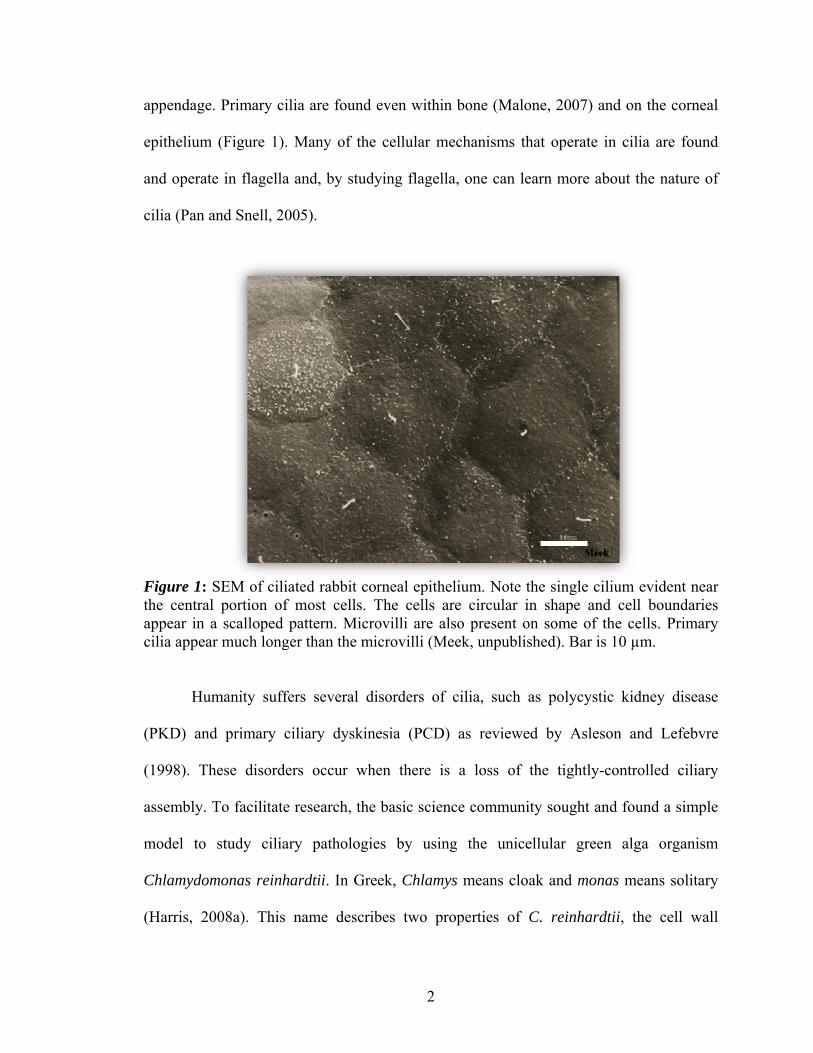
appendage. Primary cilia are found even within bone (Malone, 2007) and on the corneal
epithelium (Figure 1). Many of the cellular mechanisms that operate in cilia are found
and operate in flagella and, by studying flagella, one can learn more about the nature of
cilia (Pan and Snell, 2005).
Figure 1: SEM of ciliated rabbit corneal epithelium. Note the single cilium evident near the central portion of most cells. The cells are circular in shape and cell boundaries appear in a scalloped pattern. Microvilli are also present on some of the cells. Primary cilia appear much longer than the microvilli (Meek, unpublished). Bar is 10 µm.
Humanity suffers several disorders of cilia, such as polycystic kidney disease
(PKD) and primary ciliary dyskinesia (PCD) as reviewed by Asleson and Lefebvre
(1998). These disorders occur when there is a loss of the tightly-controlled ciliary
assembly. To facilitate research, the basic science community sought and found a simple
model to study ciliary pathologies by using the unicellular green alga organism
Chlamydomonas reinhardtii. In Greek, Chlamys means cloak and monas means solitary
(Harris, 2008a). This name describes two properties of C. reinhardtii, the cell wall
3
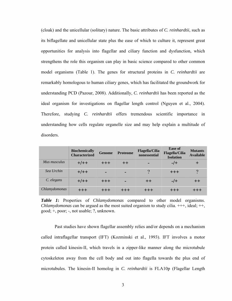
(cloak) and the unicellular (solitary) nature. The basic attributes of C. reinhardtii, such as
its biflagellate and unicellular state plus the ease of which to culture it, represent great
opportunities for analysis into flagellar and ciliary function and dysfunction, which
strengthens the role this organism can play in basic science compared to other common
model organisms (Table 1). The genes for structural proteins in C. reinhardtii are
remarkably homologous to human ciliary genes, which has facilitated the groundwork for
understanding PCD (Pazour, 2008). Additionally, C. reinhardtii has been reported as the
ideal organism for investigations on flagellar length control (Nguyen et al., 2004).
Therefore, studying C. reinhardtii offers tremendous scientific importance in
understanding how cells regulate organelle size and may help explain a multitude of
disorders.
Biochemically Characterized Genome Proteome Flagella/Cilia
nonessential
Ease of Flagella/Cilia
Isolation
Mutants Available
Mus musculus
+/++ +++ ++ - -/+ + Sea Urchin
+/++ - - ? +++ ?
C. elegans
+/++ +++ - ++ -/+ ++ Chlamydomonas
+++ +++ +++ +++ +++ +++
Table 1: Properties of Chlamydomonas compared to other model organisms. Chlamydomonas can be argued as the most suited organism to study cilia. +++, ideal; ++, good; +, poor; -, not usable; ?, unknown.
Past studies have shown flagellar assembly relies and/or depends on a mechanism
called intraflagellar transport (IFT) (Kozminski et al., 1993). IFT involves a motor
protein called kinesin-II, which travels in a zipper-like manner along the microtubule
cytoskeleton away from the cell body and out into flagella towards the plus end of
microtubules. The kinesin-II homolog in C. reinhardtii is FLA10p (Flagellar Length
4
Assembly protein 10). FLA10p carries the axoneme precursors involved in the assembly
of flagella (Qin, 2004) to the flagellar tip, where precursors are exchanged for old
particles and shuttled via a separate motor protein called cytoplasmic dynein back to the
cell body for breakdown as reviewed by Pederson and Rosenbaum (2008). There are four
long-flagella genes that have been implicated in recent studies that play a role in the
regulation of length control (Asleson and Lefebvre, 1998). The mutants of these genes,
known as lf1, lf2, lf3, and lf4, are reported to be necessary for normal flagellar length
regulation, yet, a full explanation of flagellar control of length remains incomplete
(Wilson et al., 2008).
Within the lf4 phenotype, little is known as to the exact location of FLA10p,
whereby localizing FLA10p would not only further explain the morphology of this
important long-flagella mutant but should further facilitate our understanding of FLA10p
in flagellar length control. Therefore, the purpose of this study involved the phenotypic
characterization, or in other words, a description of observable microscopic features of
the cells and flagella of lf4. And due to the lack of previous ultrastructural studies of lf4, I
sought to systematically characterize this mutant using a number of microscopic methods
including bright-field, immunofluorescence (IMF), scanning electron microscopy (SEM)
and transmission electron microscopy (TEM). In addition, I have used TEM to quantify
the number of IFT particles in wild-type and lf4 flagella.
This qualitative characterization of lf4 should facilitate our understanding in how
flagellar size is regulated, which in turn should contribute to the growing field of
translational research on human ciliary disorders. The fields of medicine and biomedical

5
science will incrementally benefit, perhaps indirectly, from this phenotypic
characterization of lf4 flagella.

6
CHAPTER II
REVIEW OF LITERATURE
Intraflagellar Transport
The ground breaking discovery by Kozminski et al. (1993) of a motility within
flagella called intraflagellar transport (IFT) revolutionized our understanding of ciliary
and flagellar assembly. This study utilized video and electron microscopy to demonstrate
the role of FLA10p and IFT in assembly and disassembly of flagella. This initial
investigation and report on IFT spawned a new field and has laid the groundwork for
understanding the mechanisms that underlie ciliopathies.
FLA10p moves IFT particles, the shape of which varies from small lollipop-like
structures to large rafts that contain 17-18 or more individual particles and are found
between the flagellar membrane and the outer-doublet microtubules (Kozminski et al,
1993). In addition to determining the presence of FLA10p, later papers reported how
FLA10p functions in IFT, which transports cargo destined for incorporation into flagella
along the axoneme microtubules much as a truck carries parts on a highway (Qui et al.,
2004).
During the same time as the initial IFT and FLA10p studies, the first
characterization of mutants defective in the ability to regulate the length of flagella were
reported in 1998 by Asleson and Lefebvre. These authors described the genetics of long-
flagella loci and the identification of the lf4 mutant. In this study they determined the
7
mean lengths of long-flagella, which are two to three times the length of wild-type
flagella.
It was not always apparent how flagella regulated the transport of IFT cargo and
regulated the assembly of flagella at a pre-set length. The mechanism of IFT was known,
but how did flagella establish a length? Marshall and coworkers (2005) described a
balance-point model to explain how lengths of organelles in general and in flagella in
particular were controlled. It involved the addition of precursors by FLA10p and removal
of turn-over particles with cytoplasmic dynein in a steady-state process. He also reported
that IFT protein quantity versus flagellar length operated independently. Marshall did not
measure the number of IFT particles per flagellar length using TEM images, as reported
in this work.
Long Flagella Specifics and Techniques
Several studies have focused on aspects of FLA10p and its role in IFT (Marshall
and Rosenbaum, 2001) for the proper length regulation of flagella. Flagella continually
are turning over, and in the absence of a mechanism to replace proteins that are lost due
to turnover, flagella disassembly exceeds flagellar assembly and therefore becomes
progressively shorter. Marshall and Rosenbaum (2001) reported that IFT was essential
for flagellar assembly and maintenance and was necessary for regulation of length.
Subsequently, it was found that a novel mitogen-activated protein (MAP) kinase was
“crucial” in determining the length of lf4 flagella (Berman et al., 2003). Here, I will
report on the first ultrastructural characterization of lf4 flagella. Moreover, biochemical
studies of lf4 flagella demonstrated a significant increase in FLA10p levels compared to
wild-type flagella (Wilson and Lefebvre, unpublished observations). To this end, I have
8
localized FLA10p in both wild-type and lf4 using IMF. These studies may help decipher
the molecular mechanisms that regulate the length and assembly of flagella. This study
will specifically aim to describe: (1) the morphology of the lf4 phenotype using bright-
field, scanning, and transmission electron microscopy; and (2) the localization of
FLA10p in the lf4 mutant using immunofluorescent microscopy. Furthermore, IFT
particles will be counted and compared in wild-type and lf4 per flagellar length, area, and
volume. It is hypothesized that the IFT particle quantity will be independent of length in
lf4, just as Marshall (2005) reported in other Chlamydomonas mutants.
9
CHAPTER III
METHODS AND MATERIALS
The research design and methodology employed cell culture techniques using C.
reinhardtii and three mammalian cell lines, plus bright-field, immunofluorescence,
scanning, and transmission electron microscopy.
Chlamydomonas Cultures
Strains of C. reinhardtii utilized included the wild-type (CC-1690 mt+ 21gr) and
lf4 mutants, such as lf4-1 arg7-, lf4-3 arg7-, lf4-6 and lf4-10. These strains were
graciously provided by Dr. Nedra Wilson of Oklahoma State University Center for
Health Sciences (OSU-CHS). Cells were maintained on TAP agar plates and transferred
to liquid cultures consisting of Medium II (Gorman and Levine, 1965). Cells were grown
under a 13:11 hour (h) light:dark cycle at 22oC with mild to moderate aeration. Some lf4
mutants, such as lf4-6, required supplementation with exogenous arginine (Sager and
Granick, 1954).
The time period of growth and other factors was determined by preliminary
experiments involving wild-type and lf4 mutants. After inoculating the liquid media, all
strains were observed and video recorded (Appendix A) at successive time points of 12,
24, 36, and 48 h. As flagella are required for cell motility, I used an estimate of overall
swimming activity as a determinant of the flagellated status of cells (Mueller et al.,
2005). Briefly, all strains were analyzed by viewing an aliquot of cells on a pre-cleaned
glass microscope slide (Anapath, Lewisville, TX), at 40x with a standard laboratory
10
dissecting microscope. Based on a gross observation, the cell swimming activity was
estimated. Wild-type and lf4 cell activity were both judged at the same setting and on the
same brand of glass slides. A high-definition digital video camera (Insignia/Best Buy,
Richfield, MN) was positioned close to the microscope eye piece and used to film the
estimated swimming activity.
Mammalian Cell Cultures
Chinese hamster ovary (CHO) cells (Puck et al., 1958), inner medullary collecting
duct (IMCD) cells (Rauchman et al., 1993), and WISH (Wistar Institute Susan Hayflick),
a human amnion epithelial cell line (Hayflick, 1961; Meek and Davis, 1986) were grown
in a Forma Scientific incubator in humidified 95% air and 5% CO2 gas at 37oC. The CHO
and WISH were purchased from the American Type Culture Collection (Manassas, VA)
and the IMCD cells were graciously provided by Dr. Rashmi Kaul of OSU-CHS.
Standard culture mediums for the respective cell lines were used (Hayflick, 1961; Meek
and Davis, 1986). The media was supplemented with fetal calf serum (Atlanta
Biologicals, Lawrenceville, GA) and antibiotic/antimycotic solutions (Gibco, Grand
Island, NY). Either CHO, IMCD or WISH cell lines were processed concurrent with C.
reinhardtii to act as experimental controls.
Bright-field Microscopy
Using either a Nikon Labophot microscope (Tokyo, Japan) with a Sony Color
Digital Camera (Tokyo, Japan) or a Zeiss Photomicroscope III (Peabody, MA) with Spot
Camera software (Diagnostic Instruments, Sterling Heights, MI), cells were viewed and
images captured under bright-field microscopy to ensure flagellation prior to processing
11
for IMF, SEM, and TEM. To fix and preserve cells, Lugol’s iodine (4% iodine crystals,
6% potassium iodide) was applied prior to image capture (Caprette, 1996).
Immunofluorescence Microscopy
A procedure for indirect immunofluorescence (IMF) was formulated based on
previously published methods (Carson et al., 1973; Huang et al., 1988; Cole et al., 1998).
In addition, the gene product of LF4, LF4p, was examined in wild-type and lf4 mutants.
Frosted, pre-cleaned, microscope slides (Anapath) were etched with three
equidistant circular wells per slide, and treated with a 1:10 dilution of poly-L-lysine
(Sigma, St. Louis, MO). In addition, 12-well slides were utilized as well (Cel-Line/Erie
Scientific Co., Madison, WI). Two methods of fixation was utilized. For method 1, in 12
mL Falcon tubes (BD Falcon, Franklin Lakes, NJ), cells were fixed for 10 minute (min)
with Carson’s normal buffered formalin (NBF) at 3.7% (Carson et al., 1973). For method
2, cells were fixed for 10 min in paraformaldehyde 1.5% (Cole et al., 1998). Other than
fixation, the methods are the same. Cells were centrifuged at 750 revolutions per minute
(RPM) for three to four min on an IEC Clinical Centrifuge (Damon, Needham, MA) and
washed with phosphate buffered saline (PBS) containing 0.3% Triton X-100 (Sigma) at
pH 7.2 (Miller et al., 2002). In humidified chambers at room temperature (RT), a 20 µL
aliquot of cells was transferred to slides and allowed to adhere for 10 to 15 min. Cells
were permeabilized in Coplin jars containing either 0.5% NP-40 for two min at RT or -
20oC 100% methanol for 10 min. For Chlamydomonas cells, chlorophyll extraction
occurred with fresh -20oC acetone for 8 to 10 min and slides were washed in PBS. Cells
were incubated for one hour with blocking buffer containing 10mM KHPO4, pH 7.2, 5%
normal horse serum (NHS) (Jackson ImmunoResearch, West Grove, PA), 5% glycerol
12
and 1% coldwater fish gelatin (Cole et al., 1998). Primary antibodies were diluted in
blocking buffer at 1:500 for the monoclonal anti-α-tubulin (Sigma); at 1:50 for LF4p
(PAS403; Dr. Nedra Wilson, OSU-CHS); at 1:100 for K2.4 (Covance, Emeryville, CA),
an anti-kinesin-II motor subunit that is homologous to FLA10p. Primary antibodies were
allowed to incubate for one hour at 37oC or overnight at 4oC. Control slides were
incubated simultaneously under the same conditions in equal volumes of blocking buffer
only (Harlow and Lane, 1988). Slides were washed three times for five minutes each in
PBS and labeled for one hour in a humidified jacket at 37oC with secondary antibody.
Secondary antibodies included Alexa Fluor 488 (Invitrogen, Eugene, OR) diluted 1:200
or 1:500 and Cy3 (Jackson ImmunoResearch) diluted 1:1000. The Cy3-labeled secondary
antibody was graciously provided by Dr. Kenneth Miller of OSU-CHS. Slides were
washed and cover-slips apposed with Prolong Gold anti-fade reagent (Invitrogen) and
sealed with clear nail polish. Images were captured on an Olympus X51 fluorescent
research microscope (Olympus, Center Valley, PA) with Spot Camera software
(Diagnostic Instruments) and analyzed when necessary with Image J software (NIH).
Scanning electron microscopy
10 mL of lf4-10 and wild-type cells were harvested by centrifugation and re-
suspended in the primary fixative of 2.0% glutaraldehyde with 0.1M cacodylate buffer
(pH 7.2-7.3) for 20 min at RT. The suspension was centrifuged and re-suspended for
washing in a 0.1M cacodylate buffer. An aliquot of cells was placed on a round, glass
coverslip, 12 mm in diameter (Anapath), which was previously coated with 0.1%
aqueous polyethyleneamine (Sigma) to which the cells were allowed to adhere for 10-15
min. The coverslips were washed in PBS and post-fixed with 1% osmium tetroxide in
13
0.1M cacodylate buffer. Cells were washed and dehydrated in a graded ethyl alcohol
(EtOH) series at 25%, 50%, 75%, 95% and 2 changes in 100%. The coverslips with cells
attached were dried with a Denton DCP-1 Critical Point Drying Apparatus (Denton
Vacuum Inc, Moorestown, NJ), placed on aluminum stubs and were coated with gold
using a Polaron SC 500 Sputter Coater (Quorum Technologies, Ringmer, UK). Images
were viewed on a Hitachi S-2300 (Hitachi, Pleasanton, CA) operating at 25 kV and
recorded on either Polaroid 72 or Fuji ISO 400, black and white instant film.
Transmission Electron Microscopy
Wild-type and lf4-10 mutants were prepared for transmission electron microscopy
using the method of Dentler and Adams (1992). Briefly, cells were fixed for 30 min with
2% glutaraldehyde in 0.1M cacodylate buffer, pH 7.2-7.3, in 1.5 mL Micro Sample
Centrifuge tubes (EMS, Ft. Washington, PA). Cells were washed, centrifuged at 750
RPM for three to four min in an IEC Clinical Centrifuge, and suspended in 3-4 mL of
0.1M cacodylate buffer. Cells were post-fixed with 1% osmium tetroxide in 0.1M sodium
cacodylate for 20 min and washed briefly in 20% EtOH. Cells were stained en bloc with
20% EtOH/uranyl acetate for 20 min at RT and dehydrated in a graded EtOH series for
10 min two times each at 50%, 70%, 90% and 100%. Cells were cleared with propylene
oxide, embedded in Epon 812 (EMS) under vacuum and polymerized at 68oC for 18 h.
Thin sections were cut with an Ultracut E Microtome (Reichert-Jung/Leica, Wetzlar,
Germany) and viewed at 80 kV on either a Zeiss T109 Electron Microscope (Zeiss,
Peabody, MA) with Gatan Digital Micrograph (Pleasanton, CA) software (see Appendix
B for an instructional video on how to operate the Zeiss T109 electron microscope) or a
Hitachi H7000 Electron Microscope (Hitachi, Pleasanton, CA), with a built-in large
14
format film camera. Negatives were processed, printed and/or scanned using standard
darkroom procedures.
Quantification
Image J (NIH) was used to set scale bars for all images. For TEM micrographs, the
scale bar was established using the known 25 nm diameter of a microtubule
(Kierszenbaum, 2007). This method allowed for the most precise measurement of flagella
and corrected for inaccuracies with magnification calibration of the electron microscope.
After establishing the scale, micrographs containing flagella in either longitudinal or
cross-section were analyzed by measuring flagellar diameter and length. All cross-
sections were cut at a thickness of 90 nm, so 90 nm was used for the determination of
volume on cross-sections. Micrographs were taken at a common magnification of
80,000x for the majority of measurements. IFT particle identification criteria included the
observation of an electron-density between the outer doublet-microtubules and the
flagellar membrane, and a minimum dimension of 25 nm, as based on measurements of
IFT particles in Pazour (1998). The data was added to a Microsoft Excel (Redmond, WA)
spreadsheet to compute area and volume. IFTs per length, area and volume were
compared in wild-type versus lf4 mutant.
15
CHAPTER IV
RESULTS
Bright-field Microscopic Observations
Observation of lf4 cells revealed flagella clearly longer than wild-type (Figures 2-
6). The lf4 cells were biflagellate and structurally appeared similar to wild-type, with the
flagella oriented at the apical end of the cell body. Fixation of cells with Lugol’s iodine
enhanced the observation of flagella and therefore the determination of whether cells
were uniformly flagellated (Figure 3). Additionally, cells were observed for their
estimated swimming activity, as this reflected their flagellated status (Table 2). After
observing swimming activity for several days, the cultures became opaque and dark
green due to large numbers of C. reinhardtii cells. This obstructed light from uniformly
reaching other cells and led to loss of synchronization of the cell cycle. Some flagellated
cells were observed as either de-flagellated or appeared motionless at all time points of
observation. The lf4-3 arg7- and lf4-6 mutants were the most active of the lf4 strains at
48 h, while lf4-10 demonstrated the least movement at all time points. Wild-type
remained actively swimming at both 24 and 48 h. It was observed that the greater the
initial inoculation of cells (≥ four loops) and the more vigorous aeration produced more
swimming activity in less time. These cultures, however, turned opaque in only two to
three days, rather than three to four days typically observed with average aeration and
inoculation.
16
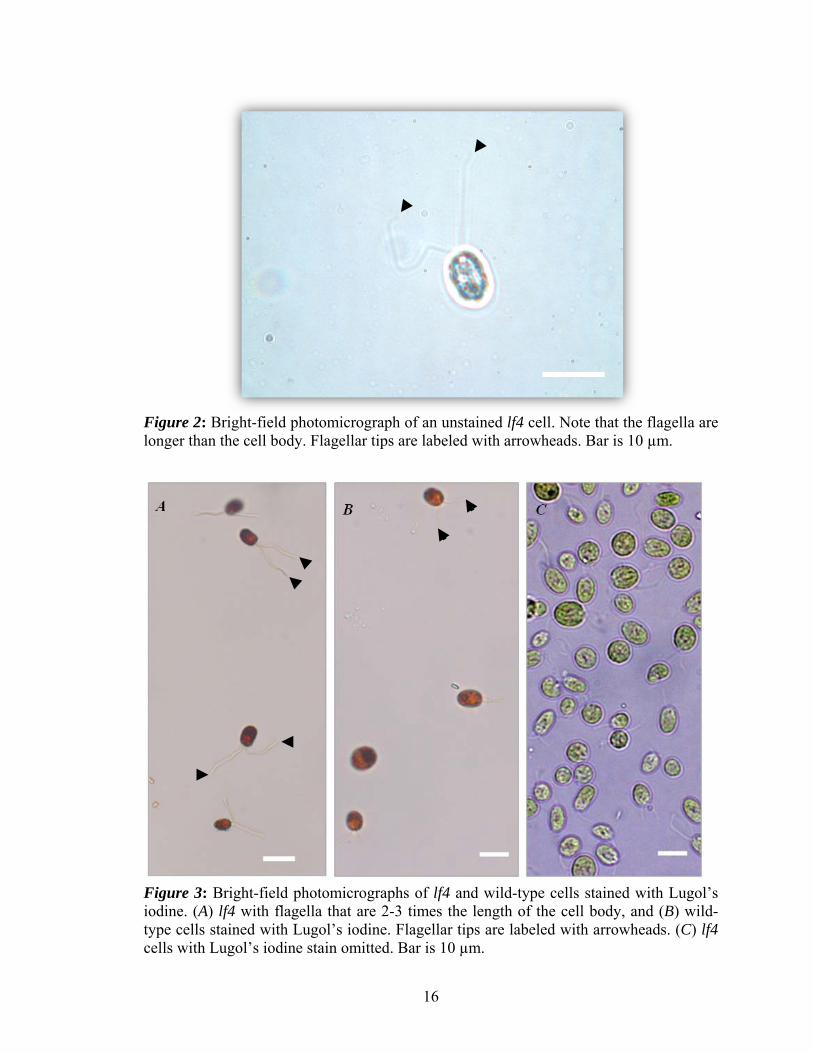
Figure 2: Bright-field photomicrograph of an unstained lf4 cell. Note that the flagella are longer than the cell body. Flagellar tips are labeled with arrowheads. Bar is 10 µm.
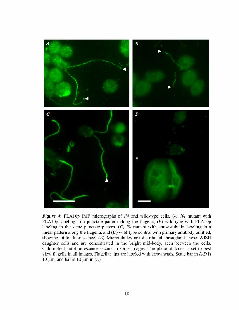
Figure 3: Bright-field photomicrographs of lf4 and wild-type cells stained with Lugol’s iodine. (A) lf4 with flagella that are 2-3 times the length of the cell body, and (B) wild-type cells stained with Lugol’s iodine. Flagellar tips are labeled with arrowheads. (C) lf4 cells with Lugol’s iodine stain omitted. Bar is 10 µm.

17
12 h 24 h 36 h 48 h
Wild-Type ++ +++ ++++ +++/+
lf4-10 - -/+ + -
lf4-1 arg 7- - -/+ + +
lf4-3 arg 7- - ++ +/++ +/++
lf4-6 - ++ +++ ++
Table 2: Estimate of swimming cells per time. At 12, 24, 36 and 48 h, wild-type and lf4 mutants are evaluated for percent of swimming activity, which indicated the presence of flagellated cells. ++++, most to all cells swimming; +++, more than majority cells swimming; ++, about half cells swimming; +, few to some cells swimming; -, little to no swimming cells.
Immunofluorescence Microscopy
As previously described by Cole and others, FLA10p localized in a punctate
pattern in both the lf4 and wild-type flagella (Figure 4, A-B; Figure 5, A & E,
respectively), and had approximately equal immunoreactivity in the microscope. Tubulin
localized in a linear pattern in the flagella (Figure 4, C). The control for FLA10p showed
little to no specific staining (Figure 4, D). WISH and IMCD cells showed microtubules
throughout the cell body and extending to the cell periphery (Figure 4, E; Figure 5, F,
respectively). IMCD cells also displayed centriole staining near the central portion of
many cells in a cell-cycle dependant manner. The LF4p was examined in wild-type and
lf4 mutant cells. LF4p localized in the proximal portion of wild-type in a punctate pattern
(Figure 5, B). The lf4 mutant appeared to have little to no specific staining for LF4p
(Figure 5, C), however, images exhibited high background fluorescence (Figure 5, B-C).
The control for LF4p showed little specific staining (Figure 5, D). Results with poly- and
monoclonal tubulin were the same (data not shown).
18
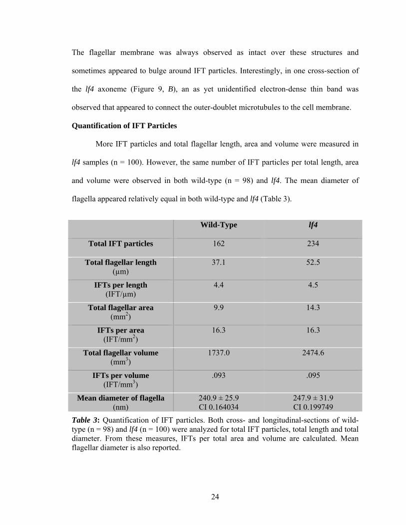
Figure 4: FLA10p IMF micrographs of lf4 and wild-type cells. (A) lf4 mutant with FLA10p labeling in a punctate pattern along the flagella, (B) wild-type with FLA10p labeling in the same punctate pattern, (C) lf4 mutant with anti-α-tubulin labeling in a linear pattern along the flagella, and (D) wild-type control with primary antibody omitted, showing little fluorescence. (E) Microtubules are distributed throughout these WISH daughter cells and are concentrated in the bright mid-body, seen between the cells. Chlorophyll autofluorescence occurs in some images. The plane of focus is set to best view flagella in all images. Flagellar tips are labeled with arrowheads. Scale bar in A-D is 10 µm; and bar is 10 µm in (E).

19
Figure 5: FLA10p versus LF4p IMF micrographs of lf4 and wild-type cells. Using Alexa Fluor 488 secondary antibodies, (A) lf4 mutant labeled with FLA10p in a punctate pattern, and compares to (B) LF4p labeling in proximal wild-type flagella with some fluorescence in the cell body. Flagellar tips are labeled with white arrowheads. (C) lf4 mutant labeled with LF4p, showing no specific staining. (D) LF4p control, with primary antibody omitted, showing no specific fluorescence. Using Cy3 secondary antibodies, (E) shows lf4 mutant labeled with FLA10p in a punctate pattern and the inset was the lf4 control with primary antibody omitted, having no specific fluorescence. (F) A confluent layer of IMCD cells labeled with α-tubulin primary and showing microtubules throughout the cytoplasm and some discrete points of fluorescence that represent centrioles (black arrowheads). Some images are overexposed to enhance visualization of flagellar staining. All bars are 10 µm.

20
Scanning Electron Microscopy
The topography of representative lf4 and wild-type cells (Figure 6, A-C) appeared
similarly oblong. The flagella of lf4 cells were observed to be grossly longer, as seen in
bright-field microscopy. The long flagella were more twisted and folded than wild-type
flagella (Figure 6, B & C). CHO cells showed intact membranes with typical surface
Figure 6: SEM images of lf4 and wild-type cells. In (A), wild-type flagella are approximately the length of a cell body. (B) In the lf4 mutant, the long flagella are apparent and folded back. Note the flagellar tips appear to taper (arrowheads). (C) A higher mag of the same lf4 cell shows some sharply demarcated bulges along the proximal flagellar membrane (thick arrows). (D) Two CHO cells show the surface topography features of round, bulbous blebs (BL) and wavy, peripheral lamellipodia (L). Scale bars in (A, B and D) are 10 µm; bar is 2 µm in (C).

21
Figure 7: SEM images of lf4 flagellar tips. The lf4 flagellar tips (arrow and inset) appear to tapper rather than end in bulbs. Bar is 10 µm.
features (Figure 6, D). In contrast to the bulbous ends of flagella with some length control
mutants, the tips of lf4 appeared tapered with no bulbs (Figure 6, B-C; and Figure 7).
Transmission Electron Microscopy
TEM images demonstrated that lf4 mutants were similar to wild-type in respect to
the cell body structures. The nucleus, nucleolus, chloroplast, ribosomes, mitochondria,
and vacuoles appeared undistinguishable from wild-type (Figure 8, A-B). Both wild-type
and lf4 had centrally located nuclei and chloroplasts that wrapped around in the
basolateral and distal ends of the cell body. In the cell apical region, the basal body
ultrastructure appeared similar (Figure 8, C-D). Representative cross-sections and
longitudinal profiles of lf4 flagella demonstrated the 9+2 axoneme ultrastructure with

22
intact inner and outer dynein arms and radial spokes. The cell membranes enveloping the
flagella were intact and IFT particles were seen between the outer-microtubule doublets
and the flagellar membranes in both lf4 and wild-type (Figure 9, A-C).
.
Figure 8: Cell body electron micrographs in lf4 (B and D) and wild-type cells (A and C). In (A), a flagellum emerges from the cell wall (CW). Chloroplasts (CL), mitochondria (M), vacuole (V), ribosomes (R), and the nucleus (N) are evident. In (B), chloroplasts fill the cell body. The basal body region in (C) depicts the transitional region (TR) and in (D), the basal body area shows two flagella and some closely related mitochondria. Ribosomes are abundant in the area around the basal bodies as seen in C and D. Bars are 0.5 µm (A), (C) and (D); and 1 µm in (B).
V

23
Figure 9: Electron micrographs of flagella found in lf4 and wild-type cells. The 9+2 microtubule arrangement is apparent in (A) wild-type and (B) lf4 cross-sections. IFT particles were seen between the outer-doublet microtubules and the cell membrane with concomitant membrane bulges (arrows). (B) An unidentified electron-dense band is seen connecting the lf4 outer-doublet microtubules to the cell membrane without the membrane bulges (arrowheads). (C) A longitudinal view of an lf4 flagellum with a large IFT raft particle (arrow). Bars are 100 nm (A) and (B); and (C) 200 nm.
24
The flagellar membrane was always observed as intact over these structures and
sometimes appeared to bulge around IFT particles. Interestingly, in one cross-section of
the lf4 axoneme (Figure 9, B), an as yet unidentified electron-dense thin band was
observed that appeared to connect the outer-doublet microtubules to the cell membrane.
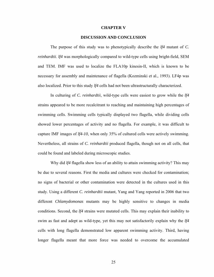
Quantification of IFT Particles
More IFT particles and total flagellar length, area and volume were measured in
lf4 samples (n = 100). However, the same number of IFT particles per total length, area
and volume were observed in both wild-type (n = 98) and lf4. The mean diameter of
flagella appeared relatively equal in both wild-type and lf4 (Table 3).
Wild-Type lf4
Total IFT particles 162 234
Total flagellar length (µm)
37.1 52.5
IFTs per length (IFT/µm)
4.4 4.5
Total flagellar area (mm2)
9.9 14.3
IFTs per area (IFT/mm2)
16.3 16.3
Total flagellar volume (mm3)
1737.0 2474.6
IFTs per volume (IFT/mm3)
.093 .095
Mean diameter of flagella (nm)
240.9 ± 25.9 CI 0.164034
247.9 ± 31.9 CI 0.199749
Table 3: Quantification of IFT particles. Both cross- and longitudinal-sections of wild-type (n = 98) and lf4 (n = 100) were analyzed for total IFT particles, total length and total diameter. From these measures, IFTs per total area and volume are calculated. Mean flagellar diameter is also reported.
25
CHAPTER V
DISCUSSION AND CONCLUSION
The purpose of this study was to phenotypically describe the lf4 mutant of C.
reinhardtii. lf4 was morphologically compared to wild-type cells using bright-field, SEM
and TEM. IMF was used to localize the FLA10p kinesin-II, which is known to be
necessary for assembly and maintenance of flagella (Kozminski et al., 1993). LF4p was
also localized. Prior to this study lf4 cells had not been ultrastructurally characterized.
In culturing of C. reinhardtii, wild-type cells were easiest to grow while the lf4
strains appeared to be more recalcitrant to reaching and maintaining high percentages of
swimming cells. Swimming cells typically displayed two flagella, while dividing cells
showed lower percentages of activity and no flagella. For example, it was difficult to
capture IMF images of lf4-10, when only 35% of cultured cells were actively swimming.
Nevertheless, all strains of C. reinhardtii produced flagella, though not on all cells, that
could be found and labeled during microscopic studies.
Why did lf4 flagella show less of an ability to attain swimming activity? This may
be due to several reasons. First the media and cultures were checked for contamination;
no signs of bacterial or other contamination were detected in the cultures used in this
study. Using a different C. reinhardtii mutant, Yang and Yang reported in 2006 that two
different Chlamydomonas mutants may be highly sensitive to changes in media
conditions. Second, the lf4 strains were mutated cells. This may explain their inability to
swim as fast and adept as wild-type, yet this may not satisfactorily explain why the lf4
cells with long flagella demonstrated low apparent swimming activity. Third, having
longer flagella meant that more force was needed to overcome the accumulated
26
attractions of the glycocalyx to the glass coverslip. So lf4 would be expected to
demonstrate less swimming activity and the minute forces between the glass and
glycocalyx may explain the motionless lf4 flagella. On the other hand, the wild-type
strain appeared to have more active cells and generally displayed greater than 50%
swimming activity from 24-72 h. Temperature, inoculation concentrations, and light:dark
cycles were not altered throughout this study and are not considered to be possible factors
influencing lf4 swimming ability. Additionally, the swimming activity was investigated
ad hoc, so it should be noted that lf4 strains were not always so resistant to start
swimming. Temporal or passage factors of maintaining cell strains may also play a role
in the swimming percentages. Future research in studying swimming activity using newly
acquired strains versus old strains, in a randomized-controlled and blind study may
facilitate in answering this question.
Under bright-field microscopy, the wild-type appeared to grossly demonstrate
flagella of expected length versus the distinctly longer lf4 mutant flagella (Asleson and
Lefebvre, 1998). Despite lower swimming percentages, the lf4 cells swam in a
characteristic jerky pattern as seen in other long-flagella mutants, while the wild-type
swimming patterns appeared to be active and without impediment (Asleson and Lefebvre,
1998). The addition of Lugol’s iodine stain made flagella more distinct and helped serve
as a quick check on approximate length of flagella (Gray, 1954) that helped ensure
cultures appeared uniform in length. In conclusion, bright-field microscopy allowed for
the gross morphological differences, such as flagellar length and estimated swimming
activity, to be catalogued between wild-type and lf4.
27
From the IMF results, the FLA10p kinesin-II showed immunoreactivity in lf4
along the length of both flagella, in a punctate pattern. Additionally, since FLA10p is
known to transport IFT particles in an anterograde fashion, the punctate staining likely
represents its presence in these areas for transporting IFT particles. LF4p also was
investigated via IMF. This protein was genetically removed to produce the lf4 mutant.
Though captured images contained background staining, this study found LF4p in
flagella of wild-type in a punctate pattern and no staining in lf4 flagella. No staining
should occur in the lf4 because the protein was knocked out, however, some images with
extremely high background demonstrated cells with some immunoreactivity. So knowing
that if any LF4p signal is observed, then this invalidates a signal seen in wild-type cells.
This variation may be due to inadequate washing, though all washing steps consisted of
three rounds of 5 min. A likely contributing factor was that the LF4p primary antibody
was not diluted in blocking buffer as were all subsequent IMF experiments. The primary
antibody itself may have been too concentrated at 1:50 or the secondary antibodies may
have been too concentrated. Perhaps all of these possibilities may explain the results
observed with LF4p.
Of the secondary antibodies utilized, the Alex Fluor 488-labeled secondary
antibody provided the best fluorescence, based on the quality of photographable images.
Cy3-labeled secondary antibody yielded good images but with a weaker signal. The Cy3
secondary was received in July 2000 and exceeded the typical shelf-life. However, no
major ill effects were observed other than mild attenuation of fluorescence intensity when
compared to Alex Fluor 488.
28
The topographic study of the lf4 mutant, using SEM, revealed an interesting
finding on long flagella. The lf4 demonstrated long flagella in a folded fashion, but
unexpectedly revealed sharply demarcated bulges along the flagella. These bulges could
represent IFT and its cargo in the assembly of long flagella. In prior ultrastructural
studies using TEM, bulges have been reported at sites of IFT particles, however the
images appeared to have smooth demarcations (Cole et al., 1998). I cannot rule out other
possibilities that may include artifact, membrane disruption from osmotic effects, or a
defective mechanism involved in IFT. These bulges were observed in the proximal region
of both wild-type and lf4 flagella, and if these observations are truly IFT particles, then
bulges would likely be observed along the full length. Further research in the assessment
of this phenotype in lf4 mutants may help to identify these structures.
Using SEM images, the lf4 flagellar tips were noted to differ from other long-
flagellar mutants, such as lf2 and lf3 (Tam et al., 2003; Tam et al. 2007). The tip bulges
reported in lf2 and lf3 were not observed in lf4 SEM images, indicating a difference in
morphology. The tapering ends of lf4 flagella indicate the lf4 mutant assembles or
disassembles flagella differently than the lf2 and lf3 mutants, in which IFT particles over-
accumulate at their bulbous ends, but not apparently so in lf4 flagella tips. Further SEM
characterizations of all four long-flagellar mutants would help resolve the differences
seen in morphology.
As observed in the cross-sectional and longitudinal electron micrographs, the
electron-dense particles are likely IFT particles. These images represent the first
ultrastructural analysis of the lf4 mutant. In comparison to wild-type, the basal bodies and
axoneme appeared the same, except for an unidentified thin band connecting the cell
29
membrane to the outer-doublet microtubules. These bands are still preliminary but
appeared at regular intervals surrounding this axoneme. Conversely, these unusual bands
were not observed nor have they been reported in wild-type flagella (Harris, 2008b). The
bands are as thin as radial spokes, but no spokes have been reported here, nor do spokes
originate in this area. Typically, radial spokes originate from the A microtubule and
interact with the central pair microtubules. So, these structures may represent an
adulteration of the lf4 axoneme. Other possibilities include artifactual observations or
merely the most proximal or distal component of IFT particles. The later seems unlikely,
as no similar structure was observed in wild-type axonemes. At this time, more research
is needed to determine what these structures may represent.
The significance of morphological assessment of lf4 flagella was viewed in light
of human ciliary disorders. The important connection linking flagella to cilia and the
multitude of ciliopathies was reviewed by Pan and Snell (2005). These authors described
the role of cilia in sensory perception and how the loss of function leads to disease.
Furthermore, they discussed PKD that most commonly strikes in adulthood and is
inherited primarily in an autosomal dominant pattern. A less common variant is
autosomal recessive and fatal to neonates (Igarashi and Somlo, 2002). The same genes
that have been attributed to PKD also are rooted in the genetics encoding primary cilia
(Yoder, 2007). Several homologous PKD genes are encoded in C. reinhardtii (Pazour,
2008), making it the ideal basic science organism for ciliopathy research.
Chodhari et al. (2004) outlined the pathogenesis of primary ciliary dyskinesia
(PCD), another ciliary-related disorder. In PCD, ciliary dysfunction occurs during
embryologic development and results in defects in left-right axis patterning (Taulman et
30
al., 2001), such as formation of dextrocardia, or a right-sided heart. A clinical triad of
signs for PCD include: recurrent sinopulmonary disease (such as chronic upper
respiratory infections), infertility and the laterality defects such as situs inversus are
found in up to 50% of PCD patients (Chodhari et al., 2004). The basic science principles
that C. reinhardtii offers in researching ciliopathies can facilitate the scientific pursuit of
treating PCD, PKD and the other disorders of cilia.
In summary, Chlamydomonas represents an ideal organism to investigate IFT,
control of flagellar length and the basic science of ciliopathies. Studying Chlamydomonas
flagella is equivalent to studying cilia and by extension to studying primary cilia. This
study observed that the lf4 flagella were grossly longer than wild-type and demonstrated
low percentages of swimming cells. FLA10p was labeled and localized to the flagella of
wild-type and lf4 in a punctate pattern. The observation that the flagella of lf4 are tapered
at their tips is in contrast to previous studies of lf2 and lf3 that form bulges at the tips of
their flagella. The ultrastructure of lf4 revealed IFT particles and the presence of an
unidentified structure between the outer-doublet microtubules and the cell membrane.
The number of IFT particles per total length was the same in wild-type and lf4 suggesting
they operate independently and confirms my hypothesis. Localization with immunogold
electron microscopy is the logical next step in furthering this phenotypic study of IFT
transport and FLA10p in the lf4 mutant of C. reinhardtii.
31
References
Alberts, B., A. Johnson, J. Lewis, M. Raff, K. Roberts, and P. Walter. 2008. The Cytoskeleton. In Molecular Biology of the Cell. B. Alberts, editor. Garland Science, Oxford, UK. 1031-1035.
Asleson, C.M., and P.A. Lefebvre. 1998. Genetic Analysis of Flagellar Length Control in Chlamydomonas reinhardtii: A New Long-Flagella Locus and Extragenic Suppressor Mutations. Genetics. 148:693-702.
Berman, S.A., N.F. Wilson, N.A. Haas, and P.A. Lefebvre. 2003. A Novel MAP Kinase Regulates Flagellar Length in Chlamaydomonas. Curr Biol. 13:1145-1149.
Caprette, D.R. 1996. Fixing Chlamydomonas to observe and preserve flagella. online: http://www.ruf.rice.edu/~bioslabs/studies/invertebrates/chlamfix.html. Updated in 2006 and accessed April 1, 2009:1-3.
Carson, F.L., J.H. Martin, and J.A. Lynn. 1973. Formalin Fixation for Electron Microscopy: A Re-evaluation. Am J Clin Pathol. 59:365-373.
Chodhari, R., H. Mitchinson, and M. Meeks. 2004. Cilia, primary ciliary dyskinesia and molecular genetics. Paediatr Respir Rev. 5:69-76.
Cole, D.G. 1999. Kinesin-II, the heteromeric kinesin. Cell Mol Life Sci. 56:217-226.
Cole, D.G., D.R. Diener, A.L. Himelblau, P.L. Beech, J.C. Fuster, and J.L. Rosenbaum. 1998. Chlamydomonas Kinesin-II-dependent Intraflagellar Transport (IFT): IFT Particles Contain Proteins Required for Ciliary Assembly in Caenorhabditis elegans Sensory Neurons. JCB. 141:993-1008.
Dentler, W.L. and C. Adams. 1992. Flagellar Microtubule Dynamics in Chlamydomonas: Cytochalasin D Induces Periods of Microtubule Shortening and Elongation; and cochicine induces disassembly of the distal, but not proximal, half of the flagellum. JCB. 117:1289-1298.
Domozych, D.S. 1999. Disruption of the Golgi apparatus and secretory mechanism in the desmid, Closterium acerosum, by brefeldin A. J Exp Biol. 50:1323-1330.
Geimer, S., and M. Melkonian. 2004. The ultrastructure of the Chlamydomonas reinhardtii basal apparatus: identification of an early marker of radial asymmetry inherent in the basal body. J. Cell Sci. 117:2663-2674.
Gorman, D.S., and R.P. Levine. 1965. Cytochrome F and Plastocyanin: Their Sequence in the Photosynthetic Electron Transport Chain of Chlamydomonas reinhardtii. Proc Natl Acad Sci USA. 54:1665-1669.
32
Gray, P. 1954. The Microtomist's Formulary and Guide. R E Krieger Pub Co, Huntington, NY. 680 pp.
Harlow, E., and D. Lane. 1988. Antibodies: A Laboratory Manual. Cold Spring Harbor Laboratory Press, Woodbury, NY. 726 pp.
Harris, E.H. 2008a. The Genus Chlamydomonas. In The Chlamydomonas Sourcebook, Volume 1. E.H. Harris, editor. Elsevier/Academic Press, Burlington, MA. 1-24.
Harris, E.H. 2008b. Motility and Behavior. In The Chlamydomonas Sourcebook, Volume 1. E.H. Harris, editor. Elsevier/Academic Press, Burlington, MA. 89-117.
Hayflick, L. 1961. The Establishment of a Line (WISH) of Human Amnion Cells in Continuous Cultivation. Exp Cell Res. 23:14-20.
Huang, B., D.M. Watterson, V.D. Lee, and M.J. Schibler. 1988. Purification and Characterization of a Basal Body-associated Ca2+-binding Protein. JCB. 107:121-131.
Igarashi, P., and S. Somlo. 2002. Genetics and Pathogenesis of Polycystic Kidney Disease. J Am Soc Nephrol. 13:2384-2394.
Kierszenbaum, A.L. 2007. Epithelium. In Histology and Cell Biology. A.L. Kierszen-baum, editor. Elsevier/Mosby, New York, NY.
Kozminski, K.G., K.A. Johnson, P. Forscher, and J.L. Rosenbaum. 1993. A motility in the eukaryotic flagellum unrelated to flagellar beating. Proc Natl Acad Sci USA. 90:5519-5523.
Lefebvre, P.A. 2008. Flagellar Length Control. In The Chlamydomonas Sourcebook Volume 3. G.B. Witman and E.H. Harris, editors. Elsevier/Academic Press, Burlington, MA. 115-129.
Malone, A.M., C.T. Anderson, P. Tummala, R.Y. Kwon, T.R. Johnston, T. Stearns, C.R. Jacobs. 2007. Primary cilia mediate mechanosensing in bone cells by a calcium-independent mechanism. Proc Natl Acad Sci USA.104:13325-13330.
Marshall, W.F., H. Qin, M.R. Brenni, and J.L. Rosenbaum. 2005. Flagellar Length Control System: Testing a Simple Model Based on Intraflagellar Transport and Turnover. Mol Biol Cell. 16:270-278.
Marshall, W.F., and J.L. Rosenbaum. 2001. Intraflagellar transport balances continuous turnover of outer doublet microtubules: implications for flagellar length control. JCB. 155:405-414.
Meek, W.D., and W.L. Davis. 1986. Fine structure and immunofluorescenct studies of the WISH cell line. In Vitro Cell Dev Biol. 22:716-724.
33
Michaud, E.J. and B.K. Yoder. 2006. The Primary Cilium in Cell Signaling and Cancer. Cancer Res. 66:6463-6467.
Miller, K.E., B.A. Richards and R.M. Kreibal. 2002. Glutamine-, glutamine synthetase-, glutamine dehydrogenase- and pyruvate carboxylase-immunoreativities in the rat dorsal root ganglion and peripheral nerve. Brain Res. 945:202-211.
Mueller, J., C.A. Perrone, R. Bower, D.G. Cole, and M.E. Porter. 2005. The FLA3 KAP Subunit Is Required for Localization of the Kinesin-2 to the Site of Flagellar Assembly and Processive Anterograde Intraflagellar Transport. Mol Biol Cell. 16:1341-1354.
Nguyen, R.L., L.W. Tam, and P.A. Lefebvre. 2004. The LF1 Gene of Chlamydomonas reinhardtii Encodes a Novel Protein Required for Flagellar Length Control. Genetics. 169:1415-1424.
Pan, J., and W.J. Snell. 2005. Chlamydomonas Shortens Its Flagella by Activating Axonemal Disassembly, Stimulating IFT Particle Trafficking, and Blocking Anterograde Cargo Loading. Dev Cell. 9:431-438.
Pan, J., and W.J. Snell. 2002. Kinesin-II Is Required for Flagellar Sensory Transduction during Fertilization in Chlamydomonas. Mol. Biol. Cell. 13:1417-1426.
Pazour, G.J. 2004. Intraflagellar Transport and Cilia-Dependent Renal Disease: The Ciliary Hypothesis of Polycystic Kidney Disease. J Am Soc Nephrol. 15:2528-2536.
Pazour, G.J., and G.B. Witman. 2008. The Chlamydomonas Flagellum as a Model for Human Ciliary Disease. In The Chlamydomonas Sourcebook, Volume 3. G.B. Witman and E.H. Harris, editors. Elsevier/Academic Press, Burlington, MA. 445-468.
Pederson, L.B. and J.L. Rosenbaum. 2008. Intraflagellar Transport (IFT): Role in Ciliary Assembly, Resorption and Signaling. Curr Top Dev Biol. 85:23-61.
Puck, T.T., S.J. Cieciura, and A. Robinson. 1958. Genetics of Somatic Mammalian Cells, III. Long-Term Cultivation of Euploid Cells from Human and Animal Subjects. J Exp Med. 108:945-956.
Qin, H., D.R. Diener, S. Geimer, D.G. Cole and J.L. Rosenbaum. 2004. Intraflagellar transport (IFT) cargo: IFT transports flagellar precursors to the tip and turnover products to the cell body. JCB 164:255-266.
Rauchman, M.I., S.K. Nigam, E. Delpire, and S.R. Gullan. 1993. An osmotically tolerant inner medullary collecting duct cell line from an SV40 transgenic mouse. Am J Physiol. 265:F416-F424.
Sager, R., and S. Granick. 1954. Nutritional control of sexuality in Chlamydomonas reinhardi. J Gen Physiol. 37:729-742.

34
Tam, L.W., W.L. Dentler, and P.A. Lefebvre. 2003. Defective flagellar assembly and length regulation in LF3 null mutants in Chlamydomonas. JCB. 163:597-607.
Tam, L.W., N.F. Wilson, and P.A. Lefebvre. 2007. A CDK-related kinase regulates the length and assembly of flagella in Chlamydomonas. JCB. 176:819-829.
Taulman, P.D., C.J. Haycraft, D.F. Balkovetz, and B.K. Yoder. 2001. Polaris, a Protein Involved in Left-Right Axis Patterning, Localized to Basal Bodies and Cilia. Mol Biol Cell. 12:589-599.
Wilson, N.F., J.K. Iyer, J.A. Buchheim, and W.D. Meek. 2008. Regulation of flagellar length in Chlamydomonas. Semin Cell Dev Biol. 19:494-501.
Yang, C., and P. Yang. 2006. The flagellar motility of Chlamydomonas pf25 mutant lacking an AKAP-binding protein is overtly sensitive to medium conditions. Mol Biol Cell. 17:227-238.
Yoder, B.K. 2007. Role of Primary Cilia in the Pathogenesis of Polycystic Kidney Disease. J Am Soc Nephrol. 18:1381-1388.
Zimmerman, K.W. 1898. Contributing to the knowledge of glands and epithelium. Arch Mikr Entwicklungsmech. 52:552-706.
35
APPENDIX
A. Growth Experiment Video: C. reinhardtii wild-type and several lf4 strains were
analyzed for percent of cells swimming per time in March 2009.
B. A Video on ‘How to Operate the OSU-CHS Zeiss T109 Electron Microscope’ by
Kevin Pargeter
VITA
Kevin Matthew Pargeter
Candidate for the Degree of Master of Science Thesis: A PHENOTYPIC STUDY OF INTRAFLAGELLAR TRANSPORT AND FLA10 IN THE lf4 MUTANT OF CHLAMYDOMONAS REINHARDTII Major Field: Biomedical Science Biographical:
Born in 1979 to Terry and Carolyn Pargeter, I was the fourth of six children to be raised in Oklahoma City, OK. Professionally my interests lie within the field of Internal Medicine and the basic science of ciliary disorders, called ciliopathies. Specifically, using the green alga Chlamydomonas reinhardtii, which has been established as the model organism for ciliopathies (Pazour 2004), I have gained a fruitful series of academic experiences in biomedical research. From the result of pursing a Master of Science concurrent with the third and fourth years of medical school, this education has greatly enriched my life and sharpened my academic goals. Ultimately, experimental and clinical techniques in transmission, scanning and immunofluorescent microscopy have become tools of my research efforts. With a D.O./M.S. education, I will pursue an internal medicine academic position at a medical center that involves responsibilities in teaching, research and patient care.
Education:
1. Expecting Doctor of Medicine at Oklahoma State University College of Osteopathic Medicine, Tulsa, OK, on May 15, 2009.
2. Expecting Master of Science at Oklahoma State University Center for Health Sciences, Tulsa, OK, on May 15, 2009.
3. Bachelor of Science in Biology at the University of Central Oklahoma, Edmond, OK, on May 2004, Cum laude.
Experience: 1. Teacher Asst, Histology Tutor & Researcher. OSU-CHS, Tulsa, May-Dec 2006. 2. U.S. Senate Intern for Senator Jim Inhofe, Washington, D.C., May-June 2004. 3. Undergrad Research Asst, UCO, Edmond, OK, Aug 2004-May 2005.
Professional Memberships: AMA, ACP, AOA, Sigma Xi, Oklahoma Microscopy Society and Oklahoma Academy of Science.

ADVISER’S APPROVAL: William D. Meek, Ph.D.
Name: Kevin Matthew Pargeter Date of Degree: May 2009 Institution: Oklahoma State University Center for Health Sciences, Tulsa, Oklahoma Title of Study: A PHENOTYPIC STUDY OF INTRAFLAGELLAR TRANSPORT
AND FLA10 IN THE lf4 MUTANT OF CHLAMYDOMONAS REINHARDTII Pages in Study: 35 Candidate for the Degree of Master of Science
Major Field: Biomedical Science Scope and Method of Study:
Chlamydomonas reinhardtii is a biflagellate, unicellular green alga that serves as an ideal basic science organism (Harris, 2008a). Compared to eukaryotic primary cilia, it shares many homologous flagellar genes with humans, of which several ciliary disorders, or ciliopathies, are known to occur (Pazour, 2008). Almost all vertebrate cells possess primary cilia, but this organelle had long been thought to have no function (Pan et al., 2005). With the discovery of intraflagellar transport (IFT), the mechanism for flagellar assembly, by Kozminski and coworkers (1993), new approaches to study ciliopathies were established. To investigate how flagellar length was controlled, long-flagellar mutants lf1, lf2, lf3 and lf4 were created. Yet little phenotypically was known about lf4. To characterize the morphology of and localize IFT proteins (FLA10p and LF4p) in the lf4 mutant, this work used bright-field microscopy, immunofluorescence microscopy (IMF), scanning electron microscopy (SEM) and transmission electron microscopy (TEM).
Findings and Conclusions:
The lf4 flagella were grossly longer than wild-type cells, which were observed to be most active at 36 h. The lf4 cells were most active between 24-36 h likely indicating the best time to harvest for experimentation. Using IMF, the FLA10p and LF4p localized to flagella in a punctate pattern, likely indicating IFT particles. No bulbous tips were observed in lf4, unlike the lf2 and lf3 mutants as reported by Tam and coworkers (2003; 2007). Multiple IFT particles were observed in cross-sectional and longitudinal flagellar ultrastructure of lf4 using TEM. Plus, an as yet unidentified electron-dense structure in an lf4 cross-section was observed to connect the outer doublet microtubules to the cell membrane. Quantification of IFT particles per length, area and volume of flagella were the same in wild-type and lf4, suggesting that IFT particle quantity is independent of length. Immunogold electron localization is the logical next step to further the phenotypic characterization of the lf4 mutant.