theses on incidence , correlation and natural history
TRANSCRIPT

“ THESES ON INCIDENCE , CORRELATION AND NATURAL HISTORY
OF EPIRETINAL MEMBRANES SURROUNDING IDIOPATHIC
MACULAR HOLES “
� Duration – 1YEAR
� AIM – to evaluate the prevalence , correlation and natural
history of EPIRETINAL MEMBRANES SURROUNDING IDIOPATHIC
MACULAR HOLES
PRIMARY OBJECTIVES:
1. To evaluate the prevalence of epiretinal membranes in eyes
with stage 2/3/4 macular holes.
2. The correlation between epiretinal membrane and visual
acuity in idiopathic macular holes
3. The correlation between size of macular hole and epiretinal
membrane
Secondary objective -To assess the increase in severity of
epiretinal membrane in follow up.
� Study design-Prospective study, observational study
� Methodology{material and methods }-All patients with
idiopathic macular hole stage 2,3,4who attended the OPD
� Inclusion criteria-All patients with symptoms of decreased
central vision and metamorphopsia and clinically proven
macular hole stage 2, 3 and 4
� Exclusion criteria-Patients who had traumatic macular hole and
macular hole due to other causes and macular edema .

Sample size- 50 patients
Age - 30 to 80 years
Gender – both genders
� Study parameters
� Detailed history including past and present illness
� Visual acuity assessment using snellens visual acuity chart
� Anterior segment examination using slit lamp including lens
and anterior vitreous .
� Fundus examination using direct and indirect ophthalmoscopic
examination.
� Stereoscopic fundus examination using slit lamp biomicroscopy
� Colour fundus photography
� Optical coherence tomography .
� Data collection and methods-All cases diagnosed as idiopathic
macular holes stage 2,3 and 4 will be registered and evaluated
subsequently at the end of 3,6 and 12 months of follow up .
� Analysis plan- to consider the importance of factors in removal
of epiretinal membrane during vitrectomy for macular hole
1
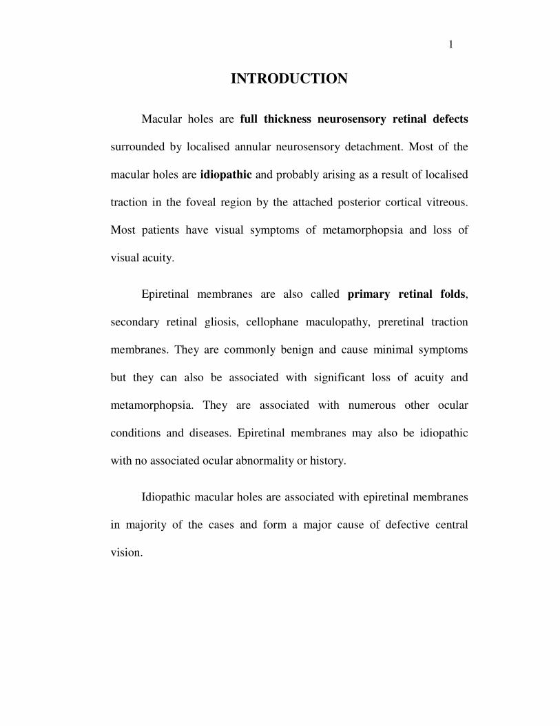
INTRODUCTION
Macular holes are full thickness neurosensory retinal defects
surrounded by localised annular neurosensory detachment. Most of the
macular holes are idiopathic and probably arising as a result of localised
traction in the foveal region by the attached posterior cortical vitreous.
Most patients have visual symptoms of metamorphopsia and loss of
visual acuity.
Epiretinal membranes are also called primary retinal folds,
secondary retinal gliosis, cellophane maculopathy, preretinal traction
membranes. They are commonly benign and cause minimal symptoms
but they can also be associated with significant loss of acuity and
metamorphopsia. They are associated with numerous other ocular
conditions and diseases. Epiretinal membranes may also be idiopathic
with no associated ocular abnormality or history.
Idiopathic macular holes are associated with epiretinal membranes
in majority of the cases and form a major cause of defective central
vision.

2
REVIEW OF LITERATURE
EPIDEMIOLOGY
Macular holes usually only affect one eye, though there is a 10 per
cent, one in ten, chance that the other eye will eventually be affected.
They mostly occur in the age group of 60-80 years. They are twice more
common in women than men.
3
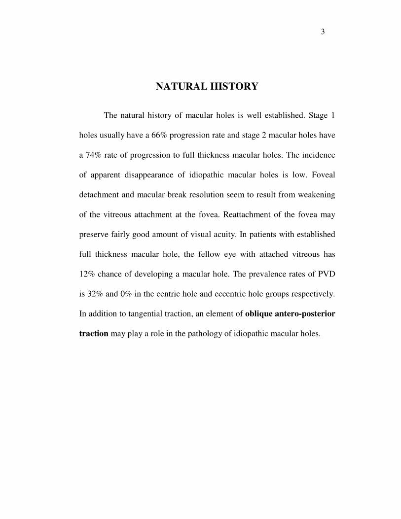
NATURAL HISTORY
The natural history of macular holes is well established. Stage 1
holes usually have a 66% progression rate and stage 2 macular holes have
a 74% rate of progression to full thickness macular holes. The incidence
of apparent disappearance of idiopathic macular holes is low. Foveal
detachment and macular break resolution seem to result from weakening
of the vitreous attachment at the fovea. Reattachment of the fovea may
preserve fairly good amount of visual acuity. In patients with established
full thickness macular hole, the fellow eye with attached vitreous has
12% chance of developing a macular hole. The prevalence rates of PVD
is 32% and 0% in the centric hole and eccentric hole groups respectively.
In addition to tangential traction, an element of oblique antero-posterior
traction may play a role in the pathology of idiopathic macular holes.

4
ANATOMY OF THE RETINA
Retina is the inner most coat of eyeball. It is a thin and delicate
transparent membrane. This layer is where the optical image is formed by
the eyes optical system. Photochemical transduction occurs so that nerve
impulses created here are transmitted along the visual pathways to the
brain for higher processing. It is thick at the posterior pole and peri-
papillary region of about 0.56mm whereas the thickness decreases to
about 0.1 mm at the ora-serrata. It is thinnest at the centre of the fovea.
The outer surface of the retina is in contact with Bruchs membrane of the
choroid and the inner surface is in contact with the vitreous body.
Retina extends more anteriorly on the medial aspect than laterally.
Ora serrata lies closer to the limbus medially. It consists of an outer
pigmented layer and an inner neurosensory layer which are derived from
the neuro-ectoderm. The ORA SERRATA is the anterior wavy edge of
the retina just posterior to the ciliary body.
Retina can be grossly divided into Optic disc, Macula lutea and rest
of the peripheral retina.

5
OPTIC DISC:
It is a pale pink, circular area of about 1.5mm in diameter. All the
layers of the retina terminate at the optic disc except the nerve fibres. The
optic disc appears white due to the absence of vascular choroid and the
presence of lamina-cribrosa and medullated nerve fibres behind it. There
is complete absence of rods and cones at the optic disc and hence
insensitive to light and is referred to as the BLIND SPOT. The area of
sclera where the optic nerve fibres exit the eye by piercing the sclera is
called the LAMINA CRIBROSA. Posterior to the optic disc, the nerve
fibers are myelinated, whereas anterior to the disc they are non
myelinated.
PHYSIOLOGICAL CUP:
It is a depression seen at the centre of the optic disc. The central
retinal vessels emerge from the centre of this cup.
MACULA LUTEA:
It is otherwise called the yellow spot. It is a relatively darker area
of about 5.5mm in diameter situated at the posterior pole just temporal to
the optic disc.
6
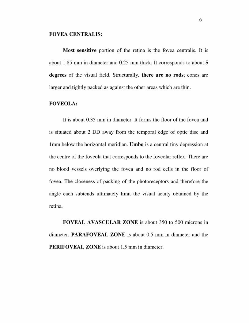
FOVEA CENTRALIS:
Most sensitive portion of the retina is the fovea centralis. It is
about 1.85 mm in diameter and 0.25 mm thick. It corresponds to about 5
degrees of the visual field. Structurally, there are no rods; cones are
larger and tightly packed as against the other areas which are thin.
FOVEOLA:
It is about 0.35 mm in diameter. It forms the floor of the fovea and
is situated about 2 DD away from the temporal edge of optic disc and
1mm below the horizontal meridian. Umbo is a central tiny depression at
the centre of the foveola that corresponds to the foveolar reflex. There are
no blood vessels overlying the fovea and no rod cells in the floor of
fovea. The closeness of packing of the photoreceptors and therefore the
angle each subtends ultimately limit the visual acuity obtained by the
retina.
FOVEAL AVASCULAR ZONE is about 350 to 500 microns in
diameter. PARAFOVEAL ZONE is about 0.5 mm in diameter and the
PERIFOVEAL ZONE is about 1.5 mm in diameter.
7
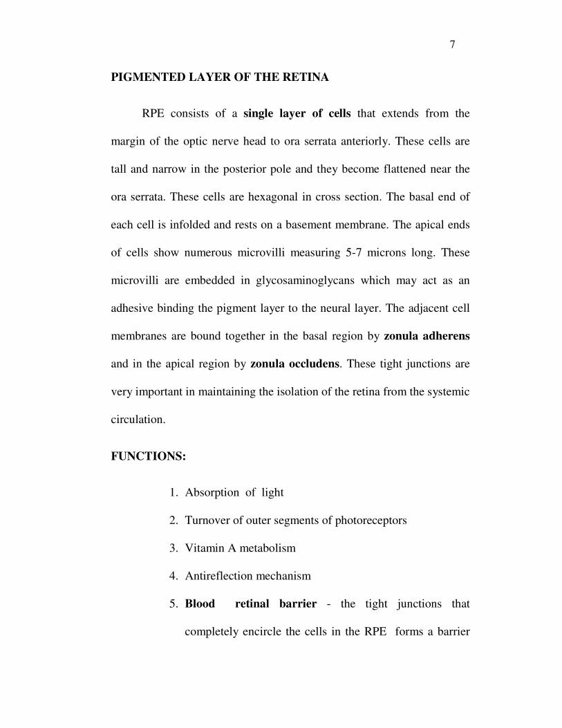
PIGMENTED LAYER OF THE RETINA
RPE consists of a single layer of cells that extends from the
margin of the optic nerve head to ora serrata anteriorly. These cells are
tall and narrow in the posterior pole and they become flattened near the
ora serrata. These cells are hexagonal in cross section. The basal end of
each cell is infolded and rests on a basement membrane. The apical ends
of cells show numerous microvilli measuring 5-7 microns long. These
microvilli are embedded in glycosaminoglycans which may act as an
adhesive binding the pigment layer to the neural layer. The adjacent cell
membranes are bound together in the basal region by zonula adherens
and in the apical region by zonula occludens. These tight junctions are
very important in maintaining the isolation of the retina from the systemic
circulation.
FUNCTIONS:
1. Absorption of light
2. Turnover of outer segments of photoreceptors
3. Vitamin A metabolism
4. Antireflection mechanism
5. Blood retinal barrier - the tight junctions that
completely encircle the cells in the RPE forms a barrier
8
that limits the flow of ions and prevents diffusion of large
molecules from the choroid capillaries to the
photoreceptors of neural retina
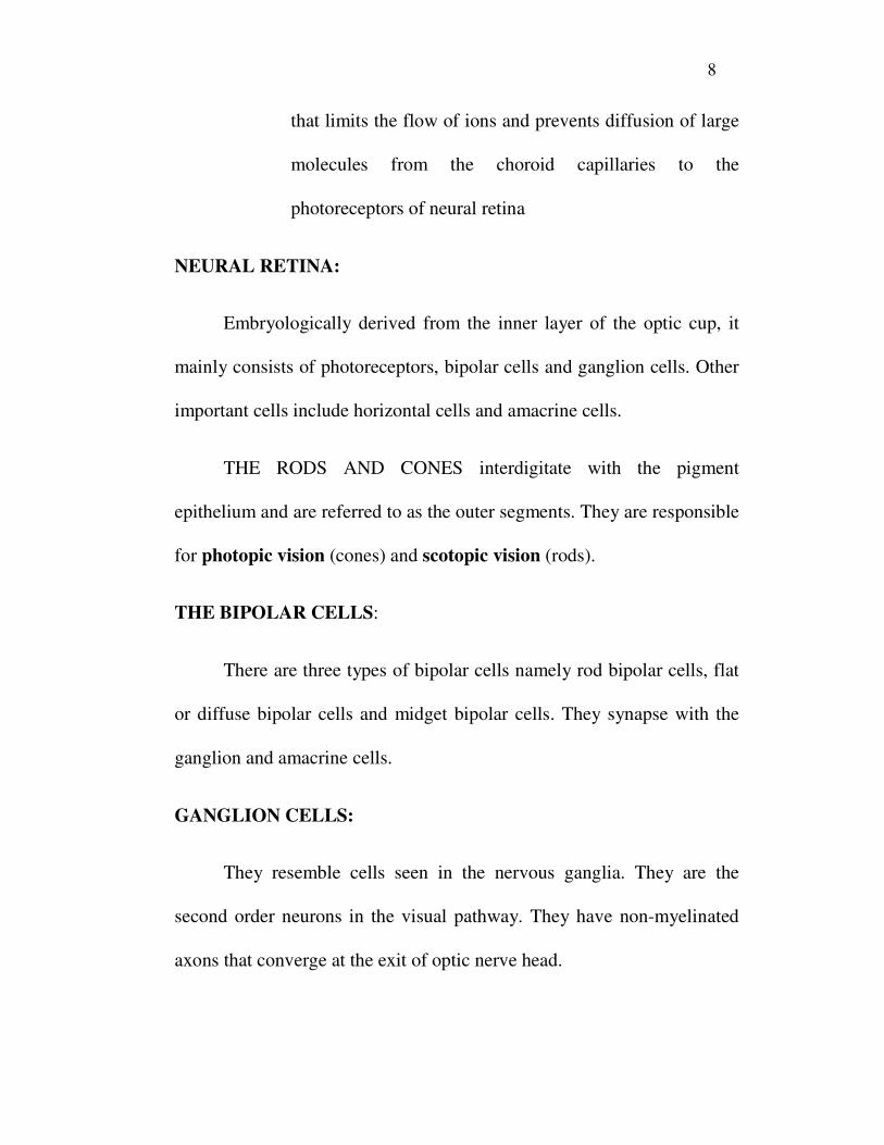
NEURAL RETINA:
Embryologically derived from the inner layer of the optic cup, it
mainly consists of photoreceptors, bipolar cells and ganglion cells. Other
important cells include horizontal cells and amacrine cells.
THE RODS AND CONES interdigitate with the pigment
epithelium and are referred to as the outer segments. They are responsible
for photopic vision (cones) and scotopic vision (rods).
THE BIPOLAR CELLS:
There are three types of bipolar cells namely rod bipolar cells, flat
or diffuse bipolar cells and midget bipolar cells. They synapse with the
ganglion and amacrine cells.
GANGLION CELLS:
They resemble cells seen in the nervous ganglia. They are the
second order neurons in the visual pathway. They have non-myelinated
axons that converge at the exit of optic nerve head.

9
The other cells include horizontal cells and amacrine cells.
Histologically, the retina consists of ten layers.
The pigment epithelium
1. The rods and cones
2. The external limiting membrane
3. The outer nuclear layer
4. The outer plexiform layer
5. The inner nuclear layer
6. The inner plexiform layer
7. The ganglion cells
8. The nerve fiber layer
9. The internal limiting membrane

10
ANATOMY OF THE MACULA
Fovea is the seat of central vision. In this area, there are no rods,
cones are larger, in abundance and tightly packed and the other areas of
retina are thin.
The foveola largely consists of cones and their nuclei covered by a
thin internal limiting membrane. All other retinal layers are very thin at
the foveolar region. In the foveolar region, the cone axons are obliquely
arranged to reach the margin of the fovea called the Henle s layer.
11
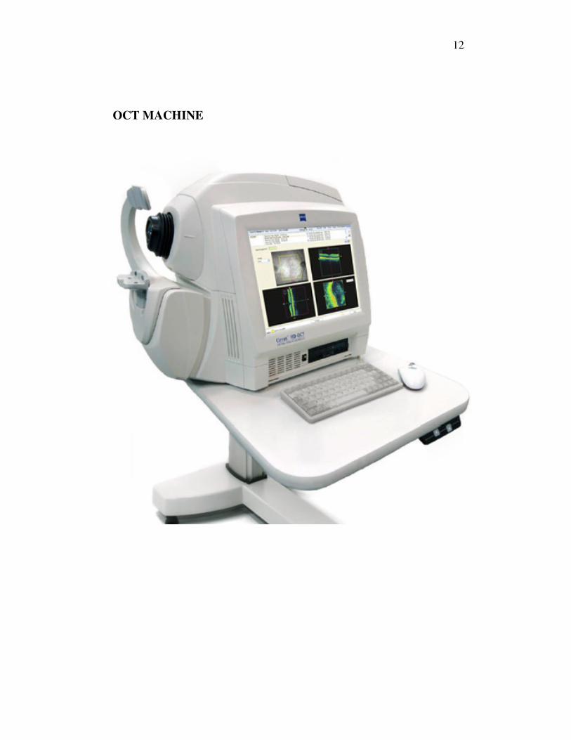
OPTICAL COHERENCE TOMOGRAPHY (OCT)
OCT is a non invasive non contact imaging system based on
interferometric optical tomographic evaluation of ocular tissues. It
produces micro resolution retinal images in vivo. It is analogous to B
scan except that it uses light instead of sound. Image resolutions to the
order of 1-15 microns have been achieved both in situ and real time.
There is an increased advantage of increased resolution and speed of
acquisition.
It is based on the principle of low coherence interferometry.
12
OCT MACHINE

13
The techniques used are
1. Time domain OCT: The path length of the reference arm is
translated longitudinally in time. The time delay of the
reflected or back scattered light from microstructures within
the tissue is used for evaluation and analysis.
2. Frequency domain OCT: The broad band interference is
acquired using spectrally spaced detectors. The depth scan is
obtained immediately using a fourier transform from the
acquired spectra without the movement of the reference arm.
The use of optical coherence tomography in diagnosing posterior
segment disorders is one of the most mature applications of OCT. OCT
has contributed to a better understanding of the pathogenesis of macular
holes, vitreomacular traction and has provided a quantitative method of
accurately detecting changes in the retinal thickness due to diabetes,
epiretinal membrane and cystoid macular edema.
The posterior boundary of the neurosensory retina is represented by
a highly reflective layer of retinal pigment epithelium and
choriocapillaris. The outer segments of photoreceptors are represented by
a dark area of minimal reflectivity just anterior to the highly reflective
14
band of RPE and chorio capillaris. The inner margin of the retina, the
nerve fibre layer is represented by a highly reflective red band due to
bright back scatter against the contrast of non reflective vitreous. The
intervening layers are represented by alternating areas of moderate to low
reflectivity.
Longitudinal surveillance of patients with macular disease requires
six radial scans centered at the fovea. Evaluation of the entire macular
region is possible using the retinal thickness map. Retinal thickness is
computed for 600 macular locations and then plotted on a false colour
topographic map.
Traditionally the diagnosis of macular holes is done using contact
lens slit lamp bio microscopy. There are a number of lesions like partial
or lamellar thickness macular holes, Pseudo holes, macular cysts that may
be difficult to distinguish from full thickness macular holes. All the above
mentioned disorders lack the characteristic full thickness defect with fluid
cuff and flask shaped appearance. OCT aids in identification and staging
of macular holes according to the GASS classification.

15
HISTORY OF MACULAR HOLES
Knapp and Noyes were the first to describe macular holes in the
late 1800s.
GASS first described a series of changes in the formation of
idiopathic macular holes. Epiretinal membrane was first described by
IWANOFF IN 1865.
TIMELINE IN THE EVOLUTION OF MACULAR HOLE
CONCEPTS
1. 1869 - KNAPP – First case description of macular hole (traumatic)
2. 1871 – NOYES – First detailed clinical description of macular
hole (traumatic)
3. 1900 – KUHNT – Atraumatic theories of cystic retinal
degeneration leading to macular hole.
4. 1901 – FUCHS AND 1907 – COATS – Early histopathologic
changes in macular hole including cystic changes.
5. 1912 – ZEEMAN – Histopathologic recognition of premacular
vitreous condensation.
6. 1924- LISTER – Vitreous forces and traction bands may cause
premacular holes.
16
7. 1967 – REESE ET AL – Vitreous separation critical to macular
hole formation.
8. 1982 – MC DONNELL ET AL – Possible female hormonal
influence on vitreous separation and macular hole formation.
9. 1983 – AVILA ET AL - Vitreous separation not necessary in the
formation of macular hole.
10. 1986 – MORGAN AND SCHATZ – Involutional macular
thinning is a premacular hole condition.
11. 1988 – GASS – Tangential traction and Gass biomicroscopic
classification of premacular and macular holes.
12. 1991 – KELLY AND WENDEL – Successful demonstration of
vitrectomy, epiretinal membrane removal and strict face down gas
tamponade could successfully treat macular holes.
13. 1995 – GASS – Centrifugal displacement of retinal receptors with
umbo dehiscence. Reappraisal of bio microscopic classification of
premacular and macular holes.

17
ETIOLOGY OF MACULAR HOLE
I. PRIMARY:
Idiopathic (70-80 %)
• Occurs due to aging unrelated to any ocular or any other
antecedent events.
II. SECONDARY:
1. Trauma
2. Hypertensive retinopathy
3. Associated with Cystoid macular oedema -
(inflammation, retinal vascular disease , macular pucker)
4. Secondary to Retinal detachment
5. Extreme myopia
6. Post LASER therapy
7. Lightning strike

18
PATHOPHYSIOLOGY OF MACULAR HOLE
Vitreo - macular traction from posterior vitreous surface is the
main event in the creation of macular hole. Antero-posterior trans-vitreal
traction by vitreous fibres extending to the vitreous base may lead to
pathologic changes that lead to full thickness macular hole. Gass and
Johnson had proposed a theory whereby shrinkage of adjacent cortical
vitreous and subsequent tangential vitreous traction first causes a
circumscribed foveolar dehiscence followed by retinal dehiscence. Then
subsequently vitreofoveal separation occurs and finally complete
posterior vitreous detachment with enlargement of the macular hole.
Guyer and Green later proposed three theories for the formation of
idiopathic senile macular hole.
1. Fluid movements and counter currents
2. Cellular remodelling of the cortical vitreous
3. Contraction of a cellular membrane on the inner surface of
the tapered cortical vitreous.
Proliferation of fibrous astrocytes and muellers cells occurs with
the formation of macular hole. This reparative tissue was previously
interpreted as operculum. Full thickness macular holes have previously
19
been documented to arise in eyes with complete PVD. So pathogenesis
other than tangential traction is likely in the formation and progression of
senile macular holes.
Hydrodynamic model of macular hole states that a macular hole
is formed or maintained by fluid flow caused by the macular retinal
pigment epithelial pump.
In reality the aetiology and pathophysiology of macular hole may
be multifactorial. Recognising the primary event probably is less
important than considering factors like vitreo-macular traction, foveolar
dehiscence etc. which can change the management and decision making.

20
PATHOGENESIS OF MACULAR HOLE

21
A. TRAUMATIC THEORY
Traumatic theory was put forth by Knapp in 1869, when he
published the first case description of a macular hole in a patient with
ocular trauma and an initial diagnosis of a macular hemorrhage. Most
other early observers attributed macular holes to ocular trauma.
Noyes published the first accurate case report and a detailed
ophthalmoscopic description of macular hole, which was secondary to
blunt trauma. He noted the difference in depth of focus from the retinal
surface to the base of the lesion and probably was the first to recognize
that the hallmark of the lesion was a full-thickness defect in retinal tissue
within the center of the macula.
In 1900 Ogilvie compiled the first case series in holes at the
macula, and proposed terminology including macular hole, as well as
floor and edge of the macular hole. Many of the first reported cases of
macular holes were in young patients, and trauma was estimated to
account for as many as 50%.
The majority of traumatic macular holes occur in men, whereas it
is now known that the majority of age related macular holes occur in
women.

22
B. CYSTOID DEGENERATION THEORY
Full-thickness and lamellar macular holes were first described by
Fuchs (1901) and Coats (1907).
Coats noted cystic intra-retinal changes adjacent to the macular
hole and concluded that these changes could be caused by trauma as well
as other mechanisms. In some cases of trauma in which there was not
immediate macular hole formation, trauma was believed to cause reactive
vasoconstriction followed by vasodilation, thus leading to cystic
degeneration of the central macula.
Blunt ocular trauma could cause immediate macular hole formation
from mechanical energy created by vitreous fluid waves and contrecoup
macular necrosis or macular laceration. Indirect ocular trauma had also
been reported to cause macular hole formation.
Recognizing that cystoid degeneration was not only due to
posttraumatic macular sequelae, Kuhnt concluded that macular holes
were caused by cystoid degeneration in the macula, not necessarily
related to trauma.
Aaberg in 1970 found that only 9% of eyes with a macular hole
were associated with trauma, compared with an earlier report of 50%. He
described macular hole and cystoid oedema in association with a variety
23
of conditions, including severe hypertension, central retinal artery
occlusion, retinal venous occlusive disease, Coats’ disease, syphilis, solar
maculopathy, arc welding maculopathy, electrocution, and vitreous
traction
C. VASCULAR THEORY
Although trauma was once believed to be the primary or sole cause
of macular holes, it was probably the histopathologic and clinical
observations of cystoid degeneration in the surrounding tissue of macular
holes that led to considerations of atraumatic causes and, in particular, the
vascular theory of pathogenesis. Coats and Kuhnt together believed that
aging-related changes of the retinal vasculature led to cystoid
degeneration and subsequent macular hole formation
D. VITREOUS THEORY
Avila and Jalkh, noted vitreoretinal traction arising from the
vitreous base and concluded that persistent vitreous-to-macula adhesions
were important in the pathogenesis of macular holes. Other proponents of
a vitreous theory emphasized that the process of vitreous separation from
the macula was the critical event in the pathogenesis of a macular hole. It
is reported that macular holes can develop despite having a pre-existing
complete posterior vitreous separation.

24
E. INVOLUTIONAL MACULAR THINNING
Morgan and Schatz proposed a mechanism that they described as
involutional macular thinning, incorporating vitreous, vascular, and cystic
degeneration theories. The macular lesion was described as “thin, mildly
atrophic fovea that has lost its normal architecture and appearance.” The
foveal lesion was a subtle abnormal depression with variably associated
retinal cystic changes or a surrounding yellow ring.

25
CLASSIFICATION OF MACULAR HOLES
STAGE – 1 MACULAR HOLES :
(Pre-macular holes, macular cysts, Involutional macular thinning)
With a unilateral stage 1 macular hole, the patient typically is
asymptomatic with both eyes open. For this reason and because they are
evanescent lesions, stage 1 macular holes are not observed commonly and
their diagnosis can be difficult. When symptoms are present, they consist
of painless metamorphopsia or decreased vision or both.
In a stage 1 macular hole, no true neural retinal defect is present.
The photoreceptor layer is believed to be intact, and no vitreo-foveal
separation has occurred. Oblique vitreous traction on the fovea is

26
believed to be the inciting event and can typically be observed on OCT.
Stage 1 holes are further divided into Stage 1a and Stage 1b, based on
clinical appearance. In a stage 1a macular hole, a small central yellow
spot is seen on ophthalmoscopy (Foveolar detachment). The fovea may
be thickened along with a loss of the normal foveal contour. In a stage 1b
macular hole, a yellow ring is visible in the foveal area (foveal
detachment).
Foveal Pseudocyst
OCT studies conclude that a stage 1a macular hole actually
represents a cystic change within the fovea, rather than a true
photoreceptor detachment from the retinal pigment epithelium (foveal
pseudo-cyst).

27
In a stage 1b macular hole, the cyst-like space is accompanied by a
foveal detachment that coalesces to a point just short of actual
dehiscence. Stage 1 holes spontaneously resolve in about 50% of eyes
with no visual sequelae. The worse the initial visual acuity, the less likely
is spontaneous resolution.

28
STAGE - 2 : MACULAR HOLES:
When peri-foveal vitreous cortex shrinks, a stage 1 hole advances
to a stage 2 hole. Stage 2 holes have a small (100–300 µm), full-
thickness neural retinal defect, either centrally or eccentrically. The
defect can be round, oval, crescentic, or horseshoe shaped. The visual
acuity typically is diminished and a pseudo-operculum, which represents
condensed vitreous, may overlie the hole. It is believed that once a stage
2 hole occurs, it nearly always progresses to stage 3, with little hope for
spontaneous visual improvement. The visual acuity with a stage 2 hole
varies between 20/50 (6/15) and 20/400 (6/120).
Pseudo operculum
29
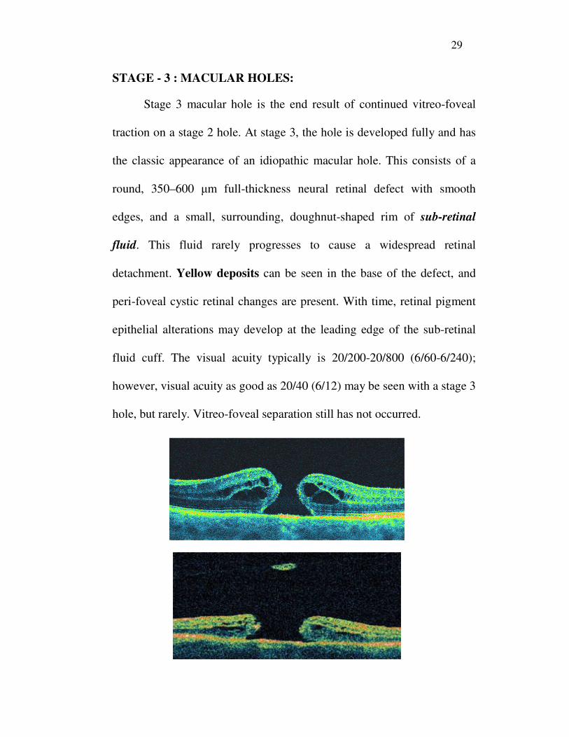
STAGE - 3 : MACULAR HOLES:
Stage 3 macular hole is the end result of continued vitreo-foveal
traction on a stage 2 hole. At stage 3, the hole is developed fully and has
the classic appearance of an idiopathic macular hole. This consists of a
round, 350–600 µm full-thickness neural retinal defect with smooth
edges, and a small, surrounding, doughnut-shaped rim of sub-retinal
fluid. This fluid rarely progresses to cause a widespread retinal
detachment. Yellow deposits can be seen in the base of the defect, and
peri-foveal cystic retinal changes are present. With time, retinal pigment
epithelial alterations may develop at the leading edge of the sub-retinal
fluid cuff. The visual acuity typically is 20/200-20/800 (6/60-6/240);
however, visual acuity as good as 20/40 (6/12) may be seen with a stage 3
hole, but rarely. Vitreo-foveal separation still has not occurred.

30
STAGE - 4 : MACULAR HOLE:
A stage 4 macular hole has all the features of a stage 3 hole, but
with complete posterior separation of the vitreous from the fovea.
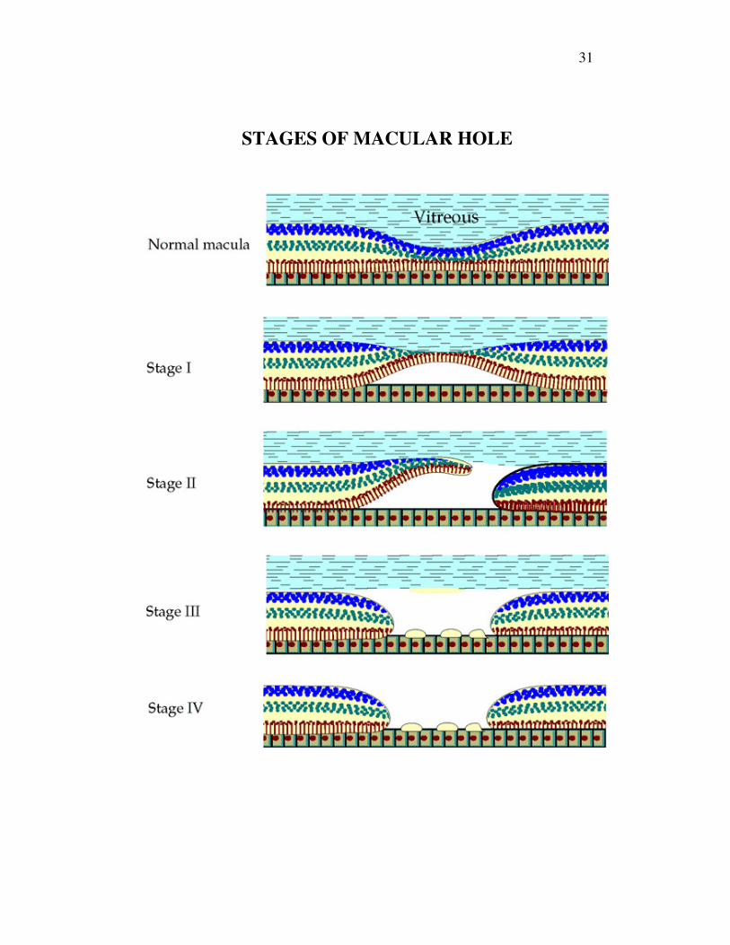
31
STAGES OF MACULAR HOLE
32
DIAGNOSIS OF A MACULAR HOLE
I. CLINICAL SYMPTOMS
II. CLINICAL SIGNS
III. FLOURESCEIN ANGIOGRAPHY
IV. OPTICAL COHERENCE TOMOGRAPHY
I. CLINICAL SYMPTOMS:
Patients usually experience visual symptoms like
metamorphopsia, loss of central vision and a central scotoma. The
visual acuity of the patient varies with the size, duration and location
and stage of the macular hole.
In patients with impending macular holes, central visual acuity
may be reduced to the range of 20/25 to 20/50. In fully developed
macular holes, central visual acuity may be reduced to the range of 20/80
to 20/200 .The central visual acuity may be correlated to the size of
neurosensory detachment surrounding the macular hole. Both the hole
size and neurosensory detachment size correlates with the duration of
symptoms.
Significant visual improvement following spontaneous closure of
macular holes is very rare.
33
II. CLINICAL SIGNS:
1. Watzke Allen test:
With the use of a thin slit beam during bio-microscopy, an absolute
scotoma can appear to the patient as a break in the beam when it is
centered over large holes (Watzke allen sign). If centered over smaller
holes, or over the surround ding neuro-sensory retinal detachment in
larger holes , only narrowing or distortion of the beam is experienced by
the patients.
2. Laser aiming beam test:
For detecting the absolute scotoma associated with smaller defects,
50 microns argon laser aiming beam is directed at the macular hole.
Disappearance of the aiming beam is noted in patients with true full
thickness macular hole, whereas in patients with epi-retinal membrane
or pseudo-macular hole, the laser aiming beam does not disappear.
3. Macular micro perimetry :
Here patient is tested in a similar fashion as a static or kinetic
perimetry. The absolute and relative scotomas are mapped directly on to
the retinal surface. This technique has demonstrated that visual loss in

34
eyes with macular holes is due to absence of retinal function and
reduction in function in the surrounding areas.
4. Flourescein angiography:
To rule out masquerading lesions as seen in cystoid macular
oedema or exudative maculopathy, flourescein angiography may be
helpful.
35
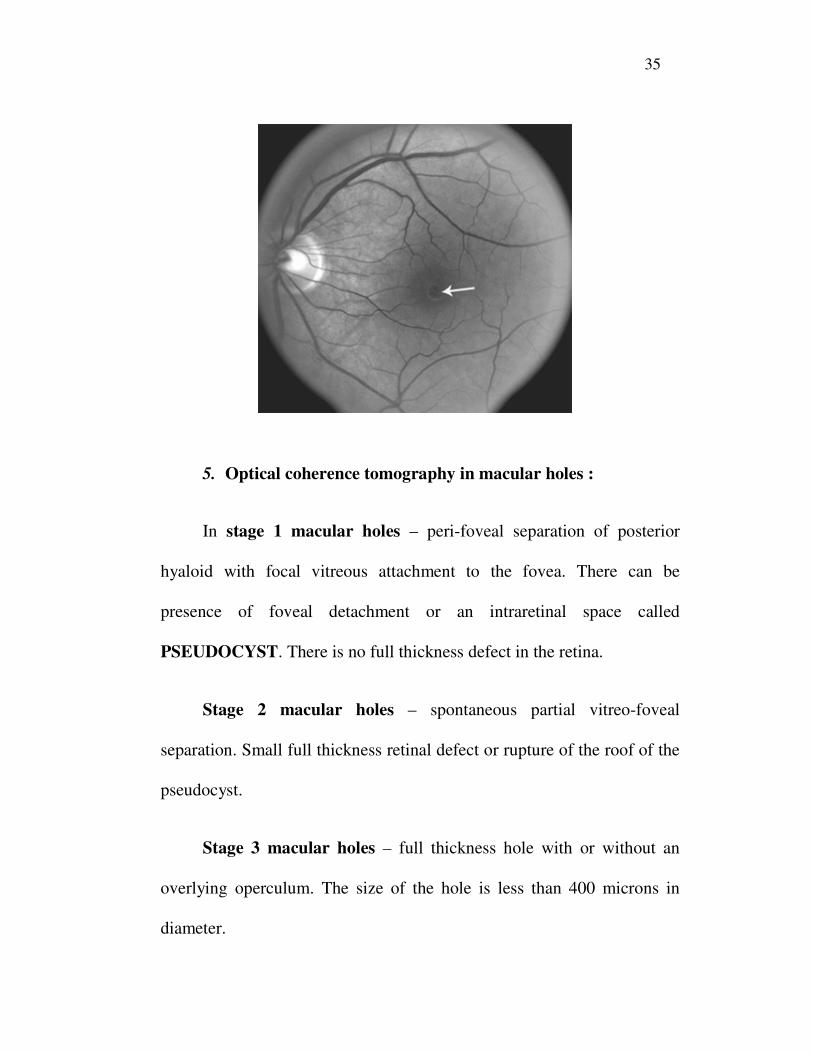
5. Optical coherence tomography in macular holes :
In stage 1 macular holes – peri-foveal separation of posterior
hyaloid with focal vitreous attachment to the fovea. There can be
presence of foveal detachment or an intraretinal space called
PSEUDOCYST. There is no full thickness defect in the retina.
Stage 2 macular holes – spontaneous partial vitreo-foveal
separation. Small full thickness retinal defect or rupture of the roof of the
pseudocyst.
Stage 3 macular holes – full thickness hole with or without an
overlying operculum. The size of the hole is less than 400 microns in
diameter.

36
Stage 4 macular holes - full thickness hole with a complete
posterior vitreous separation, the size of the hole being more than 400
microns in diameter.
PROGNOSIS:
Optical coherence tomography can also be used for the
prognostication of macular hole.
Macular hole index :
The macular hole index is the ratio of the hole height to the basal
diameter of the hole. The higher the macular hole index, the better the
visual results. If the diameter of the macular hole is more it is a bad sign.
Diameter hole index and traction hole index and the minimum
diameter of the macular hole are also used to prognosticate the results
after macular hole. A minimum diameter of less than 311 microns and a
traction hole index of more than 1.41 have been associated with a better
prognosis .
37
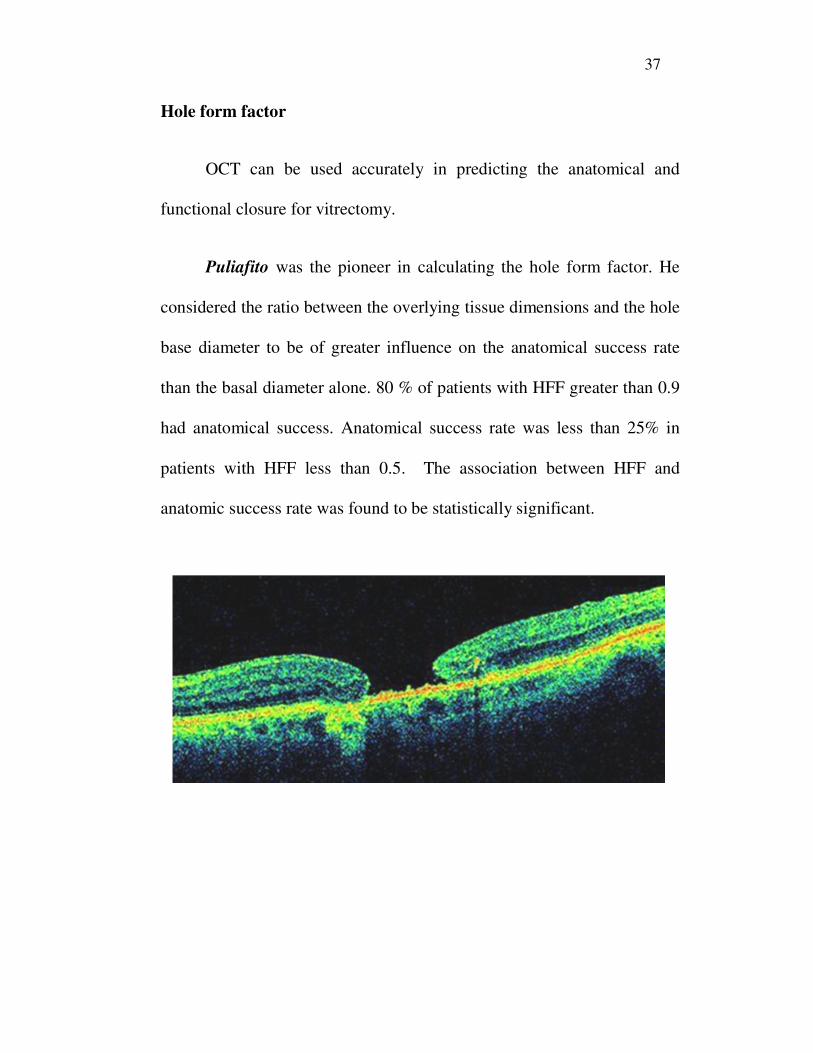
Hole form factor
OCT can be used accurately in predicting the anatomical and
functional closure for vitrectomy.
Puliafito was the pioneer in calculating the hole form factor. He
considered the ratio between the overlying tissue dimensions and the hole
base diameter to be of greater influence on the anatomical success rate
than the basal diameter alone. 80 % of patients with HFF greater than 0.9
had anatomical success. Anatomical success rate was less than 25% in
patients with HFF less than 0.5. The association between HFF and
anatomic success rate was found to be statistically significant.
38
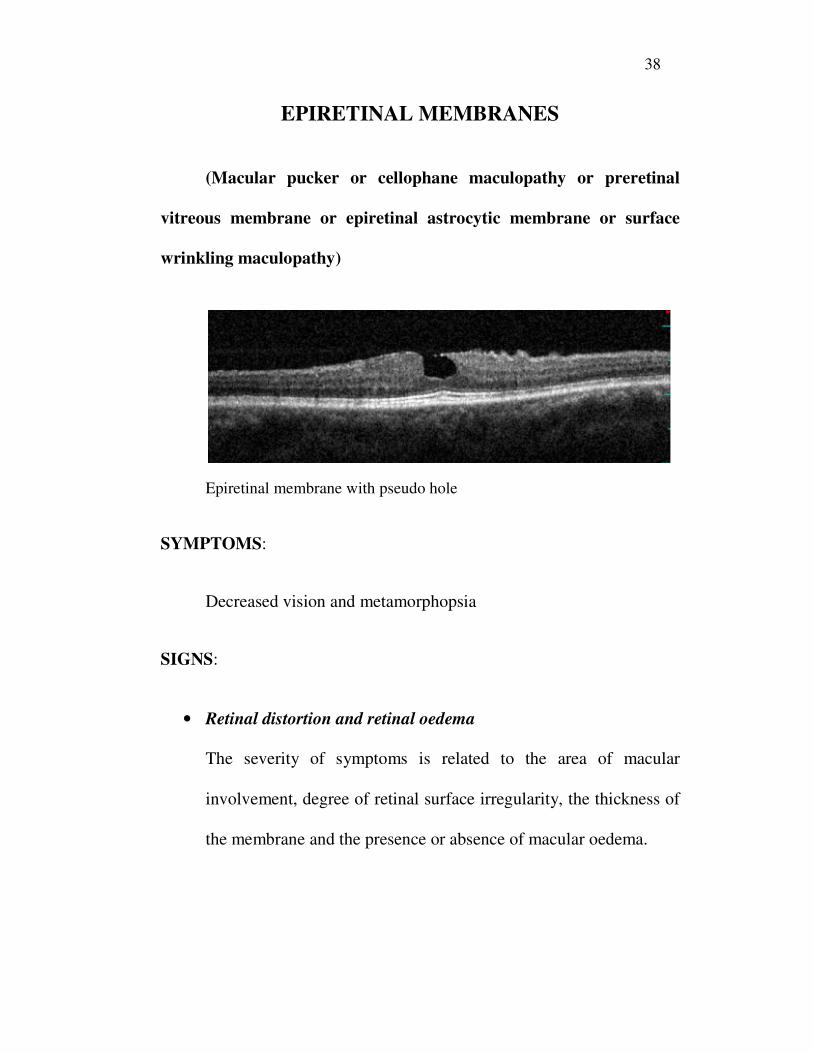
EPIRETINAL MEMBRANES
(Macular pucker or cellophane maculopathy or preretinal
vitreous membrane or epiretinal astrocytic membrane or surface
wrinkling maculopathy)
Epiretinal membrane with pseudo hole
SYMPTOMS:
Decreased vision and metamorphopsia
SIGNS:
• Retinal distortion and retinal oedema
The severity of symptoms is related to the area of macular
involvement, degree of retinal surface irregularity, the thickness of
the membrane and the presence or absence of macular oedema.
39
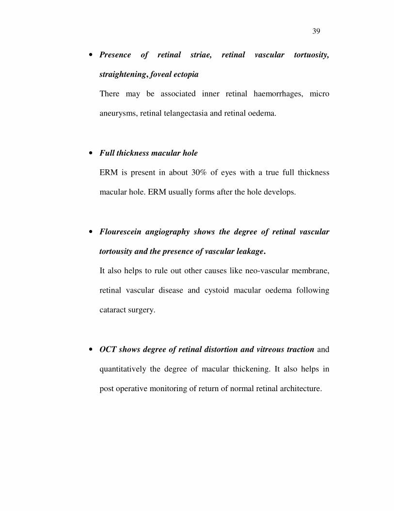
• Presence of retinal striae, retinal vascular tortuosity,
straightening, foveal ectopia
There may be associated inner retinal haemorrhages, micro
aneurysms, retinal telangectasia and retinal oedema.
• Full thickness macular hole
ERM is present in about 30% of eyes with a true full thickness
macular hole. ERM usually forms after the hole develops.
• Flourescein angiography shows the degree of retinal vascular
tortousity and the presence of vascular leakage.
It also helps to rule out other causes like neo-vascular membrane,
retinal vascular disease and cystoid macular oedema following
cataract surgery.
• OCT shows degree of retinal distortion and vitreous traction and
quantitatively the degree of macular thickening. It also helps in
post operative monitoring of return of normal retinal architecture.

40
GRADES OF EPIRETINAL MEMBRANE:
Grade 1: Presence of a cellophane membrane
Wrinkling of inner retina
No edge or ERM elevation
Grade 2: The edge of ERM is elevated
Full thickness retinal distortion
Less than half of the ERM is opaque
Grade 3: Thick membrane with more than half of ERM being opaque
Marked distortion of the retinal architecture
Obstruction of the underlying retinal architecture
41
DIFFERENTIAL DIAGNOSIS
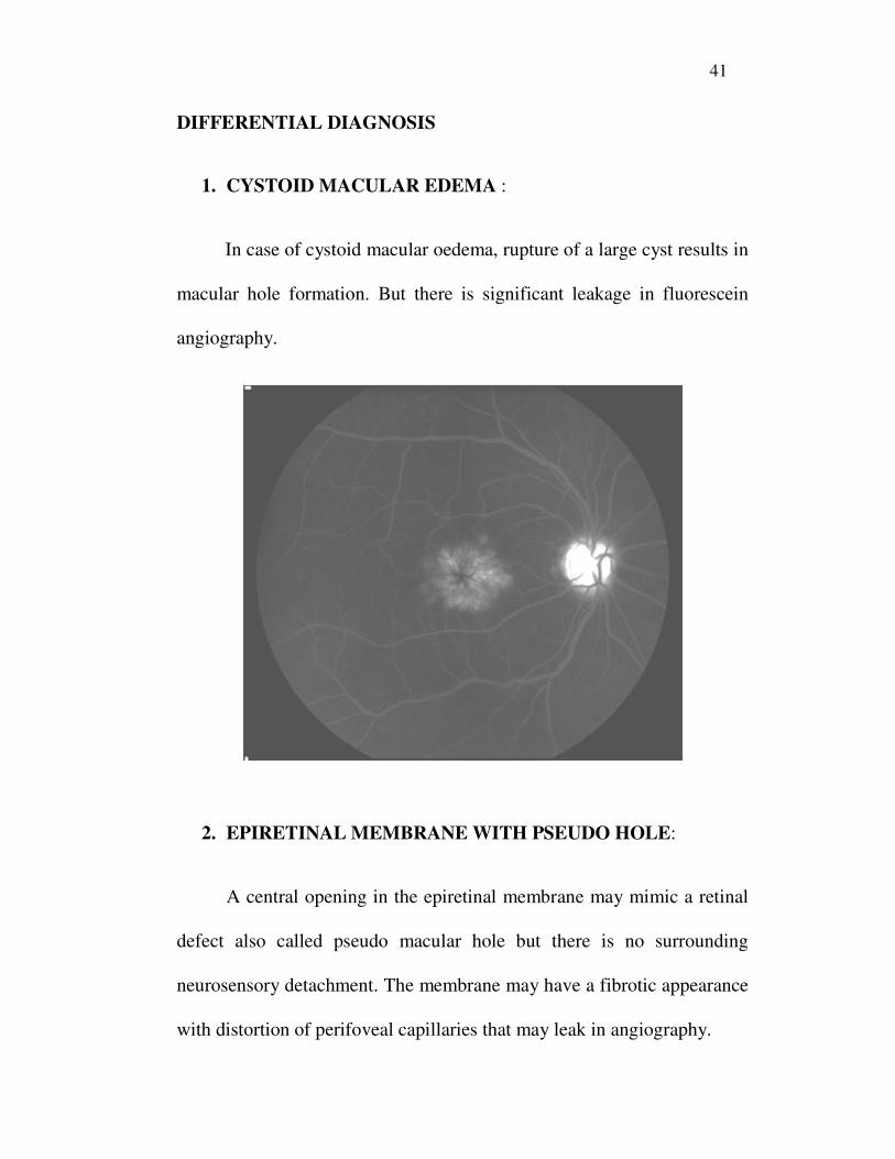
1. CYSTOID MACULAR EDEMA :
In case of cystoid macular oedema, rupture of a large cyst results in
macular hole formation. But there is significant leakage in fluorescein
angiography.
2. EPIRETINAL MEMBRANE WITH PSEUDO HOLE:
A central opening in the epiretinal membrane may mimic a retinal
defect also called pseudo macular hole but there is no surrounding
neurosensory detachment. The membrane may have a fibrotic appearance
with distortion of perifoveal capillaries that may leak in angiography.
42
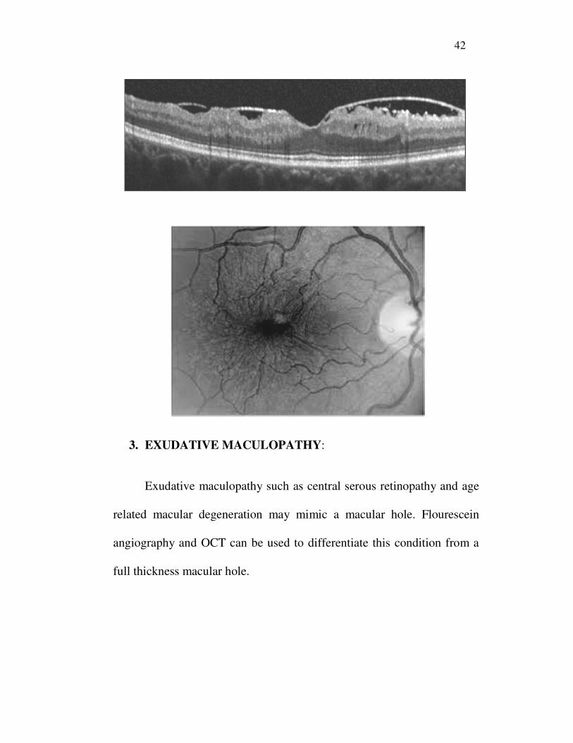
3. EXUDATIVE MACULOPATHY:
Exudative maculopathy such as central serous retinopathy and age
related macular degeneration may mimic a macular hole. Flourescein
angiography and OCT can be used to differentiate this condition from a
full thickness macular hole.
43
4. LAMELLAR MACULAR HOLE:
Some form of aborted macular hole, OCT will demonstrate the
absence of full thickness defect, possibly with vitreofoveal separation and
a pseudo operculum or epiretinal membrane.
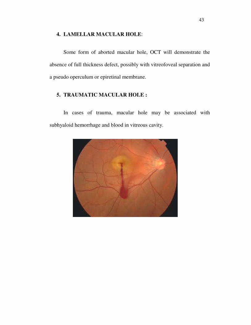
5. TRAUMATIC MACULAR HOLE :
In cases of trauma, macular hole may be associated with
subhyaloid hemorrhage and blood in vitreous cavity.
44
TREATMENT MODALITIES
INDICATIONS FOR TREATMENT:
1. Stage 3 and 4 macular holes
2. Definite symptomatic full thickness macular holes with visual
acuity in the range of 20/40 to 20/60
3. Epiretinal membrane causing trampoline traction , foveal ectopia ,
retinal vascular leakage and macular oedema
4. Epiretinal membranes causing diplopia or debilitating
metamorphopsia or severe visual decline
GENERAL SURGICAL PRINCIPLES
• Surgery performed using three port pars plana vitrectomy ,
including a separate continous infusion cannula, endoilluminator,
suction cannula and other instruments.
• After removal of the central vitreous, the posterior cortical
vitreous is identified and separated from the retinal surface.
45
• When the suction is applied to the silicone tipped cannula, the
orifice becomes occluded by the vitreous as the cannula is close to
the retinal surface called “fish strike sign”
• If posterior vitreous detachment is not present, create one by using
active suction close to the optic disc in a posteroanterior direction
• Vitrectomy has to be completed. Thorough vitrectomy can be
verified using intravitreal triamcinolone.
• Epiretinal membrane and ILM PEELING can be done at this stage
• This procedure is done using a small gauge retinal pick or a barbed
myringotomy blade.
• The ILM is removed in a circular fashion called maculorhexis
• Staining of the ILM and epiretinal membrane can be done using
dyes like ICG and brilliant blue in an air/fluid filled eye for easy
identification.

46
Staining of ILM
• ICG stains only the acellular ILM but does not stain the epiretinal
membrane. So preferential staining can be done. But this dye is
toxic to the retina. Hence not preferred
• Trypan blue and triamcinolone acetonide are alternatives to ICG.
Pre and post operative OCT images of macular hole

47
SURGICAL ADJUNCTIVES
• Intravitreal transforming growth factor
It was reported to have dose dependent efficacy and higher
anatomic success rate in eyes undergoing macular hole surgery.
• Autologous blood serum
Blood serum and concentrated platelets are said to have higher
anatomic success rate in eyes with idiopathic macular hole.
• Adjunctive laser photocoagulation
Laser photocoagulation directed only to the RPE in the base of
macular hole along with fluid gas exchange has been suggested as a
treatment to persistent macular hole following vitrectomy
• Pharmacologic PVD:
OCRIPLASMIN is a newer drug that has been used to create
pharmacologic PVD. It has been suggested as a treatment for macular
hole.
48
• Tamponade and post op positioning:
Intravitreal gas tamponade and post op face down positioning are
necessary. Previously non expansile concentrations of SF6 were used but
required post op face down positioning. In order to reduce the
dependence of surgical outcomes on prone face positioning, silicone oil is
used.
Complications of surgery
1. Iatrogenic retinal breaks
2. Intraoperative light toxicity
3. Post operative exudative detachments
4. Subretinal fibrosis
5. Epiretinal membrane formation
49
AIM OF THE STUDY
• To evaluate the incidence, prevalence and correlation of epiretinal
membranes surrounding idiopathic macular holes (stage 2/3/4)
• To find out the correlation between epiretinal membrane and visual
acuity in idiopathic macular holes
• To find the correlation between size of the macular holes and
epiretinal membrane.
• To assess the increase in severity of epiretinal membranes during
follow up.

50
INCLUSION CRITERIA
• All patients with symptoms of decreased central vision and
metamorphopsia.
• All patients presenting with complaints of central scotoma and
distortion of images.
• All patients with clinically proven idiopathic macular hole.
• All patients with OCT proven idiopathic macular hole.
EXCLUSION CRITERIA
• All patients who have previous history of trauma and an identified
aetiology of macular hole to be trauma.
• Other coexisting causes for macular hole like cystoid macular
edema and lamellar macular hole and pseudohole.
51
MATERIALS AND METHODS
The study was conducted at the Regional institute of
ophthalmology and Govt ophthalmic hospital, Chennai between Sep.2012
- Oct 2013.
Retrospective study design was followed.
50 eyes of 50 patients were included in the study. The patients
underwent a detailed history including their past and present illness,
relevant ocular history including any surgery, trauma, usage of any
topical medications for any ocular complaints etc.
Uncorrected visual acuity was assessed using Snellens visual
acuity chart and best corrected visual acuity was assessed using
Autorefractometer. Anterior segment including the anterior vitreous
face was examined using slit lamp examination techniques. Fundus
examination was performed using direct and indirect ophthalmoscopes.
Slit lamp Biomicroscopy using 90 D lenses was used to get a

52
stereoscopic view of the fundus. The findings were recorded using colour
fundus photography.
Optical coherence tomography was then performed in all patients
with the above findings. These patients were followed up and reviewed at
three months, six months and one year and the same tests were repeated
and observations recorded. Results were analysed using standard
statistical methods and conclusions derived.

53
OBSERVATIONS AND ANALYSIS
The study was done among 50 patients with idiopathic macular
hole (n=50)
30 patients included in this study were phakic and 20 were
pseudophakic.
PSEUDOPHAKICS vs PHAKICS WITH
IDIOPATHIC MACULAR HOLE
phakics
pseudophakic
54
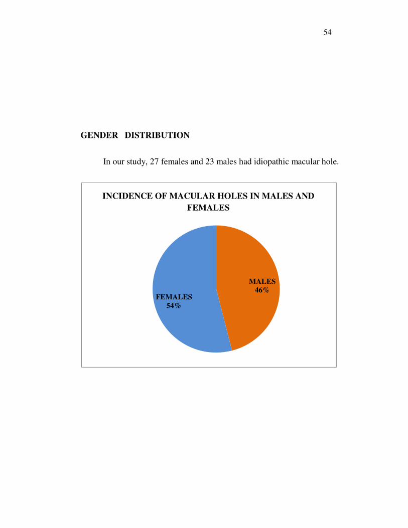
GENDER DISTRIBUTION
In our study, 27 females and 23 males had idiopathic macular hole.
MALES
46%FEMALES
54%
INCIDENCE OF MACULAR HOLES IN MALES AND
FEMALES

55
GRADING OF MACULAR HOLES
MACULAR HOLE GRADE 2 GRADE 3 GRADE 4
n 19 23 8
Number of patients with stage 2 macular holes -- 19
Number of patients with stage 3 macular holes -- 23
Number of patients with stage 4 macular holes -- 8

56
38%
46%
16%
GRADING OF MACULAR HOLES
stage 2 stage 3 stage4

57
ANALYSIS OF VARIOUS GRADES OF MACULAR HOLE
MACULAR HOLE GRADE 2 GRADE 3 GRADE 4
N 19 23 8
INITIAL MEAN V/A
5/60 NIP to
6/60 NIP
3/60 NIP to
4/60 NIP
1/60 NIP to
2/60 NIP
FOLLOW UP MEAN
V/A
5/60 NIP 4/60 NIP 1/60 NIP
ERM 4 13 6
58
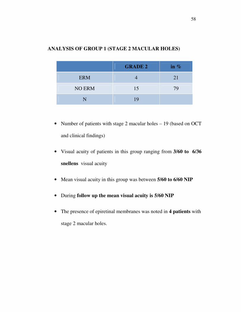
ANALYSIS OF GROUP 1 (STAGE 2 MACULAR HOLES)
GRADE 2 in %
ERM 4 21
NO ERM 15 79
N 19
• Number of patients with stage 2 macular holes – 19 (based on OCT
and clinical findings)
• Visual acuity of patients in this group ranging from 3/60 to 6/36
snellens visual acuity
• Mean visual acuity in this group was between 5/60 to 6/60 NIP
• During follow up the mean visual acuity is 5/60 NIP
• The presence of epiretinal membranes was noted in 4 patients with
stage 2 macular holes.

59
• Percentage of stage – 2 macular holes with ERM
21%
79%
ERM NO ERM

60
• Amongst those patients who had epiretinal membrane 2 were
pseudophakic and 2 were phakic.
0
0.2
0.4
0.6
0.8
1
1.2
1.4
1.6
1.8
2
pseudophakes phakics
GRADE 2 MACULAR HOLE WITH ERM
GRADE 2

61
ANALYSIS OF GROUP 2 ( STAGE 3 MACULAR HOLES)
Column1 GRADE 3 in %
ERM
13 57
NO ERM
10 43
n
23

62
• Number of patients with stage 3 macular holes – 23(n)
• Visual acuity at presentation in this group ranged from 1/60 to
5/60 NIP
• The mean visual acuity in this group was between 3/60 to 4/60
NIP
• The mean visual acuity during follow up in this group at the end of
one year is 4/60 NIP
• The presence of EPIRETINAL MEMBRANES is noted in 13
patients
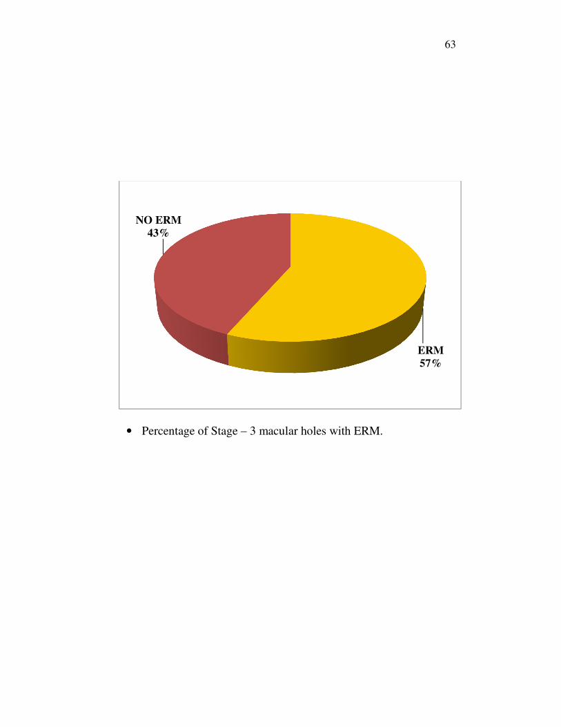
63
• Percentage of Stage – 3 macular holes with ERM.
ERM
57%
NO ERM
43%
64
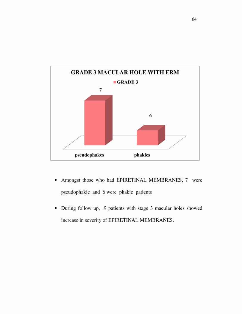
• Amongst those who had EPIRETINAL MEMBRANES, 7 were
pseudophakic and 6 were phakic patients
• During follow up, 9 patients with stage 3 macular holes showed
increase in severity of EPIRETINAL MEMBRANES.
pseudophakes phakics
7
6
GRADE 3 MACULAR HOLE WITH ERM
GRADE 3
65
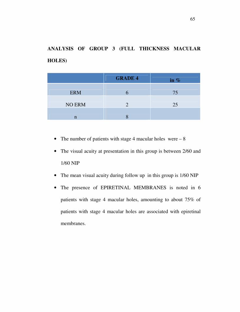
ANALYSIS OF GROUP 3 (FULL THICKNESS MACULAR
HOLES)
GRADE 4 in %
ERM
6 75
NO ERM
2 25
n
8
• The number of patients with stage 4 macular holes were – 8
• The visual acuity at presentation in this group is between 2/60 and
1/60 NIP
• The mean visual acuity during follow up in this group is 1/60 NIP
• The presence of EPIRETINAL MEMBRANES is noted in 6
patients with stage 4 macular holes, amounting to about 75% of
patients with stage 4 macular holes are associated with epiretinal
membranes.
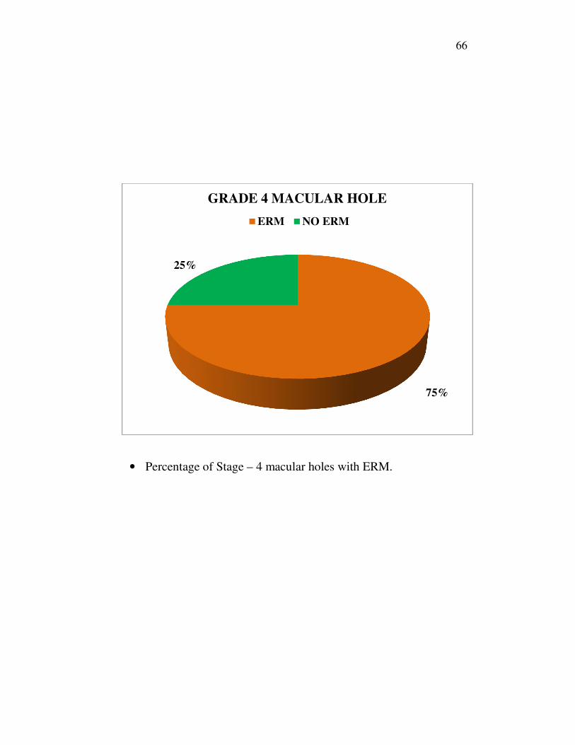
66
• Percentage of Stage – 4 macular holes with ERM.
75%
25%
GRADE 4 MACULAR HOLE
ERM NO ERM

67
• All the 6 patients in this ERM group were found to be
pseudophakics in our study.
0
1
2
3
4
5
6
pseudophakes phakics
GRADE 4 MACULAR HOLE WITH ERM
GRADE 4

68
ANALYSIS OF ERM AMONG PSEUDOPHAKICS AND PHAKICS
• Among the 20 pseudophakics, 14 patients (70 %) had epiretinal
membrane
NO ERM
30 %
ERM
70 %
PSEUDOPHAKICS WITH ERM (n=20)
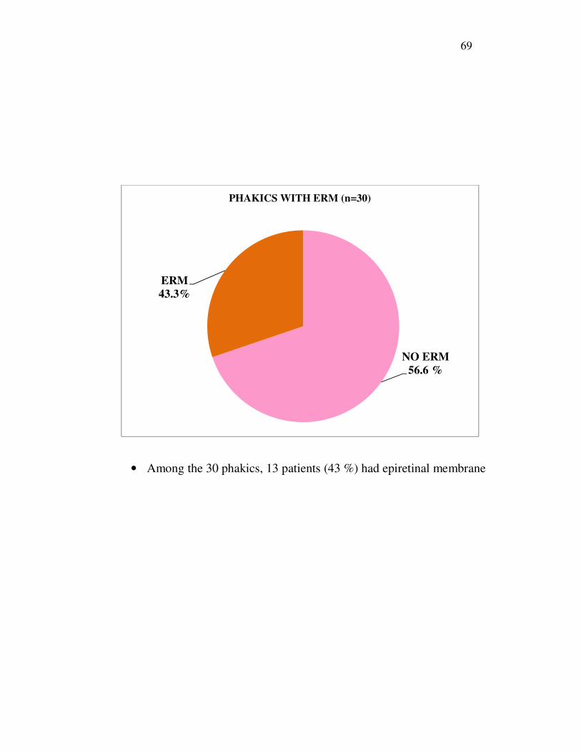
69
• Among the 30 phakics, 13 patients (43 %) had epiretinal membrane
NO ERM
56.6 %
ERM
43.3%
PHAKICS WITH ERM (n=30)

70
VISUAL ACUITY VS ERM
V/A ≤ 1/60 2/60 3/60 4/60 5/60 6/60
No. of
pts 7 9 7 12 6
8
ERM
(n in %) 85 66 72 66 16
50

71
Number of patients in each group according to visual acuity and the
incidence of epiretinal membrane.
0
2
4
6
8
10
12
Column1
NUMBER
ERM
72
INCREASE IN SEVERITY OF EPIRETINAL MEMBRANES
DURING FOLLOW UP
• 16 patients showed an increase in severity of epiretinal membranes
during follow up out of the 27.
• % of patients who showed an increase in severity of epiretinal
membranes during follow up = 60%

73
RESULTS
• In this study, out of the 50 eyes of 50 patients, 30 were phakics and
20 were pseudophakics.
• In our study, there was a slight female preponderance noted - 27
females as against 23 males with a male to female ratio of 1:1.7
• 38% of patients had stage 2 macular holes. 46% of patients had
stage 3 macular holes and 16% of patients had stage 4 macular
holes.
• Stage 4 holes had a poorer visual acuity compared to stage 2 and
stage 3 macular holes.
• Epiretinal membranes were found associated with all three stages
of macular holes. As the stage of macular hole increases, the
incidence of ERM also increases. 26% of stage 2 macular holes, 70
% of stage 3 and 75% of stage 4 macular holes were found to have
epiretinal membranes.
• 60 % of patients showed an increase in severity of epiretinal
membranes during follow up.
• The mean visual acuity in patients with stage 2 macular holes was
...... with a range of 3/60 to 6/36.
74
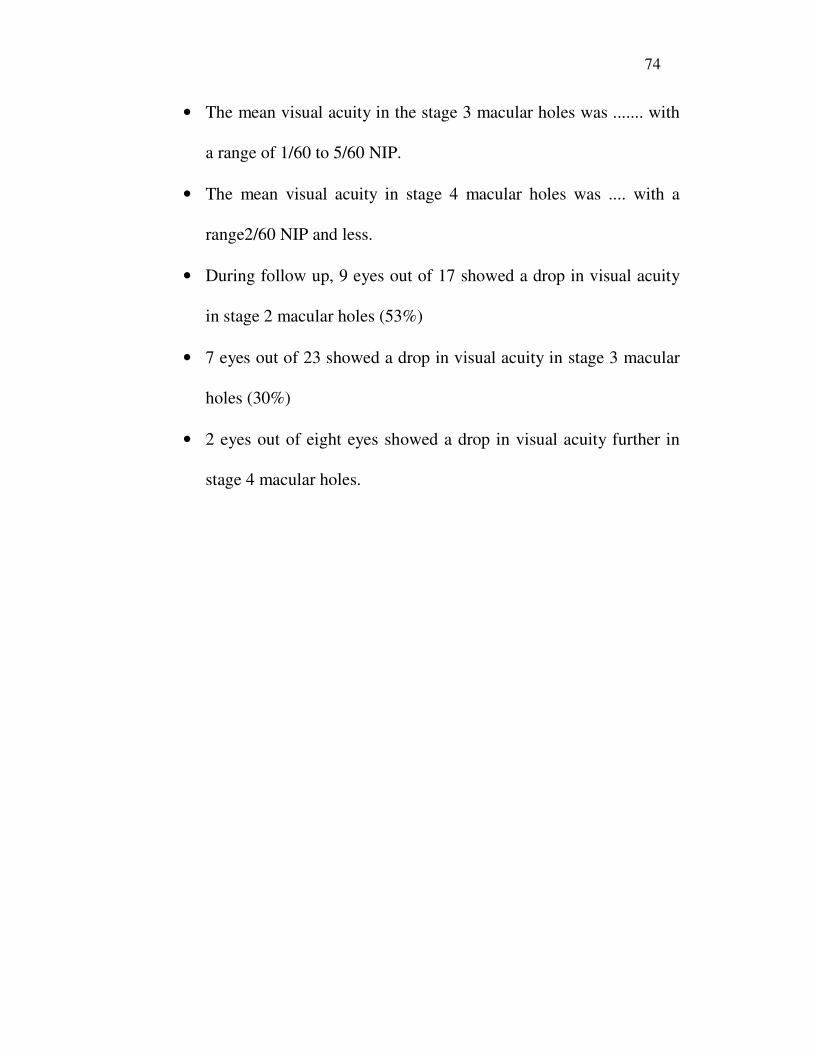
• The mean visual acuity in the stage 3 macular holes was ....... with
a range of 1/60 to 5/60 NIP.
• The mean visual acuity in stage 4 macular holes was .... with a
range2/60 NIP and less.
• During follow up, 9 eyes out of 17 showed a drop in visual acuity
in stage 2 macular holes (53%)
• 7 eyes out of 23 showed a drop in visual acuity in stage 3 macular
holes (30%)
• 2 eyes out of eight eyes showed a drop in visual acuity further in
stage 4 macular holes.
75
DISCUSSION
Idiopathic macular holes are common in the older age group. They
usually present with defective central vision and metamorphopsia. It
constitutes a fraction of ocular morbidity in the elderly.
Macular holes can occur in younger age group, though the causes
may be different like trauma, cystoid macular oedema or even a
pseudohole secondary to vasculitis or retinal degenerations.
Macular holes are significant because they cause gross loss of
central vision. The prevalence is more in females as shown in our study.
It is also more among pseudophakics than phakics. Visual acuity is
measured with snellens chart in our study and the visual acuity is very
less in stage 4 than stage 2 macular hole.
Optical coherence tomography is a very important tool in assessing
the stage of macular hole, presence and absence of epiretinal membranes
associated, hole size and also prognosticating post surgery. It also plays a
key role in assessing whether the macular hole surgery is successful or
not.
Epiretinal membranes are commonly associated with macular
holes. The incidence of epiretinal membranes increases with the stage of
76
macular hole. The severity of these epiretinal membranes also increases
during follow up in a significant number of patients eyes. Though
commonly associated with macular holes, a significant correlation has not
yet been elucidated between epiretinal membranes and idiopathic macular
holes.
In our study, we have attempted to find out the associations of
epiretinal membranes and various stages of macular holes. Epiretinal
membranes are a cause of tangential traction that plays a role in the patho
physiology of macular holes. We established that epiretinal membranes
are associated with all stages of macular holes and majority show an
increase in severity of macular holes during follow up. The incidence of
epiretinal membranes is also more among pseudophakics than phakics
who were enrolled in this study.
Stanley p azen, University of Atlanta, Florida in his journal
publication in 2000 states that no significant correlation exists between
epiretinal membranes and idiopathic macular hole with respect to visual
acuity.
With greater advancements in instrumentation and surgical
techniques, larger leaps have been made in macular hole surgery and
good results are obtained.

77
CONCLUSION
Idiopathic macular holes are common in the elderly. It is an
important cause of loss of central vision. This study shows that epiretinal
membranes are commonly associated with full thickness macular holes.
The prevalence of epiretinal membrane increases with the severity and
size of macular hole. With the advent of OCT, and finer instrumentation
greater advances have been made in macular hole surgery. Though the
prevalence of epiretinal membrane is common in idiopathic macular hole,
there is no significant correlation between epiretinal membranes in
macular hole and surgery. These factors may be considered during
vitrectomy for macular hole.

S.No Name Age/Sex Diagnosis Pseudophakic/ Phakic UCVA BCVA ERMFOLLOW
UP(3M,6M,1YR)
V/A FOLLOW
UP
1 USHA 60/F GRADE 3 PHAKIC 3/60 NIP 3M/60 + ++ 3/60 NIP
2 KALPANA 49/F GRADE 2 PHAKIC 6M/60 6M/60 - + 5/60 NIP
3 SUNDAR 70/M GRADE 4 PSEUDOPHAKIC 1M/60 1M/60 + ++ 1/60 NIP
4 RAMU 72/M GRADE 3 PSEUDOPHAKIC 4M/60 4M/60 + ++ 4/60 NIP
5 VEL 42/M GRADE 2 PHAKIC 5M/60 5M/60 _ _ 5/60 NIP
6 AYYAPAN 68/M GRADE 3 PSEUDOPHAKIC 3M/60 3M/60 + + 2/60 NIP
7 VANI 60/F GRADE 3 PHAKIC 2M/60 2M/60 _ + 2/60NIP
8 RAJESH 60/M GRADE 3 PSEUDOPHAKIC 4M/60 4M/60 + ++ 4/60 NIP
9 GOURI 52/F GRADE2 PSEUDOPHAKIC 6M/60 6/60 NIP _ + 5/60 NIP
10 SUNDAR 49/M GRADE2 PHAKIC 6/36 NIP 6/36 NIP _ + 6/60 NIP
11 DEVI 45/F GRADE 2 PHAKIC 6/60NIP 6/60 NIP _ + 6/60 NIP
12 GANGA 62/F GRADE3 PHAKIC 4/60NIP 4/60NIP _ ++ 4/60NIP
13 SEETHA 70/F GRADE3 PSEUDOPHAKIC 3/60NIP 3/60NP + + 2/60 NIP
14 PANDI 52/M GRADE2 PHAKIC 5/60 NIP 5/60 NIP _ _ 5/60 NIP
15 MANGA 50/F GRADE2 PHAKIC 6M/60
6/60WITH +1DS
6/36 _ 6/60 NIP
16 KUZHAL 60/F GRADE3 PHAKIC 2/60NIP 2/60NIP + + 2/60 NIP

17 VENU 59/M GRADE3 PHAKIC 2/60NIP 2/60NIP _ _ 2/60 NIP
18 ANGEL 65/F GRADE3 PHAKIC 5/60 NIP 5/60NIP _ _ 4/60 NIP
19 MEENA 62/F GRADE4 PSEUDOPHAKIC 1/60NIP 1/60NIP + + 1/2/60NIP
20 SANTOSH 49/M GRADE2 PSEUDOPHAKIC 4/60 NIP 4/60NIP + + 4/60NIP
21 SARAVAN 45/M GRADE2 PHAKIC 5/60 NIP 5/60 NIP _ _ 5/60 NIP
22 KUMAR 49/M GRADE2 PHAKIC 5/60 NIP 5/60 NIP _ _ 5/60NIP
23 RATNAM 54/M GRADE3 PHAKIC 4/60 NIP 4/60 NIP + ++ 3/60 NIP
24 NEELA 55/F GRADE2 PHAKIC 4M/60
4/60 WITH '+1DS
5/60 4/60 NIP
25 MEERA 64/F GRADE4 PSEUDOPHAKIC 1/60NIP 1/60 NIP ++ ++ 1/60 NIP
26 RAJAM 69/F GRADE4 PSEUDOPHAKIC 2/60 NIP 2/60 NIP + ++ 1/60NIP
27 KUPPU 72/F GRADE4 PHAKIC 2/60 NIP 2/60 NIP _ _ 2/60NIP
28 SARASU 70/F GRADE3 PHAKIC 3/60 NIP 3/60 NIP + ++ 3/60 NIP
29 SUNDARI 63/F GRADE4 PSEUDOPHAKIC 1/60NIP 1/60 NIP + ++ 1/60 NIP
30 SAKUNTA 64/F GRADE3 PHAKIC 2/60 NIP 2/60 NIP _ _ 2/60 NIP
31 SEKAR 59/M GRADE2 PHAKIC 6/60 NIP 6/60 NIP _ _ 5/60 NIP
32 CHITRAA 62/F GRADE3 PHAKIC 4/60 NIP 4/60 NIP _ + 4/60NIP

33 LAKSHMI 60/F GRADE3 PHAKIC 4/60 NIP 4/60 NIP + ++ 4/60 NIP
34 SAPNAM 71/F GRADE2 PSEUDOPHAKIC 3M/60
3/60 WITH
'+1.ODS 4/60 3/60NIP
35 KRISHNA 76/M GRADE3 PSEUDOPHAKIC 3/60 NIP 3/60 NIP ++ 2/60NIP
36 KANNAN 75/M GRADE2 PSEUDOPHAKIC 6M/60
6/60 WITH +1D
CYL 6/36 6/60 NIP
37 JANAKI 70/F GRADE4 PSEUDOPHAKIC 1/2M/60 1/2/60 NIP _ 1/2/60 NIP
38 VALLI 80/F GRADE3 PSEUDOPHAKIC 2M/60
2/60 WITH + 1DS
3/60 2/60 NIP
39 SIVA 49/M GRADE2 PHAKIC 4M/60
4/60 WITH '+1DS
5/60 4/60 NIP
40 SELVI 52/F GRADE2 PHAKIC 5m/60
5/60 WITH '+1DS
6/60 5/60 NIP
41 GANESH 60/M GRADE3 PSEUDOPHAKIC 2/60 NIP 2/60 NIP _ _ 1/60 NIP
42 SUMATHI 59/F GRADE3 PHAKIC 3M/60
3/60 WITH
'+1.ODS 4/60 3/60 NIP
43 SANTHA 54/F GRADE2 PHAKIC 4/60 NIP 4/60 NIP _ _ 4/60N IP
44 MOHAN 55/M GRADE3 PHAKIC 1/60 NIP 1/60NIP + + 1/60NIP
45 BALAJI 53/M GRADE3 PSEUDOPHAKIC 2/60NIP 2/60 NIP + ++ 2/60NIP
46 RADHA 48/F GRADE2 PHAKIC 6M/60
6/60 WITH
'+1DS6/36 + 6/60NIP
47 MANI 47/M GRADE2 PHAKIC 4/60 NIP 4/60 NIP _ _ 4/60 NIP
48 SAROJA 64/F GRADE3 PHAKIC 3/60 NIP 3/60 NIP _ _ 3/60NIP
49 BALU 62/M GRADE3 PSEUDOPHAKIC 4/60NIP 4/60 NIP + ++ 4/60NIP
50 GOPAL 60/M GRADE4 PSEUDOPHAKIC 1/60 NIP 1/60 NIP ++ ++ 1/60NIP
“ PREVALENCE , CORRELATION AND NATURAL HISTORY OF EPIRETINAL MEMBRANES SURROUNDING IDIOPATHIC MACULAR HOLES “

� GUIDE
PROF . DR.V.REVATHY
head of the department of UVEA AND RETINA SERVICES
CO GUIDES
PROF .DR . B. CHANDRASEKARAN
professor of ophthalmology ,
RIO GOH

� CO GUIDE
DR.G. BALAJI ,
assistant professor ,
RIO GOH

SYNOPSIS
� TITLE – “prevalence , correlation and estimates of epiretinal membranes surrounding idiopathic macular holes “
� Principal investigator – DR. M. ROSHNI
� GUIDE – PROF . DR. V . REVATHI,
head of the department UVEA AND RETINA SERVICES
� STUDY CENTER – regional institute of ophthalmology ,egmore , chennai

� Duration – 1YEAR � AIM – to evaluate the prevalence , correlation and natural history of EPIRETINAL MEMBRANES SURROUNDING IDIOPATHIC MACULAR HOLES
� PRIMARY OBJECTIVES- 1.to evaluate the prevalence of epiretinal membranes in eyes with stage 2/3/4 macular holes .
� 2. the correlation between epiretinal membrane and visual acuity in idiopathic macular holes
� 3.the correlation between size of macular hole and epiretinal membrane

� Secondary objective -To assess the increase in severity of epiretinal membrane in follow up.
� Study design-Prospective study, observational study
� Methodology{material and methods }-All patients with idiopathic macular hole stage 2,3,4who attended the OPD
� Inclusion criteria-All patients with symptoms of decreased central vision and metamorphopsia and clinically proven macular hole stage 2, 3 and 4
� Exclusion criteria-Patients who had traumatic macular hole and macular hole due to other causes and macular edema .

Sample size- 50 patients
Age - 30 to 80 years
Gender – both genders
� Study parameters� Detailed history including past and present illness
� Visual acuity assessment using snellens visual acuity chart
� Anterior segment examination using slit lamp including lens and anterior vitreous .
� Fundus examination using direct and indirect ophthalmoscopic examination.
� Stereoscopic fundus examination using slit lamp biomicroscopy
� Colour fundus photography
� Optical coherence tomography .
� Data collection and methods-All cases diagnosed as idiopathic macular holes stage 2,3 and 4 will be registered and evaluated subsequently at the end of 3,6 and 12 months of follow up .
� Analysis plan- to consider the importance of factors in removal of epiretinal membrane during vitrectomyfor macular hole