therapeutic potential of gamma- irradiated resveratrol in ... · pathway, and mixed lymphocyte...
TRANSCRIPT
Cell Physiol Biochem 2019;52:1117-1138DOI: 10.33594/000000076Published online: 17 April 2019 1117
Cellular Physiology and Biochemistry
Cellular Physiology and Biochemistry
© 2019 The Author(s). Published by Cell Physiol Biochem Press GmbH&Co. KG
Kim et al.: Gamma-Irradiated Resveratrol Induces Tolerance and Anti-Inflammatory Activity in Dendritic Cells
Original Paper
Accepted: 12 March 2019
This article is licensed under the Creative Commons Attribution-NonCommercial-NoDerivatives 4.0 Interna-tional License (CC BY-NC-ND). Usage and distribution for commercial purposes as well as any distribution of modified material requires written permission.
DOI: 10.33594/000000076Published online: 17 April 2019
© 2019 The Author(s)Published by Cell Physiol Biochem Press GmbH&Co. KG, Duesseldorfwww.cellphysiolbiochem.com
Therapeutic Potential of Gamma-Irradiated Resveratrol in Ulcerative Colitis via the Anti-Inflammatory Activity and Differentiation of Tolerogenic Dendritic CellsWoo Sik Kima Ha-Yeon Songa Sajid Mushtaqa Jin-Man Kimb Eui-Hong Byunc Jae-Min Yukd,e Eui-Baek Byuna
aAdvanced Radiation Technology Institute, Korea Atomic Energy Research Institute, Jeongeup, Republic of Korea, bDepartment of Pathology, College of Medicine, Chungnam National University, Daejeon, Republic of Korea, cDepartment of Food Science and Technology, Kongju National University, Gongju, Republic of Korea, dDepartment of Infection Biology, College of Medicine, Chungnam National University, Daejeon, Republic of Korea, eDepartment of Medical Science, College of Medicine, Chungnam National University, Daejeon, Republic of Korea
Key WordsResveratrol • Gamma irradiation • Tolerance • Anti-inflammatory activity • Inflammatory bowel disease
AbstractBackground/Aims: New therapeutic strategies and the development of treatments against inflammatory bowel disease (IBD) require the initiation of immune tolerance and inhibition of excessive inflammation. Resveratrol, a polyphenolic compound, is a powerful immunosuppressor, but it can lead to apoptotic death of normal cells at high concentrations. When we induced a structural modification of resveratrol by gamma irradiation, we were able to investigate the potential tolerogenic and anti-inflammatory effect of a new radiolysis product (named γ-Res) during dendritic cell (DC) activation/differentiation. Methods: The potential tolerogenic and anti-inflammatory effect of γ-Res were investigated by cytokine secretion, surface molecule expression, antigen uptake ability, antigen presenting ability, signaling pathway, and mixed lymphocyte reaction (MLR) assay using enzyme-linked immunosorbent assay (ELISA), western blot and flow cytometry. Results: LPS-activated DCs treated with γ-Res exhibited alterations in their mature and functional statuses including a strongly inhibited cytokine production, surface molecule expression, antigen-presenting ability, and activated DC-induced T cell proliferation/activation. In addition, the DCs generated by the
Dr. Eui-Baek Byun, PhD Advanced Radiation Technology Institute, Korea Atomic Energy Research InstituteJeongeup, Jeonbuk 56212 (Republic of Korea)Tel. +82 063 570 3245, Fax +82 063 570 3371, E-Mail [email protected]
WS Kim and HY Song contributed equally to this work.
Cell Physiol Biochem 2019;52:1117-1138DOI: 10.33594/000000076Published online: 17 April 2019 1118
Cellular Physiology and Biochemistry
Cellular Physiology and Biochemistry
© 2019 The Author(s). Published by Cell Physiol Biochem Press GmbH&Co. KG
Kim et al.: Gamma-Irradiated Resveratrol Induces Tolerance and Anti-Inflammatory Activity in Dendritic Cells
γ-Res treatment during DC differentiation induced a decreased surface molecule expression and increased IL-10 production without altering the levels of TNF-α and IL-12p70, thereby promoting the inhibition of T cell proliferation/activation and the induction of regulatory T cells via interaction with DCs in vitro. Furthermore, in the in vivo DSS-induced colitis model, γ-Res treatment conferred protective immunity with a decrease in IFN-γ+CD4+ and IL-17A+CD4+ T cells and imparted protection by reducing the disease activity and histological disease score and increasing the survival rate in dextran sulfate sodium (DSS)-induced colitis in mice. Conclusion: Thus, our results suggest that γ-Res may be an excellent candidate for use in IBD treatment.
Introduction
The main cause of inflammatory bowel disease (IBD) including Crohn’s disease (CD) and ulcerative colitis (UC) is not yet fully understood; however, we do know that this disease is the result of an abnormal immune response triggered by specific genetic and/or immunologic defects in the gastrointestinal tract [1]. Importantly, the immune systems involved are influenced by the dysfunction of the mucosal innate and adaptive immune cells, and these cells recognize foreign antigens from food and intestinal bacteria leading to a chronic inflammation response [1, 2]. In CD pathogenesis, IFN-γ, TNF-α, and IL-17 cytokines are mainly induced by increased intestinal expression in Th1 and Th17 cells, which may contribute to severe intestinal damage by immune cell activation and expression of apoptosis-related proteins that inhibit the expression of anti-apoptotic proteins [3-5]. On the other hand, UC is usually viewed as Th2 immunity because of the excessive expression of the principal Th2 effector cytokines (IL-5 and IL-13) and TNF-α [5], and these responses augment the ulceration and inflammation by B cell activation, activation/recruitment of eosinophils and NKT-cell-mediated cytotoxicity [6, 7]. In this regard, to solve the problems of the immune response associated with IBD, many studies have focused on effective regulation for imbalanced cytokines and immune cell populations contributing to the immunopathogenesis of IBD.
Recently, polyphenolic compounds, including resveratrol, epigallocatechin gallate (EGCG), curcumin, quercetin, and rutin, have been reported as potential candidates to help IBD. These compounds are known to interact with various immune cells. They have been proven effective in reducing excessive inflammation by regulating inflammatory mediators such as TNF-α, IFN-γ, IL-12, inducible nitric oxide synthase (iNOS), mitogen-activated protein (MAPK), and nuclear factor κ B (NF-κB) for immune cells in IBD models [8-11]. However, most polyphenols may lead to apoptosis of normal cells when used at higher concentrations [12-14], indicating that improved treatment strategies are urgently needed.
Irradiation technology is a useful tool for improving the function of polyphenols via structural modification [15-17]. For example, certain gamma-irradiated polyphenols, such as resveratrol, and luteolin, have been verified as enhancing bioactivity, such as anti-inflammatory activity and anti-melanogenic effect, and decreasing the toxicity of the parent compounds [15, 16]. However, among the gamma-irradiated polyphenols, there is still a lack of knowledge about resveratrol; it is unclear how its structure changes and whether it is involved in protection or immune evasion in the immune systems contributing to IBD.
To understand the functional roles of resveratrol transformed by gamma irradiation in the maturation and differentiation of dendritic cells (DCs) that cause inflammation in IBD, we looked at the anti-inflammatory activity of a new product of resveratrol transformed by gamma irradiation and the phenotypic and functional change induced by the treatment with the new product during DC differentiation as well as the therapeutic effect and immunological contribution caused by the treatment with this new product in an animal model of IBD.
© 2019 The Author(s). Published by Cell Physiol Biochem Press GmbH&Co. KG
Cell Physiol Biochem 2019;52:1117-1138DOI: 10.33594/000000076Published online: 17 April 2019 1119
Cellular Physiology and Biochemistry
Cellular Physiology and Biochemistry
© 2019 The Author(s). Published by Cell Physiol Biochem Press GmbH&Co. KG
Kim et al.: Gamma-Irradiated Resveratrol Induces Tolerance and Anti-Inflammatory Activity in Dendritic Cells
Materials and Methods
MiceSpecific Pathogen Free mice (female C57BL/6 (H-2Kb and I-Ab) and BALB/c (H-2Kd and I-Ad), seven
weeks of age) were used in the in vitro and in vivo experiments. These mice were purchased from Orient Bio Inc. (Seoul, Korea). All the mice were acclimated to the following controlled conditions: temperature (25 ± 2 °C), humidity (55 ± 5%), and 12 h light/dark cycle at the Central Animal Research Laboratory at the Korea Atomic Energy Research Institute (KAERI, Jeongeup, Korea). Animal experimental protocols were approved by the Institutional Animal Care and Use Committee of KAERI (KAERI-IACUC-2017-034) and performed in strict compliance with the guidelines prescribed by the committee.
Preparation of gamma-irradiated resveratrolResveratrol was purchased from R&D Systems (Minneapolis, MN, USA). The resveratrol solution (1
mg/mL) dissolved in methanol was irradiated at a dose of 50 kGy (10 kGy/h) by a cobalt-60 irradiator (point source AECL, IR-221, MDS Nordion International Co., Ltd., Ottawa, ON, Canada). The methanol was evaporated by a rotary evaporator (Tokyo Rikakikai Co., Ltd., Japan), and the irradiated resveratrol was stored at -80°C.
HPLC analysis and identification of the gamma-irradiated resveratrolFor analysis of intact resveratrol (intact-Res) and gamma-irradiated resveratrol (γ-Res), a 20 µL
sample was injected into an Agilent HPLC 1260 system (Agilent Technologies, Inc., Santa Clara, CA, USA) with a diode array detector. The chromatographic separation was performed using an Agilent Eclipse XDB-C18 column (4.6 mm × 250 mm i.d., 5 µm, Agilent Technologies). The mobile phase consisted of 0.1% formic acid in water (A) and 0.1% formic acid in acetonitrile (B). The analysis was conducted using an isocratic elution during 50 min. with A-B (75-25). For isolation of the γ-Res, purification was performed by preparative HPLC (1260 Infinity System, Agilent Technologies). The Agilent Eclipse XDB-C18 column (21.2 mm x 150 mm i.d., 7.5 µm) was used to isolate the γ-Res. Identification of the isolated γ-Res was carried out with an electrospray ionization mass spectrometer (ESI-MS), 1H-nuclear magnetic resonance (NMR), and 13C-NMR. The NMR spectra were recorded with a Bruker 500 MHz spectrometer (1H, 500 MHz and 13C, 125 MHz) using DMSO-d6.
Generation of BMDCs and gamma-irradiated resveratrol-treated DCsTo generate bone-marrow-derived dendritic cells (BMDCs), intact-Res-treated DCs (intact-Res-DCs)
and γ-Res-treated DCs (γ-Res-DCs), whole bone marrow cells from C57BL/6 mice were lysed of red blood cells (RBCs) using RBC lysing buffer (Sigma-Aldrich), and then, the lysed cells were maintained in 6-well plates (1 × 10⁶ cells/ml; 2 mL/well) and cultured at 37 °C in the presence of 5% CO2, using complete-RPMI 1640 (c-RPMI 1640, GIBCO, Carlsbad, CA, USA) containing 10% FBS (GIBCO), 100 U/mL penicillin, 100 U/mL streptomycin, 20 ng/mL GM-CSF (JW CreaGene, Daegu, Korea) and 0.5 ng/mL IL-4 (JW CreaGene). On day three of the culture, 1 mL c-RPMI 1640 medium was added to each well of the BMDC differentiation plates, and 1 mL c-RPMI 1640 medium was added to each well of the intact-Res-DCs and γ-Res-DC differentiation plates along with γ-Res and intact-Res at a concentration of 15 and 50 μg/mL. On day six of the culture, the plate media were changed with 1 mL c-RPMI 1640 medium per well. On day eight of the culture, the cells were harvested and stained with fluorescein-conjugated CD11c mAb. Over 90% of the cells expressed CD11c.
Cytotoxicity and cell viabilityTo analyze cytotoxicity, after 8 days of culturing, the BMDCs, intact-Res-DCs and γ-Res-DCs were stained
with the propidium iodide (PI; BD Bioscience, San Diego, CA, USA) method. Additionally, the cultured BMDCs were seeded in 48-well plates at a density of 2 × 10⁵ cells/well and treated with the following stimuli for 24 h: DMSO (Con), LPS (100 ng/mL; Invivogen, San Diego, CA), LPS and intact-Res (30 μg/mL), or LPS and γ-Res (30 μg/mL). The cells were washed with PBS and stained with the Annexin V/PI apoptosis detection kit (BD Bioscience) following the manufacturer’s instructions. To analyze viability, each group was treated with 10 μL EZ-Cytox kit reagent (Daeil Laboratories, Seoul, Korea), and the cells were incubated for 1 h. The absorbance was measured at 450 nm using an automated micro-plate reader (Zenyth 3100; Anthos Labtec Instruments GmbH, Salzburg, Austria).
Cell Physiol Biochem 2019;52:1117-1138DOI: 10.33594/000000076Published online: 17 April 2019 1120
Cellular Physiology and Biochemistry
Cellular Physiology and Biochemistry
© 2019 The Author(s). Published by Cell Physiol Biochem Press GmbH&Co. KG
Kim et al.: Gamma-Irradiated Resveratrol Induces Tolerance and Anti-Inflammatory Activity in Dendritic Cells
Analysis of the surface molecules on DCsTo analyze the difference in the surface molecule expression of the BMDCs and γ-Res-DCs, cells were
stimulated with LPS for 24 h. Additionally, to analyze the effect of the γ-Res treatment on the expression of surface molecules induced by the LPS-treated BMDCs, BMDCs were treated with the following stimuli for 24 h: DMSO (Con), LPS, or LPS and γ-Res, and then, the cells were stained with anti-CD80, anti-CD86, anti-MHC-I, and anti-MHC-II for 20 min. at room temperature (RT). The resulting fluorescence was collected on a FACSverse flow cytometer (BD Biosciences) and analyzed using the FlowJo software (BD Biosciences). Antibodies (Abs) were purchased from BD Biosciences.
Intracellular cytokine stainingAfter incubating with GolgiPlug for 9 h, cells were stained with a Live/Dead cell staining kit (Invitrogen,
Life Technologies, Naerum, Denmark) using DC and T cell phenotypic markers (anti-CD11c, anti-CD3, anti-CD4, and anti-CD8 Abs) for 20 min. at RT and then fixed and permeabilized with a Cytofix/Cytoperm kit (BD Bioscience) following the manufacturer’s instructions. Next, anti-TNF-a, anti-IL-12p70, anti-IFN-γ, anti-IL-17A, anti-IL-5, and anti-IL-10 Abs were stained with fluorescein-conjugated secondary Abs in a permeation buffer. The Abs for the phenotypic markers and cytokines of the DCs and T cells were purchased from BD Bioscience.
Cytokine measurementsThe levels of TNF-α, IL-12p70, IFN-γ, IL-2, and IL-10 in the supernatants were measured with an ELISA
kit (BD Bioscience) according to the manufacturer’s instructions.
Antigen-uptake capacityAfter stimulation with or without LPS, BMDCs and γ-Res-DCs were cultured in the presence of 1 mg/
ml fluorescein-conjugated dextran (40, 000 Da, Sigma-Aldrich) at 37 °C. After 30 min. of treatment, cells were washed with cold PBS and stained using anti-CD11c or DAPI and analyzed by FACSverse or LSM 800 confocal laser scanning microscope (Zeiss, Germany). Images were acquired with the LSM 800 software and processed with a LSM imager.
Isolation of T cells in splenocytesFor CD3+ T cell isolation in splenocytes, cells isolated from the mice of the DSS-induced colitis model
or naïve mice (BALB/C mice) were labeled with anti-CD16/32 and bead-conjugated anti-CD11c Abs (CD11c MicroBeads) or anti-CD3e mAbs (CD3e MicroBeads), respectively, and separated by sequential passage through LS MACS Columns. The purity of the selected CD3+ T cell fractions was >80%. The columns and antibodies for cell isolation were purchased from Miltenyi Biotec (San Diego, CA).
T cell suppression assay and mixed lymphocyte reaction (MLR)For the T cell suppression assay using allogeneic mice, CD3+ T cells separated by the MACS system were
isolated from the spleens of the BALB/C mice, stained with CFSE (5 μM) at 37 °C, and then washed with PBS (supplemented with 2% FBS) after 15 min. Next, 48-well plates were coated with anti-CD3 mAb (0.5 μg/mL) and anti-CD28 mAb (0.5 μg/mL) at 4 °C. After 24 h, 5 × 10⁵ T cells stained with CFSE were cultured onto plates coated with anti-CD3/CD28 mAbs. In addition, BMDCs and γ-Res-DCs generated from the C57BL/6 were stimulated with or without LPS for 2 h and washed with PBS. The ratio between DCs and T cells was 0.5:1.
For the MLR assay, BMDCs (from C57BL/6) were treated with LPS in the presence and absence of γ-Res for 24 h and then washed and co-cultured with CFSE-labeled T cells (from BALB/C) at a DC to T cell ratio of 0.5:1. After 3 days of co-culturing, each of the cells from the T cell suppression and MLR experiment was harvested and stained with anti-CD4 and anti-CD8 mAbs. Data were collected on a FACSverse and analyzed with the FlowJo software. The culture supernatants were harvested, and cytokine production was analyzed by ELISA.
Regulatory T cell analysisCD4+ T cells were isolated from the spleens of BALB/C mice with the Miltenyi CD4+ T cell isolation kit.
Isolated CD4+ T cells (5 × 10⁵ cells) were co-cultured with BMDC and γ-Res-DCs at a variable ratio (DC and
Cell Physiol Biochem 2019;52:1117-1138DOI: 10.33594/000000076Published online: 17 April 2019 1121
Cellular Physiology and Biochemistry
Cellular Physiology and Biochemistry
© 2019 The Author(s). Published by Cell Physiol Biochem Press GmbH&Co. KG
Kim et al.: Gamma-Irradiated Resveratrol Induces Tolerance and Anti-Inflammatory Activity in Dendritic Cells
T cell, 0.5:1) for 72 h in a 48-well plate. After 72 h, the cells were harvested and stained with anti-CD3 and anti-CD4 for 30 min. at 4 °C. The cells were fixed and permeabilized using Foxp3 Fixation/Permeabilization working solution (eBioscience) according to the manufacturer’s instructions. Following that, the cells were stained with anti-Foxp3 mAb for 30 min. at RT. Data were collected on a FACSverse and analyzed with the FlowJo software.
Antigen-presenting abilityTo investigate the difference in the peptide-MHC-I complex formation between the BMDCs and γ-Res-
DCs, each DC was stimulated with or without 100 ng/mL LPS in the presence of 500 μg/mL OVA protein (Sigma-Aldrich). Additionally, to investigate the ability of peptide-MHC-I complex formation induced by γ-Res in LPS-treated BMDCs, cells were stimulated with DMSO (Con), LPS, or LPS and γ-Res in the presence of 500 μg/mL OVA protein. OVA257-264 peptide (SIINFEKL) acts as a positive control. Next, to investigate the difference in the peptide-MHC-II complex formation between BMDCs and γ-Res-DCs, cells were treated with or without 100 ng/mL LPS in the presence of 25 μg/mL Eα44-76 peptide (RLEEFAKFASFEAQGALANIAVDKANLDVMKKR; underlined sequence bound to MHC-II). To investigate the ability of peptide-MHC-II complex formation induced by γ-Res in LPS-treated BMDCs, cells were stimulated with DMSO (Con), LPS, or LPS and γ-Res in the presence of 25 μg/mL Eα44-76 peptide. The Eα52-68 peptide (ASFEAQGALANIAVDKA) acts as a positive control. After 24 h of treatment, each cell was stained with anti-CD11c, anti-H-2Kb Ab (SIINFEKL, eBioscience) or anti-I-Ab-Eα52-68 Ab (Y-Ae; ASFEAQGALANIAVDKA, eBioscience) mAbs for 20 min. at RT. Data were collected on a FACSverse and analyzed with the FlowJo software. All peptides were synthesized by AbFrontier (Seoul, Korea).
Immunoblotting analysesTo analyze the difference in signaling mechanisms of the BMDCs and γ-Res-DCs, each cell was treated
with 100 ng/mL LPS in a time-dependent manner. Moreover, to analyze the effect of γ-Res in response to activation signals induced by LPS-treated BMDCs, BMDCs were treated with LPS in the presence and absence of γ-Res. For the cytosolic fraction, the cells were lysed with RIPA lysis buffer (Pierce, Rockford, IL, USA). The next steps were carried out as previously described [18]. Furthermore, nuclear extracts from cells were isolated with the CelLytic NuCLEAR Extraction Kit (Sigma-Aldrich) following the manufacturer’s instructions. Western blotting Abs and HRP-conjugated anti-rabbit Abs were obtained from Santa Cruz Biotechnology, Inc. (Santa Cruz, CA, USA) and Sigma-Aldrich, respectively.
DSS-induced colitis and treatment with γ-ResC57BL/6 mice were treated for 7 days with 2.5% DSS (molecular weight 36–50 kDa, MP Biomedical,
Aurora, OH, USA) dissolved in sterile water as a source of drinking water. Normal mice were given water. The mice were divided into six groups (n = 5 mice per group): (Group 1) normal, (Group 2) DSS + vehicle (2% DMSO and 0.5% Tween-20 in PBS), (Group 3) DSS + 75 mg/kg of 5-aminosalicylic acid (ASA, positive control), (Group 4) DSS + 2 mg/kg of γ-Res, and (Group 5) DSS + 5 mg/kg of γ-Res. Both ASA and γ-Res were dissolved in the vehicle solution, and this was given by oral gavage, once per day. For the survival study, five mice each in only the DSS (Group 2) and DSS + γ-Res group (Group 5) were subjected to 2.5 % DSS until day 7, and then, they were given normal distilled water and monitored for survival up to day 35.
Colitis evaluation after treatment with γ-ResThe body weights of the mice were monitored every day. The disease activity index (DAI) was calculated
by the method described by Park et al. [19]. Briefly, diarrhea (0, none; 2, loose stool; 4, watery diarrhea) and hematochezia (0, none; 2, slight bleeding; 4, gross bleeding) were observed during colitis induction for 7 days. At day 8 following the induction of colitis, the mice were sacrificed, and their entire colons were collected and cleaned of fat. For histological analysis, some of the colon was fixed in 10% buffered formalin and embedded in paraffin, and then, the sections were stained with hematoxylin and eosin (H&E). To analyze the functions of T cells sorted from the DSS-induced mice, isolated T cells were stimulated with anti-CD3/CD28 mAbs (0.5 μg/mL each) in the presence of GolgiPlug for 9 h, and then, the cytokine secretion ability for the activated T cells was analyzed using flow cytometry.
Cell Physiol Biochem 2019;52:1117-1138DOI: 10.33594/000000076Published online: 17 April 2019 1122
Cellular Physiology and Biochemistry
Cellular Physiology and Biochemistry
© 2019 The Author(s). Published by Cell Physiol Biochem Press GmbH&Co. KG
Kim et al.: Gamma-Irradiated Resveratrol Induces Tolerance and Anti-Inflammatory Activity in Dendritic Cells
StatisticsData were determined with Tukey’s multiple comparison test or unpaired t-tests using the GraphPad
Prism program (San Diego, CA, USA). The data in our graphs are expressed as the means. *p < 0.05, **p < 0.01 and ***p < 0.001 were considered statistically significant.
Results
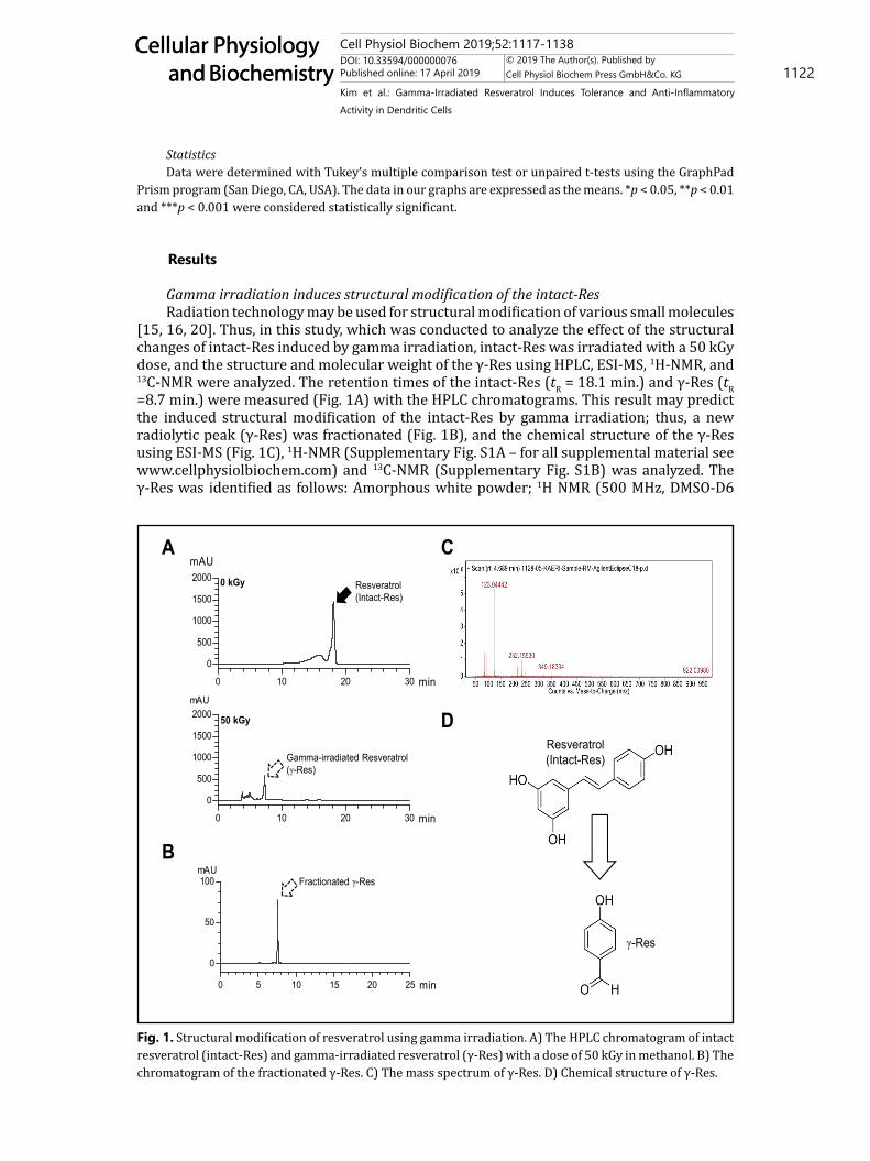
Gamma irradiation induces structural modification of the intact-ResRadiation technology may be used for structural modification of various small molecules
[15, 16, 20]. Thus, in this study, which was conducted to analyze the effect of the structural changes of intact-Res induced by gamma irradiation, intact-Res was irradiated with a 50 kGy dose, and the structure and molecular weight of the γ-Res using HPLC, ESI-MS, 1H-NMR, and 13C-NMR were analyzed. The retention times of the intact-Res (tR = 18.1 min.) and γ-Res (tR =8.7 min.) were measured (Fig. 1A) with the HPLC chromatograms. This result may predict the induced structural modification of the intact-Res by gamma irradiation; thus, a new radiolytic peak (γ-Res) was fractionated (Fig. 1B), and the chemical structure of the γ-Res using ESI-MS (Fig. 1C), 1H-NMR (Supplementary Fig. S1A – for all supplemental material see www.cellphysiolbiochem.com) and 13C-NMR (Supplementary Fig. S1B) was analyzed. The γ-Res was identified as follows: Amorphous white powder; 1H NMR (500 MHz, DMSO-D6
Fig. 1. Structural modification of resveratrol using gamma irradiation. A) The HPLC chromatogram of intact resveratrol (intact-Res) and gamma-irradiated resveratrol (γ-Res) with a dose of 50 kGy in methanol. B) The chromatogram of the fractionated γ-Res. C) The mass spectrum of γ-Res. D) Chemical structure of γ-Res.
Figure 1
0 10 20 30
0
500
1000
1500
2000mAU
min
0 10 20 30
0
500
1000
1500
2000mAU
min
Gamma-irradiated Resveratrol (γ-Res)
Resveratrol(Intact-Res)
0 kGy
50 kGy
A
B
Resveratrol (Intact-Res)
γ-Res
C
D
0 5 10 15 20 25
0
50
100mAU
min
Fractionated γ-Res
Cell Physiol Biochem 2019;52:1117-1138DOI: 10.33594/000000076Published online: 17 April 2019 1123
Cellular Physiology and Biochemistry
Cellular Physiology and Biochemistry
© 2019 The Author(s). Published by Cell Physiol Biochem Press GmbH&Co. KG
Kim et al.: Gamma-Irradiated Resveratrol Induces Tolerance and Anti-Inflammatory Activity in Dendritic Cells
TMS), ζ 9.737 (s, 1H, CHO), 7.719 (d, 2H, J = 8.4 Hz, 2, 6-H), 6.891 (m, 2H, 3, 5-H), and 5.951 (s, 1H, OH); 13C NMR (125 MHz, DMSO-D6, ppm), ζ 191.434, 164.307, 132.640, and 116.473, and MS (ESI, m/z), 123.0444 [M + H+]. The identified structure of the γ-Res is proposed in Fig. 1D.
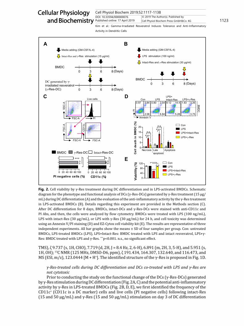
γ-Res-treated cells during DC differentiation and DCs co-treated with LPS and γ-Res are not cytotoxicPrior to conducting the study on the functional change of the DCs (γ-Res-DCs) generated
by γ-Res stimulation during DC differentiation (Fig. 2A, C) and the potential anti-inflammatory activity by γ-Res in LPS-treated BMDCs (Fig. 2B, D, E), we first identified the frequency of the CD11c+ (CD11c is a DC marker) cells and live cells (PI negative cells) following intact-Res (15 and 50 μg/mL) and γ-Res (15 and 50 μg/mL) stimulation on day 3 of DC differentiation
Fig. 2. Cell viability by γ-Res treatment during DC differentiation and in LPS-activated BMDCs. Schematic diagram for the phenotype and functional analysis of DCs (γ-Res-DCs) generated by γ-Res treatment (15 μg/mL) during DC differentiation (A) and the evaluation of the anti-inflammatory activity by the γ-Res treatment in LPS-activated BMDCs (B). Details regarding this experiment are provided in the Methods section (C). After DC differentiation for 8 days, BMDCs, intact-DCs and γ-Res-DCs were stained with anti-CD11c and PI Abs, and then, the cells were analyzed by flow cytometry. BMDCs were treated with LPS (100 ng/mL), LPS with intact-Res (30 μg/mL), or LPS with γ-Res (30 μg/mL) for 24 h, and cell toxicity was determined using an Annexin V/PI staining (D) and EZ-Cytox cell viability kit (E). The results are representative of three independent experiments. All bar graphs show the means ± SD of four samples per group. Con: untreated BMDCs, LPS-treated BMDCs (LPS), LPS+Intact-Res: BMDC treated with LPS and intact resveratrol, LPS+γ-Res: BMDC treated with LPS and γ-Res. ***p<0.001. n.s., no significant effect.
Figure 2
0 3 6 8 (Days)BMDC
0 3 6 8 (Days)
DC generated by γ-irradiated resveratrol (γ-Res-DC)
Media adding (GM-CSF/IL-4)
Intact-Res and γ-Res stimulation (15 μg/ml)
A
0 20 40 60 80 100
n.s.
*
CD11c (%)
FSC-A
SSC
-A
PI
FSC-H
FSC
-A
FSC-A
CD
11c
Live cells D
0 20 40 60 80 100
n.s.
***
PI negative cells (%)
BMDC γ-Res-DC
B
0 3 6 8 (Days)BMDC
Media adding (GM-CSF/IL-4)
LPS stimulation (100 ng/ml)
Annexin V
PI
0.97 3.84
2.06
0.80 2.54
6.84
9.12 14.4
5.28
1.83 3.26
3.75
BMD
C
LPS+Intact-Res LPS+γ-ResCon LPS
0
5
10
15
20
Necrosis Lateapoptosis
Apoptosis
***
***n.s.
n.s.
Cel
l dea
th in
BM
DC
(%)
ConLPSLPS+Intact-Res
LPS+γ-Res
0
40
80
120
***
n.s.
Viab
lility
(%) Con
LPSLPS+Intact-ResLPS+γ-Res
E
C
Intact-Res and γ-Res stimulation (30 μg/ml)
Intact-Res-DC
Cell Physiol Biochem 2019;52:1117-1138DOI: 10.33594/000000076Published online: 17 April 2019 1124
Cellular Physiology and Biochemistry
Cellular Physiology and Biochemistry
© 2019 The Author(s). Published by Cell Physiol Biochem Press GmbH&Co. KG
Kim et al.: Gamma-Irradiated Resveratrol Induces Tolerance and Anti-Inflammatory Activity in Dendritic Cells
in the presence of GM-CSF and IL-4 (Fig. 2C). As a result, on day 8 of DC differentiation, the γ-Res-DCs showed no cytotoxic effect when used at a concentration below 50 μg/mL, and there was no change in the frequency of CD11c+ cells in the γ-Res-stimulated cells compared with the non-treated BMDCs; however, γ-Res at a concentration of 50 μg/mL showed a lower number of CD11c+ cells in the γ-Res-DCs compared with the normal BMDCs (data not shown). Moreover, the intact-Res treatment during DC differentiation showed an increased cell death and decreased CD11c+ population when used at a concentration below 15 (Fig. 2C) and 50 μg/mL (data not shown). Based on these results, we found that the proper concentration of γ-Res was 15 μg/mL which had non-toxicity and no decrease in the CD11c+ numbers. Next, before identifying the anti-inflammatory activity of γ-Res in LPS-treated BMDCs, we confirmed the cytotoxic effect of γ-Res at a concentration of 30 μg/mL in LPS-treated BMDCs compared to the intact-Res and showed that the γ-Res-treated BMDCs in the presence of LPS for 24 h had no cytotoxic effect, whereas the BMDCs co-treated with LPS and intact-Res lead to increased percentages of necrosis and late apoptosis cells and a decreased cell viability (Fig. 2D, E). These results indicate that γ-Res induces a stable DC differentiation and non-toxic effect in LPS-treated DCs when used at concentrations below 15 or 30 μg/mL, respectively.
γ-Res induces the generation of IL-10-producing DCs and an anti-inflammatory action in LPS-treated BMDCsResveratrol has been reported as having anti-inflammatory activities, but high doses (≥
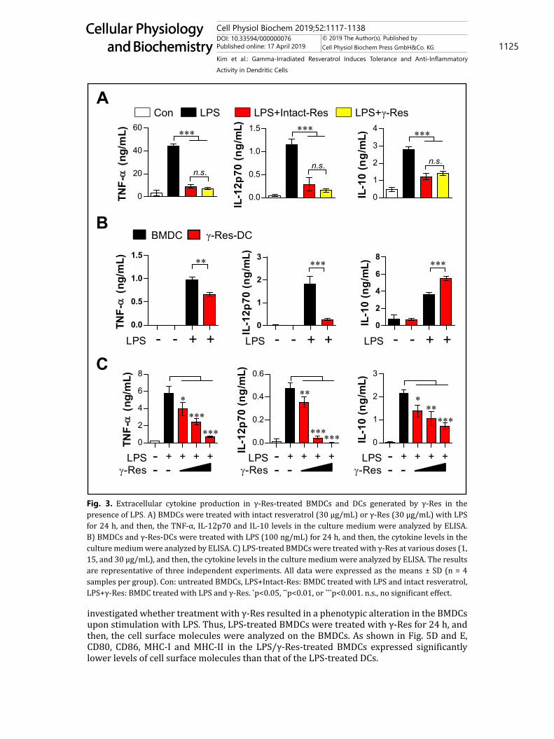
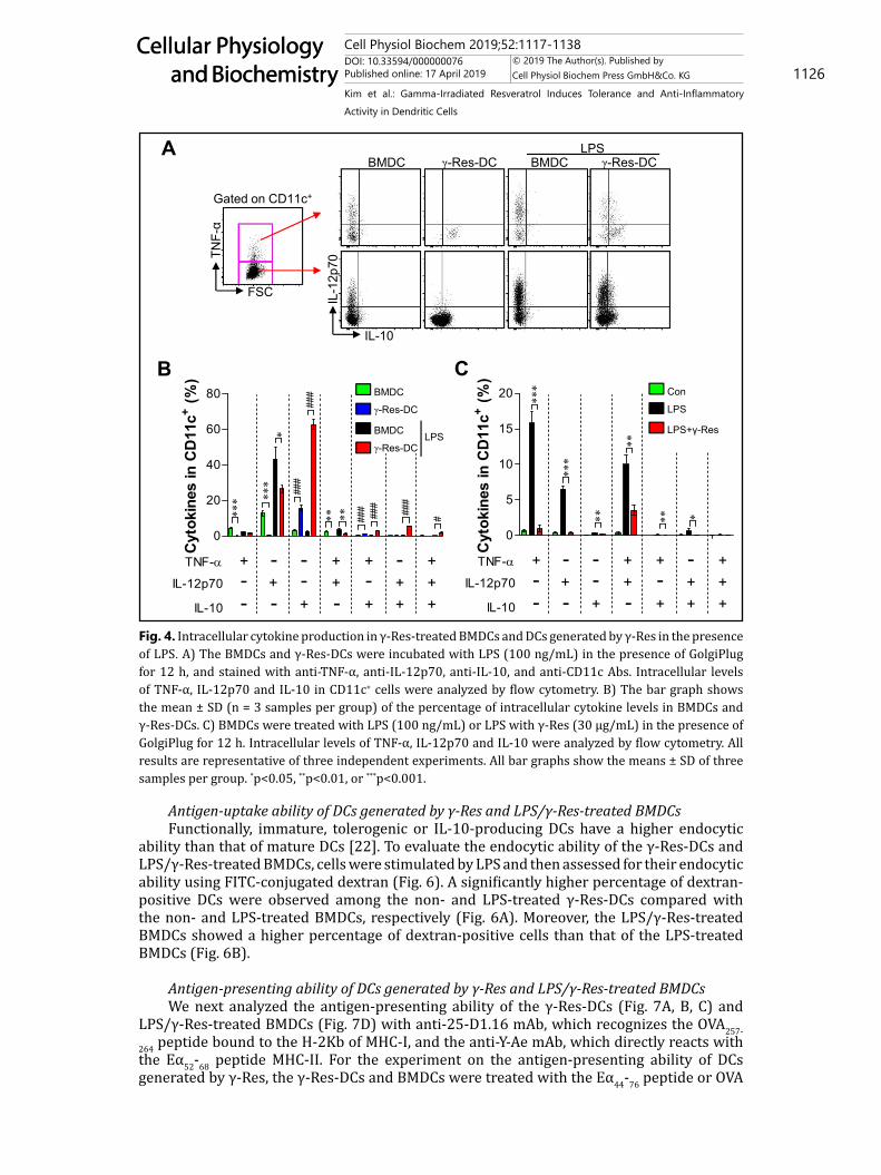
10 μg/mL) of Res induce cell death in normal cells [13]. To confirm the anti-inflammatory activity of γ-Res at a non-toxic dose, we analyzed the decreased ability of cytokines secreted by LPS-treated BMDCs after treatment with 30 μg/mL γ-Res or intact-Res for 24 h. As shown in Fig. 3A, cytokines, including TNF-α, IL-12p70 and IL-10, secreted by the LPS-treated BMDCs were reduced significantly by γ-Res or intact-Res; interestingly, γ-Res and intact-Res did not show any difference. Based on the above results, we identified the functional change (Fig. 3B) of the γ-Res-DCs and the anti-inflammatory activity (Fig. 3C) in LPS-treated BMDCs. To investigate the different ability for cytokine secretion between γ-Res-DCs and BMDCs, we analyzed the pro- (TNF-α and IL-12p70) and anti-inflammatory cytokine (IL-10) production of γ-Res-DCs and BMDCs in the presence and absence of LPS for 24 h. The LPS-treated γ-Res-DCs showed a lower production of TNF-α and IL-12p70 than that of the LPS-treated BMDCs, while IL-10 production was highly induced (Fig. 3B). Additionally, in the study on the anti-inflammatory activity of γ-Res, the LPS-treated DCs that were treated with γ-Res (1, 15, and 30 μg/mL) for 24 h showed a significant reduction in LPS-induced TNF-α, IL-12p70 and IL-10 production compared with the LPS-treated DCs (Fig. 3C). These results were confirmed by intracellular staining, which showed that the γ-Res-DCs in the presence and absence of LPS for 12 h showed an increased intracellular IL-10 expression but a low expression of intracellular TNF-α and IL-12p70 compared with the BMDCs and LPS-treated BMDCs (Fig. 4A, B). Furthermore, the LPS/γ-Res-treated BMDCs significantly decreased the LPS-induced TNF-α, IL-12p70 and IL-10 production compared with the LPS-treated DCs (Fig. 4C). These results indicate that γ-Res treatment during DC differentiation may trigger the generation of IL-10-producing DCs, and γ-Res treatment in LPS-treated BMDCs may lead to an anti-inflammatory activity.
Expression of surface molecules on DCs generated by γ-Res and LPS/γ-Res-treated BMDCsThe IL-10 production level is higher in tolerogenic DCs compared with immunogenic
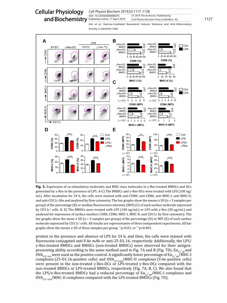
DCs [21]. Thus, we hypothesized that IL-10-producing DCs generated by γ-Res induce tolerogenic phenotypes. To validate this hypothesis, we analyzed the expression of surface molecules (CD80, CD86, MHC-I and MHC-II) on γ-Res-DCs and BMDCs after LPS treatment for 24 h. The γ-Res-DCs in the presence and absence of LPS treatment showed a low expression of surface molecules compared with the non- or LPS-treated BMDCs (Fig. 5A, B, C). We next
Cell Physiol Biochem 2019;52:1117-1138DOI: 10.33594/000000076Published online: 17 April 2019 1125
Cellular Physiology and Biochemistry
Cellular Physiology and Biochemistry
© 2019 The Author(s). Published by Cell Physiol Biochem Press GmbH&Co. KG
Kim et al.: Gamma-Irradiated Resveratrol Induces Tolerance and Anti-Inflammatory Activity in Dendritic Cells
investigated whether treatment with γ-Res resulted in a phenotypic alteration in the BMDCs upon stimulation with LPS. Thus, LPS-treated BMDCs were treated with γ-Res for 24 h, and then, the cell surface molecules were analyzed on the BMDCs. As shown in Fig. 5D and E, CD80, CD86, MHC-I and MHC-II in the LPS/γ-Res-treated BMDCs expressed significantly lower levels of cell surface molecules than that of the LPS-treated DCs.
Fig. 3. Extracellular cytokine production in γ-Res-treated BMDCs and DCs generated by γ-Res in the presence of LPS. A) BMDCs were treated with intact resveratrol (30 μg/mL) or γ-Res (30 μg/mL) with LPS for 24 h, and then, the TNF-α, IL-12p70 and IL-10 levels in the culture medium were analyzed by ELISA. B) BMDCs and γ-Res-DCs were treated with LPS (100 ng/mL) for 24 h, and then, the cytokine levels in the culture medium were analyzed by ELISA. C) LPS-treated BMDCs were treated with γ-Res at various doses (1, 15, and 30 μg/mL), and then, the cytokine levels in the culture medium were analyzed by ELISA. The results are representative of three independent experiments. All data were expressed as the means ± SD (n = 4 samples per group). Con: untreated BMDCs, LPS+Intact-Res: BMDC treated with LPS and intact resveratrol, LPS+γ-Res: BMDC treated with LPS and γ-Res. *p<0.05, **p<0.01, or ***p<0.001. n.s., no significant effect.
0
1
2
3
LPS - - + +
***
IL-1
2p70
(ng/
mL)
0.0
0.5
1.0
1.5**
LPS - - + +
TNF-
(ng/
mL)
0
2
4
6
8***
LPS - - + +IL
-10
(ng/
mL)
BBMDC γ-Res-DC
C
0
2
4
6
8
LPS-Res
-- -
+ + + +
TNF-
(ng/
mL)
****
***0.0
0.2
0.4
0.6
LPS-Res
-- -
+ + + +IL-1
2p70
(ng/
mL)
**
****** 0
1
2
3
LPS-Res
-- -
+ + + +
******
IL-1
0 (n
g/m
L)
Figure 3
0
20
40
60
n.s.
***TN
F-
(ng/
mL)
Con LPS LPS+Intact-Res LPS+γ-Res
0.0
0.5
1.0
1.5
IL-1
2p70
(ng/
mL)
n.s.
***
0
1
2
3
4
IL-1
0 (n
g/m
L)
n.s.
***
A
Cell Physiol Biochem 2019;52:1117-1138DOI: 10.33594/000000076Published online: 17 April 2019 1126
Cellular Physiology and Biochemistry
Cellular Physiology and Biochemistry
© 2019 The Author(s). Published by Cell Physiol Biochem Press GmbH&Co. KG
Kim et al.: Gamma-Irradiated Resveratrol Induces Tolerance and Anti-Inflammatory Activity in Dendritic Cells
Antigen-uptake ability of DCs generated by γ-Res and LPS/γ-Res-treated BMDCsFunctionally, immature, tolerogenic or IL-10-producing DCs have a higher endocytic
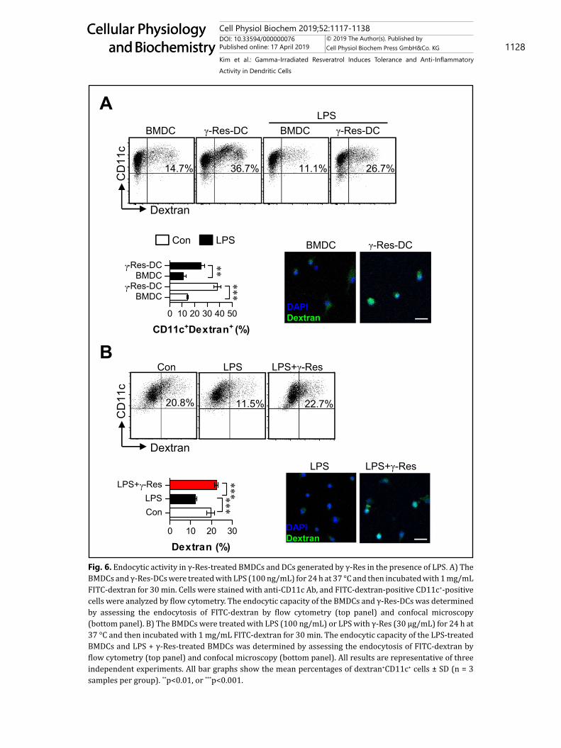
ability than that of mature DCs [22]. To evaluate the endocytic ability of the γ-Res-DCs and LPS/γ-Res-treated BMDCs, cells were stimulated by LPS and then assessed for their endocytic ability using FITC-conjugated dextran (Fig. 6). A significantly higher percentage of dextran-positive DCs were observed among the non- and LPS-treated γ-Res-DCs compared with the non- and LPS-treated BMDCs, respectively (Fig. 6A). Moreover, the LPS/γ-Res-treated BMDCs showed a higher percentage of dextran-positive cells than that of the LPS-treated BMDCs (Fig. 6B).
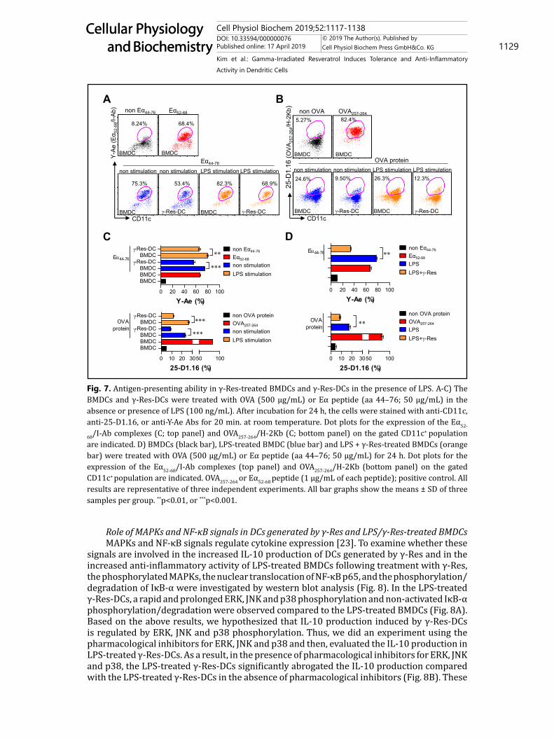
Antigen-presenting ability of DCs generated by γ-Res and LPS/γ-Res-treated BMDCsWe next analyzed the antigen-presenting ability of the γ-Res-DCs (Fig. 7A, B, C) and
LPS/γ-Res-treated BMDCs (Fig. 7D) with anti-25-D1.16 mAb, which recognizes the OVA257-
264 peptide bound to the H-2Kb of MHC-I, and the anti-Y-Ae mAb, which directly reacts with the Eα52-68 peptide MHC-II. For the experiment on the antigen-presenting ability of DCs generated by γ-Res, the γ-Res-DCs and BMDCs were treated with the Eα44-76 peptide or OVA
Fig. 4. Intracellular cytokine production in γ-Res-treated BMDCs and DCs generated by γ-Res in the presence of LPS. A) The BMDCs and γ-Res-DCs were incubated with LPS (100 ng/mL) in the presence of GolgiPlug for 12 h, and stained with anti-TNF-α, anti-IL-12p70, anti-IL-10, and anti-CD11c Abs. Intracellular levels of TNF-α, IL-12p70 and IL-10 in CD11c+ cells were analyzed by flow cytometry. B) The bar graph shows the mean ± SD (n = 3 samples per group) of the percentage of intracellular cytokine levels in BMDCs and γ-Res-DCs. C) BMDCs were treated with LPS (100 ng/mL) or LPS with γ-Res (30 μg/mL) in the presence of GolgiPlug for 12 h. Intracellular levels of TNF-α, IL-12p70 and IL-10 were analyzed by flow cytometry. All results are representative of three independent experiments. All bar graphs show the means ± SD of three samples per group. *p<0.05, **p<0.01, or ***p<0.001.
0
20
40
60
80
TNF-
IL-12p70
IL-10
+ - - + + - +- + - + - + +- - + - + + +
*** **
**
###
###
** ** ###
###
###
#
Cyt
okin
es in
CD
11c+ (%
)
ABMDC γ-Res-DC BMDC γ-Res-DC
LPS
Gated on CD11c+
FSC
TNF-
α
IL-10IL
-12p
70
BBMDC
γ-Res-DC
BMDC
γ-Res-DCLPS
0
5
10
15
20
TNF-
IL-12p70
IL-10
+ - - + + - +- + - + - + +- - + - + + +
***
***
**
**
** *
Cyt
okin
es in
CD
11c+ (%
)
Con
LPS
LPS+γ-Res
Figure 4
C
Cell Physiol Biochem 2019;52:1117-1138DOI: 10.33594/000000076Published online: 17 April 2019 1127
Cellular Physiology and Biochemistry
Cellular Physiology and Biochemistry
© 2019 The Author(s). Published by Cell Physiol Biochem Press GmbH&Co. KG
Kim et al.: Gamma-Irradiated Resveratrol Induces Tolerance and Anti-Inflammatory Activity in Dendritic Cells
protein in the presence and absence of LPS for 24 h, and then, the cells were stained with fluorescein-conjugated anti-Y-Ae mAb or anti-25-D1.16, respectively. Additionally, the LPS/γ-Res-treated BMDCs and BMDCs (non-treated BMDCs) were observed for their antigen-presenting ability according to the same method used in Fig. 7A and B (Fig. 7D). Eα52-68 and OVA257-264 were used as the positive control. A significantly lower percentage of Eα52-68/MHC-I complexes (25-D1.16-positive cells) and OVA257-264/MHC-II complexes (Y-Ae-positive cells) were present in the non-treated γ-Res-DCs or LPS-treated γ-Res-DCs compared with the non-treated BMDCs or LPS-treated BMDCs, respectively (Fig. 7A, B, C). We also found that the LPS/γ-Res-treated BMDCs had a reduced percentage of Eα52-68/MHC-I complexes and OVA257-264/MHC-II complexes compared with the LPS-treated BMDCs (Fig. 7D).
Fig. 5. Expression of co-stimulatory molecules and MHC class molecules in γ-Res-treated BMDCs and DCs generated by γ-Res in the presence of LPS. A-C) The BMDCs and γ-Res-DCs were treated with LPS (100 ng/mL). After incubation for 24 h, the cells were stained with anti-CD80, anti-CD86, anti-MHC-I, anti-MHC-II, and anti-CD11c Abs and analyzed by flow cytometry. The bar graphs show the means ± SD (n = 3 samples per group) of the percentage (B) or median fluorescence intensity (MFI) (C) of each surface molecule expressed by CD11c+ cells. D, E) The BMDCs were treated with LPS (100 ng/mL) or LPS with γ-Res (30 μg/mL) and analyzed for expression of surface markers CD80, CD86, MHC-I, MHC-II, and CD11c by flow cytometry. The bar graphs show the mean ± SD (n = 3 samples per group) of the percentage (D) or MFI (E) of each surface molecule expressed by CD11c+ cells. All results are representative of three independent experiments. All bar graphs show the means ± SD of three samples per group. **p<0.01, or ***p<0.001.
CD11c
MHC-II
MHC-I
CD86
CD80
BMDC BMDC γ-Res-DCLPSA B
0 20 40 60 80
BMDC-Res-DCBMDC
-Res-DC
******
MHC-II (%)
0 1 2 3
BMDC-Res-DCBMDC
-Res-DC
******
MHC-II (MFI)
LPSCon
C
LPSCon
γ-Res-DC
0 20 40 60 80100
BMDC-Res-DCBMDC
-Res-DC
******
CD80 (%)
0 20 40 60 80100
BMDC-Res-DCBMDC
-Res-DC
******
MHC-I (%)
0 2 4 6 8
BMDC-Res-DCBMDC
-Res-DC
******
CD80 (MFI)
0 1 2 3 4 5
BMDC-Res-DCBMDC
-Res-DC
******
MHC-I (MFI)
0 20 40 60 80100
BMDC-Res-DCBMDC
-Res-DC
******
CD86 (%)
0 2 4 6 8 10
BMDC-Res-DCBMDC
-Res-DC
******
CD86 (MFI)
0
20
40
60
80 ***
CD
80 (%
)
0
30
60
90 ***
CD
86 (%
)
0
30
60
90 ***
MH
C-I
(%)
0
20
40
60
80 ***
MH
C-II
(%)
ConLPSLPS+γ-Res
01020304050 **
CD
80 (M
FI)
0
20
40
60
80***
MH
C-II
(MFI
)(x103)(x103)
(x103) (x103)
0
40
80
120 ***
CD
86 (M
FI)
0246810 ***
MH
C-I
(MFI
)
D E
Figure 5
ConLPSLPS+γ-Res
(x102)(x102)
(x102) (x103)
Cell Physiol Biochem 2019;52:1117-1138DOI: 10.33594/000000076Published online: 17 April 2019 1128
Cellular Physiology and Biochemistry
Cellular Physiology and Biochemistry
© 2019 The Author(s). Published by Cell Physiol Biochem Press GmbH&Co. KG
Kim et al.: Gamma-Irradiated Resveratrol Induces Tolerance and Anti-Inflammatory Activity in Dendritic Cells
Fig. 6. Endocytic activity in γ-Res-treated BMDCs and DCs generated by γ-Res in the presence of LPS. A) The BMDCs and γ-Res-DCs were treated with LPS (100 ng/mL) for 24 h at 37 °C and then incubated with 1 mg/mL FITC-dextran for 30 min. Cells were stained with anti-CD11c Ab, and FITC-dextran-positive CD11c+-positive cells were analyzed by flow cytometry. The endocytic capacity of the BMDCs and γ-Res-DCs was determined by assessing the endocytosis of FITC-dextran by flow cytometry (top panel) and confocal microscopy (bottom panel). B) The BMDCs were treated with LPS (100 ng/mL) or LPS with γ-Res (30 μg/mL) for 24 h at 37 °C and then incubated with 1 mg/mL FITC-dextran for 30 min. The endocytic capacity of the LPS-treated BMDCs and LPS + γ-Res-treated BMDCs was determined by assessing the endocytosis of FITC-dextran by flow cytometry (top panel) and confocal microscopy (bottom panel). All results are representative of three independent experiments. All bar graphs show the mean percentages of dextran+CD11c+ cells ± SD (n = 3 samples per group). **p<0.01, or ***p<0.001.
Dextran
CD11c
BMDC BMDC γ-Res-DCLPS
γ-Res-DC
14.7% 36.7% 11.1% 26.7%
A
0 10 20 30 40 50
BMDC-Res-DCBMDC
-Res-DC
*****
CD11c+Dextran+ (%)
LPSCon BMDC γ-Res-DC
11.5% 22.7%
0 10 20 30
ConLPS-ResLPS+
******
Dextran (%)
Con LPS LPS+γ-Res
LPS LPS+γ-Res
DAPIDextran
Figure 6
20.8%
Dextran
CD11c
B
DAPIDextran
Cell Physiol Biochem 2019;52:1117-1138DOI: 10.33594/000000076Published online: 17 April 2019 1129
Cellular Physiology and Biochemistry
Cellular Physiology and Biochemistry
© 2019 The Author(s). Published by Cell Physiol Biochem Press GmbH&Co. KG
Kim et al.: Gamma-Irradiated Resveratrol Induces Tolerance and Anti-Inflammatory Activity in Dendritic Cells
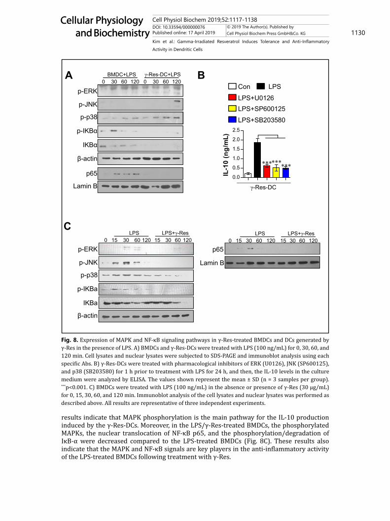
Role of MAPKs and NF-κB signals in DCs generated by γ-Res and LPS/γ-Res-treated BMDCsMAPKs and NF-κB signals regulate cytokine expression [23]. To examine whether these
signals are involved in the increased IL-10 production of DCs generated by γ-Res and in the increased anti-inflammatory activity of LPS-treated BMDCs following treatment with γ-Res, the phosphorylated MAPKs, the nuclear translocation of NF-κB p65, and the phosphorylation/degradation of IκB-α were investigated by western blot analysis (Fig. 8). In the LPS-treated γ-Res-DCs, a rapid and prolonged ERK, JNK and p38 phosphorylation and non-activated IκB-α phosphorylation/degradation were observed compared to the LPS-treated BMDCs (Fig. 8A). Based on the above results, we hypothesized that IL-10 production induced by γ-Res-DCs is regulated by ERK, JNK and p38 phosphorylation. Thus, we did an experiment using the pharmacological inhibitors for ERK, JNK and p38 and then, evaluated the IL-10 production in LPS-treated γ-Res-DCs. As a result, in the presence of pharmacological inhibitors for ERK, JNK and p38, the LPS-treated γ-Res-DCs significantly abrogated the IL-10 production compared with the LPS-treated γ-Res-DCs in the absence of pharmacological inhibitors (Fig. 8B). These
Fig. 7. Antigen-presenting ability in γ-Res-treated BMDCs and γ-Res-DCs in the presence of LPS. A-C) The BMDCs and γ-Res-DCs were treated with OVA (500 μg/mL) or Eα peptide (aa 44–76; 50 μg/mL) in the absence or presence of LPS (100 ng/mL). After incubation for 24 h, the cells were stained with anti-CD11c, anti-25-D1.16, or anti-Y-Ae Abs for 20 min. at room temperature. Dot plots for the expression of the Eα52-
68/I-Ab complexes (C; top panel) and OVA257-264/H-2Kb (C; bottom panel) on the gated CD11c+ population are indicated. D) BMDCs (black bar), LPS-treated BMDC (blue bar) and LPS + γ-Res-treated BMDCs (orange bar) were treated with OVA (500 μg/mL) or Eα peptide (aa 44–76; 50 μg/mL) for 24 h. Dot plots for the expression of the Eα52-68/I-Ab complexes (top panel) and OVA257-264/H-2Kb (bottom panel) on the gated CD11c+ population are indicated. OVA257-264 or Eα52-68 peptide (1 μg/mL of each peptide); positive control. All results are representative of three independent experiments. All bar graphs show the means ± SD of three samples per group. **p<0.01, or ***p<0.001.
8.24% 68.4%
75.3% 53.4% 82.3% 68.9%
CD11c
Y-Ae
(Eα 5
2-68
/I-Ab
)
A
C
BMDC BMDC γ-Res-DCγ-Res-DC
LPS stimulation
Eα52-68non Eα44-76
LPS stimulation
Eα44-76
non stimulation non stimulation
0 20 40 60 80 100
BMDCBMDCBMDC
-Res-DCBMDC
-Res-DC
***
**E44-76
Y-Ae (%)
5.27% 82.4%
24.6% 9.50% 26.3% 12.3%
CD11c
25-D
1.16
(OVA
257-
264/H
-2Kb
)
BMDC BMDC γ-Res-DCγ-Res-DC
LPS stimulation
OVA257-264non OVA
LPS stimulationOVA protein
non stimulation non stimulation
0 10 20 30
BMDCBMDCBMDC
-Res-DCBMDC
-Res-DC
50 100
***
***OVAprotein
25-D1.16 (%)
non OVA proteinOVA257-264
non stimulationLPS stimulation
non Eα44-76
Eα52-68
non stimulationLPS stimulation
BMDC BMDCBMDC BMDC
Figure 7
0 20 40 60 80 100
**E44-76
Y-Ae (%)
0 10 20 3050 100
**OVAprotein
25-D1.16 (%)
non Eα44-76
Eα52-68
LPSLPS+γ-Res
B
D
non OVA proteinOVA257-264
LPSLPS+γ-Res
Cell Physiol Biochem 2019;52:1117-1138DOI: 10.33594/000000076Published online: 17 April 2019 1130
Cellular Physiology and Biochemistry
Cellular Physiology and Biochemistry
© 2019 The Author(s). Published by Cell Physiol Biochem Press GmbH&Co. KG
Kim et al.: Gamma-Irradiated Resveratrol Induces Tolerance and Anti-Inflammatory Activity in Dendritic Cells
results indicate that MAPK phosphorylation is the main pathway for the IL-10 production induced by the γ-Res-DCs. Moreover, in the LPS/γ-Res-treated BMDCs, the phosphorylated MAPKs, the nuclear translocation of NF-κB p65, and the phosphorylation/degradation of IκB-α were decreased compared to the LPS-treated BMDCs (Fig. 8C). These results also indicate that the MAPK and NF-κB signals are key players in the anti-inflammatory activity of the LPS-treated BMDCs following treatment with γ-Res.
Fig. 8. Expression of MAPK and NF-κB signaling pathways in γ-Res-treated BMDCs and DCs generated by γ-Res in the presence of LPS. A) BMDCs and γ-Res-DCs were treated with LPS (100 ng/mL) for 0, 30, 60, and 120 min. Cell lysates and nuclear lysates were subjected to SDS-PAGE and immunoblot analysis using each specific Abs. B) γ-Res-DCs were treated with pharmacological inhibitors of ERK (U0126), JNK (SP600125), and p38 (SB203580) for 1 h prior to treatment with LPS for 24 h, and then, the IL-10 levels in the culture medium were analyzed by ELISA. The values shown represent the mean ± SD (n = 3 samples per group). ***p<0.001. C) BMDCs were treated with LPS (100 ng/mL) in the absence or presence of γ-Res (30 μg/mL) for 0, 15, 30, 60, and 120 min. Immunoblot analysis of the cell lysates and nuclear lysates was performed as described above. All results are representative of three independent experiments.
p65
p-ERK
p-JNK
p-p38
IKBα
p-IKBα
Figure 8
0 30 60 120 0 30 60 120BMDC+LPS γ-Res-DC+LPS
0 15 30 60LPS LPS+γ-Res
120 15 30 60 120
p65p-ERK
p-JNK
p-p38
IKBa
p-IKBa
0 15 30 60LPS LPS+γ-Res
120 15 30 60 120
0.0
0.5
1.0
1.5
2.0
2.5
*********
IL-1
0 (n
g/m
L)
Con
γ-Res-DC
LPSLPS+U0126LPS+SP600125LPS+SB203580
A B
C
β-actin
Lamin B
Lamin B
β-actin
Cell Physiol Biochem 2019;52:1117-1138DOI: 10.33594/000000076Published online: 17 April 2019 1131
Cellular Physiology and Biochemistry
Cellular Physiology and Biochemistry
© 2019 The Author(s). Published by Cell Physiol Biochem Press GmbH&Co. KG
Kim et al.: Gamma-Irradiated Resveratrol Induces Tolerance and Anti-Inflammatory Activity in Dendritic Cells
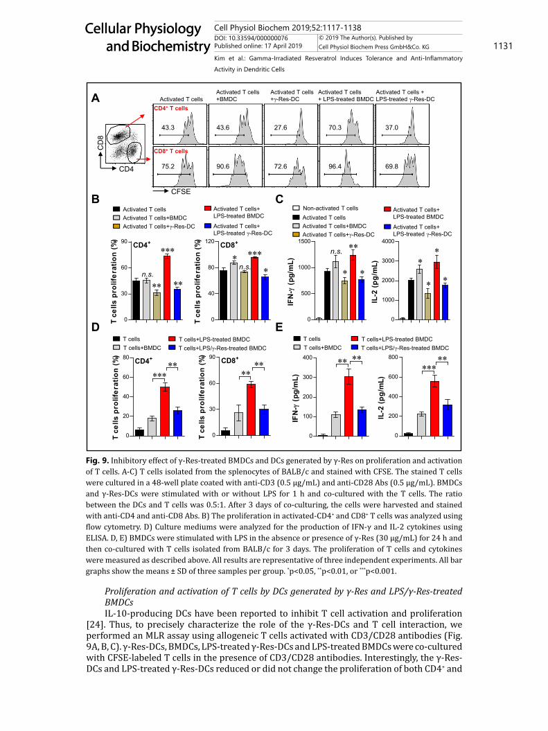
Proliferation and activation of T cells by DCs generated by γ-Res and LPS/γ-Res-treated BMDCsIL-10-producing DCs have been reported to inhibit T cell activation and proliferation
[24]. Thus, to precisely characterize the role of the γ-Res-DCs and T cell interaction, we performed an MLR assay using allogeneic T cells activated with CD3/CD28 antibodies (Fig. 9A, B, C). γ-Res-DCs, BMDCs, LPS-treated γ-Res-DCs and LPS-treated BMDCs were co-cultured with CFSE-labeled T cells in the presence of CD3/CD28 antibodies. Interestingly, the γ-Res-DCs and LPS-treated γ-Res-DCs reduced or did not change the proliferation of both CD4+ and
Fig. 9. Inhibitory effect of γ-Res-treated BMDCs and DCs generated by γ-Res on proliferation and activation of T cells. A-C) T cells isolated from the splenocytes of BALB/c and stained with CFSE. The stained T cells were cultured in a 48-well plate coated with anti-CD3 (0.5 μg/mL) and anti-CD28 Abs (0.5 μg/mL). BMDCs and γ-Res-DCs were stimulated with or without LPS for 1 h and co-cultured with the T cells. The ratio between the DCs and T cells was 0.5:1. After 3 days of co-culturing, the cells were harvested and stained with anti-CD4 and anti-CD8 Abs. B) The proliferation in activated-CD4+ and CD8+ T cells was analyzed using flow cytometry. D) Culture mediums were analyzed for the production of IFN-γ and IL-2 cytokines using ELISA. D, E) BMDCs were stimulated with LPS in the absence or presence of γ-Res (30 μg/mL) for 24 h and then co-cultured with T cells isolated from BALB/c for 3 days. The proliferation of T cells and cytokines were measured as described above. All results are representative of three independent experiments. All bar graphs show the means ± SD of three samples per group. *p<0.05, **p<0.01, or ***p<0.001.
43.3 43.6 27.6 70.3 37.0
75.2 90.6 72.6 96.4 69.8
Activated T cellsActivated T cells+BMDCActivated T cells+γ-Res-DC
Activated T cells+ LPS-treated BMDC
Activated T cells+ LPS-treated γ-Res-DC
0
40
80
120 CD8+
***n.s.
**
T ce
lls p
rolif
erat
ion
(%)
0
30
60
90 CD4+
***
n.s.
** **
T ce
lls p
rolif
erat
ion
(%)
Activated T cellsActivated T cells +BMDC
Activated T cells +γ-Res-DC
Activated T cells + LPS-treated BMDC
Activated T cells + LPS-treated γ-Res-DC
CD4+ T cells
CD8+ T cells
CD4
CD
8
CFSE
0
1000
2000
3000
4000
**
**
IL-2
(pg/
mL)
0
500
1000
1500**n.s.
* *IF
N-
(pg/
mL)
Activated T cellsActivated T cells+BMDCActivated T cells+γ-Res-DC
Activated T cells+ LPS-treated BMDC
Activated T cells+ LPS-treated γ-Res-DC
Non-activated T cells
Figure 9
D E
0
20
40
60
80 CD4+**
***
T ce
lls p
rolif
erat
ion
(%)
0
30
60
90 CD8+**
**
T ce
lls p
rolif
erat
ion
(%)
0
200
400
600
800 *****
IL-2
(pg/
mL)
0
100
200
300
400 ****
IFN
- (p
g/m
L)
T cellsT cells+BMDC
T cells+LPS-treated BMDCT cells+LPS/γ-Res-treated BMDC
T cellsT cells+BMDC
T cells+LPS-treated BMDCT cells+LPS/γ-Res-treated BMDC
A
B C

Cell Physiol Biochem 2019;52:1117-1138DOI: 10.33594/000000076Published online: 17 April 2019 1132
Cellular Physiology and Biochemistry
Cellular Physiology and Biochemistry
© 2019 The Author(s). Published by Cell Physiol Biochem Press GmbH&Co. KG
Kim et al.: Gamma-Irradiated Resveratrol Induces Tolerance and Anti-Inflammatory Activity in Dendritic Cells
CD8+ T cells and the production of IFN-γ and IL-2 compared with activated T cells, while the BMDCs and LPS-treated BMDCs, respectively, did not change or become induced (Fig. 9A, B, C). Furthermore, we evaluated the capacity of the LPS/γ-Res-treated BMDCs to induce T cell proliferation and activation using allogeneic T cells. As shown in Fig. 9D and E, T cell proliferation and production of IFN-γ and IL-2 were not induced by the LPS/γ-Res-treated BMDCs compared to the LPS-treated BMDCs. These finding indicate that the γ-Res-DCs and the presence of γ-Res in the LPS-treated BMDCs led to the inhibitory effect on the T cells.
Induction of regulatory T cells by DCs generated by γ-ResAs a next step, we investigated whether regulatory T cells (Foxp3+CD25+CD4+ T cells)
are induced during γ-Res-DC and CD4 T cell interaction because IL-10-producing DCs can induce the differentiation of naïve T cells towards regulatory T cells [21]. Thus, we evaluated the capacity of the γ-Res-DCs to induce regulatory T cells using the same experimental conditions described in Fig. 9B and C. As shown in Fig. 10, the γ-Res-DCs and LPS-treated γ-Res-DCs polarized naïve CD4+ T cells towards the regulatory T cells, while BMDCs and LPS-treated BMDCs were not induced.
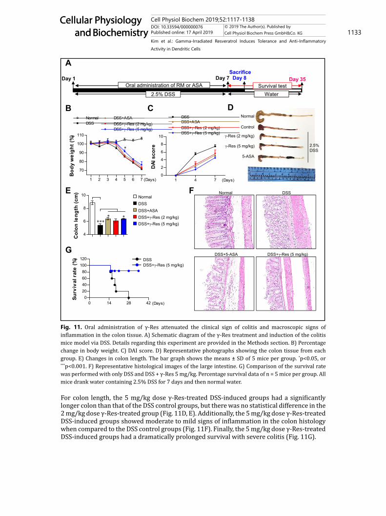
Protective effect by the γ-Res treatment in a DSS-induced colitis modelThe above results regarding the γ-Res-generated IL-10-producing DCs and anti-
inflammatory activity motivated us to test the protective efficacy of γ-Res against DSS-induced colitis. For this purpose, we evaluated the body weight, disease activity index (DAI) score, colon length, colon histology, and survival rate following γ-Res treatment in 2.5% DSS-induced mice (Fig. 11A, experimental design indicated by the schematic diagram). For body weight, the 5 mg/kg dose γ-Res-treated DSS-induced groups showed a significantly increased body weight on day 5 compared with the DSS control groups, but there was no statistical difference on the other days and in the 2 mg/kg dose γ-Res-treated group (Fig. 11B). For the DAI score, the ASA-treated (4 days; P < 0.05, 7 days; P < 0.05), 2 mg/kg (4 days; P < 0.05, 7 days; n.s.) and 5 mg/kg (4 days; P < 0.01, 7 days; P < 0.01) dose γ-Res-treated DSS-induced groups had a significantly lower score than that of the DSS control groups (Fig. 11C).
CD25
Foxp
3T cells+BMDC
T cells+γ-Res-DC
T cells+ LPS-treated BMDC
T cells+ LPS-treated γ-Res-DC
13.2% 14.7%
32.6% 42.3%
Figure 10
T cells+BMDC
T cells+γ-Res-DC
T cells+ LPS-treated BMDC
T cells+ LPS-treated γ-Res-DC
0
10
20
30
40
50
60
70 CD4+
******
CD
25+ Fo
xp3+ c
ells
(%)
Gated on CD3+CD4+ cellsA B
Fig. 10. Induction of regulatory T cells by DCs generated by γ-Res. A, B) CD4+ T cells were isolated as described in the Methods section. BMDCs and γ-Res-DCs were co-cultured with isolated CD4+ T cells for 3 days and stained with anti-CD25 and anti-Foxp3Abs. The CD25+Foxp3+CD4+ T cells were analyzed by flow cytometry. B) The bar graph shows the percentage of CD25+Foxp3+ regulatory T cells among the CD4+ T cells. All results are representative of three independent experiments. The values shown represent the mean ± SD (n = 3 samples per group). ***p<0.001.
Cell Physiol Biochem 2019;52:1117-1138DOI: 10.33594/000000076Published online: 17 April 2019 1133
Cellular Physiology and Biochemistry
Cellular Physiology and Biochemistry
© 2019 The Author(s). Published by Cell Physiol Biochem Press GmbH&Co. KG
Kim et al.: Gamma-Irradiated Resveratrol Induces Tolerance and Anti-Inflammatory Activity in Dendritic Cells
For colon length, the 5 mg/kg dose γ-Res-treated DSS-induced groups had a significantly longer colon than that of the DSS control groups, but there was no statistical difference in the 2 mg/kg dose γ-Res-treated group (Fig. 11D, E). Additionally, the 5 mg/kg dose γ-Res-treated DSS-induced groups showed moderate to mild signs of inflammation in the colon histology when compared to the DSS control groups (Fig. 11F). Finally, the 5 mg/kg dose γ-Res-treated DSS-induced groups had a dramatically prolonged survival with severe colitis (Fig. 11G).
4
6
8
10
****
*
Col
on le
ngth
(cm
)
1 2 3 4 5 6 7
70
80
90
100
110
NormalDSS
DSS+ASADSS+γ-Res (2 mg/kg)DSS+γ-Res (5 mg/kg)
*
(Days)
Bod
y w
eigh
t (%
)
1 4 70
2
4
6
8
10
DSSDSS+ASADSS+γ-Res (2 mg/kg)DSS+γ-Res (5 mg/kg)
(Days)
DAI
sco
re
Normal
Control
γ-Res (2 mg/kg)
γ-Res (5 mg/kg)
5-ASA
2.5% DSS
Day 1 Day 7 Day 8
2.5% DSS
Oral administration of RM or ASA
Sacrifice
NormalDSSDSS+ASADSS+γ-Res (2 mg/kg)DSS+γ-Res (5 mg/kg)
Figure 11A
B C D
E
Water
Day 35Survival test
G
0 14 28 420
20406080
100120
(Days)
Surv
ival
rat
e (%
)
F Normal DSS
DSS+γ-Res (5 mg/kg)DSS+5-ASA
DSS+γ-Res (5 mg/kg)DSS
Fig. 11. Oral administration of γ-Res attenuated the clinical sign of colitis and macroscopic signs of inflammation in the colon tissue. A) Schematic diagram of the γ-Res treatment and induction of the colitis mice model via DSS. Details regarding this experiment are provided in the Methods section. B) Percentage change in body weight. C) DAI score. D) Representative photographs showing the colon tissue from each group. E) Changes in colon length. The bar graph shows the means ± SD of 5 mice per group. *p<0.05, or ***p<0.001. F) Representative histological images of the large intestine. G) Comparison of the survival rate was performed with only DSS and DSS + γ-Res 5 mg/kg. Percentage survival data of n = 5 mice per group. All mice drank water containing 2.5% DSS for 7 days and then normal water.
Cell Physiol Biochem 2019;52:1117-1138DOI: 10.33594/000000076Published online: 17 April 2019 1134
Cellular Physiology and Biochemistry
Cellular Physiology and Biochemistry
© 2019 The Author(s). Published by Cell Physiol Biochem Press GmbH&Co. KG
Kim et al.: Gamma-Irradiated Resveratrol Induces Tolerance and Anti-Inflammatory Activity in Dendritic Cells
Immune responses induced by the γ-Res treatment in the DSS-induced colitis modelThe effective inhibition of activated CD4 T cell responses is an important step for
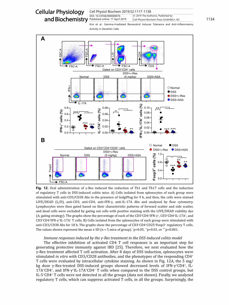
generating protective immunity against IBD [25]. Therefore, we next evaluated how the γ-Res treatment affected T cell activation. After 8 days of DSS induction, splenocytes were stimulated in vitro with CD3/CD28 antibodies, and the phenotypes of the responding CD4+ T cells were evaluated by intracellular cytokine staining. As shown in Fig. 12A, the 5 mg/kg dose γ-Res-treated DSS-induced groups showed decreased levels of IFN-γ+CD4+, IL-17A+CD4+, and IFN-γ+IL-17A+CD4+ T cells when compared to the DSS control groups, but IL-5+CD4+ T cells were not detected in all the groups (data not shown). Finally, we analyzed regulatory T cells, which can suppress activated T cells, in all the groups. Surprisingly, the
Fig. 12. Oral administration of γ-Res induced the reduction of Th1 and Th17 cells and the induction of regulatory T cells in DSS-induced colitis mice. A) Cells isolated from splenocytes of each group were stimulated with anti-CD3/CD38 Abs in the presence of GolgiPlug for 9 h, and then, the cells were stained LIVE/DEAD (L/D), anti-CD3, anti-CD4, anti-IFN-γ, anti-IL-17A Abs and analyzed by flow cytometry. Lymphocytes were then gated based on their characteristic patterns of forward scatter and side scatter, and dead cells were excluded by gating out cells with positive staining with the LIVE/DEAD viability dye (A; gating strategy). The graphs show the percentage of each of the CD3+CD4+IFN-γ+, CD3+CD4+IL-17A+, and CD3+CD4+IFN-γ+IL-17A+ T cells. B) Cells isolated from the splenocytes of each group were stimulated with anti-CD3/CD38 Abs for 18 h. The graphs show the percentage of CD3+CD4+CD25+Foxp3+ regulatory T cells. The values shown represent the mean ± SD (n = 5 mice of group). *p<0.05, **p<0.01, or ***p<0.001.
Figure 12
FSC-A
Foxp
3
Normal DSS DSS+ASADSS+γ-Res (5 mg/kg)
FSC-A
SSC
-A
FSC-H
FSC
-A
FSC-A
L/D
CD3
CD
4
IL-17A
IFN
-γ
Normal DSS DSS+ASADSS+γ-Res (5 mg/kg)
Gated on CD3+CD4+CD25+ cells
Gated on CD3+CD4+ cells
0.0
0.2
0.4
0.6
0.8
***
*** ***
IFN
- +
cells
(%)
0.00
0.02
0.04
0.06
0.08
0.10 **
*** ***
IFN
- + I
L-17
A+
cells
(%)
0.00
0.02
0.04
0.06
0.08*
* **
IL-1
7A+
cells
(%)
A
B
0.0
0.4
0.8
1.2
***
** **
Foxp
3+ cel
ls (%
)
Normal DSSDSS+γ-Res DSS+ASA
NormalDSSDSS+γ-Res DSS+ASA
Cell Physiol Biochem 2019;52:1117-1138DOI: 10.33594/000000076Published online: 17 April 2019 1135
Cellular Physiology and Biochemistry
Cellular Physiology and Biochemistry
© 2019 The Author(s). Published by Cell Physiol Biochem Press GmbH&Co. KG
Kim et al.: Gamma-Irradiated Resveratrol Induces Tolerance and Anti-Inflammatory Activity in Dendritic Cells
γ-Res-treated DSS-induced groups had dramatically increased levels of Foxp3+ regulatory T cells when compared to the DSS control group (Fig. 12B). These results indicate that the decreased T cell activation and increased regulatory T cells induced by the γ-Res treatment may contribute to the protective effect against DSS-induced colitis.
Discussion
Resveratrol, which is an anti-inflammatory component, ameliorates symptoms of inflammatory diseases, such as inflammatory bowel disease (IBD), rheumatoid arthritis, and systemic lupus erythematous, although it causes cell death when used in high concentrations [13, 26]. The results of this study demonstrated that the structurally changed compound, γ-Res, of resveratrol via gamma irradiation, which is not toxic to dendritic cells (DCs) for a high-dose treatment, has immunosuppressive properties; it induces the generation of tolerogenic DCs with decreased surface molecule expression (CD80, CD86, MHC-I, and MHC-II), and it increases IL-10 production without altering the TNF-α and IL-12p70 production and anti-inflammatory activity in activated DCs, thereby promoting the inhibition of T cell proliferation/activation and/or the induction of regulatory T cells via the interaction with DCs in vitro.
The components involved in the activation/maturation and differentiation of DCs can be used as potential candidates for valuable therapeutic targets in IBD because intestinal DCs strongly express microbial receptors, and this causes rapid inflammatory responses, including T cell activation and polarization of Th1, Th2, and Th17, mediated by the overexpression of surface molecules, cytokines, and chemokines [27, 28]. Therefore, if particular components inhibit the inflammatory response of DCs, it would be a promising treatment. Taking into account the inflammation, a recent study showed that blocking treatments for TNF-α, such as by using etanercept and adalimumab, induced excessive inflammation of IBD, translating into changes in the DC function. These treatments were shown to downregulate the expression of surface molecules, such as CD83, CD80, and CD86, on DCs and to reduce their T cell stimulatory capacity [29] Recently, granulocyte-colony stimulating factor (G-CSF), which is a proliferation, differentiation, and activation factor for hematopoietic cells, treatment in Crohn’s disease patients showed a therapeutic effect via reduction of IFN-γ-producing T cells and induction of IL-10-producing and Foxp3+-expressing CD4+ T cells [30]. The function of G-CSF as a treatment was recently reported as inducing tolerogenic phenotypes during DC differentiation, and the DCs also polarize naïve T cells to a regulatory T cell-like functional profile [31, 32]. Here, regulatory T cells (Foxp3+CD4+ T cells) have a role in immune regulation, such as inhibiting inflammation; however, reductions of these cells are associated with IBD pathogenesis [33]. The immune-modulating components involved in tolerogenic properties and anti-inflammatory activity during DC activation/differentiation may therefore serve as good candidates for IBD treatment according to their regulation of excessive inflammatory responses and induction of immune tolerance. We used γ-Res as a treatment for the DSS-induced colitis because it contributes to the generation of tolerogenic DCs and anti-inflammatory effects.
Recently, inhibitors of both MAPK and NF-κB signaling, which are key regulators of an excessive inflammatory response, and the proteins associated with the signaling have been evaluated due to their potential application in the treatment for IBD [34-36]. Takada et al. reported that Fos proteins and Bay 11-7085 are inhibitors of NF-κB signals and induce less severe clinical score and lower pro-inflammatory cytokine production in mice susceptible to DSS-induced colitis [34]. Among the MAPK inhibitor treatments, SB203580, which is a p38 inhibitor, also reduces disease activity, histological disease score, and pro-inflammatory cytokines in a DSS-induced colitis model [35]. In addition, a STAT3 inhibitor, subject to phosphorylation by JNK1/2, has been evaluated with respect to T-cell-mediated intestinal inflammation in CD patients; this treatment reduced T-cell-mediated intestinal inflammation [36].
Cell Physiol Biochem 2019;52:1117-1138DOI: 10.33594/000000076Published online: 17 April 2019 1136
Cellular Physiology and Biochemistry
Cellular Physiology and Biochemistry
© 2019 The Author(s). Published by Cell Physiol Biochem Press GmbH&Co. KG
Kim et al.: Gamma-Irradiated Resveratrol Induces Tolerance and Anti-Inflammatory Activity in Dendritic Cells
Conclusion
In conclusion, new therapeutic strategies and the development of treatments for IBD require the initiation of immune tolerance and multiple inhibitions of major inflammatory factors of IBD. Our data show that γ-Res has a critical role in anti-inflammatory activity via blocking MAPK and NF-κB signals in activated DCs and inducing tolerogenic DCs that are capable of both inhibiting T cell activation/proliferation and inducing regulatory T cells. Importantly, γ-Res treatment conferred protective immunity and imparted protection to DSS-induced colitis in mice. Although speculative, our in vivo results suggest that the inhibition of excessive inflammation by the γ-Res treatment is a result of blocking the MAPK and NF-κB signals in activated DCs and inducing tolerogenic DCs. Thus, γ-Res has the potential to be an effective treatment against IBD.
Acknowledgements
This study was supported by a National Research Foundation of Korea grant funded by the Korean Government (Grant nos. 2018M2A2B3A02072069). W. S. Kim, H.-Y. Song, and E.-B. Byun: Conceived and designed the experiments; W. S. Kim, H.-Y. Song, S. Mushtag, J.-M. Kim, J.-M. Yuk, and E.-H. Byun: Performed the experiments; W. S. Kim, S. Mushtag, J.-M. Kim, J.-M. Yuk, and E.-H. Byun: Contributed reagents/materials/analysis tools; W. S. Kim, and E.-B. Byun: Wrote the paper.
Disclosure Statement
No conflicts of interest exist.
References
1 Matricon J, Barnich N, Ardid D: Immunopathogenesis of inflammatory bowel disease. Self Nonself 2010;1:299-309.
2 Hevia A, Lopez P, Suarez A, Jacquot C, Urdaci MC, Margolles A, Sanchez B: Association of levels of antibodies from patients with inflammatory bowel disease with extracellular proteins of food and probiotic bacteria. Biomed Res Int 2014;2014:351204.
3 Kakazu T, Hara J, Matsumoto T, Nakamura S, Oshitani N, Arakawa T, Kitano A, Nakatani K, Kinjo F, Kuroki T: Type 1 T-helper cell predominance in granulomas of Crohn’s disease. Am J Gastroenterol 1999;94:2149-2155.
4 Beltran CJ, Candia E, Erranz B, Figueroa C, Gonzalez MJ, Quera R, Hermoso MA: Peripheral cytokine profile in Chilean patients with Crohn’s disease and ulcerative colitis. Eur Cytokine Netw 2009;20:33-38.
5 Silva FA, Rodrigues BL, Ayrizono ML, Leal RF: The Immunological Basis of Inflammatory Bowel Disease. Gastroenterol Res Pract 2016;2016:2097274.
6 Muzes G, Molnar B, Tulassay Z, Sipos F: Changes of the cytokine profile in inflammatory bowel diseases. World J Gastroenterol 2012;18:5848-5861.
7 Strober W, Fuss IJ: Proinflammatory cytokines in the pathogenesis of inflammatory bowel diseases. Gastroenterology 2011;140:1756-1767.
8 Mazzon E, Muia C, Paola RD, Genovese T, Menegazzi M, De Sarro A, Suzuki H, Cuzzocrea S: Green tea polyphenol extract attenuates colon injury induced by experimental colitis. Free Radic Res 2005;39:1017-1025.
9 Varilek GW, Yang F, Lee EY, deVilliers WJ, Zhong J, Oz HS, Westberry KF, McClain CJ: Green tea polyphenol extract attenuates inflammation in interleukin-2-deficient mice, a model of autoimmunity. J Nutr 2001;131:2034-2039.
Cell Physiol Biochem 2019;52:1117-1138DOI: 10.33594/000000076Published online: 17 April 2019 1137
Cellular Physiology and Biochemistry
Cellular Physiology and Biochemistry
© 2019 The Author(s). Published by Cell Physiol Biochem Press GmbH&Co. KG
Kim et al.: Gamma-Irradiated Resveratrol Induces Tolerance and Anti-Inflammatory Activity in Dendritic Cells
10 Zhang M, Deng C, Zheng J, Xia J, Sheng D: Curcumin inhibits trinitrobenzene sulphonic acid-induced colitis in rats by activation of peroxisome proliferator-activated receptor gamma. Int Immunopharmacol 2006;6:1233-1242.
11 Shapiro H, Singer P, Halpern Z, Bruck R: Polyphenols in the treatment of inflammatory bowel disease and acute pancreatitis. Gut 2007;56:426-435.
12 Matsuo M, Sasaki N, Saga K, Kaneko T: Cytotoxicity of flavonoids toward cultured normal human cells. Biol Pharm Bull 2005;28:253-259.
13 Mukherjee S, Dudley JI, Das DK: Dose-dependency of resveratrol in providing health benefits. Dose Response 2010;8:478-500.
14 Lu LY, Ou N, Lu QB: Antioxidant induces DNA damage, cell death and mutagenicity in human lung and skin normal cells. Sci Rep 2013;3:3169.
15 Badaboina S, Bai HW, Na YH, Park CH, Kim TH, Lee TH, Chung BY: Novel Radiolytic Rotenone Derivative, Rotenoisin B with Potent Anti-Carcinogenic Activity in Hepatic Cancer Cells. Int J Mol Sci 2015;16:16806-16815.
16 Byun EB, Song HY, Mushtaq S, Kim HM, Kang JA, Yang MS, Sung NY, Jang BS, Byun EH: Gamma-Irradiated Luteolin Inhibits 3-Isobutyl-1-Methylxanthine-Induced Melanogenesis Through the Regulation of CREB/MITF, PI3K/Akt, and ERK Pathways in B16BL6 Melanoma Cells. J Med Food 2017;20:812-819.
17 Wang J, Pearse DD: Therapeutic Hypothermia in Spinal Cord Injury: The Status of Its Use and Open Questions. Int J Mol Sci 2015;16:16848-16879.
18 Kim JS, Kim WS, Choi HG, Jang B, Lee K, Park JH, Kim HJ, Cho SN, Shin SJ: Mycobacterium tuberculosis RpfB drives Th1-type T cell immunity via a TLR4-dependent activation of dendritic cells. J Leukoc Biol 2013;94:733-749.
19 Park HJ, Lee SW, Kwon DJ, Heo SI, Park SH, Kim SY, Hong S: Oral administration of taheebo (Tabebuia avellanedae Lorentz ex Griseb.) water extract prevents DSS-induced colitis in mice by up-regulating type II T helper immune responses. BMC Complement Altern Med 2017;17:448.
20 Byun EB, Kim HM, Sung NY, Yang MS, Kim WS, Choi D, Mushtaq S, Lee SS, Byun EH: Gamma irradiation of aloe-emodin induced structural modification and apoptosis through a ROS- and caspase-dependent mitochondrial pathway in stomach tumor cells. Int J Radiat Biol 2018;94:403-416.
21 Raker VK, Domogalla MP, Steinbrink K: Tolerogenic Dendritic Cells for Regulatory T Cell Induction in Man. Front Immunol 2015;6:569.
22 Torres-Aguilar H, Aguilar-Ruiz SR, Gonzalez-Perez G, Munguia R, Bajana S, Meraz-Rios MA, Sanchez-Torres C: Tolerogenic dendritic cells generated with different immunosuppressive cytokines induce antigen-specific anergy and regulatory properties in memory CD4+ T cells. J Immunol 2010;184:1765-1775.
23 Zhang G, Ghosh S: Toll-like receptor-mediated NF-kappaB activation: a phylogenetically conserved paradigm in innate immunity. J Clin Invest 2001;107:13-19.
24 Corinti S, Albanesi C, la Sala A, Pastore S, Girolomoni G: Regulatory activity of autocrine IL-10 on dendritic cell functions. J Immunol 2001;166:4312-4318.
25 Larmonier CB, Shehab KW, Ghishan FK, Kiela PR: T Lymphocyte Dynamics in Inflammatory Bowel Diseases: Role of the Microbiome. Biomed Res Int 2015;2015:504638.
26 Oliveira ALB, Monteiro VVS, Navegantes-Lima KC, Reis JF, Gomes RS, Rodrigues DVS, Gaspar SLF, Monteiro MC: Resveratrol Role in Autoimmune Disease-A Mini-Review. Nutrients 2017;9:piiE1306.
27 Bates J, Diehl L: Dendritic cells in IBD pathogenesis: an area of therapeutic opportunity? J Pathol 2014;232:112-120.
28 Steinbach EC, Plevy SE: The role of macrophages and dendritic cells in the initiation of inflammation in IBD. Inflamm Bowel Dis 2014;20:166-175.
29 Baldwin HM, Ito-Ihara T, Isaacs JD, Hilkens CM: Tumour necrosis factor alpha blockade impairs dendritic cell survival and function in rheumatoid arthritis. Ann Rheum Dis 2010;69:1200-1207.
30 Mannon PJ, Leon F, Fuss IJ, Walter BA, Begnami M, Quezado M, Yang Z, Yi C, Groden C, Friend J, Hornung RL, Brown M, Gurprasad S, Kelsall B, Strober W: Successful granulocyte-colony stimulating factor treatment of Crohn’s disease is associated with the appearance of circulating interleukin-10-producing T cells and increased lamina propria plasmacytoid dendritic cells. Clin Exp Immunol 2009;155:447-456.
31 Arpinati M, Green CL, Heimfeld S, Heuser JE, Anasetti C: Granulocyte-colony stimulating factor mobilizes T helper 2-inducing dendritic cells. Blood 2000;95:2484-2490.
Cell Physiol Biochem 2019;52:1117-1138DOI: 10.33594/000000076Published online: 17 April 2019 1138
Cellular Physiology and Biochemistry
Cellular Physiology and Biochemistry
© 2019 The Author(s). Published by Cell Physiol Biochem Press GmbH&Co. KG
Kim et al.: Gamma-Irradiated Resveratrol Induces Tolerance and Anti-Inflammatory Activity in Dendritic Cells
32 Rutella S, Pierelli L, Bonanno G, Sica S, Ameglio F, Capoluongo E, Mariotti A, Scambia G, d’Onofrio G, Leone G: Role for granulocyte colony-stimulating factor in the generation of human T regulatory type 1 cells. Blood 2002;100:2562-2571.
33 Boden EK, Snapper SB: Regulatory T cells in inflammatory bowel disease. Curr Opin Gastroenterol 2008;24:733-741.
34 Takada Y, Ray N, Ikeda E, Kawaguchi T, Kuwahara M, Wagner EF, Matsuo K: Fos proteins suppress dextran sulfate sodium-induced colitis through inhibition of NF-kappaB. J Immunol 2010;184:1014-1021.
35 Hollenbach E, Neumann M, Vieth M, Roessner A, Malfertheiner P, Naumann M: Inhibition of p38 MAP kinase- and RICK/NF-kappaB-signaling suppresses inflammatory bowel disease. FASEB J 2004;18:1550-1552.
36 Atreya R, Mudter J, Finotto S, Mullberg J, Jostock T, Wirtz S, Schutz M, Bartsch B, Holtmann M, Becker C, Strand D, Czaja J, Schlaak JF, Lehr HA, Autschbach F, Schurmann G, Nishimoto N, Yoshizaki K, Ito H, Kishimoto T, Galle PR, Rose-John S, Neurath MF: Blockade of interleukin 6 trans signaling suppresses T-cell resistance against apoptosis in chronic intestinal inflammation: evidence in crohn disease and experimental colitis in vivo. Nat Med 2000;6:583-588.