therapeutic drug monitoring of antiepileptic drugs by use ... aed... · therapeutic drug monitoring...
TRANSCRIPT

REVIEW ARTICLE
Therapeutic Drug Monitoring of AntiepilepticDrugs by Use of Saliva
Philip N. Patsalos, FRCPath, PhD*† and Dave J. Berry, FRCPath, PhD†
Abstract: Blood (serum/plasma) antiepileptic drug (AED) therapeu-tic drug monitoring (TDM) has proven to be an invaluable surrogatemarker for individualizing and optimizing the drug management ofpatients with epilepsy. Since 1989, there has been an exponentialincrease in AEDs with 23 currently licensed for clinical use, andrecently, there has been renewed and extensive interest in the use ofsaliva as an alternative matrix for AED TDM. The advantages of salivainclude the fact that for many AEDs it reflects the free (pharmacolog-ically active) concentration in serum; it is readily sampled, can besampled repetitively, and sampling is noninvasive; does not require theexpertise of a phlebotomist; and is preferred by many patients,particularly children and the elderly. For each AED, this reviewsummarizes the key pharmacokinetic characteristics relevant to thepractice of TDM, discusses the use of other biological matrices withparticular emphasis on saliva and the evidence that saliva concentrationreflects those in serum. Also discussed are the indications for salivaryAED TDM, the key factors to consider when saliva sampling is to beundertaken, and finally, a practical protocol is described so as to enableAED TDM to be applied optimally and effectively in the clinicalsetting. Overall, there is compelling evidence that salivary TDM can beusefully applied so as to optimize the treatment of epilepsy withcarbamazepine, clobazam, ethosuximide, gabapentin, lacosamide,lamotrigine, levetiracetam, oxcarbazepine, phenobarbital, phenytoin,primidone, topiramate, and zonisamide. Salivary TDM of valproic acidis probably not helpful, whereas for clonazepam, eslicarbazepineacetate, felbamate, pregabalin, retigabine, rufinamide, stiripentol,tiagabine, and vigabatrin, the data are sparse or nonexistent.
Key Words: saliva, antiepileptic drugs, therapeutic drug monitoring,practical protocol for saliva antiepileptic drug monitoring
(Ther Drug Monit 2013;35:4–29)
INTRODUCTIONMeasuring antiepileptic drugs (AEDs) in serum or
plasma as an aid to personalizing drug therapy is now a well-established practice in the treatment of epilepsy, and guidelinesare published that indicate the particular features of epilepsy andthe properties of AEDs that make the practice so beneficial.1
The goal of AED therapeutic drug monitoring (TDM) is tooptimize a patient’s clinical outcome by supporting the man-agement of their medication regimen with the assistance ofmeasured drug concentrations/levels. The reason why TDMhas emerged as an important adjunct to treatment with theAEDs arises from the fact that for an individual patient identi-fying the optimal dose on clinical grounds alone can be difficultand there are many reasons for this including the following: (1)AED treatment is prophylactic and, because seizures occur atirregular intervals, it is often difficult to ascertain whether theprescribed dose will be sufficient to produce long-term seizurecontrol; (2) clinical symptoms and signs of toxicity are notalways readily detectable; (3) the correlation between AEDserum concentration and the clinical effects is much better thanthat between the dose and effect; and (4) there are no directlaboratory markers for clinical efficacy or AED toxicity.
Although reasonably well-defined reference ranges (targetranges) have been established for most of the AEDs,1–4 one sizedoes not fit all, and individual differences in the nature andseverity of epilepsy result in the effective, nontoxic AED con-centration being extremely variable; seizures in some patientscan be well managed at serum concentrations below the targetrange, whereas other patients need and tolerate drug concen-tration in excess of the range.1,2 Furthermore, many factorscause unpredictable and sometimes large differences betweenindividuals in pharmacokinetics and disposition of AEDs,which makes it impossible to predict the optimum dose fora particular patient and measuring a serum concentration willoften be the most effective way to guide treatment. Indeed, theconcept of the “individual therapeutic range” has been champ-ioned as the ideal practice parameter for bespoke AED ther-apy,1 and a similar approach has recently been advocated forpsychiatric drug therapy.5
Although AED TDM for the treatment of epilepsy wasinitially developed and validated for the few drugs that wereavailable during the 1960s–1980s, a further 17 drugs have beenintroduced since 1989 some of which are also effective formanaging other neurological disorders (Table 1). The clinicaltrials of investigational AEDs are undertaken primarily toestablish safety, ascertain pharmacokinetics, and dosagerange, their drug–drug interaction profiles, their efficacy overplacebo, and to identify acute adverse effects.6 These are the
Received for publication May 24, 2012; accepted October 19, 2012.From the *Pharmacology and Therapeutics Unit, Department of Clinical and
Experimental Epilepsy, UCL-Institute of Neurology, London, UnitedKingdom; and †Epilepsy Society, Chalfont Centre for Epilepsy, ChalfontSt Peter, United Kingdom.
The work undertaken by Professor P. N. Patsalos was performed at UCLH/UCL and received a proportion of funding from the Department ofHealth’s NIHR Biomedical Research Centre’s funding scheme. ProfessorPatsalos has received during the past year speaker’s or consultancy feesfrom the following pharmaceutical companies: Eisai, Sanofi Aventis, andUCB Pharma.
Correspondence: Philip N. Patsalos, FRCPath, PhD, Department of Clinicaland Experimental Epilepsy, UCL-Institute of Neurology, Queen Square,London WC1N 3BG, United Kingdom (e-mail: [email protected]).
Copyright © 2013 by Lippincott Williams & Wilkins
4 Ther Drug Monit � Volume 35, Number 1, February 2013
characteristics that must be documented to achieve regula-tory approval. Although serum concentration measurementsof the investigational AEDs are undertaken (often retrospec-tively) during the clinical trial process, informationregarding the serum concentration to effect/toxicity interre-lationship is rarely evaluated at this time. Although therange of serum concentrations determined at the dose rangesinvestigated during clinical trials of a new AED give someuseful information regarding a putative reference range, thecorrelation with clinical effect is rarely evaluated. Neverthe-less, this information can prove useful clinically, particu-larly when it is remembered that serum concentrationmeasurements should be used in the context of the patient’sclinical presentation (ie, treat the patient not the serum con-centration). The indications for AED TDM are shown inTable 2.
The aim of this review is to discuss the potential useof saliva as a matrix to undertake AED TDM. First, theadvantages and disadvantages of using various biologicalmatrices with particular emphasis on saliva will bereviewed. Second, for each AED, the key pharmacokineticcharacteristics relevant to the practice of TDM are pre-sented along with the evidence that saliva concentrationsreflect those in serum. Third, indications for salivary AEDTDM are emphasized along with the key factors to considerwhen saliva sampling is to be undertaken. Lastly, andfinally, a practical protocol is described so as to enable
AEDS TDM to be applied optimally and effectively in theclinical setting. Search strategy and selection criteria: Thisreview is based on published articles and searches inPubMed and Google Scholar up to April 2012, in additionto references from relevant articles. Primary sources werepreferred, but abstracts are included where no subsequentpeer reviewed article was published. Review articles ofimportance were also used. The search terms included thevarious AEDs: carbamazepine, clobazam, clonazepam,eslicarbazepine acetate, ethosuximide, felbamate, gabapen-tin, lacosamide, lamotrigine, levetiracetam, oxcarbazepine,phenobarbital, phenytoin, pregabalin, primidone, retiga-bine, rufinamide, stiripentol, tiagabine, topiramate, valproicacid, vigabatrin, and zonisamide. Also, the terms saliva,hair, cerebrospinal fluid (CSF), tears, dried spot blood, andTDM were searched.
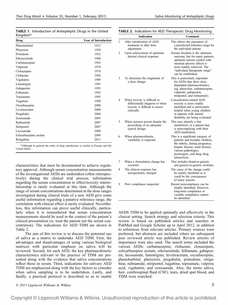
TABLE 2. Indications for AED Therapeutic Drug Monitoring
Indication Comment
1 After initialization of AEDtreatment or after doseadjustment
This allows the pursuance ofa preselected reference range forthe individual patient.
2 Upon achievement of optimumdesired clinical response
Seizure freedom is the optimumoutcome, but for many patients,optimum seizure control withminimal adverse effects ismore readily achieved. The“individual therapeutic range”can be established.
3 To determine the magnitude ofa dose change
This is particularly importantfor AEDs that show dose-dependent pharmacokinetics(eg, phenytoin, carbamazepine,valproate, gabapentin,stiripentol, and rufinamide).
4 When toxicity is difficult todifferentially diagnose or whentoxicity is difficult to assessclinically
Concentration-related AEDtoxicity is more readilyidentified and is particularlyhelpful when young childrenor patients with mentaldisability are being evaluated.
5 When seizures persist despite theprescribing of an adequate/typical dosage
This may identify a fastmetabolizer or a patient thatis noncomplying with theirAED medication.
6 When pharmacokineticvariability is expected
This is a significant category ofpatients and includes children,the elderly, during pregnancy,hepatic disease, renal disease,various pathologies,postsurgery, and drug–druginteractions.
7 When a formulation change hasoccurred
This includes brand-to-genericand generic-to-generic switches.
8 The clinical response hasunexpectantly changed
The cause of the change couldbe readily identified as itcould be the consequenceof many reasons.
9 Poor compliance suspected Recent noncompliance can bereadily identified. However,long-term compliance orvariable compliance cannotbe identified.
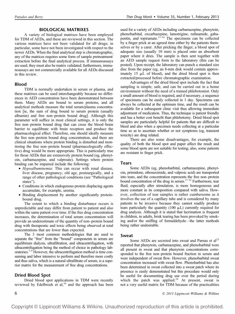
TABLE 1. Introduction of Antiepileptic Drugs in the UnitedKingdom*
Drug Year of Introduction
Phenobarbital 1912
Phenytoin 1938
Primidone 1952
Ethosuximide 1960
Carbamazepine 1963
Valproate 1974
Clonazepam 1974
Clobazam 1982
Vigabatrin 1989
Lamotrigine 1991
Gabapentin 1993
Felbamate 1993
Topiramate 1995
Tiagabine 1998
Oxcarbazepine 2000
Levetiracetam 2000
Pregabalin 2004
Zonisamide 2005
Rufinamide 2007
Stiripentol 2007
Lacosamide 2008
Eslicarbazepine acetate 2009
Retigabine 2011
*Although in general the order of drug introduction is similar in Europe and theUnited States.
Ther Drug Monit � Volume 35, Number 1, February 2013 Saliva Monitoring of Antiepileptic Drugs
� 2013 Lippincott Williams & Wilkins 5
BIOLOGICAL MATRIXESA variety of biological matrixes have been employed
for TDM of AEDs, and these are reviewed in this section. Thevarious matrices have not been validated for all drugs; inparticular, some have not been investigated with respect to thenewer AEDs. When the final analytical step is chromatographic,any of the matrices requires some form of sample pretreatment/extraction before the final analytical process. If immunoassaysare used, theymust also be matrix validated; furthermore, immu-noassays are not commercially available for all AEDs discussedin this review.
BloodTDM is normally undertaken in serum or plasma, and
these matrixes can be used interchangeably because no differ-ences in AED concentration have been demonstrated betweenthem. Many AEDs are bound to serum proteins, and allanalytical methods measure the total serum/plasma concentra-tion [ie, the sum of drug bound to serum protein (usuallyalbumin) and free non–protein bound drug]. Although thisparameter will suffice in most clinical settings, it is only thefree non–protein bound drug that can cross the blood–brainbarrier to equilibrate with brain receptors and produce thepharmacological effect. Therefore, one should ideally measurethe free non–protein bound drug concentration, and there areclinical situations where protein binding is disturbed and mon-itoring the free non–protein bound (pharmacologically effec-tive) drug would be more appropriate. This is particularly truefor those AEDs that are extensively protein bound (eg, phenyt-oin, carbamazepine, and valproate). Settings where proteinbinding can be impaired include the following:
• Hypoalbuminemia: This can occur with renal disease,liver disease, pregnancy, old age, postsurgically, and arange of other pathological conditions (see “Pathologicalstates”).
• Conditions in which endogenous protein displacing agentsaccumulate, for example, uremia.
• Binding displacement by another significantly protein-bound drug.The extent to which a binding disturbance occurs is
unpredictable and may differ from patient to patient and alsowithin the same patient over time. If the free drug concentrationincreases, the determination of total serum concentration willprovide an underestimate of the quantity of non–protein bounddrug with therapeutic and toxic effects being observed at totalconcentrations that are lower than expected.
The 3 most common methodologies that are used toseparate the “free” from the “bound” components in serum areequilibrium dialysis, ultrafiltration, and ultracentrifugation, withultracentrifugation being the method of choice in pathology lab-oratories.7–9 However, the ultracentrifugation method is time con-suming and labor intensive to perform and therefore more costlyand thus saliva, which is a natural ultrafiltrate of serum, is a supe-rior matrix for the measurement of free drug concentrations.
Dried Blood SpotDried blood spot applications in TDM were recently
reviewed by Edelbroek et al,10 and the approach has been
used for a variety of AEDs including carbamazepine, phenytoin,phenobarbital, oxcarbazepine, lamotrigine, rufinamide, gaba-pentin, and topiramate.11–16 The specimens can be collectedby a finger prick at an agreed time either by the patients them-selves or by a carer. After pricking the finger, a blood spot ofadequate size (usually 10 mm) is placed onto an absorbentpaper where it dries. The sample is then sent together withan AED sample request form to the laboratory (this can beposted). Upon receipt, the laboratory can punch a standard sizedisc from the paper (eg, an 8-mm disk would contain approx-imately 15 mL of blood), and the dried blood spot is thenextracted/processed before chromatographic examination.
Advantages of the dried blood spot include the fact thatsampling is simple, safe, and can be carried out in a homeenvironment without the need of a trained phlebotomist. Onlya small amount of blood is required, and if necessary, a sequenceof specimens can be easily collected in 1 day. Specimens canalways be collected at the optimum time, and the result can beavailable for a subsequent clinic visit thus allowing immediateadjustment of medication. Thus, the technique is patient friendlyand has a better cost benefit than phlebotomy. Dried blood spotsamples are particularly helpful for patients that are difficult tobleed and also when a specimen needs collecting at a particulartime so as to ascertain whether or not symptoms (eg, transienttoxicity) are drug related.
There are also some disadvantages, for example, thequality of both the blood spot and paper affect the result andsome blood spots are not suitable for testing; also, some patientsare resistant to finger prick.
TearsSome AEDs (eg, phenobarbital, carbamazepine, phenyt-
oin, primidone, ethosuximide, and valproic acid) are transportedinto tears, and the concentration represents the free non–proteinbound concentration of the drug in serum.17–22 Furthermore, tearfluid, especially after stimulation, is more homogeneous andmore constant in its composition compared with saliva. How-ever, collection of tear samples is rather cumbersome as itinvolves the use of a capillary tube and is considered by manypatients to be invasive because they cannot readily producetears particularly the quantity (100 mL) currently required fordrug analysis. Although it is stated that lacrimation is frequentin children, in adults, brisk tearing has been provoked by smok-ing and/or the sniffing of formaldehyde—the latter methodsbeing rather undesirable.
SweatSome AEDs are secreted into sweat and Parnas et al23
reported that phenytoin, carbamazepine, and phenobarbital wereall present in sweat and that phenytoin concentrations corre-sponded to the free non–protein bound fraction in serum andwere independent of sweat flow. However, phenobarbital sweatconcentration increased with sweat flow. Phenobarbital has alsobeen determined in sweat collected into a sweat patch where itspresence is easily demonstrated but this procedure would onlybe useful for documenting drug use over the period duringwhich the patch was applied.24 At present, sweat isnot a very useful matrix for TDM because of the practicalities
Patsalos and Berry Ther Drug Monit � Volume 35, Number 1, February 2013
6 � 2013 Lippincott Williams & Wilkins
associated with flowing sweat collection and/or the interpre-tation of the concentration if collected into a patch.
Cerebrospinal FluidMeasurement of CSF concentrations of neuroactive drugs,
including AEDs, is important because such concentrations areconsidered to reflect those occurring in the brain, which resultin the pharmacological effect of the drug (eg, anticonvulsant oradverse effects). Furthermore, CSF concentrations are consid-ered to reflect the free, non–protein bound serum concentration.
The transfer of the first-generation AEDs (carbamazepine,phenytoin, phenobarbital, and valproate) into CSF has been wellstudied, and the CSF concentration generally reflects their freenon–protein bound concentration in serum.20,21,25–28 Morerecently, some of the second-generation drugs, for example,gabapentin, oxcarbazepine, lamotrigine, levetiracetam, vigabatrin,and topiramate29–33 and third-generation AEDs, for example, esli-carbazepine acetate34 have been investigated.
Because for many AEDs the CSF concentration reflectsthe free non–protein bound drug concentration, this would prob-ably be a meaningful matrix for TDM purposes; however, forsome AEDs CSF does not reflect free serum concentration forexample, gabapentin and pregabalin.30–33 This lack of correlationis probably due to the mechanism by which gabapentin andpregabalin are distributed throughout the body, that is, theL-amino acid transporter, which is saturable so that transportationdoes not occur linearly. In the case of pregabalin, the CSF/serumarea under the concentration versus time curve 0–24 hours ratiowas 0.098 6 0.016.33 The invasive procedure (ie, lumber punc-ture) needed to collect a CSF samples negates the practical use ofCSF for AED TDM.
HairThe root of every growing hair is constantly exposed to
any drug that is circulating in the blood. The drug(s) are thussequestered into the hair structure and because head hair growsat approximately 1 cm/mo, if only a single drug exposureoccurred the portion of hair containing the drug would emergefrom the scalp after 6/7 days. This small section would thengrow away from the scalp, and by sampling the hair in 1-cmlengths, it is possible to assay the drug concentration in the hairsections and pinpoint the time of exposure. However, in patientsprescribed maintenance drug treatment, provided that they areadherent, the hair root will be exposed to a constant, steady-statedrug concentration in blood and this would be reflected by aconstant concentration of the drug along the hair shaft. Hair canthus be used to record the history of drug exposure and ascertainvariable and intermittent compliance. The AEDs that have beenreported to be transported into hair include carbamazepine,phenytoin, valproic acid, oxcarbazepine, and lamotrigine.35–39
Although hair analysis would not, therefore be helpfulfor day to day TDM, these principles have been applied todemonstrate compliance with carbamazepine and oxcarbaze-pine treatment in adult patients,39–41 compliance of carbamaze-pine and lamotrigine during pregnancy38 and to differentiatebetween chronic and acute carbamazepine intoxication.42
The disadvantages of hair analyses for AED TDM includethe fact that many factors impact on the amount of drugdeposited into hair, for example, melanin content, and whether
cosmetic hair treatments, for example, bleaching, dying, etc,remove drugs that are bound into the hair structure and thus causeinterpatient concentrations to vary significantly. Also, becausedrugs are incorporated into the hair structure, the analyticalprocess requires an initial digestion to release them before thechosen method of detection and quantification. Finally, althoughhair sampling is considered not to be invasive, it can be so forthose patients who have little hair or are indeed bald in whichcase a nonhead hair sample might be required.
SalivaSaliva was initially investigated as an alternative biolog-
ical fluid for TDM of AEDs during the 1970s; the most studieddrugs are phenytoin, phenobarbital, and carbamazepine.43–47
Saliva is once again emerging as a biological fluid that is valu-able for AED TDM and has started to be more widely usedagain because it is associated with numerous advantages overblood/serum (Table 3). Of particular advantage is that the con-centration in saliva generally reflects the free non–proteinbound pharmacologically active component in serum; salivais easier to collect than blood and patients prefer saliva sam-pling over blood sampling. Furthermore, the standard analyti-cal methods can invariably be easily adapted to accept salivaspecimens.
INDIVIDUAL AEDSIn the following section, the AEDs will be reviewed in
alphabetical order with regards to their clinical indications,key pharmacokinetic characteristics in relation to TDM ingeneral and salivary TDM in particular (Table 4), along withthe available evidence regarding the usefulness of saliva asa matrix for undertaking AED TDM.
Carbamazepine
Clinical IndicationsCarbamazepine is a first-line drug for the treatment of
partial and secondarily generalized tonic–clonic seizures andprimary generalized tonic–clonic seizures. It is also the drugof choice in the management of trigeminal neuralgia and inaddition is used in the treatment of bipolar disorder that isunresponsive to lithium. Carbamazepine is available in a vari-ety of formulations including, tablets, chewable tablets, liquidoral suspension, suppositories, and extended release tabletsand capsules.
Pharmacokinetic CharacteristicsAbsorption of carbamazepine after oral ingestion is
erratic and variable with a bioavailability of 75%–85% andTmax values that are formulation dependent.81 The drug isa powerful inducer of hepatic enzymes, and after initiationof treatment, the pharmacokinetic parameters (half-life andclearance) change considerably due to autoinduction, whichgenerally is complete in about 3 weeks.82 Protein binding is75%. Carbamazepine is extensively metabolized in the liver,primarily by CYP3A4 with some contribution by CYP2C8, tocarbamazepine–epoxide, which is pharmacologically active,equipotent to the parent drug and accumulates in serum to a
Ther Drug Monit � Volume 35, Number 1, February 2013 Saliva Monitoring of Antiepileptic Drugs
� 2013 Lippincott Williams & Wilkins 7

variable extent. Carbamazepine–epoxide is subsequently metab-olized, by epoxide hydrolase, to a pharmacologically inactive10,11-diol, which is eliminated partly unchanged and partly asa glucuronide conjugate.83 Protein binding of carbamazepine–epoxide is 50%–60%. The serum elimination half-life (t1/2)of carbamazepine in adults is 8–20 hours, whereas that ofcarbamazepine–epoxide is approximately 34 hours. Carbama-zepine is subject to many drug-drug pharmacokinetic interac-tions because it is an inducer of hepatic metabolism and also itsown metabolism is readily inhibited or induced—consequentlythere are large differences between individuals in the dose toserum concentration relationship. In addition, although there isa broad range of serum concentrations associated with an opti-mum effect, there is considerable interpatient variability in theconcentration of carbamazepine that is associated with an
optimal therapeutic response (which may in part be due to thevariation in carbamazepine–epoxide concentration). The fact thatthe dose to serum concentration relationship of carbamazepineand carbamazepine–epoxide is nonlinear, and thus unpredict-able, is the main reason why their monitoring is useful. Thecurrent reference range for carbamazepine in serum is 4–12mg/L (17–51 mmole/L), whereas carbamazepine–epoxide con-centrations are generally 5%–15% of the parent drug with a ref-erence range of up to 2.3 mg/L (up to 9 mmole/L).1
Saliva TDM for CarbamazepineThere have been many studies investigating the distribu-
tion of carbamazepine and carbamazepine epoxide in saliva andseveral have evaluated the relationship between salivary con-centration and the free, pharmacologically active, non–proteinbound concentration in serum of both adults and children withepilepsy.48–54,56,73,84–95
The salivary concentration of carbamazepine is similarto the free, non–protein bound concentration in serum withmean saliva/serum total carbamazepine concentration ratiosranging 0.26–0.44, whereas the mean saliva/serum-free car-bamazepine concentration ratios ranged 1.39–1.44. Indeed,concentrations of carbamazepine in saliva are significantlycorrelated with both serum total (r2 = 0.84–0.99) andserum-free carbamazepine concentrations (r2 = 0.91–0.99).Carbamazepine–epoxide distributes into saliva such thatthe salivary concentration is similar to the free non–proteinbound concentration in serum with mean saliva/serum totalcarbamazepine–epoxide concentration ratios of 0.31–0.55.Saliva carbamazepine–epoxide concentrations are signifi-cantly correlated with both serum total (r2 = 0.76–0.88) andserum-free (r2 = 0.75–0.98) carbamazepine–epoxide concen-trations. Thus, saliva may be used as an alternative matrix forTDM of both carbamazepine and carbamazepine–epoxide.
Clobazam
Clinical IndicationsClobazam is a 1,5-benzodiazepine drug with marked
anticonvulsant properties, which is less sedating than otherbenzodiazepines. It is licensed for use as adjunctive therapyof partial seizures or generalized seizures in patients above 3years of age and also for the management of nonconvulsivestatus epilepticus. It is also prescribed as an anxiolytic. Clobazamis available in tablet and capsule formulations.
Pharmacokinetic CharacteristicsClobazam is rapidly absorbed after oral ingestion with a
Tmax of 1–3 hours and bioavailability of .95%. Its pharma-cokinetics is linear and protein binding is 85%. Clobazam isextensively metabolized in the liver, primarily by CYP2C19and CYP3A4, to N-desmethyl clobazam, which is pharmaco-logically active, accumulates in serum to much higher con-centrations than the parent drug, and is responsible for muchof the clinical effect.96 N-desmethyl clobazam is subsequentlymetabolized by CYP2C19 and cleared from serum at a signif-icantly slower rate than the parent drug, with the half-life ofclobazam in adults being 10–30 hours, whereas the half-lifeof N-desmethyl clobazam is 36–46 hours. The protein bindingof N-desmethyl clobazam has not been reported. Clobazam is
TABLE 3. The Advantages and Disadvantages of Using Salivafor AED Therapeutic Drug Monitoring
Advantages Comments
Reflects free non–protein boundconcentration in blood
This is the ideal concentrationmeasurement in blood as it is thatcomponent (pharmacologicallyrelevant) that is accessible tothe brain where AEDs havetheir effect.
Collection is simple andnoninvasive
Avoids complications of infectionand thrombosis, which can beassociated with blood sampling.Useful for patients with needlephobias or difficult veins.
Does not require the expertiseof drawing blood
Sampling can be undertaken byspouse, partner, parent, or carer.
Cheaper than drawing blood No need for a phlebotomist, a nurse,or a doctor to bleed the patient.
Especially useful in patients withdisabilities, the elderly, andin children
Preferred by patients, parents,and carers.
Less stress, fear, and discomfort Patients are more amenable toproviding multiple samples.
Can be readily undertaken in thehome environment
Samples can be collected at the“ideal” time (trough and predose)and readily dispatched to thehospital laboratory in advanceof the patient’s clinic visit.
Disadvantages Comments
Spurious results due tocontamination (drug residuesin the mouth or leakage ofdrug-rich exudate, eg, patientswith gingivitis)
This can be avoided by sampling justbefore AED ingestion (at trough),after the mouth is rinsed or aftera few hours have elapsed sincedrug ingestion.
Saliva volume insufficient This can be overcome in thelaboratory by adding distilledwater.
Difficulty in pipetting due toviscosity of saliva
Sample may need to be rejected andpatient resampled.
AED concentration is low Analytical methods need to bespecifically developed so as to beable to measure the anticipatedlow concentrations.
Sampling may be unacceptable Some patients may refuse sampling,although in the authors’experience this has yet to happen.
Patsalos and Berry Ther Drug Monit � Volume 35, Number 1, February 2013
8 � 2013 Lippincott Williams & Wilkins

subject to many drug–drug pharmacokinetic interactions becauseits metabolism can be readily induced or inhibited—consequentlythere are large differences between individuals in the dose toserum concentration relationship. The current reference rangefor clobazam in serum is 0.03–0.3 mg/L (0.1–1.0 mmole/L),whereas that of N-desmethyl clobazam is 0.3–3.0 mg/L (1.0–10.5 mmole/L).1
Saliva TDM for ClobazamThere have been 2 studies investigating the distribution
of clobazam and N-desmethyl clobazam into saliva and therelationship between salivary clobazam/N-desmethyl cloba-zam concentrations and serum total clobazam/N-desmethylclobazam concentrations in children with epilepsy.54,55 Theseshowed that clobazam salivary concentration is similar to the
total concentration in serum and that concentrations are sig-nificantly correlated (r2 = 0.90). Also, N-desmethyl clobazamdistributes into saliva such that the salivary concentration issimilar to the total concentration in serum and that concen-trations are significantly correlated (r2 = 0.93). The excellentcorrelation between total serum clobazam/N-desmethyl cloba-zam and salivary concentrations (r = 0.9 and 0.93, respec-tively) indicates that saliva can be used as an alternativematrix for clobazam and N-desmethyl clobazam TDM.
Clonazepam
Clinical IndicationsClonazepam is licensed for the treatment of a variety of
seizure types including absence, akinetic, atonic, and myo-clonic seizures. Also, it is licensed for use in patients with
TABLE 4. Pharmacokinetic Parameters and Serum Reference Ranges for the Various AEDs Prescribed as Monotherapy to Adults
AEDTime to Steady
State (d)Serum ProteinBinding (%)
Half-Life(h)
Reference Range* PharmacologicallyActive Metabolites
That Need Monitoring
Saliva MonitoringValidated
(Key References)Mg/L Μmole/L
Carbamazepine 2–4† 75 8–20† 4–12 17–51 Carbamazepine-epoxide‡ 48–53
Clobazam 7–10§ 85 10–30 0.03–0.3 0.1–1.0 N-Desmethyl-clobazam 54,55
0.3–3.0¶ 1.0–10.5¶
Clonazepam 3–10 85 17–56 0.02–0.07 0.06–0.22 Not validated
Eslicarbazepineacetate#
3–4 30 13–20 3–35** 12–139** Eslicarbazepine Not validated
Ethosuximide 8–12 0 40–60 40–100 283–708 21,49,50,56–58
Felbamate 3–5 25 16–22 30–60 126–252 Not validated
Gabapentin 1–2 0 5–9 2–20 12–117 59,60
Lacosamide 3 90 13 61
Lamotrigine 3–8 55 15–35 2.5–15 10–59 62–64
Levetiracetam 1–2 0 6–8 12–46 70–270 65–67
Oxcarbazepine†† 2–3 40 8–15 3–35 12–139 10-Hydroxycarbazepine 68–72
Phenobarbital 15–30 55 70–140 10–40 43–172 26,50,54,73–75
Phenytoin 6–21 90 30–100‡‡ 10–20 40–79 1,50,54,56,73,76
Pregabalin 1–2 0 5–7 NE§§ NE§§ Not validated
Primidone 2–5 10 7–22 5–10¶¶ 23–46 Phenobarbital 49,50,56,73
Retigabine 1–2 80 8–10 NE§§ NE§§ Not validated
Rufinamide 1–2 35 6–10 30–40 126–168 77
Stiripentol 1–3 99 4.5–13‡‡ 4–22 17–94 Not validated
Tiagabine 1–2 96 5–9 0.02–0.2 0.05–0.53 Not validated
Topiramate 4–7 15 20–30 5–20 15–59 78
Valproic acid 2–4 90 12–16 50–100 346–693 Salivary and serumvalproic acidconcentrationsdo not correlate.
Vigabatrin 1–2 0 5–8 0.8–36 6–279 79
Zonisamide 9–12 40 50–70 10–40 47–188 80
*For clarity, values can be rounded up or down by the laboratory.†Refers to patients on chronic therapy after autoinduction is completed—values are much greater after a single dose.‡There are clinical settings where monitoring of carbamazepine-epoxide, in addition to carbamazepine, is warranted, particularly when a comedication occurs with an inhibitor ofcarbamazepine–epoxide metabolism.§Includes time to steady state for active metabolite N-desmethyl clobazam.¶Refers to values for active metabolite N-desmethyl-clobazam.#All values refer to the active metabolite eslicarbazepine.**The reference range is that quoted for the active metabolite of oxcarbazepine, namely, 10-hydroxycarbazepine because the 2 molecules are identical.††All values refer to the active metabolite 10-hydroxycarbazepine.‡‡Elimination is saturable so that half-live increases with increasing plasma concentration.§§Not established.¶¶During treatment with primidone both primidone and the pharmacologically active metabolite phenobarbital should be monitored.
Ther Drug Monit � Volume 35, Number 1, February 2013 Saliva Monitoring of Antiepileptic Drugs
� 2013 Lippincott Williams & Wilkins 9

Lennox–Gastaut syndrome and in the management of statusepilepticus. Clonazepam is available in a variety of formula-tions including tablets, a disintegrating wafer and a liquidformulation for intravenous administration.
Pharmacokinetic CharacteristicsClonazepam is rapidly absorbed after oral ingestion with a
Tmax of 1–4 hours and bioavailability of.80%. It exhibits linearpharmacokinetics and protein binding is 85%. Clonazepam isextensively metabolized in the liver, primarily by CYP3A4, to7-aminoclonazepam, which in turn is metabolized by acetyla-tion, via N-acetyl-transferase, to form 7-acetamidoclonazepam.The serum elimination half-life of clonazepam in adults is 17–56hours so that interindividual clearance is extremely variable.97–99
Clonazepam is subject to some drug–drug pharmacokineticinteractions consequent to the fact that its metabolism is readilyinduced or inhibited—therefore, large differences exist betweenindividuals in the dose to serum concentration relationship.The current reference range for clonazepam in serum is 0.02–0.07 mg/L (0.06–0.22 mmole/L).1
Saliva TDM for ClonazepamIt is not known whether clonazepam is secreted into
saliva and if it is whether the concentrations reflect those inserum. Analysis of saliva samples spiked with clonazepam andstored overnight at room temperature resulted in concentrationsthat were 76% lower compared with spiked saliva samples thatwere analyzed immediately.100 These data suggest that clona-zepam is unstable in saliva. Interestingly, clonazepam spikedinto water was stable.100
Eslicarbazepine Acetate
Clinical IndicationsEslicarbazepine acetate is licensed for the adjunctive
treatment of partial onset seizures with or without secondarygeneralization in patients with epilepsy aged 16 years andolder. The drug is available as formulations of tablet and asuspension.
Pharmacokinetic CharacteristicsEslicarbazepine acetate is a prodrug, and after oral absorp-
tion, the acetate group is rapidly and extensively metabolized byhydrolytic first pass metabolism with esterases to eslicarbaze-pine (S-licarbazepine), the S-enantiomer of the pharmacologi-cally active 10-hydroxycarbazepine metabolite of oxcarbazepine(also known as the monohydroxy derivative). After oral inges-tion, eslicarbazepine acetate is rapidly absorbed with a Tmax foreslicarbazepine of 2–3 hours and bioavailability of .90%.101
Eslicarbazepine pharmacokinetics is linear and protein bindingis 30%. In addition to eslicarbazepine, small amounts of 2 otherpharmacologically active metabolites are formed from eslicar-bazepine acetate (R-licarbazepine and oxcarbazepine), but theserepresent only approximately 6% of metabolites. Eslicarbaze-pine and its glucuronides together with minor quantities ofR-licarbazepine, oxcarbazepine, eslicarbazepine acetate, andtheir respective glucuronides are excreted in urine. Eslicarba-zepine glucuronic acid conjugation is primarily catalyzed byUGT1A4, UGT1A9, UGT2B4, UGT2B7, and UGT2B17.102
The serum half-life of eslicarbazepine in adults is 13–20hours, and it is subject to very few drug–drug pharmacoki-netic interactions.103 The current reference range for eslicar-bazepine in serum is 3–35 mg/L (12–139 mmole/L), which isbased on that for racemic 10-hydroxycarbazepine derived fromoxcarbazepine.1
Saliva TDM for Eslicarbazepine AcetateIt is not known whether eslicarbazepine acetate is secreted
into saliva or whether salivary concentrations are similar orreflect those in serum. However, because the pharmacologi-cally active metabolite, eslicarbazepine, is the same moleculeas the pharmacologically active metabolite of oxcarbazepine,10-hydroxycarbazepine, it can be expected that its transfer intosaliva will be similar to that described for 10-hydroxycarbaze-pine in “Oxcarbazepine.”
Ethosuximide
Clinical IndicationsEthosuximide is licensed for monotherapy treatment of
absence seizures in patients of all ages and is available asformulations of capsule and a syrup.
Pharmacokinetic CharacteristicsEthosuximide is rapidly absorbed after oral ingestion
with a Tmax of 1–4 hours and its bioavailability is .90%. Itspharmacokinetics is linear and it is not protein bound.104,105
Ethosuximide is extensively metabolized in the liver, primar-ily by CYP3A and to a lesser extent by CYP2E and CYP2B/C, to form isomers of 2-(1-hydroxymethyl)-2-methylsuccina-mide of which 40% are excreted as glucuronide conjugates.The serum half-life of ethosuximide in adults is 40–60 hourswith large interindividual differences in serum clearance. Fur-thermore, ethosuximide is subject to a number of drug–drugpharmacokinetic interactions because its metabolism can beboth induced and inhibited with consequent large differencesbetween individuals in the dose to serum concentration rela-tionship. The current reference range for ethosuximide inserum is 40–100 mg/L (283–708 mmole/L).1
Saliva TDM for EthosuximideThere have been several studies investigating the distri-
bution of ethosuximide into saliva and the relationship betweensaliva ethosuximide concentrations and total serum ethosuximideconcentrations in patients with epilepsy.21,49,50,56–58 Ethosuximideis not protein bound and the salivary concentration is similar tothe total serum concentration with mean saliva/serum total etho-suximide concentration ratios ranging 0.95–1.04. Indeed, salivaand serum total ethosuximide concentrations are significantlycorrelated (r2 = 0.99), and thus, saliva can be used as an alter-native matrix for ethosuximide TDM.
Felbamate
Clinical IndicationsBecause felbamate is associated with an increased risk
of aplastic anemia and hepatotoxicity, its use is restricted suchthat it is approved for use only in patients who respond inade-quately to alternative treatments and particularly in patients with
Patsalos and Berry Ther Drug Monit � Volume 35, Number 1, February 2013
10 � 2013 Lippincott Williams & Wilkins

partial seizures or Lennox–Gastaut syndrome. Felbamate isavailable as formulations of tablet and a suspension.
Pharmacokinetic CharacteristicsFelbamate is rapidly absorbed after oral ingestion with
a Tmax of 2–6 hours and a bioavailability of .90%. It haslinear pharmacokinetics and protein binding is 25%. About50% of an administered dose is metabolized in the liver,primarily by CYP3A4 and CYP2E1, to form 2 hydroxylatedmetabolites (p-hydroxy and 2-hydroxy felbamate). In additionone of the carbamate groups is hydrolyzed to an alcohol thatis further biotransformed to an acid; also a number of as yetunidentified polar metabolites are produced, some of whichare glucuronides.106,107 The development of hepatotoxicityand aplastic anemia in a few patients treated with felbamateis due to the formation of a reactive atropaldehyde metabolite,which can accumulate in some patients and cause toxicity.106
The serum half-life of felbamate in adults is 16–22 hours.Felbamate is subject to drug–drug pharmacokinetic interac-tions consequent to the fact that its metabolism is both readilyinduced and inhibited; also felbamate itself acts as an inhibitorof hepatic metabolism.108–110 Consequently, there are large dif-ferences between individuals in the dose to serum concentra-tion relationship. The current reference range for felbamate inserum is 30–60 mg/L (126–252 mmole/L).1
Saliva TDM for FelbamateIt is not known whether felbamate is secreted into saliva
and if it is whether concentrations are similar to or reflect thosein serum.
Gabapentin
Clinical IndicationsGabapentin is licensed for the monotherapy treatment
of partial seizures with or without secondary generalization inadults and children aged 12 years and above, and as an adjunc-tive treatment in adults and children aged 6 years and above.The drug is also licensed for the treatment of peripheral neu-ropathic pain. Gabapentin is available as formulations of tabletsand capsules.
Pharmacokinetic CharacteristicsGabapentin is rapidly absorbed after oral ingestion with
a Tmax of 2–3 hours. Bioavailability is 60% and is dose depen-dent with bioavailability decreasing at higher doses. Its pharma-cokinetics is nonlinear consequent to its saturable absorptionfrom the proximal small bowel primarily by the L-amino acidtransport system.111 Gabapentin is not protein bound and notmetabolized, being cleared entirely by renal excretion witha serum elimination half-life in adults of 5–9 hours.112 Neverthe-less, although gabapentin is not subject to drug–drug pharma-cokinetic interactions,103 the disposition can be extremelyvariable because of wide interindividual differences is absorp-tion.113 The fact that gabapentin is associated with nonlinearpharmacokinetics is a major reason why gabapentin monitoringis particularly valuable for patient management and thecurrent reference range for gabapentin in serum is 2–20 mg/L(12–117 mmole/L).1
Saliva TDM for GabapentinThere have been 3 studies investigating the distribution
of gabapentin in saliva—1 in healthy volunteers59 and 2 inpatients with epilepsy.60,114 Gabapentin distributes into saliva;however, mean concentrations are 2.4–10% of those observedin serum.59,60 Nevertheless, there is significant correlation (r2 .0.7) between saliva and serum total gabapentin concentra-tions114; furthermore, salivary gabapentin concentration and doseare significantly correlated (r2 = 0.77–0.95).59,60 Thus, salivamay be a useful alternative matrix for gabapentin TDM.
Lacosamide
Clinical IndicationsLacosamide is licensed for the adjunctive treatment of
partial onset seizures with or without secondary generaliza-tion in patients with epilepsy aged 16 years and older.Lacosamide is available as formulations of tablets, a solution,and a syrup.
Pharmacokinetic CharacteristicsLacosamide is rapidly absorbed after oral ingestion with
a Tmax of 1–2 hours, and its bioavailability is 100%.115 Itspharmacokinetics is linear and protein binding is controver-sial with Greenaway et al61 reporting 90% and the Summaryof Product Characteristic116 stating that serum lacosamideprotein binding is,15%. About 60% of a dose of lacosamideis hepatically metabolized, by demethylation via CYP2C19,to form O-desmethyl lacosamide.102 The serum eliminationhalf-life of lacosamide in adults is 13 hours115 and to dateno drug–drug pharmacokinetic interactions have been identi-fied.103 The current reference range for lacosamide in serum is10–20 mg/L (40–80 mmole/L).
Saliva TDM for LacosamideThere has been one study investigating the distribution
of lacosamide into saliva and the relationship between salivalacosamide concentration and both serum total and free concen-trations in adults with epilepsy.61 Lacosamide distributes intosaliva such that the salivary concentration is similar to the non–protein bound concentration in serum; mean saliva/serum-freelacosamide concentration ratios ranged 0.77–0.96. Furthermore,saliva lacosamide concentrations are significantly correlatedwith both serum total lacosamide (r2 = 0.84) and serum-freelacosamide (r2 = 0.83) concentrations. Thus, saliva should bea useful alternative matrix for lacosamide TDM.
Lamotrigine
Clinical IndicationsLamotrigine is licensed for the monotherapy treatment
of partial seizures and primary and secondarily generalizedtonic–clonic seizures in adults and children over 12 years ofage; as adjunctive treatment in adults and children over 2years of age; as adjunctive treatment of seizures associatedwith the Lennox–Gastaut syndrome in adults and childrenover 2 years of age. Lamotrigine is also licensed for thetreatment of bipolar I disorder. It is available as formulationsof tablets and dispersible chewable tablets.
Ther Drug Monit � Volume 35, Number 1, February 2013 Saliva Monitoring of Antiepileptic Drugs
� 2013 Lippincott Williams & Wilkins 11

Pharmacokinetic CharacteristicsLamotrigine is rapidly absorbed after oral ingestion
with a Tmax of 1–3 hours and its bioavailability is .95%.117
Its pharmacokinetics is linear and protein binding is 55%.Lamotrigine is extensively metabolized in the liver, primarilyby glucuronidation via UGT1A4, to form N-2 and N-5 glu-curonides.117 The serum elimination half-life of lamotrigine inadults is 15–35 hours so that interindividual clearance isextremely variable118; furthermore, it is subject to manydrug–drug pharmacokinetic interactions consequent to thefact that its metabolism can be both induced and inhibited;consequently, there are large differences between individualsin the dose to serum concentration relationship.103 The currentreference range for lamotrigine in serum is 2.5–15 mg/L(10–59 mmole/L).1
Saliva TDM for LamotrigineLamotrigine is reported to be about 55% bound to serum
proteins in patients receiving 150–300 mg/d in conjunctionwith other medication and the saliva/serum lamotrigine ratiois reported to be 0.46 in healthy subjects receiving a singledose and 0.56 in patients receiving adjunctive therapy.119,120
The excellent correlation between serum and saliva concentra-tions of lamotrigine (r = 0.95) in these early studies suggestedthat saliva could potentially be used to monitor the systemicconcentrations of lamotrigine.
A subsequent study examined the interindividual corre-lation between lamotrigine concentrations in saliva and serumtogether with the relationship between saliva concentrationand the non–protein bound lamotrigine concentration inserum.62 The authors compared both stimulated and unstimu-lated saliva from the same patients and demonstrated a goodcorrelation between lamotrigine serum concentration in bothcollection modes (r2 = 0.85, unstimulated and r2 = 0.94,stimulated). Furthermore, the study demonstrated a good cor-relation between total lamotrigine concentration in serum andthe free concentration as determined by ultrafiltration (r2 = 0.95)and equilibrium dialysis (r2 = 0.93). Lamotrigine concentrationin stimulated saliva was also significantly correlated with thefree concentration and calculation of lamotrigine protein bind-ing using the 3 alternative procedures gave the following results(mean 6 SD); 51.8% 6 13.03% (stimulated), 68.05% 6 7.59%(ultrafiltration), and 58.72%6 7.68% (equilibrium dialysis). Thedifferences in calculated binding between the 3 methods weresignificant.62
Ryan et al63 studied the relationship between serum andsalivary concentrations of lamotrigine in both pediatric andadult epilepsy populations and reported a good correlationbetween the two (r2 = 0.81–0.84) and with the saliva/serumlamotrigine concentration ratios ranging 0.40–1.19 (mean 6SD = 0.64 6 0.18). The authors concluded that althougha good correlation existed for the population at large betweensalivary and serum concentrations for lamotrigine, there iswide interpatient variability in the saliva/serum ratio. The datasuggest that salivary monitoring may play a role in the mon-itoring of lamotrigine for adult and pediatric patients.
In another study, lamotrigine concentrations weremeasured in both stimulated and unstimulated saliva along-side matching serum samples from 7 adult volunteers over
a 32-hour period after a single 50-mg dose of the drug, also insamples from 20 children and adolescents during the courseof routine AED therapy.64 In specimens collected $2 hoursafter ingestion, there was a close correlation in each individ-ual between the concentrations in stimulated and unstimulatedsaliva, which were similar. The saliva/serum lamotrigine con-centration ratio gave a mean value of 0.49 at a serum lamo-trigine concentration of 10 mg/L, and the authors concludedthat with appropriate precautions regarding the timing of sam-ple collection saliva measurements could provide a reasonablealternative to serum for TDM.64
A study in 14 healthy volunteers comparing saliva andserum lamotrigine concentrations over 96 hours after inges-tion of a single oral dose of lamotrigine reported significantcorrelation between saliva and serum (r2 = 0.677).121 Further-more, the mean saliva/serum lamotrigine concentration ratiowas 0.425 6 0.153, and the calculated protein binding fromthe concentration in saliva was 57.5 6 15.1% (mean 6 SD);thus, saliva concentrations reflect the free concentrations inserum. More recently, Mallayasamy et al122 reported a corre-lation between salivary and serum lamotrigine concentrationsof 0.683.
In summary, for lamotrigine, there are many factors thatmake TDM clinically useful, and several studies have foundgood correlation between salivary concentrations and both totaland free non–protein bound serum concentrations; thus, lamo-trigine TDM in saliva is a viable alternative to that of serum.
Levetiracetam
Clinical IndicationsLevetiracetam is licensed for the monotherapy treatment
of partial seizures with or without secondary generalization inpatients aged 16 years and older and as adjunctive treatment inadults and children from 4 years of age. The drug is alsolicensed for the adjunctive treatment of primary generalizedtonic–clonic seizures associated with idiopathic generalizedepilepsy and myoclonic seizures associated with juvenile myo-clonic epilepsy in adults and adolescents from 12 years of age.Levetiracetam is available in a variety of formulations includ-ing tablets, an oral solution, a solution for intravenous injec-tion, and an extended release tablet formulation.
Pharmacokinetic CharacteristicsLevetiracetam is rapidly absorbed after oral ingestion
with a Tmax of 1–2 hours and a bioavailability of .95%. Itspharmacokinetics is linear, and it is not protein bound.123
Approximately 30% of a dose of levetiracetam undergoesmetabolism by a cytosolic amidase enzyme to produce a car-boxylic acid metabolite (2-pyrrolidone-N-butyric acid), whichis excreted unchanged via the kidneys.124,125 Levetiracetammetabolism to the carboxylic acid is independent of the hepaticCYP system and occurs by means of a type-B esterase locatedin whole blood.126 However, the drug also undergoes a smallamount of hepatic metabolism to form 2 ring-hydroxylatedmetabolites. The serum elimination half-life of levetiracetamin adults is 6–8 hours, and it is subject to minimal drug–drugpharmacokinetic interactions.103 The current reference rangefor levetiracetam in serum is 12–46 mg/L (70–270 mmole/L).1
Patsalos and Berry Ther Drug Monit � Volume 35, Number 1, February 2013
12 � 2013 Lippincott Williams & Wilkins

Saliva TDM for LevetiracetamThere have been several studies investigating the distri-
bution of levetiracetam into saliva and the relationship betweensaliva levetiracetam and serum concentrations in adults andchildren with epilepsy and also in healthy volunteers.65–67
Levetiracetam distributes into saliva. However, there issome controversy regarding whether or not saliva and serumlevetiracetam concentrations are the same. Grim et al65 reportthat salivary concentrations are approximately 40% of thatobserved in serum, with mean saliva/serum levetiracetam con-centration ratios ranging 0.36–0.41. In contrast, Lins et al66 andMecarelli et al67 report that the concentration of levetiracetamin saliva is similar to serum with the mean saliva/serum leve-tiracetam concentration ratio being 1.0 and 1.1, respectively.Interestingly, a ratio of 1.55 was observed after ingestion of anoral solution in healthy volunteers.66 Nevertheless, saliva andserum levetiracetam concentrations are significantly correlated(r2 = 0.86–0.91) although lemon juice stimulation reduces thecorrelation from r2 = 0.91 to r2 = 0.87. Overall, the data sug-gest that saliva may be used as an alternative matrix for leve-tiracetam TDM.
Oxcarbazepine
Clinical IndicationsOxcarbazepine is licensed for the monotherapy or adjunc-
tive treatment of partial seizures with or without secondarygeneralization in patients aged 6 years or more. It is available asformulations of tablets and an oral suspension.
Pharmacokinetic CharacteristicsOxcarbazepine is a prodrug and is rapidly metabolized,
by cytosolic arylketone reductase, to a pharmacologicallyactive metabolite 10-hydroxycarbazepine (also known aslicarbazepine or monohydroxy metabolite). This metaboliteaccumulates in serum and is responsible for most of the drugeffects. The conversion of oxcarbazepine to 10-hydroxycarba-zepine is stereoselective and concentrations of the S-enantio-mer are somewhat higher than those of the R-enantiomer.127,128
After oral ingestion, oxcarbazepine is rapidly absorbed witha Tmax of 3–6 hours and a bioavailability of 100%. Oxcarba-zepine pharmacokinetics is linear and protein binding is 60%,whereas 10-hydroxycarbazepine protein binding is 40%.129 10-Hydroxycarbazepine is subsequently metabolized by conjuga-tion with glucuronic acid, and the conjugates together withsome 10-hydroxycarbazepine are excreted in urine. The serumelimination half-life of 10-hydroxycarbazepine in adults is 8–15 hours, and it is subject to many drug–drug pharmacokineticinteractions because its metabolism is both readily inhibitedand induced. Oxcarbazepine is also itself a weak inducer ofhepatic metabolism130,131; consequently, there are large differ-ences between individuals in the dose to serum concentrationrelationship. The current reference range for 10-hydroxycarba-zepine in serum is 3–35 mg/L (12–139 mmole/L).1
Saliva TDM for 10-HydroxycarbazepineThere have been several studies investigating the distri-
bution of 10-hydroxycarbazepine into saliva and the rela-tionship between salivary concentrations and serum total/free
10-hydroxycarbazepine concentrations in healthy volunteersand in adults and children with epilepsy.68–72 10-Hydroxycar-bazepine distributes into saliva; however, the extent dependson whether resting saliva or stimulated saliva is collected.Thiesohn and Heimann68 in a study of 3 healthy volunteersobserved that in resting saliva the saliva/serum ratio ranged0.3–1.7 (median 1.0). Kristensen et al69 in a study of 7 healthyvolunteers reported that in stimulated saliva (paraffin wax) themean saliva/serum ratio was 0.53. The study of Klitgaard andKristensen70 involving 17 patients with epilepsy reported thatin resting saliva the mean saliva/serum ratio was 1.01, whereasin stimulated saliva (paraffin wax), the value was 0.41. Cardotet al71 in a study of 10 patients with epilepsy reported that instimulated saliva (citric acid) the mean saliva/serum ratio was0.19. These data indicate that with greater saliva stimulationthe saliva/serum ratio decreases such that saliva 10-hydroxy-carbazepine concentrations approached the free 10-hydroxycar-bazepine concentrations in serum. This may be due to therelatively low lipid solubility of 10-hydroxycarbazepine; a char-acteristic that is not associated with carbamazepine. Unfortu-nately, increasing salivary flow has an extremely variable effecton saliva 10-hydroxycarbazepine concentration, which resultsin the saliva and serum 10-hydroxycarbazepine concentrationsnot being significantly correlated. However, when unstimulatedsaliva is collected, there is a significant correlation betweensaliva and serum total 10-hydroxycarbazepine concentrations(r2 = 0.91–0.98).68–70 Indeed, a recent study by Miles et al72 of28 children and adult patients with epilepsy reported on un-stimulated saliva whereby the mean saliva/serum ratio was0.96 and saliva 10-hydroxycarbazepine concentrations weresignificantly correlated with serum total 10-hydroxycarbazepineconcentrations (r2 = 0.94). Thus unstimulated saliva may be auseful alternative matrix for 10-hydroxycarbazepine TDM.
Phenobarbital
Clinical IndicationsPhenobarbital is licensed for the monotherapy or adjunc-
tive treatment of all forms of epilepsy, except absence seizures,in patients of any age. Phenobarbital is available in a varietyof formulations including, tablets, a solution for intravenousinjection, and an elixir formulation.
Pharmacokinetic CharacteristicsPhenobarbital is rapidly absorbed after oral ingestion
with a Tmax of 2–4 hours and a bioavailability of .90.132,133
Its pharmacokinetics is linear, and protein binding is 55%.Phenobarbital is extensively metabolized in the liver, primarilyby CYP2C9 and to a lesser extent by CYP2C19 and CYP2E1,to form 2 major metabolites, p-hydroxyphenobarbital and a9-D-glucopyranosyl phenobarbital isomer. The serum half-lifeof phenobarbital in adults is 70–140 hours so that interindivid-ual clearance is extremely variable132,133 Phenobarbital is subjectto many drug–drug pharmacokinetic interactions consequent tothe fact that it is a potent inducer of hepatic metabolism and alsoits own metabolism can be induced or inhibited. Consequently,there are large differences between individuals in the dose toserum concentration relationship. The current reference rangefor phenobarbital in serum is 10–40 mg/L (43–172 mmole/L).1
Ther Drug Monit � Volume 35, Number 1, February 2013 Saliva Monitoring of Antiepileptic Drugs
� 2013 Lippincott Williams & Wilkins 13

Saliva TDM for PhenobarbitalUsing saliva as an alternative matrix for TDM of pheno-
barbital is somewhat controversial because there is no clearconsensus regarding whether the concentration in saliva directlyreflects the concentrations of the drug in serum. A number ofstudies in both adults and children with epilepsy have demon-strated that phenobarbital distributes into saliva with saliva/totalserum phenobarbital concentration ratios ranging 0.2–0.52,whereas the mean saliva/serum-free phenobarbital concentrationratios ranged 0.63–0.68. Indeed, saliva phenobarbital concentra-tions and both serum total phenobarbital (r2 = 0.65–0.98) andserum-free phenobarbital (r2 = 0.64–0.99) concentrations aresignificantly correlated.26,49,50,53,54,56,57,73–75,134–137
Two studies have demonstrated that the distributionof phenobarbital into saliva depends on salivary pH56,136;however, other studies have not found an effect of pH.75,137
The study by McAuliffe et al56 determined phenobarbital insaliva and serum obtained simultaneously from 115 pa-tients, and a method to correct for the effect of salivarypH on drug concentration of saliva was developed. Salivaryphenobarbital concentration was found to be equivalentto the free phenobarbital concentration in serum and tocorrelate significantly with the total serum concentration.Expressed as percentage of total serum drug, the salivary(S) and serum-free (P) concentrations were: phenobarbital,S 43.1 6 5.2%, P = 40.8 6 7.9% (r = 0.91). On balance, itseems that salivary concentrations of phenobarbital corre-late with the simultaneous serum water concentrations, aftercorrecting for the effects of pH differences between salivaand serum.138
Despite the contradictory results, when equations incor-porate the relative ratio of phenobarbital pKa to the salivarypH, there is an excellent correlation between salivary and freenon–protein bound serum phenobarbital concentrations. Thus,saliva can be regarded as a useful alternative matrix for phe-nobarbital TDM.
Phenytoin
Clinical IndicationsPhenytoin is licensed for both monotherapy and adjunc-
tive therapy of clonic–clonic seizures and focal seizures inpatients of any age. It is also approved for the treatment ofseizures occurring during or after neurosurgery and/or severehead injury. In addition, it is licensed for intravenous admin-istration in the management of established status epilepticusand for monotherapy use in the treatment of trigeminal neu-ralgia. Phenytoin is available in a variety of formulationsincluding, capsules, chewable tablets, an oral suspensionand a parenteral solution formulation. More recently, fosphe-nytoin has been licensed, which is a water-soluble phenytoinphosphate prodrug that is rapidly dephosphorylated on admin-istration. It is formulated for intravenous administration tocontrol of status epilepticus and the prevention/treatment ofseizures occurring in connection with neurosurgery and/orhead trauma. It can also be substituted for oral phenytoinif oral administration is not possible and/or contraindicated.Fosphenytoin is formulated as a solution for infusion orinjection.
Pharmacokinetic CharacteristicsThe rate of absorption of phenytoin after oral ingestion
is variable and formulation dependent with a Tmax of 1–12hours.139 Bioavailability is similarly formulation dependentbut is .80%. Its pharmacokinetics is nonlinear due to satu-rable metabolism, which results in Michaelis–Menten kineticswithin the range of serum concentrations that are generallyassociated with its beneficial therapeutic effects.140 The pro-tein binding of phenytoin is 90%. Phenytoin is extensivelymetabolized in the liver, primarily by CYP2C9 andCYP2C19, to form 2 major metabolites 5-(p-hydroxyphen-yl)-5-phenylhydantoin (which undergoes partial conversionto glucuronides before renal excretion) and a dihydrodiolderivative.141 The serum elimination half-life of phenytoinin adults is 30–100 hours so that interindividual clearance isextremely variable. Phenytoin is subject to many drug–drugpharmacokinetic interactions consequent to the fact that it isa potent inducer of hepatic metabolism; also its metabolismcan be both induced and inhibited—consequently, there arelarge differences between individuals in the dose to serumconcentration relationship. The fact that phenytoin is associ-ated with nonlinear pharmacokinetics is one of the majorreasons why monitoring the drug is so useful. The currentreference range for phenytoin in serum is 10–20 mg/L (40–79 mmole/L).1
Saliva TDM for PhenytoinThere have been many studies investigating the distri-
bution of phenytoin into saliva and the relationship betweensaliva phenytoin concentration and both serum total and freephenytoin concentrations in both adults and children withepilepsy.26,49,53,54,56,57,73,76,90,95,134,135,137,142–146 Phenytoin dis-tributes into saliva such that the salivary concentration issimilar to the free non–protein bound concentration in serum.Mean saliva/serum total phenytoin concentration ratiosranged 0.09–0.13, whereas the mean saliva/serum-free phe-nytoin concentration ratios ranged 0.99–1.06. Indeed, salivaphenytoin concentrations and both serum total phenytoin(r2 = 0.85–0.99) and serum-free phenytoin (r2 = 0.96–0.99)concentrations are significantly correlated. There is a sugges-tion that the extent of phenytoin distribution in saliva dependson whether resting saliva, stimulated saliva, or reduced flowsaliva is collected. For the latter 2 situations, phenytoin concen-trations in saliva are decreased and increased, respectively.147
Thus, unstimulated saliva should be used, and this provides anextremely useful alternative matrix for phenytoin TDM.
Pregabalin
Clinical IndicationsPregabalin is licensed as adjunctive treatment of partial
seizures with or without secondary generalization in adults. Itis also licensed for the treatment of peripheral and central painand for generalized anxiety disorders. Pregabalin is availableas formulations of capsules only.
Pharmacokinetic CharacteristicsPregabalin is rapidly absorbed after oral ingestion
with a Tmax of 1–2 hours and bioavailability of .90%.148 Its
Patsalos and Berry Ther Drug Monit � Volume 35, Number 1, February 2013
14 � 2013 Lippincott Williams & Wilkins

pharmacokinetics is linear, and it is not protein bound. Prega-balin is not metabolized being cleared entirely by renal excre-tion with a serum elimination half-life of 5–7 hours in adults.148
Pregabalin is not subject to drug–drug pharmacokinetic inter-actions.103 The precise role for TDM of pregabalin has not yetbeen established, although there may be a requirement inpatients with renal impairment, to ascertain compliance, wheremalabsorption is suspected and in cases of suspected overdose.Very little information is available regarding therapeutic serumconcentrations of pregabalin; however, one report states that insamples collected at random times relative to dose frompatients maintained on 600 mg/d, serum pregabalin concentra-tions ranged from 0.9 to 14.2 mg/L. There are reports of sig-nificant toxicity in a case of self-poisoning with pregabalinalone149; also, a case of toxicity associated with therapeuticuse in a patient with renal failure although the peak drug con-centration (predialysis) in this case was only 13 mg/L.
Saliva TDM for PregabalinIt is not known whether pregabalin is secreted into
saliva and if it is whether concentrations are similar to orreflect those in serum.
Primidone
Clinical IndicationsPrimidone is licensed for the monotherapy or poly-
therapy treatment of generalized tonic–clonic seizures, psy-chomotor and focal seizures in adults and children and for themanagement of Jacksonian seizures, myoclonic jerks, andakinetic attacks. It is also licensed for the treatment of essen-tial tremor. Primidone is available as tablet formulations anda suspension.
Pharmacokinetic CharacteristicsPrimidone is rapidly absorbed after oral ingestion
with a Tmax of 2–4 hours and bioavailability of .90%.150
Its pharmacokinetics is linear and protein binding is 10%.Primidone is extensively metabolized in the liver to form 2major pharmacologically active metabolites, phenobarbitaland phenyl-ethyl-malonamide. The serum elimination half-life of primidone in adults is 7–22 hours so that interindivid-ual clearance is variable.150 Primidone is subject to manydrug–drug pharmacokinetic interactions consequent to the factthat it is a potent inducer of hepatic metabolism (via pheno-barbital). Also, its metabolism can be induced or inhibited;consequently, there are large differences between individualsin the dose to serum concentration relationship. During treat-ment with primidone, it is common practice to monitor pheno-barbital because the adverse effects from a high phenobarbitalconcentration is more likely to limit a primidone dosageincrease. The current reference range for primidone in serumis 5–10 mg/L (23–46 mmole/L).1
Saliva TDM for PrimidoneThere have been several studies that investigate the
distribution of primidone in saliva and the relationship betweensaliva primidone and serum total primidone concentrations inboth adults and children with epilepsy.26,49,50,56,57,73,151 Themean saliva/serum total primidone concentration ratios ranged
0.07–1.15. Furthermore, saliva and total serum primidone con-centrations are significantly correlated (r2 = 0.71–0.97). Sali-vary primidone concentrations seem to be flow dependent withresting saliva concentrations being approximately 38% lowerthan in flow-stimulated saliva.152 Thus, provided that samplecollection is standardized saliva may be used as an alternativematrix for primidone TDM.
Retigabine
Clinical IndicationsRetigabine is indicated as adjunctive treatment of partial
onset seizures with or without secondary generalization in adultsaged 18 years and above with epilepsy. Retigabine is availableas formulations of tablet only.
Pharmacokinetic CharacteristicsRetigabine is rapidly absorbed after oral ingestion with
a Tmax of 0.6–1.5 hours and bioavailability of about 60%.152
Its pharmacokinetics is linear and protein binding is 80%.153
Approximately 20%–30% of the administered dose of retiga-bine is eliminated renally unchanged with the remaining drugbeing biotransformed to produce the N-acetyl metabolite(which has a weak pharmacological action) and N-glucuro-nide conjugates of both parent drug and N-acetylretigabineaccumulates in plasma reaching similar concentrations to theparent drug and while the N-glucuronide metabolites are phar-macologically inactive, they may contribute to the enterohe-patic circulation of retigabine and N-acetylretigabine.102 Theserum elimination half-life of retigabine in adults is 8–10hours, and it is subject to only a few drug–drug pharmacoki-netic interactions.103 The precise role for TDM of retigabinehas not yet been established, although there may be a require-ment in patients to ascertain compliance, where malabsorp-tion is suspected and in cases of suspected toxicity. There areno data relating plasma retigabine levels with that of seizuresuppression or adverse effects.
Saliva TDM for RetigabineIt is not known whether retigabine is secreted into
saliva and if so whether concentrations are similar to or reflectthose in serum.
Rufinamide
Clinical IndicationsRufinamide is licensed for the adjunctive treatment of
seizures associated with the Lennox–Gastaut syndrome inpatients 4 years and older. Rufinamide is available as formu-lations of tablet only.
Pharmacokinetic CharacteristicsRufinamide is rapidly absorbed after oral ingestion with
a Tmax of 4–6 hours and bioavailability decreases withincreasing dose. Consequently, its pharmacokinetics is linearup to only 1600 mg/d. Protein binding is 35%.154,155 Becausefood coingestion enhances Cmax values by 50% and increasesarea under the concentration versus time curve values by33%, probably by improving the solubility of rufinamide,patients should be advised to take their rufinamide dose each
Ther Drug Monit � Volume 35, Number 1, February 2013 Saliva Monitoring of Antiepileptic Drugs
� 2013 Lippincott Williams & Wilkins 15
time in the same temporal relation to their meals to maintainsteady-state concentrations from one dose to the next.156 Ru-finamide is extensively metabolized in the liver, primarily byan amidase hydrolysis (which is not CYP dependent) thatconverts the carboxamide function to the corresponding car-boxylic acid, which is not pharmacologically active.154,157 Theacid subsequently undergoes glucuronidation before renalexcretion. The serum elimination half-life of rufinamide inadults is 6–10 hours.158 Rufinamide is subject to somedrug–drug pharmacokinetic interactions consequent to thefact that its metabolism can be both induced andinhibited103—consequently, there are large differences betweenindividuals in the dose to serum concentration relationship.Furthermore, because rufinamide is associated with nonlinearpharmacokinetics, there is an excellent rational for rufinamideTDM. The current information indicates that serum concentra-tions in the range of 30–40 mg/L (126–168 mmole/L) arerequired for seizure control in patients with Lennox–Gastautsyndrome; however, lower concentrations may prove to beeffective in other seizure types.154,155
Saliva TDM for RufinamideRufinamide distributes into saliva and preliminary
measurements in a single patient of saliva and serum samplesat steady state after 3 different rufinamide doses indicate thatthe mean saliva to serum concentration ratio was 0.66.77
Because the serum protein binding of rufinamide is 35%, thesedata indicate that salivary rufinamide concentrations reflect thenon–protein bound drug concentrations in serum.154 However,a larger population of patients will require testing to confirmthis preliminary finding and also a direct comparison betweenthe concentration in saliva and the unbound concentration inserum will need to be performed. Nevertheless, it seems thatTDM of rufinamide in saliva is likely to be a useful alternativeto serum, particularly if salivary concentrations of rufinamidereflect the free, pharmacologically active concentration in serum.
Stiripentol
Clinical IndicationsStiripentol is licensed for the adjunctive treatment of
seizures in children with severe myoclonic epilepsy in infancy(Dravet syndrome). Stiripentol is available as formulations ofcapsule- and sachet-containing granules.
Pharmacokinetic CharacteristicsStiripentol is rapidly absorbed after oral ingestion with
a Tmax of 0.5–2.0 hours; however, its bioavailability has yet tobe determined.159 Stiripentol pharmacokinetics is nonlineardue to saturable metabolism, which results in Michaelis–Menten kinetics within the range of serum concentrations thatare generally associated with beneficial therapeutic effects.160
Protein binding is 99%, and the drug is subject to extensivefirst pass metabolism.159 The serum elimination half-life ofstiripentol in adults is 4.5–13 hours, and its metabolism iscomplex with 13 different metabolites having been identi-fied.161,162 The principal hepatic enzymes involved are CY-P1A2, CYP2C19, and CYP3A4 and interindividual clearancesof stiripentol are extremely variable. Stiripentol is subject to
many drug–drug pharmacokinetic interactions consequent tothe fact that it is a potent inhibitor of hepatic metabolism andalso its own metabolism can be induced103; therefore, largedifferences occur between individuals in the dose to serumconcentration relationship. The nonlinear pharmacokinetics ofstiripentol is the primary reason why monitoring is useful.Although the reference range for stiripentol in serum is notwell defined, concentrations of 4–22 mg/L (17–94 mmole/L)correlate with control of absence seizures in children163 and inDravet syndrome concentrations of 8–12 mg/L (34–51 mmole/L)are reported to be effective.164
Saliva TDM for StiripentolIt is not known whether stiripentol is secreted into
saliva and if it is whether concentrations are similar to orreflect those in serum. However, if the concentration reflectsthe free non–protein bound concentration it will present ananalytical challenge because of the 99% binding of stiripentol.
Tiagabine
Clinical IndicationsTiagabine is licensed for the adjunctive treatment of
partial seizures with or without secondary generalization inadults and children aged 12 years and above. Tiagabine isavailable as formulations of tablet only.
Pharmacokinetic CharacteristicsTiagabine is rapidly absorbed after oral ingestion with
a Tmax of 0.5–2 hours and bioavailability is .90%. Its phar-macokinetics is linear and protein binding is 96%. Tiagabineis extensively metabolized in the liver, primarily by CYP3A4,to form two 5-oxo-tiagabine isomers together with someadditional minor metabolites.165 The serum elimination half-life of tiagabine in adults is 5–9 hours. The drug is subject todrug–drug pharmacokinetic interactions consequent to thefact its metabolism can be readily induced103; therefore, largedifferences occur between individuals in the dose to serumconcentration relationship.166 The current reference range fortiagabine is 0.02–0.2 mg/L (0.05–0.53 mmole/L).1 However,Uthman et al167 showed in their patient group that the reductionin the frequency of complex partial seizures was related to serumtiagabine concentrations with the best seizure control occurringat trough concentrations above 0.04 mg/L (0.11 mmole/L),whereas concentrations of 0.4 mg/L (1.06 mmole/L) were asso-ciated with central nervous system toxicity.
Saliva TDM for TiagabineIt is not known whether tiagabine is secreted into saliva
and if it is whether concentrations are similar to or reflectthose in serum. However, because serum concentrations are1–2 orders of magnitude less than most other AEDs and witha protein binding of 96%, measurement of the free non–pro-tein bound concentration will be analytically challenging.
Topiramate
Clinical IndicationsTopiramate is licensed for the monotherapy treatment
of generalized tonic–clonic seizures and partial seizures with
Patsalos and Berry Ther Drug Monit � Volume 35, Number 1, February 2013
16 � 2013 Lippincott Williams & Wilkins
or without secondarily generalization in adults and childrenaged 6 years and above; also as adjunctive therapy for adultsand children aged 2 years and above. It is also licensed for theadjunctive treatment of seizures associated with Lennox–Gastautsyndrome and for primary generalized tonic–clonic seizures.Topiramate is also licensed for the treatment of migraine andis available as formulations of tablet and a sprinkle capsule.
Pharmacokinetic CharacteristicsTopiramate is rapidly absorbed after oral ingestion with
a Tmax of 2–4 hours and bioavailability is .80%. Its pharma-cokinetics is linear and protein binding is 15%.168 Approxi-mately 50% of a dose of topiramate is excreted unchanged bythe kidneys with the remainder being metabolized in the liver,primarily by yet to be identified CYP isoenzymes, to formseveral oxidative metabolites some of which are conju-gated.169,170 The serum elimination half-life of topiramate inadults is 20–30 hours and the drug is subject to many drug–drug pharmacokinetic interactions consequent to the fact thatit is a weak inducer of hepatic metabolism; furthermore, itsown metabolism can be enhanced by hepatic enzyme inducersand some drugs also inhibit topiramate metabolism.103 Conse-quently, there are large differences between individuals in thedose to serum concentration relationship. The current referencerange for topiramate in serum is 5–20 mg/L (15–59 mmole/L).1
Saliva TDM for TopiramateThere has been one study investigating the distribution
of topiramate into saliva and the relationship between salivaryand serum topiramate concentration (both total and free) inadults and children with epilepsy.78 Topiramate distributesinto saliva such that the salivary concentration is similar tothe total concentration in serum with saliva/serum total con-centration ratios ranging 0.63–1.13 (mean 0.90 6 0.12). Theconcentrations of topiramate in saliva are significantly corre-lated (r2 = 0.97) with the total in serum, which enables salivato be used as an alternative matrix for topiramate TDM.
Valproate
Clinical IndicationsValproate is licensed for monotherapy or adjunctive
treatment of any form of epilepsy in patients of any age. It isalso licensed for the treatment of migraine and bipolardisorder. Valproate is available in a variety of formulationsincluding, enteric-coated tablets, chewable tablets, capsules,sustained-release tablets and microspheres, modified releasegranules, a liquid oral solution, a syrup, and a solution forintravenous injection.
Pharmacokinetic CharacteristicsValproic acid is rapidly absorbed after oral ingestion;
however, Tmax values are formulation dependent and variable(1–7 hours). Bioavailability is generally.90% but is 8%–20%lower for extended release formulations. The serum proteinbinding of valproate is normally 90% but is concentration depen-dent and saturable, which causes the pharmacokinetics of thedrug to be nonlinear.171 Valproic acid is extensively metabolizedin the liver and metabolism is complex in that it involves multiplemetabolic pathways including O-glucuronidation, b-oxidation,
v-oxidation, hydroxylation, ketone formation, and desatura-tion. In excess of 25 metabolites have been identified but val-proic acid glucuronide and 3-oxo-valproic acid are by far themost abundant. The serum elimination half-life of valproic acidin adults is 12–16 hours,172 which reflects the interindividualvariation in clearance and it is subject to many drug–drugpharmacokinetic interactions.103 Valproic acid is a potent inhib-itor of hepatic metabolism and furthermore its own metabolismcan be both induced and inhibited; consequently, there arelarge differences between individuals in the dose to serumconcentration relationship. The current reference range for totalvalproic acid in serum is 50–100 mg/L (346–693 mmole/L),1
but the measurement of free non–protein bound valproic acidconcentration is more clinically useful because of the largevariability in protein binding.
Saliva TDM for ValproateThe physicochemical properties of valproic acid (ie,
a weak acid with a pKa of 4.9), and the pH gradient betweenserum and saliva result in only small quantities of valproicacid passing into saliva; furthermore, concentrations areerratic. Stimulation by citric acid did not increase the lowsaliva valproic acid concentrations, and the correlationbetween saliva and serum valproic acid concentrations (bothtotal and free) was poor.54,173 Because salivary valproic acidconcentrations correlate poorly with total and free serum val-proic acid concentrations,54,95,173–176 saliva cannot be used asan alternative matrix for valproic acid TDM.
Vigabatrin
Clinical IndicationsVigabatrin is licensed for the adjunctive treatment of
partial seizures with and without secondary generalization. Itis also licensed for the monotherapy treatment of infantilespasms (West Syndrome). Vigabatrin is available as formu-lations of a tablet and a sachet (containing granules) only.
Pharmacokinetic CharacteristicsVigabatrin is rapidly absorbed after oral ingestion with
a Tmax of 1–2 hours and a bioavailability of 60%–80%. Itspharmacokinetics is linear, and it is not protein bound.177
Vigabatrin is not metabolized being cleared entirely by renalexcretion with a serum elimination half-life of 5–8 hours inadults.178 Vigabatrin is not subject to drug–drug pharmacoki-netic interactions.103 It acts by elevating brain g-aminobutyric acid (GABA) concentrations. GABA is the principalinhibitory neurotransmitter in the brain, and vigabatrin actsthrough a mechanism whereby it selectively and irreversiblybinds to GABA-transaminase (GABA-T) thus blocking theenzyme responsible for GABA metabolism and increasingGABA concentrations.179 GABA concentrations remain ele-vated for some time after vigabatrin can be detected in serumbecause GABA metabolism depends on resynthesis ofGABA-T.180 Because of its mechanism of action, TDM ofvigabatrin is generally considered not to be very helpfuland a wide range of trough serum concentrations have beenfound in patients successfully treated with the drug. However,TDM may be useful in patients with renal impairment, to
Ther Drug Monit � Volume 35, Number 1, February 2013 Saliva Monitoring of Antiepileptic Drugs
� 2013 Lippincott Williams & Wilkins 17

ascertain compliance, where malabsorption is suspected andin cases of suspected overdose. The current reference rangefor vigabatrin in serum is 0.8–36 mg/L (6–279 mmole/L).1
Saliva TDM for VigabatrinAfter administration of a single oral dose of vigabatrin
to 6 healthy volunteers, salivary vigabatrin concentration wasapproximately 10% of that in serum.79 Thus, salivary vigaba-trin concentrations do not reflect the free non–protein boundconcentrations in serum. There are no data as to the correla-tion between salivary and serum vigabatrin concentrations,and therefore, it is uncertain at present as to whether salivacould be used as an alternative matrix for vigabatrin TDM.
Zonisamide
Clinical IndicationsZonisamide is licensed for the adjunctive treatment of
partial seizures with and without secondary generalization inadults. Zonisamide is available only as formulations of a capsule.
Pharmacokinetic CharacteristicsZonisamide is rapidly absorbed after oral ingestion with
a Tmax of 2–5 hours and bioavailability of .90%. Its pharma-cokinetics is linear, and protein binding is 40%; also, the drughas a high affinity, low capacity-binding site on erythrocytes.181
Zonisamide is extensively metabolized in the liver, primarily byacetylation, to form N-acetyl zonisamide and reduction catalyzedby CYP3A4, to form 2-sulfamoylacetylphenol, which is sub-sequently glucuronidated.182 The serum half-life of zonisamidein adults is 50–70 hours so that interindividual clearance is vari-able.183–185 Zonisamide is subject to some drug–drug pharmaco-kinetic interactions consequent to the fact that its metabolism canbe induced or inhibited103; consequently, there are large differ-ences between individuals in the dose to serum concentrationrelationship. The current reference range for zonisamide inserum is 10–40 mg/L (47–188 mmole/L).1
Saliva TDM for ZonisamideZonisamide distributes into saliva, but limited informa-
tion is available relating salivary concentrations to those inserum. The drug seems to be actively secreted into salivabecause concentrations are reported to be higher than the freeconcentration in serum.80 More recently, Jones et al186 includedsaliva samples from patients prescribed zonisamide in a mailingstability study, and although they demonstrated that the drugwas stable in saliva, the authors did not compare the individualresults with matching serum specimens.
INDICATIONS FOR SALIVA TDMThere are numerous indications for saliva AED TDM,
and these are similar to those for serum AED TDM1:
Dose Optimization on the InitiallyPrescribed AED
Measurement of saliva concentrations of the initiallyprescribed AED can be of particular value whenever the besttherapeutic response has been achieved in an individual andmaintained for a sufficient period of time to be confident that
dosage has been optimized (ideally this would be seizurefreedom but for many patients it would entail optimumseizure control with minimal and tolerated adverse effects). Inthis situation, measuring the saliva AED concentration at astandardized sampling time will allow identification of the“individual therapeutic concentration,” which will be usefulto interpret the clinical situation should a change in responseoccur during further follow-up.1 The advantage of the “indi-vidual therapeutic concentration” approach is that it does notrely on fixed “reference ranges” and can be applied to anyAED, including all the new AEDs for some of which “refer-ence ranges” have yet to be clearly defined. Ideally, whenestablishing the individual therapeutic concentration, 2 sepa-rate determinations obtained at intervals of 2–4 months willbe preferable to a single concentration measurement, becausethis allows an estimate of any variability.
Uncontrolled SeizuresKnowledge of the individual therapeutic concentration
can greatly enhance management in patients who developbreakthrough seizures after a prolonged period of seizurecontrol. For example, after a breakthrough seizure has occurred,if the salivary concentration is much lower than the previouslydetermined individual therapeutic concentration it would sug-gest either suboptimal compliance or a clinically importantchange in the pharmacokinetics of the AED.51,187 In the settingwhereby seizures persist despite an apparently adequate dosageof an appropriate AED, saliva AED concentration monitoringwould be useful to identify potential causes of therapeutic fail-ure which may result from poor compliance (typically charac-terized by variable saliva concentrations, which increase aftersupervised drug intake) or from poor absorption, fast metabo-lism or drug interactions (typically characterized by low salivaAED concentrations).
In a series of 95 subjects with uncontrolled seizures thatwere admitted to an emergency department, saliva AED con-centrations were able to reveal that noncompliance was respon-sible for the seizures in 31% of patients.188 Zysset et al189 ina series of 13 children with epilepsy observed that salivary phe-nytoin concentrations agreed well to that of clinical judgmentabout compliance and Herkes and Eadie138 were able to identifynoncompliance as a contributor to uncontrolled seizures in theirpatients by salivary TDM.
Suspected ToxicityIf toxic symptoms are suspected in a patient, saliva
AED concentrations can aid in confirming a diagnosis of drugtoxicity. For example, Cohen et al120 showed a significantcorrelation between saliva concentrations and toxicity forphenytoin, whereas Hamilton et al190 showed that carbama-zepine saliva concentrations were significantly associatedwith adverse effects. A study of the incidence of gingivalovergrowth in patients with epilepsy was undertaken to inves-tigate whether there is a relationship between saliva (andserum) phenytoin concentrations and the extent, or prevalenceof phenytoin-induced gingival overgrowth; there was none.191
Fifty children receiving carbamazepine monotherapy weretested on a battery of cognitive and motor tests, and it wasobserved that peak and trough salivary carbamazepine
Patsalos and Berry Ther Drug Monit � Volume 35, Number 1, February 2013
18 � 2013 Lippincott Williams & Wilkins
concentrations were related to several of the test variables.192
Saliva AED monitoring is particularly valuable in patientswhose clinical status is difficult to assess, for example, youngchildren and subjects with mental and/or physical disability.In a series of 175 patients with epilepsy and mental disability,phenytoin and carbamazepine salivary concentrations wereobserved to exceed the therapeutic ranges of the 2 drugs onnumerous occasions and the authors suggest that regularsaliva TDM, along with neurological assessments should occurso as to avoid the possibility of toxic drug concentrations inthis population.193
Recently, the pharmacokinetics of carbamazepine insaliva and serum were observed to be similar in a series of 20patients experiencing acute poisoning with carbamazepineand who had been admitted to an Emergency ToxicologyUnit.194 The authors conclude that saliva carbamazepine mon-itoring can be usefully applied in managing patients experi-encing acute carbamazepine-related toxicity.
ChildrenThe serum clearance of AEDs in children is higher than
adults and also age dependent. Children may require a weightfor weight dose that is 2–3 times higher than that required toachieve the same drug concentration in an adult. Furthermore,clearance decreases gradually throughout childhood, but theprecise time course of this process is not well established andis characterized by pronounced interindividual variability.195
Thus, dosage requirements for children are constantly changingand are less predictable than in adults; therefore, AED concen-trations are particularly helpful for optimal management.196,197
Furthermore, most new AEDs are not initially approved for usein children, but many clinicians are prepared to prescribe themoff label provided TDM is available. Because of the greaterneed to monitor AED concentrations in children, there havebeen many studies of saliva AED TDM in this patientgroup.145,198,199 It is concluded that because saliva samplingis often preferred to blood sampling by children and for a childwho is frightened by the sight of needles or by the prospect ofhaving blood drawn, saliva is an acceptable alternative speci-men. In addition, for preterm infants, who have very littleblood volume and because venepuncture is an invasive pro-cedure in these infants, the use of saliva for TDM would beadvantageous; indeed such an approach has recently beendescribed for caffeine monitoring.200
PregnancyDuring pregnancy, physiological changes occur that
can alter drug absorption, distribution, metabolism, andelimination.201,202 Therefore, careful adjustments to drug dos-age may be required throughout the pregnancy to improve theefficacy and safety of prescribed medication. Gastrointestinalfunction can be prolonged (particularly during the third tri-mester) and as gestation advances the quantity of total bodywater and fat increases, which often increases the volume ofdistribution, causing drug concentrations in serum to decrease.Changes in cardiac output, ventilation, and renal/hepatic bloodflow also occur while at the same time protein concentrationsin serum decrease which results in the free (pharmacologicallyeffective) concentration of drug increasing. Renal blood flow
and glomerular filtration rate also change, which will affectserum concentrations of drugs predominantly eliminatedunchanged by the kidneys. The increased secretion of estrogenand progesterone in pregnancy affects hepatic metabolism ofdrugs in different ways; such that pregnancy can alter themetabolizing capacity of hepatic enzymes. In addition the pla-cental transport of drugs and their compartmentalization in theembryo/placenta along with metabolism by the placenta/fetuscan play an important role in modifying the pharmacokineticsof a drug during gestation.
Thus, the pharmacokinetics of many AEDs are alteredby pregnancy and most commonly serum concentrationsdecline (sometimes rapidly) throughout gestation; however,the extent of this effect varies with different AEDs andbetween patients.203 The decline in serum AED concentrationmay be insignificant in some patients and pronounced inothers, requiring dosage adjustments to maintain seizure con-trol. Monitoring drug concentrations is therefore recommendedduring pregnancy and the usefulness of salivary monitoring hasbeen well documented.74,138,204 Salivary AED monitoring isparticularly valuable for those drugs where the concentrationin saliva reflects the non–protein bound quantity in serum andalso when frequent TDM is recommended such as patients thatare sensitive to small changes in their AED concentrations.
ElderlyAdvancing age alters not only the way the body absorbs,
binds, and eliminates drugs but also the way it responds todrugs.195,205 As age increases, there is substantial interindividualand intraindividual variability in the pharmacokinetic changesresulting in large differences in the relationship between drugdose and serum concentration. The greater pharmacodynamicsensitivity observed in the elderly affects the response to anygiven serum concentration, which complicates interpretation ofTDM results.206TDM of AEDs in the elderly is particular help-ful for identifying noncompliance. Suboptimal compliance forexample, underdosing, overdosing, missed doses, or make-updoses are common in older patients and affect serum drug con-centrations and potentially the clinical response. Comorbiditiesand drug polytherapy are increased in the elderly compared withthat in other age groups; therefore, pharmacokinetic interactionsare more likely to occur and TDM helps safeguard against thesesituations.205 Because albumin concentrations are decreased inthe elderly, measurement of free non–protein bound drug con-centrations is indicated for highly protein-bound AEDs,207 andthus, salivary monitoring will be more helpful where the con-centration in saliva reflects the free non–protein bound concen-tration in serum. However, for any AED where salivaryconcentration is correlated to those in serum, saliva can beused as an alternative sample and may be preferred whenvenous access is difficult as can occur in children and theelderly.208 There have been 2 studies of the use of salivaAED measurements in the elderly whereby phenytoin wasobserved to correlate significantly with that of free non–pro-tein bound phenytoin serum concentrations.209,210
Pathological StatesIllnesses such as hepatic or renal failure, infections,
burns, stroke, cardiac failure, human immunodeficiency virus
Ther Drug Monit � Volume 35, Number 1, February 2013 Saliva Monitoring of Antiepileptic Drugs
� 2013 Lippincott Williams & Wilkins 19

infection, and other conditions can significantly affect theabsorption, distribution, elimination, and protein binding ofAEDs.142,211–215 In addition to the alterations caused by thepathological state per se, drugs used to treat these conditionscan cause interactions that also affect AED concentrations;therefore, TDM should be employed in these situations.
Whenever a concurrent condition is known or suspectedto alter protein binding, for example, renal or renal failure andafter dialysis or surgery, when hypoalbuminemia occurs, or inpatients receiving drugs that compete for protein bindingsites, for example, aspirin, naproxen, tolbutamide and phen-ylbutazone216 measurement of non–protein bound drug con-centrations is essential, particularly for extensively boundAEDs. Salivary and serum phenytoin concentrations weremeasured in 7 patients with renal failure where salivary phe-nytoin concentrations closely correlated with saliva concen-trations, and it was concluded that phenytoin therapy could bemore appropriately monitored by measurement of salivaryrather than serum drug concentrations.142 Additionally, itwas suggested that avoidance of blood sampling would bean advantage in uremic patients.
Hepatic disease can significantly alter the clearance ofAEDs that are metabolized in the liver.217 Furthermore, as theliver is the source of many proteins, serum protein bindingmay also be affected in hepatic disease and because it isimpossible to predict the extent of change in AED clear-ance.217 AED TDM (with unbound concentrations for highlybound drugs) is considered essential in patients with hepaticdisease. Although there are no reports of salivary TDM ofAEDs in hepatic disease this approach has been evaluated andproved useful for other drugs.218
Pharmacokinetic InteractionsIn patients with refractory epilepsy, multiple drug therapy
is common and monitoring serum AED concentrations is usefulin identifying pharmacokinetic drug interactions and for makingappropriate dosage adjustments. If patients treated with multipledrug therapy exhibit signs of toxicity or experience break-through seizures, AED TDM can help to ascertain which drugis more likely to be responsible for the patient’s change inclinical status.219,220 If an interaction is anticipated, it is goodpractice to measure a baseline drug concentration before addingthe new drug.221 Further TDM should be untaken at appropriatetimes after the potentially interacting agent has been added, andbased on the result, the dose of AEDs can be adjusted accord-ingly. Salivary AED monitoring has been applied to quantifypharmacokinetic interactions in patients on polytherapy regi-mens. Interactions investigated include the serum protein bind-ing displacement interaction between phenytoin and aspirin,222
phenytoin and valproate,76,223–226 and between phenytoin andazapropazone.227 The metabolic Interaction between phenytoinand erythromycin has also been investigated.228
Changes in AED Formulation/Generic Substitution
When the AED formulation prescribed for a patient ischanged, for example, when switching to/from generic for-mulations or to/from an immediate-release to a sustained-release
formulations, measuring an AED salivary concentration beforeand after the change is good practice because it identifies anydifferences in bioavailability, which would alter the steady-stateconcentrations and perhaps cause a change in the clinical statusof the patient (eg, breakthrough seizers or exacerbation ofadverse effects).229–231 In some instances, collection of$2 salivasamples at different intervals after drug intake may be desirableto fully assess any change is absorption rate.
AED PharmacokineticsBecause saliva can be collected repetitively and with
ease over prolonged periods of time, it can provide a usefulmatrix to determine the pharmacokinetic characteristics ofan AED and to compare the pharmacokinetics of differentformulations. A number of such studies are reported,including that of lamotrigine in healthy volunteers121 and pa-tients with epilepsy119 and carbamazepine and carbamazepineepoxide in children.86,232 Additionally, the serum clearance ofphenytoin and ethosuximide was reliably estimated based on asingle measurement of phenytoin or ethosuximide in saliva.233
More recently, the saliva pharmacokinetics of lacosamide werereported to be similar to that of serum and that this was the casewhen tablet and syrup formulations were compared.234
Eeg-Olofsson et al52 in an open, controlled, within-patientstudy of 12 children were able to show that saliva and serumconcentrations of both carbamazepine and carbamazepine–epoxide were related and used saliva to compare diurnalconcentrations of both moieties after ingestion of 2 differentcarbamazepine formulations (Tegretol [Novartis, Philadelphia,PA] conventional carbamazepine versus Tegretol slow-releaseformulation). Hirji et al235 similarly compared the saliva phar-macokinetics of phenytoin after ingestion of 5 different phe-nytoin formulations in 8 healthy volunteers and showed nosignificant difference in mean Tmax, Cmax, and area under theconcentration versus time curve values. Finally, the relativebioavailability of carbamazepine of 4 different immediate-release carbamazepine formulations were ascertained in a seriesof 10 healthy volunteers by use of saliva carbamazepine meas-urements.236 It was concluded that saliva is a suitable biologicalmatrix in relative bioavailability studies.
SALIVA SAMPLING CONSIDERATIONS
Saliva CollectionMost people produce 1–1.5 L of saliva per day (although
some produce larger quantities), and it can either be collectedas it is produced naturally by expectoration into a suitablebottle or drawn from under the tongue with a needlelesssyringe and transferred into a tube. Alternatively, salivary flowcan be stimulated by requiring the patient to chew an inertparafilm ball or by putting a drop of citric acid onto the tongue.Stimulated saliva is generally less viscous and so easier toanalyze, whereas unstimulated saliva may require centrifuga-tion because it often contains debris from the mouth.
In general, it is best to collect a volume of 2–3 mL,although most analytical methods require a lesser volume. Aswith blood samples, one should wait until steady state hasbeen achieved before collection (except when acute or
Patsalos and Berry Ther Drug Monit � Volume 35, Number 1, February 2013
20 � 2013 Lippincott Williams & Wilkins

transient toxicity is suspected). The use of stimulated salivahas several advantages over resting saliva: (1) a larger volumeof the sample is obtained; (2) the pH gradient between serumand saliva is smaller and the variability in saliva/serum con-centration ratios of some drugs is narrowed; and (3) it allowscollection of specimens that are not viscous or discolored andtherefore drug analysis can be undertaken more readily.173
Stimulating saliva changes the pH, and this can alter thedistribution of the drug into saliva, particularly with basicdrugs. It is necessary to validate the collection procedure andprove that the salivary excretion does not change as thesample is collected. This can be accomplished by undertaking10 successive 1-mL saliva samples and testing the first, fifth,and tenth aliquots and if the drug concentration in eachaliquot is similar, it can be concluded that the change in pHdoes not alter salivary excretion.
Saliva Collection DevisesThere are a number of commercial saliva collection
devices some of which contain a microfiber pad impregnatedwith salts (Cozart collector, Draeger drug test, Intercept, OralScreen, OralLab, Quantisal, Omnisol, OraSure, SalivaScreen,Toxiquick, Salivette, Statsure, and Sorbette). With the exceptionof Omnisol and OraSure, these devices are not primarily aimedat the TDM market and they often use a proprietary diluent tomix with the collected saliva. Typically, the absorbent pad thatis used to collect the saliva is added to the diluent, and thus, theprecise volume of saliva collected is not known, which makesthe devices unsuitable for quantitative analysis. With somedevices, the saliva can be squeezed from the pad and sampledfor quantitative analysis; however, most of the absorbing padscontain salts and salivary flow stimulants. These commercialdevices need to be evaluated/validated for application to TDMof AEDs to demonstrate that the drugs are not irreversiblybound to the device.
One such study entailed the use of the OraSure andOmnisol devices whereby the feasibility of collecting salivafor TDM from children using these salivary collecting deviceswas evaluated.237 There was no significant correlation betweenserum and saliva AED concentrations of phenobarbital, phe-nytoin, or carbamazepine. However, collection of naturallyproduced, liquid saliva in the same trial produced high corre-lation values for all 3 drugs (r2 = 0.981, 0.976, and 0.888,respectively). In contrast, in a series 48 adult patients receivingphenytoin, a significant correlation (r2 = 0.947) was observedbetween serum and saliva phenytoin concentrations whensaliva was collected by the OraSure device.238
There are some commercial systems available, whichcollect a liquid saliva specimen:1. Greiner Bio-One: This consists of 4 steps. First, the mouth
is rinsed with a proprietary cleansing solution; second, themouth is rinsed for 2 minutes to collect the saliva witha proprietary solution containing a dye; next, the mixedsaliva/dye solution is dribbled into a collection beaker; andfinally, the sample is aspirated into a vacuum collectiontube. The dye allows a spectrophotometric evaluation to beundertaken to calculate the volume of saliva collected.
2. Salicule Saliva collector: This is a small, portable collectorand dispenser that sucks an undiluted liquid saliva
specimen from the oral cavity through a self-extendabletube for expectoration and into a graduated vial.
All commercial devices will require validation for eachof the AEDs, which might be subject to salivary determina-tion to demonstrate that the drugs are not irreversibly boundto any part of the device and also to ensure that nothing isintroduced during sample collection, which interferes with theanalytical procedure. Of course, there is a substantial costassociated with such collectors and various “home-made”products have been used and evaluated over the years includ-ing a spherical pouch device, which encloses sucrose so as tocollect clean ultrafiltrate of saliva.239,240 Another approachconsists of a cotton wool ball wrapped in gauze, which issecurely attached to a string. After absorption of saliva, thecollector is placed into the barrel of a 20-mL syringe andsqueezed out.92 The former device was validated for carba-mazepine and phenytoin, whereas the later device was vali-dated for carbamazepine, carbamazepine epoxide, phenytoin,and phenobarbital.
Precautions for Saliva SamplingDuring the collection of saliva, there is risk of spurious
analytical results due to contamination from unhealthy gumsand dental caries.241,242 Ideally, a predose morning steady-statesample should be collected, which also gives the maximumtime for saliva to flush the mouth. However, it has been dem-onstrated that saliva can be contaminated for at least 2 hoursafter holding a carbamazepine tablet in the mouth for just 5seconds.243 Similarly, phenytoin may persist for up to 3 hoursafter ingestion.244 This clearly might impact on the TDM data;furthermore, if the patient is a tablet chewer or is prescribeda chewable or a liquid formulation and has dental caries, thenpockets of drug can be deposited, which could contaminate thesaliva sample and give a spuriously high result for a consider-able period of time after drug ingestion. Also, if a liquid prep-aration is prescribed and saliva collected within a couple ofhours of the dose, a false high concentration may result.66
Finally, unhealthy, bleeding gums (gingivitis and gingivalhyperplasia, which is common in patients prescribed phenyt-oin) would contaminate the saliva specimen with blood, and itmay not be possible to obtain a satisfactory salivary measure-ment in such cases.
Saliva Sample InformationFor salivary AED TDM to have maximum utility
knowledge of sampling time and a meticulous dosage historyis imperative. Sampling should occur at a steady state, whichoccurs at 5 half-lives (half-life values are shown in Table 4)after starting treatment or changing the dose. For AEDs withlong half-lives (eg, ethosuximide, phenobarbital, phenytoin,zonisamide), the fluctuation in serum (and salivary) drug con-centration during a dosing interval is negligible, and samplescan be collected at any time. For the majority of AEDs thathave shorter half-lives (eg, carbamazepine, eslicarbazepineacetate, gabapentin, lacosamide, levetiracetam, oxcarbaze-pine, pregabalin, retigabine, rufinamide, stiripentol, tiagabine,valproic acid, and vigabatrin), it is important to standardizesampling time in relation to dose. If saliva sampling is under-taken before reaching steady state so that the true steady-state
Ther Drug Monit � Volume 35, Number 1, February 2013 Saliva Monitoring of Antiepileptic Drugs
� 2013 Lippincott Williams & Wilkins 21
saliva concentration is underestimated, any dose increase thatis implemented may result in toxicity for the patient. Forcarbamazepine, it is imperative that autoinduction is allowedto complete otherwise an overestimation of the steady-state con-centration will occur, which may result in a dose that is sub-therapeutic and patients may continue to have unnecessaryseizures. The ideal saliva sampling time for all AEDs is in themorning immediately before the next oral dose (trough), butwhen this is not possible, for example, when attending an out-patient clinic, the sampling time and the time medication waslast ingested must be recorded and the information sent to thelaboratory with the specimen. If transient drug concentration–related toxicity is suspected, saliva should be sampled at the timethe patient is experiencing adverse effects.
Saliva Sample Dispatch to the LaboratoryThe collection of saliva is simple, noninvasive, and
does not require a phlebotomist; therefore a saliva specimencan be collected by patients themselves or by their carers inthe home environment and posted to the hospital laboratory.This approach has several advantages and provides opportu-nities for enhanced patient care as follows: (1) Samples can becollected at trough (ie, after an overnight fast and before themorning AED ingestion), which is the ideal time that relatesto laboratory references values, (2) samples can be collectedat a time when patients are experiencing adverse effects orbreakthrough seizures which allows a drug-related effect to beestablished, (3) samples can be collected serially to evaluatethe pharmacokinetics of a particular AED in a patient, (4) asaliva specimen can be posted to the hospital laboratory inadvance of the patient’s attendance at the clinic thus allowingthe analytical report to be ready at the time of consultationwhich improves optimization of patient care.
For salivary drug tests that are to be posted to the labo-ratory, it is also necessary to establish stability of the sampleunder postal conditions, and this has been evaluated for someAEDs including carbamazepine, gabapentin, lamotrigine, leve-tiracetam, oxcarbazepine, phenytoin, phenobarbital, topiramate,and zonisamide.93,114,186,245 The median time between saliva col-lection in the home and receipt in the laboratory was 4 days(range 1–160 days) in the study of gabapentin, lamotrigine,levetiracetam, oxcarbazepine, topiramate, and zonisamide, andall AEDs were observed to be stable.186 Similar results wereobserved by Tennison et al245 in their study of carbamazepine,phenytoin, and phenobarbital whereby the mean time betweensaliva collection in the home and receipt in the laboratory was6.5 days (range 1–20 days). The stability of the other AEDs hasyet to be investigated in this setting.
Financial ImplicationsSaliva sampling does not require a phlebotomist or a
nurse to be available and requires less costly supplies (eg,needles, blood collection tubes, gauzes). The patients whobenefit most are those that have a reduced cost of time fromwork, school, and travel, particularly those that must travelsignificant distances to the clinic or laboratory. One economicstudy compared the cost of saliva monitoring with that ofblood monitoring in cooperative and uncooperative childrenand reported that for every 1000 samples in which saliva
rather than blood was collected, there would be a cost savingof Canadian $1567 (55.4%) for cooperative children andCanadian $1822.25 (64.4%) for uncooperative children.246
Patient, Parent, and Physician PreferencesChee et al92 reported that in their series of 40 children
aged 2–15 years, each child and parent were asked to com-plete a questionnaire indicating their preference for blood orsaliva sampling without giving reasons for their preference.Of the 29 children and 30 parents who had completed thequestionnaire, 66% of children and their parents preferredsaliva sampling to blood sampling. The 24% who preferredvenepuncture had been on chronic AED treatment for manyyears and were used to having blood taken by skilled staff.Gorodischer et al246 investigated the preferences of 138 chil-dren and their parents with regards to saliva versus bloodsampling. Although 117 of parents preferred saliva sampling(3 preferred blood sampling and 1 was indifferent), only 48children preferred saliva sampling with 37 preferring bloodsampling and 12 were indifferent. Interestingly, children whopreferred saliva sampling were those who said they did notlike blood tests, whereas most that preferred blood samplingwere those who said that venepuncture did not hurt as theywere already used to it. In the study conducted by Tennisonet al245 comprising 102 patients of mean age of 27.5 years(range 1.6–89.9 years), with 42 being children less than18 years old, all parents and most children preferred salivacollection, probably because of the painless nature of the pro-cess. Of the patients studied, 32 preferred home saliva sam-pling, 5 preferred blood, and 24 had no preference.Furthermore, the majority of collectors rated saliva no moredifficult to collect than blood.245 Knott and Reynolds50 alsohighlighted that in their clinical experience most of theirpatients, particularly the children, preferred saliva to bloodsampling.
A survey of 945 child neurologists of whom 58% (544)returned the questionnaire indicated that only 0.4% hadrequested a saliva AED concentration in the last year, andonly 6% (33/544) indicated that saliva concentrations wereavailable to them.247 What was particularly informative wasthat 286/522 (55%) of responders thought that AED concen-trations in saliva achieved by a painless method as opposedto serum, which require venepuncture, would be valuable tothe care of their pediatric practice. Indeed the potential toobtain an immediate sample at home for AED concentrationdetermination at the time of a seizure or adverse effect wasconsidered to be of value by 70% of physicians (370/526).Finally, it has been estimated that 3.5% of patients have aninjury-injection phobia and salivary monitoring would alsoeliminate any risk of infection related to venepuncture.248
ANALYSIS OF AEDS IN SALIVAThe analysis of AEDs in saliva can be undertaken using
the same basic analytical methods that are applied to plasma/serum. These include immunoassays and a range of chromato-graphic procedures.53,122,191,249–252 However, it is vital that eachand every analytical method is fully validated for use with saliva
Patsalos and Berry Ther Drug Monit � Volume 35, Number 1, February 2013
22 � 2013 Lippincott Williams & Wilkins

before application with this biological matrix. The validationshould be rigorous and similar to that undertaken for serum.
Compared with serum measurement, some importantissues with respect to salivary AED determination will be therange of calibration, sensitivity requirements, and qualitycontrol. Because the salivary concentration is similar to thenon–protein bound concentration for most AEDs, the range ofcalibration will need to be considerably lower for the exten-sively bound drugs, for example, phenytoin. Furthermore,with chromatographic methods, it may be necessary to samplea larger aliquot for saliva compared with serum to achieveadequate sensitivity. For immunoassays, it would be neces-sary to adapt the operating conditions and extend the incuba-tion times to move into a lower calibration range.
Because some saliva can be extremely viscous, it is veryimportant that procedures are in place to ensure it is sampledaccurately; also, additional steps may be required to ensure thatresidual food, etc, does not interfere with the sampling process.
Each analytical run should contain internal quality con-trol specimens at 3 concentrations, which span the range ofcalibration. These QC specimens should be prepared in saliva(as should the calibrators unless an alternative matrix has beentested and shown to be equivalent). At the present time, theExternal Quality Assurance schemes for AEDs do not circulatesalivary specimens but hopefully will begin to do so once theTDM of AEDs in saliva becomes more widely used as analternative to serum.
PRACTICAL PROCEDURE FOR SALIVA AED TDMFor saliva to be useful for AED TDM, there must be
a consistent relationship between serum and saliva drugconcentrations; this could be that saliva reflects total serumconcentrations or ideally is equal to the free, pharmacolog-ically active, concentration in serum.
Ten minutes before collecting saliva, patients shouldrinse their mouths with plain water to reduce potential contam-ination with drug or food particles. With infants, uncooperativechildren and patients with learning disability the oral cavity canbe rinsed by spraying with water from a syringe. If there isevidence of particulate matter, abnormal discoloration (eg,blood contamination) or the saliva sample is abnormallyviscous, it should be discarded and a fresh specimen collected.Recollection should occur after rinsing the patients’ mouthwith water and waiting 15 minutes.
Collection of Saliva Samples
1. The ideal sampling time is after an overnight fast and justbefore drug ingestion or just before the ingestion of the nextscheduled drug dose. If this is not possible, the specimenshould be collected at least 2–3 hours after drug ingestion.
2. In the case of transient concentration-related toxicity,saliva should be collected at the time the patient is expe-riencing adverse effects.
3. If possible, stimulate salivary flow by chewing of an inertmaterial such as paraffin wax (Parafilm). Citric acid stim-ulation can be achieved by placing approximately 10 mg
of citric acid crystals or a drop of lemon juice on thetongue.
4. Saliva can be collected either by expectoration or aspiratedwith a syringe from the buccal cavity and deposited intoa suitable container or tube.
5. The saliva collected during the first 2 minutes should bediscarded.
6. The collected specimen (1–2 mL) should either be storedfrozen until dispatch to the laboratory for analysis or dis-patched on the same day. The specimen will remain stablefor several days at ambient temperatures.
Sample InformationFor the sample to be processed efficiently and for the
results to be interpretable, it is important that the followinginformation is provided on an appropriate AED TDM RequestForm: Patient name, sex, and date of birth; AEDs for whichanalysis is requested, including prescribed dose(s), informationregarding all other drugs prescribed; time of saliva sampling;time of drug ingestion; indication/reason for TDM.
Dispatch of Saliva SamplesSamples (unfrozen) should be dispatched by first class
post, using packaging currently recommended for transport-ing pathological samples by post. Alternatively, they can besent to the laboratory by courier at ambient temperature.
Receipt of Samples in the LaboratoryUpon receipt of samples in the laboratory, samples
should be transferred to a centrifugation tube and the specimencentrifuged to obtain a clear supernatant, which can sub-sequently be analyzed for AED content. Thick and grosslyclouded or discolored supernatants should be rejected.
Patient ReportFor clinicians, salivary concentrations may be somewhat
confusing because they are trained to understand and interpretserum concentrations. Although reference ranges have beensuggested for a limited number of AEDs (eg, carbamazepine,phenytoin, and phenobarbital), they have not been fullyvalidated.91 Therefore, to aid interpretation, the following pro-cedure is followed in the authors’ laboratory: The laboratory-generated report should contain all the sample informationhighlighted above along with the saliva concentration measure-ment of the requested AEDs. For AEDs whose salivary con-centrations are equal to that of the free non–protein boundconcentrations in serum (eg, carbamazepine, phenytoin), anadditional comment is included, which indicates the calculatedserum concentration equivalent. For example, “this saliva con-centration is equivalent to a total serum concentration of ???”.
SUMMARY AND CONCLUSIONSTDM using serum/plasma concentration of AEDs is
a well-established means of optimizing epilepsy treatment,and guidelines are published, which are aimed to ensureproper use of TDM.1 Although TDM is mostly undertaken inserum, many AEDs can be readily monitored using saliva.For highly protein-bound AEDs, saliva has the advantage of
Ther Drug Monit � Volume 35, Number 1, February 2013 Saliva Monitoring of Antiepileptic Drugs
� 2013 Lippincott Williams & Wilkins 23
reflecting the free non–protein bound pharmacologicallyactive concentration of drug in serum. The clinical value ofmonitoring free non–protein bound drug concentrations isparticularly applicable to AEDs such as phenytoin and val-proic acid, which are .90% protein bound.253 However,because all AEDs are either hepatically or renally excretedhepatic and/or renal disease will impact on their protein bind-ing and thus make salivary TDM particularly useful.
The primary requisite for salivary monitoring to be ofvalue is a constant or predictable relationship between the drugconcentration in saliva and the drug concentration in serum.For many AEDs (carbamazepine, clobazam, ethosuximide,gabapentin, lacosamide, lamotrigine, levetiracetam, oxcarba-zepine, phenobarbital, phenytoin, primidone, topiramate, andzonisamide), there is compelling evidence that saliva can bea useful alternative to serum for individualizing the drugtreatment of patients with epilepsy. However, salivary TDM ofvalproic acid is probably not helpful, whereas for clonazepam,eslicarbazepine acetate, felbamate, pregabalin, retigabine,rufinamide, stiripentol, tiagabine, and vigabatrin the data aresparse or nonexistent.
Salivary sampling is convenient, painless, and a non-invasive alternative to serum monitoring of AEDs. It isespecially suitable for TDM in both children and the elderly,also in those with needle phobia. The collection of saliva canbe performed with a minimum of instruction, does not requirea professional phlebotomist, is painless, incurs minimal cost,and is widely accepted by patients and physicians. SalivaryTDM will enable new approaches to treatment with strategicat-home monitoring either at the time a seizure/adverse eventoccurs or allowing more routine samples to be collected bya care giver and mailed to the laboratory so that the result isavailable in advance of a clinic visit. Furthermore, salivarycollection can be easily repeated on a daily basis if necessaryto investigate a specific clinical question or manage anintractable situation, for example, to undertake dosage adjust-ments for optimizing therapy in patients with poor seizurecontrol; in pregnant women when the time course of thechange in drug concentrations relative to dose can befollowed more closely throughout pregnancy and the post-partum period than is practical when using serum concentra-tion measurements.138
REFERENCES1. Patsalos PN, Berry DJ, Bourgeois BFD, et al. Antiepileptic drugs—best
practice guidelines for therapeutic drug monitoring: a position paper bythe subcommission on therapeutic drug monitoring, ILAE Commissionon Therapeutic Strategies. Epilepsia. 2008;49:1239–1276.
2. Johannessen SI, Battino D, Berry DJ, et al. Therapeutic drug monitoringof the newer antiepileptic drugs. Ther Drug Monit. 2003;25:347–363.
3. Krasowski MD. Therapeutic drug monitoring of the newer anti-epilepsymedications. Pharmaceuticals (Basel). 2010;3:1909–1935.
4. Brandt C, May TW. Therapeutic drug monitoring of newer antiepilepticdrugs. J Lab Med. 2011;35:161–169.
5. Hiemke C, Baumann P, Bergemann N, et al. AGNP consensus guidelinesfor therapeutic drug monitoring in psychiatry: update 2011. Pharmacop-sychiatry. 2011;44:195–235.
6. Patsalos PN. New antiepileptic drugs. Ann Clin Biochem. 1999;36:10–19.
7. Kurz H, Trunk H, Weitz B. Evaluation of methods to determine protein-binding of drugs: equilibrium dialysis, ultracentrifugation, gel filtration.Drug Res. 1977;27:1373–1380.
8. Oellerich M, Muller-Vahl H. The EMIT� ultrafiltration technique com-pared with equilibrium dialysis and ultra-centrifugation to determineprotein binding of phenytoin. Clin Pharmacokinet. 1984;9(supp 1):61–70.
9. Barre J, Chamouard JM, Houin G, et al. Equilibrium dialysis, ultrafil-tration and ultracentrifugation compared for determining the plasmaprotein binding characteristics of valproic acid. Clin Chem. 1985;30:60–64.
10. Edelbroek PM, van der Heijden J, Stolk LML. Dried blood spot meth-ods in therapeutic drug monitoring: methods, assays, and pitfalls. TherDrug Monit. 2009;31:327–336.
11. Coombes EJ, Gamlen TR, Batstone GF, et al. A phenytoin assay usingdried blood spot samples suitable for domiciliary therapeutic drug mon-itoring. Ann Clin Biochem. 1984;21:519–522.
12. la Marca G, Malvagia S, Filippi L, et al. Rapid assay of topiramate indried blood spots by a new liquid chromatography–tandem mass spec-trometric method. J Pharm Biomed Anal. 2008;48:1392–1396.
13. la Marca G, Malvagia S, Filippi L, et al. A new rapid micromethod forthe assay of phenobarbital from dried blood spots by LC–tandem massspectrometry. Epilepsia. 2009;50:2658–2662.
14. la Marca G, Malvagia S, Filippi L, et al. Rapid assay of rufinamide indried blood spots by a new liquid chromatography–tandem mass spec-trometric method. J Pharm Biomed Anal. 2011;54:192–197.
15. Wegner I, Edelbroek P, de Haan GL, et al. Drug monitoring of lamo-trigine and oxcarbazepine combination during pregnancy. Epilepsia.2010;51:2500–2502.
16. Kolocouri F, Dotsikas Y, Loukas YL. Dried plasma spots as analternative sample collection technique for the quantitative LC–MS/MS determination of gabapentin. Anal Bioanal Chem. 2010;398:1339–1347.
17. Tondi M, Mutani R, Mastropaolo C, et al. Greater reliability of tearversus saliva anticonvulsant levels. Ann Neurol. 1978;4:154–155.
18. Monaco F, Mutani R, Mastropaolo C, et al. Tears as the best practicalindicator of the unbound fraction of an anticonvulsant drug. Epilepsia.1979;20:705–710.
19. Monaco F, Piredda S, Mastropaolo C, et al. Diphenylhydantoin andprimidone in tears. Epilepsia. 1981;22:185–188.
20. Monaco F, Piredda S, Mutani R, et al. The free fraction of valproic acidin tears, saliva, and cerebrospinal fluid. Epilepsia. 1982;23:23–26.
21. Piredda S, Monaco F. Ethosuximide in tears, saliva and cerebral fluid.Ther Drug Monit. 1981;3:321–323.
22. Nakajima M, Yasmato S, Shimada K, et al. Assessment of drug con-centration in tears in therapeutic drug monitoring I: determination ofvalproic acid in tears by gas chromatography/mass spectrometry withEC/NCI mode. Ther Drug Monit. 2000;22:716–722.
23. Parnas J, Flach H, Gram L, et al. Excretion of antiepileptic drugs insweat. Acta Neurol Scand. 1978;58:197–204.
24. Kintz P, Tracqui A, Jamey C, et al. Detection of codeine and pheno-barbital in sweat collected with a sweat patch. J Anal Toxicol. 1996;20:197–201.
25. Troupin AS, Friel PN. Anticonvulsant levels in saliva, serum and cere-brospinal fluid. Epilepsia. 1975;16:223–227.
26. Schmidt D, Kupferberg H. Diphenylhydantoin, phenobarbital and pri-midone in saliva, plasma, and cerebrospinal fluid. Epilepsia. 1975;16:735–741.
27. Johannessen SI, Gerna M, Bakke J, et al. CSF concentrations and serumprotein binding of carbamazepine and carbamazepine-10,11-epoxide inepileptic patients. Br J Clin Pharmacol. 1976;3:575–582.
28. Weiser HG. Comparison of valproate concentrations in human plasma,CSF and brain tissue after administration of different formulations ofvalproate or valpromide. Epilepsy Res. 1991;9:154–159.
29. Riekkinen PJ, Ylinen A, Halonen T, et al. Cerebrospinal fluid GABAand seizure control with vigabatrin. Br J Clin Pharmacol. 1989;27:87S–94S.
30. Ben-Menachem E, Perrsson LI, Hedner T. Selected CSF biochemistryand gabapentin concentrations in the CSF and plasma in patients withpartial seizures after a single oral dose of gabapentin. Epilepsy Res.1992;11:45–49.
31. Ben-Menachem E, Soderfelt B, Hamberger A, et al. Seizure frequencyand CSF parameters in a double-blind placebo controlled trial of gaba-pentin in patients with intractable complex paryial seizures. EpilepsyRes. 1995;21:231–236.
Patsalos and Berry Ther Drug Monit � Volume 35, Number 1, February 2013
24 � 2013 Lippincott Williams & Wilkins

32. Rambeck B, Jurgens UH, May TW, et al. Comparison of brain extra-cellular fluid, brain tissue, cerebral fluid, and serum concentrations ofantiepileptic drugs measured intraoperatively in patients with intractableepilepsy. Epilepsia. 2006;47:681–694.
33. Buvanendran A, Kroin JS, Kari M, et al. Can a single dose of 300 mg ofpregabalin reach acute antihyperalgesic levels in the central nervoussystem? Reg Anesth Pain Med. 2010;35:535–538.
34. Nunes T, Rocha JF, Falcao A, et al. Steady-state plasma and cerebro-spinal fluid pharmacokinetics and tolerability of eslicarbazepine andoxcarbazepine in healthy volunteers. Epilepsia. In press.
35. Kintz P, Marescaux C, Mangin P. Testing human hair for carbamaze-pine in epileptic patients: is hair investigation suitable for drug moni-toring? Hum Exp Toxicol. 1995;14:812–815.
36. Psillakis T, Tsatsakis AM, Christodoulou P, et al. Carbamazepine levelsin head hair of patients under long-term treatment: a method to evaluatethe history of drug use. J Clin Pharm. 1999;39:55–67.
37. Mieczkowski T, Tsatsakis AM, Kruer M, et al. The concentration ofthree anti-seizure medications in hair: the effects of hair color, control-ling of dose and age. BMC Clin Pharmacol. 2001;1:2.
38. Williams J, Myson V, Steward S, et al. Self-discontinuation of antiep-ileptic medication in pregnancy: detection by hair analysis. Epilepsia.2002;43:824–831.
39. Klys M, Rojek S, Bolechala F. Determination of oxcarbazepine and itsmetabolites in postmortem blood and hair by means of liquid chroma-tography with mass detection (HPLC/APCI/MS). J ChromatogrB Analyt Technol Biomed Life Sci. 2005;825:38–46.
40. Williams, Patsalos PN, Wilson JF. Hair analysis as a potential index oftherapeutic compliance in the treatment of epilepsy. Forensic Sci Int.1997;84:113–122.
41. Williams J, Patsalos PN, Mei Z, et al. Relationship between dosage ofcarbamazepine and concentration in hair and plasma samples from a com-pliant inpatient epileptic population. Ther Drug Monit. 2001;23:15–20.
42. Mantzouranis EC, Bertsias GK, Pallis EG, et al. Hair analysis differ-entiates chronic from acute carbamazepine intoxication. Pediatr Neurol.2004;31:73–75.
43. Danhof M, Breimer DD. Therapeutic drug monitoring in saliva. ClinPharmacokinet. 1978;3:39–57.
44. Mucklow JC. The use of saliva in therapeutic drug monitoring. TherDrug Monit. 1982;4:229–247.
45. Drobitch RK, Svenson CK. Therapeutic drug monitoring in saliva.An update. Clin Pharmacokinet. 1992;23:365–379.
46. Liu H, DelgadoMR. Therapeutic drug concentration monitoring using salivasamples: focus on anticonvulsants. Clin Pharmacokin. 1999;36:453–470.
47. Baumann RJ. Salivary monitoring of antiepileptic drugs. J PharmPract. 2007;20:147–157.
48. Moreland TA, Priestman DA, Rylance GW. Saliva carbamazepine lev-els in children before and during multiple dosing. Br J Clin Pharmacol.1982;13:647–651.
49. Van HG. Comparative study of the levels of anticonvulsants and theirfree fractions in venous blood, saliva and capillary blood in man.J Pharmacol. 1984;15:27–35.
50. Knott C, Reynolds F. The place of saliva in antiepileptic drug monitor-ing. Ther Drug Monit. 1984;6:35–41.
51. Tomlin PI, McKinlay I, Smith I. A study on carbamazepine levels,including estimation of 10-11 epoxy-carbamazepine and levels in freeplasma and saliva. Dev Med Child Neurol. 1986;28:713–718.
52. Eeg-Olofsson O, Nilsson HL, Tonnby B, et al. Diurnal variation ofcarbamazepine and carbamazepine-10,11-epoxide in plasma and salivain children with epilepsy: a comparison between conventional and slow-release formulations. J Child Neurol. 1990;5:159–165.
53. Miles MV, Tennison MB, Greenwood RS, et al. Evaluation of the AmesSeralyzer for the determination of carbamazepine, phenobarbital, andphenytoin in saliva. Ther Drug Monit. 1990;12:452–460.
54. Gorodischer R, Burtin P, Verjee Z, et al. Is saliva suitable for therapeu-tic monitoring of anticonvulsants in children: an evaluation in the rou-tine clinical setting. Ther Drug Monit. 1997;19:637–642.
55. Bardy AH, Seppälä T, Salokorpi T, et al. Monitoring of concentrationsof clobazam and norclobazam in serum and saliva of children withepilepsy. Brain Dev. 1991;13:174–179.
56. McAuliffe JJ, Sherwin AL, Leppik IE, et al. Salivary levels of anticon-vulsants: a practical approach to drug monitoring. Neurology. 1977;27:409–413.
57. Horning MG, Brown L, Nowlin J, et al. Use of saliva for therapeuticdrug monitoring. Clin Chem. 1977;23:157–164.
58. Bachmann K, Schwartz J, Sullivan T, et al. Single sample estimate ofethosuximide clearance. Int J Clin Ther Toxicol. 1986;24:546–550.
59. Benetello P, Furlanut M, Fortunato M, et al. Oral gabapentin dispositionin patients with epilepsy after a high-protein diet. Epilepsia. 1997;38:1140–1142.
60. Berry DJ, Beran RG, Plunkett MJ, et al. The absorption of gabapentinfollowing high dose escalation. Seizure. 2003;12:28–36.
61. Greenaway C, Ratnaraj N, Sander JW, et al. Saliva and serum lacosa-mide concentrations in patients with epilepsy. Epilepsia. 2011;52:258–263.
62. Tsiropoulos I, Kristensen O, Klitgaard NA. Saliva and serum concen-tration of lamotrigine in patients with epilepsy. Ther Drug Monit. 2000;22:517–521.
63. Ryan M, Grim SA, Miles MV, et al. Correlation of lamotrigine con-centrations between serum and saliva. Pharmacotherapy. 2003;23:1550–1557.
64. Malone SA, Eadie MJ, Addison RS, et al. Monitoring salivary lamo-trigine concentrations. J Clin Neurosci. 2006;13:902–907.
65. Grim SA, Ryan M, Miles MV, et al. Correlation of levetiracetamconcentrations between serum and saliva. Ther Drug Monit. 2003;25:61–66.
66. Lins RL, Otoul C, De Smedy F, et al. Comparison of plasma and salivaconcentrations of levetiracetam following administration orally as a tab-let and as a solution in healthy adult volunteers. Int J Clin PharmacolTher. 2007;45:47–54.
67. Mecarelli O, Li Voti P, Pro S, et al. Saliva and serum levetiracetamconcentrations in patients with epilepsy. Ther Drug Monit. 2007;29:313–318.
68. Thiesohn M, Heimann G. Disposition of the anti-epileptic oxcarbaze-pine and its metabolites in healthy volunteers. Eur J Clin Pharmacol.1982;22:545–551.
69. Kristensen O, Klitgaard NA, Jonsson B, et al. Pharmacokinetics of10-OH-carbazepine, the main metabolite of the antiepileptic oxcarbaze-pine from serum and saliva concentrations. Acta Neurol Scand. 1983;68:145–150.
70. Klitgaard NA, Kristensen O. Use of saliva for monitoring oxcarbaze-pine therapy in epileptic patients. Eur J Clin Pharmacol. 1986;31:91–94.
71. Cardot JM, Degan F, Flesch G, et al. Comparison of plasma and salivaconcentrations of the active monohydroxy metabolite of oxcarbazepinein patients at steady state. Biopharm Drug Dispos. 1995;16:603–614.
72. Miles MV, Tang PH, Ryan MA, et al. Feasibility and limitations ofoxcarbazepine monitoring using salivary monohydroxycarbamazepine(MHD). Ther Drug Monit. 2004;26:300–304.
73. Goldsmith RF, Ouvrier RA. Salivary anticonvulsant levels in children:a comparison of methods. Ther Drug Monit. 1981;3:151–157.
74. Luoma PV, Heikkinen JE, Ylostalo PR. Phenobarbital pharmacokinet-ics and salivary and serum concentrations in pregnancy. Ther DrugMonit. 1982;4:65–68.
75. Tokugawa K, Ueada K, Fujito H, et al. Correlation between the salivaand free serum concentration of phenobarbitone in epileptic children.Eur J Pediatr. 1986;145:401–402.
76. Tsanaclis LM, Allen J, Perucca E, et al. Effect of valproate on freeplasma phenytoin concentrations. Br J Clin Pharmacol. 1984;18:17–20.
77. Mazzucchelli I, Rapetti M, Fattore C, et al. Development and validationof an HPLC–UV detection assay for the determination of rufinamide inhuman plasma and saliva. Anal Bioanal Chem. 2011;401:1013–1021.
78. Miles MV, Tang PH, Glauser TA, et al. Topiramate concentration insaliva: an alternative to serum monitoring. Pediatr Neurol. 2003;29:143–147.
79. Durham SL, Hoke JF, Chen TM. Pharmacokinetics and metabolism ofvigabatrin following a single oral dose of [14C]vigabatrin in healthymale volunteers. Drug Metab Dispos. 1993;21:480–484.
80. Kumagai N, Seki T, Yamada T, et al. Concentrations of zonisamide inserum, free fraction, mixed saliva and cerebrospinal fluid in epilepticchildren treated with monotherapy. Jpn J Psychiatry Neurol. 1993;47:291–292.
81. Rawlins MD, Collste P, Bertilsson L, et al. Distribution and eliminationkinetics of carbamazepine in man. Eur J Clin Pharmacol. 1975;8:91–96.
Ther Drug Monit � Volume 35, Number 1, February 2013 Saliva Monitoring of Antiepileptic Drugs
� 2013 Lippincott Williams & Wilkins 25
82. Kudriakova TB, Sirota LA, Rpzova GI, et al. Autoinduction and steady-state pharmacokinetics of carbamazepine and its major metabolites.Br J Clin Pharmacol. 1992;33:611–615.
83. Eichelbaum M, Tomson T, Tybnring G, et al. Carbamazepine metabo-lism in man: induction and pharmacogenetic aspects. Clin Pharmaco-kinet. 1985;10:80–90.
84. Chambers RE, Homeida M, Hunter KR, et al. Salivary carbamazepineconcentrations. Lancet. 1977;1:656–657.
85. Bartels H, Oldigs HD, Gunther E. Use of saliva in monitoring carba-mazepine medication in epileptic children. Eur J Pediat. 1977;126:37–44.
86. Westenberg HGM, van der Kleijn E, Oei TT. Kinetics of carbamazepineand carbamazepine–epoxide, determined by use of plasma and saliva.Clin Pharmacol Ther. 1978;23:320–328.
87. Paxton JW, Donald RA. Concentrations and kinetics of carbamazepinein whole saliva, parotid saliva, serum ultrafiltrate, and serum. ClinPharmacol Ther. 1980;28:695–702.
88. Kristensen O, Larsen HF. Value of saliva samples in monitoring carba-mazepine concentrations in epileptic patients. Acta Neurol Scand. 1980;61:344–350.
89. MacKichan JJ, Duffner PK, Cohen ME. Salivary concentration andplasma protein binding of carbamazepine and carbamazepine-10,11-epoxide in epileptic patients. Br J Clin Pharmacol. 1981;12:31–37.
90. Rylance GW, Moreland TA. Saliva carbamazepine and phenytoin levelmonitoring. Arch Dis Child. 1981;56:637–640.
91. Miles MV, Tennison MB, Greenwood RS. Interindividual variability ofcarbamazepine, phenobarbital, and phenytoin concentrations in saliva.Ther Drug Monit. 1991;13:166–171.
92. Chee KY, Lee D, Byron D, et al. A simple collection method for salivain children: potential for home monitoring of carbamazepine therapy.Br J Clin Pharmacol. 1995;35:311–313.
93. Rosenthal E, Hoffer E, Ben-Aryeh H, et al. Use of saliva in homemonitoring of carbamazepine levels. Epilepsia. 1995;36:72–74.
94. Vasudev A, Tripathi KD, Puri V. Correlation of serum and salivarycarbamazepine concentration in epileptic patients: implications for ther-apeutic drug monitoring. Neurol India. 2002;50:60–62.
95. Al Za’abi M, Deleu D, Batchelor C. Salivary free concentrations of anti-epileptic drugs: an evaluation in routine clinical setting. Acta NeurolBelg. 2003;103:19–23.
96. Aucamp AK. Aspects of the pharmacokinetics and pharmacodynamicsof benzodiazepines with particular reference to clobazam. Drug DevRes. 1982;2(suppl 1):117–126.
97. Kaplan SA, Alexander K, Jack ML, et al. Pharmacokinetic profile ofclonazepam in dog and humans and of flunitrazepam in dog. J PharmSci. 1974;63:527–532.
98. Berlin A, Dahlstrom H. Pharmacokinetics of the anticonvulsant drugclonazepam evaluated from single oral and intravenous doses andrepeated oral administration. Eur J Clin Pharmacol. 1975;9:155–159.
99. Walson PD, Edge JH. Clonazepam disposition in pediatric patients.Ther Drug Monit. 1996;18:1–5.
100. Hart BJ, Wilting J, de Gier JJ. The stability of benzodiazepines insaliva. Methods Find Exp Clin Pharmacol. 1988;10:21–26.
101. Almeida L, Soares-da-Silva P. Safety, tolerability, and pharmacokinet-ics profile of BIA 2-093, a novel putative antiepileptic, in a risingmultiple-dose study in young healthy humans. J Clin Pharmacol.2004;44:906–918.
102. Patsalos PN, Berry DJ. Pharmacotherapy of the third-generation AEDs:lacosamide, retigabine and eslicarbazepine acetate. Exp Opin Pharmac-other. 2012;13:699–715.
103. Johannessen Landmark C, Patsalos PN. Drug interactions involving thenew second- and third-generation antiepileptic drugs. Expert RevNeurother. 2010;10:119–140.
104. Buchanan RA, Kinkel AW, Smith TC. The absorption and excretion ofethosuximide. Int J Clin Pharmacol. 1973;7:213–218.
105. Eadie MJ, Tyrer JH, Smith GA, et al. Pharmacokinetics of drugs usedfor petit mal ‘absence’ epilepsy. Clin Exp Neurol. 1977;14:172–183.
106. Shumaker RC, Fantel C, Kelton E, et al. Evaluation of the eliminationof (14C) felbamate in healthy men. Epilepsia. 1990;31:642.
107. Thompson CD, Barthen MT, Hopper DW, et al. Quantification inpatient urine samples of felbamate and three metabolites: acid carba-mate and two mercapturic acids. Epilepsia. 1999;40:769–776.
108. Wagner ML, Graves NM, Marineau K, et al. Discontinuation of phe-nytoin and carbamazepine in patients receiving felbamate. Epilepsia.1991;32:398–406.
109. Howard JR, Dix RK, Shumaker RC, et al. Effect of felbamate oncarbamazepine pharmacokinetics. Epilepsia. 1992;33(suppl 3):84–85.
110. Ward DL, Wagner ML, Perhach JL, et al. Felbamate steady-state phar-maco-kinetics during co-administration of valproate. Epilepsia. 1991;32(suppl 3):8.
111. Gidal BE, Radulovic LL, Krugar S, et al. Inter- and intra-subject var-iability in gabapentin absorption and absolute bioavailability. EpilepsyRes. 2000;40:123–127.
112. Vollmer KO, von Hodenberg A, Kolle EU. Pharmacokinetics andmetabolism of gabapentin in rat, dog and man. Azneimittelforschung.1986;36:830–839.
113. Besag FMC, Berry DJ, Aylett SA, et al. Serum gabapentin levels con-tinue to increase with dose in the high-range in children and teenagers.Epilepsia. 2000;41(suppl florence):147.
114. Tennison MB, Miles MV, Ali I, et al. Salivary monitoring of lamotri-gine and gabapentin. Epilepsia. 1999;40(suppl 7):144.
115. Doty P, Rudd GD, Stohr T, et al. Lacosamide. Neurotherapeutics. 2007;5:145–148.
116. Vimpat, SPC. Slough, United Kingdom: UCB Pharma. Available at: http://www.medicines.org.uk/emc/medicines/21158. Accessed April 23, 2012.
117. Rambeck B, Wolf P. Lamotrigine clinical pharmacokinetics. ClinPharmacokinet. 1993;5:433–443.
118. Biton V. Pharmacokinetics, toxicology and safety of lamotrigine inepilepsy. Expert Opin Drug Metab Toxicol. 2006;2:1009–1018.
119. Trnavska Z, Krejcova H, Ykaczykovam Z, et al. Pharmacokineticsof lamotrigine (Lamictal) in plasma and saliva. Eur J Drug MetabPharmacokinet. 1991;3:211–215.
120. Cohen AF, Ashby L, Crowley D, et al. Lamotrigine (BW430C),a potential anticonvulsant. Effects on the central nervous system incomparison with phenytoin and diazepam. Br J Clin Pharmacol. 1985;20:619–629.
121. Incecayir T, Agabeyoglu I, Gucuyener K, Comparison of plasma andsaliva concentrations of lamotrigine in healthy volunteers. Arzneimittel-forschung. 2007;57:517–521.
122. Mallayasamy SR, Arumugamn K, Jain T, et al. A sensitive and selectiveHPLC method for estimation of lamotrigine in human plasma andsaliva: application to plasma–saliva correlation in epileptic patients.Arzneimittelforschung. 2010;60:599–606.
123. Patsalos PN. Pharmacokinetic profile of levetiracetam: toward idealcharacteristics. Pharmacol Ther. 2000;85:77–85.
124. Patsalos PN. Clinical pharmacokinetics of levetiracetam. Clin Pharma-cokinet. 2004;43:707–724.
125. Patsalos PN. Levetiracetam: pharmacology and therapeutics in the treat-ment of epilepsy and other neurological conditions. Rev ContempPharmacother. 2004;13:1–168.
126. Patsalos PN, Ghattaura S, Ratnaraj N, et al. In situ metabolism oflevetiracetam in blood of patients with epilepsy. Epilepsia. 2006;47:1818–1821.
127. Volosov A, Xiaodong S, Perucca E, et al. Enantioselective pharmaco-kinetics of 10-hydroxycarbazepine after oral administration of oxcarba-zepine to healthy Chinese subjects. Clin Pharmacol Ther. 1999;66:547–553.
128. Wellington K, Goa KL. Oxcarbazepine-an update of its efficacy in themanagement of epilepsy. CNS Drugs. 2001;15:137–163.
129. Patsalos PN, Elyas AA, Zakrzewska JM. Protein binding of oxcarbaze-pine and its primary active metabolite, 10-hydroxycarbazepine, inpatients with trigeminal neuralgia. Eur J Clin Pharmacol. 1990;39:413–415.
130. McKee PJ, Blacklaw J, Forrest G, et al. A double-blind, placebo-controlled interaction study between oxcarbazepine and carbamazepine,sodium valproate and phenytoin in epileptic patients. Br J ClinPharmacol. 1994;37:27–32.
131. May TW, Korn-Merker E, Rambeck B. Clinical pharmacokinetics ofoxcarbazepine. Clin Pharmacokinet. 2003;42:1023–1042.
132. Nelson E, Powell JR, Conrad K, et al. Phenobarbital pharmacokineticsand bioavailability in adults. J Clin Pharmacol. 1982;22:141–148.
133. Wilensky AJ, Friel PN, Levy RH, et al. Kinetics of phenobarbital innormal subjects and epileptic patients. Eur J Clin Pharmacol. 1982;23:87–92.
Patsalos and Berry Ther Drug Monit � Volume 35, Number 1, February 2013
26 � 2013 Lippincott Williams & Wilkins

134. Cook C, Amerson E, Pool W, et al. Phenytoin and phenobarbitalconcentrations in saliva and plasma measured by radioimmunoassay.Clin Pharmacol Ther. 1975;18:742–747.
135. Mucklow JC, Bending MR, Kahn GC, et al. Drug concentration insaliva. Clin Pharmacol Ther. 1978;24:563–570.
136. Nishihara K, Katsuyoski U, Saitoh Y, et al. Estimation of plasmaunbound phenobarbital concentration by using mixed saliva. Epilepsia.1979;20:37–45.
137. Friedman IM, Litt IF, Henson R, et al. Saliva phenobarbital andphenytoin concentrations in epileptic adolescents. J Pediatr. 1981;98:645–647.
138. Herkes GK, Eadie MJ. Possible roles for frequent salivary antiepilepticdrug monitoring in the management of epilepsy. Epilepsy Res. 1990;6:146–154.
139. Neurvonen PJ. Bioavailability of phenytoin: clinical pharmacokineticand therapeutic implications. Clin Pharmacokinet. 1979;4:91–103.
140. Richens A. Clinical pharmacokinetics of phenytoin. Clin Pharmacoki-net. 1979;4:153–169.
141. Nakajima M, Sakata N, Ohashi N, et al. Involvement of multipleUDP-glucuronosyltransferase 1A isoforms in glucuronidation of5-(4’-hydroxyphenyl)-5-phenylhydantoin in human liver microsomes.Drug Metab Dispos. 2002;30:1250–1256.
142. Reynolds F, Ziroyanis PN, Jones NF, et al. Salivary phenytoin concen-trations in epilepsy and in chronic renal failure. Lancet. 1976;II:384–386.
143. Bachmann K, Forney RBJ, Voeller K. Monitoring phenytoin salivaryand plasma utrafiltrates of pediatric patients. Ther Drug Monit. 1983;5:325–329.
144. Knott C, Williams CP, Reynolds F. Phenytoin kinetics during preg-nancy and the puerperium. Br J Obstet Gynaecol. 1986;93:1030–1037.
145. Lifshitz M, Ben-Zvi Z, Gorodischer R. Monitoring phenytoin therapyusing citric acid-stimulated saliva in infants and children. Ther DrugMonit. 1990;12:334–338.
146. Liamsuwan S, Jaiweerawattana U. Correlation between serum and sal-ivary phenytoin concentrations in Thai epileptic children. J Med AssocThai. 2011;94(suppl 3):S172–S177.
147. Kamali F, Thomas SHL. Effect of saliva flow rate on saliva phenytoinconcentrations: implications for therapeutic monitoring. Eur J ClinPharmacol. 1994;46:565–567.
148. Ben-Menachem E. Pregabalin pharmacology and its relevance to clin-ical practice. Epilepsia. 2004;45:13–18.
149. Wood DM, Berry DJ, Glover G, et al. Significant pregabalin toxicitymanaged with supportive care alone. J Med Toxicol. 2010;6:435–437.
150. Gallagher BB, Baumel IP, Mattson RH. Metabolic disposition of pri-midone and its metabolites in epileptic subjects after single and repeatedadministration. Neurology. 1972;22:1186–1192.
151. Bartels H, Gunther E, Wallis S. Flow-dependent salivary primidonelevels in epileptic children. Epilepsia. 1979;20:431–436.
152. Hermann R, Ferron GM, Erb K, et al. Effects of age and sex on thedisposition of retigabine. Clin Pharmacol Ther. 2003;73:61–70.
153. Ferron GM, Paul J, Fruncillo R, et al. Multiple-dose, linear, dose-proportional pharmacokinetics of retigabine in healthy volunteers.J Clin Pharmacol. 2002;42:175–182.
154. Perucca E, Cloyd J, Critchley D, et al. Rufinamide: clinical pharmaco-kinetics and concentration–response relationships in patients withepilepsy. Epilepsia. 2008;49:1123–1141.
155. Wheless JW, Vazquez B. Rufinamide: a novel broad-spectrum antiep-ileptic drug. Epilepsy Curr. 2010;10:1–6.
156. Cardot JM, Lecaillon JB, Czendlik C, et al. The influence of food on thedisposition of the antiepileptic rufinamide in healthy volunteers. Bio-pharm Drug Dispos. 1998;19:259–262.
157. Arroyo S. Rufinamide. Neurotherapeutics. 2007;4:155–162.158. Sachdeo RC, Rosenfeld WE, Choi L, et al. Pharmacokinetics and safety
of adjunctive rufinamide therapy in pediatric patients with epilepsy.Epilepsia. 1998;39(suppl 6):166–167.
159. Chiron C. Stiripentol. Neurotherapeutics. 2007;4:123–125.160. Levy RH, Loiseau P, Guyot M, et al. Stiripentol kinetics in epilepsy:
nonlinearity and interactions. Clin Pharmacol Ther. 1984;36:661–669.161. Levy RH, Lin HS, Blehaut HM, et al. Pharmacokinetics of stiripentol in
normal man: evidence of nonlinearity. J Clin Pharmacol. 1983;23:523–533.162. Moreland TA, Astoin J, Lepage F. The metabolic fate of stiripentol in
man. Drug Metab Dispos. 1986;14:654–662.
163. Farwell JR, Anderson GD, Kerr BM, et al. Stiripentol in atypicalabsence seizures in children: an open trial. Epilepsia. 1993;34:305–311.
164. Chiron C, Marchand MC, Tran A, et al. Stiripentol in severe myoclonicepilepsy in infancy: a randomized placebo-controlled syndrome-dedi-cated trial. Lancet. 2000;356:1638–1642.
165. Gustavson LE, Mengel HB. Pharmacokinetics of tiagabine, a g-aminobutyric acid-uptake inhibitor, in healthy subjects after single and mul-tiple doses. Epilepsia. 1995;36:605–611.
166. Wang X, Patsalos PN. The pharmacokinetic profile of tiagabine.Rev Contemp Pharmacother. 2002;12:225–233.
167. Uthman BM, Rowan J, Ahmann PA, et al. Tiagabine for complexpartial seizures. A randomized, add-on, dose–response trial. ArchNeurol. 1998;55:56–62.
168. Patsalos PN. The pharmacokinetic profile of topiramate. Rev ContempPharmacother. 1999;10:155–162.
169. Sachdeo RC, Sachdeo SK, Walker SA, et al. Steady-state pharmacoki-netics of topiramate and carbamazepine in patients with epilepsy duringmonotherapy and concomitant therapy. Epilepsia. 1996;37:774–780.
170. Britzi M, Perucca E, Soback S, et al. Pharmacokinetic and metabolicinvestigation of topiramate disposition in healthy subjects in the absenceand in the presence of enzyme induction by carbamazepine. Epilepsia.2005;46:378–384.
171. Cramer JA, Mattson RH, Bennett DM, et al. Variable free and totalvalproic acid concentrations in sole- and multi-drug therapy. Ther DrugMonit. 1986;8:411–415.
172. Perucca E, Gatti G, Frigo GM, et al. Disposition of sodium valproate inepileptic patients. Br J Clin Pharmacol. 1978;5:495–499.
173. Gorodischer R, Koren G. Salivary excretion of drugs in children:theoretical and practical issues in therapeutic drug monitoring.Dev Pharmacol Ther. 1992;19:161–177.
174. Abbott FS, Burton R, Orr J, et al. Valproic acid analysis in salivaand serum using selected monitoring (electron ionization) of the tert-butyldimethylsilyl derivatives. J Chromatogr. 1982;227:433–444.
175. Fung K, Ueda K. Saliva and serum valproic acid levels in epilepticchildren [letter]. J Pediatr. 1982;100:512.
176. Nitsche V, Mascher H. The pharmacokinetics of valproic acid after oral andparenteral administration in healthy volunteers. Epilepsia. 1982;23:153–162.
177. Richens A. Pharmacology and clinical pharmacology of vigabatrin.J Child Neurolol. 1991;6:2S7–2S10.
178. Haegele KD, Huebert ND, Ebel M, et al. Pharmacokinetics of vigaba-trin: implications of creatinine clearance. Clin Pharmacol Ther. 1988;44:558–565.
179. Patsalos PN, Duncan JS. The pharmacology and pharmacokinetics ofvigabatrin. Rev Contemp Pharmacother. 1995;6:447–456.
180. Arteaga R, Herranz JL, Valdizen EM, et al. Gamma-vinyl-GABA (vig-abatrin): relationship between dosage, plasma concentrations, plateletGABA-transaminase inhibition, and seizure reduction in children.Epilepsia. 1992;33:923–931.
181. Ito T, Yamaguchi T, Miyizaki H, et al. Pharmacokinetic studies ofAD-810, a new antiepileptic compound. Arzneimittelforschung. 1982;32:1581–1586.
182. Matsumoto K, Miyazaki H, Fujii T, et al. Absorption, distribution andexcretion of 3-(sulfamoyl[14C]methyl)-1,2-benziosoxazole (AD-810) inrats, dogs and monkeys and of AD-810 in men. Arzneimittelfoeschung.1983;33:961–968.
183. Buchanan RA, Bockbrader HN, Chang T, et al. Single- and multiple-dose pharmacokinetics of zonisamide. Epilepsia. 1996;37(suppl 5):172.
184. Kochak GM, Page JG, Buchanan RA, et al. Steady-state pharmacoki-netics of zonisamide, an antiepileptic agent for treatment of refractorycomplex partial seizures. J Clin Pharmacol. 1998;38:166–171.
185. Mimaki T. Clinical pharmacology and therapeutic drug monitoring ofzonisamide. Ther Drug Monit. 1998;20:593–597.
186. Jones MD, Ryan M, Miles MV, et al. Stability of salivary concentra-tions of the newer antiepileptic drugs in the postal system. Ther DrugMonit. 2005;27:576–579.
187. Mucklow JC, Dollery CT. Compliance with anticonvulsant therapy ina hospital clinic and community. Br J Clin Pharmacol. 1978;6:75–79.
188. Stanaway L, Lambie DG, Johnson RH. Non-compliance with anticon-vulsant therapy as a cause of seizures. N Z Med J. 1985;98:150–152.
189. Zysset T, Rudeberg A, Vassella F, et al. Phenytoin therapy for epilepticchildren: evaluation of salivary and plasma concentrations and methodsof assessing compliance. Dev Med Child Neurol. 1981;23:66–75.
Ther Drug Monit � Volume 35, Number 1, February 2013 Saliva Monitoring of Antiepileptic Drugs
� 2013 Lippincott Williams & Wilkins 27

190. Hamilton MJ, Cohen AF, Yuen AW, et al. Carbamazepine and lamo-trigine in healthy volunteers: relevance to early tolerance and clinicaltrial dosage. Epilepsia. 1993;34:166–173.
191. Ball DE, McLaughlin WS, Seymour RA, et al. Plasma and saliva con-centrations of phenytoin and 5-(4-hydroxyphenyl)-5-phenylhydantoinin relation to the incidence and severity of phenytoin-induced gingivalovergrowth in epileptic patients. J Periodontol. 1996;67:597–602.
192. Aman MG, Werry JS, Paxton JW, et al. Effects of carbamazepine onpsychomotor performance in children as a function of drug concentra-tion, seizure type, and time of medication. Epilepsia. 1990;31:51–60.
193. Aman MG, Paxton JW, Field CJ, et al. Prevalence of toxic anticonvul-sant drug concentrations in mentally retarded persons with epilepsy. AmJ Ment Defic. 1986;90:643–650.
194. Djordjevic S, Kilibarda V, Vucinic S, et al. Toxicokinetics and corre-lation of carbamazepine salivary and serum concentrations in acutepoisoning. Vosjnosanit Pregl. 2012;69:389–393.
195. Perucca E. Clinical pharmacokinetics of new generation antiepilepticdrugs at the extremes of age. Clin Pharmacokinet. 2006;45:351–363.
196. Walson PD. Role of therapeutic drug monitoring (TDM) in pediatricanti-convulsant drug dosing. Brain Dev. 1994;16:23–26.
197. Hadjiloizou SM, Bourgeois BF. Antiepileptic drug treatment in chil-dren. Exp Rev Neurother. 2007;7:179–193.
198. Mucklow JC, Bacon CJ, Hierons AM, et al. Monitoring of phenobar-bitone and phenytoin therapy in small children by salivary samples.Ther Drug Monit. 1981;3:275–277.
199. Cai WM, Zhu GZ, Chen G. Free phenytoin monitoring in serum andsaliva of patients in China. Ther Drug Monit. 1993;15:31–34.
200. de Wildt SN, Kerkvliet KTM, Wazenberg MGA, et al. Use of saliva intherapeutic drug monitoring of caffeine in preterm infants. Ther DrugMonit. 2001;23:250–254.
201. Anderson GD. Pregnancy-related changes in pharmacokinetics: a mech-anistic-based approach. Clin Pharmacokinet. 2005;44:989–1008.
202. Perucca E. Drug metabolism in pregnancy, infancy and childhood.Pharmacol Ther. 1987;34:129–143.
203. Pennell PB. Antiepileptic drug pharmacokinetics during pregnancy andlactation. Neurology. 2003;61(suppl 2):S35–S42.
204. Knott C, Reynolds F. Value of saliva anticonvulsant monitoring inpregnancy and the newborn. J Clin Chem Clin Biochem. 1989;27:227–228.
205. Cloyd JC, Lackner TE, Leppik IE. Antiepileptics in the elderly.Pharmacoepidemiology and pharmacokinetics. Arch Fam Med. 1994;3:589–598.
206. Crooks J, Stevenson IH. Drug response in the elderly: sensitivity andpharmacokinetic considerations. Age Ageing. 1981;10:73–80.
207. Wallace SM, Verbeeck RK. Plasma protein binding of drugs in theelderly. Clin Pharmacokinet. 1987;12:41–72.
208. Hockings N, Pall A, Moody J, et al. The effect of age on carbamazepinepharmacokinetics and adverse effects. Br J Clin Pharmacol. 1986;22:725–728.
209. Umstead GS, Morales M, McKercher PL. Comparison of total, free, andsalivary phenytoin concentrations in geriatric patients. Clin Pharm.1986;5:59–62.
210. Umstead GS, McKernan TM. Salivary phenytoin concentrations ingeriatric patients. Clin Pharm. 1982;1:54–58.
211. Aronsen KF, Ekelund G, Kindmark CO, et al. Sequential changes ofplasma proteins after surgical trauma. Scand J Clin Lab Invest Suppl.1972;29:127–136.
212. Bowdle IA, Neal GD, Levy RH, et al. Phenytoin pharmacokinetics inburned rats and plasma protein binding of phenytoin in burned patients.J Pharmacol Exp Ther. 1980;213:97–99.
213. Martyn JAJ, Abernethy DR, Greenblatt DJ. Plasma protein binding ofdrugs after severe burn injury. Clin Pharmacol Ther. 1984;35:535–539.
214. Burger D, Meenhorst PL, Mulder JW, et al. Therapeutic drug monitor-ing of phenytoin in patients with acquired immunodeficiency syndrome.Ther Drug Monit. 1994;16:616–620.
215. Boggs JG. Elderly patients with systemic disease. Epilepsia. 2001;42(suppl 8):18–23.
216. Perucca E. Free level monitoring of antiepileptic drugs. Clinical useful-ness and case studies. Clin Pharmacokinet. 1984;9(suppl 1):71–78.
217. Asconape JJ, Penry JK. Use of antiepileptic drugs in the presenceof liver and kidney diseases: a review. Epilepsia. 1982;23(suppl 1):S65–S79.
218. Al-Obaidy SS, Li Wan Po A, McKiernan PJ, et al. Assay of paracetamoland its metabolites in urine, plasma and saliva of children with chronicliver disease. J Pharm Biomed Anal. 1995;13:1033–1039.
219. Patsalos PN, Perucca E. Clinically important drug interactions in epi-lepsy: general features and interactions between antiepileptic drugs.Lancet Neurol. 2003;2:347–356.
220. Patsalos PN, Perucca E. Clinically important drug interactions in epi-lepsy: interactions between antiepileptic drugs and other drugs. LancetNeurol. 2003;2:473–481.
221. Patsalos PN, Froscher W, Pisani F, et al. The importance of drug inter-actions in epilepsy therapy. Epilepsia. 2002;43:365–385.
222. Paxton JW. Effects of aspirin on salivary and serum phenytoin kineticsin healthy subjects. Clin Pharmacol Ther. 1980;27:170–178.
223. Perucca E, Hebdige S, Frigo GM, et al. Interaction between phenytoinand valproic acid: plasma protein binding and metabolic effects.Clin Pharmacol Ther. 1980;28:779–789.
224. Pisani FD, Di Perri RG. Intravenous valproate: effects on plasma andsaliva phenytoin levels. Neurology. 1981;31:467–470.
225. Leonard RF, Knott PJ, Rankin GO, et al. Phenytoin-salicylate interac-tion. Clin Pharmacol Ther. 1981;29:56–60.
226. Knott C, Hamshaw-Thomas A, Reynolds F. Phenytoin–valproate inter-action: importance of saliva monitoring in epilepsy. Br Med J. 1982;284:13–16.
227. Geaney DP, Carver JG, Davies CL, et al. Pharmacokinetic investigationof the interaction of azapropazone with phenytoin. Br J Clin Pharma-col. 1983;15:727–734.
228. Bachmann K, Schwartz J, Forney R, et al. Single dose phenytoin clear-ance during erythromycin treatment. Res Commun Chem Pathol Phar-macol. 1984;46:207–217.
229. Perucca E, Albani F, Capovilla G, et al. Recommendations of the ItalianLeague against Epilepsy working group on generic products of antiep-ileptic drugs. Epilepsia. 2006;47(suppl 5):16–20.
230. Berg MJ, Gross RA, Tomaszewski KJ. Generic substitution in thetreatment of epilepsy: case evidence of breakthrough seizures. Neurol-ogy. 2008;71:525–530.
231. Fitzgerald CL, Jacobson MP. Generic substitution of levetiracetamresulting in increased breakthrough seizures. Ann Pharmacother.2011;45:e27.
232. Backman E, Dahlstrom G, Eeg-Olofsson O, et al. The 24 hour variationof salivary carbamazepine and carbamazepine-10,11-epoxide concen-trations in children with epilepsy. Pediatr Neurol. 1987;3:327–330.
233. Bachmann K, Schwartz J, Forney R, et al. Phenytoin as a probe of drugmetabolism. Predicting clearance with a salivary sample. Pharmacol-ogy. 1985;30:145–152.
234. Cawello W, Boekens H, Nickel B, et al. Tolerability, pharmacokinetics,and bioequivalence of the tablet and syrup formulations of lacosamidein plasma, saliva, and urine: saliva as a surrogate of pharmacokinetics inthe central compartment. Epilepsia. 2012. In press.
235. Hirji MR, Measuria H, Kuhn S, et al. A comparative study of the bio-availability of five different phenytoin preparations. J Pharm Pharma-col. 1985;37:570–572.
236. Ruiz ME, Conforti P, Fagiolino P, et al. The use of saliva as a biologicalfluid in relative bioavailability studies: comparison and correlation withplasma results. Biopharm Drug Dispos. 2010;31:476–485.
237. Kankirawatana P. Salivary antiepileptic drug levels in Thai children.J Med Assoc Thai. 1999;82:80–88.
238. Thieme T, Fitchen J, Bartos F, et al. Therapeutic drug monitoring usingoral samples collected with the OraSure device. Ann N Y Acad Sci.1993;694;337–339.
239. SchrammW, Annesley TM, Siegel GJ, et al. Measurement of phenytoinand carbamazepine in an ultrafiltrate of saliva. Ther Drug Monit. 1991;13:452–460.
240. Schramm W, Smith RH. An ultrafiltrate of saliva collected in situ as a bio-logical sample for diagnostic evaluation. Clin Chem. 1991;37:114–115.
241. O’Neal CL, Crouch DJ, Rollins DE, et al. Correlation of saliva codeineconcentrations with plasma concentrations after oral codeine adminis-tration. J Anal Toxicol. 1999;23:452–459.
242. Crouch DJ. Oral fluid collection: the neglected variable in oral fluidtesting. Forensic Sci Int. 2005;150:165–173.
243. Dickinson RG, Hooper WD, King AR, et al. Fallacious results frommeasuring salivary carbamazepine concentrations. Ther Drug Monit.1985;7:41–45.
Patsalos and Berry Ther Drug Monit � Volume 35, Number 1, February 2013
28 � 2013 Lippincott Williams & Wilkins

244. Ayers GJ, Burnett D. Drug formulation and salivary phenytoin meas-urements. Lancet. 1977;1:656.
245. Tennison M, Ali I, Miles MV, et al. Feasibility and acceptance ofsalivary monitoring of antiepileptic drugs via the US Postal Service.Ther Drug Monit. 2004;26:295–299.
246. Gorodischer R, Burtin P, Hwang P, et al. Saliva versus blood samplingfor therapeutic drug monitoring in children: patient and parental pref-erences and economic analysis. Ther Drug Monit. 1994;16:437–443.
247. Baumann RJ, Ryan M, Yelowitz A. Physicians preference for antiepi-leptic drug concentration testing. Pediatr Neurol. 2004;30:29–32.
248. Bienvenu OJ, Eaton WW. The epidemiology of blood-injection-injuryphobia. Psychol Med. 1998;28:1129–1136.
249. Liu H, Delgado M, Forman LJ, et al. Simultaneous determination ofcarbamazepine, phenytoin, phenobarbital, primidone and their principal
metabolites by high performance liquid chromatography with photodi-ode-array detection. J Chromatogr. 1993;616:105–115.
250. Guo T, Oswald LM, Mendu DR, et al. Determination of levetiracetamin human plasma/serum/saliva by liquid chromatography-electrospraytandem mass spectrometry. Clin Chim Acta. 2007;375:115–118.
251. Saracino MA, Tallarico K, Raggi MA. Liquid chromatographic analysisof oxcarbazepine and its metabolites in plasma and saliva after a novelmicroextraction by packed sorbent procedure. Anal Chim Acta. 2010;661:222–228.
252. Tonic-Ribarrska J, Haxhiu A, Sterjev Z, et al. Development and vali-dation of bioanalytical LC–UV method with solid-phase extraction fordetermination of valproic acid in saliva. Acta Pharm. 2012;62:211–220.
253. Lenn JN, Robertson M. Clinical utility of unbound antiepileptic drug bloodlevels in the management of epilepsy. Neurology. 1992;42:988–990.
Ther Drug Monit � Volume 35, Number 1, February 2013 Saliva Monitoring of Antiepileptic Drugs
� 2013 Lippincott Williams & Wilkins 29