theinfluenceofnasalairflowonrespiratoryandolfactoryepithel...
TRANSCRIPT
RESEARCH ARTICLE
The influence of nasal airflow on respiratory and olfactory epithelialdistribution in felidsBenison Pang1, Karen K. Yee2, Fritz W. Lischka2,*, Nancy E. Rawson2,‡, Mark E. Haskins3, Charles J. Wysocki2,3,Brent A. Craven4 and Blaire Van Valkenburgh1,§
ABSTRACTThe surface area of the maxilloturbinals and fronto-ethmoturbinals iscommonly used as an osteological proxy for the respiratory and theolfactory epithelium, respectively. However, this assumption doesnot fully account for animals with short snouts in which these twoturbinal structures significantly overlap, potentially placing fronto-ethmoturbinals in the path of respiratory airflow. In these species, it ispossible that anterior fronto-ethmoturbinals are covered with non-sensory (respiratory) epithelium instead of olfactory epithelium. In thisstudy, we analyzed the distribution of olfactory and non-sensory,respiratory epithelia on the turbinals of two domestic cats (Felis catus)and a bobcat (Lynx rufus). We also conducted a computational fluiddynamics simulation of nasal airflow in the bobcat to explore therelationship between epithelial distribution and airflow patterns. Theresults showed that a substantial amount of respiratory airflow passesover the anterior fronto-ethmoturbinals, and that contrary to whathas been observed in caniform carnivorans, much of the anteriorethmoturbinals are covered by non-sensory epithelium. This confirmsthat in short-snouted felids, portions of the fronto-ethmoturbinalshave been recruited for respiration, and that estimates of olfactoryepithelial coverage based purely on fronto-ethmoturbinal surfacearea will be exaggerated. The correlation between the shape of theanterior fronto-ethmoturbinals and the direction of respiratory airflowsuggests that in short-snouted species, CT data alone are useful inassessing airflow patterns and epithelium distribution on the turbinals.
KEY WORDS: Airflow, Ethmoturbinal, Histology, Maxilloturbinal,Respiratory physiology
INTRODUCTIONMammals vary widely in their reliance on olfaction, ranging fromthose with very limited or no sense of smell, as is the case incetaceans (Godfrey et al., 2013), to those with reduced abilities suchas semi-aquatic seals or sea lions (Negus, 1958) and visuallydominant species (Schreider and Raabe, 1981; Proctor, 1982; Smith
et al., 2004, 2007a; Hornung, 2006; Smith and Rossie, 2008), tothose with enhanced abilities such as terrestrial caniforms (e.g.canids, ursids) (Pihlström, 2008; Craven et al., 2007; VanValkenburgh et al., 2011). To understand the evolution of thisvariation, it is useful to estimate olfactory ability across a wide rangeof species, both extant and extinct. To do so, investigators haverelied on osteological proxies of olfactory ability, such as olfactorybulb size (Healy and Guilford, 1990), ethmoid plate surface area(Bhatnagar and Kallen, 1974; Pihlström et al., 2005; Pihlström,2008; Bird et al., 2014) or the area of the nasal cavity covered inolfactory epithelium (OE) (Negus, 1958; Wako et al., 1999; Roweet al., 2005; Van Valkenburgh et al., 2011; Green et al., 2012). Thelatter is typically estimated from the surface area of theethmoturbinals and frontoturbinals, a connected set of bonyscrolls that are largely housed within the caudal-most aspect ofthe nasal cavity. This is based on the assumption that theethmoturbinal and frontoturbinal scrolls are covered in sensory(olfactory) epithelium to the same degree in all species.
However, recent histological studies have shown that while thisassumption may hold for species with a long snout, it is not true ofshort-snouted species, such as small primates, bats and at least onemarsupial (Rowe et al., 2005; Smith et al., 2007a,b, 2012). In thesespecies, the first ethmoturbinal (Maier, 1993; Smith and Rossie,2006) extends far forward in the foreshortened nasal cavity, placing itwithin the respiratory airflow path, where it is covered in respiratoryepithelium rather than OE. Thus, the surface area of theethmoturbinals is an overestimate of olfactory surface area (andpresumably ability) in these species. Similarly, estimates of thesurface area available for heat andmoisture transfer during respirationbased solely on the maxilloturbinals will be underestimated.
In addition to the aforementioned osteological proxies ofolfactory ability, recent studies (Craven et al., 2007, 2010;Lawson et al., 2012) have demonstrated that the anatomicalstructure of the nasal cavity and the resulting intranasal airflowand odorant deposition patterns may also contribute significantly toolfactory ability. Specifically, Craven et al. (2007, 2010) suggestedthat two key features of the nasal cavity in keen-scented(macrosmatic) animals (including carnivorans) are the presence ofa dorsal meatus, which functions as a bypass for airflow around themaxilloturbinals, and a posteriorly located olfactory recess that maybe partially or entirely separated from the respiratory airflow path bya thin plate of bone called the transverse lamina (Negus, 1958). As aresult of this nasal morphology, airflow in the canine nose wasshown to split into distinct respiratory and olfactory flow paths, inwhich olfactory airflow is directed through the dorsal meatus to theposterior olfactory recess, where it then slowly flows anteriorlythrough the fronto-ethmoturbinal complex, eventually reaching thenasopharynx and exiting the nasal cavity (Craven et al., 2010).Additionally, Craven et al. (2010) postulated that becausemacrosmatic animals (e.g. carnivorans, ungulates, rodents andReceived 2 September 2015; Accepted 26 March 2016
1Department of Ecology and Evolutionary Biology, University of California at LosAngeles, 610 Charles Young Drive E, Los Angeles, CA 90095-7239, USA. 2MonellChemical Senses Center, 3500 Market Street, Philadelphia, PA 19104, USA.3School of Veterinary Medicine, University of Pennsylvania, Philadelphia, PA 19104,USA. 4Department of Mechanical andNuclear Engineering, The Pennsylvania StateUniversity, University Park, PA 16802, USA.*Present address: Center for Neuroscience and Regenerative Medicine (CNRM),Department for Anatomy, Physiology and Genetics, Uniformed Services Universityof the Health Sciences, 4301 Jones Bridge Road, Bethesda, MD 20814-4799, USA.‡Present address: AFB International, #3 Research Park Drive, St Charles, MO63304, USA.
§Author for correspondence ([email protected])
B.V., 0000-0002-9935-4719
1866
© 2016. Published by The Company of Biologists Ltd | Journal of Experimental Biology (2016) 219, 1866-1874 doi:10.1242/jeb.131482
Journal
ofEx
perim
entalB
iology
marsupials) all appear to possess a common gross nasal architectureconsisting of a dorsal meatus and an olfactory recess, similar nasalairflow patterns might be expected in these species. Nasal airflowstudies in the rat support this assertion (Kimbell et al., 1997; Zhaoet al., 2006; Yang et al., 2007a). In contrast, airflow patterns in thefeeble-scented (microsmatic) human nasal cavity, which lacks adorsal meatus and an olfactory recess, vary significantly from thosein macrosmatic species, largely because the olfactory region inhumans is situated within the main respiratory airflow path throughthe nasal cavity (e.g. see Keyhani et al., 1995; Subramaniam et al.,1998; Zhao et al., 2004, 2006).Although felids are considered to be macrosmats relative to
primates, including humans, they are similar to non-humanprimates in having reduced snout lengths. An earlier study thatestimated respiratory turbinal size in canids and felids, based onmaxilloturbinal bone volume, found that felids have reducedmaxilloturbinals relative to canids (Van Valkenburgh et al., 2004).By itself, this finding suggests a reduced ability to conditioninspired air as well as to retain heat and water relative to canids, butthis seems unlikely given that canids and felids share similarenvironments and have widely overlapping geographic ranges.Instead, it may be that, as in other short-snouted mammals, theanterior ethmoturbinals have been recruited to participate inrespiration rather than olfaction. To explore this possibility, weanalyzed the distribution of OE versus non-sensory epithelium inthe nasal cavity of the domestic cat, Felis catus, using standardhistological techniques and compared this with a similar analysis ofthe bobcat, Lynx rufus (Yee et al., 2016). To better understand therelationship between tissue distribution and cranial anatomy inthree dimensions, we matched selected slices from computedtomography (CT) scans of a domestic cat skull to our histologicalsections, and highlighted regions covered by OE and non-sensoryepithelia on the CT scans. We also performed a computational fluiddynamics (CFD) simulation of nasal airflow in the bobcat to assessthe correlation between airflow patterns and epithelial distributionon the turbinals.
MATERIALS AND METHODSAnimal and tissue collectionFor the domestic cat, we sampled two adults, a 10-year-old malewith normal genotype and phenotype (cat 4298) and a 6.5-month-old male (cat 6559) heterozygous for a mucopolysaccharidoses VIgenotype and normal phenotype. Histological samples wereobtained at autopsy from cats housed at the University ofPennsylvania, School of Veterinary Medicine, and euthanized
with an overdose (80 mg kg−1) of pentobarbital in accordance withprotocols approved by its Institutional Animal Care and UseCommittee and the guidelines of the American Veterinary MedicalAssociation. Experiments described in this work conformed to theNational Institutes of Health guide for the care and use of laboratoryanimals (NIH Publications 80-23, revised 1978). The anteriorportion of each head containing the nose and olfactory bulbs wasremoved and fixed in 4% paraformaldehyde in phosphate-bufferedsaline for 7 days, beginning 15–30 min after euthanasia. After7 days of fixation, the heads were decalcified in Sorenson’s solution[5% ethylenediaminetetraacetic acid (EDTA) in phosphate buffer,pH 6.8], and then cryoprotected in 10%, 20% and 30% sucroseseries. The lower jaws and muscles were removed and the noseswere frozen in M1 embedding matrix (Shandon Lipshaw,Pittsburgh, PA, USA). A series of coronal sections (16–20 μm)were made at various intervals. Sections were placed ontoSuperfrost Plus slides (Fisher Scientific, Pittsburgh, PA, USA) orStarfrost Adhesive slides (Mercedes Medical, Sarasota, FL, USA)and stored at −80°C.
For the bobcat, a single adult specimen was acquired from atrapper in Pennsylvania in accordance with the regulations of thePennsylvania Game Commission. The head was removed and thenose was flushed with 4% paraformaldehyde, and was then placedin the same fixative solution for approximately 2 weeks at 4°C. Thespecimen was then immersed for another 2 weeks in a phosphate-buffered saline solution containing approximately 0.25%Magnevist (Bayer, Germany) for high-resolution magneticresonance imaging (MRI) scanning. After MRI scanning of thenasal cavity was completed, the specimen was then shipped to theMonell Chemical Senses Center (Philadelphia, PA, USA) forhistological analysis. As described in Yee et al. (2016), the head wasimmersed in a decalcification HCl–EDTA buffer solution(Mercedes Medical) after removing the skin and soft tissuessurrounding the nose and skull, and stored at 4°C for 6 weeks, withchanges of solution every 3 to 4 days. The nose was then furthercleaned by removing the teeth, lower jaw, orbital bones andposterior regions of the skull. The nose was bisected into sagittalhalves and the right side was used for histological analysis.
Histological stainingDomestic cat slides were initially dried in an oven at 56°C overnight.After rehydration with dH2O, slides were stained with Alcian Blueat pH 2.5 for 10 min (Fisher Scientific) to label goblet cells andBowman’s glands, rinsed three times with dH2O, followed byNuclear Fast Red (Vector Laboratories, Burlingame, CA, USA) for
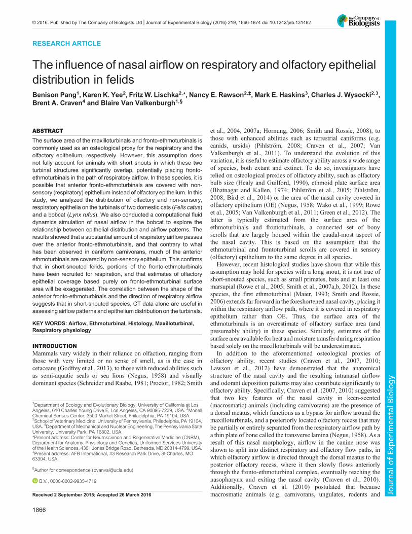
ABCDE Fig. 1. Sagittal view of the skull of a domestic cat.Locations A–E are indicated to show the locations of thescans in Fig. 2 within the nasal fossa, and are defined asthe percent of the total length of the nasal fossa fromanterior to posterior: A, 38%; B, 60%; C, 80%; D, 93%;and E, 98%.
1867
RESEARCH ARTICLE Journal of Experimental Biology (2016) 219, 1866-1874 doi:10.1242/jeb.131482
Journal
ofEx
perim
entalB
iology
3–4 min. After rinsing three times with dH2O, the slides were driedin an oven for 1 h and cleared with Histoclear (National Diagnostics,Atlanta, GA, USA) and mounted on glass coverslips with Permount(Fisher Scientific). For cat 4298, 65 slides at intervals between 160and 200 μmwere stained. Of those, 26 stained sections, between 0.5and 2.5 mm apart, were selected for quantitative measurement withthe most anterior section at approximately 14 mm posterior to the tipof the nose and the last section at a distance of 43.45 mm from thetip of the nose. For cat 6559, 26 stained slides, between 0.3 and
2.6 mm apart, with similar turbinal anatomy to that in the selectedsections from cat 4298, were chosen for staining and quantitativeanalysis.
For the bobcat, 21 slides at intervals from 200 to 400 μm werestained with Alcian Blue and Nuclear Fast Red. To further visualize
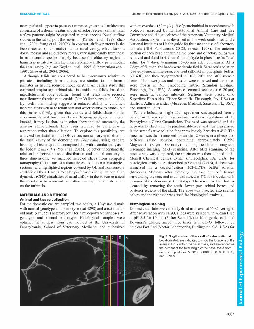
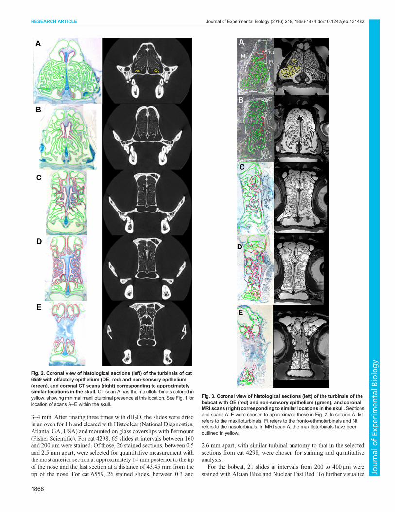
Fig. 2. Coronal view of histological sections (left) of the turbinals of cat6559 with olfactory epithelium (OE; red) and non-sensory epithelium(green), and coronal CT scans (right) corresponding to approximatelysimilar locations in the skull. CT scan A has the maxilloturbinals colored inyellow, showingminimal maxilloturbinal presence at this location. See Fig. 1 forlocation of scans A–E within the skull.
Fig. 3. Coronal view of histological sections (left) of the turbinals of thebobcat with OE (red) and non-sensory epithelium (green), and coronalMRI scans (right) corresponding to similar locations in the skull. Sectionsand scans A–E were chosen to approximate those in Fig. 2. In section A, Mtrefers to the maxilloturbinals, Ft refers to the fronto-ethmoturbinals and Ntrefers to the nasoturbinals. In MRI scan A, the maxilloturbinals have beenoutlined in yellow.
1868
RESEARCH ARTICLE Journal of Experimental Biology (2016) 219, 1866-1874 doi:10.1242/jeb.131482
Journal
ofEx
perim
entalB
iology
OE from non-sensory epithelium, adjacent sections were labeled withβ-tubulin III antibody, as has been previously done to label olfactoryneurons in cats (Lischka et al., 2008). After washing, tissue sectionswere incubatedwith a secondarymouse biotinylated antibody (VectorLaboratories), followed by the avidin-biotinylated horseradishperoxidase complex ABC Elite Kit (Vector Laboratories). Sectionswere then reacted with the chromogen diaminobenzidine (DAB;Sigma Chemicals) and 0.1% H2O2 for visualization.
Quantitative measurements of histological sectionsFor the domestic cats, selected stained sections were digitallyscanned at 1200 dpi resolution on an HP flatbed scanner to generatean 8×10 inch printed hard copy of each section. For the bobcat,stained sections were digitally captured with a RT slider SPOTcamera (Diagnostic Instruments) attached to a Nikon SMZ-Udissecting microscope. The septum, maxilloturbinal, nasoturbinaland fronto-ethmoturbinals were identified based on location in eachsection. Non-sensory epithelium (including respiratory, transitionaland stratified epithelia) and OE were identified under 40× and 60×objective magnifications by well-defined histological characteristicsof olfactory mucosa as contrasted to the adjacent respiratory mucosa(i.e. epithelial thickness, presence or absence of Alcian Blue stainedgoblet cells and/or Bowman’s glands in the underlining laminapropria). For the domestic cats, OE was identified via well-defined
histological characteristics (as described). For the bobcat, OE wasidentified by β-tubulin labeling.
Digital scans of the domestic cats and bobcat were quantitativelymeasured using the ImageProPlus 6.0 software (MediaCybernetics).The following measures were taken on each section for both sides ofthe cat noses and the right side of the bobcat nose: (1) the total length(cross-sectional perimeter) of the septum, maxilloturbinals,nasoturbinals and fronto-ethmoturbinals, and (2) the total length(cross-sectional perimeter) of OE lining each of these structures. Ourmeasurement of the fronto-ethmoturbinal complex includes all largerand smaller turbinals of the ethmoid bone (i.e. combined surface areaof the ethmoturbinals, interturbinals and frontoturbinals as defined byMaier, 1993; Smith et al., 2007b; Maier and Ruf, 2014). Theperimeter of non-sensory epithelium for each section was determinedby subtracting OE perimeter from total septal and turbinal perimeters.The surface area of a given section was calculated by multiplying theperimeter of OE for a section by the distance to the next section. TheOE surface areas were summed across all sections to give acumulative measure of OE surface area on the nasoturbinals,septum and fronto-ethmoturbinals. With these measurements, wealso calculated the proportion of the surface area of the turbinals andseptum that are covered with OE, as well as the relative OE/non-sensory epithelium ratio in the nasal fossa. As the bobcatmeasurements were taken from the right half of the nose,
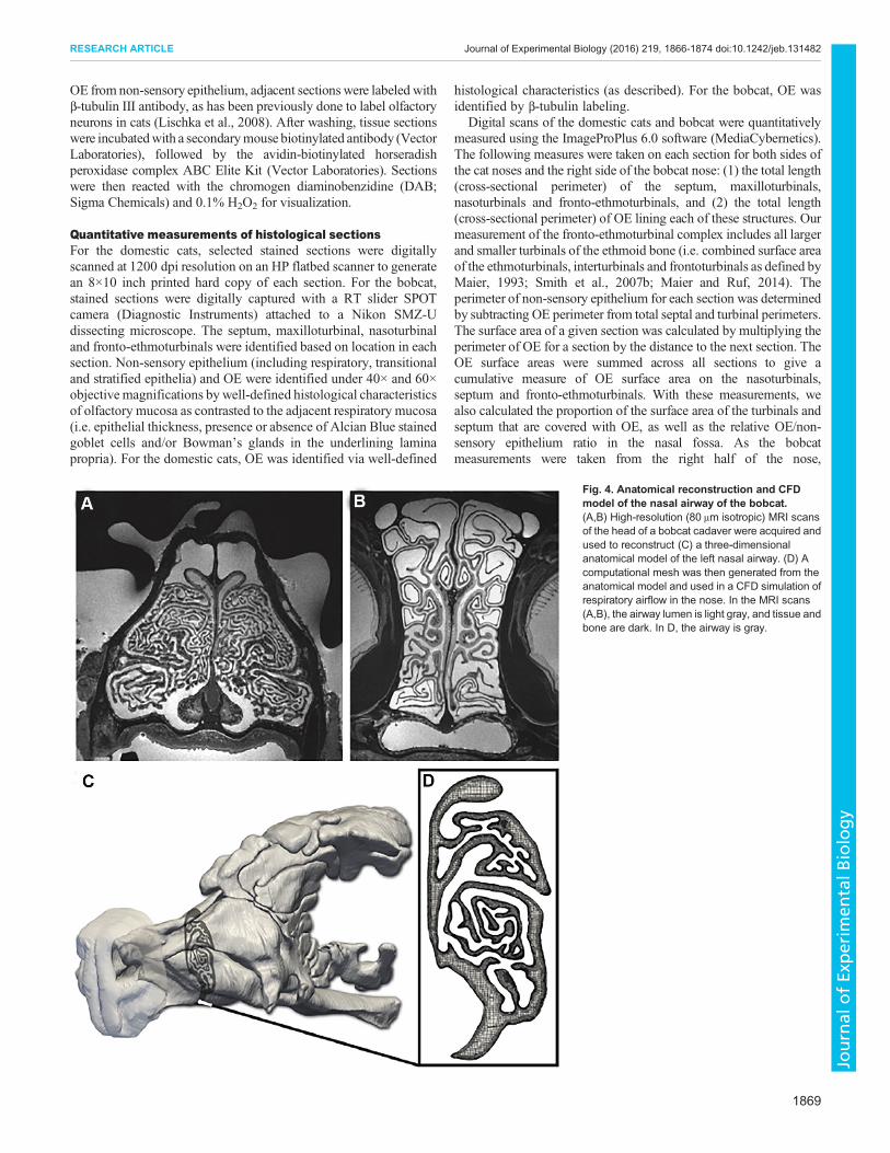
Fig. 4. Anatomical reconstruction and CFDmodel of the nasal airway of the bobcat.(A,B) High-resolution (80 μm isotropic) MRI scansof the head of a bobcat cadaver were acquired andused to reconstruct (C) a three-dimensionalanatomical model of the left nasal airway. (D) Acomputational mesh was then generated from theanatomical model and used in a CFD simulation ofrespiratory airflow in the nose. In the MRI scans(A,B), the airway lumen is light gray, and tissue andbone are dark. In D, the airway is gray.
1869
RESEARCH ARTICLE Journal of Experimental Biology (2016) 219, 1866-1874 doi:10.1242/jeb.131482
Journal
ofEx
perim
entalB
iology

cumulativemeasurements from the domestic catswere divided by twoto allow for a consistent comparison.Because the anterior tips of turbinals may have been excluded if
they occurred in the interslice intervals (0.2–0.4 mm), we estimatedthe maximum possible error. This was done by adding the perimeterof OE measured on its first occurrence to the overall sum of OE onthe septum, nasoturbinals, fronto-ethmoturbinals and cumulativetotal OE, and found the percentage difference that arose from thisaddition. The maximum OE perimeter measurement error that wasdue to possibly excluding the anterior tips of turbinals and septumbecause they ended in an interslice interval was, on average, 4.8%for the septum, 12.7% for the nasoturbinals, 0.04% for theethmoturbinals and 2.1% for the cumulative total OE surface area.
CT and MRI scansTo view the anatomy in three dimensions, high-resolution CT scansof the skull of a domestic cat (Felis catus) were acquired at theUniversity of Texas (UT) High Resolution X-Ray ComputedTomography Facility. The data set comprises 387 scans of the nasalcavity, with a slice thickness of 0.238 mm, and is available byrequest from the Digimorph library (http://www.digimorph.org/).The scans were first imported into the software program Mimics14.0 (Materialise Inc.) to view the three-dimensional data set fromcoronal, sagittal and transverse perspectives, as well as to enhancevisualization of the turbinals by increasing the contrast betweenthe bony turbinals and the air. MRI scanning for the bobcat nosewas performed on a 7-Tesla horizontal Agilent system atPennsylvania State University (PSU). Coronal CT and MRIscans were selected where the morphology of the turbinals mostclosely resembled histological sections at various locations inthe nasal fossa (Figs 1–3). This allowed for a comparison of
the relative distributions of epithelium at specific locations in thenasal fossa.
To compare the degree of overlap of the fronto-ethmoturbinalsand maxilloturbinals between a long-snouted canid and a short-snouted felid, CT scans of the skull of a Mexican gray wolf (Canislupus baileyi) (USNM 98307, Smithsonian Institution, Washington,DC, USA) were also obtained from the University of Texas andprocessed the same way as the scans of the domestic cat. A similarprocedure was performed for two additional felids, the cheetah(Acinonyx jubatus, FMNH 29635, Field Museum of NaturalHistory, Chicago, IL, USA) and the African lion (Panthera leo,MMNH 17537, University of Minnesota Museum of NaturalHistory, Minneapolis, MN, USA). Subsequently, we inspectedsagittal scans of the nasal fossa of each animal to estimate the degreeof overlap in each case.
Computational fluid dynamicsTo examine nasal airflow patterns in a short-snouted felid, weacquired high-resolution (80 μm isotropic) MRI scans (Fig. 4A,B)
Table 1. Calculated olfactory epithelium (OE) measurements for allturbinals and septum in two domestic cats and a bobcat
Cat 6559 Cat 4298 Bobcat
Total surface area of OE(mm2) (% of total OE)
OE 839.7 (100) 940 (100) 2333.9 (100)Septum OE 205.7 (24.5) 207.7 (22.1) 369.8 (15.8)Nasoturbinal OE 44.28 (5.3) 75.4 (8) 155.3 (6.7)Fronto-ethmoturbinal OE 589.7 (70.2) 656.9 (69.9) 1808.8 (77.5)
Ratio of OE to non-sensory(respiratory) epithelium
0.14 0.12 0.16
Felis catus
Canis lupus baileyi
Fig. 5. Sagittal CT scans of the turbinals of adomestic cat (Felis catus) and a Mexican gray wolf(Canis lupus baileyi). Ethmoturbinals are in red,maxilloturbinals in blue.
1870
RESEARCH ARTICLE Journal of Experimental Biology (2016) 219, 1866-1874 doi:10.1242/jeb.131482
Journal
ofEx
perim
entalB
iology

of the head of the bobcat at PSU and reconstructed a three-dimensional anatomical model of the left nasal airway (Fig. 4C)from the MRI data using the methodology of Craven and colleagues(Craven et al., 2007; Ranslow et al., 2014; Coppola et al., 2014). Ahigh-fidelity, hexahedral-dominant computational mesh (Fig. 4D)was then generated from the reconstructed nasal airway surfacemodel using snappyHexMesh, the unstructured mesh generationutility in the open-source computational continuum mechanicslibrary OpenFOAM (www.openfoam.com). The mesh containedroughly 121million computational cells, including five wall-normallayers along the airway walls to resolve near-wall velocity gradientsand a spherical refinement region around the nostril to resolve theflow as it accelerates toward and enters the naris on inspiration.A steady-state computational fluid dynamics (CFD)
simulation of laminar airflow during inspiration was performedusing OpenFOAM. As in Craven et al. (2009), thecomputational domain included the anatomical model of theleft nasal airway positioned in the center of a large rectangular
box with atmospheric pressure boundary conditions assigned tothe sides of the box. A pressure outlet boundary conditionwas specified at the nasopharynx to induce a physiologicallyrealistic respiratory airflow rate of 3.7 l min−1, which wasdetermined from the mass of the specimen (12 kg) and theallometric equation for respiratory minute volume developed byBide et al. (2000). No-slip boundary conditions were assignedon the external nose and airway walls, which were assumed tobe rigid. Additionally, as justified by Craven et al. (2009), thepresence of the thin mucus layer that lines the nasal epitheliumwas neglected.
The SIMPLE (Semi-Implicit Method for Pressure-LinkedEquations) algorithm was used to numerically solve theincompressible continuity and Navier–Stokes equations usingsecond-order accurate spatial discretization schemes. The simulationwas performed on 480 processors of a parallel computer cluster atPSU. Iterative convergenceof the SIMPLEalgorithmwas obtainedbyforcing the normalized solution residuals to be less than 10−4.Additionally, the volumetric flow rate, viscous and pressure forces,and other solution quantities were monitored to ensure solutionconvergence. Visualization and post-processing of the CFD resultswere performed using the open-source visualization softwareParaView (www.paraview.org).
RESULTSIn cat 6559, OE was distributed unequally over the septum,nasoturbinals and fronto-ethmoturbinals. Approximately 24.5% ofthe total OE could be found on the septum, with the remaining 5.3%and 70.2% lining the nasoturbinals and the fronto-ethmoturbinals,respectively (Table 1). This was similar to the distribution in cat4298, in which 22.1% of total OE was found on the septum, 8% onthe nasoturbinals and 69.9% on the fronto-ethmoturbinals, as wellas the bobcat in which 15.8% of total OE was found on the septum,6.7% on the nasoturbinals and 77.5% on the fronto-ethmoturbinals.On average, cat 6559 had a cumulative total OE surface area of839.7 mm2, while cat 4298 had a total of 940 mm2. The bobcat hada cumulative total OE surface area of 2333.9 mm2. Overall, the ratioof OE surface area to non-sensory epithelium surface area was 0.14in cat 6559, 0.12 in cat 4298 and 0.16 in the bobcat.
In both domestic cats, the general pattern of OE distribution in thenasal fossa was similar, and so here we report the results obtainedfrom cat 6559. In the rostral region, the fronto-ethmoturbinalsextended anteriorly in the snout, lying above much of themaxilloturbinal complex (Fig. 1A). However, the most anteriorappearance of OE was not on the fronto-ethmoturbinals. Instead, itfirst appeared on the roof of the nasal cavity and the dorsal arch ofthe nasoturbinals, at a distance of approximately 38% of the totallength of the nasal fossa (Fig. 2A). At this location, themaxilloturbinals were barely present. Here, the fronto-ethmoturbinals occupied much of the available space, and weretotally covered with non-sensory epithelium.
OE remained concentrated on the septum and the medial aspect ofthe nasoturbinals for the anterior 60% of the total length of the nasalfossa, at which point it began to gradually expand laterally (Fig. 2B).OE was present posterior to the internal nares within the anteriorregions of the olfactory recess, where it was predominantlydistributed on the septum, nasoturbinals and the medial portion ofthe fronto-ethmoturbinals (Fig. 2C).
Within the caudal regions of the olfactory recess, the proportionalcoverage of OE increased posteriorly until, at a position ofapproximately 90% of the total length of the nasal fossa, 48.6%of the available turbinal and septum surface area was covered by OE
Acinonyx jubatus
Panthera leo
Fig. 6. Sagittal CT scans of the turbinals of a cheetah (Acinonyx jubatus)and a lion (Panthera leo). Ethmoturbinals are in red, maxilloturbinals in blue.
1871
RESEARCH ARTICLE Journal of Experimental Biology (2016) 219, 1866-1874 doi:10.1242/jeb.131482
Journal
ofEx
perim
entalB
iology
(Fig. 2D). At location E, at a position of approximately 98% of thetotal length of the nasal fossa, OE covered 44.5% of the availablesurface area, and was split nearly evenly dorsal and ventral to thecribriform plate (Fig. 2E).In the bobcat, the broad patterns of epithelial distribution (OE
and non-sensory) are fairly similar to that of the domestic cat;Fig. 3 shows histological outlines and MRI scans of the bobcatnasal cavity at corresponding locations to that of the domestic catshown in Fig. 2. In the anterior nasal cavity of the bobcat(Fig. 3A), the fronto-ethmoturbinals are present and are coveredwith non-sensory epithelium much like in the domestic cat. OE isalso first seen in the bobcat covering the nasoturbinals and the roofof the nasal cavity. Subsequently, OE is found on the septumand nasoturbinals (Fig. 3B), and only in the olfactory recess(Fig. 3C–E) does OE occupy a sizeable portion of the availableturbinal surface area, which is again comparable to that observedin the domestic cat.Morphologically, the fronto-ethmoturbinals of the domestic cat
extended far forward in the snout and lay above much of themaxilloturbinal complex (Fig. 1A). Compared with the scanned graywolf, there was much greater overlap of the maxilloturbinal complexand the fronto-ethmoturbinals in the cat (Fig. 5). Indeed, in sagittalsections it appears that the anterior fronto-ethmoturbinals are alignedwith the path of respiratory airflow from the external nares to thenasopharynx, as are the maxilloturbinals. The anterior fronto-ethmoturbinals slope downward posteriorly, appearing to be angledtowards the internal nares (Fig. 5). This differs from the alignment ofmore posterior ethmoturbinals, which are roughly horizontal,particularly in the olfactory recess. A comparison between the
relatively long-snouted lion and short-snouted cheetah (Fig. 6)showed a similar result: there was substantially greater overlap of thefronto-ethmoturbinals and maxilloturbinals in the cheetah than in thelion, and the anterior fronto-ethmoturbinals also seem to be alignedwith the respiratory airflow path.
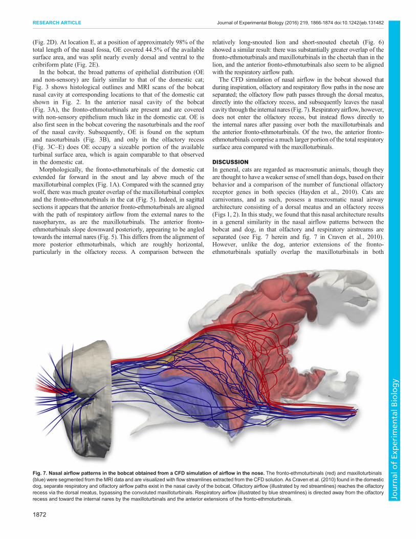
The CFD simulation of nasal airflow in the bobcat showed thatduring inspiration, olfactory and respiratory flow paths in the nose areseparated; the olfactory flow path passes through the dorsal meatus,directly into the olfactory recess, and subsequently leaves the nasalcavity through the internal nares (Fig. 7).Respiratoryairflow, however,does not enter the olfactory recess, but instead flows directly tothe internal nares after passing over both the maxilloturbinals andthe anterior fronto-ethmoturbinals. Of the two, the anterior fronto-ethmoturbinals comprise a much larger portion of the total respiratorysurface area compared with the maxilloturbinals.
DISCUSSIONIn general, cats are regarded as macrosmatic animals, though theyare thought to have aweaker sense of smell than dogs, based on theirbehavior and a comparison of the number of functional olfactoryreceptor genes in both species (Hayden et al., 2010). Cats arecarnivorans, and as such, possess a macrosmatic nasal airwayarchitecture consisting of a dorsal meatus and an olfactory recess(Figs 1, 2). In this study, we found that this nasal architecture resultsin a general similarity in the nasal airflow patterns between thebobcat and dog, in that olfactory and respiratory airstreams areseparated (see Fig. 7 herein and fig. 7 in Craven et al., 2010).However, unlike the dog, anterior extensions of the fronto-ethmoturbinals spatially overlap the maxilloturbinals in both
Fig. 7. Nasal airflow patterns in the bobcat obtained from a CFD simulation of airflow in the nose. The fronto-ethmoturbinals (red) and maxilloturbinals(blue) were segmented from the MRI data and are visualized with flow streamlines extracted from the CFD solution. As Craven et al. (2010) found in the domesticdog, separate respiratory and olfactory airflow paths exist in the nasal cavity of the bobcat. Olfactory airflow (illustrated by red streamlines) reaches the olfactoryrecess via the dorsal meatus, bypassing the convoluted maxilloturbinals. Respiratory airflow (illustrated by blue streamlines) is directed away from the olfactoryrecess and toward the internal nares by the maxilloturbinals and the anterior extensions of the fronto-ethmoturbinals.
1872
RESEARCH ARTICLE Journal of Experimental Biology (2016) 219, 1866-1874 doi:10.1242/jeb.131482
Journal
ofEx
perim
entalB
iology
felids (cat and bobcat) and are covered with respiratory epithelium(Fig. 2A,B, 3A). In this region, OE is primarily confined to thedorsal meatus, which contains the olfactory airflow stream, whilerespiratory airflow passes over the (limited) maxilloturbinals and theanterior fronto-ethmoturbinals, where OE is absent. Thus, thehistological distribution of epithelia in the bobcat nose appears to behighly correlated with the nasal airflow patterns. That is, therespiratory airflow stream passes mainly over non-sensoryepithelium, whereas OE is primarily exposed to only the olfactoryairflow stream (see Figs 3, 7).Gross nasal morphology and turbinal anatomy in the domestic
cat are very similar to that in the bobcat (Figs 2, 3), which stronglysuggests that similar nasal airflow patterns occur in the domesticcat. Given the similar epithelial distribution in the domestic cat andbobcat, the correlation between nasal histology and airflow patternsfound in the bobcat also likely applies to domestic cats.Accordingly, these data strongly suggest that the anterior fronto-ethmoturbinals have been co-opted for respiratory function in thesetwo species, and likely other similarly short-snouted felids.The degree of spatial overlap of the fronto-ethmoturbinals and
maxilloturbinals in all three short-snouted felids (domestic cat, bobcatand cheetah) was similar and much less than that observed in both thelonger-snouted lion and the long-snouted gray wolf. This suggeststhat the degree of spatial overlap of the fronto-ethmoturbinals andmaxilloturbinals is more closely correlated with relative snout lengththan with phylogenetic groupings such as feliforms versus caniforms.Given the similarity of the gross nasal architecture and the overall
nasal airflow patterns between the dog and the bobcat, we mightalso expect qualitatively similar odorant deposition patterns in theolfactory region. Lawson et al. (2012) observed that the highestodorant deposition fluxes are located anteriorly in the sensoryregion of the canine nose, along the dorsal meatus and nasal septum,which is consistent with our observation of OE being concentratedin this location in the domestic cat (Fig. 2) and bobcat (Fig. 3). In thecanine, highly soluble odorants are deposited anteriorly in thesensory region, particularly along the dorsal meatus, and less-soluble odorants are deposited more uniformly (Lawson et al.,2012). OE in the cats is concentrated on the medial aspect of theolfactory recess, with less OE being distributed peripherally; hence,there is less surface area for detecting moderately soluble andinsoluble odorants. Accordingly, compared with other animals thatpossess more peripheral OE, cats may have a reduced sense of smellfor less-soluble odorants.Compared with other species, it is interesting to note that Smith
et al. (2007a,b) also observed respiratory mucosa on fronto-ethmoturbinal I in the mouse lemur (Microcebus murinus) andnoted that it is positionedmore anteriorly and is, therefore, exposed torespiratory airflow (Smith et al., 2007b). Thus, it appears that fronto-ethmoturbinals in the mouse lemur also may have been co-opted forrespiratory air-conditioning. This suggests that fronto-ethmoturbinalsthat function in respiration may have arisen convergently in primatesand carnivorans as a result of having a shorter snout, either throughselection for enhanced jaw muscle leverage in feliforms (Bikneviciusand Van Valkenburgh, 1996) or other selection factors that led toreduced primate snouts (Smith et al., 2007b).In our study of feliform and caniform carnivorans, there appears
to be a clear relationship between turbinal architecture, nasal airflowand epithelial distribution. Recent research has shown that airflowmay influence turbinal size and shape during development(Coppola et al., 2014), which is consistent with the results of thepresent study. Inferring epithelial surface area from CT data alone isdifficult, but can be improved by taking into account nasal airflow
patterns and the variability of OE distribution in different species. Inthis study, we have shown that visual inspection of CT data can offeran initial insight into expected nasal airflow patterns and epithelialdistribution. Future work will focus on documenting OEdistribution in more short-snouted taxa and testing our hypothesesconcerning nasal airflow and odorant deposition patterns using CFD(e.g. as in Zhao et al., 2006; Yang et al., 2007a,b; Craven et al.,2009, 2010; Lawson et al., 2012).
AcknowledgementsThe authors are grateful to T. Ryan and T. Neuberger at PSU for providing CT scansof the domestic cat and MRI scans of the bobcat, respectively. We thank C. Packerfor the loan of the African lion skull, as well as the curatorial staff of the US NationalMuseum and the Field Museum of Natural History for the loans of the gray wolf andcheetah skulls, respectively. We thank A. Quigley, A. Ranslow and A. Rygg forassistance in segmenting and reconstructing the MRI data of the bobcat. We alsothank L. Lo for assistance in staining and measuring cat sections and anonymousreviewers for helpful comments on the manuscript. Histology was performed at theMonell Histology and Cellular Localization Core.
Competing interestsThe authors declare no competing or financial interests.
Author contributionsB.P. analyzed the data and wrote the manuscript. K.K.Y. performed histologicalwork, analyzed the data and wrote the manuscript. F.W.L. performed histologicalwork on the domestic cat specimens and reviewed the manuscript. N.E.R.contributed to the development of the initial hypotheses, experimental planning andmethods, and assisted with editing of the manuscript. M.E.H. provided the catsamples and reviewed the manuscript. C.J.W. was principal investigator on NSFAward 1118852 and provided intellectual and editorial input. B.A.C. was principalinvestigator on NSF Award 1120375, performed the CFD simulations and wrote themanuscript. B.V.V. was principal investigator on NSF Awards 0517748 and1119768, and wrote the manuscript.
FundingHistology was supported, in part, by funding from the National Institutes of Health–National Institute on Deafness and other Communication Disorders Core GrantP30DC011735. The study was also supported by the National Science Foundation[IOS-0517748 and IOS-1119768 to B.V.V., IOS-1118852 to C.J.W. and IOS-1120375 to B.A.C.] and the National Institutes of Health [P40-OD010939 to M.E.H.].Deposited in PMC for release after 12 months.
Data availabilityThe domestic cat CT scan data set is available by request from the Digimorph library(http://www.digimorph.org/).
ReferencesBhatnagar, K. P. and Kallen, F. C. (1974). Cribriform plate of ethmoid, olfactory
bulb and olfactory acuity in forty species of bats. J. Morphol. 142, 71-89.Bide, R. W., Armour, S. J. and Yee, E. (2000). Allometric respiration/body mass
data for animals to be used for estimates of inhalation toxicity to young adulthumans. J. Appl. Toxicol. 20, 273-290.
Biknevicius, A. R. andVanValkenburgh,B. (1996). Design for killing: craniodentaladaptations of predators. In Carnivore Behavior, Ecology, and Evolution, Vol. 2(ed. J. L. Gittleman), pp. 393-428. Ithaca, NY: Cornell University Press.
Bird, D. J., Amirkhanian, A., Pang, B. and Van Valkenburgh, B. (2014).Quantifying the cribriform plate: influences of allometry, function, and phylogeny inCarnivora. Anat. Rec. 297, 2080-2092.
Coppola, D. M., Craven, B. A., Seeger, J. and Weiler, E. (2014). The effects ofnaris occlusion on mouse nasal turbinate development. J. Exp. Biol. 217,2044-2052.
Craven, B. A., Neuberger, T., Paterson, E. G., Webb, A. G., Josephson, E. M.,Morrison, E. E. and Settles, G. S. (2007). Reconstruction and morphometricanalysis of the nasal airway of the dog (Canis familiaris) and implicationsregarding olfactory airflow. Anat. Rec. 290, 1325-1340.
Craven, B. A., Paterson, E. G., Settles, G. S. and Lawson, M. J. (2009).Development and verification of a high-fidelity computational fluid dynamicsmodel of canine nasal airflow. J. Biomech. Eng. 131, 091002.
Craven, B. A., Paterson, E. G. and Settles, G. S. (2010). The fluid dynamics ofcanine olfaction: unique nasal airflow patterns as an explanation of macrosmia.J. R. Soc. Interface 7, 933-943.
1873
RESEARCH ARTICLE Journal of Experimental Biology (2016) 219, 1866-1874 doi:10.1242/jeb.131482
Journal
ofEx
perim
entalB
iology
Godfrey, S. J., Geisler, J. and Fitzgerald, E. M. G. (2013). On the olfactoryanatomy in an archaic whale (Protocetidae, Cetacea) and the minke whaleBalaenoptera acutorostrata (Balaenopteridae, Cetacea). Anat. Rec. 296,257-272.
Green, P. A., Van Valkenburgh, B., Pang, B., Bird, D., Rowe, T. and Curtis, A.(2012). Respiratory and olfactory turbinal size in canid and arctoid carnivorans. J.Anat. 221, 609-621.
Hayden, S., Bekaert, M., Crider, T. A., Mariani, S., Murphy, W. J. and Teeling,E. C. (2010). Ecological adaptation determines functional mammalian olfactorysubgenomes. Genome Res. 20, 1-9.
Healy, S. and Guilford, T. (1990). Olfactory-bulb size and nocturnality in birds.Evolution 44, 339-346.
Hornung, D. E. (2006). Nasal anatomy and the sense of smell. Adv.Otorhinolaryngol. 63, 1-22.
Keyhani, K., Scherer, P. W. and Mozell, M. M. (1995). Numerical simulation ofairflow in the human nasal cavity. J. Biomech. Eng. 117, 429-441.
Kimbell, J. S., Godo, M. N., Gross, E. A., Joyner, D. R., Richardson, R. B. andMorgan, K. T. (1997). Computer simulation of inspiratory airflow in all regions ofthe F344 rat nasal passages. Toxicol. Appl. Pharmacol. 145, 388-398.
Lawson, M. J., Craven, B. A., Paterson, E. G. and Settles, G. S. (2012). Acomputational study of odorant transport and deposition in the canine nasal cavity:implications for olfaction. Chem. Senses 37, 553-566.
Lischka, F. W., Gomez, G., Yee, K. K., Dankulich-Nagrundy, L., Lo, L., Haskins,M.E. andRawson,N.E. (2008).Alteredolfactoryepithelial structure and function infeline models of mucopolysaccharidoses I and VI. J. Comp. Neurol. 511, 360-372.
Maier, W. (1993). Zur evolutiven und funktionellen Morphologie desGesichtsschadels der Primaten. Z. Morphol. Anthropol. 79, 279-299.
Maier, W. and Ruf, I. (2014). Morphology of the nasal capsule of primates-withspecial reference to Daubentonia and Homo. Anat. Rec. 297, 1985-2006.
Negus, V. (1958). The Comparative Anatomy and Physiology of the Nose andParanasal Sinuses. Edinburgh: E. & S. Livingstone.
Pihlstrom, H. (2008). Comparative anatomy and physiology of chemical senses inaquatic mammals. In Sensory Evolution on the Threshold (ed. J. G. M. Thewissenand S. Nummela), pp. 95-109. Berkeley, CA: University of California Press.
Pihlstrom, H., Fortelius, M., Hemila, S., Forsman, R. and Reuter, T. (2005).Scaling of mammalian ethmoid bones can predict olfactory organ size andperformance. Proc. R. Soc. B Biol. Sci. 272, 957-962.
Proctor, D. F. (1982). The upper airway. In The Nose: Upper Airway Physiology andthe Atmospheric Environment (ed. D. F. Proctor. and I. H. P. Andersen), pp. 23-43.New York, NY: Elsevier Biomedical Press.
Ranslow, A. N., Richter, J. P., Neuberger, T., Van Valkenburgh, B., Rumple,C. R., Quigley, A. P., Pang, B., Krane, M. H. and Craven, B. A. (2014).Reconstruction and morphometric analysis of the nasal airway of the white-taileddeer (Odocoileus virginianus) and implications regarding respiratory and olfactoryairflow. Anat. Rec. (Hoboken) 297, 2138-2147.
Rowe, T. B., Eiting, T. P., Macrini, T. E. andKetcham, R. A. (2005). Organization ofthe olfactory and respiratory skeleton in the nose of the gray short-tailed opossumMonodelphis domestica. J. Mamm. Evol. 12, 303-336.
Schreider, J. P. and Raabe, O. G. (1981). Anatomy of the nasal-pharyngeal airwayof experimental animals. Anat. Rec. 200, 195-205.
Smith, T. D. and Rossie, J. B. (2006). Primate olfaction: anatomy and evolution. InOlfaction and the Brain: Window to the Mind (ed. W. Brewer, D. Castle, and C.Pantelis), pp. 135-166. Cambridge: Cambridge University Press.
Smith, T. D. and Rossie, J. B. (2008). Nasal fossa of mouse and dwarf lemurs(Primates, Cheirogaleidae). Anat. Rec. (Hoboken) 291, 895-915.
Smith, T. D., Bhatnagar, K. P., Tuladhar, P. and Burrows, A. M. (2004).Distribution of olfactory epithelium in the primate nasal cavity: are microsmia andmacrosmia valid morphological concepts? Anat. Rec. A Discov. Mol. Cell. Evol.Biol. 281A, 1173-1181.
Smith, T. D., Bhatnagar, K. P., Rossie, J. B., Docherty, B. A., Burrows, A. M.,Cooper, G. M., Mooney, M. P. and Siegel, M. I. (2007a). Scaling of the firstethmoturbinal in nocturnal strepsirrhines: olfactory and respiratory surfaces. Anat.Rec. 290, 215-237.
Smith, T. D., Rossie, J. B. and Bhatnagar, K. P. (2007b). Evolution of the nose andnasal skeleton in primates. Evol. Anthropol. 16, 132-146.
Smith, T. D., Eiting, T. P. and Bhatnagar, K. P. (2012). A quantitative study ofolfactory, non-olfactory, and vomeronasal epithelia in the nasal fossa of the batMegaderma lyra. J. Mamm. Evol. 19, 27-41.
Subramaniam, R. P., Richardson, R. B., Morgan, K. T. and Kimbell, J. S. (1998).Computational fluid dynamics simulations of inspiratory airflow in the human noseand nasopharynx. Inhal. Toxicol. 10, 91-120.
Van Valkenburgh, B., Theodor, J., Friscia, A., Pollack, A. and Rowe, T. (2004).Respiratory turbinates of canids and felids: a quantitative comparison. J. Zool.264, 281-293.
Van Valkenburgh, B., Curtis, A., Samuels, J. X., Bird, D., Fulkerson, B.,Meachen-Samuels, J. and Slater, G. J. (2011). Aquatic adaptations in the noseof carnivorans: evidence from the turbinates. J. Anat. 218, 298-310.
Wako, K., Hiratsuka, H., Katsuta, O. and Tsuchitani, M. (1999). Anatomicalstructure and surface epithelial distribution in the nasal cavity of the commoncotton-eared marmoset (Callithrix jacchus). Exp. Anim. 48, 31-36.
Yang, G. C., Scherer, P. W. and Mozell, M. M. (2007a). Modeling inspiratory andexpiratory steady-state velocity fields in the Sprague-Dawley rat nasal cavity.Chem. Senses 32, 215-223.
Yang, G. C., Scherer, P. W., Zhao, K. and Mozell, M. M. (2007b). Numericalmodeling of odorant uptake in the rat nasal cavity. Chem. Senses 32, 273-284.
Yee, K. K., Craven, B. A., Wysocki, C. J. and Van Valkenburgh, B. (2016).Comparative morphology and histology of the nasal fossa in four mammals: graysquirrel, bobcat, coyote and white-tailed deer. Anat. Rec. (in press).
Zhao, K., Scherer, P. W., Hajiloo, S. A. and Dalton, P. (2004). Effect of anatomy onhuman nasal air flow and odorant transport patterns: implications for olfaction.Chem. Senses 29, 365-379.
Zhao, K., Dalton, P., Yang, G. C. and Scherer, P.W. (2006). Numerical modeling ofturbulent and laminar airflow and odorant transport during sniffing in the humanand rat nose. Chem. Senses 31, 107-118.
1874
RESEARCH ARTICLE Journal of Experimental Biology (2016) 219, 1866-1874 doi:10.1242/jeb.131482
Journal
ofEx
perim
entalB
iology