the vapb–vapc operon of acidovorax citrulli functions as a...
TRANSCRIPT
ORIGINAL RESEARCHpublished: 06 January 2016
doi: 10.3389/fmicb.2015.01499
Edited by:Martin G. Klotz,
Queens College, City Universityof New York, USA
Reviewed by:Nicolás Pinel,
Universidad EAFIT, ColombiaRamon Diaz Orejas,
Consejo Superior de InvestigacionesCientíficas, Spain
*Correspondence:Yael Helman
Specialty section:This article was submitted to
Plant Biotic Interactions,a section of the journal
Frontiers in Microbiology
Received: 04 August 2015Accepted: 11 December 2015
Published: 06 January 2016
Citation:Shavit R, Lebendiker M, Pasternak Z,
Burdman S and Helman Y (2016)The vapB–vapC Operon
of Acidovorax citrulli Functions asa Bona-fide Toxin–Antitoxin Module.
Front. Microbiol. 6:1499.doi: 10.3389/fmicb.2015.01499
The vapB–vapC Operon ofAcidovorax citrulli Functions as aBona-fide Toxin–Antitoxin ModuleReut Shavit1, Mario Lebendiker2, Zohar Pasternak1, Saul Burdman1 and Yael Helman1*
1 Department of Plant Pathology and Microbiology, The Robert H. Smith Faculty of Agriculture, Food and Environment, TheHebrew University of Jerusalem, Rehovot, Israel, 2 Protein Purification Facility, Wolfson Centre for Applied Structural Biology,Edmund J. Safra Campus, The Hebrew University of Jerusalem, Jerusalem, Israel
Toxin–antitoxin systems are commonly found on plasmids and chromosomes of bacteriaand archaea. These systems appear as biscystronic genes encoding a stable toxinand a labile antitoxin, which protects the cells from the toxin’s activity. Under specific,mostly stressful conditions, the unstable antitoxin is degraded, the toxin becomes activeand growth is arrested. Using genome analysis we identified a putative toxin–antitoxinencoding system in the genome of the plant pathogen Acidovorax citrulli. The system ishomologous to vapB–vapC systems from other bacterial species. PCR and phylogeneticanalyses suggested that this locus is unique to group II strains of A. citrulli. Usingbiochemical and molecular analyses we show that A. citrulli VapBC module is a bona-fide toxin–antitoxin module in which VapC is a toxin with ribonuclease activity that canbe counteracted by its cognate VapB antitoxin. We further show that transcription of theA. citrulli vapBC locus is induced by amino acid starvation, chloramphenicol and duringplant infection. Due to the possible role of TA systems in both virulence and dormancyof human pathogenic bacteria, studies of these systems are gaining a lot of attention.Conversely, studies characterizing toxin–antitoxin systems in plant pathogenic bacteriaare lacking. The study presented here validates the activity of VapB and VapC proteinsin A. citrulli and suggests their involvement in stress response and host–pathogeninteractions.
Keywords: Acidovorax citrulli, toxin–antitoxin, VapB, VapC
INTRODUCTION
TheGram-negative bacteriumAcidovorax citrulli is a seed-borne pathogen responsible for bacterialfruit blotch (BFB), a threatening disease of cucurbits worldwide (Schaad et al., 2003). Underfavorable conditions, this bacterium spreads rapidly throughout nurseries and in the field leading toseedling blight or, at a later stage, fruit rot. Strategies for managing BFB are limited, and althoughseed treatments reduce disease transmission, they often fail to eradicate the pathogen from theseed (Dutta et al., 2008). In addition, chemical control of the disease in the field has only limitedefficiency and to date, there are no sources of BFB resistance (Bahar et al., 2009b; Burdman andWalcott, 2012). Understanding the mechanisms that promote plant tissue colonization, virulenceand spread of A. citrulli is therefore important for developing efficient tools to manage BFB.
Frontiers in Microbiology | www.frontiersin.org 1 January 2016 | Volume 6 | Article 1499
Shavit et al. The VapBC Toxin–Antitoxin System of A. citrulli
Based on several studies examining genetic and biochemicaltraits as well as host association, A. citrulli strains have beendivided into two major groups: group I strains have beenmainly isolated from various non-watermelon hosts (mainlymelon), while group II strains have been generally isolated fromwatermelon hosts (O’Brien andMartin, 1999;Walcott et al., 2000,2004; Burdman et al., 2005).
Using genome analysis we have identified a putative VapBC-like toxin–antitoxin (TA) encoding system in the genomeof AAC00-1, a group II strain of A. citrulli (sequenced bythe Joint Genome Institute; GenBank accession NC_008752.1).Genes encoding VapBC-like TA systems are widespread inthe in the genomes of both archaea and bacteria. Thesesystems generally appear as biscystronic genes, which encodea stable toxin (VapC), and a labile antitoxin (VapB). VapBare DNA binding proteins that can also bind the VapCtoxin and inhibit its toxic activity (Robson et al., 2009).The VapC toxins are ribonucleases that belong to the PIN-domain family (a domain homologous to the N-terminaldomain of the protein PilT), which usually cleave single-stranded RNA (Arcus et al., 2009; Robson et al., 2009;Winther and Gerdes, 2011). Under specific, mostly stressfulconditions, the unstable antitoxin is degraded and the toxin isreleased from the complex leading to permanent or reversiblecell growth arrest (reviewed in Hayes, 2003; Gerdes et al.,2005).
Toxin–antitoxin encoding genes are commonly foundon plasmids and chromosomes of prokaryotes. Whilethe role of plasmid-encoded TA systems as addictivemodules has been extensively studied (Gerdes et al., 1986;Yarmolinsky, 1995; Engelberg-Kulka and Glaser, 1999;Cooper and Heinemann, 2000; Patel and Weaver, 2006),the physiological importance of chromosomally encodedTA systems is still under debate. A possible involvement inthe following mechanisms has been proposed: (i) growthmodulation under stress (Gerdes, 2000; Christensen et al.,2003; Gerdes et al., 2005); (ii) generation of persister cells(Maisonneuve et al., 2011, 2013, Gerdes and Maisonneuve,2012); (iii) genome maintenance (Szekeres et al., 2007); and(iv) programmed cell death (Engelberg-Kulka and Glaser,1999; Hazan et al., 2004; Mutschler et al., 2011; Erental et al.,2012). An additional hypothesized role, which relates to TAmodules found in pathogenic bacteria, suggests that thesesystems may be involved in growth regulation of bacteria onceinside the host cells (Hopper et al., 2000; Ren et al., 2012;De la Cruz et al., 2013). Due to this possible involvementin virulence, as well as in dormancy of human pathogenicbacteria, studies of TA systems are gaining a lot of attention.Recent studies present preliminary evidence suggesting thatsynthetic peptides can be used to modulate TA systems, thusproviding avenues for the development of novel antibacterialagents (Williams et al., 2011; Williams and Hergenrother,2012).
In contrast to the vast number of studies examining thephysiological role of TA systems in animal pathogenic bacteria,almost nothing is known about the role of these systems inplant pathogenic bacteria. We report here the molecular and
biochemical characterization of a VapBC-like module from thephytopathogenic bacterium A. citrulli.
MATERIALS AND METHODS
Bacterial Strains, Plasmids, and GrowthConditionsSince A. citrulli is a quarantine bacterium in Israel, we cannotwork with strain AAC00-1 that was isolated in the USA.Therefore, the study was performed using A. citrulli strain7a1, a group II strain isolated in Israel (Eckshtain-Levi et al.,2014), in which the vapBC operon is 100% identical to that ofstrain AAC00-1 (see Results). Unless stated otherwise, A. citrulli7a1 (Eckshtain-Levi et al., 2014) was grown in nutrient broth(NB; Difco, Franklin Lakes, NJ, USA) under constant shaking(150 rpm) or nutrient agar (NA; NB containing 15 g/l agar) at28◦C. Escherichia coli strains DH5α (Hanahan, 1983), BL21(DE3)and BL21-AI (Thermo Fisher ScientificTM, Waltham, MA, USA)were routinely grown in Lysogeny broth (LB; Difco) underconstant shaking (150 rpm) or LB agar (LB containing 15 g/lagar) at 37◦C. All strains were maintained as glycerol stocksat −80◦C.
General Molecular TechniquesKits used for plasmid and PCR product extraction andpurification were AccuPrep
R© Plasmid Mini Extraction Kitand AccuPrep R© PCR Purification Kit, respectively (BioneerCorporation, Daejeon, Republic of Korea). DNA was extractedusing the GeneEluteTM bacterial genomic DNA Kit (Sigma-Aldrich, St. Louis, MO, USA). RNA extraction was carried outusing TRI Reagent (Sigma-Aldrich). All kits were used accordingto the manufacturer’s protocols unless stated otherwise. PCRproducts were sequenced at Hy Labs (Rehovot, Israel) and datawas analyzed using the Bioedit sequence alignment editor (TomHall Ibis Biosciences, Carlsbad, CA, USA). Oligonucleotidesprimers used in this study were purchased from Sigma-Aldrichand are listed in Supplementary Table S1.
Cloning and Sequencing of vapBC Genesfrom A. citrulli 7a1Primers for amplification of vapB and vapC genes from A.citrulli 7a1 were designed according to the genome sequenceof A. citrulli AAC00-1 (GenBank NC_008752) using OligoAnalyzer 3.1 (Integrated DNA Technologies Inc, Coralville,IA, USA). PCR was performed using the REDTaq readymix (Sigma-Aldrich) in 25-μl reaction volumes. The PCRthermal profile consisted of initial denaturation for 5 minat 95◦C, followed by 35 cycles each of 30 s at 95◦C,annealing for 40 s at X◦C, and elongation at 72◦C for Ys (X and Y: annealing temperatures and elongation times,respectively, are detailed in Supplementary Table S1). A finalextension step was performed at 72◦C for 10 min. Samplesof 5 μl from each PCR reaction were run in 1% agarosegels (w/v) for 40 min at 120 V/cm. Gels were stained withethidium-bromide solution (0.5 μg/ml) and photographed with
Frontiers in Microbiology | www.frontiersin.org 2 January 2016 | Volume 6 | Article 1499
Shavit et al. The VapBC Toxin–Antitoxin System of A. citrulli
transmitted UV light at 295 nm. Cloning was carried outusing the restriction-free cloning (RF) method as described byUnger et al. (2010). High-Fidelity DNA Polymerase Phusion R©
(Bio Labs, New England, UK), was used in all RF PCRreactions.
Quantitative Real-Time PCR Analyses ofvapBC mRNA Expression LevelsQuantitative real-time PCR analyses (qRT-PCR) were performedusing the StepOnePlusTM Real-Time PCR System (AppliedBiosystems, Foster City, CA, USA) with green Fast SYBR2X (Applied Biosystems). Primers were designed usingPrimer3Input Software (v0.4.0). All values reported aregiven as relative expression of each gene compared to GAPDHmRNA expression levels. To choose the appropriate referencegene the expression levels of GAPDH, 16SrRNA, and recAmRNA were followed under the conditions used in the qRT-PCRexperiments. All samples were adjusted to 1 μg of total RNA forcDNA synthesis. mRNA levels of 16SrRNA and recA changedbetween control and stress experiments, as indicated by changesin Q-PCR cycles, by an average of 4 and 5 cycles, respectively,whereas those of GAPDH did not change over than two cycles.We therefore chose to use GAPDH as the reference gene.
For measurements of vapB and vapC mRNA levels underantibiotic stress or starvation A. citrulli 7a1 cells were grownover night in LB media at 28◦C, under constant shaking(150 rpm). After overnight growth, cultures were diluted 1:100and grown up to an OD600 of 0.5. At this time point,12.5 ng/μl chloramphenicol or 1 mg/ml serine hydroxamate(SHX) were added to the cultures according to the desiredtreatment. At various times after stress induction (0, 0.5, and1 h), 2 ml of treated culture were centrifuged (13,000 g),frozen in liquid nitrogen and kept at −20◦C till RNAextraction. Genomic DNA was eliminated by DNA-free DNase(Ambion, Austin, TX, USA). cDNA was generated using randomprimers with the High Capacity cDNA Reverse TranscriptionKit (Applied Biosystems), according to the manufacturer’sinstructions. Each sample contained 1 μg RNA in 20 μl ofreaction mix.
For in planta analyses of vapB and vapC mRNA expressionlevels,A. citrulli 7a1 was grown onNA for 48 h, resuspended fromplates in sterile distilled water (SDW) and adjusted to an OD600of 0.5 [about 108 colony forming units (CFUs)/ml] using a HeliosGamma spectrophotometer (Thermo Electron, Corp., Rochester,NY, USA). Stem inoculation experiments were performed on8-days-old watermelon (Citrullus lanatus) cv. Malali (HazeraGenetics, Co., Israel) seedlings as described (Bahar et al., 2009a).Briefly, seedlings were inoculated by placing a 5-μl drop of 108CFU/ml suspensions on the hypocotyls (at approximately 1 cmabove the soil). Then, a 25 gage needle was used to stab thestem through the drop. Seedlings were kept in the greenhouse(26–28◦C) for 6 days. At the desired time points (6 h and 1–6 days after inoculation) 1-cm segments around the inoculationpoint were cut and used for RNA extraction and bacterial cellcounts from inoculated plant tissue. Three pooled hypocotylsegments were used for each biological repeat. The segments werehomogenized and weighted. Samples for RNA extraction were
submitted to three cycles of freeze (liquid nitrogen) and thaw toensure cell breakage. RNA extraction and cDNA preparation wascarried as described above.
Cell Growth Assays for In vivo Analysesof VapBC ActivityCell growth experiments of E. coli BL21-AI cells expressingvapBC genes were carried out in 96-well microtiter plates(Thermo Fisher ScientificTM). The plasmid used for proteinexpression was pACYCDuet-1. Samples included pACYCDuet-1 plasmids expressing recombinant VapB or VapC separately,or VapB and VapC together. E. coli BL21-AI cells carrying anempty vector were grown as control. Each biological repeatconsisted of three colonies pooled into 200 μL of LB media.Following thorough mixing, 20 μL of each sample weretransferred into 180 μL LB in microtiter plates with a finalconcentration of 25 μg/ml chloramphenicol and 1% (w/v)glucose. Cell growth (OD595) was measured using an Infinite 200PRO NanoQuant instrument and i-controlTM software (Tecan,Männedorf, Switzerland). Cells were grown at 37◦C, and theoptical density at 595 nm was measured every 15 min. The plateswere shaken for a period of 15 s (linear shaking of 1 mm) beforeeach measurement. When OD595 reached 0.2, protein expressionwas induced by 0.5% (w/v) arabinose and 1 mM IPTG (finalconcentrations).
Expression and Purification ofRecombinant VapB and VapC ProteinsVapB protein with an N-terminal hexa histidine-tag (6XHis tag)was expressed in E. coli BL21 (DE3) cells using the pET15bplasmid (Merck Millipore, Billerica, MA, USA). For expressionof VapC, the VapC protein with an N-terminal 6XHis tag wasco-expressed with VapB in E. coli BL21 AI (arabinose induced)cells using plasmid pACYCDuet-1. The vapC open reading frame(ORF) was inserted within the multiple cloning site 2 (MCS-2), whereas the vapB ORF was inserted (without a tag) withinthe MCS-1. Recombinant E. coli cells were grown at 28◦C underconstant shaking at 150 rpm in LB supplemented with 1% (w/v)glucose. Induction of expression was carried out when cellsreached anOD600 of 0.5. E. coli BL21 (DE3) cells expressing VapBwere induced by 1 mM IPTG and E. coli BL21 AI, co-expressingVapC-6xHis and VapB were induced by 1 mM IPTG and 0.5%(w/v) arabinose. Growth of induced cells continued for 3 h afterwhich cells were centrifuged at 13,000 g and frozen in −20◦C tillprotein extraction.
Cell lysis was performed by sonication with the followinglysis buffer: 50 mM KH2PO4, 1.2 M NaCl, 100 mM KCl,20% glycerol, 25 mM imidazole, and 1% tritonX100 (pH7.8). The cell lysate was centrifuged at 11,000 g for 20 minat 4◦C, and the insoluble fraction (consisting of insolubleVapB and inclusion bodies of VapC) was resuspended in1% Triton (v/v) for 5 min in ice. The cell suspensionwas then centrifuged at 11,000 g for 10 min at 4◦C, andthe insoluble fraction was resuspended in 6 M urea andgently stirred for 1 h. Remaining particles were removedby centrifugation (11,000 g for 20 min) and the clarified
Frontiers in Microbiology | www.frontiersin.org 3 January 2016 | Volume 6 | Article 1499

Shavit et al. The VapBC Toxin–Antitoxin System of A. citrulli
supernatant was loaded onto a HisPurTM Ni-NTA resin (ThermoFisher ScientificTM) for separation of the VapC-6xHis fromVapB, according to the manufacturer’s instructions. Eluteddenatured protein was dialyzed in a mini GeBAflex-tube (GeneBio-Application L.T.D, Yavne, Israel) with refolding buffercontaining: 25 mM sodium phosphate buffer adjusted to pH7.5, 10 mM imidazole, 10% (v/v) glycerol and 0.5 M NaCl.The dialyzed protein suspension was then concentrated to1 mg/ml using Amicon Ultra-4 centrifugal filter unit withUltracel-10 membrane (Merck Millipore, Billerica, MA, USA).Fractions containing the desired protein were analyzed byNuPAGER© 4–12% Bis-Tris gels (Thermo Fisher ScientificTM),and were stained with InstantBlue Coommassie blue (Expedeon,Cambridge, UK), or were transferred to iBlot nitrocellulosemembranes for western blot analyses, using the iBlot Geltransfer apparatus according to the manufacturer’s instructions(Thermo Fisher Scientific). VapC refolding was carried outusing different buffers as described in Lebendiker and Danieli(2014). For verification of refolding efficiency we followed theturbidity of the protein suspension as described Lebendikerand Danieli (2014), as well as examined RNase activity(described below). For additional information on the procedureof western blot please see supplementary information ofSupplementary Figure S2.
In vitro Analyses of VapC RNase ActivityOne microgram of total RNA from A. citrulli 7a1 was added to1 μg of refolded VapC-6xHis recombinant protein in 20 μl ofreaction buffer containing 50 mM Tris-HCl and 6 mM MgCl(pH 7). The reaction was allowed to proceed for 20 min atroom temperature after which it was stopped by addition of3 μl of 6x DNA loading dye (Thermo Fisher Scientific) and1 μl of RNase inhibitor (Human Placenta RNase NEB-M0307;40 units/μl; New England Biolabs). Control reactions includedaddition of the RNase Inhibitor to the reaction medium prior tothe addition of RNA or addition of 10 mM EDTA to a reactionmedium without MgCl. Additional control samples consistedof the buffers that were used in the reaction without VapC,these were: refolding buffer used in the dialysis, reaction buffer50 mM Tris-HCl pH 7 and 6 mM MgCl. Samples from eachreaction were electrophoresed on 1.2% agarose gels for 40 minat 120 V/cm. The gels were then stained with ethidium-bromidesolution (0.5 μg/ml) and photographed with transmitted UVlight at 295 nm.
Phylogenetic Trees AnalysisVapB and VapC protein of strain AAC00-1 were BLASTed atthe NCBI protein database. All results with a score > 105were aligned with MUSCLE (Edgar, 2004) and used to createa phylogenetic tree with MEGA v6.06 (Tamura et al., 2013).The evolutionary history was inferred by using the MaximumLikelihood method based on the JTT matrix-based model.Branches with bootstrap value < 40 were collapsed.
Statistical AnalysisAll quantitative assays were analyzed using the Dunnett’s testusing JMP software (SAS Institute, Inc., Cary, NC, USA).
RESULTS
Genome Analysis of the Toxin–Antitoxin(TA) LocusAnalysis of the annotated genome of the group II strain ofA. citrulli AAC00-1 revealed the presence of a putative vapBCoperon in the chromosome of this bacterium (Aave_0579 andAave_0580;Figure 1A). SinceA. citrulli is a quarantine bacterium
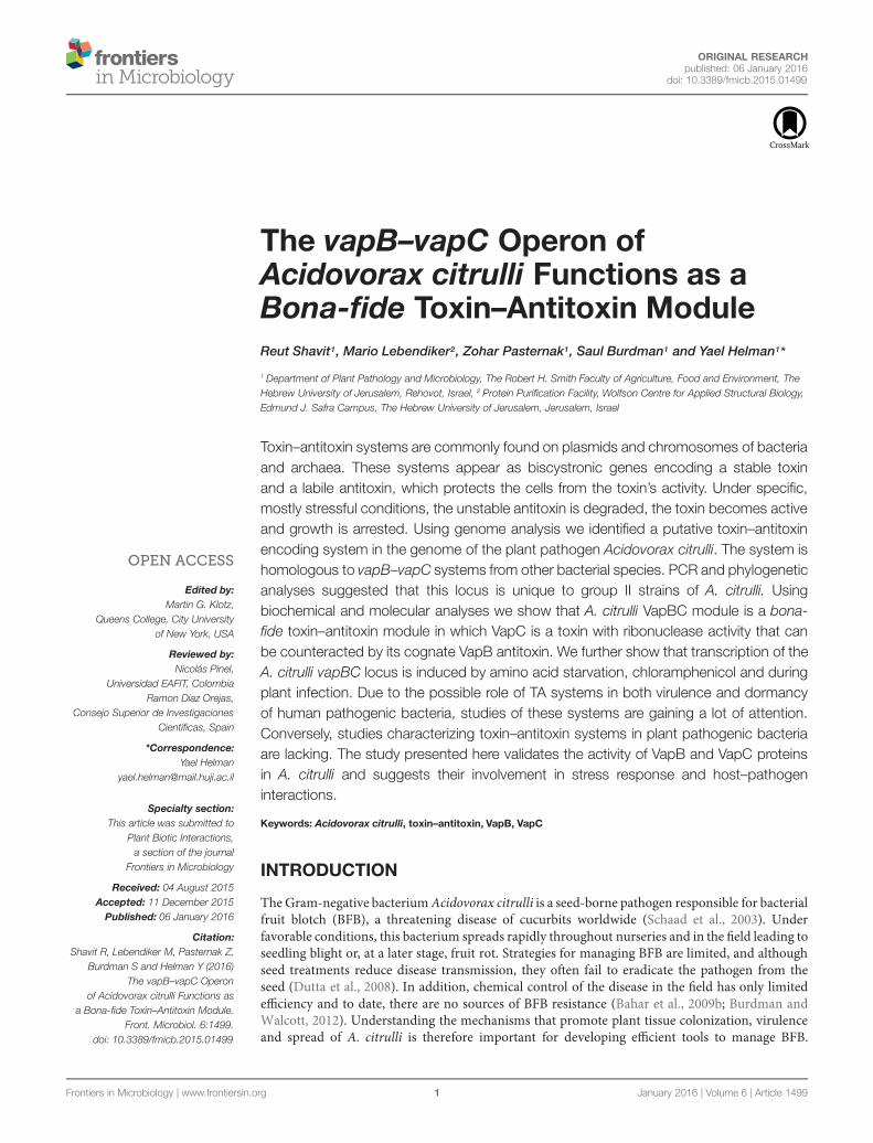
FIGURE 1 | Genetic organization of the Acidovorax citrulli AAC00-1vapBC locus. (A) Schematic representation of genes vapB and vapCencoding the antitoxin and toxin proteins, respectively (genes Aave_0579 andAave_0580, according to the annotation of the genome of strain AAC00-1).Sequence overlap between vapB and vapC is shown: the underlined ATGindicates the putative vapC start codon, while the bolded TAA indicates theputative vapB stop codon. Arrows and numbers indicate primer positionsused for electrophoresis analysis presented in (B). (B) Electrophoresed bandsfollowing PCR of cDNA products from vapB internal primers (lane 1,VapB633077 F- primer 1; VapB633412 R-primer 2); vapC internal primers(lane 2, VapB633292 F-primer 3; VapB633808 R-primer 4); and primersspanning the vapB and vapC ORFs (lane 3, VapB633077 F-primer 1;VapB633808 R-primer 4). Lanes 4–6, PCR-positive controls from PCRreactions using genomic DNA of strain 7a1 as template, in the same order ofprimers as in 1–3. Molecular weight standard of 100 base pairs increments isshown on the left (M). Negative controls with no reverse transcriptase wereused to verify that RNA samples do not contain genomic DNA (data notshown). Primers are detailed in Supplementary Table S1. Results from oneexperiment, out of three with similar results, are shown. Expression of thevapBC transcript was examined after growth for 48 h in nutrient broth.
Frontiers in Microbiology | www.frontiersin.org 4 January 2016 | Volume 6 | Article 1499
Shavit et al. The VapBC Toxin–Antitoxin System of A. citrulli
in Israel, we cannot work with strain AAC00-1 that was isolatedin the USA. Therefore, based on the AAC00-1 sequence wedesigned PCR primers that allowed us to amplify and sequencethis locus from A. citrulli strain 7a1, a group II strain isolatedin Israel (Eckshtain-Levi et al., 2014). Sequence of the TA locusof strain 7a1 revealed that it is 100% identical to that of strainAAC00-1. This sequence was deposited in the NCBI databaseunder GenBank accessions KT149413 and KT149414 for vapBand vapC, respectively. Sequence analyses of the putative toxingene indicated that it possibly encodes a VapC-likemember of thePIN domain superfamily of ribonucleases. The putative antitoxingene was shown to encode a transcriptional regulator/antitoxinwith an AbrB-like domain commonly found in VapB antitoxinencoding genes. Sequence analyses also indicated that theputative translational start codon for vapC overlaps with thetranslational stop codon of vapB, providing a strong indication oftranslational coupling (Figure 1A). Reverse transcriptase (RT)-PCR experiments using primers coding for a joint segmentfrom the end of the antitoxin and beginning of the toxin geneconfirmed that these genes are expressed in A. citrulli, andthat their expression occurs in a single transcriptional unit(Figure 1B).
We have recently sequenced the genome of M6, a group Istrain of A. citrulli. Sequence analysis of the M6 draft genomerevealed that it does not carry a vapBC-like locus. To assesswhether this finding applies broadly to differences betweengroups I and II strains, of A. citrulli we employed PCR analysesto assess presence/absence of the TA locus in 15 group I strainsand 12 group II strains (including M6 and 7a1, respectively).Similarly, to the results obtained from analysis of strains 7a1 andM6, the vapBC TA locus was shown to be present in all testedgroup II strains and absent in all tested group I strains (Table 1).Importantly, the strains selected for this analysis were isolatedfrom various geographical locations and belong to different pulsefield gel electrophoresis (PFGE)-based haplotypes (Table 1), thusincreasing the broad significance of this finding.
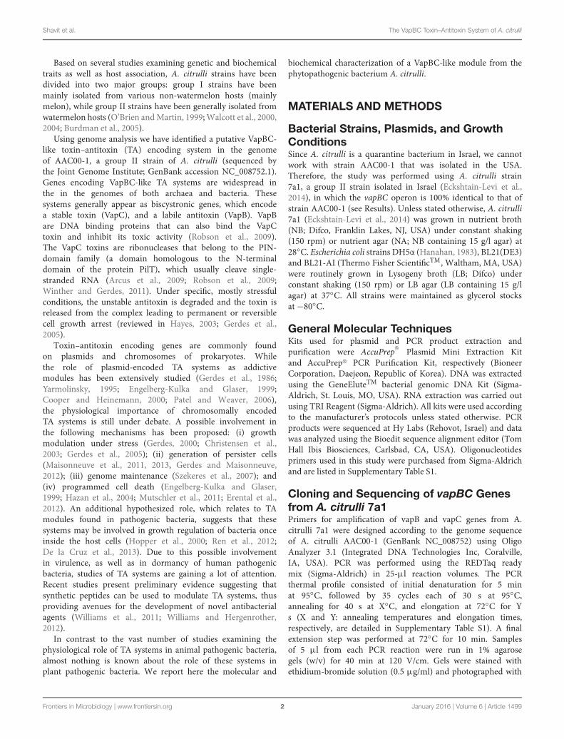
Phylogenetic analysis of A. citrulli AAC00-1 VapC revealedthat the A. citrulli toxin protein closely clusters with similarproteins from three Xanthomonas species, namely X. axonopodispv. vasculorum, X. cassavae andX. axonopodis pv. citri (Figure 2).This group ofA. citrulli and xanthomonads are branched togetherwith a bigger cluster containing nine species, most of thempathogenic ones (eight out of nine). Phylogenic analyses of theVapB antitoxin protein of A. citrulli AAC00-1 revealed similarresults to those of the VapC toxin, clustering it with similarproteins of other Xanthomonas sp. (Supplementary Figure S1).BLAST analysis of the A. citrulli DNA sequence containingboth vapB and vapC genes, indicated that the only significantsimilarities found (E-value < 1) were from Xanthomonas strains(data not shown), which also group with the corresponding genesin the protein trees (Figure 2; Supplementary Figure S1).
Ectopic Expression of the TA Module:Activity Assays and Growth RegulationTo further characterize the A. citrulli TA module weaimed at expressing the toxin and antitoxin proteins in
E. coli BL21 cells. The antitoxin was successfully expressedin E. coli cells using the pET15b expression plasmid(Supplementary Figure S2). Repeated attempts to expressthe toxin on its own, using various expression plasmidsin E. coli failed, suggesting a lethal activity of VapC (datanot shown). We therefore used the pACYCDuet-1 plasmidfor coupled expression of both the toxin (with His-tag)and the antitoxin (no tag). Co-expression of the antitoxinprotein abolished the lethal effect of expressing the toxinalone, allowing expression of the latter in E. coli cells(Supplementary Figure S2).
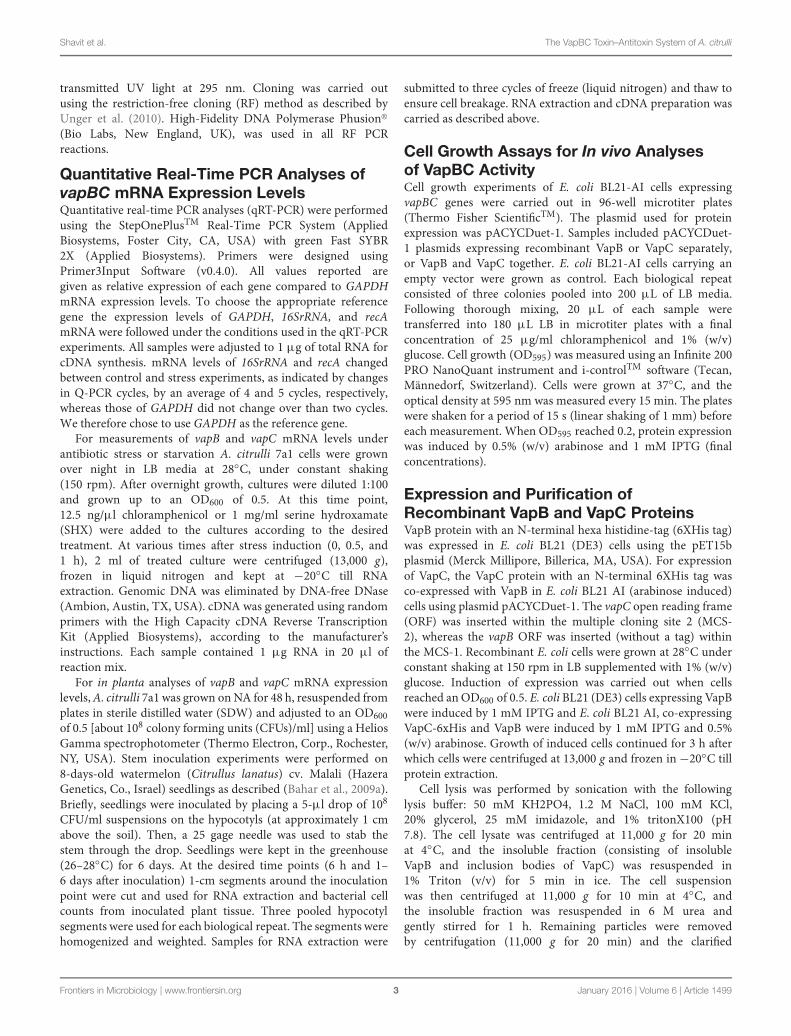
The VapC PIN-domain-containing toxins are known tofunction as ribonucleases (Arcus et al., 2011). We thereforeinvestigated whether the A. citrulli VapC protein exhibits anRNase catalytic activity. VapC from the co-purified VapB-VapC-His complex was obtained by denaturing Ni-NTAchromatography and subsequent refolding. The purified VapC-His recombinant protein efficiently degraded a cellular RNApreparation from A. citrulli including the 23S and 16Sribosomal RNAs (Figure 3). In contrast, addition of an RNaseinhibitor or EDTA blocked RNA degradation (Figure 3),further corroborating the VapC-like nature of the toxin as aMg2+/Mn2+-dependent ribonuclease.
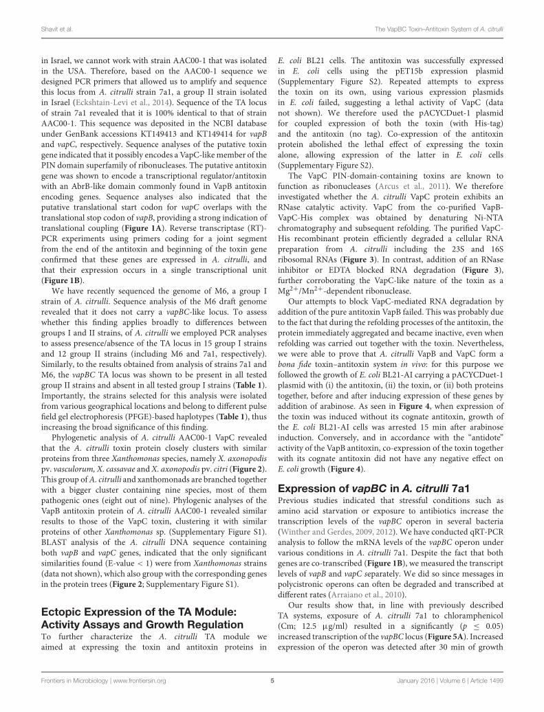
Our attempts to block VapC-mediated RNA degradation byaddition of the pure antitoxin VapB failed. This was probably dueto the fact that during the refolding processes of the antitoxin, theprotein immediately aggregated and became inactive, even whenrefolding was carried out together with the toxin. Nevertheless,we were able to prove that A. citrulli VapB and VapC form abona fide toxin–antitoxin system in vivo: for this purpose wefollowed the growth of E. coli BL21-AI carrying a pACYCDuet-1plasmid with (i) the antitoxin, (ii) the toxin, or (ii) both proteinstogether, before and after inducing expression of these genes byaddition of arabinose. As seen in Figure 4, when expression ofthe toxin was induced without its cognate antitoxin, growth ofthe E. coli BL21-AI cells was arrested 15 min after arabinoseinduction. Conversely, and in accordance with the “antidote”activity of the VapB antitoxin, co-expression of the toxin togetherwith its cognate antitoxin did not have any negative effect onE. coli growth (Figure 4).
Expression of vapBC in A. citrulli 7a1Previous studies indicated that stressful conditions such asamino acid starvation or exposure to antibiotics increase thetranscription levels of the vapBC operon in several bacteria(Winther and Gerdes, 2009, 2012). We have conducted qRT-PCRanalysis to follow the mRNA levels of the vapBC operon undervarious conditions in A. citrulli 7a1. Despite the fact that bothgenes are co-transcribed (Figure 1B), we measured the transcriptlevels of vapB and vapC separately. We did so since messages inpolycistronic operons can often be degraded and transcribed atdifferent rates (Arraiano et al., 2010).
Our results show that, in line with previously describedTA systems, exposure of A. citrulli 7a1 to chloramphenicol(Cm; 12.5 μg/ml) resulted in a significantly (p ≤ 0.05)increased transcription of the vapBC locus (Figure 5A). Increasedexpression of the operon was detected after 30 min of growth
Frontiers in Microbiology | www.frontiersin.org 5 January 2016 | Volume 6 | Article 1499

Shavit et al. The VapBC Toxin–Antitoxin System of A. citrulli
TABLE 1 | List of Acidovorax citrulli group I and group II strains examined for the presence of the vapBC locus by PCR analyses.
Strain name Group PFGE haplotypea Reference/source Toxin–antitoxin module
AACAU-2 I B4 (L) Walcott et al., 2004 −AACAU-9 I B5 (M) Walcott et al., 2004 −AAC98-17 I B6 (N) Walcott et al., 2000 −AAC200-23 I B8 (P) Walcott et al., 2004 −AAC201-16 I B11 (V) Walcott et al., 2004 −AAC200-30 I B10 (S) Walcott et al., 2004 −AAC92-300 (ATCC29625) I B3 (K) Walcott et al., 2000 −AAC201-15 I B11 (V) Walcott et al., 2004 −AAC92-305 I B2 (I) Walcott et al., 2000 −AAC201-22 I B1 (F) Walcott et al., 2004 −AAC202-66 I B12 (X) Walcott et al., 2004 −M1 I B21 (Y) Burdman et al., 2005 −M4 I B21 (Y) Burdman et al., 2005 −M6 I B21 (Y) Burdman et al., 2005 −5 I B21 (Y) Eckshtain-Levi et al., 2014 −AAC92-17 II A4 (D) Walcott et al., 2000 +W4 II A13 (E2) Burdman et al., 2005 +W6 II A20 (Z) Burdman et al., 2005 +7a1 II A23 Eckshtain-Levi et al., 2014 +AAC94-39 II A7 (J) Walcott et al., 2000 +AAC201-19 II A2 (B) Walcott et al., 2004 +AAC202-69 II A11 (W) Walcott et al., 2004 +AAC94-87 II A6 (G) Walcott et al., 2000 +SaticoyB II A8 (Q) Walcott et al., 2004 +AAC201-20 II A3 (C) Walcott et al., 2004 +AAC94-55 II A5 (E) Walcott et al., 2000 +AAC94-48 II A9 (U) Walcott et al., 2000 +aHaplotypes based on PFGE analyses of a wide collection of A. citrulli strains (R. Walcott, personal communication). When available, letters inside parentheses indicatethe previous haplotype designation (Walcott et al., 2000, 2004; Burdman et al., 2005).
in the presence of the antibiotic. Notably, the increase in vapBtranscript level was higher than that of vapC. At 30 min ofexposure to Cm, the mRNA levels of the vapB antitoxin wereabout seven times higher (7.3 ± 1) than those measured at timezero, while the mRNA levels of the vapC toxin increased byabout four folds (3.8 ± 0.3). After 1 h of exposure to Cm, vapBmRNA levels increased by 8.5 ± 1.4 and those of vapC increasedby 3.7 ± 0.5 in comparison to those measured at time zero(Figure 5A).
Induction of vapBC also occurred when A. citrulli cells wereexposed to amino acid starvation imposed by the addition ofSHX, an inhibitor of seryl-tRNA charging (Tosa and Pizer, 1971).After 30 min of exposure to SHX a significant (p ≤ 0.05) increasewas recorded only for vapB (2.4 ± 0.55). Nevertheless, after 1 hof exposure to SHX both vapB (5.3 ± 0.22) and vapC (3.3 ± 0.8)transcript levels significantly (p ≤ 0.05) increased compared totime zero (Figure 5B).
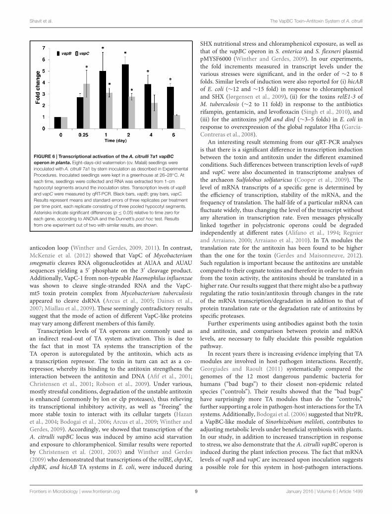
One of the hypothesized roles for TA systems is that theyare used to regulate the growth of pathogens once inside thehost. We therefore followed the transcription levels of A. citrullivapBC genes at various time points after stem inoculation ofmelon seedlings, performed as described (Bahar et al., 2009a).Our results indicate that the A. citrulli vapBC operon is activatedduring the infection process (Figure 6). In contrast to animal
pathogens, most plant pathogenic bacteria, including A. citrulli,colonize extracellular spaces within the plant tissue (Alfanoand Collmer, 1996). Therefore, these results correspond tointeraction of the bacteria with the plant tissue and not toan intracellular behavior. At 6 h after inoculation, vapC levelsincreased significantly (p ≤ 0.05) as compared to vapC levels attime zero (3.9 ± 1.2). At this time, a higher level of expressionrelative to time zero was measured for vapB (2.7 ± 0.3),though not significant. At longer time points, the levels ofvapB transcripts increased more than those of vapC. At 1 and2 days after inoculation (d.a.i.), vapC levels were still significantly(p ≤ 0.05) higher than those at time zero but they hardly changed(3 ± 0.4 and 2.8 ± 0.3, respectively), whereas those of vapBcontinued to increase during these days (5 ± 1.6 and 5.6 ± 0.7folds, respectively). A trend was observed indicating reduction ofvapC expression with time, and at 4 and 6 d.a.i., the expressionof this gene was not significantly different from that measuredat time zero (2.2 ± 0.7 and 1.2 ± 0.04 folds, respectively). Incontrast, at 4 and 6 d.a.i. vapB levels were still significantly(p ≤ 0.05) higher relative to time zero (3.9 ± 0.8 and 3.6 ± 0.3folds, respectively). Notably, beyond 6 d.a.i, the seedlings wereseverely affected by the bacterium and accurate sampling was notpossible beyond this time. Importantly, although the transcriptlevels of the vapBC operon increased during the first 2 days of
Frontiers in Microbiology | www.frontiersin.org 6 January 2016 | Volume 6 | Article 1499

Shavit et al. The VapBC Toxin–Antitoxin System of A. citrulli
FIGURE 2 | Phylogenetic analyses of VapC-like toxin proteins. The evolutionary history was inferred by using the Maximum Likelihood method based on theJTT matrix-based model. The percentage of trees (out of 100 bootstraps) in which the associated taxa clustered together is shown next to the branches; brancheswith bootstrap value <40 were collapsed. A. citrulli AAC00-1 is underlined. P, pathogenic; E, environmental.
infection, bacterial counts increased from ∼8 × 10⁵ CFU/cmhypocotyl at the inoculation time to approximately 1.3× 10⁸ and4.4 × 108 CFU/cm hypocotyl at 2 and 4 d.a.i, respectively, thusindicating that no apparent growth arrest of A. citrulli occurredduring the time of vapC expression.
DISCUSSION
We identified a VapBC-like toxin–antitoxin module in thegenome of the group II strain of A. citrulli, AAC00-1. Geneticanalysis of the vapBC locus from various A. citrulli strains, withdistinguished genetic backgrounds, supports this locus existsonly in group II strains of A. citrulli. Sequence analyses ofthe A. citrulli vapBC locus revealed that the only significantsimilarities (E-value < 1) from the available database are fromseveral Xanthomonas species and pathovars and this is supportedby phylogenetic relatedness at the VapB and VapC protein levels.These results suggest that the vapBC in group II strains of
A. citrulli and in some Xanthomonas species were obtainedthrough horizontal gene transfer. In A. citrulli, acquisition of thisTA module possibly occurred after the splitting of this speciesinto group I and group II strains. The opposite possibility-namely, that the vapBC operon was present in group I A. citrulli,but lost through time- cannot be discarded but is less likelydue to the stabilizing addictive characteristics of TA genesystems (Hayes, 2003; Magnuson, 2007; Saavedra De Bast et al.,2008).
The high relatedness between A. citrulli and xanthomonadsvapBC sequences is interesting but not surprising. For instance,based on genetic composition and regulation, A. citrulli andXanthomonas species possess a highly similar type III secretionapparatus (Bahar and Burdman, 2010). Moreover, a recent studyrevealed that the majority of genes encoding putative type IIIsecreted effectors in A. citrulli are highly similar of knownxanthomonads effectors (Eckshtain-Levi et al., 2014). Due tothe common phytopathogenic nature of Xanthomonas and someAcidovorax species, it is plausible to assume that horizontal gene
Frontiers in Microbiology | www.frontiersin.org 7 January 2016 | Volume 6 | Article 1499

Shavit et al. The VapBC Toxin–Antitoxin System of A. citrulli
FIGURE 3 | RNase activity of recombinant A. citrulli VapC-6xHisprotein on a preparation of cellular RNA of A. citrulli (1 µg/lane). RNAwas incubated for 20 min at room temperature in each treatment; Marker,(100 bp increments). Treatments: degradation of RNA by recombinantVapC-6xHis; inhibition of VapC activity by RNase inhibitor; inhibition of VapCactivity by addition of 10mM EDTA; and three negative controls without VapC:refolding buffer, 50 mM Tris-HCl buffer and MgCl 6 mM solution. Results fromone experiment, out of three with similar results, are shown.
FIGURE 4 | Growth curve of Escherichia coli BL21-AI transformed withpACYCDuet-1 plasmids expressing recombinant VapB or VapCseparately, or VapB and VapC together. E. coli cells carrying the emptyvector were grown as control. Cells were grown in LB media at 37◦C, withlinear shaking (1 mm) for 15 s every 15 min. Arrow indicates expressioninduction by 1 mM IPTG and 0.5% (w/v) arabinose. Growth curves wereinitiated by adding three colonies to each well, n = 12 (each replicateconsisting of three pooled colonies); error bars are standard error of mean.Results from one representative experiment, out of three with similar resultsare shown.
transfer events have occurred among member of these genera oramong ancestral species.
In this study we present molecular and biochemical evidencedemonstrating that the A. citrulli vapBC genes indeed encode a
FIGURE 5 | Transcriptional activation of vapBC in A. citrulli 7a1 uponexposure to (A) antibiotic stress (12.5 µg/ml chloramphenicol) and (B)nutritional stress (1 mg/ml serine hydroxamate). Cells were grown in LBmedium containing the above stressors, with constant shaking (150 rpm) at28◦C. Black bars, vapB; gray bars, vapC. Transcription levels of these geneswere measured by Quantitative-Real Time-PCR (qRT-PCR). Results representmeans and standard errors of six replicates per treatment per time point.Asterisks indicate significant differences (p ≤ 0.05) relative to time zero foreach gene, according to ANOVA and the Dunnett’s post hoc test. Resultsfrom one experiment, out of three with similar results, are shown.
bona fide VapBC TA module. We show that ectopic expressionof VapC toxin in E. coli succeeded to inhibit cell growth andthat this inhibition was counteracted by the expression of thecognate VapB antitoxin. We also showed that, as suggestedby the presence of a PIN domain sequence, VapC indeedhas ribonuclease activity, efficiently degrading a cellular RNApreparation from A. citrulli, including the 23S and 16S ribosomalRNAs. Characterization of VapC proteins from the entericpathogenic bacteria Salmonella enterica and Shigella flexnerirevealed they do not cleave mRNAs but rather act as site-specific riboendonucleases that cleave initiator tRNA fMet in the
Frontiers in Microbiology | www.frontiersin.org 8 January 2016 | Volume 6 | Article 1499
Shavit et al. The VapBC Toxin–Antitoxin System of A. citrulli
FIGURE 6 | Transcriptional activation of the A. citrulli 7a1 vapBCoperon in planta. Eight-days-old watermelon (cv. Malali) seedlings wereinoculated with A. citrulli 7a1 by stem inoculation as described in ExperimentalProcedures. Inoculated seedlings were kept in a greenhouse at 26–28◦C. Ateach time, seedlings were collected and RNA was extracted from 1-cmhypocotyl segments around the inoculation sites. Transcription levels of vapBand vapC were measured by qRT-PCR. Black bars, vapB; gray bars, vapC.Results represent means and standard errors of three replicates per treatmentper time point, each replicate consisting of three pooled hypocotyl segments.Asterisks indicate significant differences (p ≤ 0.05) relative to time zero foreach gene, according to ANOVA and the Dunnett’s post hoc test. Resultsfrom one experiment out of two with similar results, are shown.
anticodon loop (Winther and Gerdes, 2009, 2011). In contrast,McKenzie et al. (2012) showed that VapC of Mycobacteriumsmegmatis cleaves RNA oligonucleotides at AUAA and AUAUsequences yielding a 5′ phosphate on the 3′ cleavage product.Additionally, VapC-1 from non-typeable Haemophilus influenzaewas shown to cleave single-stranded RNA and the VapC-mt5 toxin protein complex from Mycobacterium tuberculosisappeared to cleave dsRNA (Arcus et al., 2005; Daines et al.,2007; Miallau et al., 2009). These seemingly contradictory resultssuggest that the mode of action of different VapC-like proteinsmay vary among different members of this family.
Transcription levels of TA operons are commonly used asan indirect read-out of TA system activation. This is due tothe fact that in most TA systems the transcription of theTA operon is autoregulated by the antitoxin, which acts asa transcription repressor. The toxin in turn can act as a co-repressor, whereby its binding to the antitoxin strengthens theinteraction between the antitoxin and DNA (Afif et al., 2001;Christensen et al., 2001; Robson et al., 2009). Under various,mostly stressful conditions, degradation of the unstable antitoxinis enhanced (commonly by lon or clp proteases), thus relievingits transcriptional inhibitory activity, as well as “freeing” themore stable toxin to interact with its cellular targets (Hazanet al., 2004; Bodogai et al., 2006; Arcus et al., 2009; Winther andGerdes, 2009). Accordingly, we showed that transcription of theA. citrulli vapBC locus was induced by amino acid starvationand exposure to chloramphenicol. Similar results were reportedby Christensen et al. (2001, 2003) and Winther and Gerdes(2009) who demonstrated that transcriptions of the relBE, chpAK,chpBK, and hicAB TA systems in E. coli, were induced during
SHX nutritional stress and chloramphenicol exposure, as well asthat of the vapBC operon in S. enterica and S. flexneri plasmidpMYSF6000 (Winther and Gerdes, 2009). In our experiments,the fold increments measured in transcript levels under thevarious stresses were significant, and in the order of ∼2 to 8folds. Similar levels of induction were also reported for (i) hicABof E. coli (∼12 and ∼15 fold) in response to chloramphenicoland SHX (Jørgensen et al., 2009), (ii) for the toxins relE1-3 ofM. tuberculosis (∼2 to 11 fold) in response to the antibioticsrifampin, gentamicin, and levofloxacin (Singh et al., 2010), and(iii) for the antitoxins yefM and dinJ (∼3–5 folds) in E. coli inresponse to overexpression of the global regulator Hha (García-Contreras et al., 2008).
An interesting result stemming from our qRT-PCR analysesis that there is a significant difference in transcription inductionbetween the toxin and antitoxin under the different examinedconditions. Such differences between transcription levels of vapBand vapC were also documented in transcriptome analyses ofthe archaeon Sulfolobus solfataricus (Cooper et al., 2009). Thelevel of mRNA transcripts of a specific gene is determined bythe efficiency of transcription, stability of the mRNA, and thefrequency of translation. The half-life of a particular mRNA canfluctuate widely, thus changing the level of the transcript withoutany alteration in transcription rate. Even messages physicallylinked together in polycistronic operons could be degradedindependently at different rates (Alifano et al., 1994; Regnierand Arraiano, 2000; Arraiano et al., 2010). In TA modules thetranslation rate for the antitoxin has been found to be higherthan the one for the toxin (Gerdes and Maisonneuve, 2012).Such regulation is important because the antitoxins are unstablecompared to their cognate toxins and therefore in order to refrainfrom the toxin activity, the antitoxins should be translated in ahigher rate. Our results suggest that there might also be a pathwayregulating the ratio toxin/antitoxin through changes in the rateof the mRNA transcription/degradation in addition to that ofprotein translation rate or the degradation rate of antitoxins byspecific proteases.
Further experiments using antibodies against both the toxinand antitoxin, and comparison between protein and mRNAlevels, are necessary to fully elucidate this possible regulationpathway.
In recent years there is increasing evidence implying that TAmodules are involved in host-pathogen interactions. Recently,Georgiades and Raoult (2011) systematically compared thegenomes of the 12 most dangerous pandemic bacteria forhumans (“bad bugs”) to their closest non-epidemic relatedspecies (“controls”). Their results showed that the “bad bugs”have surprisingly more TA modules than do the “controls,”further supporting a role in pathogen-host interactions for the TAsystems. Additionally, Bodogai et al. (2006) suggested that NtrPR,a VapBC-like module of Sinorhizobium meliloti, contributes toadjusting metabolic levels under beneficial symbiosis with plants.In our study, in addition to increased transcription in responseto stress, we also demonstrate that the A. citrulli vapBC operon isinduced during the plant infection process. The fact that mRNAlevels of vapB and vapC are increased upon inoculation suggestsa possible role for this system in host-pathogen interactions.
Frontiers in Microbiology | www.frontiersin.org 9 January 2016 | Volume 6 | Article 1499

Shavit et al. The VapBC Toxin–Antitoxin System of A. citrulli
Despite the observed activation of the vapBC operon in plantaduring the first 2 days after inoculation, no growth inhibitionof A. citrulli was observed within the plant at these stages.It is possible that the observed increase in vapBC expressionoccurred only in a small proportion of the bacterial populationand therefore, it is not reflected in overall growth parameters.Indeed, several studies show that heterogeneous activation of theTA system increases the percentage of persistent cells within apopulation, thereby providing individuals within the colony withmeans to avoid the damage caused by stress and host defenseresponses (Maisonneuve et al., 2011, 2013; Fasani and Savageau,2013). In this regard, a study by De la Cruz et al. (2013), whoexamined the involvement of the TA system in the virulenceof Salmonella enterica subsp. enterica serovar Typhimurium (S.Typhimurium) in mice, showed that increased expression of theTA module in S. Typhimurium is transient upon infection andthat expression varies between different locations within the host.The authors also showed that the toxin itself is necessary butnot sufficient to limit bacterial growth and that an additionalfactor synergizes with the toxin activity under conditions ofmouse infection. Additionally, in a recent study presented byLobato-Márquez et al. (2015) it was shown that both type Iand type II toxins are essential for survival of S. Typhimuriuminside fibroblasts, irrespective of the growth rate. Interestingly,the authors showed that a vapC mutant of S. Typhimuriumexhibited up to 80% decrease in the rate of intracellular survivalcompared to the wild-type strain. Their results suggest thatthere is a specialization of distinct TA modules for regulatingintracellular activity of pathogenic bacteria, and thus progressionof infection.
Further studies examining the expression of the vapBC lociin different locations within the plant and in other tissuesare required to further understand the role of this system inplant infection. Additionally, construction and characterizationofA. citrullimutants impaired in the expression of the TAmodulecould provide insights as to the possible role of this system in suchinteractions. Albeit, it should be noted that in some studies of
various TA systems no apparent phenotype of TA mutants wasever detected, although overexpression of the toxin componentnegatively affected growth in vitro (Tsilibaris et al., 2007; Robsonet al., 2009).
Characterization of TA modules in plant pathogenic bacteriais lacking behind that of human pathogens. Here we show thatA. citrulli possesses a TA module similar to that of knownhuman pathogens such as M. tuberculosis, Salmonella, andH. influenzae. To the best of our knowledge, this is the firstreport showing expression of a TA module during infection ofa plant pathogenic bacterium. We aim to further examine themechanism, role and activation pathway of the A. citrulli TAmodule upon plant infection. Moreover, due to the presence ofTA modules in other plant pathogenic species, and particularlyin several xanthomonads, the relevance of the findings presentedin this study is much beyond the A. citrulli-cucurbit pathosystem.Further studies will provide important insights into the role of TAsystems in plant-microbe interactions, as well as provide valuableinformation on the regulation and function of TA systems inmicrobial populations in general.
ACKNOWLEDGMENTS
We thank R. Walcott for kindly supplying DNA samples ofvarious A. citrulli strains for this study, O. Ostersetzer-Biran andL. Sultan for their valuable assistance in the procedure of proteinpurification and O. Avidan for helpful advice on the constructionof the restriction free plasmids. This research was funded by grant1396/10 from the Israeli Science Foundation (ISF).
SUPPLEMENTARY MATERIAL
The Supplementary Material for this article can be foundonline at: http://journal.frontiersin.org/article/10.3389/fmicb.2015.01499
REFERENCES
Afif, H., Allali, N., Couturier, M., and Van Melderen, L. (2001). The ratiobetween CcdA and CcdB modulates the transcriptional repression of theccd poison-antidote system. Mol. Microbiol. 41, 73–82. doi: 10.1046/j.1365-2958.2001.02492.x
Alfano, J. R., and Collmer, C. (1996). Bacterial pathogens in plants: life up againstthe wall. Plant Cell 8, 1683–1698. doi: 10.1105/tpc.8.10.1683
Alifano, P., Bruni, C. B., and Carlomagno, M. S. (1994). Control ofmRNA processing and decay in prokaryotes. Genetica 94, 157–172. doi:10.1007/BF01443430
Arcus, V. L., McKenzie, J. L., Robson, J., and Cook, G. M. (2011). The PIN-domainribonucleases and the prokaryotic VapBC toxin-antitoxin array. Protein Eng.Des. Sel. 24, 33–40. doi: 10.1093/protein/gzq081
Arcus, V. L., Rainey, P. B., and Turner, S. J. (2005). The PIN-domaintoxin-antitoxin array in mycobacteria. Trends Microbiol. 13, 360–365. doi:10.1016/j.tim.2005.06.008
Arcus, V. L., Robson, J., McKenzie, J. L., Cursons, R., and Cook, G. M. (2009).The vapBC operon from Mycobacterium smegmatis is an autoregulated toxin-antitoxin module that controls growth via inhibition of translation. J. Mol. Biol.390, 353–367. doi: 10.1016/j.jmb.2009.05.006
Arraiano, C. M., Andrade, J. M., Domingues, S., Guinote, I. B.,Malecki, M., Matos, R. G., et al. (2010). The critical role of RNAprocessing and degradation in the control of gene expression.FEMS Microbiol. Rev. 34, 883–923. doi: 10.1111/j.1574-6976.2010.00242.x
Bahar, O., and Burdman, S. (2010). Bacterial fruit blotch: a threat to the cucurbitindustry. Isr. J. Plant Sci. 58, 19–31. doi: 10.1560/IJPS.58.1.19
Bahar, O., Goffer, T., and Burdman, S. (2009a). Type IV pili are required forvirulence, twitching motility, and biofilm formation of Acidovorax avenaesubsp. citrulli. Mol. Plant Microbe Interact. 22, 909–920. doi: 10.1094/MPMI-22-8-0909
Bahar, O., Kritzman, G., and Burdman, S. (2009b). Bacterial fruit blotchof melon: screens for disease tolerance and role of seed transmission inpathogenicity. Eur. J. Plant Pathol. 123, 71–83. doi: 10.1007/s10658-008-9345-7
Bodogai, M., Ferenczi, S., Bashtovyy, D., Miclea, P., Papp, P., and Dusha, I.(2006). The ntrPR operon of Sinorhizobium meliloti is organized and functionsas a toxin-antitoxin module. Mol. Plant Microbe Interact. 19, 811–822. doi:10.1094/MPMI-19-0811
Burdman, S., Kots, N., Kritzman, G., and Kopelowitz, J. (2005). Molecular,physiological, and host-range characterization of Acidovorax avenae subsp.
Frontiers in Microbiology | www.frontiersin.org 10 January 2016 | Volume 6 | Article 1499
Shavit et al. The VapBC Toxin–Antitoxin System of A. citrulli
citrulli isolates from watermelon and melon in Israel. Plant Dis. 89, 1339–1347.doi: 10.1094/PD-89-1339
Burdman, S., and Walcott, R. (2012). Acidovorax citrulli: generating basic andapplied knowledge to tackle a global threat to the cucurbit industry.Mol. PlantPathol. 13, 805–815. doi: 10.1111/j.1364-3703.2012.00810.x
Christensen, S. K., Mikkelsen, M., Pedersen, K., and Gerdes, K. (2001). RelE, aglobal inhibitor of translation, is activated during nutritional stress. Proc. Natl.Acad. Sci. U.S.A. 98, 14328–14333. doi: 10.1073/pnas.251327898
Christensen, S. K., Pedersen, K., Hansen, F. G., and Gerdes, K. (2003). Toxin-antitoxin loci as stress-response-elements: ChpAK/MazF and ChpBK cleavetranslated RNAs and are counteracted by tmRNA. J. Mol. Biol. 332, 809–819.doi: 10.1016/S0022-2836(03)00922-7
Cooper, C. R., Daugherty, A. J., Tachdjian, S., Blum, P. H., and Kelly, R. M. (2009).Role of vapBC toxin-antitoxin loci in the thermal stress response of Sulfolobussolfataricus. Biochem. Soc. Trans. 37, 123–126. doi: 10.1042/BST0370123
Cooper, T. F., and Heinemann, J. A. (2000). Postsegregational killing doesnot increase plasmid stability but acts to mediate the exclusion ofcompeting plasmids. Proc. Natl. Acad. Sci. U.S.A. 97, 12643–12648. doi:10.1073/pnas.220077897
Daines, D. A., Wu, M. H., and Yuan, S. Y. (2007). VapC-1 of nontypeableHaemophilus influenzae is a ribonuclease. J. Bacteriol. 189, 5041–5048. doi:10.1128/JB.00290-07
De la Cruz, M. A., Zhao, W., Farenc, C., Gimenez, G., Raoult, D., Cambillau, C.,et al. (2013). A toxin-antitoxin module of Salmonella promotes virulence inmice. PLoS Pathog. 9:e1003827. doi: 10.1371/journal.ppat.1003827
Dutta, B., Genzlinger, L. L., and Walcott, R. R. (2008). Localization of Acidovoraxavenae subsp. citrulli (Aac), the bacterial fruit blotch pathogen in naturallyinfested watermelon seed. Phytopathology 98, S49–S49.
Eckshtain-Levi, N., Munitz, T., Zivanovic, M., Traore, S. M., Spröer, C., Zhao, B.,et al. (2014). Comparative analysis of type III secreted effector genesreflects divergence of Acidovorax citrulli strains into three distinct lineages.Phytopathology 104, 1152–1162. doi: 10.1094/PHYTO-12-13-0350-R
Edgar, R. C. (2004).MUSCLE:multiple sequence alignment with high accuracy andhigh throughput. Nucl. Acids Res. 32, 1792–1797. doi: 10.1093/nar/gkh340
Engelberg-Kulka, H., and Glaser, G. (1999). Addiction modules and programmedcell death and antideath in bacterial cultures. Annu. Rev. Microbiol. 53, 43–70.doi: 10.1146/annurev.micro.53.1.43
Erental, A., Sharon, I., and Engelberg-Kulka, H. (2012). Two programmedcell death systems in Escherichia coli: an apoptotic-like death is inhibitedby the mazEF-mediated death pathway. PLoS Biol. 10:e1001281. doi:10.1371/journal.pbio.1001281
Fasani, R. A., and Savageau, M. A. (2013).Molecular mechanisms of multiple toxin-antitoxin systems are coordinated to govern the persister phenotype. Proc. Natl.Acad. Sci. U.S.A. 110, 2528–2537. doi: 10.1073/pnas.1301023110
García-Contreras, R., Zhang, X. S., Kim, Y., and Wood, T. K. (2008). Proteintranslation and cell death: the role of rare tRNAs in biofilm formationand in activating dormant phage killer genes. PLoS ONE 3:e2394. doi:10.1371/journal.pone.0002394
Georgiades, K., and Raoult, D. (2011). Genomes of the most dangerousepidemic bacteria have a virulence repertoire characterized by fewergenes but more toxin-antitoxin modules. PLoS ONE 6:e17962. doi:10.1371/journal.pone.0017962
Gerdes, K. (2000). Toxin-antitoxin modules may regulate synthesis ofmacromolecules during nutritional stress. J. Bacteriol. 182, 561–572. doi:10.1128/JB.182.3.561-572.2000
Gerdes, K., Christensen, S. K., and Lobner-Olesen, A. (2005). Prokaryotictoxin-antitoxin stress response loci. Nat. Rev. Microbiol. 3, 371–382. doi:10.1038/nrmicro1147
Gerdes, K., and Maisonneuve, E. (2012). Bacterial persistence and toxin-antitoxinloci. Annu. Rev. Microbiol. 66, 103–123. doi: 10.1146/annurev-micro-092611-150159
Gerdes, K., Rasmussen, P. B., and Molin, S. (1986). Unique type of plasmidmaintenance function: postsegregational killing of plasmid-free cells. Proc. Natl.Acad. Sci. U.S.A. 83, 3116–3120. doi: 10.1073/pnas.83.10.3116
Hanahan, D. (1983). Studies on transformation of Escherichia coli with plasmids.J. Mol. Biol. 166, 557–580. doi: 10.1016/S0022-2836(83)80284-8
Hayes, F. (2003). Toxins-antitoxins: plasmidmaintenance, programmed cell death,and cell cycle arrest. Science 301, 1496–1499. doi: 10.1126/science.1088157
Hazan, R., Sat, B., and Engelberg-Kulka, H. (2004). Escherichia coli mazEF-mediated cell death is triggered by various stressful conditions. J. Bacteriol. 186,3663–3669. doi: 10.1128/JB.186.11.3663-3669.2004
Hopper, S., Wilbur, J. S., Vasquez, B. L., Larson, J., Clary, S., Mehr, I. J.,et al. (2000). Isolation of Neisseria gonorrhoeae mutants that show enhancedtrafficking across polarized T84 epithelial monolayers. Infect. Immun. 68,896–905. doi: 10.1128/IAI.68.2.896-905.2000
Jørgensen, M. G., Pandey, D. P., Jaskolska, M., and Gerdes K. (2009) HicAof Escherichia coli defines a novel family of translation-independent mRNAinterferases in bacteria and archaea. J. Bacteriol. 191, 1191–1199. doi:10.1128/JB.01013-08
Lebendiker, M., and Danieli, T. (2014). Production of prone-to-aggregate proteins.FEBS Lett. 588, 236–246. doi: 10.1016/j.febslet.2013.10.044
Lobato-Márquez, D., Moreno-Córdoba, I., Figueroa, V., Díaz-Orejas, R., andGarcía-del Portillo, F. (2015). Distinct type I and type II toxin-antitoxinmodules control Salmonella lifestyle inside eukaryotic cells. Sci. Rep. 5:9374 doi:10.1038/srep09374
Magnuson, R. D. (2007). Hypothetical functions of toxin-antitoxin systems.J. Bacteriol. 189, 6089–6092. doi: 10.1128/JB.00958-07
Maisonneuve, E., Castro-Camargo, M., and Gerdes, K. (2013). (p)ppGpp controlsbacterial persistence by stochastic induction of toxin-antitoxin activity.Cell 154,1140–1150. doi: 10.1016/j.cell.2013.07.048
Maisonneuve, E., Shakespeare, L. J., Jørgensen, M. G., and Gerdes, K. (2011).Bacterial persistence by RNA endonucleases. Proc. Natl. Acad. Sci. U.S.A. 108,13206–13211. doi: 10.1073/pnas.1100186108
McKenzie, J. L., Robson, J., Berney, M., Smith, T. C., Ruthe, A., Gardner,P. P., et al. (2012). A VapBC toxin-antitoxin module is a posttranscriptionalregulator of metabolic flux in mycobacteria. J. Bacteriol. 194, 2189–2204. doi:10.1128/JB.06790-11
Miallau, L., Faller, M., Chiang, J., Arbing, M., Guo, F., Cascio, D., et al. (2009).Structure and proposed activity of a member of the VapBC family of toxin-antitoxin systems. VapBC-5 from Mycobacterium tuberculosis. J. Biol. Chem.284, 276–283. doi: 10.1074/jbc.M805061200
Mutschler, H., Gebhardt, M., Shoeman, R. L., and Meinhart, A. (2011).A novel mechanism of programmed cell death in bacteria by toxin-antitoxin systems corrupts peptidoglycan synthesis. PLoS Biol. 9:e1001033. doi:10.1371/journal.pbio.1001033
O’Brien, R. G., and Martin, H. L. (1999). Bacterial blotch of melons caused bystrains of Acidovorax avenae subsp. citrulli.Aust. J. Exp. Agric. 39, 479–485. doi:10.1071/EA98172
Patel, S., and Weaver, K. E. (2006). Addiction toxin Fst has unique effects onchromosome segregation and cell division in Enterococcus faecalis and Bacillussubtilis. J. Bacteriol. 188, 5374–5384. doi: 10.1128/JB.00513-06
Regnier, P., and Arraiano, C. M. (2000). Degradation of mRNA inbacteria: emergence of ubiquitous features. Bioessays 22, 235–244. doi:10.1002/(SICI)1521-1878(200003)22:3<235::AID-BIES5>3.0.CO;2-2
Ren, D., Walker, A. N., and Daines, D. A. (2012). Toxin-antitoxin loci vapBC-1and vapXD contribute to survival and virulence in nontypeable Haemophilusinfluenzae. BMCMicrobiol. 12:263. doi: 10.1186/1471-2180-12-263
Robson, J., McKenzie, J. L., Cursons, R., Cook, G. M., and Arcus, V. L. (2009).The vapBC operon from Mycobacterium smegmatis is an autoregulated toxin-antitoxin module that controls growth via inhibition of translation. J. Mol. Biol.390, 353–367. doi: 10.1016/j.jmb.2009.05.006
Saavedra De Bast, M., Mine, N., and Van Melderen, L. (2008). Chromosomaltoxin-antitoxin systems may act as antiaddiction modules. J. Bacteriol. 190,4603–4609. doi: 10.1128/JB.00357-08
Schaad, N. W., Postnikova, E., and Randhawa, P. S. (2003). “Emergence ofAcidovorax avenae subsp. citrulli as a crop threatening disease of watermelonand melon,” in Pseudomonas Syringae and Related Pathogens, eds N. S.Iacobellis, A. Collmer, S. W. Hutcheson, J. W. Mansfield, C. E. Morris, J.Murillo, et al. (Dordrecht: Kluwer Academic Publishers), 573–581.
Singh, R., Barry, C. E., and Boshoff, H. I. (2010). The three RelE homologs ofMycobacterium tuberculosis have individual, drug-specific effects on bacterialantibiotic tolerance. J. Bacteriol. 1279–1291. doi: 10.1128/jb.01285-09
Szekeres, S., Dauti, M., Wilde, C., Mazel, D., and Rowe-Magnus, D. A. (2007).Chromosomal toxin-antitoxin loci can diminish large-scale genome reductionsin the absence of selection.Mol. Microbiol. 63, 1588–1605. doi: 10.1111/j.1365-2958.2007.05613.x
Frontiers in Microbiology | www.frontiersin.org 11 January 2016 | Volume 6 | Article 1499
Shavit et al. The VapBC Toxin–Antitoxin System of A. citrulli
Tamura, K., Stecher, G., Peterson, D., Filipski, A., and Kumar, S. (2013). MEGA6:molecular evolutionary genetics analysis version 6.0. Mol. Biol. Evol. 30, 2725–2729. doi: 10.1093/molbev/mst197
Tosa, T., and Pizer, L. I. (1971). Biochemical bases for the antimetabolite action ofL-serine hydroxamate. J. Bacteriol. 106, 972–982.
Tsilibaris, V., Maenhaut-Michel, G., Mine, N., and Van Melderen, L. (2007).What is the benefit to Escherichia coli of having multiple toxin-antitoxinsystems in its genome? J. Bacteriol. 189, 6101–6108. doi: 10.1128/JB.00527-07
Unger, T., Jacobovitch, Y., Dantes, A., Bernheim, R., and Peleg, Y. (2010).Applications of the Restriction Free (RF) cloning procedure for molecularmanipulations and protein expression. J. Struct. Biol. 172, 34–44. doi:10.1016/j.jsb.2010.06.016
Walcott, R. R., Fessehaie, A., and Castro, A. C. (2004). Differences in pathogenicitybetween two genetically distinct groups of Acidovorax avenae subsp.citrulli on cucurbit hosts. J. Phytopathol. 152, 277–285. doi: 10.1111/j.1439-0434.2004.00841.x
Walcott, R. R., Langston, D. B., Sanders, F. H., and Gitaitis, R. D. (2000).Investigating intraspecific variation of Acidovorax avenae subsp. citrulli usingDNA fingerprinting and whole cell fatty acid analysis. Phytopathology 90,191–196. doi: 10.1094/PHYTO.2000.90.2.191
Williams, J. J., Halvorsen, E. M., Dwyer, E. M., DiFazio, R. M., and Hergenrother,P. J. (2011). Toxin-antitoxin (TA) systems are prevalent and transcribedin clinical isolates of Pseudomonas aeruginosa and methicillin-resistantStaphylococcus aureus. FEMS Microbiol. Lett. 322, 41–50. doi: 10.1111/j.1574-6968.2011.02330.x
Williams, J. J., and Hergenrother, P. J. (2012). Artificial activation of toxin–antitoxin systems as an antibacterial strategy. Trends Microbiol. 20, 291–298.doi: 10.1016/j.tim.2012.02.005
Winther, K. S., and Gerdes, K. (2009). Ectopic production of VapCs fromEnterobacteria inhibits translation and trans-activates YoeB mRNA interferase.Mol. Microbiol. 72, 918–930. doi: 10.1111/j.1365-2958.2009.06694.x
Winther, K. S., and Gerdes, K. (2011). Enteric virulence associated protein VapCinhibits translation by cleavage of initiator tRNA. Proc. Natl. Acad. Sci. U.S.A.108, 7403–7407. doi: 10.1073/pnas.1019587108
Winther, K. S., and Gerdes, K. (2012). Regulation of enteric vapBC transcription:induction by VapC toxin dimer-breaking.Nucleic Acids Res. 40, 4347–4357. doi:10.1093/nar/gks029
Yarmolinsky,M. B. (1995). Programmed cell death in bacterial populations. Science267, 836–837. doi: 10.1126/science.7846528
Conflict of Interest Statement: The authors declare that the research wasconducted in the absence of any commercial or financial relationships that couldbe construed as a potential conflict of interest.
Copyright © 2016 Shavit, Lebendiker, Pasternak, Burdman and Helman. Thisis an open-access article distributed under the terms of the Creative CommonsAttribution License (CC BY). The use, distribution or reproduction in other forumsis permitted, provided the original author(s) or licensor are credited and that theoriginal publication in this journal is cited, in accordance with accepted academicpractice. No use, distribution or reproduction is permitted which does not complywith these terms.
Frontiers in Microbiology | www.frontiersin.org 12 January 2016 | Volume 6 | Article 1499