the urinary system (uropoetic system) · the urinary system the kidneys are situated in the...
TRANSCRIPT

LECTURE ON THE
URINARY SYSTEM(Uropoetic System)
AN OVERVIEW
Dr HAMIADJI

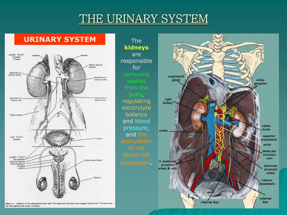
THE URINARY SYSTEM
The kidneys
are responsible
for removing wastes
from the body,
regulating electrolyte
balanceand blood pressure, and the
stimulation of red
blood cell
production.
URINARY SYSTEM
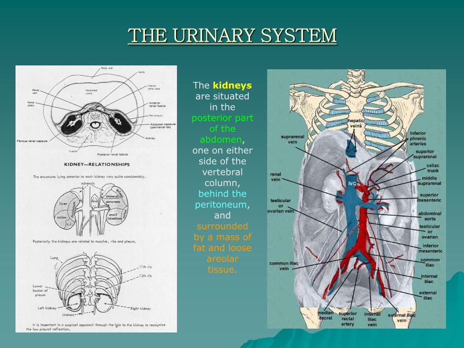
THE URINARY SYSTEM
The kidneysare situated
in the posterior part
of the abdomen,
one on either side of the vertebral column,
behind the peritoneum,
and surrounded by a mass of fat and loose
areolar tissue.

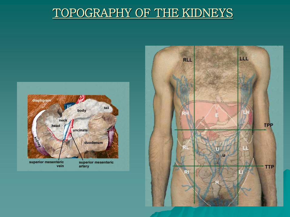
TOPOGRAPHY OF THE KIDNEYS
STUDY THE RELATIONS TO THE SURROUNDING ORGANS / STRUCTURES
TOPOGRAPHY OF THE KIDNEYS
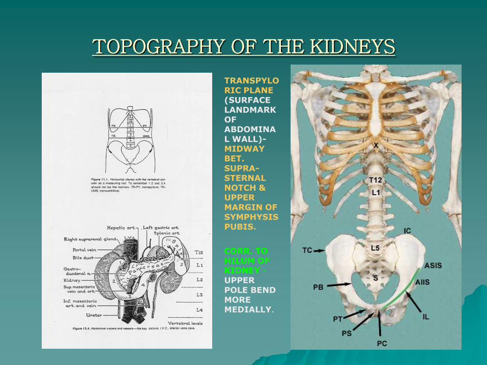
TOPOGRAPHY OF THE KIDNEYS
TRANSPYLORIC PLANE(SURFACE LANDMARK OF ABDOMINAL WALL)-MIDWAY BET. SUPRA-STERNAL NOTCH & UPPER MARGIN OF SYMPHYSIS PUBIS.
CORR. TO HILUM OF KIDNEY -UPPER POLE BEND MORE MEDIALLY.

SURFACE MARKINGS OF KIDNEYS
The kidneys lie partly under cover of the lower thoracic ribs. Renal trauma should be suspected in all injuries associated
with trauma to the posterior lower thorax or upper abdomen.
RENAL ANGLE-Between LOWER BORDER OF 12 RIB & LATERAL BORDER OF ERECTOR SPINAE MUSCLE-RENAL COLIC PAIN STARTS
FROM THIS ANGLE DOWN & FORWARDS TO GROIN
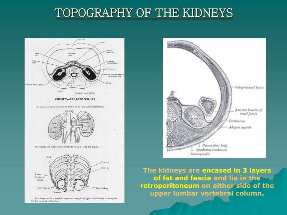
TOPOGRAPHY OF THE KIDNEYS
The kidneys are encased in 3 layers of fat and fascia and lie in the
retroperitoneum on either side of the upper lumbar vertebral column.

TOPOGRAPHY OF THE KIDNEYS
The adipose capsule, which
is thickest at the margins of the kidney and is prolonged through the
hilum into the
renal sinus.
Above the suprarenal gland the two layers of the renal fascia fuse, and unite with the fascia of the diaphragm; below they remain separate,
and are gradually lost in the subperitoneal fascia of the iliac fossa.
The kidney and the adipose capsule are
enclosed in a sheath of
fibrous tissue continuous with
the subperitoneal fascia, and named the
renal fascia

TOPOGRAPHY OF THE KIDNEYS
The retro-peritoneal space
retroperitoneal
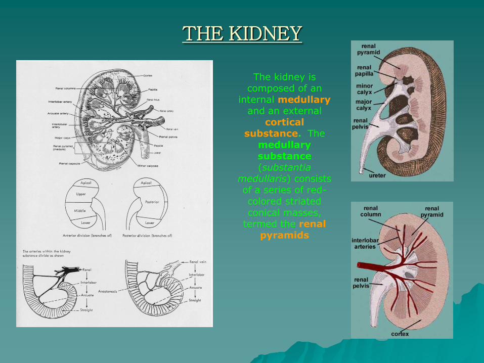
THE KIDNEY
The kidney is composed of an
internal medullaryand an external
cortical substance. The
medullary substance(substantia
medullaris) consists of a series of red-colored striated conical masses,
termed the renal pyramids
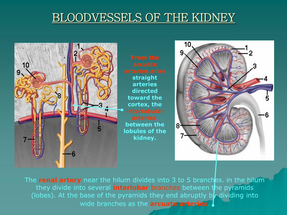
BLOODVESSELS OF THE KIDNEY
The renal artery near the hilum divides into 3 to 5 branches. in the hilum they divide into several interlobar branches between the pyramids
(lobes). At the base of the pyramids they end abruptly by dividing into
wide branches as the arcuate arteries.
From the arcuate
arteries arisestraight arteries directed
toward the cortex, theinterlobular
arteries, between the lobules of the
kidney.
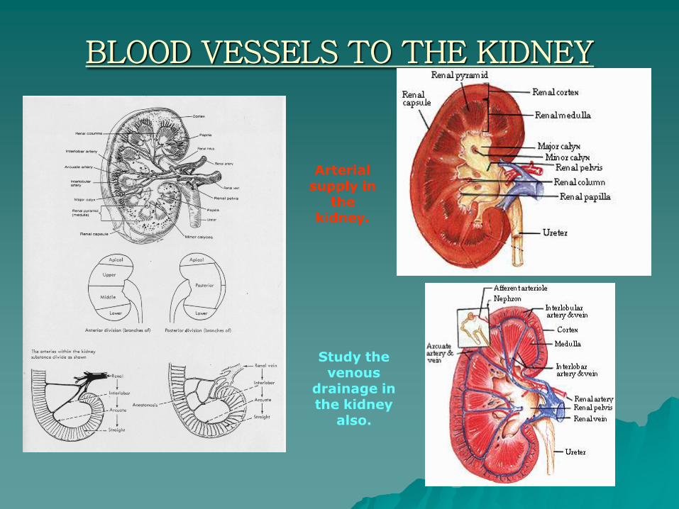
BLOOD VESSELS TO THE KIDNEY
Study the venous
drainage in the kidney
also.
Arterial supply in
the kidney.

THE KIDNEY
This fissure is named the hilum,
and transmits
the vessels, nerves, and
ureter. Above the hilum the
medial border is in
relation with the
suprarenal gland;
below the hilum, with
the ureter.
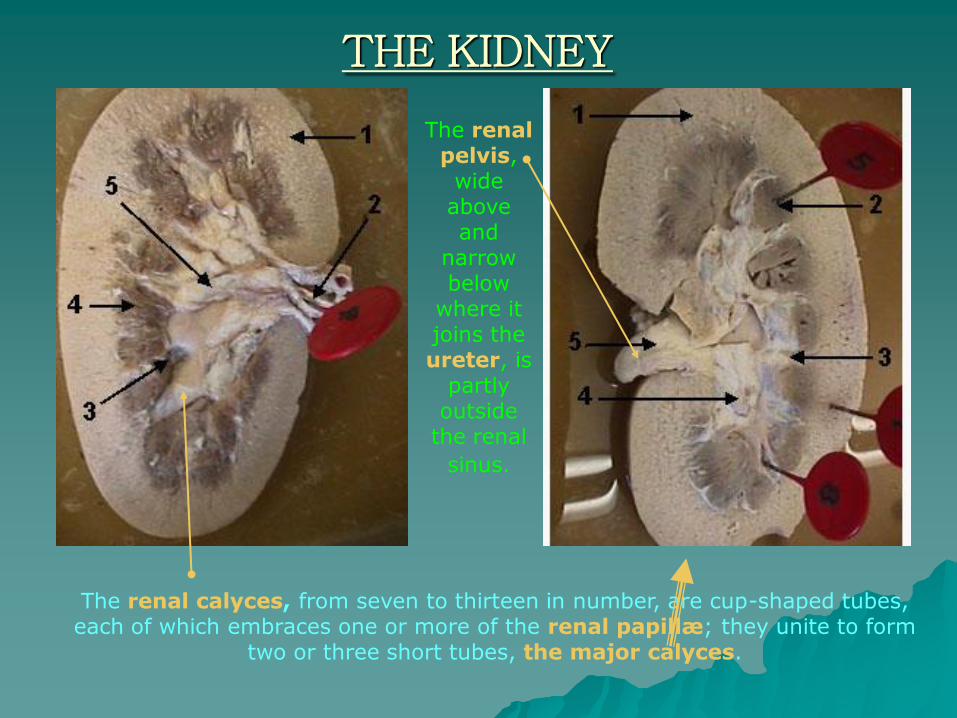
THE KIDNEY
The renal calyces, from seven to thirteen in number, are cup-shaped tubes, each of which embraces one or more of the renal papillæ; they unite to form
two or three short tubes, the major calyces.
The renal pelvis, wide above and
narrow below
where it joins the ureter, is
partly outside
the renal
sinus.
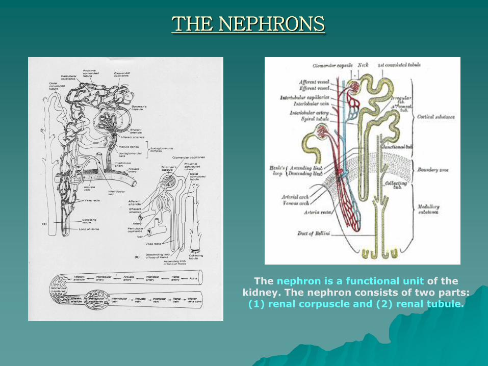
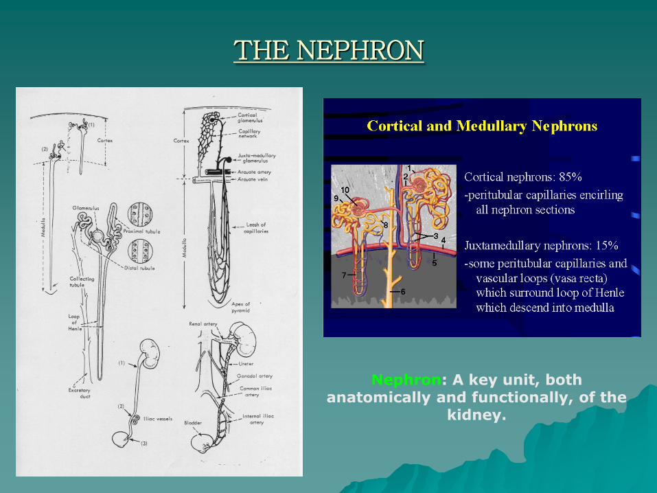
THE NEPHRONS
The nephron is a functional unit of the kidney. The nephron consists of two parts: (1) renal corpuscle and (2) renal tubule.
THE NEPHRON
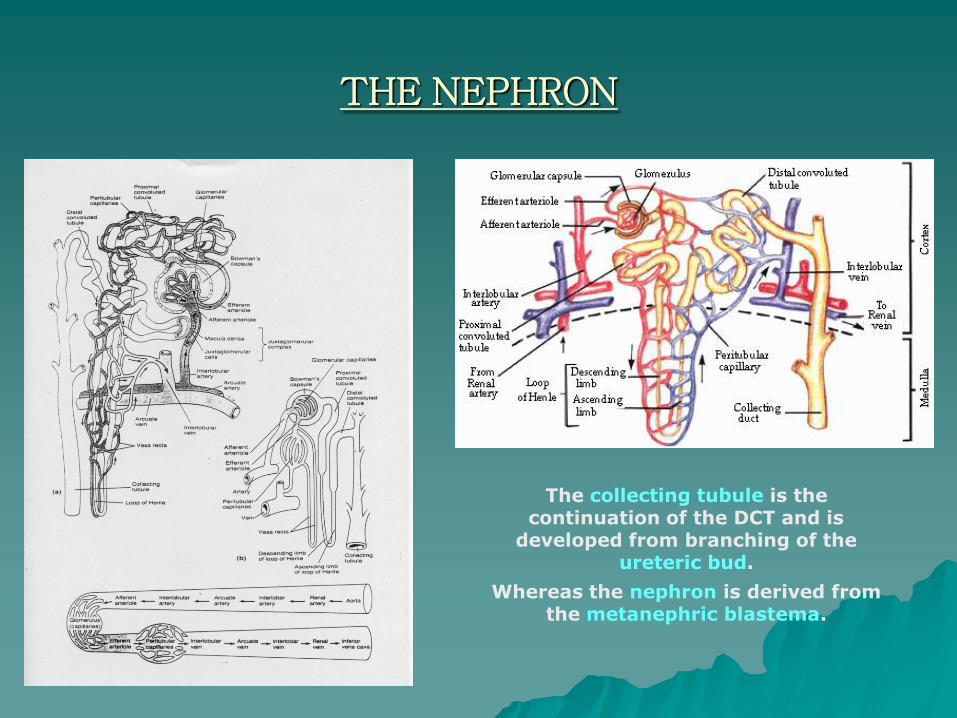
The collecting tubule is the continuation of the DCT and is
developed from branching of the ureteric bud.
Whereas the nephron is derived from the metanephric blastema.

THE NEPHRON
The juxtaglomerular apparatus is a collective term referring to the cells
near a structure called the glomerulus in the kidney.
THE NEPHRON
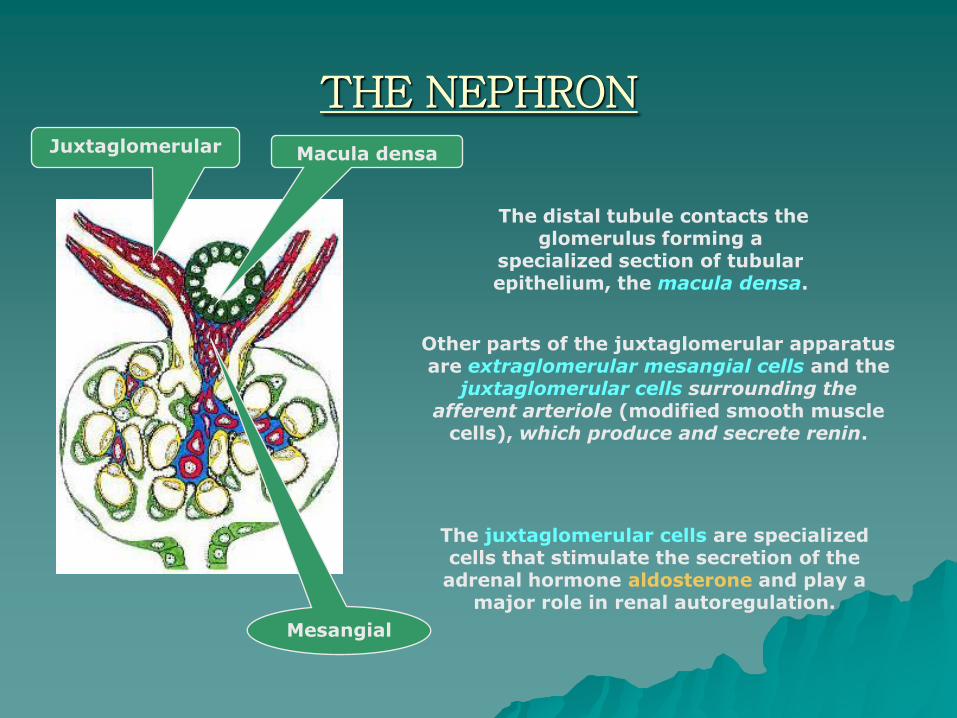
Other parts of the juxtaglomerular apparatus are extraglomerular mesangial cells and the
juxtaglomerular cells surrounding the afferent arteriole (modified smooth muscle
cells), which produce and secrete renin.
The distal tubule contacts the glomerulus forming a
specialized section of tubular epithelium, the macula densa.
Macula densaJuxtaglomerular
Mesangial
The juxtaglomerular cells are specialized cells that stimulate the secretion of the adrenal hormone aldosterone and play a
major role in renal autoregulation.
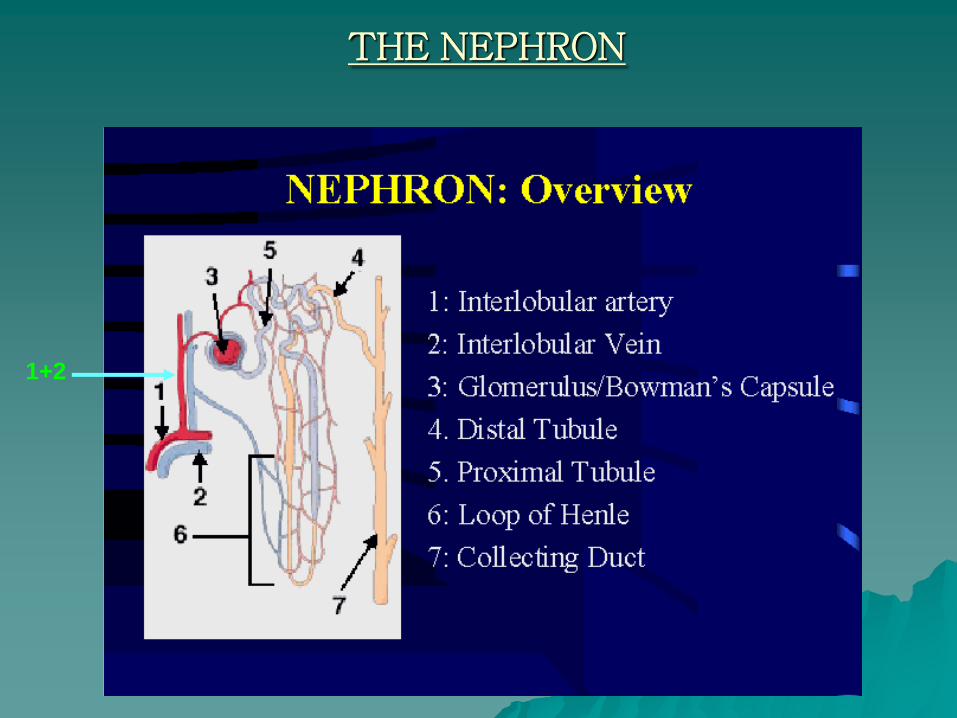
THE NEPHRON
1+2
THE NEPHRON
Nephron: A key unit, both anatomically and functionally, of the
kidney.

THE RENAL CORPUSCLE

The capsule of Bowman consiste of two layers: outer parietal and inner visceral
layer. The epithelium of the inner layer, the podocytes, surround and and closely
invest the capillary loops. Podocytes have many cytoplasmic extensions and small
processes called pedicles.
The narrow slits between the packed pedicles are called the
slit diaphragm.The capillaries of the
glomerulus are fenestrated capillaries having pores
between the endothelial cells.

THE GLOMERULUS
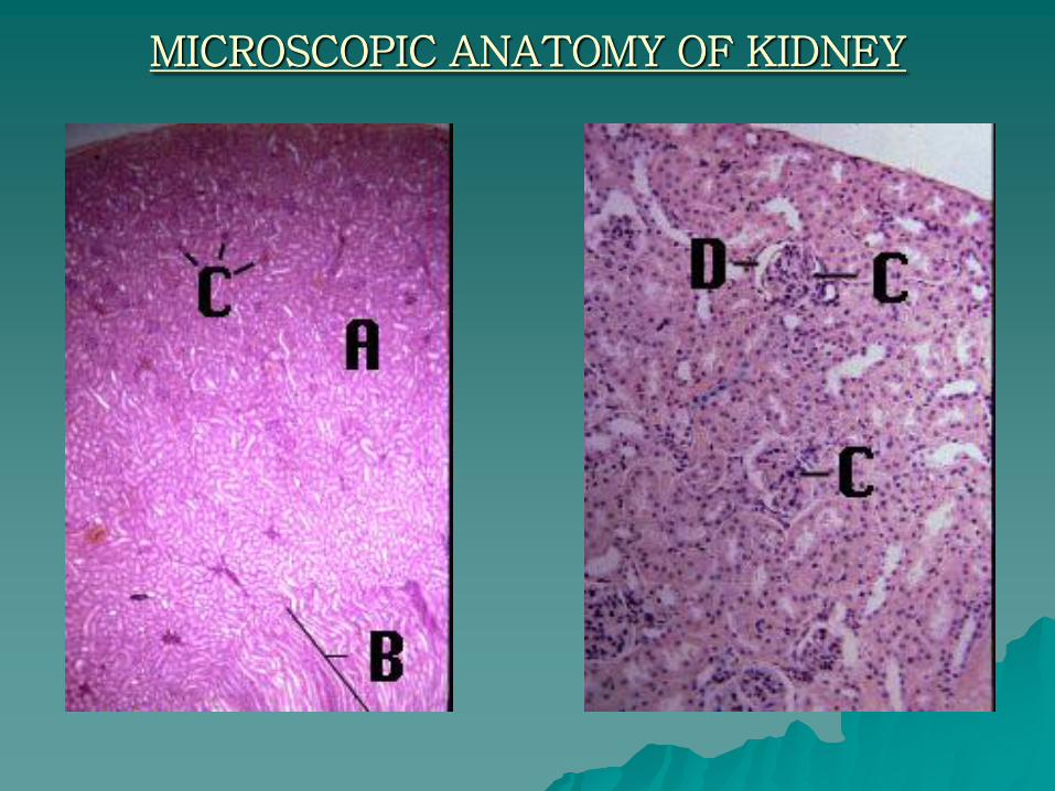
MICROSCOPIC ANATOMY OF KIDNEY
DEVELOPMENT OF KIDNEY
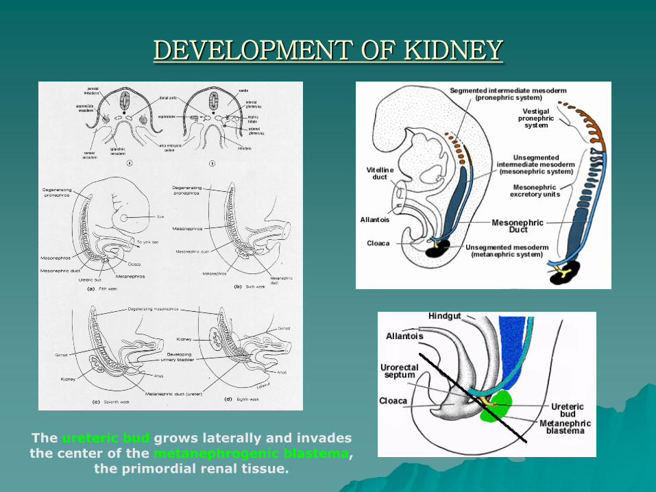
DEVELOPMENT OF KIDNEY
The ureteric bud grows laterally and invades the center of the metanephrogenic blastema,
the primordial renal tissue.

DEVELOPMENT OF KIDNEY
The ureteric bud divides and branches forming the renal
pelvis, infundibulae, calyces, and collecting tubules which
will provide a conduit for urine drainage in the mature kidney.
The metanephrogenic blastema forms glomeruli, proximal tubules and distal
tubules.
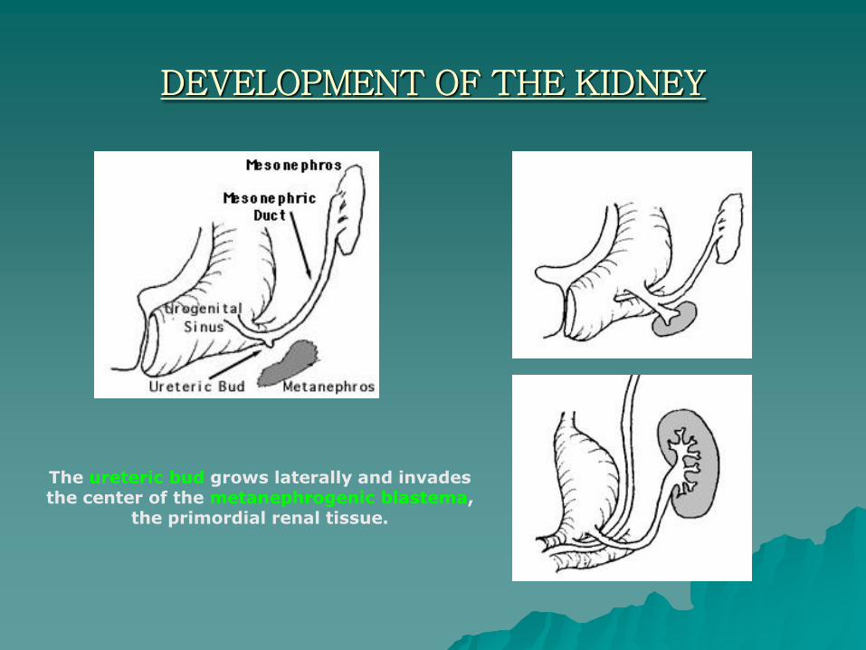
DEVELOPMENT OF THE KIDNEY
The ureteric bud grows laterally and invades the center of the metanephrogenic blastema,
the primordial renal tissue.

THE URETER
The uretersare about 25
cm long. Muscles in the ureter
walls constantly tighten and
relax to force urine
downward to
the bladder.
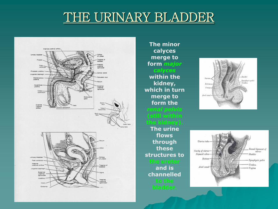
THE URINARY BLADDER
The minor calyces
merge to form major
calyceswithin the
kidney, which in turn
merge to form the
renal pelvis(still within the kidney).
The urine flows
through these
structures to the ureter
and is channelled
to the bladder.
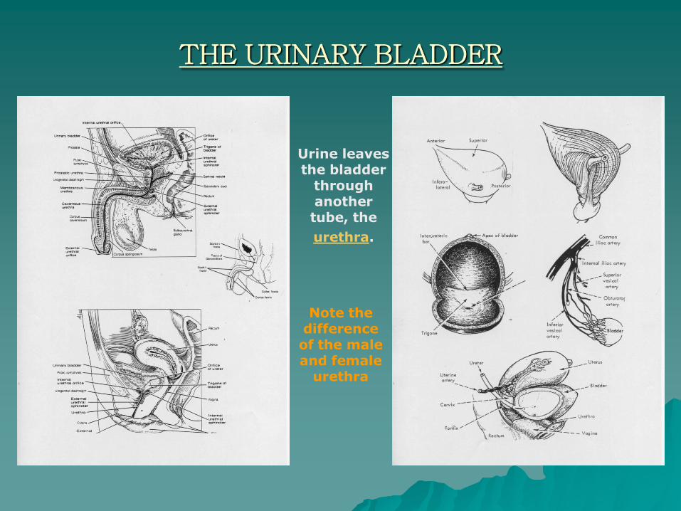
THE URINARY BLADDER
Urine leaves the bladder
through another tube, the
urethra.
Note the difference of the male and female
urethra
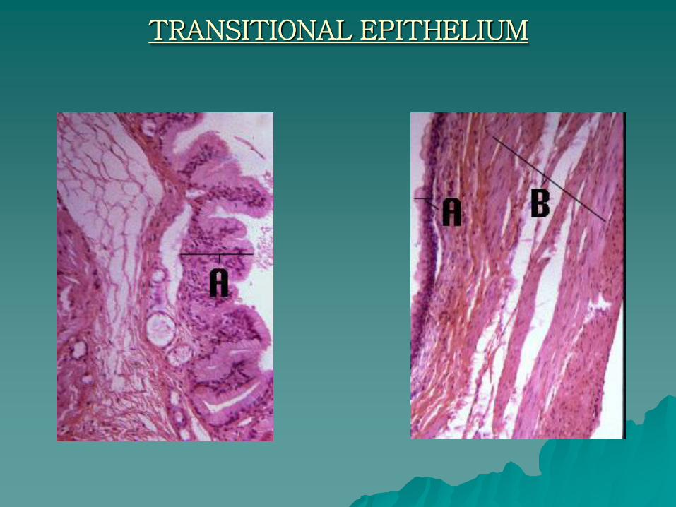
TRANSITIONAL EPITHELIUM
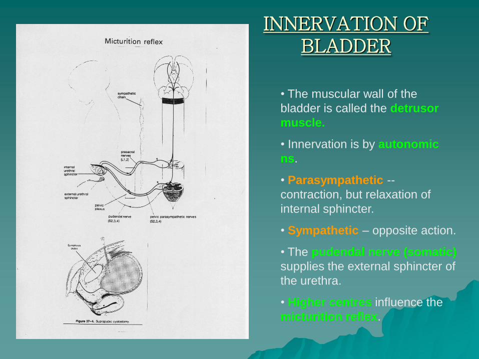
INNERVATION OF BLADDER
• The muscular wall of the
bladder is called the detrusor
muscle.
• Innervation is by autonomic
ns.
• Parasympathetic --
contraction, but relaxation of
internal sphincter.
• Sympathetic – opposite action.
• The pudendal nerve (somatic)
supplies the external sphincter of
the urethra.
• Higher centres influence the
micturition reflex.
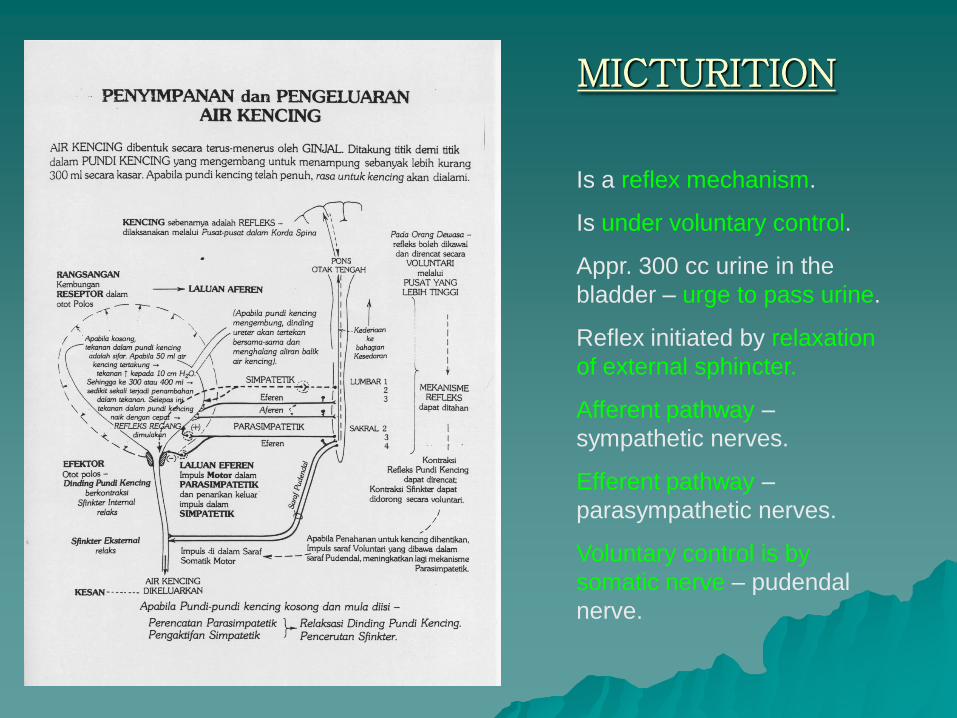
MICTURITION
Is a reflex mechanism.
Is under voluntary control.
Appr. 300 cc urine in the
bladder – urge to pass urine.
Reflex initiated by relaxation
of external sphincter.
Afferent pathway –
sympathetic nerves.
Efferent pathway –
parasympathetic nerves.
Voluntary control is by
somatic nerve – pudendal
nerve.

THANK YOU
The fear of he Lord teaches a man wisdom, and humility comes before honor. (Prov 15:33)
Pleasant words are a honeycomb, sweet to the soul and healing to the bones. (Prov 16:24)
A cheerful look brings joy to the heart, and good news gives health to the bones. (Prov 15:30)
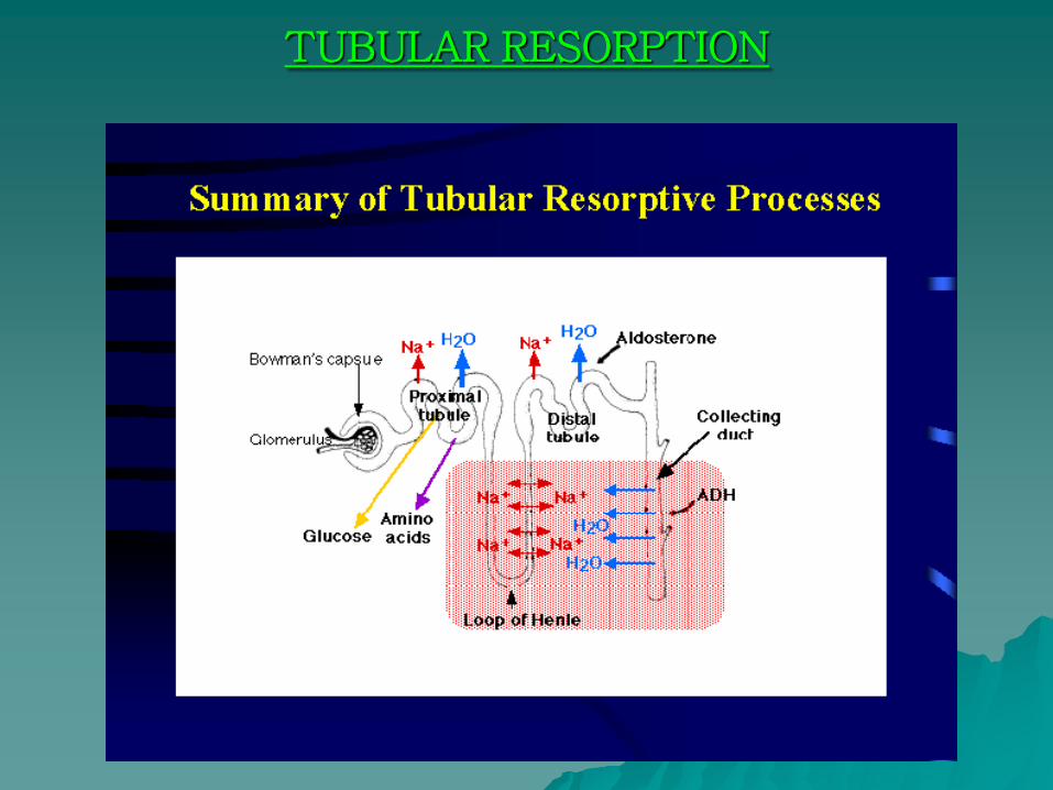
TUBULAR RESORPTION
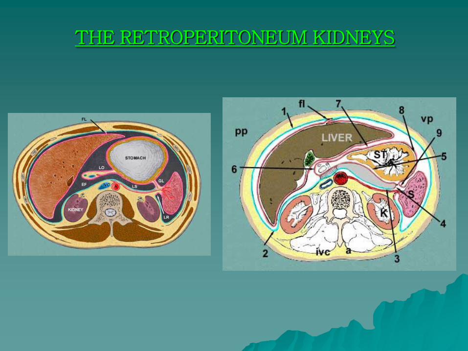
THE RETROPERITONEUM KIDNEYS
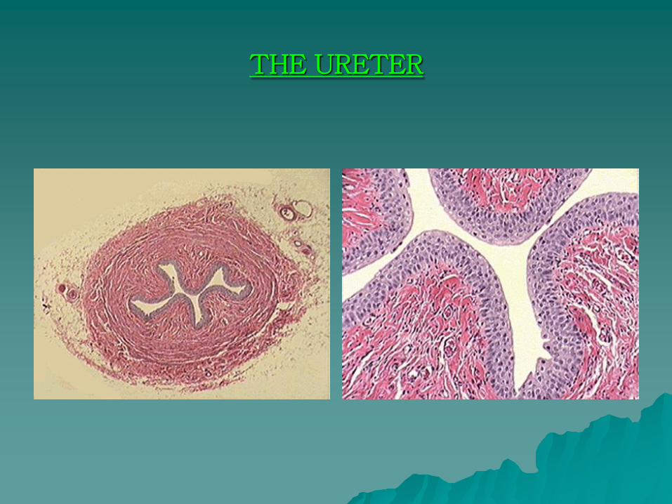
THE URETER

