the unique phylogenetic position of a novel tick-borne ... · sftsv, severe fever with...
TRANSCRIPT
The Unique Phylogenetic Position of a Novel Tick-BornePhlebovirus Ensures an Ixodid Origin of the Genus Phlebovirus
Keita Matsuno,a,b Masahiro Kajihara,c Ryo Nakao,d Naganori Nao,e Akina Mori-Kajihara,c Mieko Muramatsu,c Yongjin Qiu,f
Shiho Torii,g Manabu Igarashi,b,c Nodoka Kasajima,c Keita Mizuma,a Kentaro Yoshii,h Hirofumi Sawa,b,g Chihiro Sugimoto,b,i
Ayato Takada,b,c Hideki Ebiharaj
aLaboratory of Microbiology, Faculty of Veterinary Medicine, Hokkaido University, Sapporo, Hokkaido, JapanbDivision of International Services, Global Institution for Collaborative Research and Education (GI-CoRE),Hokkaido University, Sapporo, Japan
cDivision of Global Epidemiology, Hokkaido University Research Center for Zoonosis Control, Sapporo, JapandLaboratory of Parasitology, Faculty of Veterinary Medicine, Hokkaido University, Sapporo, Hokkaido, JapaneDepartment of Virology III, National Institute of Infectious Diseases, Tokyo, JapanfHokudai Center for Zoonosis Control in Zambia, Hokkaido University Research Center for Zoonosis Control,University of Zambia, Lusaka, Zambia
gDivision of Molecular Pathobiology, Hokkaido University Research Center for Zoonosis Control, Sapporo,Japan
hLaboratory of Public Health, Faculty of Veterinary Medicine, Hokkaido University, Sapporo, Hokkaido, JapaniDivision of Collaboration and Education, Hokkaido University Research Center for Zoonosis Control, Sapporo,JapanjDepartment of Molecular Medicine, Mayo Clinic, Rochester, Minnesota, USA
ABSTRACT The recent emergence of novel tick-borne RNA viruses has complicatedthe epidemiological landscape of tick-borne infectious diseases, posing a significantchallenge to public health systems that seek to counteract tick-borne diseases. Theidentification of two novel tick-borne phleboviruses (TBPVs), severe fever withthrombocytopenia syndrome virus (SFTSV) and Heartland virus (HRTV), as causativeagents of severe illness in humans has accelerated the investigation and discoveriesof novel TBPVs. In the present study, we isolated a novel TBPV designated Mukawavirus (MKWV) from host-questing Ixodes persulcatus females captured in Japan.Genetic characterization revealed that MKWV is a member of the genus Phlebovirusin the family Phenuiviridae. Interestingly, MKWV is genetically distinct from otherknown TBPVs and shares a most recent common ancestor with mosquito/sandfly-borne (insect-borne) phleboviruses. Despite its genetic similarity to insect-bornephleboviruses, the molecular footprints of its viral proteins and its biological charac-teristics define MKWV as a tick-borne virus that can be transmitted to mammals. Aphylogenetic ancestral-state reconstruction for arthropod vectors of phlebovirusesincluding MKWV based on viral L segment sequences indicated that ticks likely har-bored ancestral phleboviruses that evolved into both the tick-borne and MKWV/insect-borne phlebovirus lineages. Overall, our findings suggest that most of thephlebovirus evolution has occurred in hard ticks to generate divergent viruses,which may provide a seminal foundation for understanding the mechanisms under-lying the evolution and emergence of pathogenic phleboviruses, such as Rift Valleyfever virus and SFTSV/HRTV.
IMPORTANCE The emergence of novel tick-borne RNA viruses causing severe illnessin humans has complicated the epidemiological landscape of tick-borne diseases, re-quiring further investigation to safeguard public health. In the present study, we dis-covered a novel tick-borne phlebovirus from Ixodes persulcatus ticks in Japan. Whileits viral RNA genome sequences were similar to those of mosquito/sandfly-borne vi-ruses, molecular and biological footprints confirmed that this is a tick-borne virus.
Received 3 May 2018 Accepted 22 May2018 Published 13 June 2018
Citation Matsuno K, Kajihara M, Nakao R, NaoN, Mori-Kajihara A, Muramatsu M, Qiu Y, Torii S,Igarashi M, Kasajima N, Mizuma K, Yoshii K,Sawa H, Sugimoto C, Takada A, Ebihara H. 2018.The unique phylogenetic position of a noveltick-borne phlebovirus ensures an ixodid originof the genus Phlebovirus. mSphere 3:e00239-18. https://doi.org/10.1128/mSphere.00239-18.
Editor James M. Pipas, University of Pittsburgh
Copyright © 2018 Matsuno et al. This is anopen-access article distributed under the termsof the Creative Commons Attribution 4.0International license.
Address correspondence to Hideki Ebihara,[email protected].
K. Matsuno and M. Kajihara contributed equallyto this work.
RESEARCH ARTICLEEcological and Evolutionary Science
crossm
May/June 2018 Volume 3 Issue 3 e00239-18 msphere.asm.org 1
on June 3, 2020 by guesthttp://m
sphere.asm.org/
Dow
nloaded from

The unique evolutionary position of the virus allowed us to estimate the ancestralphlebovirus vector, which was likely a hard tick. Our findings may provide a betterunderstanding of the evolution and emergence of phleboviruses associated withemerging infectious diseases, such as severe fever with thrombocytopenia syndrome(SFTS) and Heartland virus disease.
KEYWORDS ancestral state, evolution, phylogenetic analysis, tick-borne phlebovirus
Emerging and reemerging infectious diseases caused by arthropod-borne viruses(arboviruses) have become significant global public health concerns in recent
decades. In particular, the emergence of mosquito-borne viruses such as Chikungunyavirus (1), West Nile virus (2), and Zika virus (3), which have spread to new geographiclocations with increased pathogenic potential, demonstrated that arboviruses poseongoing and future global public health threats. This is due to the intricacies ofhost-vector-virus interactions and the capacity for viruses to adapt to new vectors oramplifying hosts, driving the selection of novel strains/variants with pandemic/epi-demic potential (4–7).
In 2009, two novel tick-borne phleboviruses (TBPVs), severe fever with thrombocy-topenia syndrome virus (SFTSV) (8) and Heartland virus (HRTV) (9), were recognized tocause severe and often fatal human illnesses resembling viral hemorrhagic fever withthrombocytopenia, lymphocytopenia, hemophagocytic syndrome, and multiorgan fail-ure, resulting in fatality rates of up to 30%. HRTV was identified in the United States,whereas SFTSV was identified in East Asian countries, including China and Japan (10)and South Korea (11). Due to the similarities in the clinical features of TBPV diseases andsevere cases of tick-borne bacterial/rickettsial diseases, such as human monocyticehrlichiosis and human granulocyte anaplasmosis, newly emerging TBPVs have furthercomplicated the epidemiological landscape of tick-borne infectious diseases, posing asignificant challenge to public health authorities and health care providers in theirefforts to prevent, diagnose, and treat tick-borne infectious diseases.
A phlebovirus that has enveloped spherical virus particles, containing tripartite andsingle-stranded RNAs (L, M, and S segments) with negative-sense and/or ambisensepolarity, belongs to the genus Phlebovirus in the family Phenuiviridae, order Bunyavirales(formally known as the family Bunyaviridae) (12). Currently, the Phenuiviridae familyconsists of two species of TBPVs (i.e., SFTS phlebovirus and Uukuniemi phlebovirus) andseveral species of sandfly-borne and mosquito-borne viruses. Within the order Bunya-virales, the genus Phlebovirus is unique in that it contains human-pathogenic virusestransmitted by a wide range of arthropod vectors, either phlebotomine sandflies,mosquitos, or ticks. Recent advances in genome sequencing technology have allowedus to identify several tentative TBPV species/groups that are genetically distinct fromthe representative TBPV species Uukuniemi phlebovirus, revealing a greater diversityamong TBPVs than among mosquito/sandfly-borne phleboviruses, which form a singleclade with relatively low genetic divergence (13–17). Indeed, phylogenetic analyseshave revealed that there are at least four TBPV lineages based on genetic/serologicalsimilarities (14): SFTSV/HRTV, belonging to the SFTS group and associated with hem-orrhagic fever-like illness; Bhanja virus (BHAV) and Bhanja group viruses, associatedwith sporadic febrile illness cases; and Kaisodi virus (KSOV) and Uukuniemi virus (UUKV)in the Kaisodi and Uukuniemi groups, respectively, which have not been recognized ashuman pathogens. The mechanism(s) underlying the emergence of various TBPVs,especially two genetically related, highly pathogenic TBPVs (SFTSV and HRTV) on twodifferent continents, however, has not yet been revealed.
In the present study, we successfully isolated a novel TBPV named Mukawa virus(MKWV) from Ixodes persulcatus in Japan. We have characterized the molecular andbiological properties of the novel virus using virological and bioinformatics approachesand revealed that MKWV possesses the molecular and biological signatures of bothtick-borne and mosquito/sandfly-borne phleboviruses. A better understanding of theunique evolutionary traits of MKWV will contribute to the identification of the molec-
Matsuno et al.
May/June 2018 Volume 3 Issue 3 e00239-18 msphere.asm.org 2
on June 3, 2020 by guesthttp://m
sphere.asm.org/
Dow
nloaded from

ular determinants responsible for the evolution of viruses in the genus Phlebovirus andthe emergence of novel pathogenic phleboviruses, especially TBPVs.
RESULTSTick collection and isolation of MKWV. The southwestern part of Japan is an area
of endemicity of SFTS (10) (Fig. 1a). The RNA genome of SFTSV has been detected inmultiple species of ticks in the area of endemicity, and the seroprevalence of SFTSV inwild animals, such as Shika deer and raccoons, has also been confirmed (18). To gainfurther insights into the ecology and epidemiology of TBPVs and to identify novelTBPVs other than SFTSV in Japan, we therefore collected ticks outside the SFTS area ofendemicity (i.e., Hokkaido prefecture). In 2013, adult host-questing ticks were collectedby the flagging method in Mukawa, Hokkaido, Japan (Fig. 1a), and tick species wereidentified morphologically. Total RNA was extracted from a total of 66 individual ticks
FIG 1 Detection and isolation of MKWV. (a and b) Ticks (Ixodes persulcatus) collected in Mukawa, Hokkaido, Japan (a), were genetically screened for tick-bornephleboviruses (TBPVs) by using the pan-TBPV RT-PCR (b). Numbers at top are sample number designations. (c) The nucleotide sequences of amplified DNAfragments were phylogenetically analyzed with those of other phleboviruses, and all MKWV fragments clustered together in the independent branch. (d) Virionsof the MKWV isolate MKW73, harvested from the ISE6 tick embryonic cell supernatant, were morphologically analyzed using scanning electron microscopy. Bar,100 nm. SFTSV, severe fever with thrombocytopenia syndrome virus; RVFV, Rift Valley fever virus.
A Novel Virus Unveiling Phlebovirus Origins
May/June 2018 Volume 3 Issue 3 e00239-18 msphere.asm.org 3
on June 3, 2020 by guesthttp://m
sphere.asm.org/
Dow
nloaded from

and subjected to reverse transcription-PCR (RT-PCR) to amplify the phlebovirus L gene(14). Fragments of approximately 500 bp were amplified from three RNA samples fromfemale I. persulcatus specimens (samples 69, 73, and 74) (Fig. 1b). Following nucleotidesequencing, a phylogenetic tree was constructed (Fig. 1c). Interestingly, the phyloge-netic analysis of the partial L segment RT-PCR amplicons indicated that the all threeRT-PCR products were derived from viruses belonging to the genus Phlebovirus thatformed a cluster independent of any known phleboviruses. The detected phleboviruseshave been tentatively designated MKWVs.
To isolate MKWVs, homogenates of RT-PCR-positive ticks were used to inoculatecultured cells (Vero E6, DH82, Huh7, and ISE6 cells) and newborn BALB/c mice. Thereplication of two isolates, MKW73 and MKW74, was confirmed by RT-PCR only in thesupernatants of tick-derived ISE6 cells without cytopathic effects. Replication of MKWVswas not detected by RT-PCR in any mammal-derived cell lines or in newborn mice. Theproduction of virions in the supernatant of ISE6 cells infected with the isolate MKW73was confirmed by transmission electron microscopy (Fig. 1d). The virions of MKW73 areenveloped spherical particles (average diameter, 100 nm) with surface spike proteins,indicating morphological similarity with bunyaviruses (19).
Gene structure/organization of MKWV. For the removal of endogenous virusesfrom ISE6 cells (20, 21), MKW73 was passaged in Huh7 cells once, and the removal ofendogenous viruses was confirmed by RT-PCR (data not shown). MKW73 RNA extractedfrom the supernatant of Huh7 cells was subjected to deep sequencing. The sequencingreads were de novo assembled into contigs, three of which were associated withphleboviruses according to a BLAST search of GenBank. After the sequencing ofgenomic RNA termini by rapid amplification of cDNA ends (RACE) and resequencing ofregions with low-quality scores by Sanger sequencing, the complete nucleotide se-quence of each segment of MKW73 was obtained; large (L), medium (M), and small (S)RNA segments were composed of 6,443, 3,327, and 1,907 nucleotides, respectively(Table 1).
The deduced amino acid sequences of the L and M segment open reading frames(ORFs) showed homology with those of L proteins (RNA-dependent RNA polymerase)and surface glycoproteins (polyproteins Gn and Gc) from known phleboviruses, respec-tively. The S segment of the isolate possessed two ORFs in ambisense. A BLAST searchindicated that the deduced amino acid sequences of the ORFs were related to thoseof nucleocapsid proteins (N) and nonstructural proteins (NSs) from phleboviruses.The sequences of the L, M, and S genomic termini of MKW73 were identical tocorresponding conserved sequences among phleboviruses (i.e., 3=-UGUGU andACACA-5=), which may allow each segment of RNA to form panhandle structures.Thus, the genetic characteristics of MKWV clearly indicated that it is classified intothe genus Phlebovirus.
Phylogenetic characterization of MKWV. To further determine the molecularrelationship between MKWV and the other phleboviruses, including both tick-borneand mosquito/sandfly-borne phleboviruses, phylogenetic trees were constructed withMrBayes based on the nucleotide sequences of the L, M, and S segments (Fig. 2; see alsoFig. S1, S2, and S3 in the supplemental material) and using Gouléako virus (22) andCumuto virus (23) in the genus Goukovirus as an outgroup. The phylogenetic treesconstructed from the full-length nucleotide sequences of the L, M, and S genome
TABLE 1 Genome structure of Mukawa virus
RNA segment Length (base) Protein(s) ORF position (length) (bp)a
Terminal sequence (5=–3=)
5= 3=L 6,443 RdRp 19–6312 (6,294) ACACAAAGTCCG GCGCCTTTGTGTM 3,327 Gn and Gc 22–3243 (3,222) ACACAAAGACCG CCGTCTTTGTGTS 1,907 N 36–776 (741) ACACAAAGACCC GGGTCTTTGTGT
NSs 1871–852 (1,020)aIn cRNA, not including stop codon.
Matsuno et al.
May/June 2018 Volume 3 Issue 3 e00239-18 msphere.asm.org 4
on June 3, 2020 by guesthttp://m
sphere.asm.org/
Dow
nloaded from
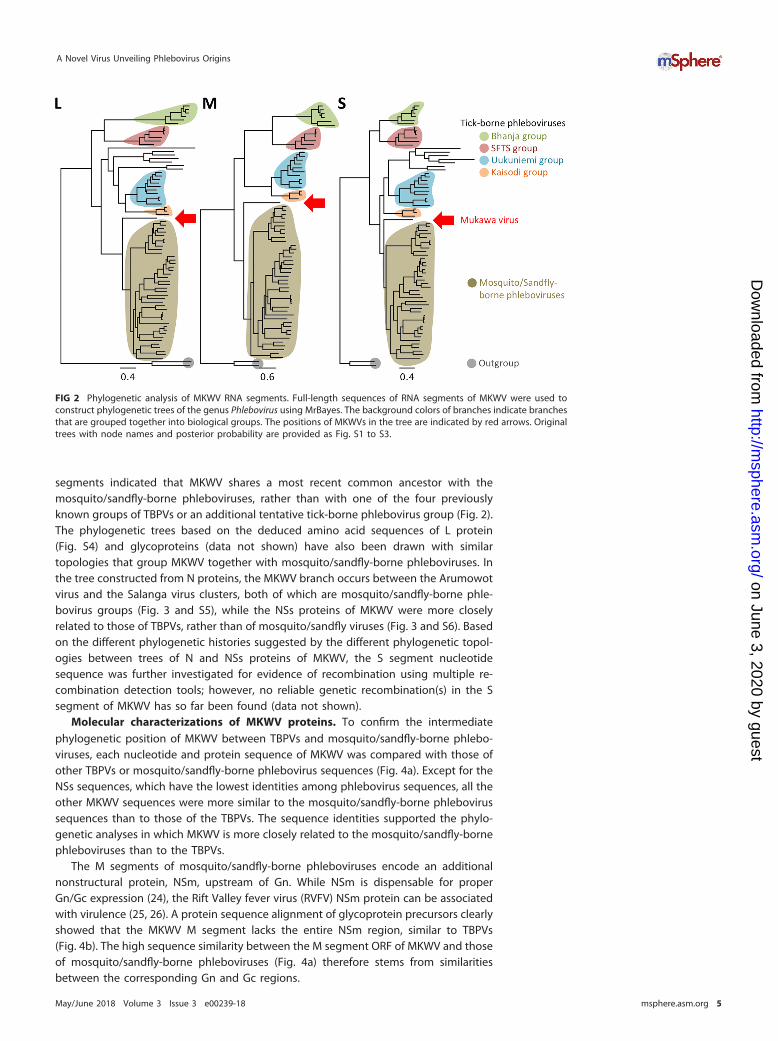
segments indicated that MKWV shares a most recent common ancestor with themosquito/sandfly-borne phleboviruses, rather than with one of the four previouslyknown groups of TBPVs or an additional tentative tick-borne phlebovirus group (Fig. 2).The phylogenetic trees based on the deduced amino acid sequences of L protein(Fig. S4) and glycoproteins (data not shown) have also been drawn with similartopologies that group MKWV together with mosquito/sandfly-borne phleboviruses. Inthe tree constructed from N proteins, the MKWV branch occurs between the Arumowotvirus and the Salanga virus clusters, both of which are mosquito/sandfly-borne phle-bovirus groups (Fig. 3 and S5), while the NSs proteins of MKWV were more closelyrelated to those of TBPVs, rather than of mosquito/sandfly viruses (Fig. 3 and S6). Basedon the different phylogenetic histories suggested by the different phylogenetic topol-ogies between trees of N and NSs proteins of MKWV, the S segment nucleotidesequence was further investigated for evidence of recombination using multiple re-combination detection tools; however, no reliable genetic recombination(s) in the Ssegment of MKWV has so far been found (data not shown).
Molecular characterizations of MKWV proteins. To confirm the intermediatephylogenetic position of MKWV between TBPVs and mosquito/sandfly-borne phlebo-viruses, each nucleotide and protein sequence of MKWV was compared with those ofother TBPVs or mosquito/sandfly-borne phlebovirus sequences (Fig. 4a). Except for theNSs sequences, which have the lowest identities among phlebovirus sequences, all theother MKWV sequences were more similar to the mosquito/sandfly-borne phlebovirussequences than to those of the TBPVs. The sequence identities supported the phylo-genetic analyses in which MKWV is more closely related to the mosquito/sandfly-bornephleboviruses than to the TBPVs.
The M segments of mosquito/sandfly-borne phleboviruses encode an additionalnonstructural protein, NSm, upstream of Gn. While NSm is dispensable for properGn/Gc expression (24), the Rift Valley fever virus (RVFV) NSm protein can be associatedwith virulence (25, 26). A protein sequence alignment of glycoprotein precursors clearlyshowed that the MKWV M segment lacks the entire NSm region, similar to TBPVs(Fig. 4b). The high sequence similarity between the M segment ORF of MKWV and thoseof mosquito/sandfly-borne phleboviruses (Fig. 4a) therefore stems from similaritiesbetween the corresponding Gn and Gc regions.
FIG 2 Phylogenetic analysis of MKWV RNA segments. Full-length sequences of RNA segments of MKWV were used toconstruct phylogenetic trees of the genus Phlebovirus using MrBayes. The background colors of branches indicate branchesthat are grouped together into biological groups. The positions of MKWVs in the tree are indicated by red arrows. Originaltrees with node names and posterior probability are provided as Fig. S1 to S3.
A Novel Virus Unveiling Phlebovirus Origins
May/June 2018 Volume 3 Issue 3 e00239-18 msphere.asm.org 5
on June 3, 2020 by guesthttp://m
sphere.asm.org/
Dow
nloaded from

For comparisons among the N and Gc proteins of phleboviruses, MKWV proteinstructures were predicted, as were the structures of the viral proteins of TBPVs, i.e.,Bhanja virus and Uukuniemi virus. The protein models predicted in the presentstudy mostly exhibited the same structural orientations, such as helix and sheets, asin the original N and Gc structures of RVFV proteins, respectively. In order to identifysimilar three-dimensional regions/domains that might not be observed in thesequence alignments between MKWV proteins and TBPV proteins, the amino acidsin the predicted models were compared with those of a representative TBPV of theSFTS group, SFTSV, and a representative mosquito-borne phlebovirus, RVFV (Fig. 4cand d). As shown in the phylogenetic analysis, the N protein structures of MKWVand Uukuniemi virus were more closely related to that of RVFV than to that of SFTSV(Fig. 4c). In terms of the structure of the MKWV Gc protein, it was more similar tothe structure of the RVFV Gc protein than to the other two Gc structures, andidentical amino acids between MKWV and SFTSV seemed to predominate in domainII of Gc (Fig. 4d).
Biological characterizations of MKWV. To confirm the arthropod host(s) of MKWV,the replication of MKW73 was assessed in three different cell lines: human-derivedHuh7 cells, tick-derived ISE6 cells, and mosquito-derived C6/36 cells (Fig. 5a). MKWVreplication in each cell line was monitored for a maximum of 14 days until a monolayerof cells was disrupted. In the Huh7 and ISE6 cells, MKW73 replicated to maximum titersof 107 50% tissue culture infectious doses (TCID50)/ml equivalent and 106 TCID50/mlequivalent, respectively. In contrast, C6/36 cells did not support the efficient reproduc-tion of progeny infectious virions, and a slight increase of viral RNA was detectedthroughout the observation period. Next, the growth levels of MKWV, UUKV, and TOSVwere compared in ISE6 cells and sandfly-derived LL-5 cells (Fig. 5b). ISE6 cells producedlarger amounts of TBPVs (i.e., MKWV and UUKV) than LL-5 cells, which supported higherreplication of sandfly-borne TOSV than ISE6 cells.
In addition, MKW73 was passaged in Huh7 cells and inoculated into newborn and3-week-old C57BL/6J mice, and virus replication was monitored (Fig. 5c). All newborn
FIG 3 Phylogenetic analysis of MKWV N and NSs proteins. Deduced amino acid sequences of the MKWVnucleoprotein (N) and nonstructural protein (NSs) were used to construct phylogenetic trees of the genusPhlebovirus by using MrBayes. The background colors of branches indicate branches that are groupedtogether into biological groups. The positions of MKWV in the tree are indicated by red arrows. Theoriginal trees with node names are provided as Fig. S4 and S5.
Matsuno et al.
May/June 2018 Volume 3 Issue 3 e00239-18 msphere.asm.org 6
on June 3, 2020 by guesthttp://m
sphere.asm.org/
Dow
nloaded from

FIG 4 Molecular characterization of MKWV. (a) Nucleotide sequence identities of open reading framesor deduced amino acid sequence identities of viral proteins (RNA-dependent RNA polymerase [L],
(Continued on next page)
A Novel Virus Unveiling Phlebovirus Origins
May/June 2018 Volume 3 Issue 3 e00239-18 msphere.asm.org 7
on June 3, 2020 by guesthttp://m
sphere.asm.org/
Dow
nloaded from

mice infected with MKWV died by 9 days postinoculation (dpi). At 3 dpi, three infectedmice from each group were sacrificed to examine viral replication in their brains.Productive viral replication was detected in the brains of MKWV-infected mice. In theyoung mice, productive replication of MKWV was also detected in the spleen at 3 and7 dpi following intraperitoneal inoculation, indicating that MKWV replicated in miceand systematically spread within the body.
FIG 4 Legend (Continued)glycoprotein [G], nucleoprotein [N], or nonstructural protein [NSs]) were determined for the comparisonsof MKWV with tick-borne phleboviruses (tick virus) and mosquito/sandfly-borne phleboviruses (mosquitovirus). The mean identity of each group is presented as a horizontal bar. Significant differences betweenthe identity values of tick virus comparisons and mosquito virus comparisons are indicated by asterisks(P � 0.05). (b) Alignments of deduced amino acid sequences of phlebovirus glycoproteins. Boxes indicatealigned amino acids, and bars indicate gaps in each sequence. (c and d) Structures of N protein (c) andmembrane glycoprotein Gc (d) of three tick-borne phleboviruses, MKWV, Bhanja virus, and Uukuniemivirus, were predicted based on the crystal structures of RVFV proteins. Amino acid residues identical tothose of RVFV and SFTSV are shown in blue and red, respectively. Amino acid residues shared with bothRVFV and SFTSV are shown in dark gray. The inset view of the surface loop of domain II in the MKWV Gcis indicated by a black square.
FIG 5 In vitro and in vivo replication of MKWV and interferon-antagonism activity of MKWV NSs. (a) MKWV isolate MKW73was used to inoculate cells derived from human (Huh7), tick (ISE6), or mosquito (C6/36). The supernatant of each cell linewas collected every 2 days from immediately after inoculation (day 0) to 14 days postinoculation (dpi). (b) Phleboviruseswere inoculated into cells derived from tick (ISE6) or sandfly (LL-5), and the supernatant of each cell line was harvested at7 dpi. RNA extracted from the supernatant was subjected to quantitative RT-PCR to calculate the viral titer. Each data pointindicates the mean from triplicate experiments, and error bars indicate standard deviations (SDs). (c) MKWV MKW73 andSFTSV isolate YG1 were used to inoculate 1-day-old mice (SM) and 3-week-old mice (YM). Tissues were collected at 3, 7,and/or 9 dpi, and viral titers were calculated as the 50% tissue culture infectious dose (TCID50/gram). Error bars indicateSDs. (d and e) Luciferase activities under the control of the beta interferon promoter (d) and interferon-sensitive responseelement (ISRE) (e) were compared in NSs-expressing 293 cells stimulated by the expression of an activated form of RIG-I(d) and recombinant alpha interferon (e), respectively. All tests were done in triplicate, and the means � SDs are shown.
Matsuno et al.
May/June 2018 Volume 3 Issue 3 e00239-18 msphere.asm.org 8
on June 3, 2020 by guesthttp://m
sphere.asm.org/
Dow
nloaded from

Phlebovirus NSs proteins have been recognized as significant virulence factors dueto their ability to counteract host innate antiviral responses (27, 28). To determine therole of MKWV NSs proteins in their viral replication in human cells, inhibitory effects ofNSs on the activities of the beta interferon promoter (Fig. 5d) or interferon-sensitiveresponse element (ISRE) (Fig. 5e) were measured in human embryonic kidney 293 cellstransiently expressing MKWV NSs and compared to those of cells expressing SFTSV NSs.MKWV NSs significantly blocked beta interferon promoter activity induced by RIG-Isignaling but not ISRE activity, while SFTSV NSs inhibited both. During the transientexpression of MKWV NSs in the cells, transcriptional shutoff, a significant function ofRVFV NSs, was not detected.
Ancestral-state reconstruction of the genus Phlebovirus. For predicting thearthropod host(s) of ancestral phleboviruses, ancestral-state reconstruction was per-formed using the likelihood reconstruction method in Mesquite with a phylogenetictree constructed based on nucleotide sequences of the L segment of the phleboviruses(Fig. 6). In the analysis, only the viruses with clearly identified arthropod vectors wereused. The most recent ancestor of the MKWV and the mosquito/sandfly-borne phle-boviruses, as well as more ancient ancestors of phleboviruses, was predicted to beharbored by ticks, not insects. The ancestor of the genus Phlebovirus, which wasdetermined by using a defined outgroup (Fig. 2), was also predicted to be a tick-bornevirus.
DISCUSSION
The identification of SFTSV and HRTV stressed the potential public health threat ofTBPVs. In addition to these novel pathogens, the identification of novel phleboviruseshas been sporadically reported worldwide owing to the recent advancements and
FIG 6 Phlebovirus ancestral-state reconstruction. The phylogenetic tree based on the nucleotidesequence of the L segment was used for ancestral-state reconstitution. Phleboviruses were divided intotwo states (insect-borne and tick-borne), and the state of each ancestor was predicted using themaximum likelihood method. The likelihood of each node or each ancestor is indicated by a pie chart(white, insect; black, tick). Abbreviations: BHAV, Bhanja virus; LSV, Lone Star virus; HIGV, Hunter Islandvirus; SFTSV, severe fever with thrombocytopenia syndrome virus; GAV, Grand Arbaud virus; UUKV,Uukuniemi virus; RMLV, RML-105355; SILV, Silverwater virus; MUKV (MKWV), Mukawa virus; TOSV, Toscanavirus; AMTV, Arumowot virus; SALV, Salehabad virus; AGUV, Aguacate virus; DURV, Durania virus; SSTV,sandfly Sicilian Turkey virus; KARV, Karimabad virus; RVFV, Rift Valley fever virus; PTV, Punta Toro virus;NIQV, Nique virus; CDUV, Candiru virus; MCRV, Mucura virus; MUNV, Munguba virus.
A Novel Virus Unveiling Phlebovirus Origins
May/June 2018 Volume 3 Issue 3 e00239-18 msphere.asm.org 9
on June 3, 2020 by guesthttp://m
sphere.asm.org/
Dow
nloaded from

increasing availability of new sequencing technologies. Despite our increased knowl-edge of phleboviruses, their ecology and evolutionary dynamics remain poorly under-stood. In the present study, we identified a novel TBPV, designated MKWV, which wasfound to occupy a unique evolutionary position among phleboviruses. To date, theevolutionary history of TBPVs and mosquito/sandfly-borne phleboviruses has beencompletely unknown, though it was suspected that the ancestor of phlebovirusesmight have been an arthropod virus (15, 29). MKWV is the first TBPV to exhibit geneticsimilarity to mosquito/sandfly-borne phleboviruses. Despite the fact that MKWV sharescommon ancestry with and is phylogenetically closely related to mosquito/sandfly-borne phleboviruses, MKWV replicated at low levels in the mosquito-derived C6/36 cellsand sandfly-derived LL-5 cells but replicated at higher levels in the tick (ISE6) andmammalian (Huh7) cells, like tick-borne UUKV, which was reported to replicate in Aedesaegypti mosquitos by experimental infections (30). Based on the discovery of thisphylogenetically unique virus, the evolutionary history of phleboviruses with theirvectors could be revisited with the stronger support values on trees. These resultsconfirm that the ancestor of phleboviruses was a tick-borne virus that later generatedvarious TBPVs and an ancestor of MKWV (Fig. 7). Further, the ancestor of MKWV evolvedinto mosquito/sandfly-borne phleboviruses and the current MKWV. Since more diver-gent insect-borne phleboviruses have not yet been reported in insect vectors, the “tickorigin” hypothesis is consistent with our current knowledge of phleboviruses. More-over, the Uukuniemi group viruses have been isolated from Ixodes hard ticks and Argassoft ticks (17), suggesting that the ancestral phlebovirus might retain the replicationcapability in hard ticks and soft ticks, as well as insects.
The topologies of phlebovirus trees were nearly identical among three RNA seg-ments as well as three viral proteins (i.e., L, glycoprotein, and NSs), indicating the stableassociation among the three viral proteins. The topology of the N protein tree, however,in which some TBPVs, including MKWV, clustered with the mosquito/sandfly-bornephleboviruses, was unique. The difference between the N protein phylogeny and thoseof the other proteins and nucleotides may indicate a historical recombination event(s)or different selection pressures in the S segment, although no genetic recombinationcould be identified in the current analysis. Further discoveries of novel phlebovirusescould shed additional light on the evolutional history of phleboviruses.
I. persulcatus, the tick species from which MKWV was isolated, is a common tickdistributed across the Hokkaido prefecture and the northern part of the main island ofJapan. I. persulcatus infests humans and animals, frequently resulting in cases of Lymedisease in Hokkaido (31). While several studies have been conducted on the bacterialpathogens carried by this tick species (32–34), there have previously been no reports ofdiseases indicating associations with viral pathogens. MKWV is likely to be transmittedto mammals through tick bites and to establish infections, as suggested by the growthof MKWV in a human-derived cell line and mice and by the anti-innate immunefunction of MKWV NSs in human cells. Based on these findings, it is conceivable that
FIG 7 Hypothetical pathways of phlebovirus evolution. Based on our current knowledge of phlebovi-ruses, the ancestor of phleboviruses is likely to be a tick-borne virus. The ancestral phlebovirus evolvedinto divergent viruses in ticks, and a variety of tick-borne viruses have been generated. MKWV andmosquito/sandfly-borne phleboviruses might be produced from the ancestor of Mukawa virus, and thus,less diversity can be observed among these insect-borne viruses than tick-borne phleboviruses.
Matsuno et al.
May/June 2018 Volume 3 Issue 3 e00239-18 msphere.asm.org 10
on June 3, 2020 by guesthttp://m
sphere.asm.org/
Dow
nloaded from

wild animals such as local deer and rodents may be infected with MKWV through tickbites and act as intermediate hosts, similar to the way in which SFTSV infects wildanimals and livestock in regions of endemicity (35–37). Furthermore, the replicationcompetence of MKWV in human-derived Huh7 cells at 37°C and ISE6 cells at 34°Cindicates the potential of MKWV to replicate in both arthropod and mammalian hosts(29). A serological study of MKWV in humans and animals is thus warranted forfurthering our understanding of the epidemiology of the virus. Moreover, it is highlyplausible that a large number of phleboviruses remain unknown, and further intensivestudies may reveal the evolutional mechanism(s) of the emergence of human-pathogenic phleboviruses.
MATERIALS AND METHODSTick collection and sample preparation. Adult host-questing ticks were captured using the
flagging method in Mukawa, Hokkaido, Japan (Fig. 1a) (latitude 42.61 north, longitude 141.95 east) inMay 2013. Collected ticks were morphologically identified as I. persulcatus, Ixodes ovatus, Haemaphysalisjaponica, or Haemaphysalis megaspinosa under a stereomicroscope. Ten males and 10 females ofI. persulcatus, 12 males and 12 females of I. ovatus, three females of H. japonica, and 10 males and 10females of H. megaspinosa were used in the following experiments. Each tick was homogenized twicewith 100 �l of plain Dulbecco’s modified Eagle’s medium (DMEM) using a homogenizer (Tomy Seiko) at3,000 rpm. Total RNAs were extracted from 50 �l of the homogenates by using the blackPREP tickDNA/RNA kit (Analytik Jena) according to the manufacturer’s protocol, and remaining lysate sampleswere stored at �80°C until use for virus isolation.
One-step RT-PCR. We utilized a previously reported one-step RT-PCR system that detects a wide-range of TBPVs from RNA samples from collected ticks (14). Briefly, one-step RT-PCR was performed withthe PrimeScript One Step RT-PCR kit, version 2 (Dye Plus) (TaKaRa), with 1 �l of total tick RNA and 4 pmolof primers, HRT-GOUL2759F (5=-CAGCATGGIGGIYTIAGRGAAATYTATGT-3=) and HRT-GOUL3276R (5=-GAWGTRWARTGCAGGATICCYTGCATCAT-3=) in 10 �l of reaction solution with the following incubationprogram: 50°C for 30 min; 94°C for 2 min; 40 cycles of 94°C for 30 s, 55°C for 30 s, and 72°C for 30 s; and72°C for 5 min. Amplified RT-PCR products were purified and sequenced with a 3130 Genetic Analyzer(ABI, Thermo Fisher Scientific).
Cells and viruses. Vero E6 (African green monkey kidney), Huh7 (human hepatocellular carcinoma),and DH82 (canine macrophage) cells were grown in DMEM supplemented with fetal calf serum (FCS).ISE6 (Ixodes scapularis embryo) cells were grown in modified L-15B medium supplemented with 10% FCSand 5% tryptose phosphate broth (Sigma) at 34°C (38). C6/36 (Aedes albopictus larva) cells were grownin minimal essential medium (MEM) supplemented with 10% FCS and L-glutamine at 28°C. SFTSV (strainYG1, kindly provided by Ken Maeda, Yamaguchi University, Japan) was propagated in Vero E6 cells (10).
Virus isolation. Tenfold-diluted tick lysates in DMEM supplemented with 10% FCS, 2 mML-glutamine, 50 U/ml penicillin, 50 �g/ml streptomycin, and 25 �g/ml gentamicin (Sigma) were filteredand used to inoculate cultured cells. Cells were cultured for 14 days, and RNAs were purified fromsupernatants using the QIAamp viral RNA minikit (Qiagen). Isolates from two I. persulcatus samples weretentatively designated MKWVs. Blind passages were carried out once or twice. Virus shedding in thesupernatants was confirmed by RT-PCR. Filtrates were also used to inoculate newborn BALB/c miceintracerebrally. The mice were observed for manifestations of clinical signs. At 14 dpi, mice showing noclinical sign were sacrificed, and their brains were sampled. Viral replication in brains was examined usingRT-PCR.
Electron microscopy. The supernatant of TBPV-positive cells was harvested and pelleted by ultra-centrifugation (27,000 rpm, 90 min) through 25% sucrose in phosphate-buffered saline (PBS). Virionswere resuspended in PBS and adsorbed to collodion-carbon-coated copper grids. Virions negativelystained with 2% phosphotungstic acid solution (pH 5.8) were examined under an H-7650 transmissionelectron microscope (Hitachi) at 80 kV.
Determination of full-length viral genome sequences. Viral RNA extracted from purified virionswas subjected to double-stranded cDNA synthesis using a cDNA synthesis kit (Moloney murine leukemiavirus [M-MLV] version; TaKaRa). Pyrosequencing analysis was performed on a 454 Genome SequencerJunior (GS-Junior; Roche) according to the manufacturer’s protocol. After trimming low-quality reads, theresulting reads were de novo assembled using Newbler (Roche) with default parameters. Both the 5= and3= terminus regions of each segment were amplified by the RACE method (39), and the completenucleotide sequence of each segment was obtained.
Multiple sequence alignment and phylogenetic analysis. The nucleotide sequences of the L genefragments of approximately 500 bp amplified by RT-PCR were aligned with representative sequencesfrom other known phleboviruses available from GenBank by using MUSCLE as implemented in MEGA,version 6 (40). Multiple sequence alignments were modified manually to correct unreliable alignments.Phylogenetic trees were constructed using the maximum likelihood (ML) method. For ML analysis, theTamura-Nei model with gamma-distributed invariant sites (G�I) built into MEGA 6 was used. Therobustness of each node was tested by 1,000 bootstrap replicates. The full-length nucleotide sequenceof each RNA segment and deduced amino acid sequence of each viral protein of MKWV and therepresentative phleboviruses were subjected to the multiple sequence alignment using MEGA as well asClustal Omega (41) as implemented in CLC Main Workbench (Qiagen). The aligned sequences were usedfor phylogenetic tree construction by using MEGA (ML and neighbor-joining methods) and MrBayes 3.2.2
A Novel Virus Unveiling Phlebovirus Origins
May/June 2018 Volume 3 Issue 3 e00239-18 msphere.asm.org 11
on June 3, 2020 by guesthttp://m
sphere.asm.org/
Dow
nloaded from

(42) as a plug-in of Geneious (Biomatters Ltd.) with the GTR�G�I substitution model. The trees obtainedby three independent methods were compared with each other to confirm the same topologies.Ancestral-state reconstruction was performed with Mesquite (43).
Experimental infection of mice with TBPVs. MKWV (5.0 � 104 TCID50) and SFTSV (3.6 � 104 TCID50)were intracerebrally inoculated into nine and six newborn mice (C57BL/6J), respectively. Three mice ofeach group were sacrificed at 3 dpi, and their brains were sampled. The remaining mice were observedfor their clinical symptoms. MKWV (2.5 � 105 TCID50) and SFTSV (1.8 � 105 TCID50) were intraperitoneallyinoculated into 11 3-week-old C57BL/6J mice. Three mice were sacrificed at 3 and 7 dpi, and their brains,kidneys, lungs, livers, and spleens were aseptically collected. The remaining mice were observed forclinical signs, and their body weights were recorded for 14 days. Viral replication in each tissue wasexamined as described above, and the titers were calculated as TCID50 per gram of tissue. Experimentalinfections were carried out in the biosafety level 3 facility at the Hokkaido University Research Center forZoonosis Control, Japan, strictly according to the Guidelines for Proper Conduct of Animal Experimentsof the Science Council of Japan. The protocol was approved by the Hokkaido University Animal Care andUse Committee. All efforts were made to minimize suffering.
Growth kinetics of MKWV in various cells. MKWV was inoculated into Huh7, ISE6, and C6/36 cellsat a multiplicity of infection (MOI) of 1. After a 1-h incubation to allow viruses to attach, cells were rinsedonce with their original medium and incubated for up to 14 days in the same medium with 10% FCS.Supernatants were collected every 2 days after the inoculation (0 to 14 days), and RNAs were extractedusing TRIzol LS reagent (Thermo Fisher Scientific) according to the manufacturer’s protocol. Viral RNAsin the supernatant were quantified as equivalent to TCID50 by using real-time PCR with the KAPA ProbeFast qPCR kit (KAPA Biosystems) (primer and probe sequences available upon request).
Innate immune signaling inhibition assays. 293 cells maintained in DMEM-10% FCS were tran-siently transfected with an expression plasmid of MKWV NSs-FLAG or SFTSV NSs-FLAG together with areporter plasmid (pI125luc or pISRE-Luc) expressing firefly luciferase under the control of the betainterferon promoter or ISRE, respectively, and a control-luciferase-expressing plasmid, pGL4.75[hRluc/CMV] (Promega). The cells transfected with pI125luc were also transfected with an expression plasmid ofthe human RIG-I CARD domain (pEF-BOS RIG-IN) to activate the interferon-production signaling. The cellstransfected with pISRE-Luc were treated with recombinant human alpha interferon (R&D Systems) 6 hprior to harvest. Cells were lysed at 24 h posttransfection, and the luciferase activity was measured byusing the Dual-Glo luciferase assay system (Promega) with a Modulus microplate luminometer.
Accession number(s). The pyrosequencing data and complete genome sequences were depositedin the Sequence Read Archive (SRA) under accession number DRA002645 (raw reads) and in EMBL/GenBank/DDBJ under accession numbers LC063768 to LC063770 (complete genome sequences).
SUPPLEMENTAL MATERIALSupplemental material for this article may be found at https://doi.org/10.1128/
mSphere.00239-18.FIG S1, PDF file, 0.1 MB.FIG S2, PDF file, 0.1 MB.FIG S3, PDF file, 0.1 MB.FIG S4, PDF file, 0.1 MB.FIG S5, PDF file, 0.1 MB.FIG S6, PDF file, 0.1 MB.
ACKNOWLEDGMENTSWe thank Hiroko Miyamoto, Aiko Ohnuma, and Tatsuyuki Osuga at the Hokkaido
University Research Center for Zoonosis Control for their help and assistance. We aregrateful to Sonja M. Best (Innate Immunity and Pathogenesis Unit, Rocky MountainLaboratories, DIR, NIAID, NIH) for providing the reporter plasmids and RIG-IN expressionplasmid used in this study. We also thank the World Reference Center for EmergingViruses and Arboviruses (WRCEVA) for providing us with Uukuniemi virus, Toscana virus,and LL-5 cells.
This study was partly supported by the Akiyama Life Science Foundation, MEXT/JSPS KAKENHI (grant numbers JP17KT0045, JP16K18791, JP16H06431, JP16H06429,JP16K21723, JP16H05805, and JP15K18778), the Japan Initiative for Global ResearchNetwork on Infectious Diseases (J-GRID) and the Japan Initiative for Progress of Re-search on Infectious Disease for Global Epidemic (J-PRIDE) (AMED grant no.JP18fm0108008 and JP17fm0208001, respectively), AMED/Japan International Cooper-ation Agency (JICA) within the framework of the Science and Technology ResearchPartnership for Sustainable Development (SATREPS), and the Fusion-H program fromHokkaido University. The funders had no role in study design, data collection andinterpretation, or the decision to submit the work for publication.
Matsuno et al.
May/June 2018 Volume 3 Issue 3 e00239-18 msphere.asm.org 12
on June 3, 2020 by guesthttp://m
sphere.asm.org/
Dow
nloaded from

REFERENCES1. Morrison TE. 2014. Reemergence of Chikungunya virus. J Virol 88:
11644 –11647. https://doi.org/10.1128/JVI.01432-14.2. Murray KO, Mertens E, Desprès P. 2010. West Nile virus and its emer-
gence in the United States of America. Vet Res 41:67. https://doi.org/10.1051/vetres/2010039.
3. Yadav S, Rawal G, Baxi M. 2016. Zika virus: an emergence of a newarbovirus. J Clin Diagn Res 10:DM01–DM03. https://doi.org/10.7860/JCDR/2016/19170.8133.
4. Weaver SC, Forrester NL. 2015. Chikungunya: evolutionary history andrecent epidemic spread. Antiviral Res 120:32–39. https://doi.org/10.1016/j.antiviral.2015.04.016.
5. Tsetsarkin KA, Chen R, Yun R, Rossi SL, Plante KS, Guerbois M, ForresterN, Perng GC, Sreekumar E, Leal G, Huang J, Mukhopadhyay S, Weaver SC.2014. Multi-peaked adaptive landscape for Chikungunya virus evolutionpredicts continued fitness optimization in Aedes albopictus mosquitoes.Nat Commun 5:4084. https://doi.org/10.1038/ncomms5084.
6. Liu Y, Liu J, Du S, Shan C, Nie K, Zhang R, Li XF, Zhang R, Wang T, Qin CF,Wang P, Shi PY, Cheng G. 2017. Evolutionary enhancement of Zika virusinfectivity in Aedes aegypti mosquitoes. Nature 545:482– 486. https://doi.org/10.1038/nature22365.
7. Brault AC. 2009. Changing patterns of West Nile virus transmission:altered vector competence and host susceptibility. Vet Res 40:43.https://doi.org/10.1051/vetres/2009026.
8. Yu XJ, Liang MF, Zhang SY, Liu Y, Li JD, Sun YL, Zhang L, Zhang QF,Popov VL, Li C, Qu J, Li Q, Zhang YP, Hai R, Wu W, Wang Q, Zhan FX,Wang XJ, Kan B, Wang SW, Wan KL, Jing HQ, Lu JX, Yin WW, Zhou H,Guan XH, Liu JF, Bi ZQ, Liu GH, Ren J, Wang H, Zhao Z, Song JD, He JR,Wan T, Zhang JS, Fu XP, Sun LN, Dong XP, Feng ZJ, Yang WZ, Hong T,Zhang Y, Walker DH, Wang Y, Li DX. 2011. Fever with thrombocytopeniaassociated with a novel bunyavirus in China. N Engl J Med 364:1523–1532. https://doi.org/10.1056/NEJMoa1010095.
9. McMullan LK, Folk SM, Kelly AJ, MacNeil A, Goldsmith CS, Metcalfe MG,Batten BC, Albariño CG, Zaki SR, Rollin PE, Nicholson WL, Nichol ST. 2012.A new phlebovirus associated with severe febrile illness in Missouri. NEngl J Med 367:834 – 841. https://doi.org/10.1056/NEJMoa1203378.
10. Takahashi T, Maeda K, Suzuki T, Ishido A, Shigeoka T, Tominaga T, KameiT, Honda M, Ninomiya D, Sakai T, Senba T, Kaneyuki S, Sakaguchi S,Satoh A, Hosokawa T, Kawabe Y, Kurihara S, Izumikawa K, Kohno S,Azuma T, Suemori K, Yasukawa M, Mizutani T, Omatsu T, Katayama Y,Miyahara M, Ijuin M, Doi K, Okuda M, Umeki K, Saito T, Fukushima K,Nakajima K, Yoshikawa T, Tani H, Fukushi S, Fukuma A, Ogata M,Shimojima M, Nakajima N, Nagata N, Katano H, Fukumoto H, Sato Y,Hasegawa H, Yamagishi T, Oishi K, Kurane I, Morikawa S, Saijo M. 2014.The first identification and retrospective study of severe fever withthrombocytopenia syndrome in Japan. J Infect Dis 209:816 – 827. https://doi.org/10.1093/infdis/jit603.
11. Kim KH, Yi J, Kim G, Choi SJ, Jun KI, Kim NH, Choe PG, Kim NJ, Lee JK, OhMD. 2013. Severe fever with thrombocytopenia syndrome, South Korea,2012. Emerg Infect Dis 19:1892–1894. https://doi.org/10.3201/eid1911.130792.
12. Adams MJ, Lefkowitz EJ, King AMQ, Harrach B, Harrison RL, Knowles NJ,Kropinski AM, Krupovic M, Kuhn JH, Mushegian AR, Nibert M, Sa-banadzovic S, Sanfaçon H, Siddell SG, Simmonds P, Varsani A, ZerbiniFM, Gorbalenya AE, Davison AJ. 2017. Changes to taxonomy and theInternational Code of Virus Classification and Nomenclature ratified bythe International Committee on Taxonomy of Viruses (2017). Arch Virol162:2505–2538. https://doi.org/10.1007/s00705-017-3358-5.
13. Matsuno K, Weisend C, Travassos da Rosa APA, Anzick SL, Dahlstrom E,Porcella SF, Dorward DW, Yu XJ, Tesh RB, Ebihara H. 2013. Characteriza-tion of the Bhanja serogroup viruses (Bunyaviridae): a novel species ofthe genus Phlebovirus and its relationship with other emerging tick-borne phleboviruses. J Virol 87:3719 –3728. https://doi.org/10.1128/JVI.02845-12.
14. Matsuno K, Weisend C, Kajihara M, Matysiak C, Williamson BN, SimuunzaM, Mweene AS, Takada A, Tesh RB, Ebihara H. 2015. Comprehensivemolecular detection of tick-borne phleboviruses leads to the retrospec-tive identification of taxonomically unassigned bunyaviruses and thediscovery of a novel member of the genus Phlebovirus. J Virol 89:594 – 604. https://doi.org/10.1128/JVI.02704-14.
15. Li CX, Shi M, Tian JH, Lin XD, Kang YJ, Chen LJ, Qin XC, Xu J, Holmes EC,Zhang YZ. 2015. Unprecedented genomic diversity of RNA viruses in
arthropods reveals the ancestry of negative-sense RNA viruses. Elife4:8783. https://doi.org/10.7554/eLife.05378.
16. Tokarz R, Williams SH, Sameroff S, Sanchez Leon M, Jain K, Lipkin WI.2014. Virome analysis of Amblyomma americanum, Dermacentor varia-bilis, and Ixodes scapularis ticks reveals novel highly divergent vertebrateand invertebrate viruses. J Virol 88:11480 –11492. https://doi.org/10.1128/JVI.01858-14.
17. Palacios G, Savji N, Travassos da Rosa A, Guzman H, Yu X, Desai A, RosenGE, Hutchison S, Lipkin WI, Tesh R. 2013. Characterization of the Uuku-niemi virus group (Phlebovirus: Bunyaviridae): evidence for seven distinctspecies. J Virol 87:3187–3195. https://doi.org/10.1128/JVI.02719-12.
18. National Institute of Infectious Diseases. 2016. Severe fever with throm-bocytopenia syndrome (SFTS) in Japan, as of February 2016. Infectiousagents surveillance report. National Institute of Infectious Diseases, To-kyo, Japan.
19. Plyusnin A, Beaty BJ, Elliott RM, Goldbach R, Kormelink R, Lundkvist A,Schmaljohn CS, Tesh RB. 2011. Family Bunyaviridae, p 725–741. In KingA, Lefkowitz E, Adams MJ, Carstens EB (ed), Virus taxonomy: ninth reportof the International Committee on Taxonomy of Viruses, 1st ed. Elsevier,San Diego, CA.
20. Alberdi MP, Dalby MJ, Rodriguez-Andres J, Fazakerley JK, Kohl A, Bell-Sakyi L. 2012. Detection and identification of putative bacterial endo-symbionts and endogenous viruses in tick cell lines. Ticks Tick Borne Dis3:137–146. https://doi.org/10.1016/j.ttbdis.2012.05.002.
21. Nakao R, Matsuno K, Qiu Y, Maruyama J, Eguchi N, Nao N, Kajihara M,Yoshii K, Sawa H, Takada A, Sugimoto C. 2017. Putative RNA viralsequences detected in an Ixodes scapularis-derived cell line. Ticks TickBorne Dis 8:103–111. https://doi.org/10.1016/j.ttbdis.2016.10.005.
22. Marklewitz M, Handrick S, Grasse W, Kurth A, Lukashev A, Drosten C,Ellerbrok H, Leendertz FH, Pauli G, Junglen S. 2011. Gouleako virusisolated from West African mosquitoes constitutes a proposed novelgenus in the family Bunyaviridae. J Virol 85:9227–9234. https://doi.org/10.1128/JVI.00230-11.
23. Auguste AJ, Carrington CVF, Forrester NL, Popov VL, Guzman H, WidenSG, Wood TG, Weaver SC, Tesh RB. 2014. Characterization of a novelnegevirus and a novel bunyavirus isolated from Culex (Culex) declaratormosquitoes in Trinidad. J Gen Virol 95:481– 485. https://doi.org/10.1099/vir.0.058412-0.
24. Gerrard SR, Bird BH, Albariño CG, Nichol ST. 2007. The NSm proteins ofRift Valley fever virus are dispensable for maturation, replication andinfection. Virology 359:459 – 465. https://doi.org/10.1016/j.virol.2006.09.035.
25. Won S, Ikegami T, Peters CJ, Makino S. 2007. NSm protein of Rift Valleyfever virus suppresses virus-induced apoptosis. J Virol 81:13335–13345.https://doi.org/10.1128/JVI.01238-07.
26. Bird BH, Maartens LH, Campbell S, Erasmus BJ, Erickson BR, Dodd KA,Spiropoulou CF, Cannon D, Drew CP, Knust B, McElroy AK, Khristova ML,Albariño CG, Nichol ST. 2011. Rift valley fever virus vaccine lacking theNSs and NSm genes is safe, nonteratogenic, and confers protection fromviremia, pyrexia, and abortion following challenge in adult and pregnantsheep. J Virol 85:12901–12909. https://doi.org/10.1128/JVI.06046-11.
27. Kainulainen M, Lau S, Samuel CE, Hornung V, Weber F. 2016. NSsvirulence factor of Rift Valley fever virus engages the F-box proteinsFBXW11 and �-TRCP1 to degrade the antiviral protein kinase PKR. J Virol90:6140 – 6147. https://doi.org/10.1128/JVI.00016-16.
28. Wuerth JD, Weber F. 2016. Phleboviruses and the type I interferonresponse. Viruses 8:174. https://doi.org/10.3390/v8060174.
29. Marklewitz M, Zirkel F, Kurth A, Drosten C, Junglen S. 2015. Evolutionaryand phenotypic analysis of live virus isolates suggests arthropod originof a pathogenic RNA virus family. Proc Natl Acad Sci U S A 112:7536 –7541. https://doi.org/10.1073/pnas.1502036112.
30. Raikova AP, Klimenko SM, Kostyrko IN, Gromashevskii VL, L’vov DK. 1971.The ability of Sumah virus of the Ukuniemi group to reproduce in Aedesaegypti mosquitoes. Vopr Virusol 16:731–735.
31. Miyamoto K, Hashimoto Y. 1998. Prevention of Lyme borreliosis infectionafter tick bites. Kansenshogaku Zasshi 72:512–516. https://doi.org/10.11150/kansenshogakuzasshi1970.72.512.
32. Nakao R, Abe T, Nijhof AM, Yamamoto S, Jongejan F, Ikemura T, Sug-imoto C. 2013. A novel approach, based on BLSOMs (batch learningself-organizing maps), to the microbiome analysis of ticks. ISME J7:1003–1015. https://doi.org/10.1038/ismej.2012.171.
A Novel Virus Unveiling Phlebovirus Origins
May/June 2018 Volume 3 Issue 3 e00239-18 msphere.asm.org 13
on June 3, 2020 by guesthttp://m
sphere.asm.org/
Dow
nloaded from

33. Murase Y, Konnai S, Hidano A, Githaka NW, Ito T, Takano A, Kawabata H,Ato M, Tajima T, Tajima M, Onuma M, Murata S, Ohashi K. 2011. Molec-ular detection of Anaplasma phagocytophilum in cattle and Ixodes per-sulcatus ticks. Vet Microbiol 149:504 –507. https://doi.org/10.1016/j.vetmic.2010.11.025.
34. Isogai E, Isogai H, Masuzawa T, Postic D, Baranton G, Kamewaka Y,Kimura K, Nishikawa T, Fuji N, Ishii N, Ohno S, Yamaguti N. 1996. Borreliaburgdorferi sensu lato in an endemic environment: wild sika deer (Cervusnippon yesoensis) with infected ticks and antibodies. Microbiol Immunol40:13–19. https://doi.org/10.1111/j.1348-0421.1996.tb03311.x.
35. Li Z, Hu J, Bao C, Li P, Qi X, Qin Y, Wang S, Tan Z, Zhu Y, Tang F, ZhouM. 2014. Seroprevalence of antibodies against SFTS virus infection infarmers and animals, Jiangsu, China. J Clin Virol 60:185–189. https://doi.org/10.1016/j.jcv.2014.03.020.
36. Liu JW, Wen HL, Fang LZ, Zhang ZT, He ST, Xue ZF, Ma DQ, Zhang XS,Wang T, Yu H, Zhang Y, Zhao L, Yu XJ. 2014. Prevalence of SFTSV amongAsian house shrews and rodents, China, January–August 2013. EmergInfect Dis 20:2126 –2128. https://doi.org/10.3201/eid2012.141013.
37. Xing Z, Schefers J, Schwabenlander M, Jiao Y, Liang M, Qi X, Li C, GoyalS, Cardona CJ, Wu X, Zhang Z, Li D, Collins J, Murtaugh MP. 2013. Novel
bunyavirus in domestic and captive farmed animals, Minnesota, USA.Emerg Infect Dis 19:1487–1489. https://doi.org/10.3201/eid1909.130165.
38. Munderloh UG, Kurtti TJ. 1989. Formulation of medium for tick cell culture.Exp Appl Acarol 7:219–229. https://doi.org/10.1007/BF01194061.
39. Li Z, Yu M, Zhang H, Wang HY, Wang LF. 2005. Improved rapid ampli-fication of cDNA ends (RACE) for mapping both the 5= and 3= terminalsequences of paramyxovirus genomes. J Virol Methods 130:154 –156.https://doi.org/10.1016/j.jviromet.2005.06.022.
40. Tamura K, Stecher G, Peterson D, Filipski A, Kumar S. 2013. MEGA6:molecular evolutionary genetics analysis version 6.0. Mol Biol Evol 30:2725–2729. https://doi.org/10.1093/molbev/mst197.
41. Sievers F, Higgins DG. 2014. Clustal Omega, accurate alignment of verylarge numbers of sequences. Methods Mol Biol 1079:105–116. https://doi.org/10.1007/978-1-62703-646-7_6.
42. Huelsenbeck JP, Ronquist F. 2001. MrBayes: Bayesian inference of phy-logenetic trees. Bioinformatics 17:754 –755. https://doi.org/10.1093/bioinformatics/17.8.754.
43. Maddison WP, Maddison DR. 2016. Mesquite: a modular system forevolutionary analysis, v 3.11. http://www.mesquiteproject.org.
Matsuno et al.
May/June 2018 Volume 3 Issue 3 e00239-18 msphere.asm.org 14
on June 3, 2020 by guesthttp://m
sphere.asm.org/
Dow
nloaded from