the trojan horse: survival tactics of pathogenic mycobacteria in macrophages
TRANSCRIPT
The Trojan horse: survival tactics ofpathogenic mycobacteria inmacrophagesLiem Nguyen and Jean Pieters
Biozentrum, University of Basel, Klingelbergstrasse 70, 4056 Basel, Switzerland
Mycobacterium tuberculosis, the causative agent of
tuberculosis, has infected billions of people worldwide.
A key to the success of M. tuberculosis and related
pathogenic mycobacteria lies in their ability to persist
within the hostile environment of the host macrophage.
After internalization by macrophages, most microbes
are rapidly transported to lysosomes in which they are
destroyed. By contrast, pathogenic mycobacteria pre-
vent fusion of phagosomes with lysosomes, thereby
surviving intracellularly. Recent progress in understand-
ing the molecular biology of host–mycobacteria inter-
actions is providing insights into these survival tactics.
Pathogenic mycobacteria: survival within macrophages
The first barrier that amicrobial pathogenmust face wheninfecting multicellular organisms is the innate immunedefense system. A key cell involved in vertebrate immun-ity is the macrophage; this ‘professional phagocyte’recognizes microbes and engulfs them into vacuoles calledphagosomes. Phagosomes then fuse with lysosomes(phagosome maturation), resulting in degradation of thecargo. To survive this primary defense system, intracellu-lar pathogens have developed several different strategies;whereas some pathogens escape macrophage phagocytosisby promoting their uptake by less-bactericidal cells, othersdevelop abilities to tolerate the hostile environmentwithin lysosomes [1].
Pathogenic mycobacteria do not avoid capture bymacrophages or tolerate the lysosomal environment.Instead, they survive the bactericidal milieu of themacrophage by blocking fusion of their intracellularniche – the phagosome – with late endosomes andlysosomes [2–4]. The success at establishing and main-taining a protected niche inside this hostile environmentseems to be crucial for the persistence of pathogenicmycobacteria because they manage to survive for pro-longed periods of time inside host cells.
Phagocytosis of pathogenic mycobacteria
As intracellular pathogens, mycobacteria are activelyinternalized by macrophages. Uptake through phago-cytosis requires recognition of mycobacteria by receptor
Corresponding author: Pieters, J. ([email protected]).Available online 7 April 2005
www.sciencedirect.com 0962-8924/$ - see front matter Q 2005 Elsevier Ltd. All rights reserved
molecules exposed at the macrophage cell surface. Theseinclude complement receptors (CRs), the mannose recep-tor, immunoglobulin fragment carrying the constantregion of the heavy chain (Fc) receptors and scavengerreceptors [5]. For certain bacteria, the route of entrydefines their final fate; for example, Salmonella typhi thatenters murine macrophages through CR3 resides withinlysosomes, whereas entering through CR1 enables it tosurvive in phagosomes [6]. Pathogenic mycobacteria,however, are internalized into macrophages throughdifferent receptors without affecting their subsequentsurvival [5,7]. Although the precise uptake mechanismin vivo remains to be established, one of the mostimportant receptors in vitro is CR3 [8–10]. Inhibition ofthis receptor by antibody binding or by blocking its lectinsite leads to strong reduction of phagocytosis of myco-bacteria [11,12].
Phagocytosis of mycobacteria by CR3 requires theaccumulation of the plasma membrane steroid cholesterolat the site of entry [11]. Depletion of cholesterol by amixture of the cholesterol-reducing drug lovastatin andthe cholesterol chelator methyl-b-cyclodextrin results in adefect in uptake of mycobacteria. The role of cholesterol inCR3-mediated uptake is specific for mycobacteria becausecholesterol-depleted macrophages can still internalizeother microorganisms. However, the precise function ofcholesterol remains unknown. It might be involved in thesignal-transduction reactions following the engagementof CR3 [8,13]. Alternatively, the presence of cholesterolmight aid the rearrangement of the membrane–cyto-skeleton network during phagocytosis. Interestingly,cholesterol-rich domains that are formed at the leadingedge of moving endothelial cells change the microviscosityof the membrane at the front, thus enabling efficientformation of the actin network required to push theleading-edge membrane forward [14]. Similarly, choles-terol might help to increase the viscosity of the membranethat is in contact with the hydrophobic mycobacterial cellwall, thereby accelerating phagocytic uptake. Moreover,rearrangements of the actin network at the phagocytosingcup might also help to engulf the bacteria.
Inhibition of phagosome maturation
Phagosome maturation refers to a process in whichmaterial that is internalized by phagosomes is deliveredto lysosomes, either through a series of membrane
Review TRENDS in Cell Biology Vol.15 No.5 May 2005
. doi:10.1016/j.tcb.2005.03.009
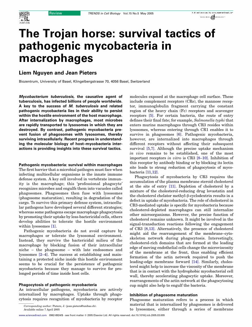
Box 1. Proteins containing FYVE and PX domains
The FYVE domain is composed of w70 amino acids rich in cysteine
and forms a zinc finger. Its name is derived from the first letters of the
first four proteins found to share the domain (Fab1p, YOTB, Vac1p
and EEA1). This domain binds to PI3P and is required for the
localization of proteins to early-endosomal membranes (Figure I).
The PX domain contains w120 amino acids, with the polyproline
motif PXXP in the middle, and is found primarily in the phagocyte
NADH oxidase (Phox). This domain binds preferentially to PI3P but
can also bind to other phosphoinositides. Many PX-binding proteins
have been reported to have key roles in endosomal trafficking and
protein sorting.
FYVE and PX domain proteins have essential roles in endosomal
fusion. It has been proposed that the requirement of PI3P for these
proteins to bind to the endosomal membrane enables their selective
recruitment to early endosomes because PI3P is present exclusively
on early-endosomal membranes. These proteins, in turn, regulate
the trafficking and sorting of select hydrolases from the trans-Golgi
network to endosomes.
FYVE FYVEZn Zn
PI3PPI3P
Hydrocarboncore
Interface
Figure I. Binding of dimeric Hrs protein to membranes through its interactions
with PI3P. FYVE domains are shown in green; PI3P is shown in white and red;
and phosphate groups are shown in red and yellow.White spheres indicate zinc
molecules. Reproduced, with permission, from Ref. [32].
Review TRENDS in Cell Biology Vol.15 No.5 May 2005270
budding and fusion steps involving vesicular transport orthrough a gradual acquisition of lysosomal markermolecules [15,16]. Pathogenic mycobacteria use severalstrategies to circumvent such lysosomal delivery, and themolecules that are involved in these processes are of bothhost and mycobacterial origin.
Role of host proteins
Cholesterol is essential for association of the host proteincoronin 1, also known as P57 or tryptophan-aspartate-containing coat protein (TACO), to mycobacterial phago-somes [11,17]. Coronin 1 was identified in a proteomicsearch for host proteins that are involved in prevention ofphagosome maturation. This 50-kDa protein contains anN-terminal tryptophan aspartate 40 (WD40)-repeatdomain and a coiled-coil sequence of 30 amino acidsat the C terminus. It belongs to a conserved family ofWD-repeat actin-binding proteins that is found predomin-antly in eukaryotes [18]. Whereas some coronins areexpressed ubiquitously (coronins 2, 3 and 7), others areexpressed in a tissue-specific fashion; coronin 2SE (or pp66)is expressed in epithelial tissues, coronins 4 and 5 areexpressed in the brain, and coronin 1 is expressedexclusively in leukocytes [17,18].
Members of the coronin protein family have beenshown to function in actin-cytoskeleton remodeling,phagocytosis, cell division and polarity, and vesiculartrafficking from the endoplasmic reticulum to the Golgi[19]. In murine macrophages, coronin 1 is retained exclu-sively on phagosomes of macrophages infected with livemycobacteria. In macrophages infected with dead myco-bacteria, coronin 1 rapidly dissociates from phagosomes,followed by fusion of the non-coated phagosome tolysosomes and the subsequent degradation of the myco-bacteria, which suggests an essential role for coronin 1 inprotection of the mycobacterial phagosome from fusion tolysosomes [17,20]. Interestingly, in macrophages that aregenerated by activation with phorbol esters, interferon(IFN)-g or human serum, coronin 1 is retained onlypartially, which is consistent with the majority of themycobacteria internalized by these cells being transferredto lysosomal organelles [21]. Kupffer cells, the majormacrophages in the liver, do not express coronin 1 and candestroy live mycobacteria within lysosomes effectively.Thus, the ability of pathogenic mycobacteria to recruithost components to protect the mycobacterial habitat fromlysosomal fusion is a perfect example of how pathogenicmicroorganisms have evolved to live inside host cells thatare designed to eliminate them.
Role of host phosphoinositides
Phagosome maturation requires the biosynthesis ofphosphatidylinositol 3-phosphate (PI3P) [22]. This phos-phoinositide is a membrane-trafficking regulatory lipidessential for phagolysosome biosynthesis. PI3P is syn-thesized by PI3-kinase on early-endosomal and phago-somal membranes and was observed to move on thephagosomal membrane in temporally scheduled waves[23,24]. PI3P functions as a ligand for many proteinscontaining FYVE or phox homology (PX) domains thatmight be involved in phagosome maturation [25,26]
www.sciencedirect.com
(Box 1). An example of a PI3P-binding protein ishepatocyte growth factor (HGF)-regulated tyrosine kinasesubstrate (Hrs), which has a role in the regulation oftraffic in the endosomal pathway [27–30]. Depletion of Hrscauses arrest of phagosome maturation, and Hrs associ-ates with phagosomes before fusion to late endosomes orlysosomes [31]. This association requires the interactionof the FYVE domain of Hrs with phagosomal PI3P [32](Box 1). Interestingly, when pathogenic mycobacteriareside within phagosomes, Hrs dissociates [31], possiblybecause of the ability of pathogenic mycobacteria to alterthe wave of PI3P [24].
PI3P is also essential for the elevation of Ca2C
concentration in the cytosol that stimulates the activityof PI3-kinase. This kinase, in turn, raises the concen-tration of PI3P on the phagosomal membrane, whichmodulates phagosomematuration [33]. This feedback-loopcircuit seems to be targeted by pathogenic mycobacteria toprevent the fusion of phagosomes to late endosomes andlysosomes.
Role of the mycobacterial cell wall
Both host-cell and mycobacterial lipids are known tomodulate the intracellular fate of pathogenic myco-bacteria [34,35]. Notably, the lipid-rich cell wall ofmycobacteria has been recognized as providing the bacilli

Review TRENDS in Cell Biology Vol.15 No.5 May 2005 271
with an effective innate immune defense against lyso-somal degradation [34,36]. TheMycobacterium tuberculosisPI3P analog glycosylated phosphatidylinositol lipoarabi-nomannan (ManLAM) has been reported to block phago-some maturation [37]. Phagosomes containing latex beadscovered with ManLAM acquire features of mycobacterialphagosomes, including inhibition of phagosomal acidifica-tion and fission of late-endosomal markers. ManLAMprobably blocks a PI3P-dependent pathway involved inthe transport of cargo between the trans-Golgi networkand phagosomes: a transport step required for phagolyso-some biogenesis [37,38]. The rise in Ca2C concentration inthe macrophage cytosol required for accumulation ofPI3P on the phagosomal membrane is also inhibited byManLAM [39]. ManLAM might exert these activities bycompeting with PI3P for binding to proteins containingFYVE and PX domains, thereby preventing their recruit-ment to phagosomes. Alternatively, ManLAM mightinterfere with the waves of PI3P on the phagosomalmembrane [24].
In contrast to the inhibitory effect of ManLAM onphagosome–lysosome fusion, phosphatidylinositol manno-side (PIM), another mycobacterial cell-wall component,enhances the fusion of phagosomes to early endosomes[40]. Although the molecular partners of the different lipidmoieties that modulate fusion events within the phago-some–lysosome pathway remain to be established, thesemycobacterial cell-wall components could ensure a safeintracellular haven while continuing to provide nutrientsto resident mycobacteria.
Role of macrophage activation
Considering the multiple ways in which mycobacteria canremain viable inside macrophage phagosomes, how canthese pathogens be destroyed? One answer to thisquestion lies in host innate immune defense mechanismsthat come into operation after activation of macrophages.One of the most important macrophage-activating mol-ecules involved in combating pathogenic mycobacteria isIFN-g, which is secreted by activated T cells and naturalkiller cells during infection [41,42]. Bothmice and humanswith genetic defects in IFN-g signaling are extremelysusceptible to mycobacterial diseases [43,44]. Manycellular processes such as antigen presentation, leuko-cyte–endothelium cell interactions, cell growth and apop-tosis, and phagosome–lysosome fusion can be modulatedthrough the activity of IFN-g [45–47]. The molecularmechanisms involved in such modulation are, however,largely unknown [48]. Recently, the IFN-g-induced hostguanosine triphosphatase protein LRG-47 was implicatedin the regulation of phagosome maturation; LRG-47associates with phagosomes to promote maturation.Interestingly, LRG-47-depleted macrophages activatedby IFN-g fail to kill infected bacteria because of theirimpaired maturation. Consequently, mice lacking LRG-47cannot control mycobacterial replication, confirming theimportance of phagosome maturation in host control oftuberculosis [49].
In addition to modulation of the adaptive immuneresponse, macrophage activation can be mediated throughactivities of the toll-like receptors (TLRs), of which TLR-2
www.sciencedirect.com
and TLR-4 have key roles [50,51]. Many mycobacterialcell-wall constituents such as PIM and the non-cappedlipoarabinomannan (AraLAM), and triacetylated peptidesfrom the mycobacterial 19- and 33-kDa lipoproteins havebeen shown to activate TLRs [50,51]. TLR-2, probably incoordination with TLR-1 and TLR-6, recognizes soluble,heat-stable mycobacteria-derived material, whereasTLR-4 recognizes heat-sensitive cell-associated com-ponents [52]. Activation of TLRs leads to the activationof mitogen-activated protein kinases (MAPKs) [53]. Thesekinases initiate signaling cascades to activate transcrip-tion factors, leading to synthesis of the cytokine tumornecrosis factor (TNF)-a, chemokines and other inflamma-tory mediators, in addition to a burst of nitric oxideproduction [54–56]. In turn, these effectors modulate theantibacterial and inflammatory immune responses ofmacrophages, including accelerated phagosome matu-ration [57]. These pathways are likely to be exploited bypathogenic mycobacteria to promote mycobacterial survi-val inside macrophages [56] and, indeed, ManLAM fromM. tuberculosis inhibits the activation of MAPK in humanmonocytes [58].
Mycobacterial protein kinase G
In addition to cell-wall lipids, mycobacterial proteins areinvolved in the arrest of phagosome maturation [59,60]. Acomprehensive screen revealed several proteins that arepotentially involved in this process. Disruption of theseproteins led to defects in preventing acidification ofphagosomes and to attenuation of intracellular survival[59,60]. An independent search for mycobacterial proteinsinvolved in signal-transduction reactions with potential tomodulate phagosome–lysosome fusion suggested thatmycobacterial kinase (or kinases) might be involved inthis modulation. Treatment of Mycobacterium bovisBacillus Calmette–Guerin (BCG) with a eukaryotic pro-tein kinase Ca (PKCa) inhibitor before infection ofmacrophages leads to immediate transport of the myco-bacteria to lysosomes, suggesting the involvement of aPKCa-like kinase activity in blocking lysosomal delivery(Figure 1). Interestingly, 11 different eukaryotic-likeprotein kinases are encoded by pathogenic mycobacteria[61,62], and a ClustalW (http://www.ebi.ac.uk/clustalw/)alignment of these kinases with mammalian PKCarevealed closest homology to mycobacterial protein kinaseG (PknG). Whereas most mycobacterial kinases arepredicted to be transmembrane proteins with an intra-cellular kinase domain, two of these kinases – PknG andprotein kinase K – lack such a transmembrane segment.Interestingly, PknG is retained in the downsized genomeof the obligate intracellular pathogen Mycobacteriumleprae that carries the minimum set of mycobacterialgenes necessary for intracellular survival. This suggeststhat PknG provides an important function for mycobac-terial survival in the host [63].
To analyze its role in mycobacterial survival inside hostcells, PknG was disrupted in M. bovis BCG using phage-mediated homologous recombination. The resultingM. bovis BCG pknG-deleted mutant was immediatelytransferred to lysosomes, and the mutant mycobacteriacould not survive inside macrophages [59] (Figure 2).

(a) (b)
Figure 1. Involvement of kinase activity in lysosomal delivery of pathogenic mycobacteria. (a) Live untreated Mycobacterium bovis BCG. (b) Effect of the PKCa inhibitor
chelerythrine on lysosomal delivery of M. bovis BCG. Incubation of mycobacteria [expressing green fluorescent protein (GFP)] with chelerythrine leads to enhanced
lysosomal delivery comparedwith that in (a). Lysosomeswere visualized using antibodies against the lysosomalmarker lysosomal-associatedmembrane protein 1 (LAMP1),
followed by secondary antibodies coupled to Alexa Fluor 568.
Review TRENDS in Cell Biology Vol.15 No.5 May 2005272
Subcellular-localization studies suggest that PknG ispresent in the phagosomal lumen, in addition to in thecytosol of infected macrophages, suggesting that patho-genic mycobacteria actively secrete PknG. These datasuggest that PknG is a mycobacterial virulence factor thatis delivered into the cytosol of host cells and mightinterfere with the regulation of phagosome–lysosometransfer (Figure 3).
Notably, PknG lacks signal sequences required for itstranslocation by the classical Sec secretion system (Box 2).However, many pathogenic microbes deliver virulencefactors into host cells by specialized secretion mechanisms[64]. The type III secretion system (Box 2), derived fromthe ancestral flagellum, is used by many Gram-negativepathogenic bacteria to transfer into host cells effectorsthat, in turn, prevent the host response to infection by
LAMP–568
M. bovis BCG
M. bovis BCG ∆pknG
Figure 2. Effect of PknG deletion on mycobacterial intracellular trafficking. Bone marrow
pknG-deletedmutant (DpknG) expressing GFP for one hour, followed by a two-hour chas
the lysosomalmarker LAMP1, followed by secondary antibodies coupled to Alexa Fluor 5
colocalized mostly within LAMP1-positive compartments. Scale barZ10 mm. Reproduce
www.sciencedirect.com
inhibiting signaling pathways [65]. Conjugation systems,which were designed for genetic exchange, have also beenexploited by Gram-negative bacteria to deliver virulencefactors into hosts [66]. Similarly, the Gram-positivepathogen Streptococcus pyogenes has been shown todeliver its toxin inside the host through a cytolysin-mediated process [67]. Also, pathogenic mycobacteriapossess specialized secretion systems required for theirvirulence, such as proteins encoded in the RD1 (region ofdeletion in M. bovis BCG) region of the M. tuberculosisgenome. These proteins comprise a secretion apparatusthat is essential for the secretion of the major T-cellantigenic proteins ESAT-6 and CFP-10, which have nodetectable signal sequence [68,69]. In the non-pathogenicMycobacterium smegmatis, the RD1 proteins form aDNA-conjugation system, which suggests that pathogenic
GFP Merge
macrophages were infected with Mycobacterium bovis BCG or an M. bovis BCG
e. Cells were processed as described in Ref. [59] and stained with antibodies against
68 (LAMP–568). WhereasM. bovis BCG resided in LAMP1-negative vacuoles,DpknG
d, with permission, from Ref. [59]. q (2004) AAAS (http://www.sciencemag.org).

TRENDS in Cell Biology
pknG mutantmycobacteria
Pathogenic mycobacteria
MycobacterialphagosomePhagosomal
vacuole
PknG
Lysosome
Other microbes
Figure 3. Involvement of PknG in mycobacterial trafficking in macrophages. Whereas most microbes and non-pathogenic mycobacteria are rapidly transferred to lysosomes,
pathogenic mycobacteria remain within phagosomes. PknG is secreted by pathogenic mycobacteria into the phagosomal lumen and cytosol to block the fusion of
phagosomes to lysosomes. Deletion of the pknG gene results in rapid lysosomal transfer of the resulting mutant bacteria.
Review TRENDS in Cell Biology Vol.15 No.5 May 2005 273
mycobacteria have adapted an ancestral conjugationsystem for protein secretion [70]. This evolutionaryconservation has also been observed for type IVsecretion systems found in pathogenic Gram-negativebacteria [64]. Whereas the RD1 region is absent fromthe genome of M. bovis BCG, five RD1-homologousregions are distributed among different loci in thegenome of M. tuberculosis [71]. It remains to be
Box 2. Bacterial secretion systems
To perform extracellular activities, bacteria have developed several
systems for secreting their proteins to the surrounding environment.
At least five major protein-secretion systems have been described.
Type I protein-secretion systemA secretion system composed of three proteins forming a channel that
spans the inner membrane, periplasmic space and outer membrane. It
is a one-step continuous secretion. The secretion signal is present in
the C-terminal region of the secreted protein but secretion does not
involve proteolytic processing of secreted proteins. In Escherichia coli,
the most-studied type I secreted protein is a-haemolysin [80,81].
Type II protein-secretion systemA secretion system that employs a two-step mechanism and is
dependent on the Sec machinery. The Sec machinery is composed of
an ATPase, SecA, several integral inner-membrane proteins (SecD,
SecE, SecF, SecG and SecY) and a signal peptidase. First, secreted
proteins pass across the inner membrane by the Sec machinery,
during which the cleavage of a signal sequence at the N terminus
occurs. Subsequently, crossing the outer membrane is mediated by a
dedicated secretion apparatus composed of multiple proteins. The
type II protein-secretion system is exemplified by the secretion of a
starch-hydrolyzing lipoprotein called pullulanase from Klebsiella
oxytoca [82].
Type III protein-secretion systemA system in which secretion occurs in a continuous process without
www.sciencedirect.com
investigated whether or not PknG is secreted throughan RD1-like secretion machinery.
The ability of PknG to block lysosomal deliverysuggests that this kinase might be a valuable target inthe development of compounds that could induce myco-bacterial death inside macrophages. A screen for PknGinhibitors identified a tetrahydrobenzothiophene thatspecifically inhibits the kinase activity of PknG. When
the presence of periplasmic intermediates. This secretion does not
involve proteolytic processing of secreted proteins. The system
involves at least 20 proteins, including cytoplasmic, inner-membrane
and outer-membrane proteins, many of which have homologs
involved in flagellar biogenesis. This secretion system is used by
pathogens to inject their proteins into eukaryotic cells. The type III
secretion system was identified in pathogenic Yersinia spp. for the
secretion of Yop proteins [83].
Type IV protein-secretion system
A secretion system related to the bacterial DNA conjugative transfer
system that enables the transfer of nucleoprotein DNA-conjugation
intermediates or proteins into the extracellular milieu or directly into
host cells. Crossing the outer membrane involves pore formation by
the C-terminal region of the proteins involved. The prototypical type IV
secretion system is that of the Agrobacterium tumefaciens nucleo-
protein tDNA-transfer system [84].
Type V protein-secretion system
A secretion system also referred to as the auto-transporter system. The
auto-transporter proteins have a tripartite unifying structure: the
N-terminal leader sequence, the secreted mature protein (passenger
domain) and a C-terminal domain that forms a b-barrel pore to enable
secretion of the passenger protein. Proteins secreted in thismanner are
implicated as putative virulence factors in many pathogens. The first
type V protein-secretion system to be described was that of gonococcal
immunoglobulin A1 protease [85].

Review TRENDS in Cell Biology Vol.15 No.5 May 2005274
added to infected macrophages, this compound inducesthe fusion of phagosomes to lysosomes and mediates thekilling of mycobacteria inside macrophages [59]. Onepossible advantage of targeting PknG is that this willenable the macrophage to carry out its natural anti-bacterial activity of redirecting intracellularly survivingmycobacteria to lysosomes. Also, by targeting a secretedmolecule such as PknG, transport of antimycobacterialcompounds through the extremely impermeable myco-bacterial cell wall can be circumvented, greatly improvingthe accessibility of the compounds to their target.
The downstream targets of PknG are currentlyunknown. It is, however, interesting to note that mam-malian PKCa, besides being associated with phagosomalmembranes itself, modulates the association of p57, thehuman homolog of coronin 1, to phagosomes [72,73].Considering the importance of PKCa in membrane-trafficking events and its possible involvement in phago-some maturation, PknG might modulate signalingpathways involved in phagolysosome biogenesis by com-peting with host PKCa for substrate binding.
Mycobacterial resistance to destruction
Lysosomal delivery and subsequent degradation by resi-dent hydrolytic enzymes do not constitute the onlymechanism for degrading internalized microbes. Reduc-tion in pH and increase in the local concentrations ofreactive oxygen and nitrogen species such as nitric oxidethat accompany lysosomal cargo delivery also aid microbedegradation. In macrophages, nitric oxide is generated byinducible nitric oxide synthase (iNOS), which is a cytosolicenzyme that catalyzes the conversion of L-arginine toL-citrulline and nitric oxide. Nitric oxide is extremelybactericidal [74], and mice lacking iNOS succumb rapidlyto M. tuberculosis [75]. Not surprisingly, pathogenicmycobacteria have evolved ways of countering thedestructive effects of nitric oxide and preventing theassembly of iNOS with phagosomes, possibly throughrearrangement of the actin cytoskeleton, and this mightlead to a reduction in the local concentration of nitricoxide [76].
Unexpectedly, potential damage by nitric oxide can alsobe countered by the mycobacterial proteasome [77]. Theproteasome is a multisubunit molecular machine that ishighly conserved from archaebacteria to humans and isresponsible for the proteolysis of cytoplasmic proteins.Whereas the proteasome in eukaryotes is involved inmultiple pathways such as the cell cycle, development anddifferentiation, immune and inflammatory responses, andsignal transduction, the function of the proteasome inprokaryotes is less clear [78]. M. tuberculosis has adaptedthe proteasome machinery to protect itself from the killingeffect of nitric oxide [77,79]. Deletion of genes that encodeproteins involved in the formation of the proteasomecauses hypersensitivity of the bacilli to nitric oxide. Itremains to be elucidated whether protease activity isinvolved in the resistance to reactive nitrogen intermedi-ates; because the precise nitric-oxide-mediated bacteri-cidal mechanisms are unknown, the demonstration thatdeletion of a putative proteasome subunit attenuatesvirulent M. tuberculosis might, therefore, provide clues to
www.sciencedirect.com
the destruction mechanisms involved in nitric-oxide-induced mycobacterial cell death.
Concluding remarks
Interactions between pathogenic mycobacteria andmacro-phages represent an interesting example of the co-evolution of infectious microbial agents with their hosts.Mycobacteria, probably evolved from saprophytes in soil tobecome pathogens, have gained several mechanisms topersist inside macrophages. Besides directing mycobac-terial entry into macrophages, these tactics include signal-transduction-mediated modulation of fusion–fission reac-tions to ensure residence within the safe haven of themycobacterial phagosome.
It is becoming clear that, for a full understanding ofthese host-evasion strategies, a better understanding ofmycobacterial physiology is needed, both inside andoutside host macrophages. Knowledge of the mycobacter-ial tactics for survival inside macrophages might help notonly to control tuberculosis and other mycobacterialdiseases but also to uncover novel aspects of host cellbiology.
Acknowledgements
We thank Giorgio Ferrari for expert technical assistance, and Guy R.Cornelis for critical comments about the manuscript. Current support isprovided by grants from the Swiss National Science Foundation and theWorld Health Organization to J.P.
References
1 Finlay, B.B. and Cossart, P. (1997) Exploitation of mammalian hostcell functions by bacterial pathogens. Science 276, 718–725
2 Armstrong, J.A. and Hart, P.D.A. (1971) Response of culturedmacrophages to Mycobacterium tuberculosis, with observations onfusion of lysosomes with phagosomes. J. Exp. Med. 134, 713–740
3 Mwandumba, H.C. et al. (2004)Mycobacterium tuberculosis resides innonacidified vacuoles in endocytically competent alveolar macro-phages from patients with tuberculosis and HIV infection.J. Immunol. 172, 4592–4598
4 Russell, D.G. (2001)Mycobacterium tuberculosis: here today, and heretomorrow. Nat. Rev. Mol. Cell Biol. 2, 569–577
5 Ernst, J.D. (1998) Macrophage receptors for Mycobacterium tubercu-losis. Infect. Immun. 66, 1277–1281
6 Meresse, S. et al. (1999) Controlling the maturation of pathogen-containing vacuoles: a matter of life and death. Nat. Cell Biol. 1,E183–E188
7 Zimmerli, S. et al. (1996) Selective receptor blockade duringphagocytosis does not alter the survival and growth ofMycobacteriumtuberculosis in human macrophages. Am. J. Respir. Cell Mol. Biol. 15,760–770
8 Peyron, P. et al. (2000) Nonopsonic phagocytosis of Mycobacteriumkansasii by human neutrophils depends on cholesterol and ismediated by CR3 associated with glycosylphosphatidylinositol-anchored proteins. J. Immunol. 165, 5186–5191
9 Velasco-Velazquez, M.A. et al. (2003) Macrophage–Mycobacteriumtuberculosis interactions: role of complement receptor 3. Microb.Pathog. 35, 125–131
10 Ferguson, J.S. et al. (2004) Complement protein C3 binding toMycobacterium tuberculosis is initiated by the classical pathway inhuman bronchoalveolar lavage fluid. Infect. Immun. 72, 2564–2573
11 Gatfield, J. and Pieters, J. (2000) Essential role for cholesterol in entryof mycobacteria in macrophages. Science 288, 1647–1650
12 Cywes, C. et al. (1997) Nonopsonic binding of Mycobacteriumtuberculosis to complement receptor type 3 is mediated by capsularpolysaccharides and is strain dependent. Infect. Immun. 65, 4258–4266
13 Gatfield, J. et al. (2000) Immunity against extracellular pathogens.Protoplasma 210, 99–107

Review TRENDS in Cell Biology Vol.15 No.5 May 2005 275
14 Vasanji, A. et al. (2004) Polarization of plasma membrane micro-viscosity during endothelial cell migration. Dev. Cell 6, 29–41
15 Murphy, R.F. (1991) Maturation models for endosome and lysosomebiogenesis. Trends Cell Biol. 1, 77–82
16 Griffiths, G. and Gruenberg, J. (1991) The arguments for pre-existingearly and late endosomes. Trends Cell Biol. 1, 5–9
17 Ferrari, G. et al. (1999) A coat protein on phagosomes involved in theintracellular survival of mycobacteria. Cell 97, 435–447
18 de Hostos, E.L. (1999) The coronin family of actin-associated proteins.Trends Cell Biol. 9, 345–350
19 Rybakin, V. et al. (2004) Coronin 7, themammalian POD-1 homologue,localizes to the Golgi apparatus. FEBS Lett. 573, 161–167
20 Tailleux, L. et al. (2003) Constrained intracellular survival ofMycobacterium tuberculosis in human dendritic cells. J. Immunol.170, 1939–1948
21 Schuller, S. et al. (2001) Coronin is involved in uptake of Mycobacter-ium bovis BCG in human macrophages but not in phagosomemaintenance. Cell. Microbiol. 3, 785–793
22 Roth, M.G. (2004) Phosphoinositides in constitutive membrane traffic.Physiol. Rev. 84, 699–730
23 Wurmser, A.E. et al. (1999) Phosphoinositide 3-kinases and theirFYVE domain-containing effectors as regulators of vacuolar/lysosomalmembrane trafficking pathways. J. Biol. Chem. 274, 9129–9132
24 Chua, J. and Deretic, V. (2004) Mycobacterium tuberculosis repro-grams waves of phosphatidylinositol 3-phosphate on phagosomalorganelles. J. Biol. Chem. 279, 36982–36992
25 Itoh, T. and Takenawa, T. (2002) Phosphoinositide-binding domains:functional units for temporal and spatial regulation of intracellularsignalling. Cell. Signal. 14, 733–743
26 Birkeland, H.C. and Stenmark, H. (2004) Protein targeting toendosomes and phagosomes via FYVE and PX domains. Curr. Top.Microbiol. Immunol. 282, 89–115
27 Komada, M. and Soriano, P. (1999) Hrs, a FYVE finger proteinlocalized to early endosomes, is implicated in vesicular traffic andrequired for ventral folding morphogenesis.Genes Dev. 13, 1475–1485
28 Chin, L.S. et al. (2001) Hrs interacts with sorting nexin 1 andregulates degradation of epidermal growth factor receptor. J. Biol.Chem. 276, 7069–7078
29 Raiborg, C. and Stenmark, H. (2002) Hrs and endocytic sorting ofubiquitinated membrane proteins. Cell Struct. Funct. 27, 403–408
30 Raiborg, C. et al. (2002) Hrs sorts ubiquitinated proteins into clathrin-coated microdomains of early endosomes. Nat. Cell Biol. 4, 394–398
31 Vieira, O.V. et al. (2004) Acquisition of Hrs, an essential component ofphagosomal maturation, is impaired by mycobacteria. Mol. Cell. Biol.24, 4593–4604
32 Mao, Y. et al. (2000) Crystal structure of the VHS and FYVE tandemdomains of Hrs, a protein involved in membrane trafficking and signaltransduction. Cell 100, 447–456
33 Fratti, R.A. et al. (2001) Role of phosphatidylinositol 3-kinase andRab5 effectors in phagosomal biogenesis and mycobacterial phago-some maturation arrest. J. Cell Biol. 154, 631–644
34 Glickman, M.S. et al. (2000) A novel mycolic acid cyclopropanesynthetase is required for cording, persistence, and virulence ofMycobacterium tuberculosis. Mol. Cell 5, 717–727
35 Anes, E. et al. (2003) Selected lipids activate phagosome actinassembly and maturation resulting in killing of pathogenic mycobac-teria. Nat. Cell Biol. 5, 793–802
36 Briken, V. et al. (2004) Mycobacterial lipoarabinomannan and relatedlipoglycans: from biogenesis to modulation of the immune response.Mol. Microbiol. 53, 391–403
37 Fratti, R.A. et al. (2003) Mycobacterium tuberculosis glycosylatedphosphatidylinositol causes phagosome maturation arrest. Proc. Natl.Acad. Sci. U. S. A. 100, 5437–5442
38 Sturgill-Koszycki, S. et al. (1996) Mycobacterium-containing phago-somes are accessible to early endosomes and reflect a transitionalstate in normal phagosome biogenesis. EMBO J. 15, 6960–6968
39 Vergne, I. et al. (2003) Tuberculosis toxin blocking phagosomematuration inhibits a novel Ca2C/calmodulin-PI3K hVPS34 cascade.J. Exp. Med. 198, 653–659
40 Vergne, I. et al. (2004) Mycobacterium tuberculosis phagosomematuration arrest: mycobacterial phosphatidylinositol analog phos-phatidylinositol mannoside stimulates early endosomal fusion. Mol.Biol. Cell 15, 751–760
www.sciencedirect.com
41 Adams, D.O. and Hamilton, T.A. (1992) Molecular basis of macro-phage activation: diversity and origin. In The Macrophage(Lewis, C.E. and McGee, J.O.D., eds), pp. 75–114, Oxford UniversityPress
42 Stout, R.D. (1993) Macrophage activation by T cells: cognate and non-cognate signals. Curr. Opin. Immunol. 5, 398–403
43 Huang, S. et al. (1993) Immune response in mice that lack theinterferon-g receptor. Science 259, 1742–1745
44 Jouanguy, E. et al. (1997) Partial interferon-g receptor 1 deficiency ina child with tuberculoid bacillus Calmette-Guerin infection and asibling with clinical tuberculosis. J. Clin. Invest. 100, 2658–2664
45 Boehm, U. et al. (1997) Cellular responses to interferon-g. Annu. Rev.Immunol. 15, 749–795
46 Via, L.E. et al. (1998) Effects of cytokines on mycobacterial phagosomematuration. J. Cell Sci. 111, 897–905
47 Montaner, L.J. et al. (1999) Type 1 and type 2 cytokine regulation ofmacrophage endocytosis: differential activation by IL-4/IL-13 asopposed to IFN-g or IL-10. J. Immunol. 162, 4606–4613
48 Cosma, C.L. et al. (2003) The secret lives of the pathogenicmycobacteria. Annu. Rev. Microbiol. 57, 641–676
49 MacMicking, J.D. et al. (2003) Immune control of tuberculosis byIFN-g-inducible LRG-47. Science 302, 654–659
50 Krutzik, S.R. and Modlin, R.L. (2004) The role of Toll-like receptors incombating mycobacteria. Semin. Immunol. 16, 35–41
51 Quesniaux, V. et al. (2004) Toll-like receptor pathways in the immuneresponses to mycobacteria. Microbes Infect. 6, 946–959
52 Means, T.K. et al. (1999) Human toll-like receptors mediate cellularactivation byMycobacterium tuberculosis. J. Immunol. 163, 3920–3927
53 Guha, M. and Mackman, N. (2001) LPS induction of gene expressionin human monocytes. Cell. Signal. 13, 85–94
54 Chan, E.D. et al. (1999) Systematic evaluation of the mitogen-activated protein kinases in the induction of iNOS by tumor necrosisfactor-a and interferon-g. Chest 116(Suppl. 1), S91–S92
55 Chen, C. et al. (1999) Involvement of p38 mitogen-activated proteinkinase in lipopolysaccharide-induced iNOS and COX-2 expression inJ774 macrophages. Immunology 97, 124–129
56 Schorey, J.S. and Cooper, A.M. (2003) Macrophage signalling uponmycobacterial infection: the MAP kinases lead the way. Cell.Microbiol. 5, 133–142
57 Schaible, U.E. et al. (1998) Cytokine activation leads to acidificationand increases maturation of Mycobacterium avium-containing phago-somes in murine macrophages. J. Immunol. 160, 1290–1296
58 Knutson, K.L. et al. (1998) Lipoarabinomannan of Mycobacteriumtuberculosis promotes protein tyrosine dephosphorylation and inhi-bition of mitogen-activated protein kinase in human mononuclearphagocytes. Role of the Src homology 2 containing tyrosine phospha-tase 1. J. Biol. Chem. 273, 645–652
59 Walburger, A. et al. (2004) Protein kinase G from pathogenicmycobacteria promotes survival within macrophages. Science 304,1800–1804
60 Pethe, K. et al. (2004) Isolation of Mycobacterium tuberculosismutants defective in the arrest of phagosome maturation. Proc.Natl. Acad. Sci. U. S. A. 101, 13642–13647
61 Cole, S.T. et al. (1998) Deciphering the biology of Mycobacteriumtuberculosis from the complete genome sequence.Nature 393, 537–544
62 Av-Gay, Y. and Everett, M. (2000) The eukaryotic-like Ser/Thr proteinkinases of Mycobacterium tuberculosis. Trends Microbiol. 8, 238–244
63 Cole, S.T. et al. (2001) Massive gene decay in the leprosy bacillus.Nature 409, 1007–1011
64 Yeo, H.J. andWaksman, G. (2004) Unveiling molecular scaffolds of thetype IV secretion system. J. Bacteriol. 186, 1919–1926
65 Cornelis, G.R. (2002) Yersinia type III secretion: send in the effectors.J. Cell Biol. 158, 401–408
66 Cascales, E. and Christie, P.J. (2003) The versatile bacterial type IVsecretion systems. Nat. Rev. Microbiol. 1, 137–149
67 Madden, J.C. et al. (2001) Cytolysin-mediated translocation (CMT): afunctional equivalent of type III secretion in Gram-positive bacteria.Cell 104, 143–152
68 Stanley, S.A. et al. (2003) Acute infection and macrophage subversionbyMycobacterium tuberculosis require a specialized secretion system.Proc. Natl. Acad. Sci. U. S. A. 100, 13001–13006
69 Pym, A.S. et al. (2003) Recombinant BCG exporting ESAT-6 confersenhanced protection against tuberculosis. Nat. Med. 9, 533–539

Review TRENDS in Cell Biology Vol.15 No.5 May 2005276
70 Flint, J.L. et al. (2004) The RD1 virulence locus of Mycobacteriumtuberculosis regulates DNA transfer in Mycobacterium smegmatis.Proc. Natl. Acad. Sci. U. S. A. 101, 12598–12603
71 Gey Van Pittius, N.C. et al. (2001) The ESAT-6 gene cluster ofMycobacterium tuberculosis and other high GCC Gram-positivebacteria. Genome Biol 2, RESEARCH0044
72 Itoh, S. et al. (2002) The role of protein kinase C in the transientassociation of p57, a coronin family actin-binding protein, withphagosomes. Biol. Pharm. Bull. 25, 837–844
73 Ng Yan Hing, J.D. et al. (2004) Proteomic analysis reveals a role forprotein kinase C-a in phagosome maturation. Biochem. Biophys. Res.Commun. 319, 810–816
74 MacMicking, J. et al. (1997) Nitric oxide and macrophage function.Annu. Rev. Immunol. 15, 323–350
75 MacMicking, J.D. et al. (1997) Identification of nitric oxide synthase asa protective locus against tuberculosis. Proc. Natl. Acad. Sci. U. S. A.94, 5243–5248
76 Miller, B.H. et al. (2004) Mycobacteria inhibit nitric oxide synthaserecruitment to phagosomes during macrophage infection. Infect.Immun. 72, 2872–2878
77 Darwin, K.H. et al. (2003) The proteasome of Mycobacteriumtuberculosis is required for resistance to nitric oxide. Science 302,
Five things you might no
1.
Elsevier is a founder member of the WHO’s HINARI and AGORA ini
access to scientific literature. More than 1000 journals, including the T
at significantly re
2.
The online archive of Elsevier’s premier Cell Press journal collection w
recent archive, including Cell,Neuron, Immunity and Current Biology
sites 12 months after artic
3.
Have you contributed to anElsevier journal, book or series?Did you kn
stand-alone CDs when ordered directly from us?
+1 800 782 4927 (US) or +1 800 460 3110
or +44 1865 474 010 (
4.
Elsevier has a long tradition of liberal copyright policies and formany y
and the posting of final papers on internal servers. Now, Elsevier has
the final text version of their papers on both their person
5.
The Elsevier Foundation is a knowledge-centered foundationmaking
culturally rich global organization, the Foundation has funded, for ex
Philadelphia, provided storybooks to children in Cape Town, sponsor
Brigham and Women’s Hospital and given funding to the 3rd Intern
www.sciencedirect.com
1963–196678 Baumeister, W. et al. (1998) The proteasome: paradigm of a self-
compartmentalizing protease. Cell 92, 367–38079 Pieters, J. and Ploegh, H. (2003) Chemical warfare and mycobacterial
defense. Science 302, 1900–190280 Thanabalu, T. et al. (1998) Substrate-induced assembly of a contigu-
ous channel for protein export from E. coli: reversible bridging of an
inner-membrane translocase to an outer membrane exit pore. EMBO
J. 17, 6487–649681 Gentschev, I. et al. (2002) The E. coli a-hemolysin secretion system
and its use in vaccine development. Trends Microbiol. 10, 39–4582 Takizawa, N. and Murooka, Y. (1985) Cloning of the pullulanase gene
and overproduction of pullulanase in Escherichia coli and Klebsiella
aerogenes. Appl. Environ. Microbiol. 49, 294–29883 Michiels, T. et al. (1990) Secretion of Yop proteins by Yersiniae. Infect.
Immun. 58, 2840–284984 Koukolikova-Nicola, Z. and Hohn, B. (1993) How does the T-DNA of
Agrobacterium tumefaciens find its way into the plant cell nucleus?
Biochimie 75, 635–63885 Pohlner, J. et al. (1987) Gene structure and extracellular secretion of
Neisseria gonorrhoeae IgA protease. Nature 325, 458–462
t know about Elsevier
tiatives, which enable the world’s poorest countries to gain free
rends and Current Opinion collections, will be available for free or
duced prices.
ill become freely available from January 2005. Free access to the
, will be available on both ScienceDirect and the Cell Press journal
les are first published.
ow that all our authors are entitled to a 30%discount on books and
For more information, call our sales offices:
(Canada, South & Central America)
rest of the world)
ears has permitted both the posting of preprints on public servers
extended its author posting policy to allow authors to freely post
al websites and institutional repositories or websites.
grants and contributions throughout the world. A reflection of our
ample, the setting up of a video library to educate for children in
ed the creation of the Stanley L. Robbins Visiting Professorship at
ational Conference on Children’s Health and the Environment.