the surgical management - orion group · the surgical management of trigeminal schwannomas ... the...
TRANSCRIPT
Case Report
www.orion-group.net/journals The ORION. Vol 32, Issue 2, May 2009 www.orion-group.net/medicaljournal
The surgical management of trigeminal schwannomas Alam S, Khair A, Hassan R, Munir SF, Wakil
Introduction Schwannomas are tumors of the nerve sheath that usually exhibit benign behavior1,5,8. Benign trigeminal schwannoma of the trigeminal nerve comprises only 0.2% to 0.4% of all intracranial tumors and primarily arises in the gasserian ganglion2,9,11,12. Trigeminal schwannomas are benign tumors of schwann cell origin, are relatively rare and much less common than acoustic neuroma5,8,12. Most trigeminal neurinomas irrespective of the site of spread have an association with this region of the nerve5. The tumor grows larger and spreads in the available spaces7. The Meckel's cave can accommodate a large amount of the tumor, which bloats up the cave7. The tumor being soft is unable to open up the dural sheath beyond the ganglion into the roots. This may be the reason that in most of the cases tumor does not extend beyond the dilated cave3,7. The tumor presses the adjacent normal fifth nerve, most of which is clinically involved by direct pressure of the tumor7. In the posterior fossa the trigeminal neurinomas are located intradurally7. The part in proximity to the brain stem is in most cases like any other extra-axial tumor with a well-defined plane of cleavage7,9. In general these tumors involve the adjoining cranial nerves, blood vessels and brain only by displacement and not by invasion7,10. We retrospectively analyzed the clinical profiles of 6 patients who were surgically treated for trigeminal schwannomas. The aim of this study was to
analyze the presenting clinical and radiological features of these tumors, to establish factors that might affect surgical decision-making, to critically evaluate the appropriate surgical route, depending on tumor location and to assess the long-term outcomes after radical tumor resection. Surgical anatomy Recent advances in understanding the microsurgical anatomy of skull base structures are hallmarks of modern neurosurgery6. The trigeminal nerve has an extensive anatomic course2,6. Comprehensive knowledge of trigeminal nerve anatomy facilitates understanding of the relationship between the brainstem, skull base and facial area2. The trigeminal nerve trifurcates into ophthalmic, maxillary and mandibular nerves distal to the trigeminal ganglion2. The ophthalmic nerve passes forward in the lateral wall of the cavernous sinus. It gains access into the orbit via the superior orbital fissure. The ophthalmic nerve then divides to supply sensation to the eyeball, lacrymal glands, conjunctiva, part of the nasal mucosa, skin of the nose, eyelid and forehead3,6. Classification of tumor extension Trigeminal schwannomas may originate from the root, the ganglion or the peripheral branches of the trigeminal nerve8. Jefferson initially divided these tumors into 4 groups depending on their anatomical location: Posterior fossa (Root type), Combined posterior fossa-middle fossa (Dumbbell type), Middle fossa (Ganglion type), and Peripheral (Division type)8. Samii et al13 classified the tumor extension into 4 categories based on radiological findings: Type A- Intracranial tumor predominantly in the middle fossa; Type B- Intracranial tumor predominantly in the posterior fossa; Type C- Intracranial dumbbell-shaped tumor in the middle and posterior fossa; and Type D- Extracranial tumor with intracranial extensions13.
The ORION Medical Journal 2009 May;32(2):659-662
1. Dr. Shamsul Alam, MBBS, MS (Neurosurgery), Assistant Professor, Department of Neurosurgery, BSMMU 2. Dr. Abul Khair, MBBS, FCPS, WHO fellow in Neurosurgery, Associate Professor, Department of Neurosurgery, BSMMU 3. Dr. Rashidul Hassan, MBBS, FCPS Resident, Department of Neurosurgery, BSMMU 4. Dr. SK Farhad Munir, MBBS Resident, Department of Neurosurgery, BSMMU 5. Dr. Wakil, MBBS Resident, Department of Neurosurgery, BSMMU
Case Report
www.orion-group.net/journals The ORION. Vol 32, Issue 2, May 2009 www.orion-group.net/medicaljournal
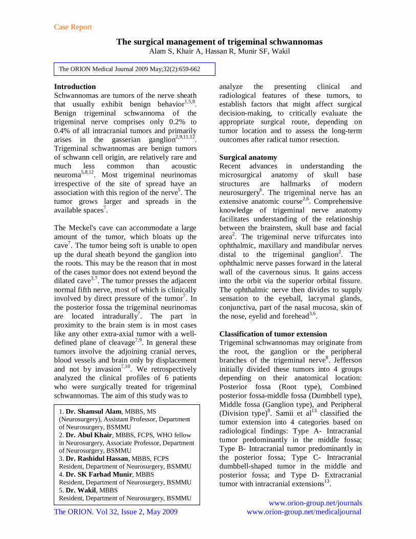
Figure 1, 2 : Shows the direction of growth of trigeminal schwannoma
Methods This series includes 6 patients who were surgically treated between 2005 and 2008 at BSMMU. No patients received a diagnosis of NF2. Results Total tumor excision was possible in 4 patients, whereas total removal were not achieved in 2 patients. The extent of resection was graded according to the surgeons' impressions, confirmed by postoperative imaging in all patients. "Total" resection was defined as complete resection of the tumor and its capsule. "Radical subtotal" was assigned to the resection when tumor capsule fragments remained on vital structures. When the tumor capsule remained in the cavernous sinus or on the brain stem, the resection was graded as "Subtotal"2,3. Out of the six patients one undergone subtotal resection, two patient required a staged procedure with a large dumbbell type lesion. There were no operation related death or mortality.
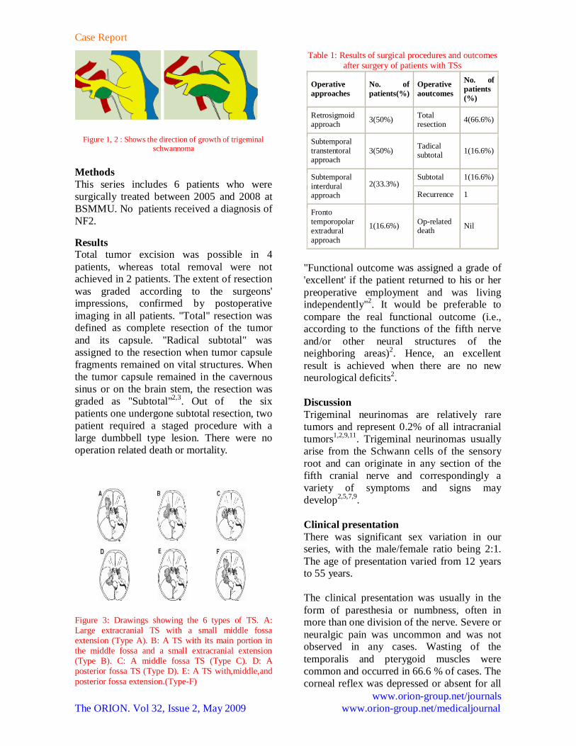
Figure 3: Drawings showing the 6 types of TS. A: Large extracranial TS with a small middle fossa extension (Type A). B: A TS with its main portion in the middle fossa and a small extracranial extension (Type B). C: A middle fossa TS (Type C). D: A posterior fossa TS (Type D). E: A TS with,middle,and posterior fossa extension.(Type-F)
Table 1: Results of surgical procedures and outcomes after surgery of patients with TSs
Operative approaches
No. of patients(%)
Operative aoutcomes
No. of patients (%)
Retrosigmoid approach 3(50%) Total
resection 4(66.6%)
Subtemporal transtentoral approach
3(50%) Tadical subtotal 1(16.6%)
Subtotal 1(16.6%) Subtemporal interdural approach
2(33.3%) Recurrence 1
Fronto temporopolar extradural approach
1(16.6%) Op-related death Nil
"Functional outcome was assigned a grade of 'excellent' if the patient returned to his or her preoperative employment and was living independently"2. It would be preferable to compare the real functional outcome (i.e., according to the functions of the fifth nerve and/or other neural structures of the neighboring areas)2. Hence, an excellent result is achieved when there are no new neurological deficits2. Discussion Trigeminal neurinomas are relatively rare tumors and represent 0.2% of all intracranial tumors1,2,9,11. Trigeminal neurinomas usually arise from the Schwann cells of the sensory root and can originate in any section of the fifth cranial nerve and correspondingly a variety of symptoms and signs may develop2,5,7,9. Clinical presentation There was significant sex variation in our series, with the male/female ratio being 2:1. The age of presentation varied from 12 years to 55 years. The clinical presentation was usually in the form of paresthesia or numbness, often in more than one division of the nerve. Severe or neuralgic pain was uncommon and was not observed in any cases. Wasting of the temporalis and pterygoid muscles were common and occurred in 66.6 % of cases. The corneal reflex was depressed or absent for all
Case Report
www.orion-group.net/journals The ORION. Vol 32, Issue 2, May 2009 www.orion-group.net/medicaljournal
patients. The symptoms of involvement of adjacent cranial nerves in the cavernous sinus and in the cerebellopontine angle have been frequently reported in 2 cases. These symptoms probably because of the large sizes of the tumors encountered in this series. The large tumor size was also responsible for the relatively infrequently encountered symptoms of increased intracranial pressure and ophthalmoscopically demonstrated papilledema, which were observed in 4 cases (66.6%). One patient demonstrated contralateral hemiparesis and pyramidal signs related to severe compression of the brainstem. The unusual symptom of pathological laughter was observed in one case of large, dumbbell-shaped tumors. The clinical features of slowly progressive symptoms and the predominant presence of trigeminal nerve-related symptoms of numbness and muscle wasting are usually diagnostic5,7.
Table 2: Preoperative clinical symptoms in 6 patients
with TSs
Symptom No. of Patients(%) Symptom No. of
Patients(%)
Trigeminal hypesthesia 6(100%) Diplopia 2(33.3%)
Facial hypesthesia 4(66.6%) Ataxia 2(33.3%)
Headache 3(50%) Pathological laughter 1(16.6%)
Hearing symptoms 3(50%)
Seizure Nil
Hemiparesis & increased ICP with papilledema
2(33.3%)
Table 3:Shows the distribution of cases
Case No. Age/sex Presentation Location Name of
operaton Complications Follow up
1. 28/F Headache, facial hypoacesthesia
Middle fossa
Fronto temporopolar extradural approach
No Lost from follow up
2. 55/M Deafness, facial hypoaesthesia, afaxia
Posterior fossa
Stages 1.Petrosigmoid approach 1.Subtemporal transtentorail
No 3 yrs no recurrence
3. 35/M Headache visual blurring Combined
Extended subtmeporal transtential approach
3rd nerve palsy, tinnitus
2yrs norecurrence
4. 25/M
Facial hypoaesthesia, facial asymmetry due to atrophy of muscle
Middle fossa
Subtemporal interdural approach
No 1 yrs no recurrence
5. 12/F
Ataxia,deafness, respiratory distress, dysphagia
Combined
Stages 1.Petrosigmoid approach 1.Subtemporal transtentorail
Facial palsy, exposure keratitis, dysphagia
9 months recurrence
6. 35/M Dysphasia, facial asymmetry
Combined
Extended subtmeporal transtential approach
No no recurrence
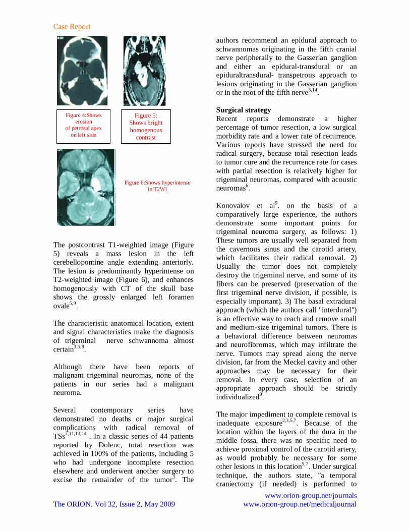
Radiological features Erosion of the petrous apex, as noted on plain x-rays or computed tomographic scans, was uniformly observed for larger tumors, and this finding was of diagnostic significance5,7 (figure 4). Lesions are usually isodense on unenhanced CT but may reveal variable attenuation. There is usually homogenous enhancement with contrast9,11. Because of its multiplanar capability, exquisite anatomic detail and characteristic tissue signal intensity, MRI is helpful in the differential diagnosis of primary tumours of the trigeminal nerve and Meckel's cave and in the evaluation of tumor involvement for preoperative planning7,9. MRI with contrast enhancement is preferable to CT scanning because of multiplanar capability and absence of Hounsfield artifact from the skull base11,14. In addition MRI is sensitive for detection of additional neuromas, which is a consideration in neurofibromatosis (NF2) patients5,6.
Trigeminal schwannomas show homogeneity on T1-weighted images and variable heterogeneity on T2-weighted images with prolongation of T1 and T2 relaxation times5,7. There is usually intense heterogeneous enhancement with gadolinium. MRI signal characteristics are similar to those of acoustic schwannoma; the key to this diagnosis is the neuroanatomic localization along the fifth nerve pathway5,7,11,13.
Case Report
www.orion-group.net/journals The ORION. Vol 32, Issue 2, May 2009 www.orion-group.net/medicaljournal
Figure 6:Shows hyperintense in T2WI
The postcontrast T1-weighted image (Figure 5) reveals a mass lesion in the left cerebellopontine angle extending anteriorly. The lesion is predominantly hyperintense on T2-weighted image (Figure 6), and enhances homogenously with CT of the skull base shows the grossly enlarged left foramen ovale5,9. The characteristic anatomical location, extent and signal characteristics make the diagnosis of trigeminal nerve schwannoma almost certain3,5,8. Although there have been reports of malignant trigeminal neuromas, none of the patients in our series had a malignant neuroma. Several contemporary series have demonstrated no deaths or major surgical complications with radical removal of TSs7,11,13,14 . In a classic series of 44 patients reported by Dolenc, total resection was achieved in 100% of the patients, including 5 who had undergone incomplete resection elsewhere and underwent another surgery to excise the remainder of the tumor3. The
authors recommend an epidural approach to schwannomas originating in the fifth cranial nerve peripherally to the Gasserian ganglion and either an epidural-transdural or an epiduraltransdural- transpetrous approach to lesions originating in the Gasserian ganglion or in the root of the fifth nerve3,14. Surgical strategy Recent reports demonstrate a higher percentage of tumor resection, a low surgical morbidity rate and a lower rate of recurrence. Various reports have stressed the need for radical surgery, because total resection leads to tumor cure and the recurrence rate for cases with partial resection is relatively higher for trigeminal neuromas, compared with acoustic neuromas6. Konovalov et al9. on the basis of a comparatively large experience, the authors demonstrate some important points for trigeminal neuroma surgery, as follows: 1) These tumors are usually well separated from the cavernous sinus and the carotid artery, which facilitates their radical removal. 2) Usually the tumor does not completely destroy the trigeminal nerve, and some of its fibers can be preserved (preservation of the first trigeminal nerve division, if possible, is especially important). 3) The basal extradural approach (which the authors call "interdural") is an effective way to reach and remove small and medium-size trigeminal tumors. There is a behavioral difference between neuromas and neurofibromas, which may infiltrate the nerve. Tumors may spread along the nerve division, far from the Meckel cavity and other approaches may be necessary for their removal. In every case, selection of an appropriate approach should be strictly individualized9. The major impediment to complete removal is inadequate exposure2,3,5,7. Because of the location within the layers of the dura in the middle fossa, there was no specific need to achieve proximal control of the carotid artery, as would probably be necessary for some other lesions in this location5,7. Under surgical technique, the authors state, "a temporal craniectomy (if needed) is performed to
Figure 4:Shows erosion
of petrosal apex on left side
Figure 5: Shows bright homogenous
contrast

Case Report
www.orion-group.net/journals The ORION. Vol 32, Issue 2, May 2009 www.orion-group.net/medicaljournal
obtain a flat viewing angle across the floor of the middle fossa." Naturally, it is always needed, because this provides better access to the parasellar region and does not necessitate any retraction of the brain. Another statement, "If necessary, the middle meningeal artery is ligated to increase exposure of the lateral middle fossa floor"3,5 . Retrosigmoid approach This approach is performed by placing the patient in the dorsal (mastoid) position with the head turned to the opposite side and the ipsilateral shoulder elevated. A linear incision is placed 4 cm behind the external auditory canal. The asterion is exposed to determine the junction of the tranverse and sigmoid sinuses. A craniectomy 4 cm in diameter is performed, with the superior and anterior margins bordering the transverse and sigmoid sinuses, respectively. The dura mater is opened parallel to the sigmoid sinus; CSF is drained from the cerebellomedullary cistern; and CNs VII-XI are identified. The tumor is thereby exposed near the tentorium margin. After intracapsular tumor debulking, microsurgical radical removal is accomplished3. Lateral basal subtemporal approach The skin incision extends from in front of the ear and travels superiorly and posteriorly. The branches of the facial nerve are saved by working deep to the fascial layers. The exposure is centered on the external ear canal. The temporalis muscle is elevated and displaced anteriorly. The basal extension of the exposure was achieved by resection of the roots of the zygomatic arch, roof of the external ear canal and superior third of the mastoid bone (Figure 7). The temporalis muscle was rotated anteriorly and was thus away from the field. The exposure was centered over the external ear canal in line with the petrous apex. The direction of the approach to the tumor was the shortest and perpendicular from the surface and avoided any neural or vascular manipulation. Inclusion of mastoidectomy in the exposure added the advantages of petrosal approach5,7.
Subtemporal transtentorial approach This approach provides a limited view of the underside of the tumor and its relationship to the vessels and cranial nerves below. A better view of the tumor's relationship to cranial nerves VI, VII, and VIII, the vertebrobasilar system and the anteroinferior cerebellar artery is provided via the suboccipital portion of the combined petrosal approach2,3,5,7,14. Combinned (retrosigmoid with subtemporal transtentorial approach) This is the combination of subtemporal and lateral suboccipital approach for dumbbell shaped schwannoma. This approach do not need drilling of petrosal bone and exposure of sigmoid sinus, hence less time consuming, disadvantage is cerebellar retraction2,3. Frontotemporal extradural temporopolar approach Lesions of the orbital-cavernous and ganglion types were approached via an extradural approach to the cavernous sinus. With the head in three-pin fixation, supine, rotated approximately 30 degrees, a pterional skin incision is made. The scalp and temporalis muscle are then reflected anteriorly in one layer. In selected cases, a two-layer scalp flap is fashioned to retract the temporalis muscle inferiorly and posteriorly. This maneuver provides a widened corridor to the anterior middle fossa, necessary in approaching larger tumors. A pterional craniotomy is performed, which typically measures 3 x 5 cm. A temporal craniectomy (if needed) is performed to obtain a flat viewing angle across the floor of the middle fossa. The dura is then elevated from the sphenoid ridge and
Figure 7: Shows extent of craniotomy in lateral
basal subtemporal approach
Figure 8:Shows operative picture of trigeminal
schwonnoma in frontotemparopola
approach
Case Report
www.orion-group.net/journals The ORION. Vol 32, Issue 2, May 2009 www.orion-group.net/medicaljournal
medial frontal fossa. Elevation of the dura of the middle fossa proceeds laterally, to the foramen spinosum. If necessary, the middle meningeal artery is ligated to increase exposure of the lateral middle fossa floor2,3,5,7. Combined petrosal approach Patients with lesions that involve both the cavernous sinus and posterior fossa, the so-called "dumbbell" type, underwent surgical resection via the combined petrosal approach. The patient is placed in the three-quarter lateral position. An "L" shaped craniotomy is made around the ear to expose temporal and retromastoid dura. A mastoidectomy is then performed, preserving the structures of the bony labyrinth. Extradurally, the petrous apex medial to the internal auditory canal is fenestrated to create a window via the middle fossa trajectory to the posterior fossa3,7,14. Conclusion On the basis of our limited experience, we believe that the best treatment for TSs is complete microsurgical removal of the lesion and that this treatment should be considered the gold standard therapeutic modality for the majority of cases. Hypesthesia, to some degree, is common after surgery, at least in the early postoperative period. Bibiloraphy 1. Bhatjiwale MG, Nadkarni TD, Desai KI,
Goel A: Pathological laughter as a presenting symptom of massive trigeminal neuromas: Report of four cases. Neurosurgery 2000;47:469-471.
2. Day JD, Fukushima T: The surgical management of trigeminal neuromas. Neurosurgery 1998;42:233-241.
3. Dolenc VV: Anatomy and Surgery of the Cavernous Sinus. Vienna: Springer-Verlag, 1989.
4. Dolenc VV: Frontotemporal epidural approach to trigeminal neurinomas. Acta Neurochir (Wien) 1994;130:55-65.
5. Goel A, Muzumdar D, Raman C: Trigeminal neuroma: analysis of surgical experience with 73 cases. Neurosurgery 2003;52:783-790.
6. Goel A: Infratemporal fossa interdural approach for trigeminal schwannomas. Acta Neurochir (Wien) 1995;136:99-102.
7. Goel A, Nadkarni T: Basal lateral subtemporal approach for trigeminal schwannomas: Report of an experience with 18 cases. Acta Neurochir (Wien) 1999;141:711-719.
8. Jefferson G: The trigeminal neurinomas with some remarks on malignant invasion of the gasserian ganglion. Clin Neurosurg 1953;1:11-54.
9. Konovalov AN, Spallone A, Mukhamedjanov DJ, Tcherekajev VA, Makhmudov UB: Trigeminal neurinomas. A series of 111 surgical cases from a single institution. Acta Neurochir (Wien) 1996;138:1027-1035.
10. Lang J: Anatomy of the posterior cranial fossa, in Sekhar LN, Janecka IP (eds): Surgery of Cranial Base Tumors. New York: Raven Press, 1993:131-146.
11. Lesoin F, Rousseaux M, Villette L, Autricque A, Dhellemmes P, Pellerin P, et al: Neurinomas of the trigeminal nerve. Acta Neurochir (Wien) 1986;82:118-122.
12. Mc Cormick PC, Bello JA, Post KD: Trigeminal schwannoma. Surgical series of 14 cases with review of the literature. J Neurosurg 1988;69:850-860.
13. Samii M, Migliori MM, Tatagiba M, Babu R: Surgical treatment of trigeminal schwannomas. J Neurosurg 1995;82:711-718.
14. Yasui T, Hakuba A, Kim SH, Nishimura S: Trigeminal schwannomas: Operative approach in eight cases. J Neurosurg 1989;71:506-511.