the structure and mechanism of protein ...ressources.unisciel.fr/biocell/chap11/res/76-review...b...
TRANSCRIPT

P1: PSA/dat P2: ars/rpk
March 18, 1998 14:1 Annual Reviews AR056-06
Annu. Rev. Biophys. Biomol. Struct. 1998. 27:133–64Copyright c© 1998 by Annual Reviews Inc. All rights reserved
THE STRUCTURE ANDMECHANISM OF PROTEINPHOSPHATASES: Insights intoCatalysis and Regulation
David Barford, Amit K. Das, and Marie-Pierre EgloffLaboratory of Molecular Biophysics, University of Oxford, Oxford, OX1 3QU,United Kingdom; e-mail: [email protected]
KEY WORDS: protein Ser/Thr phosphatases, protein tyrosine phosphatases, proteinphosphorylation, signal transduction, metalloenzymes
ABSTRACT
Eukaryotic protein phosphatases are structurally and functionally diverse en-zymes that are represented by three distinct gene families. Two of these, thePPP and PPM families, dephosphorylate phosphoserine and phosphothreonineresidues, whereas the protein tyrosine phosphatases (PTPs) dephosphorylatephosphotyrosine amino acids. A subfamily of the PTPs, the dual-specificityphosphatases, dephosphorylate all three phosphoamino acids. Within each fam-ily, the catalytic domains are highly conserved, with functional diversity endowedby regulatory domains and subunits. The protein Ser/Thr phosphatases are met-alloenzymes and dephosphorylate their substrates in a single reaction step usinga metal-activated nucleophilic water molecule. In contrast, the PTPs catalyzedephosphorylation by use of a cysteinyl-phosphate enzyme intermediate. Thecrystal structures of a number of protein phosphatases have been determined,enabling us to understand their catalytic mechanisms and the basis for substraterecognition and to begin to provide insights into molecular mechanisms of proteinphosphatase regulation.
CONTENTS
PERSPECTIVES AND OVERVIEW. . . . . . . . . . . . . . . . . . . . . . . . . . . . . . . . . . . . . . . . . . . . 134
PROTEIN SERINE/THREONINE PHOSPHATASES OF THE PPP FAMILY. . . . . . . . . . . . 136
1331056-8700/98/0610-0133$08.00
Ann
u. R
ev. B
ioph
ys. B
iom
ol. S
truc
t. 19
98.2
7:13
3-16
4. D
ownl
oade
d fr
om a
rjou
rnal
s.an
nual
revi
ews.
org
by I
NSE
RM
-mul
ti-si
te a
ccou
nt o
n 04
/18/
07. F
or p
erso
nal u
se o
nly.

P1: PSA/dat P2: ars/rpk
March 18, 1998 14:1 Annual Reviews AR056-06
134 BARFORD ET AL
Structure and Catalytic Mechanism. . . . . . . . . . . . . . . . . . . . . . . . . . . . . . . . . . . . . . . . . . 137Phosphatase Regulation. . . . . . . . . . . . . . . . . . . . . . . . . . . . . . . . . . . . . . . . . . . . . . . . . . . 140Regulatory Subunits of PP1. . . . . . . . . . . . . . . . . . . . . . . . . . . . . . . . . . . . . . . . . . . . . . . . . 142Model of PP1c-Phospho-Inhibitor 1 Complex. . . . . . . . . . . . . . . . . . . . . . . . . . . . . . . . . . 145
PROTEIN SERINE/THREONINE PHOSPHATASES OF THE PPM FAMILY. . . . . . . . . . . 146
PROTEIN TYROSINE PHOSPHATASES. . . . . . . . . . . . . . . . . . . . . . . . . . . . . . . . . . . . . . . . 148Structure. . . . . . . . . . . . . . . . . . . . . . . . . . . . . . . . . . . . . . . . . . . . . . . . . . . . . . . . . . . . . . . . 150Catalytic Mechanism. . . . . . . . . . . . . . . . . . . . . . . . . . . . . . . . . . . . . . . . . . . . . . . . . . . . . . 152Substrate Specificity. . . . . . . . . . . . . . . . . . . . . . . . . . . . . . . . . . . . . . . . . . . . . . . . . . . . . . . 155Regulation . . . . . . . . . . . . . . . . . . . . . . . . . . . . . . . . . . . . . . . . . . . . . . . . . . . . . . . . . . . . . . 156
PERSPECTIVES AND OVERVIEW
The regulation of cellular processes in response to external stimuli is fundamen-tal to all living processes. It is now recognized that the reversible phosphoryla-tion of proteins is an essential element of the numerous and varied mechanismsthat have evolved to communicate these stimuli across a cell’s surface andsubsequently to cause changes in the activities and functions of intracellularproteins. Changes in the state of protein phosphorylation are regulated by twotypes of enzyme activities: those catalyzed by the protein kinases, which co-valently attach a phosphate group to an amino acid side chain, and the reverseactivity of protein phosphatases.
The attachment or removal of a phosphate group from a protein may haveprofound effects on that protein’s activities and properties. For example, pro-tein phosphorylation allows the regulation of enzymic activities by mediatingallosteric conformational changes, or by directly blocking access to enzymecatalytic sites (64, 65). More recently it has been realized that an essentialfeature of signaling by protein phosphorylation is to modulate the formation ofprotein-protein interactions, a process that is coordinated by signaling modulessuch as src-homology domain 2 and phosphotyrosine binding domains, and by14-3-3 proteins that recognize phosphotyrosine and phosphoserine residues,respectively, within sequence-specific contexts (89).
Nearly all aspects of cell life are regulated by reversible protein phosphory-lation. Some examples include metabolic processes, gene regulation, cell cyclecontrol, transport and secretory processes, the organization of the cytoskeleton,and cell adhesion. Reflecting the diversity and breadth of functions regulated byprotein phosphorylation, a large proportion of intracellular proteins (30%) aresubject to reversible protein phosphorylation, and it is perhaps not surprisingthat higher eukaryotes encode approximately 2000 and 1000 protein kinase andphosphatase genes, respectively, corresponding to 3% of their genomes (60).
Protein kinases and phosphatases belong to a small number of gene superfam-ilies, with each member of the gene superfamily being related by divergent evo-lution. This is a characteristic of most signal transduction proteins. The protein
Ann
u. R
ev. B
ioph
ys. B
iom
ol. S
truc
t. 19
98.2
7:13
3-16
4. D
ownl
oade
d fr
om a
rjou
rnal
s.an
nual
revi
ews.
org
by I
NSE
RM
-mul
ti-si
te a
ccou
nt o
n 04
/18/
07. F
or p
erso
nal u
se o
nly.

P1: PSA/dat P2: ars/rpk
March 18, 1998 14:1 Annual Reviews AR056-06
PROTEIN PHOSPHATASES 135
Table 1 Nomenclature of protein phosphatases
PPP family
Catalytic subunit Regulatory subunits
PP1c GM, GL, M110 + M21, NIPP-1, RIPP-1, R110, p53BP2,L5, sds22, RB gene product, inhibitor-1, DARPP-32, inhibitor-2,splicing factor, kinesin-like protein, (Herpes simplex), R5
PP2Ac A subunit (PR65)B subunit (PR55, PR72, PR61), eRF1, PTPA, SET,
polyoma middle and small T antigens, SV40 small T antigen
PP2B B-subunit, calmodulin, AKAP-79
Novel protein phosphatases of the PPP familyPPP1: PPY, Ppz1, Ppz2, Ppq1PPP2A: PP4, PP6, PPV 6A, sit4, Ppc1, Ppg1PPP5: PP5, RdgC
PP2CArabidopsis ABI1Arabidopsis KAPP-1Pyruvate dehydrogenase phosphataseBacillus subtilis SpoIIE phosphatase
Protein–tyrosine phosphatase
Tyrosine-specific phosphatasesCytosolic, nonreceptor forms
PTP1B, SHP-1, SHP-2Receptor-like, transmembrane forms
CD45, RPTPµ, RPTPα
Dual-specificity phosphatasesCDC25Kinase-associated phosphataseMAP kinase phosphatase-1
γ
PPM family
134.5
family
phosphatases are defined by three structurally distinct gene families (Table 1).The PPP and PPM families encode protein Ser/Thr phosphatases, whereas theprotein tyrosine phosphatase (PTP) family includes both tyrosine-specific anddual-specificity phosphatases. Within each superfamily, considerable structuraldiversity is generated by the attachment of regulatory and targeting domainsand/or subunits to the protein catalytic domain. Regulatory subunits and do-mains serve to localize the protein to particular subcellular localizations andmodulate protein specificity, functions that are regulated by allosteric modifi-cation using second messengers and reversible protein phosphorylation.
Ann
u. R
ev. B
ioph
ys. B
iom
ol. S
truc
t. 19
98.2
7:13
3-16
4. D
ownl
oade
d fr
om a
rjou
rnal
s.an
nual
revi
ews.
org
by I
NSE
RM
-mul
ti-si
te a
ccou
nt o
n 04
/18/
07. F
or p
erso
nal u
se o
nly.

P1: PSA/dat P2: ars/rpk
March 18, 1998 14:1 Annual Reviews AR056-06
136 BARFORD ET AL
The state of cellular protein phosphorylation at any time is a dynamic processthat depends on the activities of protein kinases and protein phosphatases fortheir appropriate substrates. To understand these overall processes, we needto understand (a) the mechanisms by which protein kinases and protein phos-phatases recognize their substrates, (b) their catalytic mechanisms, (c) howthese enzymes are targeted to particular subcellular locations, and finally, (d )how these three latter processes are regulated. In this review we will describerecent progress made towards understanding these questions in relation to theprotein phosphatases at a molecular level.
PROTEIN SERINE/THREONINE PHOSPHATASESOF THE PPP FAMILY
The protein Ser/Thr phosphatases PP1, PP2A, and PP2B of the PPP family,together with PP2C of the PPM family, account for the majority of the proteinserine/threonine phosphatase activity in vivo (Table 1, above). While PP1,PP2A, and PP2B share a common catalytic domain of 280 residues, theseenzymes are most divergent within their noncatalytic N- and C-termini and aredistinguished by their associated regulatory subunits to form a diverse varietyof holoenzymes.
PP1 is involved in controlling multiple cellular functions including glycogenmetabolism, muscle contraction, cell cycle progression, neuronal activities,and the splicing of RNA. These different processes are regulated by distinctPP1 holoenzymes in which the same catalytic subunit (PP1c) is complexed todifferent regulatory and targeting subunits (42, 59). Similar to PP1, the di-verse functions of PP2A in vivo result from the (at least) 15 distinct regulatoryB-subunits that individually assemble with a core heterodimer consisting ofPP2Ac and a 65-kDa A-subunit (106). Specific roles for each of the PP2Aholoenzymes are not well delineated, although PP2A regulates processes that in-clude metabolism, cell signaling and the cell cycle, and the control of telomeraseactivity. Mutations in the PP2A 55-kDa B-subunit lead to defects in cell divisionin Drosophilaembryos (80), whereas the association of the small T-antigen ofSV40 and polyoma viruses to the core heterodimer inhibits PP2A dephospho-rylation of MEK and MAP kinase with resultant activation of the MAP kinasepathway (97). PP2B is characterized by its dependence on Ca2+ for activity.The enzyme consists of an A-subunit with an N-terminal catalytic domain anda C-terminal regulatory region containing binding sites for the PP2B B-subunitand calmodulin, and at the extreme C-terminus, an autoinhibitory sequence.Maximal Ca2+ stimulation of PP2B requires association of Ca2+ to both theB-subunit and to calmodulin. PP2B plays a crucial role in the Ca2+ signalingcascade of activated T-cells. Increases in T-cell Ca2+ concentrations promoted
Ann
u. R
ev. B
ioph
ys. B
iom
ol. S
truc
t. 19
98.2
7:13
3-16
4. D
ownl
oade
d fr
om a
rjou
rnal
s.an
nual
revi
ews.
org
by I
NSE
RM
-mul
ti-si
te a
ccou
nt o
n 04
/18/
07. F
or p
erso
nal u
se o
nly.

P1: PSA/dat P2: ars/rpk
March 18, 1998 14:1 Annual Reviews AR056-06
PROTEIN PHOSPHATASES 137
by antigen presentation to the T-cell receptor stimulates PP2B to dephosphory-late the cytosolic subunit of the transcription factor NFAT1 (61). Dephosphory-lated NFAT1 translocates into the nucleus (probably as a complex with PP2B,94) where, in concert with other transcription factors, it induces expression ofthe IL-2 gene, one of the early genes in the T-cell activation pathway. Inhibitionof this Ca2+ signaling cascade by immunosuppressant drugs suppresses T-cellactivation.
PP1 and PP2A are specifically and potently inhibited by a variety of natu-rally occurring toxins such as okadaic acid, a diarrhetic shellfish poison andstrong tumor promoter, and microcystin, a liver toxin produced by blue-greenalgae (77). Whereas PP2B is only poorly inhibited by the toxins that affectPP1 and PP2A, it was recently defined as the immunosuppressive target ofFK506 and cyclosporin in association with their major cellular binding proteins,thecis-transpeptidyl prolyl isomerases FKBP12 and cyclophilin, respectively(74).
Novel protein phosphatases of the PPP family have been isolated using molec-ular cloning screening techniques (24) (Table 1, above). These proteins areexpressed at low abundance, with some having targeting domains attached tothe phosphatase catalytic domains in addition to regulatory subunits. One ex-ample, PP5, contains three tetratricopeptide repeat (TPR) domains fused to theN-terminus of the phosphatase domain (20). The TPR repeats are assumedto form protein-protein interactions. Recent data support this notion. For ex-ample, it has been found that the TPR domain of PP5 interacts with the TPRbinding site of the protein chaperone hsp90 as a complex with PP5, hsp90, andthe glucocorticoid receptor (GR) (95). This association is likely to be importantin vivo, because 35% of the hsp90.GR is found in a complex with PP5. TheTPR domains of PP5 may directly regulate the phosphatase activity, becauseproteolytic removal of the TPR domain of PP5 results in a 25-fold enhancementof activity (19). A similar enhancement of activity is observed with unsaturatedfatty acids such as arachadonic acid and phosphatidyl inositol, which bind tothe TPR domain, allosterically stimulating phosphatase activity (19).
Structure and Catalytic MechanismX-ray crystallographic structural data are available for PP1c (39, 40, 49) andPP2B (50, 69), and the crystallization of the PP2A B-subunit has been reportedrecently. Not surprisingly (given the high level of sequence similarity shared bymembers of the PPP family), PP1c and PP2B share a common catalytic domainstructure and architecture.
The catalytic domain fold of PP1c and PP2B consists of a centralβ-sandwichof two mixedβ-sheets surrounded on one side by sevenα-helices and on theother by a subdomain comprising threeα-helices and a three-strandedβ-sheet
Ann
u. R
ev. B
ioph
ys. B
iom
ol. S
truc
t. 19
98.2
7:13
3-16
4. D
ownl
oade
d fr
om a
rjou
rnal
s.an
nual
revi
ews.
org
by I
NSE
RM
-mul
ti-si
te a
ccou
nt o
n 04
/18/
07. F
or p
erso
nal u
se o
nly.

P1: PSA/dat P2: ars/rpk
March 18, 1998 14:1 Annual Reviews AR056-06
138 BARFORD ET AL
Figure 1 Protein phosphatase 1α in complex with microcystin LR (MCLR). MCLR interacts withthe hydrophobic groove (via the Adda side chain), the metal sites (via a carboxylate group and acarbonyl oxygen of the toxin), and to Cys 273 (via the Mdha side chain). This structure, combinedwith the PP1-tungstate complex structure, reveals that microcystin inhibits the activity of PP1 bydirectly blocking substrate binding to the catalytic site.
(Figure 1). The interface of the threeβ-sheets at the top of theβ-sandwichcreates a shallow catalytic site channel. Amino acid residues present on loopsemanating fromβ-strands of this centralβ-sandwich are responsible for coor-dinating a pair of metal ions to form a binuclear metal center.
Crystallographic data on PP1c (39, 49) and PP2B (50, 69) provided the firstcompelling information for the role of metal ions in the catalytic reaction of thePPP family. The identity of the two metal ions is slightly controversial. Proton-induced x-ray emission spectroscopy performed on PP1c crystals producedfrom protein expressed inEschericia coliindicated that the metal ions wereFe2+ (or Fe3+) and Mn2+ (39), whereas atomic absorption spectroscopy ofbovine brain PP2B indicated a stoichiometric ratio of Zn2+ and iron (68, 111).There have also been conflicting reports concerning the iron oxidation states,
Ann
u. R
ev. B
ioph
ys. B
iom
ol. S
truc
t. 19
98.2
7:13
3-16
4. D
ownl
oade
d fr
om a
rjou
rnal
s.an
nual
revi
ews.
org
by I
NSE
RM
-mul
ti-si
te a
ccou
nt o
n 04
/18/
07. F
or p
erso
nal u
se o
nly.

P1: PSA/dat P2: ars/rpk
March 18, 1998 14:1 Annual Reviews AR056-06
PROTEIN PHOSPHATASES 139
with both Fe2+ and Fe3+ observed (111). Native active PP2B is most likelyto contain Fe2+, and the time- and Ca2+/calmodulin-dependent inactivationobserved for PP2B results from oxidation of the Fe-Zn center that is preventedby superoxide dismutase in vivo and in vitro (108). Oxidation of the binuclearmetal center and phosphatase inactivation may represent a novel mechanismfor PP2B regulation by redox potential during oxidative stress (108).
The structures of PP1c with tungstate (39) and PP2B with phosphate (50)indicated that two oxygens of the oxyanion coordinate the metal ions. Two watermolecules, one of which is a metal-bridging water molecule, contribute to theoctahedral hexacoordination of the metal ions. The metal coordinating residues,aspartates, histidines, and asparagines, are invariant amongst all the PPP familymembers. These residues, together with Arg and His residues that interact withthe phosphate group of the phosphorylated residue, occur within five conservedsequence motifs. These motifs are found in the sequences of other enzymes,including the purple acid phosphatase (99), that catalyze phosphoryl transferreactions to water, suggestive of divergent evolution of these enzymes froman ancestral metallophosphoesterase (75). Consistent with roles in catalysis,mutation of these residues profoundly reduces catalytic rate (58, 113, 122).
Most data are consistent with the hypothesis that PPPs catalyze dephosphor-ylation in a single step with a metal-activated water molecule or hydroxide ion.The most compelling evidence for this notion is that the purple acid phosphatase,related to the PPPs at the catalytic site (99), catalyzes dephosphorylation withinversion of configuration of the oxygen geometry of the phosphate ion (85).This indicates that a phosphoryl-enzyme intermediate would not occur, con-sistent with the inability of PP2B to catalyze transphosphorylation reactions(79), and unlike the PTPs, the lack of detectable phosphoryl-enzyme reactionintermediates. The two metal-bound water molecules are within van der Waalsdistance of the phosphorus atom of phosphate bound to the catalytic site, oneof which is the likely metal-activated nucleophile. Insight into the mechanismof the PPP-catalyzed dephosphorylation reaction has been provided by a recentstudy employing a phosphate monoester that is coordinated by two octahedralCo(III) centers which are also joined by two hydroxide bridges. This dinuclearCo(III) complex accelerates hydrolysis of the phosphate monoester at a greaterrate than any other known chemical system.Para-nitrophenol phosphate, forexample, is hydrolyzed with a rate constant of 445 M−1 s−1. It was proposedthat the mechanism of hydrolysis ofpara-nitrophenol phosphate involved ametal-bridging hydroxide that attacked the phosphate and displaced the phe-nolate. This hypothesis was supported by the finding that18O is incorporatedinto the product by labeling the bridging hydroxides with18OH. The bridginghydroxides do not exchange with solvent owing to the inertness of the binu-clear Co(III) complex. In contrast, no label is incorporated into the phosphate
Ann
u. R
ev. B
ioph
ys. B
iom
ol. S
truc
t. 19
98.2
7:13
3-16
4. D
ownl
oade
d fr
om a
rjou
rnal
s.an
nual
revi
ews.
org
by I
NSE
RM
-mul
ti-si
te a
ccou
nt o
n 04
/18/
07. F
or p
erso
nal u
se o
nly.

P1: PSA/dat P2: ars/rpk
March 18, 1998 14:1 Annual Reviews AR056-06
140 BARFORD ET AL
when the solvent is labeled with18OH2; hence the incoming nucleophile mustbe provided by a bridging hydroxide ion.
Further catalytic rate enhancement is provided by other catalytic site residues.For example, it is likely that the catalytic site Arg residues (Arg 96 and Arg 221of PP1) stabilize the pentacoordinate transition state as loss of these residuesdramatically reduces Kcat (58). In addition, His 125, which forms an ion pairwith an invariant Asp residue, is ideally poised to donate a proton to the leavinggroup oxygen atom of the P-O scissile bond, a role consistent with the 106-foldloss of activity in PP1 (58, 113) and bacteriophage lambda phosphatase mutantsof this residue (122). It is worth noting that the oxygen atom of phosphatehydrogen bonded to His 125 is directly opposite and in-line with the metalbridging water molecule.
Phosphatase RegulationThe C-terminal regions of PPP catalytic domains appear to be crucially impor-tant for communicating regulatory signals to the catalytic site. For instance,CDK-2 phosphorylation of a C-terminal Thr residue of PP1c inhibits PP1 in acell-cycle–dependent manner (34, 110), an event that is an important processof cell cycle regulation. CDK2 phosphorylation and inhibition of PP1 preventsthe reversal of CDK2-mediated phosphorylation of the retinoblastoma geneproduct, which must be phosphorylated for cells to enter the S-phase of the cellcycle (7). Methylation of the C-terminal Leu residue of PP2Ac moderately ac-tivates catalytic activity (43), and there are reports of tyrosine phosphorylationof the C-terminus of PP2Ac (18). The exact role of PP2A phosphorylation isnot understood, but modulation of PP2Ac-regulatory subunit interactions is apossibility. The regulatory B-subunit of PP2B binds to a B-subunit bindinghelix of the A subunit, immediately C-terminal to the phosphatase catalyticdomain (50, 69).
The mechanisms of inhibition of PP1c by microcystin LR (MCLR), andPP2B (both by its natural inhibitor, the autoinhibitory domain, and the FKBP12/FK506 complex) have been defined by the structure of the PP1-MCLR com-plex (49) and from two PP2B structures (50, 69). Microcystin LR is a complexcyclic heptapeptide that interacts with three distinct regions on the surface ofPP1c (Figure 1, above). One of these involves a carboxylate and carbonyl groupof the toxin that interacts with two of the metal-bound water molecules, henceblocking substrate binding directly. Other sites consist of a protein hydrophobicgroove, and theβ12/β13 loop where the Sγ atom of Cys 273 of PP1c formsa covalent bond to one of the side chains of the toxin, and the side chain ofTyr 272 packs against a leucine residue of the toxin. Interestingly, the confor-mation of MCLR does not change from its solution structure when in complexwith PP1c (3), whereas theβ12/β13 loop of PP1 undergoes a conformational
Ann
u. R
ev. B
ioph
ys. B
iom
ol. S
truc
t. 19
98.2
7:13
3-16
4. D
ownl
oade
d fr
om a
rjou
rnal
s.an
nual
revi
ews.
org
by I
NSE
RM
-mul
ti-si
te a
ccou
nt o
n 04
/18/
07. F
or p
erso
nal u
se o
nly.

P1: PSA/dat P2: ars/rpk
March 18, 1998 14:1 Annual Reviews AR056-06
PROTEIN PHOSPHATASES 141
change that avoids steric conflict between Tyr 276 of PP1 and the Mdha sidechain of MCLR (D Barford and M-P Egloff, unpublished information). Thesolution structure of another PP1/PP2A toxin, the heptapeptide motuporin, re-sembles that of MCLR, suggesting a similar model of interaction between PP1and this inhibitor (3).
The role of theβ12/β13 loop as a site of interaction with toxins was an-ticipated earlier from the finding that the substitution of Cys 269 of PP2A toGly within theβ12/β13 loop causes resistance to okadaic acid. The equivalentresidue in PP1 is a Phe, and replacing this with a Cys enhances okadaic acidbinding to PP1 (115). Other studies showed that mutation of Tyr 272 of theβ12/β13 loop causes a dramatic loss of potency of okadaic acid, caliculyn A,and tautomyocin as well as microcystin and motuporin (114). These findingssuggest that toxins of PP1 and PP2A, despite their dissimilar structures, inter-act with their targets through theβ12/β13 loop. Significantly, the equivalentloop appears to play a role in interactions of PP2B with immunosuppressant-immunophilin complexes.
In the structure of the full-length PP2B holoenzyme, the autoinhibitory do-main lies over the substrate binding channel of the catalytic domain in such away that a Glu side-chain hydrogen bonds with two of the metal-bound wa-ter molecules (69). This interaction sterically hinders substrate binding and isreminiscent of the mechanism of inhibition of PP1 by MCLR (49).
Neither FKBP12 nor FK506 individually are capable of associating withPP2B; their interaction with PP2B requires a composite recognition surface ofthe binary complex. The quaternary complex of the PP2B holoenzyme withFKBP12 and FK506 indicates that the conformation of FKBP12 is nearly iden-tical to the form in the binary complex of FKPB12 and FK506, with minorflexibilities observed in the His 87 to Ile 90 loop of FKBP12 (50, 69). Themajor site of interaction on PP2B is the base of the B-subunit binding helixof the A subunit, together with the B-subunit and a minor interaction with theβ12/β13 loop of the catalytic site. FK506 is situated 25A from the catalyticsite and cannot participate directly in phosphatase inhibition. FKBP12 ster-ically blocks access to the catalytic site for large macromolecules, althoughsince its closest approach to the catalytic site is 10A, it does not prevent de-phosphorylation of small molecule substrates such aspara-nitrophenol phos-phate. The conformation of neither microcystin nor the FKBP12-FK506 com-plex changes significantly on binding to their cellular targets PP1 (3) and PP2B(103), respectively, perhaps explaining their high affinity binding. It is likelythat the mechanism of inhibition of PP2B by FKBP12-FK506 is similar tothat for the cyclosporin/cyclophilin complex. Both these complexes competefor binding to PP2B, and mutations of theβ12/β13 loop of yeast PP2B re-sults in cyclosporin resistance (17). In the brain, PP2B forms a complex with
Ann
u. R
ev. B
ioph
ys. B
iom
ol. S
truc
t. 19
98.2
7:13
3-16
4. D
ownl
oade
d fr
om a
rjou
rnal
s.an
nual
revi
ews.
org
by I
NSE
RM
-mul
ti-si
te a
ccou
nt o
n 04
/18/
07. F
or p
erso
nal u
se o
nly.

P1: PSA/dat P2: ars/rpk
March 18, 1998 14:1 Annual Reviews AR056-06
142 BARFORD ET AL
AKAP-79, an A-kinase anchoring protein, that is a noncompetitive inhibitor ofPP2B (23).
Regulatory Subunits of PP1Multiple proteins are substrates for PP1 and PP2A, the reversible phosphory-lation of which are responsible for regulating diverse cellular functions. Thisseemingly paradoxical situation was resolved by the discovery that distinctforms of PP1 and PP2A holoenzymes occur in vivo, where the same catalyticsubunit is complexed to different regulatory or targeting subunits (42, 59). ForPP1, it has been shown that the latter class of subunit confer in-vivo substratespecificity by directing particular PP1 holoenzymes to a subcellular locationand by enhancing or suppressing activity towards different substrates. Forexample, the M110 subunit, responsible for the association of PP1c with themyofibrils of skeletal muscle and smooth muscle, stimulates the activity of PP1towards myosin light chain and suppresses activity towards glycogen phospho-rylase (63). Similarly, GL, which targets PP1 to liver glycogen, suppresses PP1phosphorylase phosphatase activity (63). Besides defining specificity, regula-tory subunits allow the activity of PP1c to be modulated by reversible proteinphosphorylation and second messengers in response to extracellular stimuli.For example, the GM subunit of PP1G is phosphorylated by PKA at Ser 67, anevent that leads to the dissociation of PP1c from GM and hence glycogen (27).Rho-mediated regulation of smooth muscle contraction and focal adhesions isexerted in part by Rho-GTP regulated protein kinase (ROK)-mediated phospho-rylation of the M110 subunit, inactivating the myosin light-chain phosphataseactivity of PP1M (67). Currently,∼15 PP1-regulatory subunits are charac-terized (Table 1, above), but with the recent use of novel techniques such asmicrocystin-Sepharose affinity chromatography and the yeast–two hybrid sys-tem, this number will be greatly exceeded and at least 100 novel PP1-bindingproteins have already been identified (16).
The binding of targeting subunits to the catalytic subunit of PP1 (PP1c) ismutually exclusive, suggesting one or more common or overlapping bindingsite(s), and it is a little surprising that there are no overall sequence similaritiesamongst most of the PP1-binding subunits. Recent experiments have begun toshed light on the molecular basis for the recognition of regulatory subunits byPP1c. Deletion mutagenesis of PP1 targeting subunits, together with syntheticpeptides representing sequences within these subunits, has enabled the PP1cbinding region of many PP1 regulatory subunits to be identified. PP1c is targetedto glycogen in liver and to glycogen particles and the sarcoplasmic reticulum inmuscle by tissue-specific glycogen-binding subunits (G-subunits) (35, 84, 101).Comparison of GM and GL sequences identified three short, highly conservedregions, one being residues 63–86 of GM (35), the region that encompasses
Ann
u. R
ev. B
ioph
ys. B
iom
ol. S
truc
t. 19
98.2
7:13
3-16
4. D
ownl
oade
d fr
om a
rjou
rnal
s.an
nual
revi
ews.
org
by I
NSE
RM
-mul
ti-si
te a
ccou
nt o
n 04
/18/
07. F
or p
erso
nal u
se o
nly.

P1: PSA/dat P2: ars/rpk
March 18, 1998 14:1 Annual Reviews AR056-06
PROTEIN PHOSPHATASES 143
the site of PKA phosphorylation. A peptide corresponding to this sequence(GM[63–75] peptide) disrupts the interaction of PP1c with GL, and also otherPP1-binding subunits such as M110 and p53BP2 (40). This result implies thatPP1c contains a recognition site for this peptide that overlaps with, or is identicalto, its binding sites(s) for other regulatory subunits.
The recently determined crystal structure of PP1c in complex with theGM[63–75] peptide revealed a critical sequence motif important for the inter-action of regulatory subunits with PP1c (40). Residues 64–69 of the GM[63–75]peptide are bound in an extended conformation to a hydrophobic channel withinthe C-terminal region of PP1c that is formed at the interface of the twoβ-sheetsof the β-sandwich opposite to the catalytic site channel. Three residues ofthe peptide are incorporated into one of the PP1cβ-sheets as a sixthβ-strandparallel to the edgeβ-strand (β14). The predominant interactions between thepeptide and PP1c involve Val 66 and Phe 68 of the peptide, which interactwith hydrophobic residues of the channel together with Arg 64 and Arg 65that form electrostatic interactions with acidic residues at the N-terminus of thechannel.
The Val 66 and Phe 68 residues of the GM[63–75] peptide are critical in medi-ating the interactions of GM and PP1c, because substitution of either of theseresidues for alanine abolishes (in the case of Phe) or severely reduces (in thecase of Val) the ability of GM[63–75] peptides to bind PP1c. Interestingly, it wasnoted that the sequence RVSF that binds GM to PP1c is invariant within GL andrelated to a sequence (KVKF) within the N-terminal 38 residues of the M110subunit. The M110[1–38]peptide mimics the M110 subunit by enhancing the PP1activity towards myosin light chain (63); however, this activity is abolished ifthe VKF residues related to VSF of GM are removed (40). Hence, a motif RVXFfound in GM and M110 is responsible for targeting these subunits to the samerecognition site on PP1c in a mutually exclusive manner. Other data (reviewedin 40) indicated that relatively short fragments of PP1-binding subunits—forexample, NIPP-1 (9, 105), p53BP2 (53), and an RNA splicing factor (55)—thatretain the ability to bind to PP1 contain an R/KVXF motif. It is therefore likelythat these proteins interact with PP1 in a similar manner to that of GM. Therealization that PP1 contains a binding site for the sequence RVXF provided abasis for understanding earlier published work concerning DARPP-32 and pro-tein inhibitor-1, two proteins that become highly potent, nM, inhibitors of PP1cwhen phosphorylated on a Thr residue by PKA (Thr 35 of inhibitor-1, Thr 34 ofDARPP-32). Previous studies had revealed that the N-terminal 8–38 residues ofinhibitor-1 and DARPP-32 mimicked the full-length proteins in their ability toinhibit PP1c. Moreover, the identical sequence KIQF (similar to the R/KVXFmotif ) at the N-terminus of this sequence is necessary for mediating the inhibi-tion of PP1 by these proteins. Loss of Ile 10 of the KIQF sequence of inhibitor-1
Ann
u. R
ev. B
ioph
ys. B
iom
ol. S
truc
t. 19
98.2
7:13
3-16
4. D
ownl
oade
d fr
om a
rjou
rnal
s.an
nual
revi
ews.
org
by I
NSE
RM
-mul
ti-si
te a
ccou
nt o
n 04
/18/
07. F
or p
erso
nal u
se o
nly.

P1: PSA/dat P2: ars/rpk
March 18, 1998 14:1 Annual Reviews AR056-06
144 BARFORD ET AL
(2), or deletion of the motif, disrupts the inhibitory effects on PP1c by phospho-inhibitor-1 (41). A similar result was found on disrupting the equivalent residue(Ile 9) of DARPP-32 (33, 54). It was also discovered using biosensor studiesthat inhibitor-1 and DARPP-32 bind to PP1c in both their phosphorylated andnonphosphorylated states. Phosphorylation enhances binding affinity of bothinhibitors by only two- to fourfold, whereas it causes a 106-fold increase in theinhibitory potency of DARPP-32. Moreover, although dephospho-inhibitor-1binds to PP1, it is not a PP1c inhibitor (33, 41). These results were interpreted toindicate that inhibitor-1 and DARPP-32 bind to PP1c through two low-affinitysites, one that encompasses the sequence KIQF (similar to the RVXF motif ofGM and M110), and another that includes the phosphorylated Thr residue andwhich presumably binds at the catalytic site. A peptide fragment of DARPP-32(residues 8 to 38) completely antagonizes the inhibitory properties of phos-phorylated DARPP-32 at a concentration of 15µM (70), presumably becauseof competition between the peptide and protein at the RVxF binding site ofPP1c that recognizes the DARPP-32 KIQF sequence.
The identification of a PP1 binding site for the RVxF motif of regulatorysubunits (situated opposite to the catalytic site) explains the results that PP1cimmobilized to microcystin-Sepharose affinity columns maintains an intactregulatory subunit binding site (83). It also explains how fragments of unphos-phorylated DARPP-32 (residues 8–38) antagonize the inhibitory properties ofphospho-DARPP-32 without affecting the inhibitory properties of microcystin,okadaic acid, or caliculyn A (70). An interaction between a relatively shortamino acid sequence on PP1 binding subunits with PP1c explains not only theobservation that multiple PP1-binding subunits exist, with no overall sequencesimilarities or shared domains, but also how the binding of targeting subunitsto PP1 appears to be mutually exclusive. However, there is evidence that theRVXF motif binding site is not sufficient for high-affinity binding for all sub-units, similarly to the situation for inhibitor-1 and DARPP-32, and a secondlow-affinity site is present on the GM and M110 subunits (56, 63, 109).
The site of PKA phosphorylation on GM, Ser 67, interacts with a Met residueand is in close proximity to a region of negative electrostatic potential on the pro-tein surface. Phosphorylation of Ser 67 would introduce a bulky charged residueat this site that is energetically unfavorable, and this probably accounts for thedissociation of PP1c from phosphorylated GM. A similar mechanism of con-trol may also operate for other PP1-regulatory subunits. For example, NIPP-1,a nuclear RNA-binding protein, inhibits PP1 with an inhibitory constant of1 pM (9). Phosphorylation of NIPP-1 by PKA and/or casein kinase 2 in vitroabolishes this inhibition (8) and these sites have been recently mapped to acentral acidic region near to the RVXF-motif (104).
Ann
u. R
ev. B
ioph
ys. B
iom
ol. S
truc
t. 19
98.2
7:13
3-16
4. D
ownl
oade
d fr
om a
rjou
rnal
s.an
nual
revi
ews.
org
by I
NSE
RM
-mul
ti-si
te a
ccou
nt o
n 04
/18/
07. F
or p
erso
nal u
se o
nly.

P1: PSA/dat P2: ars/rpk
March 18, 1998 14:1 Annual Reviews AR056-06
PROTEIN PHOSPHATASES 145
Model of PP1c-Phospho-Inhibitor 1 ComplexUsing the knowledge that the KIQF sequence of inhibitor-1 and DARPP-32binds to the RVXF binding site on PP1c, a plausible model of a complex ofPP1c with phospho-inhibitor-1 has been constructed (Figure 2).
Secondary structure predictions of inhibitor 1 suggested that residues 9 to14 and 23 to 31 adoptβ-strand andα-helical conformations, respectively. Theprediction of the sequence KIQF as aβ-strand is consistent with our assumptionthat this region of inhibitor-1 adopts the same conformation as RVSF of theGM peptide when bound to the VxF recognition site of PP1c. The residuesRRPpTP encompassing the pThr 35 site were positioned within the catalyticsite channel in an extended conformation, with the phosphate group of thepThr 35 occupying the phosphate binding site of the catalytic site. The four
Figure 2 Structure of PP1c in complex with the residues 8 to 38 of phosphorylated inhibitor-1(dark shading). The Ile 10 and Phe 12 residues interact at the (R/K)VXF motif of PP1, whereasthe pThr-35 residue binds to the catalytic site.
Ann
u. R
ev. B
ioph
ys. B
iom
ol. S
truc
t. 19
98.2
7:13
3-16
4. D
ownl
oade
d fr
om a
rjou
rnal
s.an
nual
revi
ews.
org
by I
NSE
RM
-mul
ti-si
te a
ccou
nt o
n 04
/18/
07. F
or p
erso
nal u
se o
nly.
P1: PSA/dat P2: ars/rpk
March 18, 1998 14:1 Annual Reviews AR056-06
146 BARFORD ET AL
consecutive Arg residues N-terminus to pThr 35 interact with Asp and Gluresidues within an acidic groove of PP1c formed from theβ7/β8 loop on oneside and theβ10/β11 loop andβ11 strand on the other, similar to that proposedby Goldberg et al (49) for their model of DARPP-32 bound to PP1c.
We propose that residues 20 to 30 of inhibitor-1 form an amphipathic helixthat folds around the edge of theβ-sandwich of PP1c. The N-terminus ofthis helix is disrupted by prolines at residues 19 and 23. Pro 19 and Pro 15are probably responsible for introducing turns into the polypeptide chain thatallows theβ-strand encompassing the KIQF sequence (residues 9 to 14) toconnect with theα-helix. The model of the phospho-inhibitor 1-PP1c complexis shown in Figure 2.
A final comment is that the mode of interaction between PP1c and a shortpeptide is similar to that observed in complexes of phosphotyrosine binding do-mains (PTB) (121) and PDZ domains (36) with their cognate peptide ligands.In these complexes, short peptides of 4-6 residues engage the protein by form-ing antiparallel hydrogen bonding interactions with edgeβ-strands that occurwithin aβ-barrel. Formation of hydrogen-bonds between edgeβ-strands is ob-served at protein interfaces within numerous protein-protein complexes. Theseinclude (a) the streptococcal protein-G domain interaction with the CH domainof IgG (31), (b) the Ras-binding domain of Raf kinase with Rap1A (86), and(c) the interaction of p27Kip1 with Cdk2 within a ternary p27Kip1-cyclin A-Cdk2complex (91).
PROTEIN SERINE/THREONINE PHOSPHATASESOF THE PPM FAMILY
Protein phosphatases of the PPM family are present in both eukaryotes andprokaryotes whose defining member is PP2C. Within the PPM family, thePP2C domain occurs in numerous structural contexts that reflect functionaldiversity. For example, the PP2C domain of theArabidopsisABI1 gene isfused with EF hand motifs (73, 81) whereas in KAPP-1, a kinase interactiondomain that associates with a phosphorylated receptor precedes the phosphatasedomain (98). Other less closely related examples include the Ca2+ stimulatedmitochondrial pyruvate dehydrogenase phosphatase, which contains a catalyticsubunit sharing 22% sequence identity with mammalian PP2C (72), and theSpoIIE phosphatase ofB. subtilis, which has ten membrane spanning regionspreceding the PP2C-like catalytic domain (13, 37). A surprising homologue isa 300-residue region of yeast adenylyl cyclase, present immediately N-terminalto the cyclase catalytic domain, which shares sequence similarity with PP2C(66). This domain may function to mediate Ras-GTP activation of adenylylcyclase activity and is not known to possess protein phosphatase activity.
Ann
u. R
ev. B
ioph
ys. B
iom
ol. S
truc
t. 19
98.2
7:13
3-16
4. D
ownl
oade
d fr
om a
rjou
rnal
s.an
nual
revi
ews.
org
by I
NSE
RM
-mul
ti-si
te a
ccou
nt o
n 04
/18/
07. F
or p
erso
nal u
se o
nly.

P1: PSA/dat P2: ars/rpk
March 18, 1998 14:1 Annual Reviews AR056-06
PROTEIN PHOSPHATASES 147
In eukaryotes, one of the roles of PP2C is to reverse protein kinase cascadesthat become activated as a result of stress. For example, in mammalian hep-atocytes, PP2C prevents inhibition of cholesterol and fatty acid biosynthesisresulting from elevated AMP/ATP ratios (82). Wip1, a novel PP2C enzyme,is induced in response to ionizing radiation in a p53-dependent manner, andectopic expression ofwip1 in human cells suppressed colony formation (44).These results suggest that Wip1 might mediate growth inhibitory effects inresponse to DNA damage. The fission yeast enzyme negatively regulates thePBS2/HOG1-MAP kinase pathway that is activated in response to osmotic andheat shock (78). This may be achieved via dephosphorylation of a protein sub-strate downstream of the PBS2/HOG1-MAP kinase pathway (48).ArabidopsisPP2C is essential for transducing a signal by the hormone abscissic acid, lead-ing to maintenance of seed dormancy, stomatal closure, and growth inhibition(73, 81). The role of PP2C-like protein phosphatases in regulating stress re-sponse pathways is also conserved in prokaryotes. The protein phosphataseSpoIIE controls the sporulation ofBacillus subtilisby dephosphorylating anantitranscription factor SpoIIAA, reversing the actions of the SpoIIAB proteinkinase in a process that is governed by the ADP/ATP ratio (37).
The sequences of protein phosphatases of the PPM family share no similaritywith those of the PPP family; however, the structures of these two families arestrikingly similar (Figure 3) (26). Mammalian PP2C consists of two domains:an N-terminal catalytic domain with 6α-helices and 11β-strands, commonto all members of the PP2C family, and a 90-residue C-terminal domain of3α-helices, characteristic of mammalian PP2Cs (see again Figure 3). The cata-lytic domain is dominated by a central, buriedβ-sandwich formed by the asso-ciation of two antiparallelβ-sheets, both of which are flanked by a pair of anti-parallelα-helices inserted between the two centralβ-strands. The C-terminaldomain is formed from three antiparallelα-helices remote from the catalyticsite, suggesting a role in defining substrate specificity rather than catalysis.
At the catalytic site of PP2C, two Mn2+ ions within a binuclear metal centerare coordinated by four invariant aspartate residues and a nonconserved Gluresidue. These residues are situated at the top of a centralβ-sandwich. Sixwater molecules coordinate the two metal ions. One of these water moleculesbridges the two metal ions and four form hydrogen bonds to a phosphate ion atthe catalytic site. Dephosphorylation is probably catalyzed by metal-activatedwater molecules that act as nucleophiles and general acids in a similar mecha-nism to that proposed for the PPP family. Substitution of Asp residues of theyeast PP2C homologue, TPD1, and theB. subtilisSpoIIE phosphatase predictedby sequence alignments to be equivalent to metal coordinating Asps of humanPP2C abolishes catalytic activity, supporting a role for metal ions in catalysisand the classification of SpoIIE as a PP2C-like protein phosphatase.
Ann
u. R
ev. B
ioph
ys. B
iom
ol. S
truc
t. 19
98.2
7:13
3-16
4. D
ownl
oade
d fr
om a
rjou
rnal
s.an
nual
revi
ews.
org
by I
NSE
RM
-mul
ti-si
te a
ccou
nt o
n 04
/18/
07. F
or p
erso
nal u
se o
nly.

P1: PSA/dat P2: ars/rpk
March 18, 1998 14:1 Annual Reviews AR056-06
148 BARFORD ET AL
Figure 3 Human protein phosphatase 2C. The catalytic domain consists of a centralβ-sandwichsurrounded byα-helices. The Mn2+ ions,spheres, are coordinated by Asp and Glu residues fromthe centralβ-sandwich structure.
PROTEIN TYROSINE PHOSPHATASES
The evolution of reversible tyrosine phosphorylation is linked to the develop-ment of multicellular organisms, being intimately associated with transmem-brane signaling events. Numerous extracellular stimuli—for example hor-mones, growth factors, antigens, and cell-cell and cell-matrix interactions—activate receptor protein tyrosine kinases (PTKs) and/or receptor-associatedsoluble protein tyrosine kinases, leading to an increase in the levels of cellulartyrosine phosphorylation and the triggering of downstream signaling pathways.These pathways frequently involve the generation of second messengers, reg-ulation of Ser/Thr phosphorylation, and G-protein activation. The importanceof tyrosine phosphorylation in mediating intercellular communications is re-flected in the increasing number and diversity of protein tyrosine kinases andphosphatases with an increase in eukaryotic complexity. Thus, multicellularorganisms may encode approximately 500 PTKs, with perhaps a few hundredPTPs within their genomes. In contrast, yeasts encode no authentic PTKs andonly two PTPs.
Similarly to the PTKs, diversity of the PTP family includes receptor-liketransmembrane proteins and soluble cytosolic proteins (Table 1). Each PTP
Ann
u. R
ev. B
ioph
ys. B
iom
ol. S
truc
t. 19
98.2
7:13
3-16
4. D
ownl
oade
d fr
om a
rjou
rnal
s.an
nual
revi
ews.
org
by I
NSE
RM
-mul
ti-si
te a
ccou
nt o
n 04
/18/
07. F
or p
erso
nal u
se o
nly.

P1: PSA/dat P2: ars/rpk
March 18, 1998 14:1 Annual Reviews AR056-06
PROTEIN PHOSPHATASES 149
contains a highly conserved catalytic domain of∼240 residues that shares highsequence similarity throughout the family. One of the hallmarks of the RPTPs isthat most possess (with the exception of RPTPβ) two PTP domains arranged intandem. The biological significance of this is not understood. For some, such asCD45, the membrane distal domain is inactive, and lacks many of the residuesrequired for catalytic activity (6). Since the structures of PTP catalytic domainsare highly conserved, the structural and functional diversity within the familyis generated by noncatalytic regulatory and targeting domains attached to theN and C termini of the catalytic domain. Such domains function not only to reg-ulate PTP catalytic activity but also to target the enzyme to particular subcellularlocations (87). The net effect of these domains is to confer in vivo substratespecificity upon PTPs, and this is well exemplified by PTP1B and the SH2-domain containing PTPs, SHP-1, and SHP-2. The catalytic domain of PTP1Bshares an average of 40% sequence identity with other members of the familyand in vitro catalyzes the dephosphorylation of a wide spectrum of tyrosinephosphorylated peptides and proteins, although with some preference for pTyrresidues immediately C-terminal to Asp and Glu containing sequences. Ec-topic expression of the PTP1B catalytic domain within eukaryotic cells causesuniform reduction in the levels of tyrosine phosphorylation.
Moreover, so-called substrate trapping mutants of PTP1B (D181A) have beengenerated that inactivate the enzyme but allow formation of stable phosphatase-substrate complexes. When expressed in cells, the catalytic domain of PTP1BD181A associates with numerous tyrosine phosphorylated proteins (45). Incontrast, expression of the full-length PTP1B D181A mutant, which containsa C-terminal endoplasmic reticulum targeting domain (46), restricts the asso-ciation of PTP1B to only three proteins, one of which is the EGF receptor.The related SH2 domain-containing PTPs, SHP-1, and SHP-2, negatively andpositively regulate signaling pathways downstream of activated cell-surface re-ceptors, respectively (reviewed in 87). Within these proteins, the N-terminalSH2 domains serve the dual roles of targeting and regulation. Stimulation ofreceptor-associated PTKs and concomitant phosphorylation of receptor tyro-sine residues leads to the engagement of pTyr residues within sequence-specificcontexts, by the phosphatase SH2 domains. Hence the enzymes are recruited totheir cellular substrates at the cell surface, and this is accompanied by a confor-mational change within the protein that activates PTP catalytic activity. Othernoncatalytic domains of the non-transmembrane PTPs include domains withhomology to cytoskeletal and lipid binding domains, SH3 domain-recognitionpolyproline sequences, and PEST sequences.
Receptor-like PTPs (RPTPs) possess the potential to communicate trans-membrane signals via modulation of the activity of their intracellular cat-alytic domains and resultant change of cellular tyrosine phosphorylation. Theregulation of cell adhesion may be an important function of one group of
Ann
u. R
ev. B
ioph
ys. B
iom
ol. S
truc
t. 19
98.2
7:13
3-16
4. D
ownl
oade
d fr
om a
rjou
rnal
s.an
nual
revi
ews.
org
by I
NSE
RM
-mul
ti-si
te a
ccou
nt o
n 04
/18/
07. F
or p
erso
nal u
se o
nly.

P1: PSA/dat P2: ars/rpk
March 18, 1998 14:1 Annual Reviews AR056-06
150 BARFORD ET AL
four RPTPs that possess extracellular segments with fibronectin-type III andimmunoglobulin-domains, hence sharing structural similarity to cell-adhesionmolecules of the N-CAM and V-CAM families. Overexpression of the extra-cellular segments of RPTPµ and RPTPκ on the surface of insect cells causeshomotypic interactions (14, 92). For RPTPµ, RPTPκ, and RPTPλ, an inter-action with the cadheren/β-catenin complex has been demonstrated in vitro,consistent with the subcellular localization of these RPTPs to cell adherensjunctions in vivo (15, 21, 47). It is likely that these RPTPs regulate the level oftyrosine phosphorylation within adherens junctions and the associated interac-tion of these junctions with the cytoskeleton.
A subfamily of the tyrosine-specific PTPs is termed the dual-specificity phos-phatases (DSPs), so called because in vitro these enzymes catalyze the de-phosphorylation of all three phospho-amino acids, although they show strongpreferences for particular protein substrates in vitro and in vivo. Examplesinclude the cell cycle regulators CDC25 and kinase associated phosphatase(KAP) and MAP kinase phosphatases. These enzymes are related to the PTPsby their possession of the conserved PTP signature motif, similarities of cat-alytic mechanism, and (it is now known) similarities in tertiary structure.
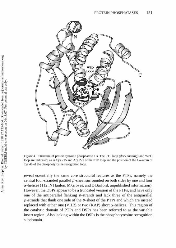
StructureProtein tyrosine phosphatases and the dual-specificity phosphatases are char-acterized by the PTP signature motif (I/V)HCXAGXGR(S/T) containing thecatalytically essential Cys and Arg residues. Within the PTPs and DSPs, thesignature motif is situated at the center of the molecule, at the base of the cat-alytic site. The sequence forms the C-terminus of aβ-strand, a loop connectingtheβ-strand with anα-helix, and the first turn of theα-helix (Figure 4).
The three-dimensional structures of all PTP catalytic domains known share acommon architecture of a central, highly twistedβ-sheet of nineβ-strands withfour central parallelβ-strands flanked by antiparallelβ-strands (5, 100). Thissheet is surrounded byα-helices with four on one side and two on the oppositeside. Additional secondary structural elements present N- and C-terminal to theconserved PTP domain are accommodated within this domain. For example,in common with the other eukaryotic PTPs, two N-terminalα-helices (α-1′ andα-2′) pack againstα-5 andα-6 of PTP1B. The PTP signature motif, locatedat the base of the catalytic site cleft, is surrounded by four loops, three ofwhich provide residues necessary for catalysis and substrate specificity. One ofthese loops, which forms part of the phosphotyrosine recognition region locatedN-terminus to theβ-sheet, is critical in defining the specificity of the PTPs forphosphotyrosine (62).
The catalytic domains of the dual-specificity phosphatases VHR and KAP,despite sharing no sequence-similarity between themselves or with the PTPs,
Ann
u. R
ev. B
ioph
ys. B
iom
ol. S
truc
t. 19
98.2
7:13
3-16
4. D
ownl
oade
d fr
om a
rjou
rnal
s.an
nual
revi
ews.
org
by I
NSE
RM
-mul
ti-si
te a
ccou
nt o
n 04
/18/
07. F
or p
erso
nal u
se o
nly.
P1: PSA/dat P2: ars/rpk
March 18, 1998 14:1 Annual Reviews AR056-06
PROTEIN PHOSPHATASES 151
Figure 4 Structure of protein tyrosine phosphatase 1B. The PTP loop (dark shading) and WPDloop are indicated, as is Cys 215 and Arg 221 of the PTP loop and the position of the Cα-atom ofTyr 46 of the phosphotyrosine recognition loop.
reveal essentially the same core structural features as the PTPs, namely thecentral four-stranded parallelβ-sheet surrounded on both sides by one and fourα-helices (112; N Hanlon, M Groves, and D Barford, unpublished information).However, the DSPs appear to be a truncated version of the PTPs, and have onlyone of the antiparallel flankingβ-strands and lack three of the antiparallelβ-strands that flank one side of theβ-sheet of the PTPs and which are insteadreplaced with either one (VHR) or two (KAP) shortα-helices. This region ofthe catalytic domain of PTPs and DSPs has been referred to as the variableinsert region. Also lacking within the DSPs is the phosphotyrosine recognitionsubdomain.
Ann
u. R
ev. B
ioph
ys. B
iom
ol. S
truc
t. 19
98.2
7:13
3-16
4. D
ownl
oade
d fr
om a
rjou
rnal
s.an
nual
revi
ews.
org
by I
NSE
RM
-mul
ti-si
te a
ccou
nt o
n 04
/18/
07. F
or p
erso
nal u
se o
nly.

P1: PSA/dat P2: ars/rpk
March 18, 1998 14:1 Annual Reviews AR056-06
152 BARFORD ET AL
Catalytic MechanismMuch is now understood concerning the mechanism of PTP catalysis and speci-ficity for pTyr-containing peptides. The phosphate group of pTyr is coordinatedby main-chain amide groups and the Arg side chain of the PTP motif so that thephosphorus atom is situated adjacent to the Sγ -atom of the catalytic Cys residue(62). Engagement of phosphopeptides to PTP1B promotes a major conforma-tional change of one of the catalytic site loops (the WPD loop) consisting ofresidues 179 to 187 that shift by as much as 8A to close over the phenyl ring ofpTyr and allow the side chain of Asp 181 to act as a general acid in the catalyticreaction. The Arg 221 side chain reorients to optimize salt-bridge interactionswith the phosphate bound to the catalytic site. This shift is coupled to motionof the WPD loop via a hydrogen bond between NH2 of Arg 221 and the car-bonyl oxygen of Pro 180, and hydrophobic interactions between the aliphaticmoiety of Arg 221 and the side chain of Trp 179. These interactions and thehydrophobic packing between Phe 182 and the phenyl ring of pTyr stabilizethe closed, catalytically competent conformation of the loop. The phosphoty-rosine dephosphorylation reaction commences with nucleophilic attack by theSγ -atom of the catalytic cysteine on the pTyr phosphorus atom. Cleavage ofthe scissile P-O bond is facilitated by protonation of the phenolic oxygen byAsp 181 with the consequent formation of a phospho-cysteine intermediate.This transient intermediate is hydrolyzed by an activated water molecule. Phos-phoryl transfer reaction to a water molecule catalyzed by PTPs is highly specificbecause PTPs are unable to phosphorylate a range of primary alcohols and otherphosphoryl-acceptors (Z-Y Zhang, personal communication). The structure ofa PTP1B-tungstate complex suggests that in the presence of a phosphate, theWPD loop opens, allowing product release (5).
Numerous kinetic data support the reaction mechanism outlined above (see29 for a review). Cysteinyl-phosphate intermediates have been trapped by rapiddenaturation of PTPs and a dual-specificity phosphatase (VHR) during cat-alytic turnover (22, 52, 90, 120). Moreover, substitution of the catalytic Cysresidue for a serine abolishes catalytic activity and the formation of a cysteinyl-phosphate intermediate (52). The nucleophilicity of the Cys residues resultsfrom its close proximity to main-chain amide groups and a hydrogen bond withthe side chain of Ser 222 of the PTP signature motif, and has an unusually lowpKa of 4.6 (116). The catalytic Asp residue (Asp 181 of PTP1B) contributesto the basic limb of the pH activity profile, and its substitution to Ala causes a105-fold reduction in kcat, suggestive of a role as an acid catalyst (45, 119). Thisimplies that Asp 181 is necessary for the first step of the reaction, namely cleav-age of the pTyr P-O bond and intermediate formation, a notion consistent withthe finding that Asp 181 mutants of PTP1B allow phosphorylated substrates toform stable complexes with the enzyme in vivo (45). The consequence of the
Ann
u. R
ev. B
ioph
ys. B
iom
ol. S
truc
t. 19
98.2
7:13
3-16
4. D
ownl
oade
d fr
om a
rjou
rnal
s.an
nual
revi
ews.
org
by I
NSE
RM
-mul
ti-si
te a
ccou
nt o
n 04
/18/
07. F
or p
erso
nal u
se o
nly.

P1: PSA/dat P2: ars/rpk
March 18, 1998 14:1 Annual Reviews AR056-06
PROTEIN PHOSPHATASES 153
catalytic role played by the catalytic Asp residue (Asp 181 of PTP1B) is thatthe rate of hydrolysis of a range of aryl-phosphate esters is independent of thepKa of the leaving-group residue (29).
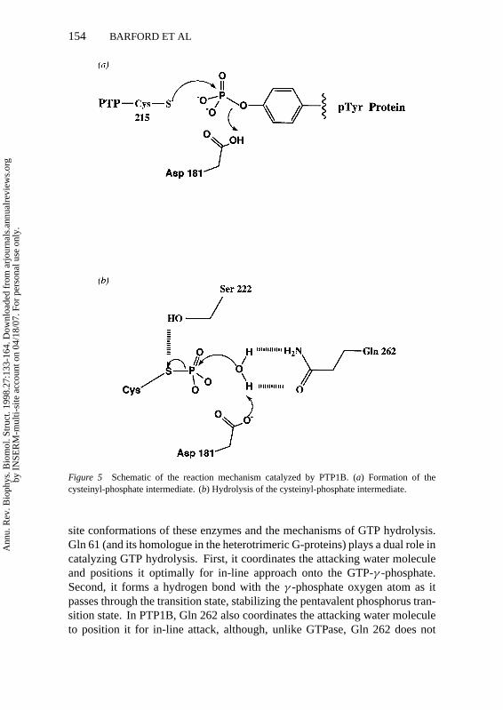
The structures of PTP complexes with vanadate provided insights into thesecond step of the reaction, namely the hydrolysis of the cysteinyl-phosphateintermediate (28). Vanadate forms a covalent bond with the Sγ -atom of thenucleophilic Cys residue to produce a pentavalent trigonal bipyramidal config-uration that is analogous to the transition state. The apical oxygen atom, whichmost closely resembles the attacking nucleophilic water molecule, forms a pairof hydrogen bonds to Asp 181 and to Gln 262. This observation led to thehypothesis that the role of Gln 262 is to position a water molecule for nucle-ophilic attack onto the cysteinyl-phosphate intermediate, and hence substitutionof this residue for Ala should decrease the rate of cysteinyl-phosphate hydroly-sis without affecting the rate of its formation. In this situation, the rate-limitingstep becomes that of phosphocysteine hydrolysis, and hence the intermedi-ate should accumulate. Direct visualization of this intermediate was achievedwithin PTP1B Q262A molecules by soaking crystals at 4◦C in a large molarexcess ofpara-nitrophenol phosphate (a pTyr analogue) and freezing the crys-tals at 100 K to trap, at steady state, the accumulation of the intermediate. Thestructure of the PTP1B-cysteinyl-phosphate intermediate demonstrates that awater-molecule hydrogen bonded to Asp 181 of the closed WPD loop was sit-uated above the phosphate group of the cysteinyl-phosphate residue. However,the position of the water molecule was not ideal for in-line attack onto the phos-phorus atom of the intermediate, being displaced from such a position by 1A.It is likely that this accounts for the reduced rate of hydrolysis of the PTP1Bmutant, and the shift of this water molecule is probably caused by the loss ofhydrogen bonding to the Gln 262 residue. Each step of the reaction pathwaycatalyzed by the PTPs may now be delineated in detail. A schematic is outlinedin Figure 5 (ADB Pannifer and D Barford, unpublished MS).
The reactions catalyzed by PTPs share many features in common with thatof GTP hydrolysis by the GTPases. For example, the role of Gln 262 in po-sitioning a water molecule for nucleophilic attack onto the phosphocysteineintermediate of PTP1B is reminiscent of the Gln residue at the catalytic site ofmost GTPases, including the Ras family and Gα-subunits of the heterotrimericG-proteins. Mutation of the catalytic site Gln 61 residue in Ras causes cellu-lar transformation (4) and a 10-fold rate-reduction of GTP hydrolysis (12, 30).Similarly, mutations of the equivalent Gln residue within the catalytic sites ofGαs and Giα reduces the intrinsic rate of GTP hydrolysis and are associatedwith thyroid and pituitary tumors (71, 76). Crystal structures of complexesof Gαt and Giα with GDP, Mg2+ and AlF4− (25, 96) and a recent structureof a Ras-RasGAP, GDP, Mg2+, AlF3 complex (93) have revealed the active
Ann
u. R
ev. B
ioph
ys. B
iom
ol. S
truc
t. 19
98.2
7:13
3-16
4. D
ownl
oade
d fr
om a
rjou
rnal
s.an
nual
revi
ews.
org
by I
NSE
RM
-mul
ti-si
te a
ccou
nt o
n 04
/18/
07. F
or p
erso
nal u
se o
nly.
P1: PSA/dat P2: ars/rpk
March 18, 1998 14:1 Annual Reviews AR056-06
154 BARFORD ET AL
Figure 5 Schematic of the reaction mechanism catalyzed by PTP1B. (a) Formation of thecysteinyl-phosphate intermediate. (b) Hydrolysis of the cysteinyl-phosphate intermediate.
site conformations of these enzymes and the mechanisms of GTP hydrolysis.Gln 61 (and its homologue in the heterotrimeric G-proteins) plays a dual role incatalyzing GTP hydrolysis. First, it coordinates the attacking water moleculeand positions it optimally for in-line approach onto the GTP-γ -phosphate.Second, it forms a hydrogen bond with theγ -phosphate oxygen atom as itpasses through the transition state, stabilizing the pentavalent phosphorus tran-sition state. In PTP1B, Gln 262 also coordinates the attacking water moleculeto position it for in-line attack, although, unlike GTPase, Gln 262 does not
Ann
u. R
ev. B
ioph
ys. B
iom
ol. S
truc
t. 19
98.2
7:13
3-16
4. D
ownl
oade
d fr
om a
rjou
rnal
s.an
nual
revi
ews.
org
by I
NSE
RM
-mul
ti-si
te a
ccou
nt o
n 04
/18/
07. F
or p
erso
nal u
se o
nly.

P1: PSA/dat P2: ars/rpk
March 18, 1998 14:1 Annual Reviews AR056-06
PROTEIN PHOSPHATASES 155
hydrogen-bond to the oxygens of the cysteinyl phosphate. In addition to acommon Gln residue, both PTPs and GTPases use an essential Arg residue thatcoordinates and stabilizes the pentavalent phosphorus intermediate (25, 93, 96).
Substrate SpecificityPTPs are absolutely specific for pTyr-containing proteins, being unable to de-phosphorylate pSer and pThr proteins (102). Hydrolysis of small-moleculephosphate monoesters such as free pSer and pThr proceeds at a measurable (al-though extremely low) rate, some 105-fold lower than that of pTyr hydrolysis.Amino acids N- and C-terminal to the pTyr residue confer additional bindingaffinity, and for PTP1B and a number of other PTPs, a preference is displayedfor peptides with acidic residues preceding the pTyr residue and C-terminal hy-drophobic residues (6, 117, 118). The molecular basis for substrate specificityresults from the dimensions of the catalytic site cleft, which measures 9A fromits base, where the nucleophilic Cys residue is positioned, to its entrance. A keystructural component of the catalytic cleft is provided by the phosphotyrosinerecognition loop formed from a conserved sequence KNRY (residues 43–46of PTP1B). The Tyr residue of this motif packs against the phenyl ring of apTyr substrate and is critical in defining the depth of the catalytic site cleft (62)(Figure 4, above). Other nonpolar residues present within the pTyr recognitionloop, the WPD and PTP loops interact with the phenyl ring. An Asp residuepresent within the pTyr recognition loop forms a bifurcated hydrogen bond tothe main-chain amide groups of the pTyr and P+1 residues of the peptide. Thisforces the pTyr residue to adopt a helical conformation and insert into the cat-alytic site cleft. Acidic residues present N-terminal to the pTyr residue interactwith basic residues on the phosphatase surface, explaining the preference of thisenzyme for peptides with acidic residues in these positions. The significance ofthe depth of the catalytic site cleft as a determinant of substrate specificity waselegantly demonstrated in a study showing that theYersiniaPTP was capableof dephosphorylating straight-chain peptide-bound aliphatic phosphates of thegeneral structure: (Glu)4-NH-(CH2)n-PO3. The most efficient substrate is onewith seven methylenes, that is, exactly as long as a tyrosine residue (38).
The DSPs differ from the PTPs because of their ability to dephosphorylatepSer and pThr proteins. A comparison of VHR with PTP1B and theYersiniaPTP revealed the expected similarities in the structure of the PTP motif atthe base of the catalytic site (112). An Asp residue (Asp 92) is equivalentto the catalytic Asps of PTPs within both the three-dimensional structure andapproximately 30 residues N-terminal to the PTP motif. The catalytic siteof KAP shows more similarities to the PTPs than does VHR (N Hanlon,M Groves, D Barford, unpublished information). For instance, in KAP, asin the PTPs, a Glu residue forms an ion-pair with the catalytic site Arg residue,
Ann
u. R
ev. B
ioph
ys. B
iom
ol. S
truc
t. 19
98.2
7:13
3-16
4. D
ownl
oade
d fr
om a
rjou
rnal
s.an
nual
revi
ews.
org
by I
NSE
RM
-mul
ti-si
te a
ccou
nt o
n 04
/18/
07. F
or p
erso
nal u
se o
nly.

P1: PSA/dat P2: ars/rpk
March 18, 1998 14:1 Annual Reviews AR056-06
156 BARFORD ET AL
and an equivalent Gln residue to the nucleophilic water coordinating Gln residueof PTPs (Gln 262 of PTP1B) is present. The most significant difference be-tween the PTPs and DSPs is the absence of the pTyr recognition domain withinthe DSPs, the consequence of which is to produce a much shallower and opencatalytic site with a depth of 6A, permitting the hydrolysis of the shorter pSerand pThr residues.
RegulationPTPs are efficient catalysts, with constitutive activity of these proteins caus-ing the rapid dephosphorylation of intracellular pTyr residues and consequentdisruption of signal transduction pathways. Interestingly, this is a mechanismexploited byYersiniabacteria, which inject proteins encoded by the Yop genes,including the YopH PTP, into macrophages, hence preventing immune-directedbacterial phagocytosis and leading to diseases such as the bubonic plague andtuberculosis (51, 11).
The isolated catalytic domains of most PTPs demonstrate activity, hence PTPregulation—at least in part—requires inhibition of this activity. Such a role maybe played by the regulatory domains attached to the PTP catalytic domains. Forthe SH2 domain-containing PTPs, SHP-1 and SHP-2, numerous data supportthe notion that the N-terminal SH2 domains inhibit catalytic activity, most likelyby binding to the PTP domain and blocking substrate access to the catalyticsite. Engagement of phosphorylated peptides by the SH2 domains releases thisinhibition with concomitant activation of the PTP’s activity.
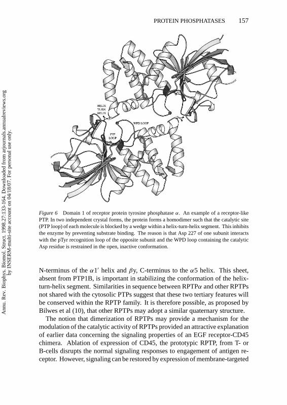
Structural support for such a mechanism of control came unexpectedly withthe crystal structure of the membrane proximal domain (domain 1) of RPTPα
(10). This structure revealed that the molecule was a homodimer in two inde-pendent crystal forms (Figure 6).
The biological significance of this structure lay in the observation that thecatalytic site of each subunit was sterically blocked by the insertion of a wedgeconnecting two helices,α1′ andα2′, within a helix-turn-helix segment imme-diately preceding the PTP catalytic domain from the opposite subunit of thedimer. Residues of this wedge directly interact with the equivalent catalyticsite residues which were part of the phosphopeptide binding site of PTP1B.Moreover, the WPD loop is stabilized in the open conformation and is steri-cally prevented from adopting the catalytically closed conformer. The overalltertiary structure of RPTPα is very similar to that of PTP1B; however, twodifferences in tertiary structure between these two proteins are significant inpromoting the dimeric structure that is observed for RPTPα. One is a two-residue insertion within the wedge connectingα1′ with α2′ of RPTPα. Thesetwo residues participate at the dimeric interface of RPTPα and insert into thecatalytic site. The second is a smallβ-sheet formed byβx at the immediate
Ann
u. R
ev. B
ioph
ys. B
iom
ol. S
truc
t. 19
98.2
7:13
3-16
4. D
ownl
oade
d fr
om a
rjou
rnal
s.an
nual
revi
ews.
org
by I
NSE
RM
-mul
ti-si
te a
ccou
nt o
n 04
/18/
07. F
or p
erso
nal u
se o
nly.
P1: PSA/dat P2: ars/rpk
March 18, 1998 14:1 Annual Reviews AR056-06
PROTEIN PHOSPHATASES 157
Figure 6 Domain 1 of receptor protein tyrosine phosphataseα. An example of a receptor-likePTP. In two independent crystal forms, the protein forms a homodimer such that the catalytic site(PTP loop) of each molecule is blocked by a wedge within a helix-turn-helix segment. This inhibitsthe enzyme by preventing substrate binding. The reason is that Asp 227 of one subunit interactswith the pTyr recognition loop of the opposite subunit and the WPD loop containing the catalyticAsp residue is restrained in the open, inactive conformation.
N-terminus of theα1′ helix andβy, C-terminus to theα5 helix. This sheet,absent from PTP1B, is important in stabilizing the conformation of the helix-turn-helix segment. Similarities in sequence between RPTPα and other RPTPsnot shared with the cytosolic PTPs suggest that these two tertiary features willbe conserved within the RPTP family. It is therefore possible, as proposed byBilwes et al (10), that other RPTPs may adopt a similar quaternary structure.
The notion that dimerization of RPTPs may provide a mechanism for themodulation of the catalytic activity of RPTPs provided an attractive explanationof earlier data concerning the signaling properties of an EGF receptor-CD45chimera. Ablation of expression of CD45, the prototypic RPTP, from T- orB-cells disrupts the normal signaling responses to engagement of antigen re-ceptor. However, signaling can be restored by expression of membrane-targeted
Ann
u. R
ev. B
ioph
ys. B
iom
ol. S
truc
t. 19
98.2
7:13
3-16
4. D
ownl
oade
d fr
om a
rjou
rnal
s.an
nual
revi
ews.
org
by I
NSE
RM
-mul
ti-si
te a
ccou
nt o
n 04
/18/
07. F
or p
erso
nal u
se o
nly.

P1: PSA/dat P2: ars/rpk
March 18, 1998 14:1 Annual Reviews AR056-06
158 BARFORD ET AL
Fig
ure
7Se
quen
ceal
ignm
ento
fre
pres
enta
tive
RPT
PD
1sin
the
regi
ons
that
form
the
dim
erin
terf
aces
ofR
PTPα
and
RPT
Pµ.
Top
left
,βx-
helix
-tu
rn-h
elix
segm
ent.
Top
righ
t,re
sidu
esof
the
pTyr
reco
gniti
onlo
opof
the
cata
lytic
site
.B
otto
m,r
esid
ues
from
β8
toth
eW
PDlo
opof
the
cata
lytic
site
.R
esid
ues
ofR
PTPµ
D1
and
RPT
PαD
1th
atfo
rmin
tera
ctio
nsat
thei
rre
spec
tive
dim
erin
terf
aces
are
indi
cate
dw
ithve
rtic
alar
row
s(t
op)
and
star
s(b
otto
m),
resp
ectiv
ely.
The
resi
dues
that
form
the
dim
erin
terf
ace
ofR
PTPµ
D1
are
poor
lyco
nser
ved
thro
ugho
utth
efa
mily
,whe
reas
resi
dues
ofth
eR
PTPα
D1
inte
rfac
ear
epo
orly
cons
erve
dw
ithin
the
helix
-tur
n-he
lixse
gmen
t,bu
twel
lcon
serv
edw
ithin
the
cata
lytic
site
.
Ann
u. R
ev. B
ioph
ys. B
iom
ol. S
truc
t. 19
98.2
7:13
3-16
4. D
ownl
oade
d fr
om a
rjou
rnal
s.an
nual
revi
ews.
org
by I
NSE
RM
-mul
ti-si
te a
ccou
nt o
n 04
/18/
07. F
or p
erso
nal u
se o
nly.

P1: PSA/dat P2: ars/rpk
March 18, 1998 14:1 Annual Reviews AR056-06
PROTEIN PHOSPHATASES 159
constructs containing the catalytic domain of CD45. For example, a chimericmolecule where the extracellular and transmembrane segments of CD45 werereplaced with those of the EGF receptor was able to restore signaling in the ab-sence, but not in the presence, of EGF (32). In other words, EGF, the extracellu-lar ligand of the chimeric molecule, blocked signaling from the phosphatase do-main. It was proposed that in the presence of growth factor, dimerization of theextracellular segment of the chimera is induced that causes a trans-membranesignal resulting in dimerization of the PTP catalytic domain with concomitantinhibition of PTP activity. Interestingly, the sequence of the helix-turn-helixsegment of CD45 is highly conserved with that of RPTPα, and therefore it isreasonable to suggest that CD45 may dimerize similarly to RPTPα.
For RPTPs with lower sequence similarities to RPTPα, dimerization involv-ing the helix-turn-helix and catalytic sites would appear to be less likely. Thisassumption is based on the observation that residues of the wedge, which arevariable amongst more distantly related RPTPs, interact with invariant catalyticsite residues within the RPTPα dimer (Figure 7).
Direct support for this notion is provided by the crystal structure of RPTPµ
D1 (57). RPTPµ D1 is a homodimer; however, although the tertiary structuresof RPTPαD1 and RPTPµD1 are very similar, the quaternary structures of thesetwo proteins are different. Neither the catalytic site nor the N-terminal helix-turn-helix segment of RPTPµ D1 participates in protein-protein interactions.The catalytic site of RPTPµD1 is unhindered and adopts an open conformationsimilar to that of the cytosolic PTP, PTP1B. The dimer interface of RPTPµ
involved residues fromβ8 andβ10 of the variable insert region.
ACKNOWLEDGMENTS
The work in D Barford’s laboratory has been supported by grants from theMRC, Wellcome Trust, and European Union. We thank Andrew Pannifer, NeilHanlon, Matthew Groves, and Kurt Hoffman for discussions.
Visit the Annual Reviews home pageathttp://www.AnnualReviews.org.
Literature Cited
1. Adler E, Donella-Deana A, Arigoni F,Pinna LA, Stragier P. 1996. Structural re-lationship between a bacterial develop-ment protein and eukaryotic PP2C phos-phatases.Mol. Microbiol. 23:57–62
2. Aitken A, Cohen P. 1982. Isolation andcharacterisation of active fragments ofprotein phosphatase inhibitor-1 from rab-bit skeletal muscle.FEBS Lett.147:54–58
3. Bagu JR, Sonnichsen FD, Williams D,Anderson RJ, Sykes BD, Holmes CFB.1995. Comparison of the solution struc-tures of microcystin-LR and motuporin.Nat. Struct. Biol.2:114–16
4. Barbacid M. 1987. Ras genes.Annu. Rev.Biochem.56:779–827
5. Barford D, Flint AJ, Tonks NK. 1994.Crystal structure of human protein
Ann
u. R
ev. B
ioph
ys. B
iom
ol. S
truc
t. 19
98.2
7:13
3-16
4. D
ownl
oade
d fr
om a
rjou
rnal
s.an
nual
revi
ews.
org
by I
NSE
RM
-mul
ti-si
te a
ccou
nt o
n 04
/18/
07. F
or p
erso
nal u
se o
nly.

P1: PSA/dat P2: ars/rpk
March 18, 1998 14:1 Annual Reviews AR056-06
160 BARFORD ET AL
tyrosine phosphatase 1B.Science263:1397–1404
6. Barford D, Jia Z, Tonks NK. 1995. Pro-tein tyrosine phosphatases take off.Nat.Struct. Biol.2:1043–53
7. Berndt N, Dohadwala M, Liu CWY. 1997.Constitutively active protein phosphatase1α causes Rb-dependent G1 arrest in hu-man cancer cells.Curr. Biol. 7:375–86
8. Beullens M, Van Eynde A, Bollen M,Stalmans W. 1993. Inactivation of nu-clear inhibitory polypeptides of proteinphosphatase-1 (NIPP-1) by protein kinaseA. J. Biol. Chem.268:13172–77
9. Beullens M, Van Eynde A, Stalmans W,Bollen M. 1992. The isolation of novelinhibitory polypeptides of protein phos-phatase 1 from bovine thymus nuclei.J.Biol. Chem.267:16538–44
10. Bilwes AM, Den Hertog J, Hunter T, NoelJP. 1996. Structural basis for inhibitionof receptor protein-tyrosine phosphatase-alpha by dimerisation.Nature 382:555–59
11. Bliska JB. 1995. Crystal structure ofthe Yersinia protein tyrosine phosphatase.Trends Microbiol.3:125–27
12. Bollag G, McCormick F. 1991. Differen-tial regulation of RasGAP and neurofibro-matosis gene products.Nature351:575–78
13. Bork P, Brown NP, Hegyi H, Shultz J.1996. The protein phosphatase 2C (PP2C)superfamily: detection of bacterial homo-logues.Protein Sci.5:1421–25
14. Brady-Kalnay SM, Flint AJ, Tonks NK.1993. Homophilic binding of PTPµ, areceptor-type protein tyrosine phospha-tase, can mediate cell-cell aggregation.J.Cell Biol. 122:961–72
15. Brady-Kalnay SM, Rimm DL, Tonks NK.1995. Receptor protein tyrosine phospha-tase PTPµ associates with cadherins andcatenins in vivo.J. Cell Biol.130:977–86
16. Campos M, Fadden P, Alms G, Qian Z,Haystead TAJ. 1996. Identification of pro-tein phosphatase-1-binding proteins bymicrocystin-biotin affinity chromatogra-phy.J. Biol. Chem.271:28478–84
17. Cardenas ME, Muir RS, Breuder T,Heitman J. 1995. Targets of immunophi-lin-immunosuppressant complexes aredistinct highly conserved regions of cal-cineurin A.EMBO J.14:2772–83
18. Chen J, Martin BL, Brautigan DL. 1992.Regulation of protein serine-threoninephosphatase type 2A by tyrosine phos-phorylation.Science257:1261–64
19. Chen MX, Cohen PTW. 1997. Activationof protein phosphatase 5 by limited pro-teolysis or the binding of polyunsaturated
fatty acids to the TPR domain.FEBS Lett.400:136–40
20. Chen MX, McPartlin AE, Brown L, ChenYH, Barker HM, Cohen PTW. 1994.A novel human protein serine/threoninephosphatase which possesses four tetra-tricopeptide repeat motifs and localises tothe nucleus.EMBO J.13:4278–90
21. Cheng J, Wu K, Armanini M, O’RourkeN, Dowbenko D, Lasky LA. 1997. Anovel protein-tyrosine phosphatase re-lated to the homotypically adhering k andµ receptors.J. Biol. Chem.272:7264–77
22. Cho H, et al. Isolation and structural elu-cidation of a novel phosphocysteine inter-mediate in the LAR protein tyrosine phos-phatase enzymatic pathway. 1992.J. Am.Chem. Soc.114:7296–98
23. Coghlan VM, Perrino BA, Howard M,Langeberg LK, Hicks JB, et al. 1995. As-sociation of protein kinase A and proteinphosphatase 2B with a common anchor-ing protein.Science267:108–11
24. Cohen PTW. 1997. Novel protein serine/threonine phosphatases: variety is thespice of life. Trends Biochem. Sci.22:245–51
25. Coleman DE, Berghuis AM, Lee E, Lin-der ME, Gilman AG, Sprang SR. 1994.Structures of active conformations of Giαand the mechanism of GTP hydrolysis.Science265:1405–12
26. Das AK, Helps NR, Cohen PTW, BarfordD. 1996. Crystal structure of the proteinserine/threonine phosphatase 2C at 2.0Aresolution.EMBO J.15:6798–809
27. Dent P, Lavoinne A, Nakielny A, Caud-well FB, Watt P, Cohen P. 1990. Themolecular mechanism by which insulinstimulates glycogen synthesis in mam-malian skeletal muscle.Nature348:302–8
28. Denu JM, Lohse DL, Vijayalakshmi J,Saper MA, Dixon JE. 1996. Visualiza-tion of intermediate and transition-statestructures in protein tyrosine phosphatasecatalysis.Proc. Natl. Acad. Sci.93:2493–98
29. Denu JM, Stuckey JA, Saper MA, DixonJE. 1996. Form and function in proteinphosphorylation.Cell 87:361–64
30. Der CJ, Finkel T, Cooper GM. 1986. Bio-logical and biochemical properties of hu-man RasH genes mutated at codon 61.Cell 44:167–76
31. Derrick JP, Wigley DB. 1992. Crystalstructure of a streptococcal protein G do-main bound to an Fab fragment.Nature359:752–54
32. Desai DM, Sap J, Schessinger J, Weiss
Ann
u. R
ev. B
ioph
ys. B
iom
ol. S
truc
t. 19
98.2
7:13
3-16
4. D
ownl
oade
d fr
om a
rjou
rnal
s.an
nual
revi
ews.
org
by I
NSE
RM
-mul
ti-si
te a
ccou
nt o
n 04
/18/
07. F
or p
erso
nal u
se o
nly.

P1: PSA/dat P2: ars/rpk
March 18, 1998 14:1 Annual Reviews AR056-06
PROTEIN PHOSPHATASES 161
A. 1993. Ligand-mediated negative reg-ulation of a chimeric transmembrane re-ceptor tyrosine phosphatase.Cell73:541–54
33. Desdouits F, Cheetham JJ, Huang H-B,Jwon Y-G, da Cruz e Silva EF, et al.1995. Mechanism of inhibition of pro-tein phosphatase 1 by DARPP-32: stud-ies with recombinant DARPP-32 and syn-thetic peptides.Biochem. Biophys. Res.Comm.206:653–58
34. Dohadwala M, da Cruz e Silva EF, HallFL, Williams RT, Carbonara-Hall DA,et al. 1994. Phosphorylation and inacti-vation of protein phosphatase 1 by cyclindependent kinases.Proc. Natl. Acad. Sci.91:6408–12
35. Doherty MJ, Moorhead G, Morrice N,Cohen P, Cohen PTW. 1995. Aminoacid sequence and expression of the hep-atic glycogen-binding GL-subunit of PP1.FEBS Lett.375:294–98
36. Doyle DA, Lee A, Lewis J, Kim E,Sheng M, MacKinnon R. 1996. Crystalstructures of a complexed and peptide-free membrane protein-binding domain:molecular basis of peptide recognition byPDZ.Cell 85:1067–76
37. Duncan L, Alper S, Arigoni F, LosickR, Stragier P. 1995. Activation of cell-specific transcription by a serine phos-phatase at the site of asymmetric division.Science270:641–44
38. Dunn D, Chen L, Lawrence DS, Zhang Z-Y. 1996. The active site specificity of theYersinia protein tyrosine phosphatase.J.Biol. Chem.271:168–73
39. Egloff M-P, Cohen PTW, Reinemer P,Barford D. 1995. Crystal structure of thecatalytic subunit of human protein phos-phatase 1 and its complex with tungstate.J. Mol. Biol.254:942–59
40. Egloff M-P, Johnson DF, Moorhead G,Cohen PTW, Cohen P, Barford D. 1997.Structural basis for the recognition of reg-ulatory subunits by the catalytic subunit ofprotein phosphatase 1.EMBO J.16:1876–87
41. Endo S, Zhou X, Connor J, Wang B,Shenolikar S. 1996. Multiple structuralelements define the specificity of re-combinant human inhibitor-1 as a pro-tein phosphatase-1 inhibitor.Biochem-istry 35:5220–28
42. Faux MC, Scott JD. 1996. More on targetwith protein phosphorylation: conferringspecificity by location.Trends Biochem.Sci.21:312–15
43. Favre B, Zoinierowicz S, Turowski P,Hemmings BA. 1994. The catalytic sub-unit of protein phosphatase 2A is car-
boxyl-methylated in vivo.J. Biol. Chem.269:16311–17
44. Fiscella M, Zhang HL, Fan S, SakaguchiK, Shen S, et al. 1997. Wip1, a novel hu-man protein phosphatase that is inducedin response to ionising radiation in a p53-dependent manner.Proc. Natl. Acad. Sci.94:6048–53
45. Flint AJ, Tiganis T, Barford D, TonksNK. 1997. Development of “substratetrapping” mutants to identify physiolog-ical substrates of protein tyrosine phos-phatases.Proc. Natl. Acad. Sci.94:1680–85
46. Frangioni JV, Beahm PH, Shifrin V,Jost CA, Neel BG. 1992. The nontrans-membrane protein tyrosine phosphatasePTP1B localises to the endoplasmic retic-ulum via its 35 amino acid C-terminal se-quence.Cell 68:545–60
47. Fuchs M, Muller T, Lerch MM, UllrichA. 1996. Association of human protein-tyrosine phosphatase k with membersof the armadillo family.J. Biol. Chem.271:16712–19
48. Gaits F, Shiozaki K, Russell P. 1997. Pro-tein phosphatase 2C acts independentlyof stress-activated kinase cascade to reg-ulate stress response in fission yeast.J.Biol. Chem.272:17873–79
49. Goldberg J, Huang H, Kwon Y, Green-gard P, Nairn AC, Kuriyan J. 1995.Three dimensional structure of the cat-alytic subunit of protein serine/threoninephosphatase-1.Nature376:745–53
50. Griffith JP, Kim JL, Kim EE, SintchakMD, Thomson JA, et al. 1995. X-raystructure of calcineurin inhibited bythe immunophilin-immunosuppressantFKBP12-FK506 complex.Cell 82:507–22
51. Guan KL, Dixon JE. 1990. Protein ty-rosine phosphatase activity of an essen-tial virulence determinant inYersinia.Science249:553–56
52. Guan KL, Dixon JE. 1991. Evidenceof protein tyrosine phosphatase catalysisproceeding via a cysteine-phosphate in-termediate.J. Biol. Chem.266:17026–30
53. Helps N, Barker H, Elledge SJ, CohenPTW. 1995. Protein phosphatase 1 inter-acts with p53BP2, a protein which bindsto the tumour supressor p53.FEBS Lett.377:295–300
54. Hemmings HC Jr, Nairn AC, Elliot JR,Greengard P. 1990. Synthetic peptide ana-logs of DARPP-32, an inhibitor of pro-tein phosphatase-1.J. Biol. Chem.265:20369–76
55. Hirano K, Erdodi F, Patton JG, HartshorneDJ. 1996. Interaction of protein phospha-
Ann
u. R
ev. B
ioph
ys. B
iom
ol. S
truc
t. 19
98.2
7:13
3-16
4. D
ownl
oade
d fr
om a
rjou
rnal
s.an
nual
revi
ews.
org
by I
NSE
RM
-mul
ti-si
te a
ccou
nt o
n 04
/18/
07. F
or p
erso
nal u
se o
nly.

P1: PSA/dat P2: ars/rpk
March 18, 1998 14:1 Annual Reviews AR056-06
162 BARFORD ET AL
tase type 1 with a splicing factor.FEBSLett.389:191–94
56. Hirano K, Phan BC, Hartshorne DJ. 1997.Interactions of the subunits of smoothmuscle myosin phosphatase.J. Biol.Chem.272:3683–88
57. Hoffmann KMV, Tonks NK, Barford D.1997. The crystal structure of domain 1 ofreceptor protein tyrosine phosphataseµ.J. Biol. Chem.272:27505–8
58. Huang H-B, Horiuchi A, Goldberg J,Greengard P, Nairn AC. 1997. Site-direc-ted mutagenesis of amino acid residues ofprotein phosphatase 1 involved in catal-ysis and inhibitor binding,Proc. Natl.Acad. Sci.94:3530–35
59. Hubbard MJ, Cohen P. 1993. On targetwith a new mechanism for the regula-tion of protein phosphorylation.Trends.Biochem. Sci.18:172–77
60. Hunter T. 1995. Protein kinases and phos-phatases: the yin and yang of proteinphosphorylation and signalling.Cell 80:225–36
61. Jain J, McCaffrey PG, Liner Z, KerppolaTK, Lambert JN, et al. 1993. Calcineurinphosphatase activity in T lymphocytes isinhibited by FK506 and cyclosporin A.Nature365:352–55
62. Jia Z, Barford D, Flint AJ, Tonks NK.1995. Structural basis for phosphotyro-sine peptide recognition by protein tyro-sine phosphatase 1B.Science268:1754–58
63. Johnson DF, Moorhead G, Caudwell FB,Cohen P, Chen Y-H, et al. 1996. Identi-fication of protein phosphatase-1-bindingdomains on the glycogen and myofibril-lar targetting subunits.Eur. J. Biochem.239:317–25
64. Johnson LN, Barford D. 1993. The effectsof phosphorylation on the structures andfunction of proteins.Annu. Rev. Biophys.Biomol. Struct.22:199–232
65. Johnson LN, O’Reilly M. 1996. Controlby phosphorylation.Curr. Opin. Struct.Biol. 6:762–69
66. Kataoka T, Broek D, Wigler M. 1985.DNA sequence and characterization of theS. cerevisiaegene encoding adenylate cy-clase.Cell 43:493–505
67. Kimura K et al. 1996. Regulation ofmyosin phosphatase by Rho and Rho-associated kinase (Rho-kinase).Science273:245–48
68. King MM, Huang CY. 1984. The cal-modulin-dependent activation and deacti-vation of the phosphoprotein calcineurinand the effect of nucleotides, pyrophos-phate, and divalent metal ions.J. Biol.Chem.259:8847–56
69. Kissinger CR, Parge HE, Knighton DR,Lewis CT, Pelletier LA, et al. 1995. Crys-tal structure of human calcineurin andthe human FKBP12-FK506-calcineurincomplex.Nature378:641–44
70. Kwon Y-G, Huang H-B, Desdouits F,Girault J-A, Greengard P, Nairn AC.1997. Characterisation of the interactionbetween DARPP-32 and protein phos-phatase 1 (PP-1): DARPP-32 peptidesantagonize the interaction of PP-1 withbinding proteins.Proc. Natl. Acad. Sci.94:3536–41
71. Landis CA, Masters SB, Spada A, PaceAM, Bourne HR, Vallar L. 1989. GTPasesinhibitory mutations activate the chain ofGαs and stimulate adenylyl cyclase in hu-man pituitary tumours.Nature340:692–96
72. Lawson JE, Niu X-D, Browning KS, Tro-ng HL, Yan J, Reed LJ. 1993. Molecu-lar cloning and expression of the catalyticsubunit of bovine pyruvate dehydroge-nase phosphatase and sequence similaritywith protein phosphatase 2C.Biochem-istry 32:8987–93
73. Leung J, Bouvier-Durand M, Morris P-C, Guerrier D, Chefdor F, Giraudat J.1994. Arabidopsis ABA response geneABI1: Features of a calcium-modulatedprotein phosphatase.Science264:1448–52
74. Liu J, Farmer JD, Lane WS, FriedmanJ, Weissman I, Schreiber SL. 1991. Cal-cineurin is a common target of cyclo-philin-cyclosporin A and FKBP-FK506complexes.Cell 66:807–15
75. Lohse DL, Denu JM, Dixon JE. 1995.Insights derived from the structures ofthe Ser/Thr phosphatases calcineurin andprotein phosphatase 1.Structure3:987–90
76. Lyons J et al. 1990. Two G protein onco-genes in human endocrine tumors.Sci-ence249:655–59
77. MacKintosh C, MacKintosh RW. 1994.Inhibitors of protein kinases and phos-phatases.Trends Biochem. Sci.17:444–47
78. Maeda T, Wurgler-Murphy SM, Saito H.1994. A two-component system that reg-ulates an osmosensing MAP kinase cas-cade in yeast.Nature369:242–45
79. Martin BL, Graves DJ. 1994. Isotope ef-fects on the mechanism of calcineurincatalysis: kinetic solvent isotope and iso-tope exchange studies.Biochim. Biophys.Acta1206:136–42
80. Mayer-Jaekel RE, Okhura H, GomesR, Sunkel CE, Baumgartner S, et al.1993. The 55 kD regulatory subunit of
Ann
u. R
ev. B
ioph
ys. B
iom
ol. S
truc
t. 19
98.2
7:13
3-16
4. D
ownl
oade
d fr
om a
rjou
rnal
s.an
nual
revi
ews.
org
by I
NSE
RM
-mul
ti-si
te a
ccou
nt o
n 04
/18/
07. F
or p
erso
nal u
se o
nly.

P1: PSA/dat P2: ars/rpk
March 18, 1998 14:1 Annual Reviews AR056-06
PROTEIN PHOSPHATASES 163
Drosophilaprotein phosphatase 2A is re-quired for anaphase.Cell 72:621–33
81. Meyer K, Leube MP, Grill E. 1994. A pro-tein phosphatase 2C involved in ABA sig-nal transduction inArabidopsis thaliana.Science264:1452–55
82. Moore F, Weekes J, Hardie DG. 1991. Ev-idence that AMP triggers phosphorylationas well as direct allosteric activation of ratliver AMP-activated protein kinase.Eur.J. Biochem.199:691–97
83. Moorhead G, MacKintosh C, MorriceN, Cohen P. 1995. Purification of the hep-atic glycogen-associated form of proteinphosphatase-1 by microcystin-Sepharoseaffinity chromatography. FEBS Lett.362:101–5
84. Moorhead G, MacKintosh CW, MorriceN, Gallagher T, MacKintosh C. 1994. Pu-rification of type 1 protein (serine/threo-nine) phosphatases by microcystin-Se-pharose affinity chromatography.FEBSLett.356:46–50
85. Mueller EG, Crowder MW, Averill BA,Knowles JR. 1993. Purple acid phos-phatase: a diron enzyme that catalyses adirect phospho group transfer to water.J.Am. Chem. Soc.115:2974–75
86. Nassar N, Horn G, Herrman C, Scherer A,McCormick F, Wittinghofer A. 1995. The2.2 A crystal structure of the ras-bindingdomain of the serine/threonine kinase c-Raf1 in complex with Rap1A and a GTPanalogue.Nature375:554–60
87. Neel BG, Tonks NK. 1997. Protein tyro-sine phosphatases in signal transduction.Curr. Opin. Cell Biol.9:193–204
88. Deleted in proof89. Pawson T. 1995. Protein modules and sig-
naling networks.1995.Nature 373:573–80
90. Pot DA, Woodford TA, Remboutsika E,Huan RS, Dixon JE. 1991. Cloning, bac-terial expression, purification, and char-acterisation of the cytoplasmic domain ofrat LAR, a receptor-like protein tyrosinephosphatase.J. Biol. Chem.266:19688–96
91. Russo AA, Jeffry PD, Patten AK, Mas-sague J, Pavletich NP. 1996. Crystal struc-ture of the p27Kip1 cyclin-dependent-kinase inhibitor bound to the cyclinA-Cdk2 complex.Nature382:325–31
92. Sap JY, Jiang YP, Friedlander D, GrumetM, Schlessinger J. 1994. Receptor tyro-sine phosphatase RPTP-k mediates ho-mophilic binding.Mol. Cell Biol. 14:1–9
93. Scheffzek K, Ahmadian MR, KabschW, Wiesmuller L, Lautwein A, et al.1997. The Ras-RasGAP complex: struc-
tural basis for GTPase activation and itsloss in oncogenic Ras mutants.Science277:333–45
94. Shibasaki F, Price ER, Milan D, McKeonF. 1996. Role of kinases and the phos-phatase calcineurin in the nuclear shut-tling of transcription factor NF-AT4.Na-ture382:370–73
95. Silverstein AM Galigniana Chen M-SOwens-Grillo JK Chinkers M, Pratt WB.1997. Protein phosphatase 5 is a ma-jor component of glucocorticoid receptor-hsp90 complexes with properties of anFK506-binding immunophilin.J. Biol.Chem.272:16224–30
96. Sondek J, Lambright DG, Noel JP, HammHE, Sigler PB. 1994. GTPase mecha-nism of G proteins from the 1.7-A crystalstructure of transducin GDPAlF4-. Nature372:276–29
97. Sontag E, Fedorov S, Kamibayashi C,Robbins D, Cobb M, Mumby M. 1993.The interaction of SV40 small tumor anti-gen with protein phosphatase 2A stim-ulates the MAP kinase pathway and in-duces cell proliferation.Cell 75:887–97
98. Stone JM, Collinge MA, Smith RD, HornMA, Walker JC. 1994. Interaction ofprotein phosphatase with anArabidopsisserine-threonine receptor kinase.Science266:793–95
99. Strater N, Klabunde T, Tucker P, WitzelH, Krebs B. 1995. Crystal structure of apurple acid phosphatase containing a di-nuclear Fe(III)-Zn(II) active site.Science268:1489–92
100. Stuckey JA, Schubert HL, Fauman EB,Zhang ZY, Dixon JE, Saper MA. 1994.Crystal structure ofYersiniaprotein ty-rosine phosphatase at 2.5A and thecomplex with tungstate.Nature370:571–75
101. Tang PM, Bondor JA, Swiderek KM,dePaoli-Roach AA. 1991. Molecularcloning and expression of the regula-tory (RGl) subunit of glycogen-associatedprotein phosphatase.J. Biol. Chem.266:15782–89
102. Tonks NK, Diltz CD, Fischer EH. 1988.Purification of the major protein tyrosinephosphatase of human placenta.J. Biol.Chem.263:6722–30
103. Van Duyne GD, Standaert RF, KarplusPA, Schreiber SL, Clardy J. 1991. Atomicstructure of FKB-FK506, an immuno-philin-immunosuppressant complex.Sci-ence252:839–42
104. Van Eynde A, Beullens M, Stalmans W,Bollen M. 1994. Full activation of a nu-clear species of PP1 by phosphorylation
Ann
u. R
ev. B
ioph
ys. B
iom
ol. S
truc
t. 19
98.2
7:13
3-16
4. D
ownl
oade
d fr
om a
rjou
rnal
s.an
nual
revi
ews.
org
by I
NSE
RM
-mul
ti-si
te a
ccou
nt o
n 04
/18/
07. F
or p
erso
nal u
se o
nly.

P1: PSA/dat P2: ars/rpk
March 18, 1998 14:1 Annual Reviews AR056-06
164 BARFORD ET AL
with protein kinase A and casein kinase2. Biochem. J.297:447–49
105. Van Eynde A, Wera S, Beullens M, Tor-rekens S, Van Leuven F, et al. 1995.Molecular cloning of NIPP-1, a nuclearinhibitor of protein phosphatase-1, re-veals homology with polypeptides in-volved in RNA processing.J. Biol. Chem.270:28068–74
106. Wera S, Hemmings BA. 1995. Serine/threonine protein phosphatases.Biochem.J. 311:17–29
107. Deleted in proof108. Wang X, Culotta VC, Lee CB. 1996. Su-
peroxide dismutase protects calcineurinfrom inactivation.Nature383:434–37
109. Wu J, Kleiner U, Brautigan DL. 1996.Protein phosphatase type-1 and glycogenbind to a domain in the skeletal muscleregulatory subunit containing conservedhydrophobic sequence motifs.Biochem-istry 35:13858–64
110. Yamano H, Ishii K, Yanagida M. 1994.Phosphorylation of dis2 protein phos-phatase at the C-terminal cdc2 consensusand its potential role in cell cycle regula-tion. EMBO J.13:5310–18
111. Yu L, Haddy A, Rusnak F. 1995. Evidencethat calcineurin accommodates an activesite binuclear metal center.J. Am. Chem.Soc.117:10147–48
112. Yuvaniyama J, Denu JM, Dixon JE, SaperMA. 1996. Crystal structure of the dualspecificity protein phosphatase VHR.Sci-ence272:1328–31
113. Zhang J, Zhang Z, Brew K, Lee EYC.1996. Mutational analysis of the catalyticsubunit of muscle protein phosphatase 1.Biochemistry35:6276–82
114. Zhang L, Zhang Z, Long F, Lee EYC.1996. Tyrosine-272 is involved in the in-
hibition of protein phosphatase-1 by mul-tiple toxins.Biochemistry35:1606–11
115. Zhang Z, Zhao S, Long F, Zhang L,Bai G, et al. 1994. A mutant of proteinphosphatase-1 that exhibits altered toxinsensitivity. J. Biol. Chem.269:16997–17000
116. Zhang Z-Y, Dixon JE. 1993. Active sitelabelling of the Yersinia protein tyro-sine phosphatase: the determination ofthe pKa of the active site cysteine and thefunction of the conserved histidine 402.Biochemistry32:9340–45
117. Zhang Z-Y et al. 1994. Substrate speci-ficity of the protein tyrosine phosphatases.Proc. Natl. Acad. Sci. USA90:4446–50
118. Zhang Z-Y, Maclean D, McNamara DJ,Sawyer TK, Dixon JE. 1994. Protein ty-rosine phosphatase substrate specificity:size and phosphotyrosine positioning re-quirements in peptide substrates.Bio-chemistry33:2285–90
119. Zhang Z-Y, Wang Y, Dixon JE. 1994. Dis-secting the catalytic mechanism of pro-tein-tyrosine phosphatases.Proc. Natl.Acad. Sci.91:1624–28
120. Zhou G, Denu JM, Wu L, Dixon JE.1994. The catalytic role of Cys 124 in thedual specificity phosphatase VHR.J. Biol.Chem.269:28084–90
121. Zhou MM, Ravichandran KS, OlejniczakEF, Petros AM, Meadows RP, et al. 1995.Structure and ligand recognition of thephosphotyrosine binding domain of SHC.Nature378:584–92
122. Zhuo S, Clemens JC, Stone RL, DixonJE. 1994. Mutational analysis of a Ser/Thrphosphatase: identification of residuesimportant in phosphoesterase binding andcatalysis.J. Biol. Chem.269:26234–38
Ann
u. R
ev. B
ioph
ys. B
iom
ol. S
truc
t. 19
98.2
7:13
3-16
4. D
ownl
oade
d fr
om a
rjou
rnal
s.an
nual
revi
ews.
org
by I
NSE
RM
-mul
ti-si
te a
ccou
nt o
n 04
/18/
07. F
or p
erso
nal u
se o
nly.

Annual Review of Biophysics and Biomolecular Structure Volume 27, 1998
CONTENTSThe Stereochemical Mechanism of the Cooperative Effects in Hemoglobin Revisited, M. F. Perutz, A. J. Wilkinson, M. Paoli, G. G. Dodson
1
The Three-Dimensional Structure of the Ribosome and Its Components, Peter B. Moore 35
Signaling Complexes: Biophysical Constraints on Intracellular Communication, Dennis Bray 59
Spatio-Temporal Resolution of Exocytosis from Individual Cells, Eric R. Travis, R. Mark Wightman 77
Minor Groove-Binding Architectural Proteins: Structure, Function, and DNA Recognition, Carole A. Bewley, Angela M. Gronenborn, G. Marius Clore
105
The Structure and Mechanism of Protein Phosphatases: Insights into Catalysis and Regulation, David Barford, Amit K. Das, Marie-Pierre Egloff
133
Biosensors in Chemical Separations, Harvey A. Fishman, Daniel R. Greenwald, Richard N. Zare 165
Simulation of Prokaryotic Genetic Circuits, Harley H. McAdams, Adam Arkin 199
DNA Nanotechnology: Novel DNA Constructions, Nadrian C. Seeman 225
Inhibitors of HIV-1 Protease: A Major Success of Structure-Assisted Drug Design, Alexander Wlodawer, Jiri Vondrasek 249
Structure, Dynamics, and Function of Chromatin In Vitro, J. Widom 285
Cytochrome C Oxidase: Structure and Spectroscopy, H. Michel, J. Behr, A. Harrenga, A. Kannt 329
The Use of 2H,13C,15N Multidimensional NMR To Study the Structure and Dynamics of Proteins, Kevin H. Gardner, Lewis E. Kay 357
RNA Recognition by RNP Proteins During RNA Processing, Gabriele Varani, Kiyoshi Nagai 407
On the Characteristics of Functional Magnetic Resonance Imaging of the Brain, S. Ogawa, R. S. Menon, S.-G. Kim, K. Ugurbil 447
Crystallographic Structures of the Hammerhead Ribozyme: Relationship to Ribozyme Folding and Catalysis, Joseph E. Wedekind, David B. McKay
475
Pleckstrin Homology Domains: A Common Fold with Diverse Functions, M. J. Rebecchi, S. Scarlata 503
Ann
u. R
ev. B
ioph
ys. B
iom
ol. S
truc
t. 19
98.2
7:13
3-16
4. D
ownl
oade
d fr
om a
rjou
rnal
s.an
nual
revi
ews.
org
by I
NSE
RM
-mul
ti-si
te a
ccou
nt o
n 04
/18/
07. F
or p
erso
nal u
se o
nly.