the spectrum of gallbladder disease - connecticut … · pathophysiology diagnostic ... gallbladder...
TRANSCRIPT

The Spectrum of
Gallbladder Disease
Rebecca Kowalski, M.D.
October 18, 2017

Overview
A (brief) history of gallbladder surgery
Anatomy
Anatomical variations
Physiology
Pathophysiology
Diagnostic imaging of the gallbladder
Natural history of cholelithiasis
Case presentations of the spectrum of gallstone disease
Summary

History of Gallbladder Surgery
Gallbladder Surgery: A Relatively Recent Change
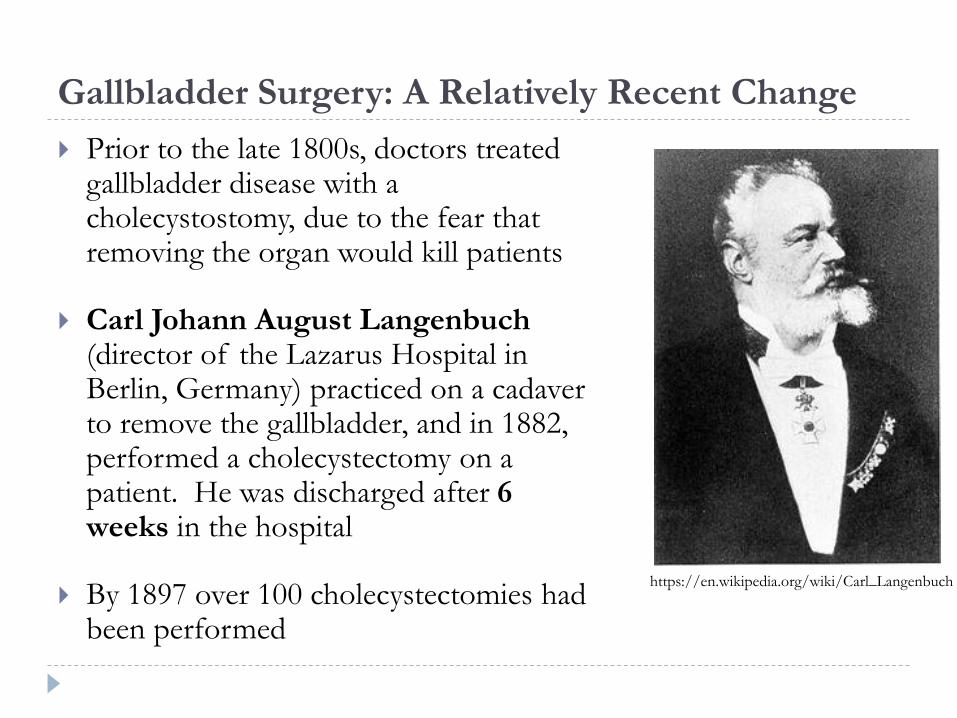
Prior to the late 1800s, doctors treated gallbladder disease with a cholecystostomy, due to the fear that removing the organ would kill patients
Carl Johann August Langenbuch(director of the Lazarus Hospital in Berlin, Germany) practiced on a cadaver to remove the gallbladder, and in 1882, performed a cholecystectomy on a patient. He was discharged after 6 weeks in the hospital
By 1897 over 100 cholecystectomies had been performed
https://en.wikipedia.org/wiki/Carl_Langenbuch

Gallbladder Surgery: A Relatively Recent Change
In 1985, Erich Mühe removed a patient’s gallbladder laparoscopically in Germany
In 1987, Philippe Mouret (a French gynecologic surgeon) performed a laparoscopic cholecystectomy
In 1992, the National Institutes of Health (NIH) created guidelines for laparoscopic cholecystectomy in the United States, essentially transforming surgical practice
Erich Muhehttps://openi.nlm.ni
h.gov/detailedresult.
php?img=PMC30152
44_jsls-2-4-341-
g01&req=4
Philippe Mourethttps://www.pinterest.com
/pin/58195020154734720/

Anatomy and Abnormal Anatomy


http://accesssurgery.mhmedical.com/content.aspx?bookid=1202§ionid=71521210
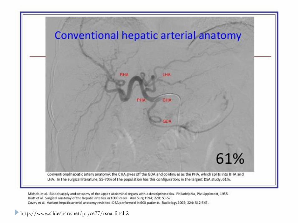
http://www.slideshare.net/pryce27/rsna-final-2
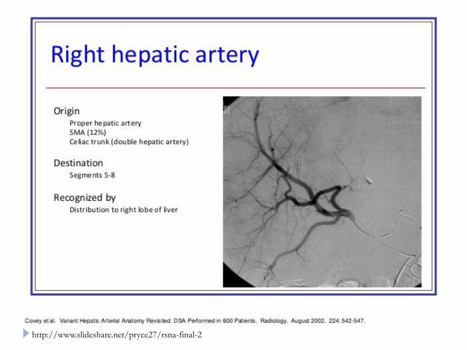
http://www.slideshare.net/pryce27/rsna-final-2

http://www.slideshare.net/pryce27/rsna-final-2

Physiology

http://www.nature.com/nrm/journal/v2/n9/fig_tab/nrm0901_657a_F3.html
a

Simplified overview of the bile acid biosynthesis pathway derived from cholesterol
Lisa D. Beilke et al. Drug Metab Dispos 2009;37:1035-1045
http://dmd.aspetjournals.org/content/37/5/1035
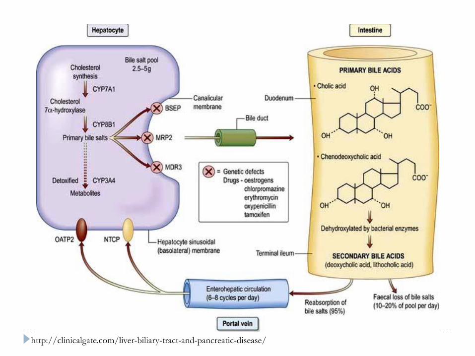
http://clinicalgate.com/liver-biliary-tract-and-pancreatic-disease/

http://clinicalgate.com/liver-biliary-tract-and-pancreatic-disease/

Gallbladder Function: Absorption and Secretion
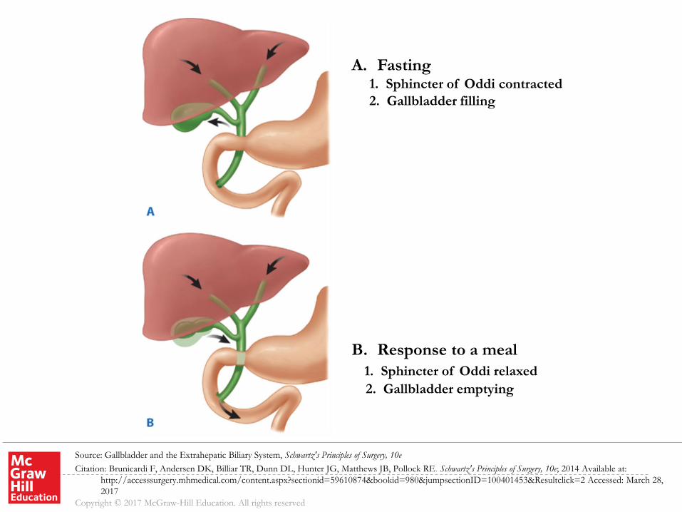
Main function of the gallbladder is to concentrate and store
hepatic bile and to deliver bile into the duodenum in
response to a meal
In the fasting state, approximately 80% of the bile secreted
by the liver is stored in the gallbladder
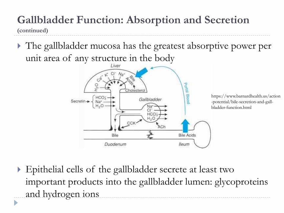
Gallbladder Function: Absorption and Secretion(continued)
The gallbladder mucosa has the greatest absorptive power per
unit area of any structure in the body
Epithelial cells of the gallbladder secrete at least two
important products into the gallbladder lumen: glycoproteins
and hydrogen ions
https://www.barnardhealth.us/action
-potential/bile-secretion-and-gall-
bladder-function.html

https://courses.washington.edu/conj/bess/bile/bile.html

http://www.austincc.edu/apreview/PhysText/Digestive.html
Source: Gallbladder and the Extrahepatic Biliary System, Schwartz's Principles of Surgery, 10e
Citation: Brunicardi F, Andersen DK, Billiar TR, Dunn DL, Hunter JG, Matthews JB, Pollock RE. Schwartz's Principles of Surgery, 10e; 2014 Available at:
http://accesssurgery.mhmedical.com/content.aspx?sectionid=59610874&bookid=980&jumpsectionID=100401453&Resultclick=2 Accessed: March 28,
2017
Copyright © 2017 McGraw-Hill Education. All rights reserved
A. Fasting1. Sphincter of Oddi contracted
2. Gallbladder filling
B. Response to a meal
1. Sphincter of Oddi relaxed
2. Gallbladder emptying
Pathophysiology

Gallstone Types
Gallstones form as a result of solids settling out of solution
Major organic solutes in bile are bilirubin, bile salts, phospholipids, and cholesterol
Gallstones are classified by their cholesterol content
Cholesterol stones
Western countries: approximately 80% of gallstones
Pigment stones
Black
• Western countries: approximately 15-20%
Brown
• Only a small percentage of stones in Western countries

Cholesterol Stones Pure cholesterol stones are uncommon and account for less than 10% of all
stones Typically occur as single large stones with smooth surfaces
Most other cholesterol stones contain variable amounts of bile pigments and calcium, but are always greater than 70% cholesterol by weight Usually multiple stones of varying sizes
May be hard and faceted, or irregular, mulberry-shaped, and soft
Colors range from whitish yellow and green to black
Less than 10% of cholesterol stones are radiopaque
Source: Gallbladder and the Extrahepatic Biliary System, Schwartz's Principles of Surgery, 10e
Citation: Brunicardi F, Andersen DK, Billiar TR, Dunn DL, Hunter JG, Matthews JB, Pollock RE. Schwartz's Principles of Surgery, 10e; 2014 Available at:
http://accesssurgery.mhmedical.com/ViewLarge.aspx?figid=100401457&gbosContainerID=0&gbosid=0 Accessed: March 28, 2017
Copyright © 2017 McGraw-Hill Education. All rights reserved

Cholesterol Supersaturation
The common primary event in the formation of cholesterol stones is supersaturation of bile with cholesterol
High bile cholesterol levels and cholesterol gallstones are considered as one disease
Cholesterol is highly nonpolar and insoluble in water and bile
Cholesterol solubility depends on the relative concentration of cholesterol, bile salts, and lecithin (the main phospholipid in bile)
Supersaturation is caused by cholesterol hypersecretionrather than by a reduced secretion of phospholipid or bile salts

Source: Gallbladder and the Extrahepatic Biliary System, Schwartz's Principles of Surgery, 10e
Citation: Brunicardi F, Andersen DK, Billiar TR, Dunn DL, Hunter JG, Matthews JB, Pollock RE. Schwartz's Principles of Surgery, 10e; 2014 Available at:
http://accesssurgery.mhmedical.com/ViewLarge.aspx?figid=100401457&gbosContainerID=0&gbosid=0 Accessed: March 28, 2017
Copyright © 2017 McGraw-Hill Education. All rights reserved

Pigment Stones
Pigment stones contain less than 20% cholesterol and are
dark because of the presence of calcium bilirubinate
Black stones and brown stones have very little in common
aside from cholesterol content and should be considered
separate entities
https://www.flickr.com/photos/jian-hua_qiao_md/3953725382

Black Pigment Stones
Typically small, brittle, black, and sometimes spiculated
Formed by supersaturation of calcium bilirubinate,
carbonate, and phosphate
Most often secondary to hemolytic disorders such as hereditary
spherocytosis and sickle cell disease, and in those with cirrhosis
Almost always form in the gallbladder
In Asian countries such as Japan, black stones account for a
much higher percentage of gallstones than in the Western
hemisphere

Brown Pigment Stones
Typically less than 1 cm in diameter, brownish-yellow, soft, and often mushy
Can form either in the gallbladder or in the bile ducts
Typically secondary to bacterial infection caused by bile stasis
Precipitated calcium bilirubinate and bacterial cell bodies compose the major part of the stone
Typically found in the biliary tree of Asian populations and are associated with stasis secondary to parasite infection
In Western populations, brown stones occur as primary bile duct stones in patients with biliary strictures or other common bile duct stones that cause stasis and bacterial contamination

Diagnostic Imaging of the Gallbladder

Plain Abdominal X-Ray
X-ray
10-15% of gallstones seen on X-ray
Air in the biliary tree
http://www.slideshare.net/shaffar75/ultrasound-of-the-gallbladder
http://medlibes.com/entry/porcelain-gallbladder
Porcelain gallbladder

Ultrasound
Gold standard for diagnosis of cholelithiasis
~85% sensitive for gallstones
False negative results in only 5% of patients (small stones or
contracted gallbladder)
Typically misses stones in the CBD
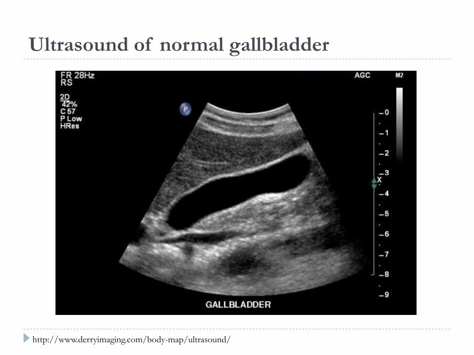
Ultrasound of normal gallbladder
http://www.derryimaging.com/body-map/ultrasound/

Ultrasound of gallbladder with gallstones
https://commons.wikimedia.org/wiki/File:Ultrasound_image_of_gallbladder_stone_Gallstone_091937515.jpg

Ultrasound of gallbladder “sludge”
http://www.radiologytutorials.com/main.cgi?tut=/main.cgi&frame=main&tt=1&s=2&t=80
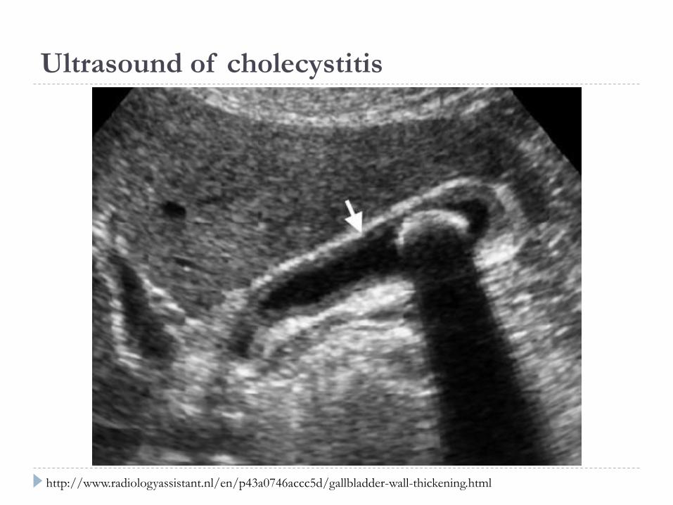
Ultrasound of cholecystitis
http://www.radiologyassistant.nl/en/p43a0746accc5d/gallbladder-wall-thickening.html

Ultrasound vs. CT of Cholecystitis
http://www.radiologyassistant.nl/en/p43a0746accc5d/gallbladder-wall-thickening.html
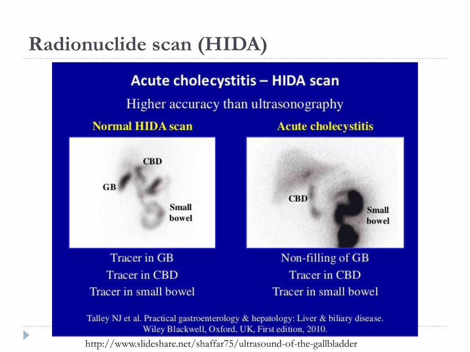
Radionuclide scan (HIDA)
http://www.slideshare.net/shaffar75/ultrasound-of-the-gallbladder

Magnetic Resonance
Cholangiopancreatography (MRCP)
https://mrimaster.com/anatomy/biliary%20system%20anatomy%20(mrcp)/
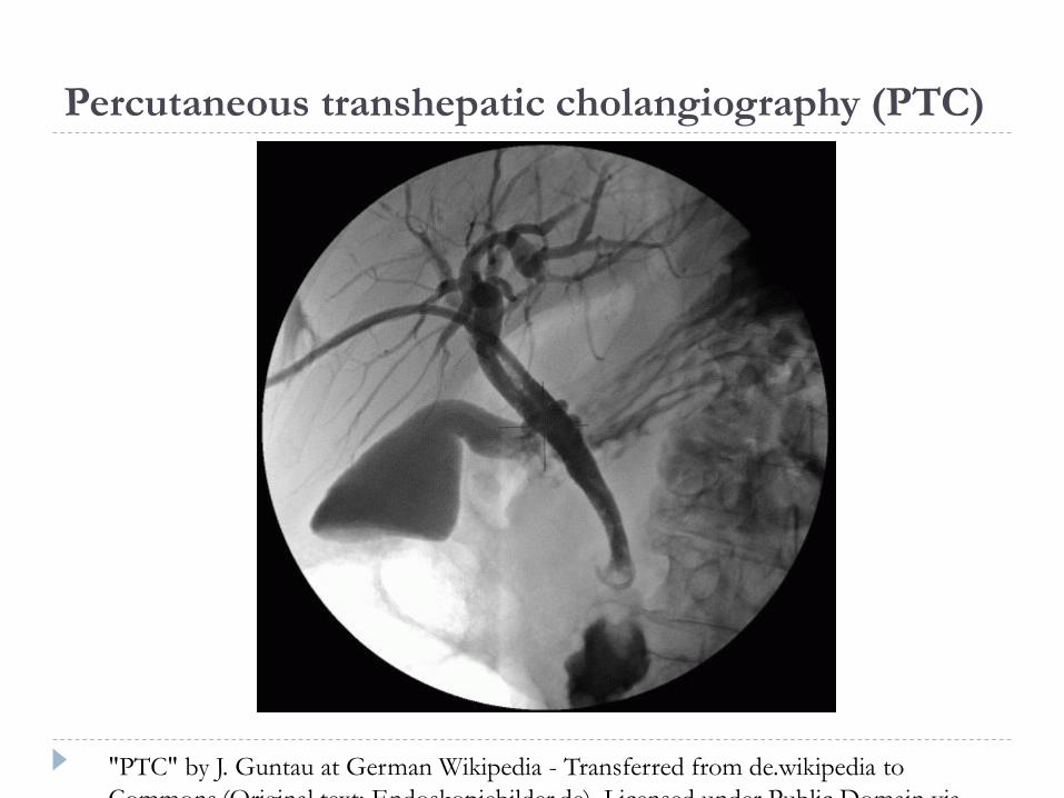
Percutaneous transhepatic cholangiography (PTC)
"PTC" by J. Guntau at German Wikipedia - Transferred from de.wikipedia to
Commons.(Original text: Endoskopiebilder.de). Licensed under Public Domain via
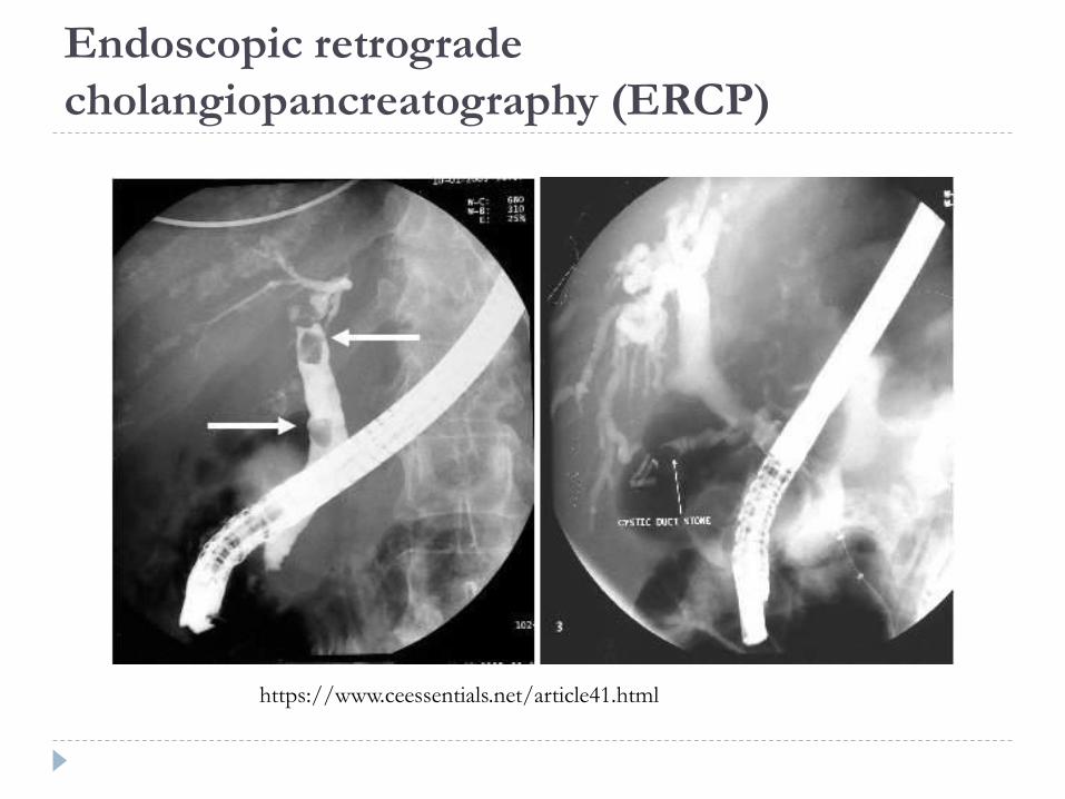
Endoscopic retrograde
cholangiopancreatography (ERCP)
https://www.ceessentials.net/article41.html

Natural History of Cholelithiasis

Spectrum of Gallbladder Disease
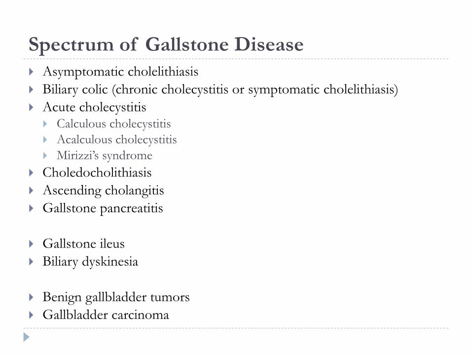
Spectrum of Gallstone Disease
Asymptomatic cholelithiasis
Biliary colic (chronic cholecystitis or symptomatic cholelithiasis)
Acute cholecystitis Calculous cholecystitis
Acalculous cholecystitis
Mirizzi’s syndrome
Choledocholithiasis
Ascending cholangitis
Gallstone pancreatitis
Gallstone ileus
Biliary dyskinesia
Benign gallbladder tumors
Gallbladder carcinoma

Case #1
53 year old female presented for evaluation after she
underwent an echocardiogram which incidentally identified
a 3-cm gallstone
She denied abdominal pain, nausea, vomiting, diarrhea, or
constipation
RUQ US demonstrated cholelithiasis (a large gallstone
measuring 3 cm) without gallbladder wall thickening,
pericholecystic fluid, or a sonographic Murphy’s sign


Asymptomatic cholelithiasis Approximately 30% of people with cholelithiasis end up having
surgery
Symptoms of gallstone disease generally do not change in severity
Each year, approximately 2% of patients with asymptomatic gallstones develop symptoms
Presence of gallstones in a person with asymptomatic or mildly symptomatic disease is not an indication for cholecystectomy
Reasons to recommend cholecystectomy in asymptomatic cholelithiasis
Large stones (> 2 cm in diameter) – they produce acute cholecystitis more often than small stones
Calcified gallbladder – often associated with carcinoma

Case #2
29 year old female presented complaining of episodic
abdominal pain and abdominal bloating approximately 1
hour after eating
RUQ US demonstrated cholelithiasis with a 1.6 cm gallstone
and diffuse gallbladder wall thickening without
pericholecystic fluid, and a negative sonographic Murphy’s
sign
HIDA demonstrated chronic cholecystitis (non-filling of the
gallbladder at 45 minutes, but eventual filling at 4 hours)

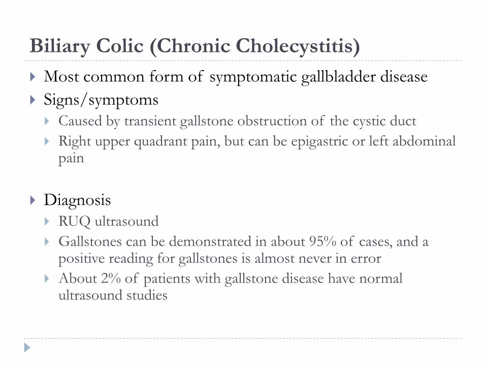
Biliary Colic (Chronic Cholecystitis)
Most common form of symptomatic gallbladder disease
Signs/symptoms
Caused by transient gallstone obstruction of the cystic duct
Right upper quadrant pain, but can be epigastric or left abdominal pain
Diagnosis
RUQ ultrasound
Gallstones can be demonstrated in about 95% of cases, and a positive reading for gallstones is almost never in error
About 2% of patients with gallstone disease have normal ultrasound studies
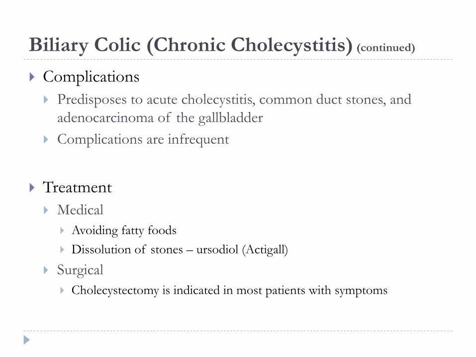
Biliary Colic (Chronic Cholecystitis) (continued)
Complications
Predisposes to acute cholecystitis, common duct stones, and
adenocarcinoma of the gallbladder
Complications are infrequent
Treatment
Medical
Avoiding fatty foods
Dissolution of stones – ursodiol (Actigall)
Surgical
Cholecystectomy is indicated in most patients with symptoms
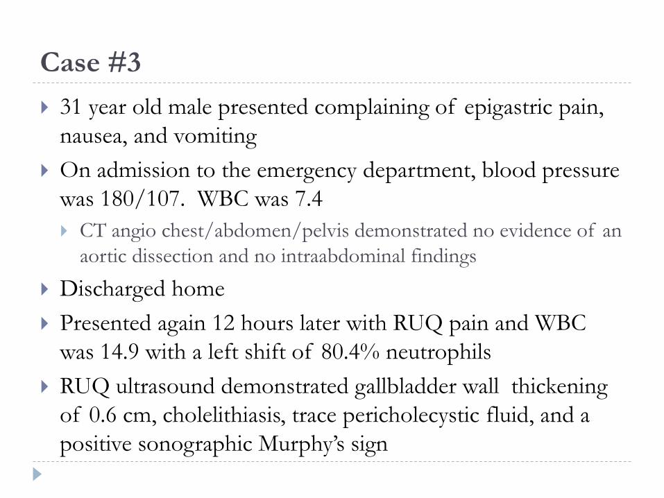
Case #3
31 year old male presented complaining of epigastric pain,
nausea, and vomiting
On admission to the emergency department, blood pressure
was 180/107. WBC was 7.4
CT angio chest/abdomen/pelvis demonstrated no evidence of an
aortic dissection and no intraabdominal findings
Discharged home
Presented again 12 hours later with RUQ pain and WBC
was 14.9 with a left shift of 80.4% neutrophils
RUQ ultrasound demonstrated gallbladder wall thickening
of 0.6 cm, cholelithiasis, trace pericholecystic fluid, and a
positive sonographic Murphy’s sign

Acute Cholecystitis
In 80% of cases, acute cholecystitis results from obstruction
of the cystic duct by a gallstone
Pathologic changes in the gallbladder
Gangrene and perforation may occur as early as 3 days after
onset, but most perforations occur during the second week
In cases that resolve spontaneously, acute inflammation has
largely cleared by 4 weeks
About 20% of cases of acute cholecystitis occur in the
absence of cholelithiasis (acalculous cholecystitis)
Most cases occur in patients hospitalized with some other illness
Common in trauma victims and patients receiving TPN

Acute Cholecystitis (continued)
Signs/symptoms
RUQ pain and tenderness
Murphy’s sign
Inspiratory arrest with right subcostal region palpation during inspiration
Nausea and vomiting are present in about half of patients, but the vomiting is rarely severe
Fever (usually 38°C to 38.5°C)
High fever and chills are uncommon and should suggest the possibility of complications or an incorrect diagnosis
Labs
WBC elevated
LFTs should be normal
Imaging
US
HIDA
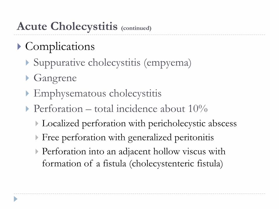
Acute Cholecystitis (continued)
Complications
Suppurative cholecystitis (empyema)
Gangrene
Emphysematous cholecystitis
Perforation – total incidence about 10%
Localized perforation with pericholecystic abscess
Free perforation with generalized peritonitis
Perforation into an adjacent hollow viscus with
formation of a fistula (cholecystenteric fistula)

Acute Cholecystitis (continued)
Treatment
IV fluids
IV antibiotics
Laparoscopic (open) cholecystectomy
Percutaneous cholecystostomy
Prognosis
Overall death rate of acute cholecystitis is about 5%

Algorithm for Management of Acute Cholecystitis
Adapted from:
RUQ pain,
tenderness,
RUQ US
positive
Non-
operative
management
If US
equivocal,
HIDA
Late
cholecystectomy
Good
operative
risk
Advanced
disease,
toxic
Early
cholecystectomy
(or
cholecystostomy)
No
No
Prompt
improvement
Yes
No
Diagnostic
Yes

Case #4
46 year old female presented complaining of RUQ pain
Labs
WBC count 6.8
Total bilirubin 5.8
Alkaline phosphatase 182
AST 237
ALT 516
RUQ US demonstrated cholelithiasis without gallbladder
wall thickening or pericholecystic fluid, and a slightly dilated
CBD (0.8 cm) without dilated intrahepatic bile ducts
ERCP demonstrated a stone in the distal CBD, which was
removed

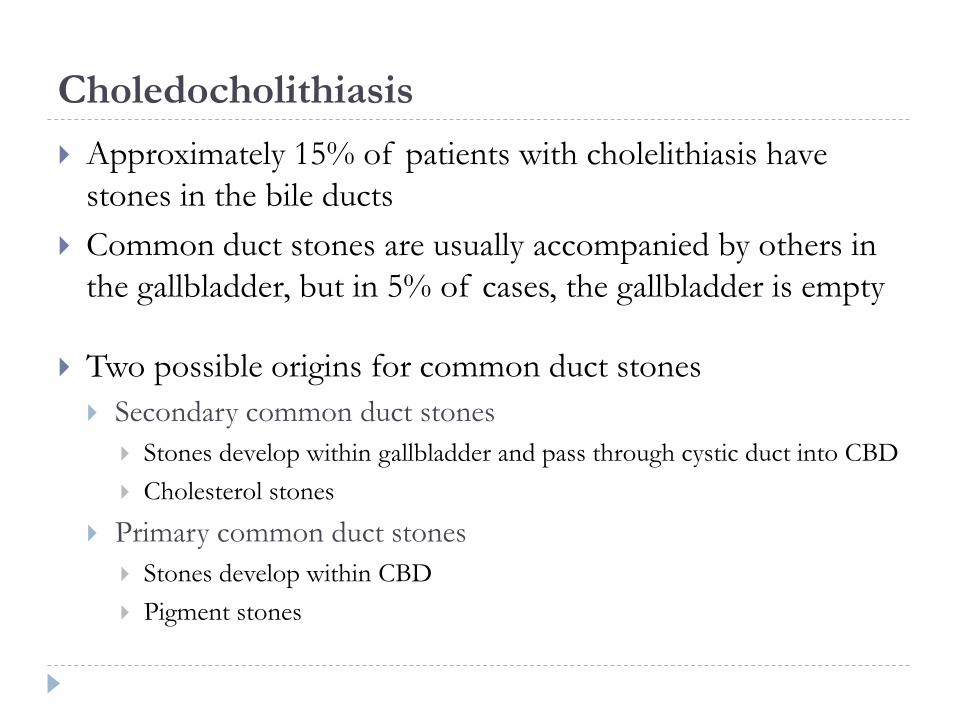
Choledocholithiasis
Approximately 15% of patients with cholelithiasis have
stones in the bile ducts
Common duct stones are usually accompanied by others in
the gallbladder, but in 5% of cases, the gallbladder is empty
Two possible origins for common duct stones
Secondary common duct stones
Stones develop within gallbladder and pass through cystic duct into CBD
Cholesterol stones
Primary common duct stones
Stones develop within CBD
Pigment stones

Choledocholithiasis (continued)
Symptoms
RUQ, epigastric or sub-sternal pain with referred pain to the right
scapula
Intermittent chills/fever
Jaundice
Transient darkening of urine
Pruritis
Labs
AST/ALT, Alkaline phosphatase, and bilirubin all elevated
WBC may be elevated or normal

Choledocholithiasis (continued)
Imaging
X-ray
US
CT
MRCP
ERCP

Choledocholithiasis (continued)
Complications
Intra-hepatic abscesses
Hepatic failure or secondary biliary cirrhosis
Acute pancreatitis
Erosion of a CBD stone through the ampulla gallstone ileus
Hemorrhage (hemobilia)
Treatment
Antibiotics if cholangitis is suspected
ERCP
Cholecystectomy with cholangiogram
Common bile duct exploration

Natural History of Choledocholithiasis
Gallbladder
stones
100
Cholangitis
3
Biliary
colic
2
Pancreatitis
1
Suppurativ
e
cholangitis
Jaundice
3
Asymptomatic
common duct
stones
6
Common
bile duct
stones
15
Rare
Adapted from:

Case #5
28 year old male presented complaining of severe epigastric abdominal pain, nausea, vomiting, and chills
History of a laparoscopic cholecystectomy 3 weeks prior
Febrile (101.4 °F)
Labs
WBC 10.8
Total bilirubin 7.1
Alkaline phosphatase 199
AST 130
ALT 408
CT abdomen/pelvis demonstrated intra- and extra-hepatic biliary ductal dilatation
ERCP demonstrated a stone in the CBD, which was extracted

Ascending cholangitis
Bacterial infection of obstructed biliary ducts
Causes
Choledocholithiasis
Biliary stricture
Neoplasm
Less common:
Chronic pancreatitis, ampullary stenosis, pancreatic pseudocyst, duodenal diverticulum, congenital cyst, and parasitic invasion
Iatrogenic cholangitis may complicate transhepatic or T-tube cholangiography
Higher chance of ascending cholangitis once the duct is colonized with bacteria
Predominant organisms (in decreasing frequency) are E. coli, Klebsiella, Pseudomonas, Enterococci, and Proteus

Ascending cholangitis (continued)
Diagnosis is mostly clinical, although RUQ ultrasound can demonstrate dilated intra- and extra-hepatic ducts
Charcot’s triad – present in only 70% of cases
RUQ pain
Fever
Jaundice
Reynolds’ pentad
Charcot’s triad
Altered mental status
Hypotension
Ascending cholangitis (continued)
Treatment
Antibiotics
ERCP
PTC
Common bile duct exploration

Case #6
46 year old female presented complaining of two days of severe abdominal pain, which started in the epigastrium and became diffuse, associated with nausea and vomiting
Labs WBC 21.2
Total bilirubin 2.3
Alkaline phosphatase 126
AST 99
ALT 170
Lipase 8059
RUQ US demonstrated a distended gallbladder with mild wall thickening, small pericholecystic fluid, non-mobile stones, and a mildly dilated CBD (0.9 cm)
MRCP demonstrated acute pancreatitis with extensive peripancreaticinfiltration and acute peripancreatic fluid collections, cholelithiasis, choledocholithiasis, and mild biliary dilatation


Gallstone Pancreatitis
Acute pancreatitis due to biliary obstruction
Diagnosis is mostly clinical (history, physical exam, labs)
RUQ ultrasound demonstrates cholelithiasis
CT can be used to find necrotizing pancreatitis, fluid collections,
or other complications but is not technically required to make the
diagnosis
Treatment is supportive initially
Cholecystectomy with cholangiogram prior to discharge
from the hospital if the pancreatitis is mild or moderate to
avoid a recurrent episode
~30% will recur within 6 weeks if CBD is not cleared prior to
discharge

Gallstone Ileus
A mechanical intestinal obstruction caused by a large
gallstone lodged in the lumen
It occurs more often in women, and the average patient age
is about 70 years
Usually presents with obvious small bowel obstruction
The obstructing gallstone enters the intestine through a
cholecystenteric fistula located in the duodenum, colon, or,
rarely, the stomach or jejunum
Stones that cause gallstone ileus are almost always 2.5 cm or
more in diameter

Gallstone Ileus (continued)
Imaging
Abdominal X-ray may show a radiopaque gallstone
Pneumobilia will be seen in about 40% of cases
http://radiopaedia.org/articles/gallstone-ileus
Gallstone Ileus (continued)
Treatment
Emergency laparotomy and removal of the obstructing stone
through a small proximal enterotomy
Leave gallbladder alone at emergency laparotomy
The death rate from gallstone ileus remains about 20%,
largely because of the poor general condition of elderly
patients at the time of laparotomy

Summary
RUQ pain? Fever?Elevated
WBC?
Elevated
LFTs?Diagnosis? Treatment?
Biliary colicYes
(intermittent)No No No US
Laparoscopic
cholecystectomy
Acute
cholecystitis
Yes
(constant)Yes Yes No
US
HIDA
1) Antibiotics
2) Laparoscopic
cholecystectomy
3) Percutaneous
cholecystostomy
Choledocholithiasis Yes No No Yes MRCP
1)ERCP
2) Laparoscopic
cholecystectomy
Ascending
cholangitisYes Yes Yes Yes
Clinical
US
1) Antibiotics
2) ERCP
3) PTC
4) CBD exploration
Gallstone
pancreatitis
Yes
(Epigastric)Maybe Yes Yes
Clinical
US
CT
1)Supportive
2) Laparoscopic
cholecystectomy
Post-Cholecystectomy Syndrome
A heterogeneous group of disorders affecting patients who continue to complain of symptoms after cholecystectomy
The usual reason for incomplete relief after cholecystectomy is that the preoperative diagnosis of chronic cholecystitis was incorrect
An organic cause for the symptoms is more likely to be discovered in patients with severe episodic pain than in those with other complaints
Abnormal liver function studies, jaundice, and cholangitis are other manifestations that indicate residual biliary disease
Patients with suspicious findings should be studied by ERCP or PTC
Benign Gallbladder Tumors
The differentiation from gallstones is based upon observing
whether a shift in position of the projections follows
changes in posture of the patient, since stones are not fixed
Cancer should be suspected in any polypoid lesion that
exceeds 1 cm in diameter
Polyps
Adenomyomatosis
Adenomas

Gallbladder Carcinoma
An uncommon neoplasm that occurs in elderly patients
Associated with gallstones in 70% of cases
The risk of malignant degeneration correlates with the
length of time gallstones have been present
Prevalence in women compared to men is approximately 2:1
Histology – adenocarcinoma is the most common
Scirrhous (60%)
Papillary (25%)
Mucoid (15%)
Gallbladder Carcinoma (continued)
Early direct invasion of the liver and hilar structures and by metastases to the common duct lymph nodes, liver, and lungs
Carcinoma can be incidentally found after cholecystectomy, where the tumor is confined to the gallbladder
Symptoms
Right upper quadrant pain
Obstruction of the cystic duct by tumor sometimes initiates an attack of acute cholecystitis
Other cases present with obstructive jaundice and, occasionally, cholangitis due to secondary involvement of the common duct

Gallbladder carcinoma – Imaging

Gallbladder Carcinoma:
Complications and Prevention
Complications
Intra-hepatic, pericholecystic or within the gallbladder abscesses
Prevention?
Incidence of gallbladder cancer has decreased in recent years as
the frequency of cholecystectomy has increased
Estimated that one case of gallbladder cancer is prevented for
every 100 cholecystectomies performed

Gallbladder Carcinoma: Surgical Treatment
In the few cases when cancer has not penetrated the muscularis mucosae, cholecystectomy alone should suffice
Small invasive carcinoma discovered by the pathologist
Reoperation to perform a wedge resection of the liver bed plus regional lymphadenectomy
Localized carcinoma
Cholecystectomy along with en bloc wedge resection of an adjacent 3-5 cm of normal liver and dissection of the lymph nodes in the hepatoduodenal ligament
More extensive hepatectomies (e.g., right lobectomy) are not worthwhile
There is little that surgery can offer in cases with hepatic metastases or more distant spread

Gallbladder Carcinoma: Prognosis
Radiotherapy and chemotherapy are not effective
palliative measures
About 85% of patients are dead within a year
after diagnosis
The 10% of patients who survive more than 5
years:
Carcinoma was an incidental finding during
cholecystectomy for symptomatic gallstone disease
An aggressive resection has removed all gross tumor

References
CURRENT Diagnosis & Treatment: Surgery, 14e
Gerard M. Doherty
Schwartz’s Principles of Surgery, 10e
F. Charles Brunicardi, Dana K. Andersen, Timothy R.
Billiar, David L. Dunn, John G. Hunter, Jeffrey B. Matthews,
Raphael E. Pollock
The History of Medicine: The Galling Gallbladder.
http://columbiasurgery.org/news/2015/06/11/history-
medicine-galling-gallbladder

#ILookLikeASurgeon #NYerORCoverChallenge