the special senses - unit: (hk24cy008)
TRANSCRIPT

1
THE SPECIAL SENSES - Unit: (HK24CY008)
INTRODUCTION
The Special Senses include sight, smell (olfaction), taste (gustation), hearing and
equilibrium (balance) and touch.
The senses exist to provide information about the environment. Sensory receptors, either
on the surface of the relevant organ or within its tissues, receive and translate stimuli into
meaningful information. The body can then respond accordingly.
The Eye is the organ of sight
The Ear is the organ of hearing and equilibrium (balance)
The Tongue is the organ of taste
The Nose is the organ of smell (as well as respiration)
The Skin or Integument is the organ of touch

2
THE EYE – STRUCTURE AND FUNCTION
In ancient civilisations, the eyes were considered to be the gateway to the soul. In health
and disease, they often serve as a window, giving us clues to illnesses situated in other parts
of the body.
For instance, a yellow tinge on the white of the eyes may indicate jaundice; fatty deposits on
the eyelids may suggest high cholesterol levels; atherosclerosis and hypertensive changes to
the arterial circulation may also be observed in the circulation of the retina.

3
The eyes are innervated by the optic nerve (the second cranial nerve), and movement is
controlled by six external muscles that have the ability to move the eyeball in a number of
directions. These muscles are innervated by the third, fourth and sixth cranial nerves.
Structurally, the two eyes function separately but their activities are co-ordinated so that
they work as a pair. It is possible to see with only one eye, but abilities such as three
dimensional vision and judgement of distance are only possible when both eyes work
together.
The adult eyeball measures about 2.5cm in diameter. Only the anterior (front), one-sixth is
exposed - the remainder is recessed and protected by the orbit, into which it fits.
Anatomically, the wall of the eyeball consists of three layers or tunics: fibrous tunic,
vascular tunic and retina.
Fibrous Tunic
This is the outer layer of the eyeball, and it consists of the anterior cornea and posterior
sclera.
The cornea is a transparent coat that covers the coloured iris. Because it is curved
(convex), the cornea helps focus light onto the retina by refracting or bending the light rays.
The cornea is covered by the conjunctiva; a fine transparent membrane.
The sclera, the “white” of the eye, is a layer of dense connective tissue made up mostly of
collagen fibres and fibroblasts. The sclera covers the eyeball (except for the cornea) –
giving the eyeball its shape, making it more rigid, and protecting its inner parts. The sclera
also provides the attachment point for the muscles that move the eye. At the junction of
the sclera and cornea, is an opening known as the scleral venous sinus (canal of Schlemm,
named after a German anatomist). This is where the aqueous humour drains back into the
venous circulation.
Vascular Tunic
The vascular tunic, or uvea, is the middle layer of the eyeball and has three parts – choroid,
ciliary body and iris.
The choroid lines most of the internal surface of the sclera and is rich in blood vessels,
providing nutrients to the posterior surface of the retina. Light enters the eye through the
pupil, stimulates the nerve endings in the retina and is then absorbed by the choroid.

4
In the anterior portion of the vascular tunic, the choroid becomes the ciliary body, which
consists of the ciliary processes and the ciliary muscle. The ciliary processes are
protrusions, or folds, on the internal surface of the ciliary body; they contain epithelial cells
that secrete aqueous humour.
Aqueous humour supplies nutrients and removes waste from the transparent structures in
the front of the eye that have no blood supply, ie the cornea, lens and lens capsule. There is
a continuous production and drainage of the aqueous humour, but the intraocular pressure
remains constant - if there is an increase in pressure it causes Glaucoma. The ciliary
processes also attach to suspensory ligaments, which connect to the lens. The ciliary muscle
is a circular band of smooth muscle that alters the shape of the lens, adapting it for near or
far vision.
The Iris, or coloured part of the eyeball, is shaped like a flattened doughnut. It is
suspended between the cornea and the lens and attached to the ciliary processes. A principal
function of the iris is to regulate the amount of light entering the vitreous chamber of the
eyeball through the pupil, ie the hole in the centre of the iris.
Autonomic reflexes regulate pupil diameter in response to light levels, meaning that the pupil
dilates the more light stimulus it receives.
To nourish the lens and cornea, the ciliary body secretes a fluid called aqueous humour into
the posterior chamber; this then passes through the pupil into the anterior chamber. The
cavity behind the lens is filled with a gel, known as the vitreous body, which helps to maintain
the shape of the eyeball. It is a soft, colourless, transparent, jelly-like substance composed
of 99% water, some salts and mucoprotein. The eye keeps its shape because of the
intraocular pressure exerted by the vitreous body and the aqueous fluid.
The colour of the iris is genetically determined and depends on the number of pigment cells
present. Albinos have no pigment cells, and people with blue eyes have fewer than those with
brown eyes.
Just posterior to the pupil and iris, within the cavity of the eyeball is the lens; a highly
elastic, circular, biconvex, transparent body. The lens fine-tunes focussing of light rays onto
the retina to facilitate clear vision.
Retina
The third, and inner layer of the eyeball, is the retina. It lines the posterior three-quarters
of the eyeball and is the beginning of the visual pathway. The surface of the retina is the
only place in the body where blood vessels can be viewed directly and examined for
pathological changes (with an ophthalmoscope), such as those that occur with hypertension or
diabetes mellitus.

5
The retina is made up of layers of nerve cell bodies and their fibres, lying on a pigmented
layer of epithelial cells which attach it to the choroid. The layer that contains light sensitive
cells (photoreceptors) is the layer of rods and cones.
Each retina has about 6 million cones and 120 million rods. Rods have a low light threshold
allowing us to see in dim light, such as moonlight. Because they do not provide colour vision, in
dim light, we see only shades of grey. Rhodopsin is a pigment present in rods, and it is
degraded or bleached when exposed to light - its regeneration requires the presence of
Vitamin A.
Brighter lights stimulate the cones, which contain other pigments and have a higher
threshold to produce colour vision. Most of our visual experiences are mediated by the cone
system, the loss of which produces blindness. In contrast, a person who loses rod vision,
mainly has difficulty seeing in dim light and thus should not drive at night.
Light passes through the cornea, pupil, lens and vitreous humour to reach the retina. Images
relayed to the retina are transported via the optic nerve, with the nerves of each eye
converging at the optic chasm and connecting to the brain.
Near the centre of the posterior part of the retina, there is a yellow looking area known as
the macula lutea, and in the centre of this area, there is a little depression called fovea
centralis, consisting of only cone-shaped cells. Towards the anterior part of the retina,
there are fewer cone than rod shaped cells. About 0.5cm to the nasal side of the macula
lutea all the nerve fibres of the retina converge to form the optic nerve. The optic nerve
then passes through the sphenoid bone to reach the cerebral cortex in the occipital lobe of
the cerebrum. The small area of the retina where the optic nerve leaves the eye is the optic
disc or blind spot, so called because it has no light-sensitive cells.
ACCESSORY ORGANS OF THE EYE

6
Eyebrows
Eyelids and Eyelashes
Lacrimal Apparatus
The Eyebrows - protect the anterior aspect of the eyeball from sweat, dust and other
foreign bodies.
The Eyelids and Eyelashes - protect the eye from injury:
Reflex closure of the lids occurs when the conjunctiva or eyelashes are touched, or when
an object comes close to the eye, or a bright light shines into the eye.
Blinking at about 3-7 second intervals spreads tears and Meibomian secretions over the
cornea, preventing drying.
Meibomian glands secrete an oily substance in the free margin of the eyelids, to prevent
evaporation of tears.
Eyelids are lined with a fine transparent membrane known as the conjunctiva.
The Lacrimal glands are situated at the upper corner of the orbit - the cavity at the front
of the skull housing the eyeball. Tears are secreted by the lacrimal gland - these tears form
a protective film and lubricate the eye. The blinking movement of the eyelid moves the fluid
toward its drainage point, the nasolacrimal duct, which opens to the nose. Tears are
composed of water, mineral salts, antibodies, and lysozyme, a bactericidal enzyme.
SIGHT

7
Light waves travel at a speed of 186,000 miles (300,000 kilometres) per second. Light is
reflected into the eyes by objects within the field of vision. The left and right eyes have
slightly different fields of vision, which overlap and merge together to focus on an object,
allowing us to discern distance and 3-D structure. This is known as binocular vision.
When an object is viewed, the light reflected off the object enters the eye - with the
amount of light entering the eye, being governed by the iris, ie the coloured part of the eye
which lies behind the cornea. Light rays passing through the cornea are adjusted to bring the
rays closer together; these rays then pass through the pupil to the lens. Muscular action can
alter the shape of the lens to allow focus on distant and near objects. The light rays then
pass through the vitreous humour to the retina.
As the light rays travel through the eye to the retina, they are concentrated and bent
(refracted), so that the image received on the back of the retina is an inverted, transposed
version of the object in sight.
The photoreceptors of the retina, called rods and cones, are activated by the light rays and
send nerve impulses along the cells of the optic nerve. The optic nerve (cranial nerve II)
transmits the impulses to the thalamus, where some of the processing of the visual
information is conducted by the lateral geniculate nuclei of the thalamus.
The nerve fibres of the optic nerve from the nasal side of each retina cross over to the
opposite side. The fibres from the temporal side do not cross, but continue backwards on the
same side. The visual information is then relayed to the visual cortex in the occipital lobe of
the brain for interpretation.
The visual cortex interprets and makes sense of nerve impulses sent from the eyes via the
optic nerves and thalamus. This is where the messages from the right and left eyes are
merged to form one image and the object seen is recognised.
The visual association cortex processes more complex features of the visual stimulus, such
as colour and movement. The images received on the retina are upside down, but the brain
automatically corrects these images.

8
Summary
We see because the structure of the eye is specially designed to bend and concentrate light
rays to form a tiny image of a seen object on the back of the eye. This is then transported
to the brain in the form of nerve impulses. Light rays entering the eye strike the cornea,
which bends (refracts) the rays bringing them closer together. The rays then pass through
the lens, which focuses the rays on the back of the retina. The retina consists of a layer of
light sensitive cells – rods and cones – which when stimulated by light, send electrical signals
along the cells of the optic nerves to the brain.
THE NOSE – STRUCTURE AND FUNCTION
The nose is part of the respiratory system and also the special sense organ of smell.
A combination of bone and cartilage make up the nose. The nasal bone and maxillae comprise
the bony component of the external structure, while the nostrils are formed by cartilage.
The nasal septum, which divides the two nostrils, is a combination of bone and cartilage. The
bones surrounding the nasal cavities are the vomer and parts of the frontal, ethmoid,
maxillary and sphenoid bones. The floor of the nasal cavities forms the roof of the palate.
The nasal cavities lead to the trachea via the pharynx.

9
Olfactory bulb
Olfactory
Receptor cells in the
nasal epithelium
Olfactory
Epithelium
Cilia in mucous
membrane

10
Inhaled air is warmed and moistened in the nose, and any foreign particles are trapped by
the hairs lining the nostrils. In the nasal cavities, there are curved plates, called the
conchae; these conchae increase the surface area available to warm and moisten the inhaled
air. The nasal cavity is lined with mucous membrane (respiratory mucosa), which changes to
olfactory mucosa in the upper region of each nasal cavity. The respiratory mucosa has a
covering of minute hairs that capture foreign particles and send them to the nasopharynx.
The olfactory mucosa contains special nerve called cells chemoreceptors, used to determine
smell.
SMELL
The sense of smell is the most immediate of the senses and closely allied to the sense of
taste. Odour molecules enter the nostrils and pass into the nasal cavity.
Nasal epithelium at the back of the nasal cavity contains tens of millions of olfactory
receptors. Olfactory receptors are neurons (chemoreceptors), and each neuron has several
cilia, called olfactory hairs, protruding from the dendrites. These hairs are positioned in the
olfactory mucosa covering the nasal epithelium.
Odours are absorbed by the nerve cells, triggering nerve impulses, which are transmitted
along the axons of the olfactory receptors that, in turn, unite to form the olfactory nerves.
The olfactory nerves terminate in the brain in paired masses of grey matter called the
olfactory bulbs. The olfactory bulbs are positioned beneath the frontal lobes of the
cerebrum.
Nerve impulses from the olfactory bulb are conducted along the olfactory nerve (cranial
nerve I). The impulses are then relayed to the olfactory cortex, the limbic system and the
hypothalamus in the brain, for identification.

11
The limbic system plays an important role in memory and emotion. The olfactory bulb is
closely linked to the limbic system, in particular with the hippocampus and amygdala, which is
why smells often trigger memories of past places and feelings. Some smells stimulate the
limbic system to activate the hypothalamus and pituitary gland, which triggers the release of
hormones associated with appetite and emotional responses. The olfactory system was
thought capable of building a library of more than 10,000 odours but latest research
discounts this and estimates that the 400 nasal nerve receptors can detect 1 trillion or more
different scents .
On first exposure to an odour the perception of it is quick and sharp, however after a short
time, the ability to perceive it fades. This “tiring” of the sense of smell is known as anosmia.
Food smells can stimulate the production of saliva. However, someone with a cold whose
sense of smell is reduced, and complains of loss of the taste sensation, is in fact
experiencing a blockage of olfaction.
THE TONGUE – STRUCTURE AND FUNCTION
The tongue is the muscular organ of taste. It has a dorsum or upper surface, a base
attached to the floor of the mouth, a soft lower surface and a tip.
Since it is required to perform a number of different functions, including chewing, swallowing
and speaking, the tongue has a range of movements. The muscles in the tongue are arranged
in three directions, allowing the tongue to shorten, narrow and thin out; the muscles are
called the intrinsic tongue muscles. The extrinsic tongue muscles are attached to the jaw,
skull, palate and hyoid bones; these muscles allow the tongue to change position, moving it
forward, backward, upward and downward. The upper side of the tongue, the dorsum, is
covered with three types of papillae (small projections): filiform, fungiform and vallate
papillae. Taste buds are found in both the fungiform and vallate papillae; they are also found
on the palate, epiglottis and pharynx. An adult has about 9,000 taste buds. Each taste bud
consists of groups of receptor cells, and each of these has fine hair-like projections (called
micro-villi) sticking out into the surface of the tongue through fine pores in the surface of
the papilla. The receptor cells link up with a network of nerve fibres. The soft underside of
the tongue is kept moist by secretions from the salivary glands.
Taste
The sense of taste, also known as gustation, is aided by the sense of smell. In fact, 80
percent of the sensation of taste is due to smell.
Like smell, the taste mechanism is triggered by the chemical content of substances in food
and drink. Saliva, released by the salivary glands, mixes with food - dry food has little
flavour, the more liquid the better the sensation of taste. When the mixture comes into

12
contact with the taste buds, they are activated, which then sends nerve impulses to the
brain.
There are four basic taste types: sweet, sour, salty, and bitter and in recent years a
savoury taste has been identified and called Umami. Certain areas of the tongue are more
sensitive than others to these particular taste types. The taste buds at the front of the
tongue are most sensitive to sweet and salty, with the tip of the tongue being particularly
sensitive to sweet foods. The taste buds located at the sides of the tongue are best at
detecting sour tastes, while bitterness is best registered at the back of the tongue. Some
of the stronger tastes, such as the hot flavour of spicy food are due to stimulation of pain-
sensitive nerve endings in the tongue. To protect the body from poisonous substances, when
foul tasting food is eaten, it stimulates the gagging reflex that may lead to vomiting.
Three cranial nerves, the facial nerve (cranial nerve VII), the glossopharyngeal nerve (cranial
nerve IX) and the vagus nerve (cranial nerve X) are involved in the sense of taste. Taste
buds on the front part of the tongue activate the facial nerve; taste buds on the back of the
tongue activate the glossopharyngeal nerve; and the vagus nerve is triggered by the taste
buds found in the throat. Nerve impulses are initiated by the activated taste buds and are
transmitted along these three nerves to the brain. Therefore, nerve impulses from the
receptor cells in the taste buds are sent via the cranial nerves to the parietal lobe of the
cerebrum which is responsible for taste identification.
13
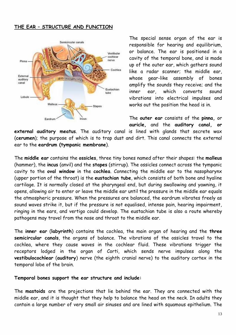
THE EAR – STRUCTURE AND FUNCTION
The special sense organ of the ear is
responsible for hearing and equilibrium,
or balance. The ear is positioned in a
cavity of the temporal bone, and is made
up of the outer ear, which gathers sound
like a radar scanner; the middle ear,
whose gear-like assembly of bones
amplify the sounds they receive; and the
inner ear, which converts sound
vibrations into electrical impulses and
works out the position the head is in.
The outer ear consists of the pinna, or
auricle, and the auditory canal, or
external auditory meatus. The auditory canal is lined with glands that secrete wax
(cerumen); the purpose of which is to trap dust and dirt. This canal connects the external
ear to the eardrum (tympanic membrane).
The middle ear contains the ossicles, three tiny bones named after their shapes: the malleus
(hammer), the incus (anvil) and the stapes (stirrup). The ossicles connect across the tympanic
cavity to the oval window in the cochlea. Connecting the middle ear to the nasopharynx
(upper portion of the throat) is the eustachian tube, which consists of both bone and hyaline
cartilage. It is normally closed at the pharyngeal end, but during swallowing and yawning, it
opens, allowing air to enter or leave the middle ear until the pressure in the middle ear equals
the atmospheric pressure. When the pressures are balanced, the eardrum vibrates freely as
sound waves strike it, but if the pressure is not equalised, intense pain, hearing impairment,
ringing in the ears, and vertigo could develop. The eustachian tube is also a route whereby
pathogens may travel from the nose and throat to the middle ear.
The inner ear (labyrinth) contains the cochlea, the main organ of hearing and the three
semicircular canals, the organs of balance. The vibrations of the ossicles travel to the
cochlea, where they cause waves in the cochlear fluid. These vibrations trigger the
receptors lodged in the organ of Corti, which sends nerve impulses along the
vestibulocochlear (auditory) nerve (the eighth cranial nerve) to the auditory cortex in the
temporal lobe of the brain.
Temporal bones support the ear structure and include:
The mastoids are the projections that lie behind the ear. They are connected with the
middle ear, and it is thought that they help to balance the head on the neck. In adults they
contain a large number of very small air sinuses and are lined with squamous epithelium. The

14
mastoid process is a rounded projection of the mastoid portion of the temporal bone,
posterior to the external auditory meatus. It serves as a point of attachment for several
neck muscles.
The styloid process (styl-=stake or pole) projects inferiorly from the inferior surface of the
temporal bone, and serves as a point of attachment for muscles and ligaments of the tongue
and neck. Between the styloid process and the mastoid process, is the stylomastoid foramen.
At the floor of the cranial cavity is the petrous portion of the temporal bone. This portion is
triangular and located at the base of the skull between the sphenoid and occipital bones. The
petrous portion houses both the internal ear and the middle ear.
Hearing
Sound waves are produced by the vibrations of air molecules. The size and energy of these
waves determines loudness, which is measured in decibels.
The auricle (pinna) directs sound waves into the external auditory canal. When sound waves
strike the tympanic membrane (eardrum), the alternating high and low pressure of the air
cause the eardrum to vibrate back and forth. The distance it moves, which is very small,
depends on the intensity and frequency of sound waves. The eardrum vibrates slowly in
response to low frequency sounds and rapidly in response to high frequency sounds.
The central area of the eardum connects to the malleus (ossicle), which also starts to
vibrate. The vibration is transmitted from the malleus to the incus and then to the stapes.
As the stapes moves back and forth, it pushes the membrane of the oval window (entrance to
the cochlea) in and out. The oval window vibrates about 20 times more vigorously than the
eardrum because the ossicles efficiently transmit small vibrations spread over a large
surface area (eardrum) into larger vibrations of a smaller surface area (oval window).
The movement of the oval window sets up fluid pressure waves in the perilymph of the
cochlea. Perilymph is the watery fluid between the bony and membranous labyrinths. In the
membranous labyrinth the watery fluid is called endolymph. As the oval window bulges
inward, it continues to cause pressure waves in the perilymph until they reach the round
window, causing it to bulge outward into the middle ear. As the pressure waves deform the
walls of the cochlea, pressure waves are formed in the endolymph inside the cochlea duct.
The pressure waves in the endolymph cause the hair cells, (receptor cells), of the spiral
organ (organ of Corti) to move. This movement leads to the generation of nerve impulses in
cochlea nerve fibres. The hair cells transduce mechanical vibrations into electrical signals
which are sent along the vestibulocochlear nerve (cranial nerve VIII) to the hearing area in
the cerebrum, where sound is perceived, and to various nuclei in the pons varolii and the
midbrain.

15
Balance
Within the ear, movement is registered by the vestibular apparatus that includes semi-
circular canals and otolith organs. The otolith organs (the utricle and saccule) are hollow sacs
containing a gelatinous fluid. Tiny hair cells are anchored to the inner surface of the organs
known as the maculae, and above them lie crystals of calcium carbonate, the otoliths. When
the head moves, the otoliths change position, activating the hair cells, which send impulses to
the brain. The brain then triggers the body’s reflex mechanisms to correct the position of
the body.
The hair-like nerve cells in the semi-circular ducts are also triggered by fluid (endolymph)
movement, causing the nerves to send impulses to the brain. The nerves of the semi-circular
canals and otolith organs transmit impulses via the vestibulocochlear nerve to the
cerebellum. While the eyes transmit visual information about the body’s position, the semi-
circular canals and otolith organs, along with the nerves and muscles that control motor co-
ordination and movement, maintain the body’s balance.

16
SKIN – STRUCTURE AND FUNCTION
(also refer to the handout on the Integumentary system)
The skin is a protective organ that covers the body, and it is the body’s largest organ. The
function of the skin is to provide a protective layer against injury and attack, extremes of
temperature and invading organisms, such as viruses and bacteria. The skin is also involved in
body temperature regulation (normal body temperature 37C), vitamin D production, and
ultraviolet (radiation) protection. It detects pain and touch.
Within its three layers - the epidermis, dermis and subcutaneous tissue - the skin contains
specialised structures, including nerve receptors, hair follicles, sweat glands and
sebaceous glands.
The outer layer of the skin, the epidermis is itself divided into five layers, with each of
the sub-layers performing a specific function. There are no blood vessels or nerve endings in
the epidermis, and its surface is ridged by projections of cells in the dermis called papillae.
The pattern of ridges is different in every individual and the impression made by them is the
fingerprint.
The bottom layer of the epidermis, (stratum basale), is responsible for the production of
melanin from the melanocytes, which absorbs dangerous ultraviolet light and creates the
pigment which gives skin its tanned appearance after exposure to sunlight. The amount of
melanin varies in different parts of the body, with very little melanin in the soles of the feet
and palms of the hands; it also varies from person to person, although the number of
melanocytes is fairly constant in all people.
The level of oxygenation of haemoglobin, and the amount of blood circulating in the dermis,
gives the skin its pink colour. Bile pigments in blood, and carotenes in subcutaneous fat, give
the skin a yellowish colour.
The layers of the epidermis are:

17
Stratum corneum – The thickest outside part of the epidermis, it is waterproof and
composed of dead keratinocytes
Stratum lucidum - The clear transparent layer present in skin of fingertips, palms and soles.
Composed of dead keratinocytes
Stratum granulosum – Thickest on palms and soles, keratinisation begins here, and also
lamellar granules that produce a lipid-rich, water repellent secretion.
Stratum spinosum – Active layer, contains keratinocytes, and projections of the
melanocytes, producing melanin, plus Langerhans cells (immune response cells formed in the
bone marrow that migrate to the epidermis).
Stratum germinativum (basale) – The deepest section and where new cells are formed. It
contains melanocytes and keratinocytes, together with Langerhans and Merkel cells.
The outermost layer (stratum corneum) provides the main defence against skin infection.
This outer layer consists mainly of dead cells, which are constantly sloughed off, and then
replaced by the cells of the underlying layer, ensuring that the skin is constantly being
renewed.
The cells of the epidermis are produced in a specific way, starting in the deepest layer of
the epidermis, known as the germinative layer (Stratum germinativum). The cells obtain their
nutrients from the dermis, via the flow of interstitial fluid arising from this layer. Waste
products are removed by the lymph vessels.
After formation these cells are pushed towards the surface of the skin. As they move up the
epidermis, they slowly ‘die’, losing their cytoplasm as they ‘dry up’, and in doing so become
flatter in shape. These are the cells that form the stratum granulosum and stratum lucidum.

18
As the epidermal cells ‘die’ off, the contents are replaced by the protein keratin which
toughens cells, and helps to waterproof and protect the skin. Keratin is produced by
keratinocytes.
Eventually, these cells reach the uppermost layer, known as the stratum corneum (horny
layer). At this point, the cells are flat, thin and fully keratinised. This is the layer that is
constantly being rubbed off and shed by the body in a process known as desquamation.
The maintenance of a healthy epidermis is dependent upon:
Continual cell division in the germinative layer
Effective keratinisation of the cells
Desquamation of keratinised cells from the stratum corneum
The structures, lying within the layers of the skin, each perform a supportive role for the
skin. The hair follicles hold the hairs that offer protection to the skin and form an insulating
layer in cold conditions. Usually allied to the hair follicles are the sebaceous glands, and
these release sebum - a liquid that lubricates and softens the skin. The sweat glands
secrete a watery fluid onto the surface of the skin, and are found almost everywhere on the
body. When the body becomes overheated, the sweat glands are activated, releasing sweat
to cool the skin.
The dermis is tough and elastic, composed of collagen fibres and interlaced with elastic
fibres. Rupture of the elastic fibres occurs when the skin is over-stretched, resulting in
permanent striae or stretch marks.
Collagen fibres bind water and give skin its tensile strength, but as this ability declines with
age, wrinkles develop.
The structures in the dermis are:
Blood vessels – capillaries and arterioles, to feed the dermis and epidermis
Lymph vessels – draining interstitial fluid from the dermis and epidermis
Sensory (somatic nerve endings) for sensations of pain, pressure, temperature and touch
Sweat glands and their ducts – to regulate temperature, and excrete water and sodium
Hair roots, hair follicles and hairs – see integumentary system

19
The arrectores pilorum (arerector pili - arrect=to raise pili=hair) – involuntary muscles,
attached to the hair follicles, and stimulated by sympathetic nerve fibres in response to
fear and cold.
Sebaceous glands – producing sebum to protect skin
Nails (see Integumentary system)
The subcutaneous layer (not strictly part of the skin), is also called the hypodermis, and
consists of areolar and adipose tissues. Fibres that extend from the dermis, anchor the skin
to the subcutaneous layer, which in turn, attaches to underlying tissues and organs. The
subcutaneous layer acts as storage for fat and contains large blood vessels that supply the
skin.

20
Touch
The sense of touch is provided by the nervous system and allows a variety of different
sensations to be perceived. It also plays a protective role in maintaining awareness of the
perimeters of the body, and allows early detection of invasion. Another important
consideration, is the role that the sense of touch can play in promoting healing, good health
and recovery from illness.
Sensations of touch generally result from stimulation of tactile (sensory) receptors in the
skin, or subcutaneous layer, called cutaneous receptors. These receptors allow tactile
sensations (touch, pressure, vibration), thermal sensations (hot and cold), and pain to be
detected. The more receptors present in an area of skin, the more sensitive that area of
skin will be. A further characteristic of most sensory receptors is adaptation. As a result of
adaptation, the perception of a sensation may disappear, even though the stimulus persists.
For example, when first stepping into a hot shower, the water may feel very hot, but soon
the sensation decreases to one of comfortable warmth, even though the stimulus (the water
temperature) does not change.
Receptors vary in how quickly they adapt. Rapidly adapting (phasic) receptors adapt very
quickly, and are specialised for signalling changes in a particular stimulus – these receptors
are associated, in particular, with pressure, touch and smell. Slowly adapting (tonic)
receptors adapt slowly and continue to trigger nerve impulses as long as the stimulus
persists. Slowly adapting receptors monitor stimuli associated with pain, body position and
chemical composition of the blood.
Crude touch is the ability to perceive that something has contacted the skin, even though its
exact location, shape, size or texture cannot be determined.
Discriminative touch provides specific information about a touch sensation, such as exactly
what point on the body is touched, plus the shape, size and texture of the source of
stimulation.
Rapidly Adapting Touch Receptors
Corpuscles of Touch or Meissner corpuscles are receptors for discriminative touch that are
located in the dermal papillae of hairless skin, especially in the fingertips and palms. Each
corpuscle is an egg-shaped mass of dendrites enclosed by a capsule of connective tissue.
Because corpuscles of touch are rapidly adapting receptors, they generate nerve impulses
mainly at the onset of a touch. They account for 40 per cent of the tactile receptors in the
hands, and are also abundant in the eyelids, tip of the tongue, lips, nipples, soles, clitoris and

21
tip of the penis. Hair root plexuses are rapidly adapting touch receptors found in hairy skin
(see Integumentary system).
Slowly Adapting Touch Receptors
Type I cutaneous mechanoreceptors, also known as tactile or Merkel discs, function in
discriminative touch. Merkel discs are saucer-shaped, flattened, free nerve endings that
contact Merkel cells of the stratum basale. These mechanoreceptors are plentiful in the
fingertips, lips and external genitalia, and represent 25 per cent of the tactile receptors in
the hands.
Type II cutaneous mechanoreceptors or Ruffini corpuscles are elongated, encapsulated
receptors located deep in the dermis and in ligaments and tendons as well. They account for
20 per cent of the sensory receptors in the hands, and they are abundant on the plantar
aspects of the feet. Although structurally similar to other tactile receptors, the function of
Ruffini corpuscles is not well understood. They are most sensitive to the type stretching
that occurs as digits or limbs are moved.
Pressure is a sustained sensation that is felt over a larger area than touch. Receptors that
contribute to sensations of pressure include corpuscles of touch, Type I mechanoreceptors
and lamellated corpuscles. Lamellated or Pacinian corpuscles are large oval structures
composed of a connective tissue capsule, layered like an onion, enclosing a dendrite. Like
corpuscles of touch, lamellated corpuscles adapt rapidly. They are widely distributed in the
body – in the sub-cutaneous layer and sometimes the dermis; in sub-mucosal tissues that
underlie mucous and serous membranes; around joints, tendons and muscles; in the
periosteum, and in the mammary glands, external genitalia, and certain viscera such as the
pancreas and urinary bladder.
Sensations of vibration result from rapidly repetitive sensory signals from tactile receptors.
The receptors for vibration sensations are corpuscles of touch and lamellated corpuscles.
Whereas corpuscles of touch can detect lower frequency vibrations, lamellated corpuscles
detect the higher frequency vibrations.
The itch sensation results from stimulation of free nerve endings by certain chemicals, often
as a result of a local inflammatory response.
Thermal sensations can be perceived as cold or warm.
Thermoreceptors are free nerve endings that have receptive fields about 1mm in diameter
on the surface of the skin. Cold receptors are located in the stratum basale of the
epidermis, whereas, warm receptors are located in the dermis. Both cold and warm receptors
adapt rapidly at the onset of a stimulus, but continue to generate impulses at a lower
frequency throughout a prolonged stimulus. Temperatures below 10C and above 48C

22
stimulate mainly nociceptors, rather than thermoreceptors, thereby producing painful
sensations.
Pain sensation is indispensable for survival. It serves a protective function by signalling the
presence of noxious, tissue damaging conditions. Nociceptors are the receptors for pain and
are free nerve endings found in every tissue of the body except the brain.
Proprioceptive sensations inform us of the degree to which muscles are contracted, the
amount of tension present in tendons, the positions of joints and the orientation of the head
relative to the ground, and during movement. Such sensations also report the rate of
movement of one body part in relation to others, so that we can walk, type or dress without
using our eyes.
Kinaesthesia is the perception of body movements. Proprioceptive sensations also allow us to
estimate the weight of objects and determine the muscular effort necessary to perform a
task.
The receptors for proprioception are called proprioceptors, they adapt slowly and slightly to
adjust the body to ensure co-ordination. Hairs of the inner ear are proprioceptors that
provide information for maintaining balance.
The three types of proprioceptors are:
Muscle Spindles are specialised groups of muscle fibres, interspersed among and oriented
parallel to, regular skeletal muscle fibres - they monitor changes in the length of skeletal
muscles. Information from muscle spindles arrives at the cerebral cortex, allowing
perception of limb position and also passes to the cerebellum, where it aids in the co-
ordination of muscle contraction. Impulses from muscle spindles also constitute the afferent
part of stretch reflexes. Because the stimulus for the reflex is stretching of muscle, the
reflex helps avoid injury by preventing the over-stretching of muscles.
Golgi Tendon organs are proprioceptors found at the junction of a tendon and a muscle.
Tendon organs protect tendons, and their associated muscles, from damage due to excessive
tension. They also decrease muscle tension, by causing muscle relaxation when muscle force
becomes too great.
Joint Kinaesthetic Receptors are present within and around the articular capsules of
synovial joints, and there are several types of joint kinaesthetic receptors:
Free nerve endings and type II cutaneous mechanoreceptors (Ruffini corpuscles) in
the capsules of joints respond to pressure.
Small lamellated (pacinian) corpuscles in the connective tissue, outside articular
capsules, respond to acceleration and deceleration of joints during movement.

23
Articular ligaments contain receptors, similar to tendon organs, that adjust reflex
inhibition of the adjacent muscles, when excessive strain is placed on a joint.
Kinaesthesia, therefore, is the awareness of directions of movement, whereas proprioception
is the awareness of the precise position of body parts.
Summary of Main Sensory Receptors
Proprioceptors Provide information about body position, muscle tension and the
position and movement of joints
Mechanoreceptors Detect mechanical pressure or stretching.
Activation of mechanoreceptors gives rise to perceptions of touch,
pressure, vibration, proprioception, hearing and equilibrium
Thermoreceptors Detect changes in temperature
Nociceptors Respond to stimuli resulting from physical or chemical damage to
tissues, giving rise to the sensation of pain
Photoreceptors Detect light that strikes the retina of the eye
Chemoreceptors Detect chemicals in the mouth (taste), nose (smell) and body fluids
Summary of Receptors for Somatic Sensations
Receptor Type Location Sensations
Tactile Receptors:
Corpuscles of touch Dermal papillae of Touch, pressure,
(Meissner corpuscles) hairless skin slow vibrations
Hair Root Plexuses Hair follicles Touch
Type I cutaneous Epidermis Touch and Pressure
Mechanoreceptors
(tactile or Merkel disc)
Type II cutaneous Dermis, ligaments Stretching of skin
Mechanoreceptors tendons
(Ruffini corpuscles)
Lamellated (Pacinian) Subcutaneous layer, Pressure, fast

24
Corpuscles dermis, submucosal tissue vibrations, tickling
Joints, periosteum, viscera
Itch and Tickle receptors Skin, mucous membranes Itching, tickling
Thermoreceptors:
Warm and cold receptors Skin, mucous membranes Warmth or cold
of mouth, vagina, anus
Pain Receptors:
Nociceptors As above Pain
Proprioreceptors:
Muscle Spindles Skeletal muscle Muscle length
Tendon Organs (Golgi) Junction of tendons Muscle tension
and muscle
Joint Kinaesthetic receptors Lamellated corpuscles Joint position
Ruffini corpuscles and movement
Tendon organs,
free nerve endings
References
Hickson, I. (1998) The Eye. Available at: http://academia.hixie.ch/bath/eye/home.html
(Accessed: 25 February 2013).
NHS Choices (No date) Health A-Z - Conditions and treatments. Available at:
http://www.nhs.uk/Conditions/Pages/hub.aspx (Accessed: 02 May 2012).
Rhinoplasty Online (2013) Anatomy of the Nose. Available at:
http://www.rhinoplastyonline.com/anatomynose.html (Accessed: 25 February 2013).
Scioly.org (2012) Optics. Available at: http://scioly.org/wiki/index.php/Optics (Accessed:
25 February 2013).
Tortoria , G. and Grabowski, S. (2000) Principles of Anatomy and Physiology. 9thedn. New
York: John Wiley and Sons.