the signal recognition particle receptor is a complex that
TRANSCRIPT

The Signal Recognition Particle Receptor Is a Complex That Contains Two Distinct Polypeptide Chains Shoji Ta j ima , L e a n d e r Lauffer , Vi rg in ia L. P a t h , a n d Peter Wal ter
Department of Biochemistry and Biophysics, University of California Medical School, San Francisco, California 94143-0448
Abstract. Signal recognition particle (SRP) and SRP receptor are known to be essential components of the cellular machinery that targets nascent secretory pro- teins to the endoplasmic reticulum (ER) membrane. Here we report that the SRP receptor contains, in ad- dition to the previously identified and sequenced 69- kD polypeptide (Qt-subunit, SRet), a 30-kD 13-subunit (SRJ]).
When SRP receptor was purified by SRP-Sepharose affinity chromatography, we observed the co-purifica- tion of two other ER membrane proteins. Both pro- teins are ~30 kD in size and are immunologically dis- tinct from each other, as well as from SRa and SRP proteins. One of the 30-kD proteins (SR~) forms a tight complex with SR~t in detergent solution that is
stable to high salt and can be immunoprecipitated with antibodies to either SRct or SR~. Both subunits are present in the ER membrane in equimolar amounts and co-fractionate in constant stoichiometry when rough and smooth liver microsomes are separated on sucrose gradients. We therefore conclude that SRI3 is an integral component of SRP receptor. The presence of SRI~ was previously masked by proteolytic break- down products of SRct observed by others and by the presence of another 30-kD ER membrane protein (mp30) which co-purifies with SRct. Mp30 binds to SRP-Sepharose directly and is present in the ER mem- brane in several-fold molar excess of SRa and SRI3. The affinity of mp30 for SRP suggests that it may serve a yet unknown function in protein translocation.
N " ASCENT secretory proteins are targeted specifically to
the endoplasmic reticulum (ER) ~ membrane. Two components, the signal recognition particle (SRP)
and the SRP receptor (or docking protein [23]), are known constituents of the cellular targeting apparatus responsible for this protein sorting event (30). SRP binds to signal se- quences within the nascent polypeptide chain as it emerges from the ribosome (18, 19) and causes an arrest or pause of protein synthesis. Then, when the ribosome-bound SRP in- teracts with the ER membrane, the elongation arrest is released (27). The ribosome engages in a functional ribo- some-membrane junction that translocates the growing polypeptide chain across the membrane by a mechanism that is, as yet, poorly understood.
The SRP receptor was functionally defined as an activity residing in a microsomal membrane protein that would re- lease the elongation arrest (27). This activity was purified by SRP-Sepharose affinity chromatography and attributed to a 69-kD ER membrane protein (12), which we will henceforth refer to as the Q-subunit of the SRP receptor (SRet). Indepen- dent evidence for the involvement of SRt~ in protein translo- cation was provided by proteolytic dissection experiments. Treatment of microsomal membranes with a variety of pro- teases leads to the release of a 52-kD cytoplasmic domain of
~1. Abbreviations used in this paper: ER, endoplasmic reticulum; mp30, 30- kD ER membrane protein; SRtt and SRI3, ¢t- and 13-subunits of the SRP receptor; SRP, signal recognition particle.
SRa from the membrane and a concomitant loss of the abil- ity of these membranes to translocate secretory proteins. Readdition of the purified 52-kD cytoplasmic domain to pro- teolyzed microsomes will reconstitute functional SRP recep- tor and restore the translocation activity of the vesicles (22, 31). Recently, we determined the primary sequence of SRct from cDNA clones. We established that SRct is anchored to the ER membrane by its amino-terminal region and that the membrane anchor fragment and the 52-kD cytoplasmic do- main jointly contribute to a functionally important region, which is highly charged and may function as the SRP binding site (20). Here we report that the SRP receptor contains an additional subunit of •30 kD that has not been separated from SRtt in previous studies.
Materials and Methods
Materials [aSS]Methionine (800 Ci/mmol) was purchased from Amersham Corp., Arlington Heights, IL; Na~25I (100 mCi/ml) from New England Nuclear, Boston, MA; Nikkol (octa-ethylene-mono-n-dodecyl ether) from Nikko Chemicals Co., Ltd., Tokyo, Japan; nitrocellulose filters from Schleicher & Schuell, Inc., Keene, NH; Trasylol (10,000 kallikrein inhibition units per ml) from FBA Pharmaceuticals, New York, NY; TPCK-trypsin from Wor- thington Biochemical Corp., Freehold, NJ; aminopentyl agarose, elastase, papain, chymotrypsin, and protease inhibitors from Sigma Chemical Co., St. Louis, MO; Freund's complete and incomplete adjuvant, anti-mouse Ig and anti-rabbit Ig antibodies from Cappel Laboratories, Malvern, PA;
© The Rockefeller University Press, 0021-9525/86/10/1167/12 $1.00 The Journal of Cell Biology, Volume 103, October 1986 1167-1178 1167
on January 9, 2019jcb.rupress.org Downloaded from http://doi.org/10.1083/jcb.103.4.1167Published Online: 1 October, 1986 | Supp Info:

CNBr-activated Sepharose CL-4B, CM-Sepharose, and protein A-Sephar- ose from Pharmacia Fine Chemicals, Uppsala, Sweden; Affigel 10, DEAE Affigel Blue, hydroxylapatite from Bio-Rad Laboratories, Richmond, CA.
Preparations of Microsomal Membranes, SRP, and Salt-extracted Microsomal Membranes These preparations were performed as previously described (28, 29).
Preparation of Monoclonal Antibodies to SR a A 6-wk-old BALB/c mouse was first immunized by an injection into the footpad of 50 gg of a 52-kD proteolytic fragment of SRct emulsified with Freund's complete adjuvant. The 52-kD fragment of SRct was purified after elastase digestion of rough microsomal membranes as described (26) and further purified by preparative SDS PAGE (17). Boost immunizations were performed at 2-wk intervals intraperitoneally by injecting 50-100 Ixg of purified SRP receptor (see below) emulsified with incomplete adjuvant. Spleen cells were fused to the myeioma cell line, SP2/o, using polyethylene glycol. The fusion and subsequent selection of hybrydomas in hypoxan- thine/aminopterin/thymidine medium were performed as described else- where (8). Positive clones were detected by Western blotting using alkaline phosphatase-coupled second antibody. The series of cloning gave more than 10 individual clones that recognized two distinct epitopes on SRct (see Results). Subclasses of the monoclonal antibodies were determined using a kit purchased from Boehringer Mannheim Biochemicals, Indianapolis, IN. Hybridoma cells were propagated as ascites tumors. IgG secreted into the ascites fluid was purified on DEAE Afligel Blue (7).
A hybridoma cell line that secretes IgGl, which recognizes a 220-kD cytoskeletal protein, was a generous gift from Dr. David Gard (Department of Biochemistry, University of California at San Francisco).
Preparation of Polyclonal Antibodies Polyclonal antibodies against SRtt, SRI3, and mp30 were raised in rabbits. SRa and SRI] were purified by immunoaffinity chromatography and mp30 by affinity chromatography on SRP-Sepharose (see below), followed by preparative SDS PAGE (17). Primary immunization involved 100-I, tg subcu- taneous injections of each protein emulsified with Freund's complete adju- vant. For boost immunizations 100-txg antigen emulsified with incomplete adjuvant was injected every 2 wk until a serum tiler was observed. Anti- SRet and anti-SRl~ antibodies were immunoselected by antigen coupled to Sepharose (24). Anti-mp30 antiserum was generally not immunoselected, but IgG was purified using a DEAE Affigel Blue column.
Coupling of Proteins to Gel Matrix Each protein fraction, either antigen or antibody, was coupled to CNBr- activated Sepharose CL-4B as described in the Pharmacia manual except that 0.1 M sodium phosphate (pH 6.5) was used as coupling buffer. Anti- SRct monocional antibody recognizing epitope A was also coupled to Affigel 10 as described in the Bio-Rad manual.
Purification of SRP Receptor SRP receptor was purified by two different methods. The first method in- volved chromatography on aminopentyl agarose, hydroxylapatite, and SRP-Sepharose as described by Gilmore and Blobel (11), with the follow- ing modification. During detergent extraction of salt-extracted rnicrosomal membranes, we used additional protease inhibitors (0.1 mM diisopropyl fluorophosphate and 10 U/ml of Trasylol).
For the second method, we used our monoclonal antibody against SRct as affinity adsorbent. Salt- and EDTA-extracted canine pancreatic micro- somal membranes were detergent extracted as described above, except that 0.5 mM glutathione was used instead of 1 mM dithiothreitol (DTT) to keep the disulfide bonds of the antibodies intact. In the next step, 80 ml of deter- gent extract (1,000 eq/ml; 1 eq is the material that is derived from 1 gl of rough microsomal membranes at a concentration of 50 A2s0 units/ml [281) was loaded onto 2 ml of IgG-Sepharose (4 mg of monoclonal antibody rec- ognizing epitope A coupled per milliliter of resin). The column was washed with 10 column volumes of 250 mM sucrose, 50 mM triethanolamine-HOAc (pH 7.5), 500 mM potassium acetate (KOAc), 1% Nikkol, and 0.5 mM glu- tathione. SRP receptor was eluted with 3 column volumes of 4.5 M mag- nesium chloride, 50 mM triethanolamine-HOAc (pH 7.5), 0.5% Nikkol, and 0.5 mM glutathione. The eluate was dialyzed overnight at 4°C against 1 liter of 250 mM sucrose, 50 mM triethanolamine-HOAc (pH 7.5), and
1 mM DTT. The dialyzed sample was loaded onto 2 ml of CM-Sepharose that had been equilibrated with 250 mM sucrose, 25 mM Hepes-KOH (pH 7.5), 10 mM KOAc, 0.5 % Nikkol, and I mM DTr. The column was washed with 5 column volumes of the equilibration buffer. SRP receptor was eluted with 2 column volumes of 500 mM KOAc in the same buffer. The SRP re- ceptor preparation was active when assayed for arrest releasing activity. In a quantitative assay half-maximal arrest release was obtained at 9 aM SRct in the presence of 8 nM SRP. This amounts to a similar specific activity as obtained after purification on SRP-Sepharose. As discussed in this paper, SRP receptor is partially dissociating during the purification procedure. Ac- cording to quantitative measurements, immunopurified SRP receptor sam- pies contained on average 0.46 mol of SRI3 per mol of SRa.
Immunoblotting Immunoblotting was done as described by Fisher et al. (9) with the follow- ing exceptions. To detect the primary antibody, 100,000 cpm/lane of 125I- labeled second antibody was used. Secondary antibodies were labeled using chloramine T (6). Whenever monoclonal antibodies were used as primary antibodies, SDS was excluded from all of the buffers. For the quantitation of the specific protein bands, the immunoblotted nitrocellulose was cut and radioactivity in the bands was determined in a Beckman gamma well scintil- lation counter.
Assays for Arrest Releasing Activity of SRP Receptor These assays were performed as previously described (26).
Quantitation of SRa and SR~ SRa and SR~ that had been electroeluted from preparative gels by SDS PAGE were quantitated by amino acid analysis. The proteins were hydro- lyzed in 6 N HC1 in the presence of phenol in vacuo at 108°C for 24 h. Phen- ylisothiocyanate-derivatized amino acids were analyzed and quantitated by high performance liquid chromatography as described (16). Under the given hydrolysis condition cysteine and tryptophan residues are degraded. Also, a slight loss of threonine and serine residues may have occurred. We esti- mate that these degradations effect the quantitation of SRI~ to <10%. For SRct the primary sequence is known from eDNA cloning and verifies this assumption.
Subfractionation of Rat Liver Microsomes Subfractionation was performed according to the method of Beaufay et al. (3). Female Wistar rats that weighed 150-200 g were starved for 18 h. Rats were killed and the livers removed. A liver that weighed 6 g was homog- enized in 54 ml of 0.25 M sucrose in 10 mM Tris-HCl (pH 7.5) with four strokes in a motor-driven Teflon homogenizer. The homogenate was centri- fuged at 3,000 rpm (1,000 g , ) for 10 min in a Beckman J-21 rotor (Beck- man Instruments, Inc., Palo Alto, CA), and floated fat was removed. The supernatant fraction was then centrifuged again at 10,000 rpm (10,000 g,0 for 10 min in the same rotor. The supernatant fraction was centrifuged in a Beckman type 50.2 Ti rotor at 40,000 rpm (140,000 g~) for 60 rain. The pellet was suspended in 25 ml of homogenizing buffer and repelleted as above. The final pellet was resuspended in 6 ml of homogenization buffer. An aliquot of this microsomal fraction (1 ml) was loaded onto a 12 ml su- crose gradient (d = 1.100-1.250 g/n'd) containing a 0.5 ml sucrose cushion (d = 1.34 g/ml), and centrifuged at 4°C in a Beckman SW 40 rotor at 40,000 rpm (200,000 g,v) for 20 h. The gradient was fractionated into 0.8 ml per tube. Sucrose concentrations were determined by refractometry (Atago), protein concentration was determined by the method of Schaffner and Weissman (25), RNA by Fleck and Munro (10), and phospholipid phospho- rus by the method of Ames and Dubin (2) after extraction of phospholipid by chloroform/methanol (5). SRct, SRI~, and ribophorin II were determined by Western blotting using 125I-labeled secondary antibodies. Monoclonal antibodies recognizing ribophorin II were generous gifts of Dr. Gert Kreibich (New York University). NADPH cytochrome c reductase activity was determined as described (4).
Results
Chromatography on SRP-Sepharose as a key purification step was used by Gilmore et al. (12) as the original purifica- tion procedure for SRP receptor. When we purified SRP
The Journal of Cell Biology, Volume 103, 1986 1168

Figure 1. Purification of SRP receptor on SRP-Sepharose. Key fractions of the purification procedure (outlined schematically in A) were subjected to electrophoresis on 10-15% SDS polyacrylamide gels. The following samples were loaded: lanes 1, detergent extract of salt- extracted microsomes that was loaded onto aminopentyl agarose; lanes 2, flow-through fraction from aminopentyl agarose that was loaded onto hydroxylapatite; lanes 3, flow-through fraction from hydroxylapatite; lanes 4, eluate from hydroxylapatite that was loaded onto SRP- Sepharose; lanes 5, flow-through fraction from SRP-Sepharose. This fraction was re-loaded onto a fresh SRP-Sepharose column (see lanes 7and 8). Lanes 6, eluate from SRP-Sepharose; lanes 7, flow-through fraction from SRP-Sepharose after reloading of the first flow-through shown in lanes 5; lanes 8, eluate from SRP-Sepharose after reloading of the first flow-through fraction shown in lanes 5. All samples were loaded at 10 eq, except lanes 4-8 in A, where the load was increased to 50 eq. (A) Coomassie Blue-stained SDS gel. Downward arrows indicate SRct (upper) and SRI~ (lower), respectively. Upward arrows indicate mp30. (B) The SDS gel was blotted onto nitrocellulose and probed with immunoselected rabbit polyclonal anti-SRet Ig. Bound Ig was detected after incubation with a J2~I-labeled secondary antibody and autoradiography (see Materials and Methods). (C) Same as B, but immunoselected polyclonal anti-SRl~ was used to probe the blot. (D) Same as B, but polyclonal anti-mp30 IgG was used to probe the blot. Only the relevant sections of the blots are shown in B-D. Regions above and below the shown sections were blank.
receptor from a detergent extract of canine microsomal membranes, we noticed the purification of other polypep- tides in addition to the previously described and sequenced 69-kD polypeptide (SRa). Fig. 1 A shows the polypeptide
profiles of the key fractions in the purification procedure. Elution from SRP-Sepharose (Fig. 1 A, lane 6) reproducibly yielded SR~t (upper downward arrow) as well as two promi- nent, closely spaced bands with a molecular mass of •30 kD
Tajima ~t al. Quaternary Structure of SRP Receptor 1169

Figure 2. Characterization of antibodies to SRI3 and mp30. SRP receptor purified by immunoaflinity chromatography (see Materials and Methods and Fig. 3) (lanes 1, 4, and 7), SRI3 purified by preparative SDS PAGE (see Materials and Methods) (lanes 2, 5, and 8), and mp30 purified by preparative SDS PAGE (lanes 3, 6, and 9) were displayed on a 10-15 % SDS polyacryamide gel. The gel was either stained with Coomassie Blue (lanes 1-3) or blotted onto nitrocellulose and probed with immunoselected rabbit anti- SRI3 (lanes 4-6) or anti-mp30 (lanes 7-9) (see Fig. 1).
(lower arrows) and a number of minor polypeptides. For rea- sons that are discussed below we refer to the upper, more fuzzy 30-kD band (Fig. 1 A, lanes 4 and 6, lower downward arrows) as the 13-subunit of the SRP receptor (SR[3) and to the sharp lower band (Fig. 1 A, lanes 4-& upward arrows) as mp30. None of the proteins bound to control Sepharose columns that contained immobilized bovine serum albumin (data not shown).
To further investigate the nature of these polypeptides, we raised in rabbits polyclonal antibodies against SRI~ and mp30 after purification of the denatured antigens by preparative SDS PAGE (see Materials and Methods). Fig. 2 shows a characterization of the obtained antisera. SRI~ (Fig. 2, lanes 2, 5, and 8) and mp30 (Fig. 2, lanes 3, 6, and 9) were purified by preparative SDS PAGE, re-electrophoresed, and blotted onto nitrocellulose. SRP receptor (purified on anti- SRct Sepharose and therefore devoid of mp30, see below) is analyzed in lanes 1, 4, and 7. Lanes 1-3 show the Coomassie Blue-stained gel before blotting, lanes 4-6 the blot probed with anti-SRI3, and lanes 7-9 the blot probed with anti- mp30. It is apparent from these data that anti-SRI3 does not recognize mp30 (compare Fig. 2, lanes 4 and 5 with lane 6) and anti-mp30 is also not cross-reactive with SRI~ (compare Fig. 2, lane 9 with lanes 7 and 8). The higher molecular weight band in lane 9 is apparently a dimer of mp30 that is not dissociated in SDS. This irreversible oligomerization (oligomers up to penta- and hexamers of mp30 are detectable by stain and immunoblot in overloaded gels) was induced
during purification by preparative SDS PAGE and is not de- tectable if the eluate of the SRP-Sepharose is analyzed directly (see Fig. 1 D, lanes 6and 8). Note that neither anti- SRI3 (Fig. 2, lane 4) nor anti-mp30 (Fig. 2, lane 7) recog- nizes SR~t.
The availability of specific antibodies allowed us to detect and estimate the amounts of SR0t (Fig. 1 B), SRI3 (Fig. 1 C), and mp30 (Fig. 1 D) in various fractions of the SRP receptor purification procedure. All three proteins fractionate like ER membrane proteins in the earlier steps of the purification. They are not extracted by EDTA or high salt but require de- tergents to be solubilized (data not shown). The detergent ex- tract was fractionated on aminopentyl agarose followed by hydroxylapatite. Note that SRtt and mp30 are quantitatively recovered in the eluate of the hydroxylapatite column (Fig. 1, B and D, lanes 4), whereas some SRI3 (about one-third of the load) is also found in the flow-through fraction of this column (Fig. 1 C, lane 3). 2 Upon chromatography on SRP- Sepharose we found that SRI~ is quantitatively retained by the affinity column (Fig. 1 C, compare lanes 4 and 5) and then co-eluted with SRct (Fig. 1, B and C, lanes 6). In contrast, only a fraction of mp30 binds to SRP-Sepharose and the bulk is recovered in the flow-through fraction (Fig. 1 D, lanes 4-6). When the flow-through fraction (Fig. 1, A and D, lanes 5) was re-applied to a fresh SRP-Sepharose column, an equivalent amount of mp30 bound to the column and could be eluted (Fig. 1, A and D, lanes 8). A large amount of mp30 was still recovered in the flow-through of this second passage (Fig. 1, A and D, lanes 7). Thus we conclude that mp30 inter- acts with SRP-Sepharose directly and not through SR~t or SRI3, because binding occurs even in the absence of these two proteins. When an SRP-Sepharose column was saturated with the load fraction, we found that the affinity column binds approximately equal molar amounts of SRQt, SRI3, and mp30. From the Coomassie staining intensity of mp30 and from its distribution into various fractions during the purification, we estimate that mp30 is present in microsomal membranes in roughly 5-20-fold molar excess of SRcx and SRI3 (see below).
Purification of SRP receptor by a different strategy pro- vided us with direct evidence that SRct and SRI3 interact with each other. Fig. 3 A shows a Coomassie Blue-stained SDS gel monitoring the purification of the SRP receptor on a Sepharose column containing a covalently bound mono-
2. Partial dissociation of SRa and SR~3 was observed upon chromatography on hydroxylapatite (Fig. 1, B and C, lanes 3), CM-Sepharose (Fig. 3, B and C, lanes 4), and upon immunopurification with antibodies to SRa (Fig. 3, B and C, lanes 2) and SRI~ (Fig. 8, B and C). A rough estimate shows that about one-third of SRI3 dissociates from SRa in each of the described chro- matography steps in Figs. 1 and 3 (with the notable exception of SRP Sepha- rose, see below). Almost 80% of SRct was dissoeiated when SRP receptor was immunoprecipitated with polyclonal anti-SRl~ (Fig. 8, B and C). We interpret this finding to mean that during these fractionations some SRct and SRI3 are dissociating due to an irreversible denaturation of either protein that is induced by its interaction with the chromatography resins or antibodies. The dissociations are only induced during the binding steps of SRP recep- tor; no further dissociation was observed upon subsequent washing steps. This indicates that the observed effects are not due to an intrinsically weak affinity of the two subunits for each other. Similar "induced dissociations" have been observed previously, e.g., for the El and E2 coat proteins of Sem- liki Forest virus, where both proteins clearly exist as a heterodimer, yet fail to be immunoprecipitable as such (32). Note that no dissociation was ob- served when SRP receptor was chromatographed on SRP-Sepharose (Fig. 1, B and C, lanes 5). On this column SRP receptor is retained presumably through a functional interaction that does not perturb SRP receptor structure.
The Journal of Cell Biology, Volume 103, 1986 1170
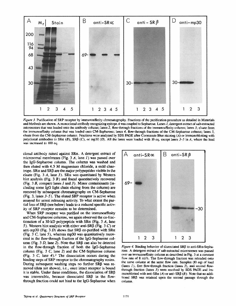
Figure 3. Purification of SRP receptor by immunoaflinity chromatography. Fractions of the purification procedure as detailed in Materials and Methods are shown. A monoclonal antibody recognizing epitope A was coupled to Sepharose. Lanes 1, detergent extract of salt-extracted microsomes that was loaded onto the antibody column; lanes 2, flow-through fractions of the immunoaffinity column; lanes 3; eluate from the immunoatlinity column that was loaded onto CM-Sepharose; lanes 4, flow-through fractions of the CM-Sepharose column; lanes 5, eluate from the CM-Sepharose column. Fractions were analyzed by SDS PAGE after Coomassie Blue staining (A) or immunoblotting with polyclonal antibodies to SR~t (B), SRI3 (C), or mp30 (D). All the lanes were loaded with 10 eq, except lanes 3-5 in A, where the load was increased to 100 eq.
clonal antibody raised against SRa. A detergent extract of microsomal membranes (Fig. 3 A, lane 1) was passed over the IgG-Sepharose column. The column was washed and then eluted with 4.5 M magnesium chloride, a mild chao- trope. SRa and SRI3 are the major polypeptides visible in the eluate (Fig. 3 A, lane 3). SRct was quantitated by Western blot analysis (Fig. 3 B) and found quantitatively recovered (Fig. 3 B, compare lanes 1 and 3). Minor contaminants (in- cluding some IgG light chain eluting from the column) are removed by subsequent chromatography on CM-Sepharose (Fig. 3, lanes 3-5) . The eluted SRP receptor is active when assayed for arrest releasing activity. To what extent the par- tial loss of SRI3 (see below) leads to a reduced specific activ- ity of SRP receptor remains to be determined.
When SRP receptor was purified on the immunoatlinity and CM-Sepharose columns, we again observed the co-frac- tionation of a 30-kD polypeptide with SR~t (Fig. 3 A, lane 5). Western blot analysis with either anti-SRI3 (Fig. 3 C) or anti-mp30 (Fig. 3 D) shows that SRI3 co-purified with SRct (Fig. 3 C, lane 5), whereas rap30 was quantitatively recov- ered in the flow-through fraction of the IgG-Sepharose col- umn (Fig. 3 D, lane 2). Note that SRI3 can also be detected in the flow-through fraction of both the IgG-Sepharose column (Fig. 3 C, lane 2) and the CM-Sepharose column (Fig. 3 C, lane 4). 2 The dissociation occurs during the binding steps of SRP receptor to the chromatography resins. During subsequent washing steps no further SRI3 was re- moved (data not shown), i.e., once intact receptor is bound it is stable. Under these conditions, the dissociation of SRI3 was irreversible, because dissociated SRI~ in the flow- through fraction could not bind to the IgG-Sepharose when
Figure 4. Binding behavior of dissociated SRI3 to anti-SRa-Sepha- rose. A detergent extract of salt-extracted microsomes was passed over an immunoaliinity column as described in Fig. 3 at a constant flow rate of 8 ml/h. The flow-through fraction was reloaded onto the same column at the same flow rate. Samples (10 eq) of load (lanes 1), first flow-through fraction (lanes 2), and second flow- through fraction (lanes 3) were resolved by SDS PAGE and im- munoblotted with anti-SRa (,4) or anti-SRl~ (B). Note that no addi- tional SRI3 was retained upon the second passage through the column.
Tajima et al. Quaternary Structure of SRP Receptor 1171

Figure 5. One-dimensional peptide map- ping of SRa. SRP receptor (150 Ixg/ml, prepared by immunoaflinity chromatog- raphy followed by CM-Sepharose chro- matography), was digested with TPCK- trypsin (T, lanes 2), papain in the pres- ence of 30 mM cysteine (P, lanes 3), elastase (E, lanes 4), chymotrypsin (C, lanes 5), or no proteases (lanes l). All proteases were added to 1:500 (wt/wt), except for elastase which was added to 1:100 (wt/wt). Digestions were for 1 h at 37°C in the elution buffer of the CM- Sepharose column (see Materials and Methods). Digests derived from 0.5 Ixg of SRP receptor were displayed by SDS PAGE and blotted onto nitrocellulose. Blots were probed with monoclonal an- tibodies recognizing epitope A (A) or epitope B (B) on SRct.
re-applied to the same column (Fig. 4 B, compare lanes 2 and 3).
To eliminate the possibility that SRI3, rather than interact- ing with SRct, somehow interacted with the quenched CNBr- activated Sepharose or the particular IgG1, we performed the following control experiments. First, anti-SRa was coupled
to a different matrix (Affi Gel 10) and used as the affinity ab- sorbent. We found that the elution behaviors of SRa and SRI3 were indistinguishable from that shown in Fig. 3 (data not shown), indicating that SRI3 is not bound due to interaction with the gel matrix.
Second, different monoclonal antibodies were used as the
Figure 6. Chromatography of SRP receptor on control immunoaflinity columns. Immunoaffinity columns were prepared by either coupling a monoclonal IgG1 recognizing epitope B on SRct (A) or a control IgGl recognizing a 220-kD cytoskeletal protein (a gift of Dr. D. Gard) (B) to CNBr-activated Sepharose. Coupling densities were 2 and 5 mg per ml of resin, respectively. A detergent extract of microsomal vesicles was loaded onto the columns and eluted as described in Fig. 3. Samples were displayed by SDS PAGE and Coomassie Blue staining (lanes 1-3), or immunoblotting with anti-SRct (lanes 4-6) or anti-SRI3 (lanes 7-9) (see Fig. 1). Lanes 1, 4, and 7show the detergent extract, lanes 2, 5, and 8 show the flow-through fractions, and lanes 3, 6, and 9 show the column eluates. Each lane was loaded with 10 eq, except lanes 3 which were loaded with 100 eq.
The Journal of Cell Biology, Volume 103, 1986 1172

affinity adsorbent for SRP receptor. 10 different hybridoma cell lines producing monoclonal antibodies to SRct were originally isolated. We could group these monoclonal anti- bodies into two distinct groups that recognize different epi- topes (epitope A and epitope B) on SRt~ using a one-dimen- sional peptide mapping approach. Purified SRP receptor (Fig. 3, lane 5) was subjected to limited proteolysis using a variety of different proteases. Fragments were fractionated by SDS PAGE and blotted onto nitrocellulose. Very discrete and characteristic patterns were obtained when the blot was probed with either anti-SRct recognizing epitope A (Fig. 5 A) or anti-SR~t recognizing epitope B (Fig. 5 B). Thus, both epitopes mark physically separate locations on the SRa poly- peptide. In the immunopurification of SRP receptor shown in Fig. 3, an IgG1 recognizing epitope A was used. When we repeated the experiment using an IgG1 recognizing epitope B, identical results were obtained (Fig. 6 A). As expected, if a control IgG1 Sepharose column was used, neither SRa nor SRI3 bound to the column (Fig. 6 B).
Previously, Hortsch et al. also reported a method using a monoclonal antibody as affinity probe for the purification of SR~t (14). In addition to SRa these investigators also found a peptide of Mr 27,000 in their column eluate, but concluded that the peptide was a degradation product of SR~t. This con- clusion was based on their observation that a band of ,,o27 kD was recognized by their monoclonal antibodies to SRa (12B4 and 12E3). Furthermore, Hortsch et al. raised a poly- clonal rabbit antibody against this 27-kD polypeptide and found it cross-reactive to SRtz. We have obtained samples of their antibodies and used them to probe our SRP receptor preparation (Fig. 7, lane 1). We conclude that: (a) There is no cross-reacting material in the 30-kD range when the blot is probed with 12B4 or 12E3 (Fig. 7, lanes 3 and 4), i.e., SRI3 is not detected by these antibodies. The visible minor break- down product of SR~t migrates at *40 kD. (b) The poly- clonal anti-27-kD serum recognizes both SR~t and SRI3 (Fig. 7, lane 2). This indicates that breakdown product(s) of SR~t must have been present in the 30-kD range during antigen preparation by Hortsch et al., which then elicited the ob- served immune response against SR~t. These data also dem- onstrate that SRI3 is present in the SRP receptor preparations of Hortsch et al., since their anti-27-kD serum recognizes it (Fig. 7, lane 2). Occasionally, we also observed a minor 30- kD breakdown product of SR~t in SRP receptor preparations (e.g., Fig. 5 B, lane 1 ). This degradation was prevented in subsequent preparations by the inclusion of protease in- hibitors during the detergent extraction (see Materials and Methods; Fig. 7).
The data presented so far demonstrate that SRa and SR~ reproducibly co-purify by different isolation procedures. If both polypeptides exist as a complex, we can expect also to immunopurify SR~t with anti-SRIL Fig. 8 shows the results from such an experiment. Rabbit anti-SRI3 Ig was prepared by immunopurification (see Materials and Methods). The Ig fraction was coupled to CNBr-activated Sepharose. A deter- gent extract from microsomal membranes was then passed over the immunoaffinity column (Fig. 8 A, lanes 1 and 2). The column was washed (Fig. 8 A, lane 3) and then eluted with 4.5 M magnesium chloride (Fig. 8 A, lane 4). SR~t (Fig. 8 A, lane 5) and SRI3 (Fig. 8 A, lane 6), but no mp30 (not shown), were detected in the eluate, i.e., both subunits of SRP receptor also co-purify by this technique.
Figure 7. Immunoblot analysis of various antisera and antibodies. Immunopurified SRP receptor (0.5 gg SRct per lane) was subjected to SDS PAGE and stained with Coomassie Blue (lane 1) or im- munoblotted (see Fig. 1) with the following antibodies: lane 2, polyclonal rabbit antiserum raised against a 27-kD protein (14), lanes 3 and 4, monoclonal antibodies 12114 and 12E3 (14), and lanes 5 and 6, monoclonal antibodies described in this paper recognizing epitope A or epitope B on SRa, respectively. Anti-27-kD serum, 12134, and 12E3 ascites fluids were obtained from Drs. M. Hortsch and D. Meyer.
The result presented in Fig. 8 A is of a qualitative nature. We performed a series of immunoadsorption experiments at- tempting to quantitate the amount of SRa that can be co- immunoprecipitated by anti-SRlL In Fig. 8 B and C a typical experiment is shown. Anti-SRl$ was titrated into an extract of microsomal membranes, immune complexes were precip- itated with protein A-Sepharose, and both the pellet (Fig. 8, B and C, lanes 1-3) and the supernatant fractions (Fig. 8, B and C, lanes 4 and 5) were analyzed by SDS PAGE followed by Western blotting. Blots were either probed with anti-SRa (Fig. 8 B) or anti-SR[3 (Fig. 8 C). As expected, we depleted the supernatant fractions of SRI3 with increasing antibody concentrations (Fig. 8 C, lanes 4-6) and recovered SRI~ in the corresponding pellet fractions (Fig. 8 C, lanes 1-3). However, SRct was only incompletely precipitated (Fig. 8 B, compare lane 3 with lane 6), even at the higher antibody con- centrations where SRI$ was largely depleted from the ex- tracts. We estimate that only ,o20% of SR~t is immuno- precipitable with anti-SRI3. We interpret these results that anti-SRI3 (which is a polyclonal antibody and therefore pre-
Tajima et al. Quaternary Structure of SRP Receptor 1173

Figure & Co-fractionation of SRa by immunoadsorption on anti-SR~l. (A) An immunoaflinity column was prepared by coupling 0.5 mg of immunoselected polyclonal rabbit anti-SRI3 to 0.8 ml of CNBr-activated Sepharose. A detergent extract of microsomes (9,000 eq) was chromatographed on this column as described in Fig. 3. Fractions were subjected to SDS PAGE and stained with Coomassie Blue (lanes 1-4). Lane 1 shows the load, lane 2 the flow-through fraction, lane 3 the wash fraction, and lane 4 the eluate of the immunoaffinity column. SRP receptor was detected in the eluate fraction by immunohlot analysis with anti-SRct (lane 5) or anti-SR~ (lane 6). Samples of 10 eq were loaded in lanes 1 and 2, and 100 eq in lanes 3-6. Both SRa and SR~ were also detected in the flow-through fraction (not shown), indicating that the column was saturated under the given conditions. Also, the eluate fraction consistently contained other polypeptides unrelated to the SRP receptor subunits (lane 4, SRct and SR~ are indicated by arrows). This may be a result of the low coupling density of the antibodies on the resin and/or the use of a polyclonal Ig fraction. (B and C) Immunoprecipitations of SRP receptor from a microsomal detergent extract. Increasing amounts of immunoselected polyclonal rabbit anti-SR~ (no Ig [lanes 1 and 4], 10 gg Ig [lanes 2 and 5], and 23 gg Ig [lanes 3 and 6]), were incubated with 6 eq of detergent extract (final volume 100 Ixl) at room temperature for 6 h. A 20-1tl aliquot of protein A-Sepharose was added. The gel matrix was pelleted and washed once with a phosphate-buffered saline solution containing 0.1% Nikkol detergent. Pellets (lanes 1-3) and combined wash and supernatant fractions (lanes 4-6) were subjected to SDS PAGE and blotted onto nitrocellulose. Blots were probed with anti-SRct (B) or anti-SRI3 (C), respectively. The positions of SRtt and SRI3 are indicated by their respective molecular weights. The heavy bands marked with an asterisk that are present in the pellet fractions correspond to protein A that has leaked off the Sepharose resin during SDS PAGE sample preparation and that binds antibodies on the nitrocellulose blot. IgG heavy chain migrates in the same position and may contribute to the signal in the lanes where antibody was added. Another minor band (indicated with a diamond in C, lanes 5 and 6) corresponds to the IgM heavy chain. This was shown by probing an equivalent blot with an anti-IgM specific secondary antibody (not shown). As expected, this band is found in the supernatant fraction only, since IgM does not bind to protein A. Both the asterisk and diamond bands are less pronounced in B, because the blot was probed with a mouse monoclonal IgG1 and a secondary anti-mouse IgG antibody.
sumably binds to multiple epitopes on SRI3) causes a simi- lar dissociation of the two subunits as we also obse rved- although to a lesser extent - for anti-SRct, 2
Given the complication of incomplete immunoprecipita- tions it became important to demonstrate that SRct and SRI3 form a complex when their structures were not perturbed by bound antibodies. We therefore subjected purified SRP re- ceptor (Fig. 9 A) or a detergent extract of microsomal mem- branes (Fig. 9 B) to velocity sedimentation analysis. Under both conditions SRtl and SRI~ co-sedimented almost indis- tinguishably from one another, but clearly off-set from mp30 (Fig. 9 B). The peaks that we obtained when crude extracts
were centrifuged (Fig. 9 B) were always sharper than those obtained from purified SRP receptor (Fig. 9 A), indicating that some aggregation may have occurred in the purified sample. Comparison of SRP receptor with sedimentation marker proteins showed that SRP receptor sediments with a velocity similar to that of ovalbumin (S = 3.7). Given that ovalbumin (43 kD) is only half the size of SRP receptor (SRct + SRI3 = 100 kD), this anomolous sedimentation must be due to effects caused by bound detergent and/or an extended structure of the SRP receptor that deviates from that of a spherical particle.
Next we determined the absolute amounts of SRa and SRI3
The Journal of Cell Biology, Volume 103, 1986 1174
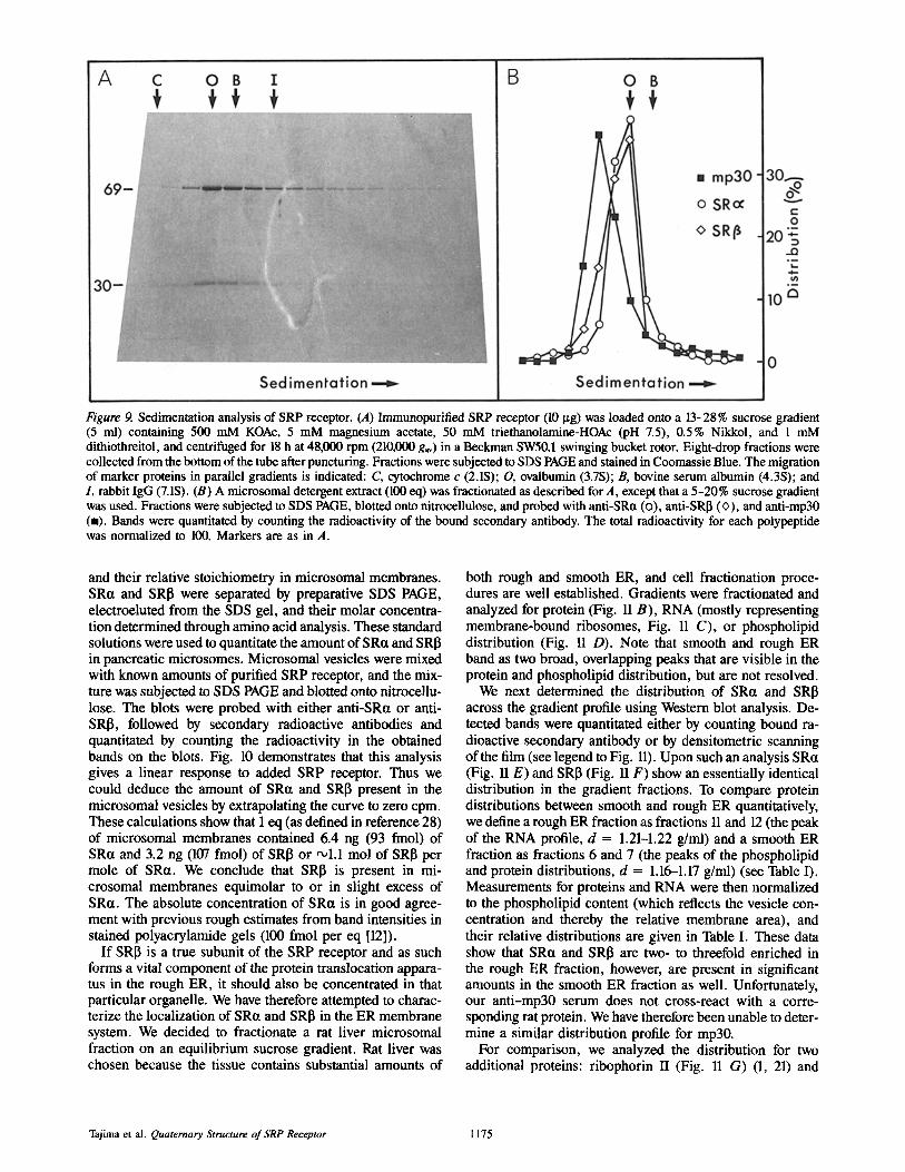
Figure 9. Sedimentation analysis of SRP receptor. (A) Immunopurified SRP receptor (10 ~tg) was loaded onto a 13-28% sucrose gradient (5 ml) containing 500 mM KOAc, 5 mM magnesium acetate, 50 mM triethanolamine-HOAc (pH 7.5), 0.5% Nikkol, and 1 mM dithiothreitol, and centrifuged for 18 h at 48,000 rpm (210,000 gay) in a Beckman SW50.1 swinging bucket rotor. Eight-drop fractions were collected from the bottom of the tube after puncturing. Fractions were subjected to SDS PAGE and stained in Coomassie Blue. The migration of marker proteins in parallel gradients is indicated: C, cytochrome c (2.1S); O, ovalbumin (3.7S); B, bovine serum albumin (4.3S); and I, rabbit IgG (7.1S). (B) A microsomal detergent extract (100 eq) was fractionated as described for A, except that a 5-20% sucrose gradient was used. Fractions were subjected to SDS PAGE, blotted onto nitrocellulose, and probed with anti-SRct (o), anti-SRI3 (<>), and anti-mp30 (m). Bands were quantitated by counting the radioactivity of the bound secondary antibody. The total radioactivity for each polypeptide was normalized to 100. Markers are as in A.
and their relative stoichiometry in microsomal membranes. SR~t and SRI~ were separated by preparative SDS PAGE, electroeluted from the SDS gel, and their molar concentra- tion determined through amino acid analysis. These standard solutions were used to quantitate the amount of SRct and SRI3 in pancreatic microsomes. Microsomal vesicles were mixed with known amounts of purified SRP receptor, and the mix- ture was subjected to SDS PAGE and blotted onto nitrocellu- lose. The blots were probed with either anti-SRct or anti- SRI3, followed by secondary radioactive antibodies and quantitated by counting the radioactivity in the obtained bands on the blots. Fig. 10 demonstrates that this analysis gives a linear response to added SRP receptor. Thus we could deduce the amount of SR~t and SRI3 present in the microsomal vesicles by extrapolating the curve to zero cpm. These calculations show that 1 eq (as defined in reference 28) of microsomal membranes contained 6.4 ng (93 fmol) of SRct and 3.2 ng (107 fmol) of SRI~ or ,,o1.1 mol of SRI3 per mole of SR~t. We conclude that SRI3 is present in mi- crosomal membranes equimolar to or in slight excess of SRa. The absolute concentration of SRa is in good agree- ment with previous rough estimates from band intensities in stained polyacrylamide gels (100 fmol per eq [12]).
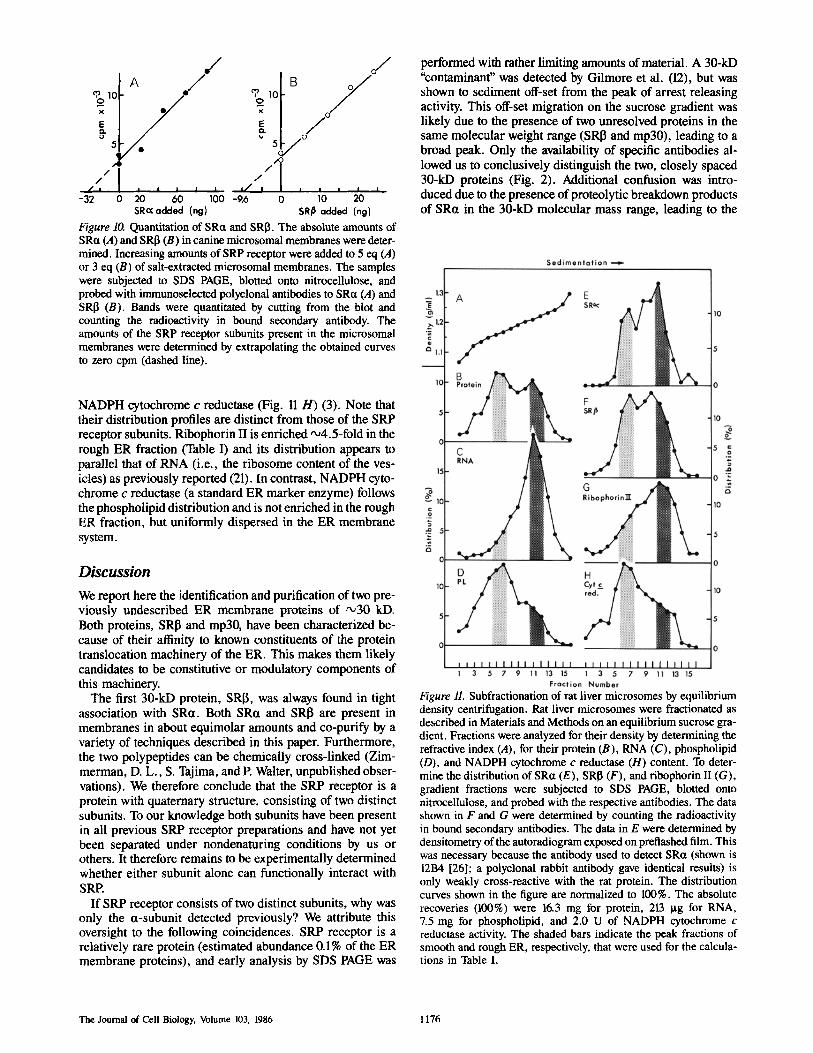
If SRI] is a true subunit of the SRP receptor and as such forms a vital component of the protein translocation appara- tus in the rough ER, it should also be concentrated in that particular organelle. We have therefore attempted to charac- terize the localization of SRct and SRI5 in the ER membrane system. We decided to fractionate a rat liver microsomal fraction on an equilibrium sucrose gradient. Rat liver was chosen because the tissue contains substantial amounts of
both rough and smooth ER, and cell fractionation proce- dures are well established. Gradients were fractionated and analyzed for protein (Fig. 11 B), RNA (mostly representing membrane-bound ribosomes, Fig. 11 C), or phospholipid distribution (Fig. 11 D). Note that smooth and rough ER band as two broad, overlapping peaks that are visible in the protein and phospholipid distribution, but are not resolved.
We next determined the distribution of SRct and SRI3 across the gradient profile using Western blot analysis. De- tected bands were quantitated either by counting bound ra- dioactive secondary antibody or by densitometric scanning of the film (see legend to Fig. 11). Upon such an analysis SRa (Fig. 11 E) and SRI5 (Fig. 11 F) show an essentially identical distribution in the gradient fractions. To compare protein distributions between smooth and rough ER quantitatively, we define a rough ER fraction as fractions 11 and 12 (the peak of the RNA profile, d = 1.21-1.22 g/ml) and a smooth ER fraction as fractions 6 and 7 (the peaks of the phospholipid and protein distributions, d = 1.16-1.17 g/ml) (see Table I). Measurements for proteins and RNA were then normalized to the phospholipid content (which reflects the vesicle con- centration and thereby the relative membrane area), and their relative distributions are given in Table I. These data show that SRa and SRI3 are two- to threefold enriched in the rough ER fraction, however, are present in significant amounts in the smooth ER fraction as well. Unfortunately, our anti-mp30 serum does not cross-react with a corre- sponding rat protein. We have therefore been unable to deter- mine a similar distribution profile for mp30.
For comparison, we analyzed the distribution for two additional proteins: ribophorin II (Fig. 11 G) (1, 21) and
Tajima et al. Quaternary Structure of SRP Receptor 1175
/
J - - ~ " | i I
-32 0 60 100 SRc~ added (ng)
,o B/o/ / 5 j °
/ / /
I ~ ' t a I I I I
-9.6 o 10 20 SR/~ added (ng)
Figure 10. Quantitation of SRct and SRI3. The absolute amounts of SRa (A) and SRI3 (B) in canine microsomal membranes were deter- mined. Increasing amounts of SRP receptor were added to 5 eq (A) or 3 eq (B) of salt-extracted microsomal membranes. The samples were subjected to SDS PAGE, blotted onto nitrocellulose, and probed with immunoselected polyclonal antibodies to SRa (A) and SR[3 (B). Bands were quantitated by cutting from the blot and counting the radioactivity in bound secondary antibody. The amounts of the SRP receptor subunits present in the microsomal membranes were determined by extrapolating the obtained curves to zero cpm (dashed line).
performed with rather limiting amounts of material. A 30-kD "contaminant" was detected by Gilmore et al. (12), but was shown to sediment off-set from the peak of arrest releasing activity. This off-set migration on the sucrose gradient was likely due to the presence of two unresolved proteins in the same molecular weight range (SRI3 and mp30), leading to a broad peak. Only the availability of specific antibodies al- lowed us to conclusively distinguish the two, closely spaced 30-kD proteins (Fig. 2). Additional confusion was intro- duced due to the presence of proteolytic breakdown products of SRa in the 30-kD molecular mass range, leading to the
NADPH cytochrome c reductase (Fig. 11 H) (3). Note that their distribution profiles are distinct from those of the SRP receptor subunits. Ribophorin II is enriched ,x,4.5-fold in the rough ER fraction (Table I) and its distribution appears to parallel that of RNA (i.e., the ribosome content of the ves- icles) as previously reported (21). In contrast, NADPH cyto- chrome c reductase (a standard ER marker enzyme) follows the phospholipid distribution and is not enriched in the rough ER fraction, but uniformly dispersed in the ER membrane system.
D i s c u s s i o n
We report here the identification and purification of two pre- viously undescribed ER membrane proteins of ~30 kD. Both proteins, SRI3 and mp30, have been characterized be- cause of their affinity to known constituents of the protein translocation machinery of the ER. This makes them likely candidates to be constitutive or modulatory components of this machinery.
The first 30-kD protein, SRI3, was always found in tight association with SRa. Both SRa and SRI~ are present in membranes in about equimolar amounts and co-purify by a variety of techniques described in this paper. Furthermore, the two polypeptides can be chemically cross-linked (Zim- merman, D. L., S. Tajima, and P. Walter, unpublished obser- vations). We therefore conclude that the SRP receptor is a protein with quaternary structure, consisting of two distinct subunits. To our knowledge both subunits have been present in all previous SRP receptor preparations and have not yet been separated under nondenaturing conditions by us or others. It therefore remains to be experimentally determined whether either subunit alone can functionally interact with SRP.
If SRP receptor consists of two distinct subunits, why was only the a-subunit detected previously? We attribute this oversight to the following coincidences. SRP receptor is a relatively rare protein (estimated abundance 0.1% of the ER membrane proteins), and early analysis by SDS PAGE was
Figure 11. Subfractionation of rat liver microsomes by equilibrium density centrifugation. Rat liver microsomes were fractionated as described in Materials and Methods on an equilibrium sucrose gra- dient. Fractions were analyzed for their density by determining the refractive index (A), for their protein (B), RNA (C), phospholipid (D), and NADPH cytochrome c reductase (H) content. To deter- mine the distribution of SRct (E), SRI3 (F), and ribophorin II (G), gradient fractions were subjected to SDS PAGE, blotted onto nitrocellulose, and probed with the respective antibodies. The data shown in F and G were determined by counting the radioactivity in bound secondary antibodies. The data in E were determined by densitometry of the autoradiogram exposed on pretiashed film. This was necessary because the antibody used to detect SRa (shown is 12B4 [26]; a polyclonal rabbit antibody gave identical results) is only weakly cross-reactive with the rat protein. The distribution curves shown in the figure are normalized to 100%. The absolute recoveries (100%) were 16.3 mg for protein, 213 Ixg for RNA, 7.5 mg for phospholipid, and 2.0 U of NADPH cytochrome c reductase activity. The shaded bars indicate the peak fractions of smooth and rough ER, respectively, that were used for the calcula- tions in Table I.
The Journal of Cell Biology, Volume 103, 1986 1176

Table L Distribution of Protein, RNA, Phospholipid, SRa, SR[3, Ribophorin II, and NADPH Cytochrome c Reductase between Rough and Smooth Microsomal Fractions.
Smooth ER fraction Rough ER fraction
Rough ER fraction
Smooth ER fraction
Protein 0 .60 1.12 1.9 RNA 0.31 2.33 7.5 Phospholipid 1.00 1.00 1.0 SRct 0 .60 1.70 2.8 SRI3 0.61 1.28 2.1 Ribophorin II 0 .34 1.53 4.5 N A D P H cytochrome c reductase 1.04 1.18 1.1
The data were taken from Fig. 11. Smooth and rough ER fraction indicate the percent recovery into fractions (6 + 7) and into fractious (11 + 12), respectively, that were normalized to the phospholipid in these fractions. The ratio of rough ER fraction to smooth ER fraction shows the relative enrichment of each component into the rough ER fraction.
report by Hortsch et al. claiming that the 30-kD band was a breakdown product of SRet (14). These results are recon- ciled with the data shown in Fig. 7 that are described in the Results section. We therefore feel that our results are not in conflict with previous reports, but rather that data that appear conflicting on first sight can be rationalized satisfactorily.
Our finding that the SRP receptor is a two subunit protein refines but does not drastically change our views on its struc- ture or membrane disposition. In the primary sequence of SRa a predominantly basic domain consisting of mixed charge clusters was suggested to provide a binding site for SRP that could function through a direct interaction with 7SL RNA (20). This conjecture remains a viable hypothesis and is not affected by the presence of the additional subunit. In the primary sequence of SRct we also identified two poten- tial membrane-spanning segments. Each of these regions- if they formed a membrane-spanning a-helix-would place a positively charged amino acid in the middle of the hydropho- bic core of the membrane. Association of SRct with SRI3 could provide the necessary countercharges and stabilize the receptor-membrane interaction. SRI3 remains intact and membrane associated after proteolytic removal of the cyto- plasmic domain of SRct (data not shown). Therefore it is pos- sible that SRI3, in conjunction with the remaining fragment of SRct, could provide the binding site to which the 52-kD cytoplasmic fragment of SRct can rebind noncovalenfly to re- store a functional receptor.
The second newly identified 30-kD membrane protein, mp30, was purified on SRP-Sepharose and binds to SRP directly. This interaction also appears to be specific. On the SRP-Sepharose column, binding of mp30 saturated at the same molar stoichiometry as the SRP-SRP receptor interac- tion. SRP receptor and mp30 must however bind to different sites on SRP since they do not compete with each another for binding (Fig. 1). Furthermore, mp30 was eluted from SRP-Sepharose under the same conditions used to elute SRP receptor by increasing the magnesium concentration in the buffer from 5 to 25 mM, while the monovalent cation con- centration was adjusted to keep the ionic strength constant. These conditions, originally described by Gilmore et al. (11), appear to be subtle, possibly affecting conformational changes in SRP, and are unlikely to cause elution if binding were due to nonspecific ionic interactions. Nevertheless, we have presently no means to distinguish whether the binding affinity reflects a physiologically meaningful interaction or is merely fortuitous. Purified mp30 in detergent solution had
no elongation arrest releasing activity when assayed in vitro, nor did it measurably promote or inhibit the activity of SRP receptor (Lauffer, L., unpublished observations). Antibod- ies to mp30 did not inhibit the protein translocation activity of microsomal membranes (Tajima, S., unpublished obser- vations). We do not know, however, whether our antibodies are directed towards cytoplasmically exposed epitopes on mp30. Thus, while mp30 remains a good candidate for a functionally important protein, functional assays will be re- quired to assess its putative role in protein translocation. One can speculate that additional SRP-binding proteins in the ER membrane could act, for example, to locally increase the SRP concentration. Alternatively they may directly partici- pate in the SRP targeting cycle or be involved in recycling SRP after it has interacted with its receptor.
SRP receptor was previously claimed to be a marker pro- tein specific for the rough membranes of the ER (13, 15). Smooth and rough ER form a continuous membrane system and are morphologically and experimentally distinguished by their density of membrane-bound ribosomes. Most if not all of these ribosomes are actively translating proteins, and translation appears to be coupled to translocation of the nascent polypeptides across the ER membrane. Upon sepa- ration of smooth and rough ER by equilibrium sucrose gra- dient centrifugation it was unexpected, in contrast to the above-mentioned claim, to find that SRP receptor was pres- ent at relatively high concentrations in the light ER fractions. Both ribosomes (measured as RNA) and ribophorin II (a protein that was suggested to function as a ribosome receptor [32] show a much more skewed distribution toward the heav- ier gradient fractions than does SRP receptor. This implies that a population of SRP receptor (amounting to about half the SRP receptor molecules in a rat liver cell) exists in regions of the ER with a low ribosome density and a low ribophorin concentration. These sites may represent regions of the ER to which newly initiated polysomes are targeted. Ribophorins and possibly other ribosome-binding proteins may act subsequently to stabilize those ribosomes that are al- ready functionally engaged on the membrane to eventually establish a classical ribosome-membrane junction.
We thank Drs. G. Kreibich and D. Meyer for gifts of antibodies to ribopho- fin and SRP receptor, respectively, and Ms. Vivian Siegel for many helpful discussions relating to the work.
This work was supported by National Institutes of Health grant GM- 32384 and funds from the Chicago Community Trust/Searle Scholars Pro- gram. S. Tajima is the recipient of a Fogarty Public Health Service Interna-
Tajima et al. Quaternary Structure of SRP Receptor 1177

tional Research Fellowship (FO5 TW03646). L. Lauffer is a fellow of the Jane Coffin Childs Memorial Fund for Medical Research.
Received for publication 10 July 1986, and in revised form 24 July 1986.
References
1. Amar-Costesec, A., J. A. Todd, and G. Kreibich. 1984. Segregation of the polypeptide translocation apparatus to regions of the endoplasmic retieulum containing ribophorins and ribosomes. I. Functional tests on rat liver micro- somal subfractions. J. Cell Biol. 99:2247-2253.
2. Ames, B. N., and D. T. Dubin. 1960. The role of polyamines in the neu- tralization of bacteriophage deoxyribonucleic acid. J. Biol. Chem. 235:769- 775.
3. Beanfay, H., A. Amar-Costesec, D. Thines-Sempoux, M. Wibo, M. Robbi, and J. Berthet. 1974. Analytical study of microsomes and isolated sub- cellular membranes from rat liver. III. Subfractionation of the microsomal frac- tion by isopycnic and differential centrifugation in density gradients. J. Cell Biol. 61:213-231.
4. Beaufay, H., A. Amar-Costesec, E. Feytmans, D. Thines-Sempoux, M. Wibo, M. Robbi, and J. Berthet. 1974. Analytical study of microsomes and iso- lated subcellular membranes from rat liver. I. Biochemical methods. J. Cell Biol. 61:188-200.
5. Bligh, E. G., and W. J. Dyer. 1959. A rapid method of total lipid extrac- tion and purification. Can. J. Biochem. Physiol. 37:911-917.
6. Broome, S., and W. Gilbert. 1978. Immunological screening method to detect specific translation products. Proc. Natl. Acad. Sci. USA. 75:2746-2749.
7. Bruek, C., D. Portetelle, C. Glineur, and A. Bollen. 1982. One-step purification of mouse monoclonal antibodies from ascitic fluid by DEAE Afli- Gel Blue chromatography. J. lmmunol. Methods. 53:313-319.
8. Fazekas de St. Groth, S., and D. Scheidegger. 1980. Production of monoclonal antibodies: strategy and tactics. ,L Iraraunol. Methods. 35:1-21.
9. Fisher, P. A., M. Berrios, and G. BIobel. 1982. Isolation and character- ization of a proteinaceous subnuclear fraction composed of nuclear matrix, pe- ripheral lamina, and nuclear pore complexes from embryos of Drosophila me- lanogaster. J. Cell Biol. 92:674-686.
10. Fleck, A., and H. N. Munro. 1962. The precision of ultravioletabsorp- tion measurements in the Schrnidt-Thannhauser procedure from nucleic acid es- timation. Biochim. Biophys. Acta. 55:571-583.
11. Gilmore, R., and G. Blobel. 1983. Transient involvement of signal rec- ognition particle and its receptor in the microsomal membrane prior to protein translocation. Cell. 35:677-685.
12. Gilmore, R., P. Walter, and G. Blobel. 1982. Protein translocation across the endoplasmic reticulum. II. Isolation and characterization of the signal recognition particle receptor. J. Cell Biol. 95:470-477.
13. Hortsch, M., and D. I. Meyer. 1985. Immunochemical analysis of rough and smooth microsomes from rat liver. Segregation of docking protein in rough membranes. Eur. J. Biochem. t50:559-564.
14. Hortsch, M., D. Avossa, and D. I. Meyer. 1985. A structural and func- tional analysis of the docking protein. Characterization of active domains by proteolysis and specific antibodies. J. Biol. Chem. 260:9137-9145.
15. Hortsch, M., G. Griftiths, and D. I. Meyer. 1985. Restriction of docking protein to the rough endoplasmic reticulum: immunochemical localization in rat liver. Eur. J. Cell Biol. 38:271-279.
16. Hunkapiller, M. W., and L. E. Hood. 1983. Analysis of Phenylthio- hydantoins by ultrasensitive gradient high-performance liquid chromatography. Methods Enzymol. 91:486-493.
17. Hunkapillar, M. W., E. Lujan, F. Ostrander, and L. E. Hood. 1983. Iso- lation of microgram quantities of protein from polyacrylamide gels for amino acid sequence analysis. Methods Enzymol. 91:227-236.
18. Krieg, U., P. Walter, and A. E. Johnson. 1986. Photocrosslinking of the signal sequence of nascent preprolactin to the 54 kDa polypeptide of the signal recognition particle. Proc. Natl. Acad. Sci. USA. In press.
19. Kurzchalia, T. V., M. Wiedmann, A. S. Girshovich, E. S. Bochkareva, H. Bielka, and T. A. Rapaport. 1986. The signal sequence of nascent preprolac- tin interacts with the 54 kD polypeptide of the signal recognition particle. Na- ture (Lord.). 320:634-636.
20. Lauffer, L., P. D. Garcia, R. N. Harkins, L. Coussens, A. Ullrich, and P. Walter. 1985. Topology of signal recognition particle receptor in endoplas- mic reticulum membrane. Nature (Lord.). 318:334-338.
21. Marcantonio, E. E., A. Amoar-Costesec, and G. Kreibich. 1984. Segre- gation of the polypeptide translocation apparatus to regions of the endoplasmic reticulum containing ribophorins and ribosomes. II. Rat liver microsomal sub- fractions contain equimolar amounts of ribophorins and ribosomes. J. Cell Biol. 99:2254-2259.
22. Meyer, D. I., and B. Dobberstein. 1980. A membrane component essen- tial for vectorial translocation of nascent proteins across the endoplasmic reticu- lure: requirements for its extraction and reconstitution with the membrane. J. Cell Biol. 87:498-502.
23. Meyer, D. I., E. Krause, and B. Dobberstein. 1982. Secretory protein translocation across membranes --- the role of the 'docking protein'. Nature (Lond.). 297:647-650.
24. Mihara, K., G. Blobel, and R. Sato. 1982. In vitro synthesis and integra- tion into mitocbondria of porin, a major protein of the outer mitochondria mem- brane of Saccharomyces cerevisiae. Proc. Natl. Acad. Sci. USA. 79:7102- 7106.
25. Schaffner, W., and C. Weissmann. 1973. A rapid, sensitive, and specific method for the determination of protein in dilute solution. Anal. Biochem. 56:502-514.
26. Siegel, V., and P. Walter. 1985. Elongation arrest is not a prerequisite for secretory protein translocation across the microsomal membrane. J. Cell Biol. 100:1913-1921.
27. Waiter, P., and G. Blobel. 1981. Translocation of proteins across the en- doplasmic reticulum. III. Signal recognition protein (SRP) causes signal sequence- dependent and site-specific arrest of chain elongation that is released by microsomal membranes. J. Cell Biol. 91:557-561.
28. Walter, P., and G. Blobel. 1983. Preparation of microsomal membranes for cotranslational protein translocation. Methods Enzymol. 96:84-93.
29. Walter, P., and G. Blobel. 1983. Signal recognition particle: a ribonu- cleoprotein required for cotranslational transloeation of proteins, isolation and properties. Methods Enzymol. 96:682-691.
30. Walter, P., R. Gilmore, and G. Blobel. 1984. Protein translocation across the endoplasmic reticulum. Cell. 38:5-8.
31. Walter, P., R. C. Jackson, M. M. Marcus, V. R. Lingappa, and G. Blobel. 1979. Tryptic dissection and reconstitution of translocation activity for nascent presecretory proteins across microsomal membranes. Proc. Natl. Acad. Sci. USA. 76:1795-1799.
32. Ziemiecki, A., and H. Garoff. 1978. Subunit composition of the mem- brane glycoprotein complex of Semliki Forest virus. J. Mol. Biol. 122:259- 269.
The Journal of Cell Biology, Volume 103, 1986 1178