the shoulder - mccc - mercer countybehrensb/documents/theshoulder.pdf · leads to chronic...
TRANSCRIPT

PTA 216

Many structures that perform many movements
Injuries inside or outside of the joint capsule
Magee, 2008. pg. 231
Composed of 4 articulations between the
sternum, humerus, scapula, and clavicle.
3 synovial joints
Glenohumeral joint
Acromioclavicular joint (AC joint)
Sternoclavicular joint (SC joint)
2 functional articulations
Suprahumeral/subacromial
Scapulothoracic
Dutton, 2012. pg. 353

Dutton, 2012. pg. 354

Articulation between the articular end of the
clavicle, the clavicular notch of the
manubrium of the sternum, and the cartilage
of the first rib
Motions include:
Elevation and Depression
Protraction and Retraction
Axial rotation
Dutton, 2012. pg. 354
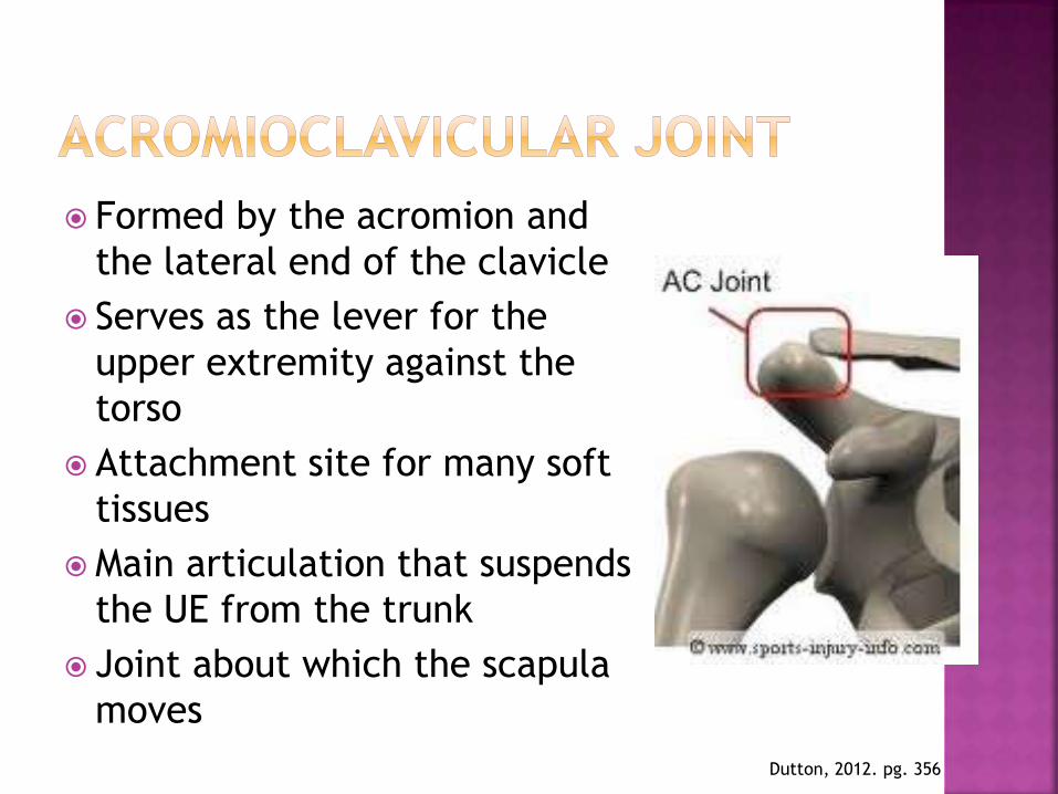
Formed by the acromion and
the lateral end of the clavicle
Serves as the lever for the
upper extremity against the
torso
Attachment site for many soft
tissues
Main articulation that suspends
the UE from the trunk
Joint about which the scapula
moves
Dutton, 2012. pg. 356

Ball and socket joint
Relatively unstable requiring assistance from other structures Labrum
Glenohumeral ligaments Superior, middle, and inferior
Coracoacromial ligament
Coracoclavicular ligaments
Joint capsule
Muscular dynamic stabilizers Rotator cuff, biceps tendon, muscles of scapular
motion
Dutton, 2012. pg. 357

Dutton, 2012. pg. 361

Functionally acts as a joint, but lacks
anatomic characteristics of a synovial joint
Lacks ligamentous support
Relies solely on muscular support between
the scapula and thorax
Dutton, 2012. pg. 356
Motions that occur
Elevation, Depression, Protraction, Retraction
Seen with clavicular motion at the SC joint, when the
humerus moves, and shoulder shrugging
Upward and Downward rotation
Seen with clavicular motion at the AC joint and with
humerus movement
Winging and Tipping
Seen with motions of the AC joint and humerous
movment
Dutton, 2012. pg. 356

Boundaries are formed by:
Greater tuberosity of the humeral head,
inferiorly
Coracoid process, anteromedially
Coracoacromial arch, superiorly
Dutton, 2012. pg. 363

Synchronized motion that occurs between
the glenoid cavity and the humerus during
arm elevation
Allows the glenoid to stay centered under the
humeral head which resists downward (inferior)
dislocation
Ratio of ROM is 2:1
Every 2 degrees of abduction, there should be 1
degree of scapular upwards rotation
Dutton, 2012. pg. 363
3 main groups of muscles
Thoracoscapular
Rhomboids, levator scapulae, serratus anterior,
and trapezius muscles
Thoracohumeral
Latissimus dorsi and pectoralis major
Scapulohumeral
Supraspinatus, infraspinatus, teres minor,
subscapularis, and deltoid
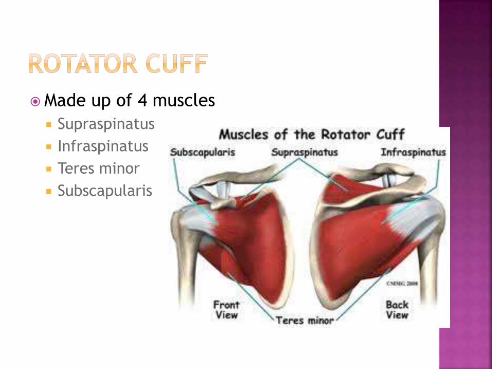
Made up of 4 muscles
Supraspinatus
Infraspinatus
Teres minor
Subscapularis
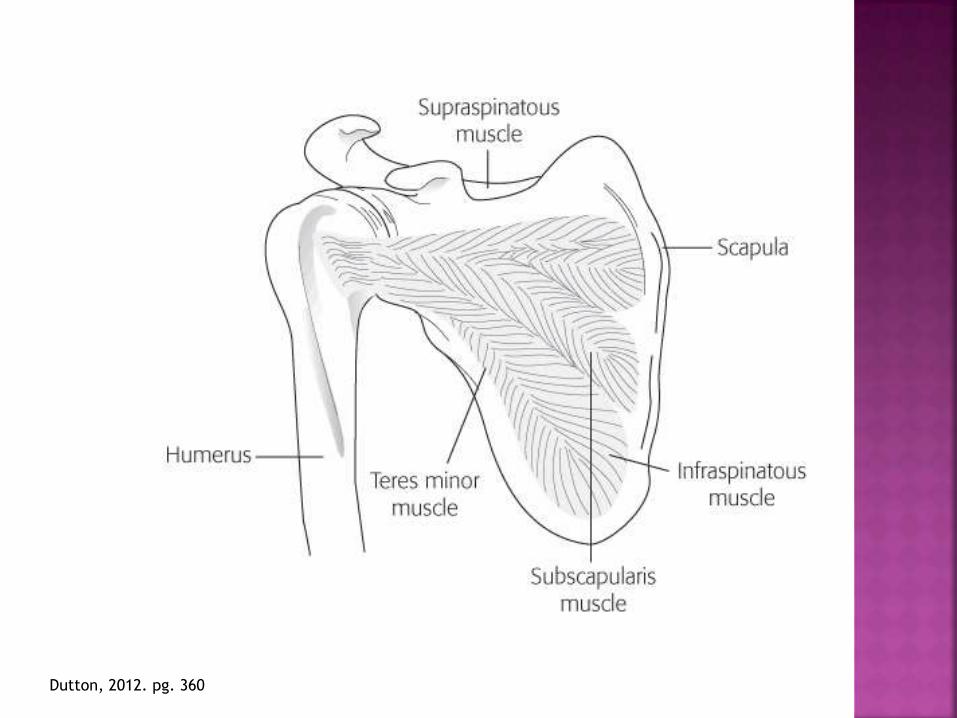
Dutton, 2012. pg. 360

Inflammation of the tendon
Most common forms:
Bicep’s Tendonitis
Supraspinatus Tendonitis
Rotator Cuff Tendonitis

Decrease pain and inflammation
Modalities as needed
Increase flexibility
Manual intervention
Increase joint stability
Initiate therapeutic exercise as tolerated
Joint stabilization activity

Increased superior translation with shoulder
elevation resulting in encroachment of the
coracoacromial arch producing compression
of the suprahumeral structures
2 separate types
Primary
Secondary
Dutton, 2012. pg. 377
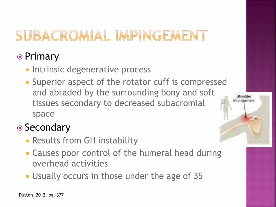
Primary
Intrinsic degenerative process
Superior aspect of the rotator cuff is compressed
and abraded by the surrounding bony and soft
tissues secondary to decreased subacromial
space
Secondary
Results from GH instability
Causes poor control of the humeral head during
overhead activities
Usually occurs in those under the age of 35
Dutton, 2012. pg. 377

Stage I: (under 25 years of age)
Edema and hemorrhage
Pain with shoulder ABDuction over 90 degrees
Considered reversible at this stage
Typically responds to PT intervention
Shankman, 2011. pg. 349
Dutton, 2012. pg. 379
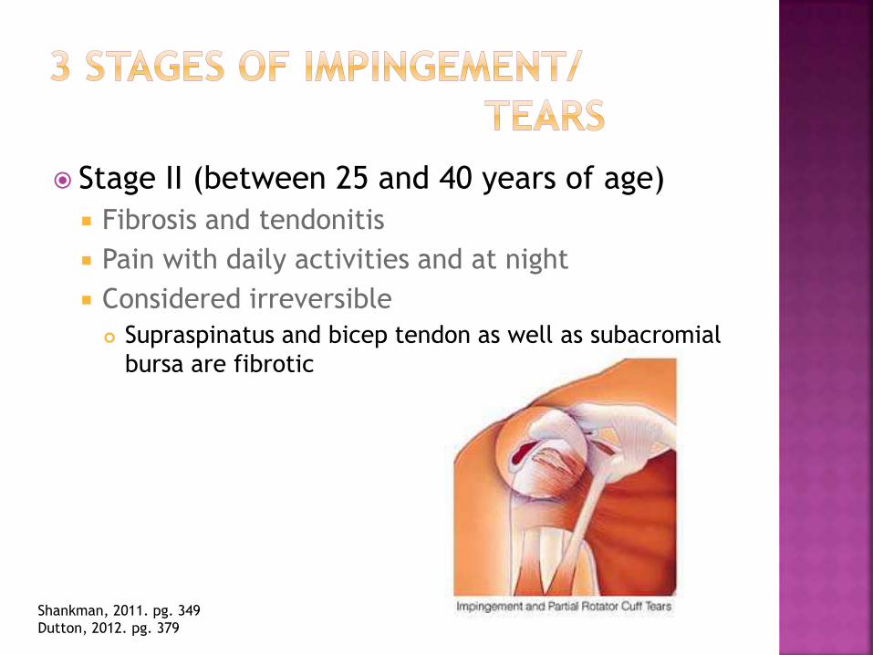
Stage II (between 25 and 40 years of age)
Fibrosis and tendonitis
Pain with daily activities and at night
Considered irreversible
Supraspinatus and bicep tendon as well as subacromial
bursa are fibrotic
Shankman, 2011. pg. 349
Dutton, 2012. pg. 379

Stage III (over 40 years of age)
Long history of shoulder pain
Significant muscle weakness
Tendon degeneration
Rotator cuff tears
Rotator cuff ruptures
Shankman, 2011. pg. 349
Dutton, 2012. pg. 379
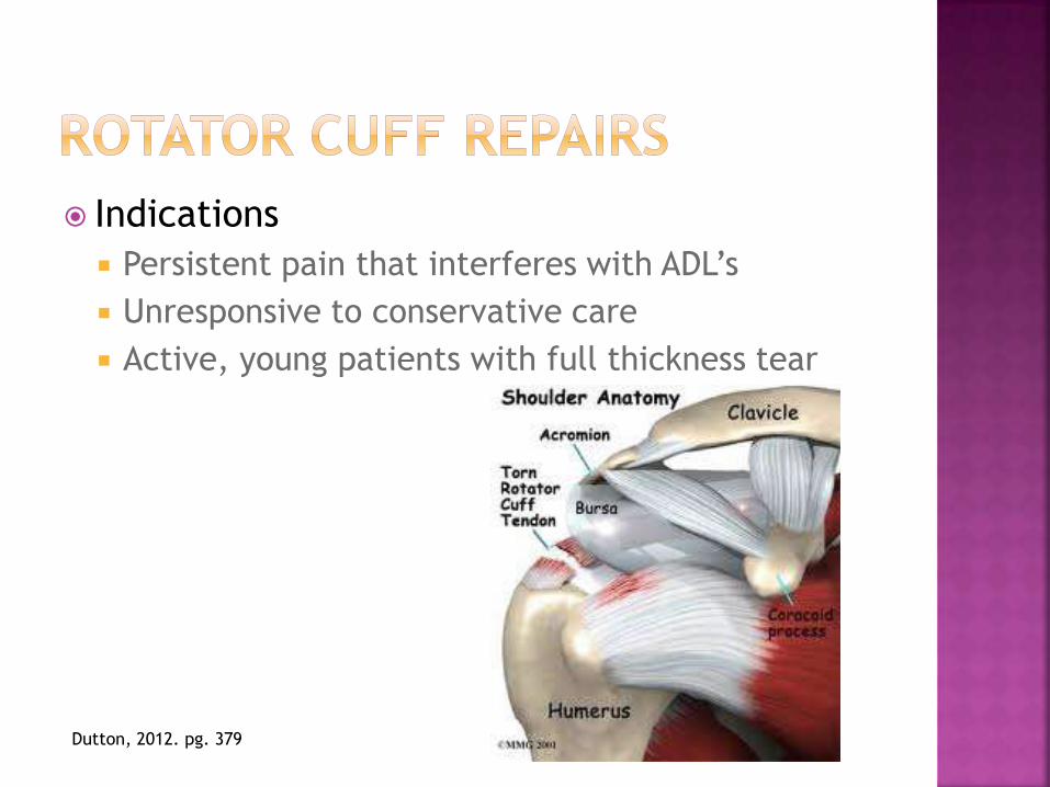
Indications
Persistent pain that interferes with ADL’s
Unresponsive to conservative care
Active, young patients with full thickness tear
Dutton, 2012. pg. 379

Vertical incision made over anterior shoulder
Deltoid is divided allowing access to rotator cuff and subacromial space
Anterior/inferior acromioplasty is performed
Humeral head is roughened
Holes are drilled for sutures
Sutures in place attaching tendon to bone
Dutton, 2012. pg. 379
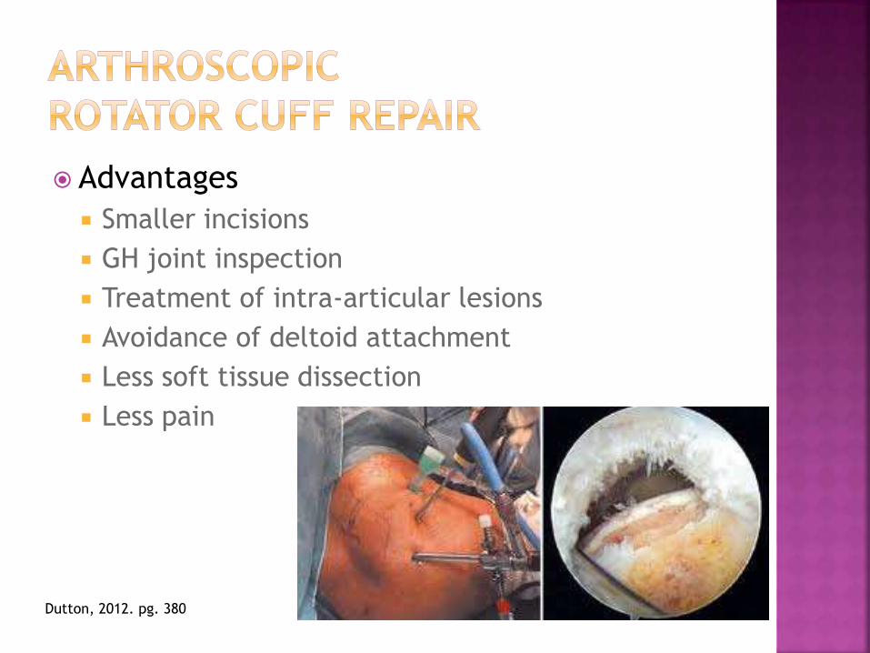
Advantages
Smaller incisions
GH joint inspection
Treatment of intra-articular lesions
Avoidance of deltoid attachment
Less soft tissue dissection
Less pain
Dutton, 2012. pg. 380

Period of immobilization (depending on MD)
Gentle range of motion
Glenohumeral
scapulothoracic
Strengthening as per MD protocol
Manual intervention
General standards:
-Improvement in ROM noted for approx.
6 months
-Return to strength in 12 months

Shoulder is the most commonly dislocated
joint in the body
Men more often than women
Anterior: shoulder ABDuction, extension, and
external rotation
Posterior: shoulder ABDuction, flexion, and
internal rotation
Shankman, 2011. pg. 354

Generalized capsular laxity
Leads to chronic subluxation/dislocation
Anterior
Posterior
Inferior
Primary complaint is pain
Possible instability complaints
Dutton, 2012. pg. 380

Most common direction of instability
Repetition towards anterior apprehension
position
External rotation and horizontal abduction
Patient complaints
Pain with overhead movement
Impingement like symptoms
Positions of abduction and external rotation
Dutton, 2012. pg. 380
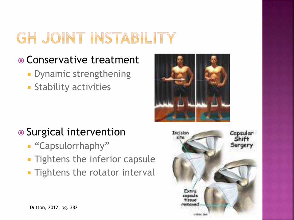
Conservative treatment
Dynamic strengthening
Stability activities
Surgical intervention
“Capsulorrhaphy”
Tightens the inferior capsule
Tightens the rotator interval
Dutton, 2012. pg. 382

Fibrocartilagenous tissue that deepens the
glenoid cavity of the scapula
Injury occurs with trauma or with repetitive
movement
Magee, 2008. pg. 231
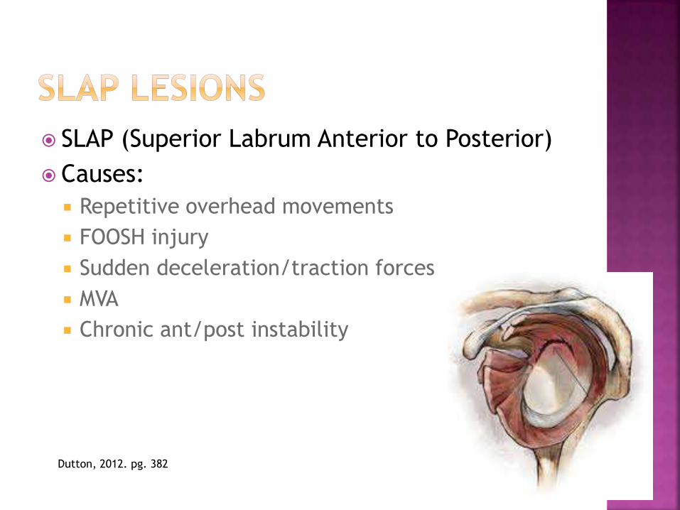
SLAP (Superior Labrum Anterior to Posterior)
Causes:
Repetitive overhead movements
FOOSH injury
Sudden deceleration/traction forces
MVA
Chronic ant/post instability
Dutton, 2012. pg. 382
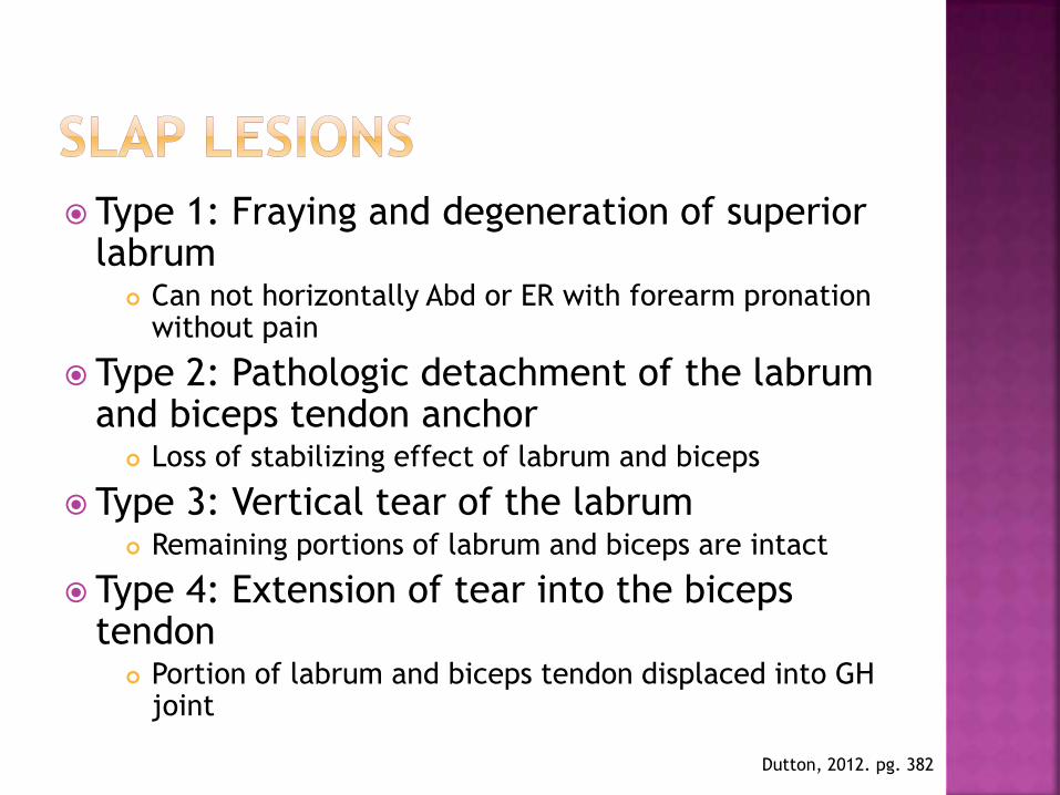
Type 1: Fraying and degeneration of superior labrum
Can not horizontally Abd or ER with forearm pronation without pain
Type 2: Pathologic detachment of the labrum and biceps tendon anchor
Loss of stabilizing effect of labrum and biceps
Type 3: Vertical tear of the labrum Remaining portions of labrum and biceps are intact
Type 4: Extension of tear into the biceps tendon
Portion of labrum and biceps tendon displaced into GH joint
Dutton, 2012. pg. 382

Avulsion of the anterior inferior labrum from
the glenoid rim
Requires surgical stabilization
“TUBS” procedure
Traumatic
Unidirectional instability
Bankart lesion requiring
Surgery
Dutton, 2012. pg. 380

Compression fracture on the posterior
humeral head at the site where the humeral
head impacted the inferior glenoid rim
Dutton, 2012. pg. 381

Conservative treatment is attempted first
Avoidance of provocative position
Gentle ROM/ submaximal isometric exercises
Scapular stability exercises
Closed chain exercises
Improve scapulohumeral rhythm
Open chain activities
Dutton, 2012. pg. 382
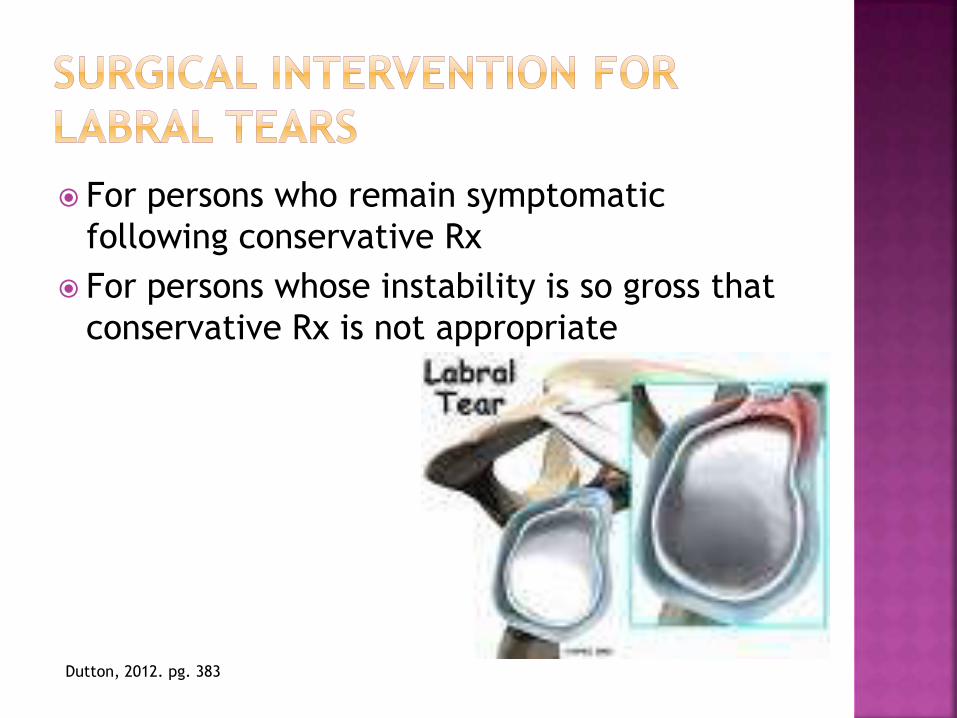
For persons who remain symptomatic
following conservative Rx
For persons whose instability is so gross that
conservative Rx is not appropriate
Dutton, 2012. pg. 383

Most common fracture of the humerus
Results from direct blow to anterior, lateral,
or posterolateral humerus or FOOSH
Represent a major morbidity in the elderly
population
Involve the proximal third of the humerus
Dutton, 2012. pg. 391

Non-displaced fractures:
Immobilization x 2-3 weeks
Gentle ROM
Therapeutic exercise as indicated by physician
Displaced fractures:
Classified into categories
Greater tuberosity, lesser tuberosity, surgical neck,
and anatomic neck
ORIF
Allows progression of ROM and strengthening quicker
due to stabilization of fracture
Shankman, 2011. pg. 361
Dutton, 2012. pg. 391
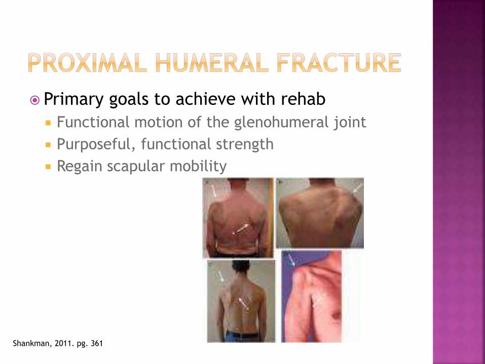
Primary goals to achieve with rehab
Functional motion of the glenohumeral joint
Purposeful, functional strength
Regain scapular mobility
Shankman, 2011. pg. 361
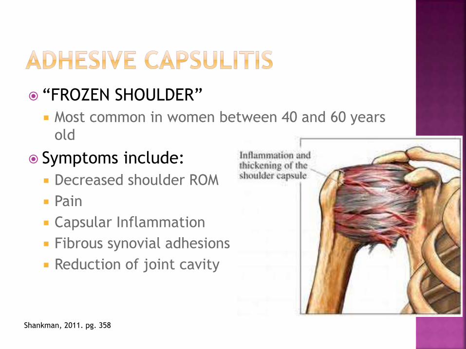
“FROZEN SHOULDER”
Most common in women between 40 and 60 years
old
Symptoms include:
Decreased shoulder ROM
Pain
Capsular Inflammation
Fibrous synovial adhesions
Reduction of joint cavity
Shankman, 2011. pg. 358
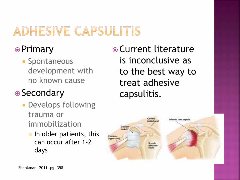
Primary
Spontaneous
development with
no known cause
Secondary
Develops following
trauma or
immobilization
In older patients, this
can occur after 1-2
days
Current literature
is inconclusive as
to the best way to
treat adhesive
capsulitis.
Shankman, 2011. pg. 358

Joint manipulation under anesthesia to
increase mobility

Acromioplasty:
Surgical removal of a piece of bone to allow for
increased space within the joint space
Distal clavicle resection:
Removal of the end of the clavicle closest to the
acromion to alleviate pain and loss of motion

Removal of the humeral head and
glenoid and replaced with metal or
plastic
TSR requires intact
rotator cuff in
order to provide
return to
functional activity
Shankman, 2011. pg. 362
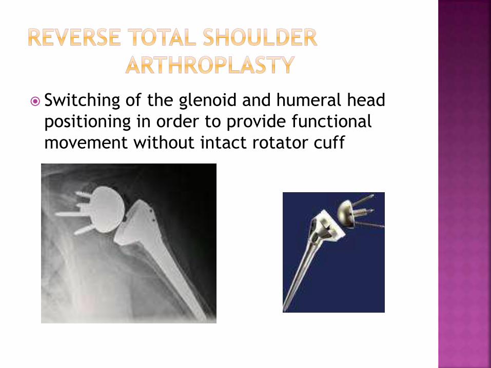
Switching of the glenoid and humeral head
positioning in order to provide functional
movement without intact rotator cuff

Immobilization
Early range of motion
Progressive exercises
Functional return of the affected arm can be
expected around 6 months post-operatively
Shankman, 2011. pg. 362

The patient stands with both shoulders
ABDucted to 90 degrees first with their
thumbs up
The tester provides a downward force on the
arms and notes the patient’s strength
Next, the patient elevates the arms to 90
degrees of ABDuction and 30 degrees of
horizontal ADDuction with thumbs down
The tester provides downward pressure on
the arms and notes the patient’s strength.
Cook, 2013. pg. 166
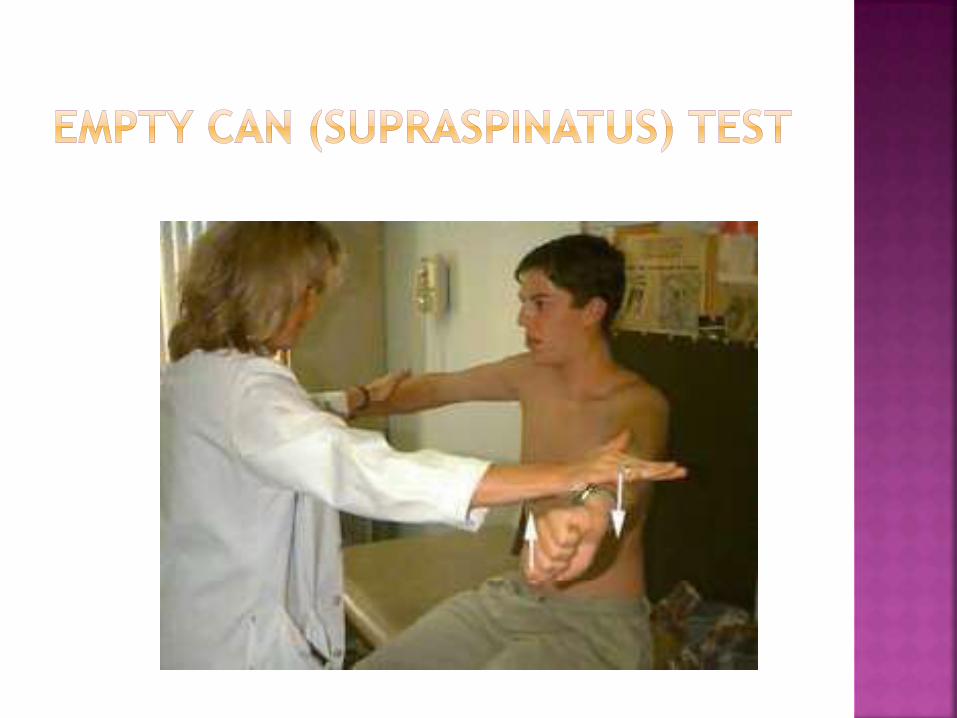

Increased weakness in the empty can
position vs the full can position with or
without complaints of pain is indicative of a
positive result.
Cook, 2013. pg. 166

The patient sits on a table with the involved
shoulder flexed to 90 degrees, the elbow in
full extension, and the forearm in supination.
The tester places one hand on the volar
aspect of the patient’s forearm and the other
on the proximal aspect of the patient’s
humerus and resists the patient’s attempt to
flex the humerus
Konin, 2006. pg. 27

A positive test is indicated by pain in the
bicipital groove that may suggest bicipital
tendonitis

The patient sits on a table while the tester
passively ABDucts the arm to 90 degrees
The patient is asked to slowly lower their
arm to the side
If the patient is unable to slowly lower the
arm to their side or experiences significant
pain with task, this is a positive result
indicating supraspinatus pathology
Cook, 2013. pg. 170
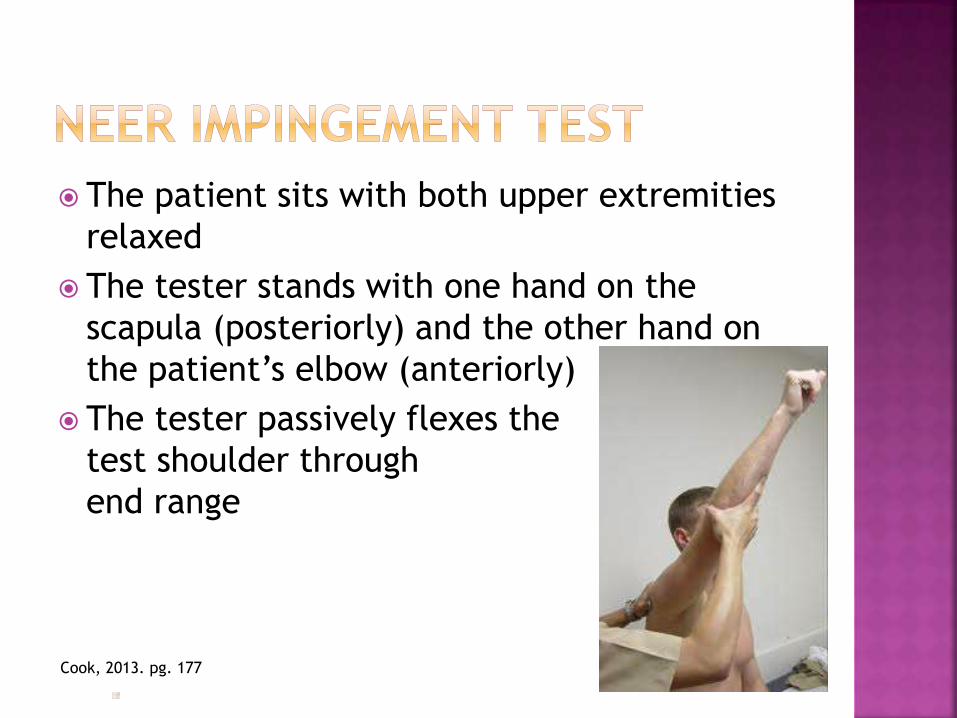
The patient sits with both upper extremities
relaxed
The tester stands with one hand on the
scapula (posteriorly) and the other hand on
the patient’s elbow (anteriorly)
The tester passively flexes the
test shoulder through
end range
Cook, 2013. pg. 177

Pain and/or apprehension with forward
flexion are indicative of shoulder
impingement, specifically the supraspinatus
and biceps long head tendons
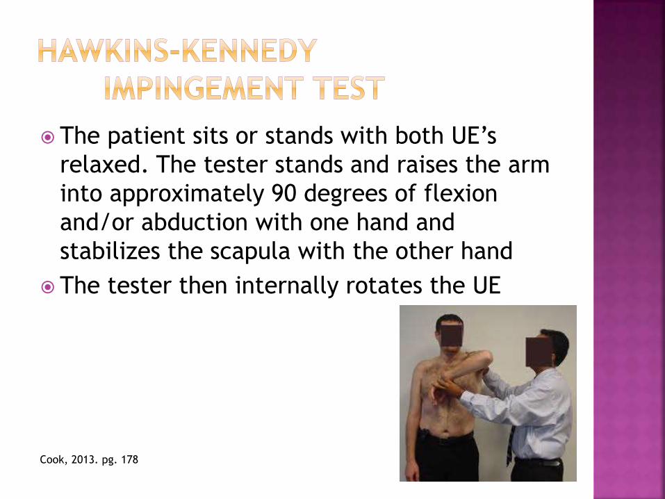
The patient sits or stands with both UE’s
relaxed. The tester stands and raises the arm
into approximately 90 degrees of flexion
and/or abduction with one hand and
stabilizes the scapula with the other hand
The tester then internally rotates the UE
Cook, 2013. pg. 178

Shoulder pain and apprehension are
indicative of supraspinatus tendon
impingement
This test is considered the most
sensitive for assessing
subacromial impingement

The patient lies supine on the table
The tester places the patient’s arm in 90
degrees of abduction and 90 degrees of
elbow flexion and then slowly externally
rotates the shoulder
The patient demonstrating
apprehension is indicative of a
positive test. This position mimics
the positioning of an anterior
dislocation, recreating instability
Cook, 2013. pg. 189
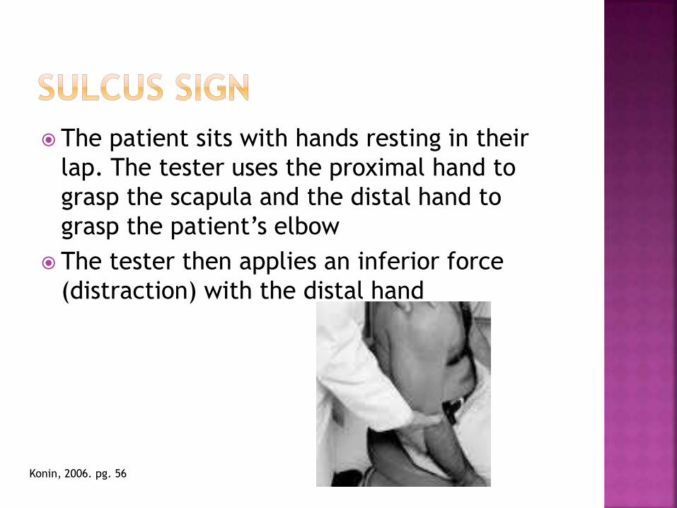
The patient sits with hands resting in their
lap. The tester uses the proximal hand to
grasp the scapula and the distal hand to
grasp the patient’s elbow
The tester then applies an inferior force
(distraction) with the distal hand
Konin, 2006. pg. 56

Excessive inferior humeral head translation
with a visible or palpable “step-off” is
indicative of inferior and/or multi-directional
instability
The patient stands with the arm in 90
degrees of flexion, 30-45 degrees of
horizontal ADDuction and maximal internal
rotation while the tester grasps their wrist
The patient tries to flex and horizontally
ADDuct the arm against the tester’s
resistance
The test is then repeated with the arm in
external rotation
Konin, 2006. pg. 74

Pain and/or clicking in an internally rotated
position but absent in the externally rotated
position is indicative of a SLAP lesion
Stretching and Exercises
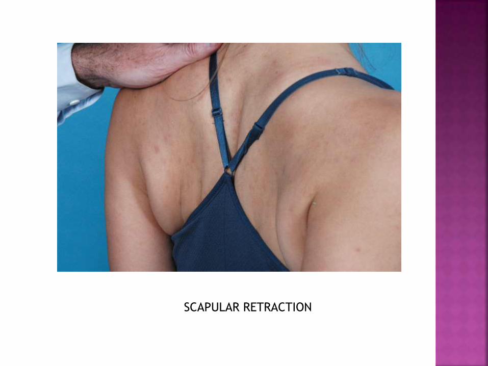
SCAPULAR RETRACTION

INTERNAL ROTATION STRETCH

INFERIOR CAPSULAR STRETCH

POSTERIOR CAPSULE STRETCH
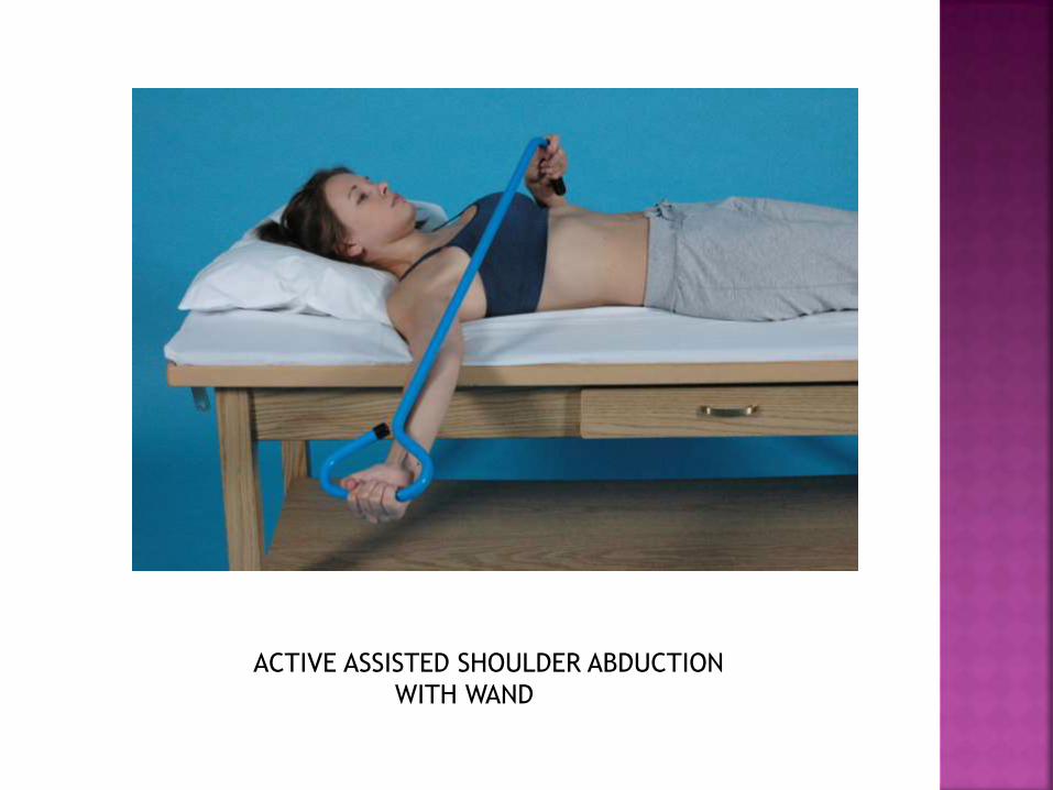
ACTIVE ASSISTED SHOULDER ABDUCTION
WITH WAND
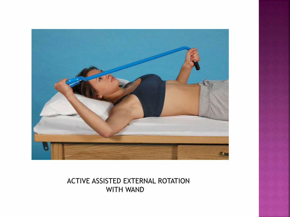
ACTIVE ASSISTED EXTERNAL ROTATION
WITH WAND

MANUAL INTERVENTION

PASSIVE FLEXION

PASSIVE EXTERNAL ROTATION

PASSIVE INTERNAL ROTATION
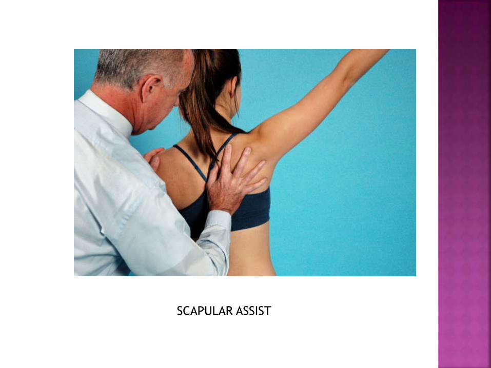
SCAPULAR ASSIST

PEC MINOR STRETCH

Shankman, Fundamental Orthopedic Management for the Physical Therapist Assistant, 3rd edition. Mosby.2011
Konin, Wiksten, Isear, Brader, Special Tests for Orthopedic Examination, 3rd edition. Slack. 2006
Magee, Orthopedic Physical Assessment, 5th edition. Saunders. 2008
Dutton, Orthopaedics for the Physical Therapist Assistant. Jones&Bartlett. 2012
Cook, Hegedus, Orthopedic Physical Examination Tests, 2nd edition. Pearson. 2013