the shoulder complex - asht 2016_htrc... · hand therapy review course curtis national hand center...
TRANSCRIPT

Hand Therapy Review CourseCurtis National Hand Center
Baltimore, MDOctober 7‐9. 2016
Shoulder Anatomy and KinesiologyRomina Astifidis MS, PT, CHT
The Shoulder Complex
Bones
•Clavicle• Connects axial skeleton and upper limb
• Serves as attachment site for muscles controlling upper extremity
• Protects the neurovascular bundle from neck to arm
30°with 130°
Bones
• Scapula• Lies over ribs 2‐7• 30 anterior to the coronal plane, 10 on the frontal tilt.
• Provides a stability for shoulder complex
• Serves as an attachment site for muscles
• Transmits energy proximal to distal• Landmarks: Spine, Acromionprocess, Glenoid fossa, Coracoidprocess
Inferior angle
Lateral border
Medial border
Superior angle
Acrom.
Definitions of Scapular Motion
Adduct/Retract
Abduct/Protract
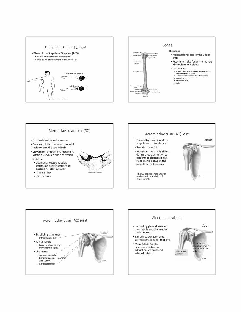
Functional Biomechanics2
• Plane of the Scapula or Scaption (POS)• 30‐45 anterior to the frontal plane• True plane of movement of the shoulder
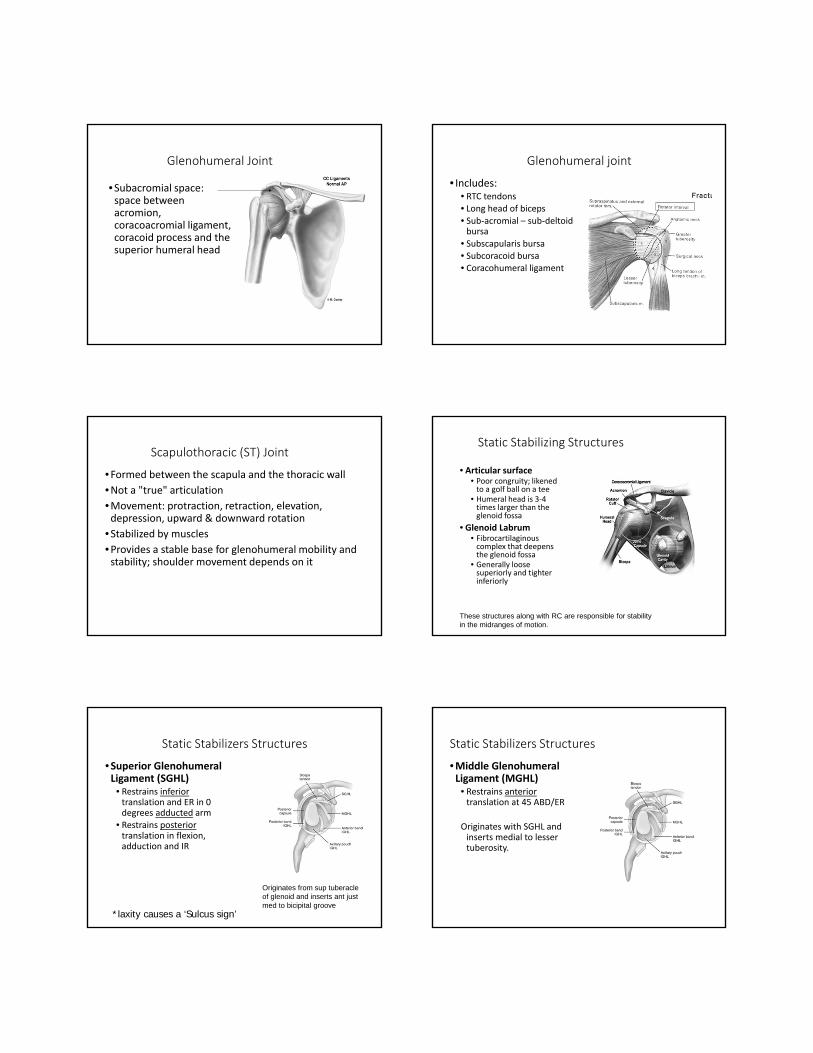
Bones
• Humerus
• Proximal lever arm of the upper limb
• Attachment site for prime movers of shoulder and elbow
• Landmarks• Greater tubercle: insertion for supraspinatus, infraspinatus, teres minor
• Lesser tubercle: insertion for subscapularis
• Surgical neck
• Anatomical neck
• Shaft
Sternoclavicular Joint (SC)
• Proximal clavicle and sternum
• Only articulation between the axial skeleton and the upper limb
• Movement: protraction, retraction, rotation, elevation and depression
• Stability: • Ligaments: costoclavicular, sternoclavicular (anterior and posterior), interclavicular
• Articular disk• Joint capsule
Acromioclavicular (AC) Joint
• Formed by acromion of the scapula and distal clavicle
• Synovial plane joint
• Movement: Primarily slides during shoulder motion to conform to changes in the relationship between the scapula & the humerus
The AC capsule limits anterior and posterior translation of distal clavicle.
Acromioclavicular (AC) joint
• Stabilizing structures• Intraarticular disk
• Joint capsule• Loose to allow sliding movement of joint
• Ligaments• Acromioclavicular
• Coracoclavicular (Trapezoid and Conoid)
• Coracoacromial
T C
Glenohumeral joint
• Formed by glenoid fossa of the scapula and the head of the humerus
• Ball and socket joint that sacrifices stability for mobility
• Movement: flexion, extension, abduction, adduction, external and internal rotation
5° tilt helps to keep humerus in socket with arm at side25% or 1/3
contact
Glenohumeral Joint
•Subacromial space: space between acromion, coracoacromial ligament, coracoid process and the superior humeral head
Glenohumeral joint
• Includes:• RTC tendons• Long head of biceps• Sub‐acromial – sub‐deltoid bursa
• Subscapularis bursa• Subcoracoid bursa• Coracohumeral ligament
Scapulothoracic (ST) Joint
•Formed between the scapula and the thoracic wall
•Not a "true" articulation•Movement: protraction, retraction, elevation, depression, upward & downward rotation
•Stabilized by muscles
•Provides a stable base for glenohumeral mobility and stability; shoulder movement depends on it
Static Stabilizing Structures
• Articular surface• Poor congruity; likened to a golf ball on a tee
• Humeral head is 3‐4 times larger than the glenoid fossa
• Glenoid Labrum• Fibrocartilaginouscomplex that deepens the glenoid fossa
• Generally loose superiorly and tighter inferiorly
These structures along with RC are responsible for stability in the midranges of motion.
Static Stabilizers Structures
•Superior Glenohumeral Ligament (SGHL)
• Restrains inferiortranslation and ER in 0 degrees adducted arm
• Restrains posteriortranslation in flexion, adduction and IR
*laxity causes a ‘Sulcus sign’
Originates from sup tuberacle of glenoid and inserts ant just med to bicipital groove
Static Stabilizers Structures
•Middle GlenohumeralLigament (MGHL)
• Restrains anterior translation at 45 ABD/ER
Originates with SGHL and inserts medial to lesser tuberosity.
Static Stabilizing Structures
•Inferior glenohumeral ligament (IGHL)•Primary anterior restraint in position of 90abduction, 90 ER‐ Anterior portion
•Thickened bands that form a hammock to support the humerus in the axillary pouch
•Primary restraint to posterior translation in ABD/IR‐ Posterior portion
• Primary restraint to inferior translation with arm at 90 degrees ABD
Tension of ligaments
LigamentShoulder motions to pull the ligament taut
Humeral head motion to pull the ligament taut
Superior glenohumeral lig. Full adduction Inferior or anterior glide
Middle glenohumeral lig. External rotation Anterior glide
Inferior glenohumeral lig.Anterior bandPosterior band
Abduction and external rotationAbduction and internal rotation
Non specific
Coracohumeral lig.Extremes of flexion, extension, or external rotation
Inferior glide
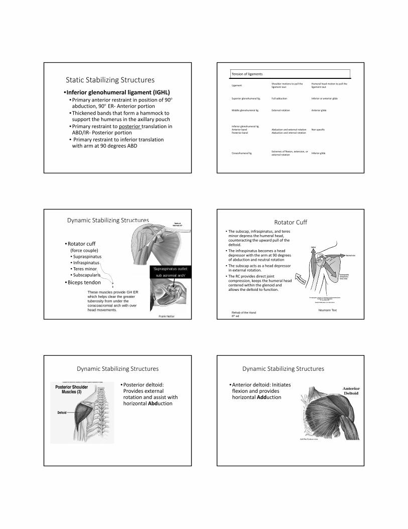
Dynamic Stabilizing Structures
•Rotator cuff (force couple)• Supraspinatus • Infraspinatus• Teres minor• Subscapularis
•Biceps tendon
Frank Netter
‘Supraspinatus outlet:
sub acromial arch’
These muscles provide GH ER which helps clear the greater tuberosity from under the coracoacromial arch with over head movements.
Rotator Cuff• The subscap, infraspinatus, and teres minor depress the humeral head, counteracting the upward pull of the deltoid.
• The infraspinatus becomes a head depressor with the arm at 90 degrees of abduction and neutral rotation
• The subscap acts as a head depressor in external rotation.
• The RC provides direct joint compression, keeps the humeral head centered within the glenoid and allows the deltoid to function.
Neumann TextRehab of the Hand 6th ed
Dynamic Stabilizing Structures
•Posterior deltoid: Provides external rotation and assist with horizontal Abduction
Dynamic Stabilizing Structures
•Anterior deltoid: Initiates flexion and provides horizontal Adduction
Dynamic Stabilizing Structures
•Middle Deltoid: initiates Abduction primarily to 90 degrees while producing upward shear of the humeral head
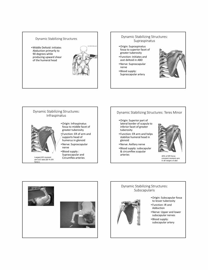
Dynamic Stabilizing Structures: Supraspinatus
•Origin: Supraspinatus fossa to superior facet of greater tuberosity
•Function: Initiates and asst deltoid in ABD
•Nerve: Suprascapularnerve
•Blood supply: Suprascapular artery
Dynamic Stabilizing Structures: Infraspinatus
•Origin: Infraspinatus fossa to middle facet of greater tuberosity
•Function: ER of arm and supports head of humerus in glenoid
•Nerve: Suprascapularnerve
•Blood supply : Suprascapular and Circumflex arteriesLargest ER moment
arm at 0 abd (60 % ER force.)
Dynamic Stabilizing Structures: Teres Minor
•Origin: Superior part of lateral border of scapula to inferior facet of greater tuberosity
•Function: ER arm and helps stabilize humeral head in glenoid
•Nerve: Axillary nerve•Blood supply: subscapular & circumflex scapular arteries
40% of ER force-constant moment arm in all ranges of abd
Dynamic Stabilizing Structures: Subscapularis
•Origin: Subscapular fossa to lesser tuberosity
•Function: IR and Adduction
•Nerve: Upper and lower subscapular nerves
•Blood supply: subscapular artery
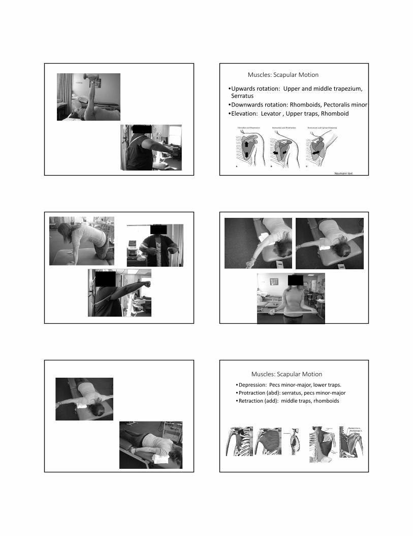
Muscles: Scapular Motion
•Upwards rotation: Upper and middle trapezium, Serratus
•Downwards rotation: Rhomboids, Pectoralis minor
•Elevation: Levator , Upper traps, Rhomboid
Neumann text
Muscles: Scapular Motion
•Depression: Pecs minor‐major, lower traps.
•Protraction (abd): serratus, pecs minor‐major
•Retraction (add): middle traps, rhomboids
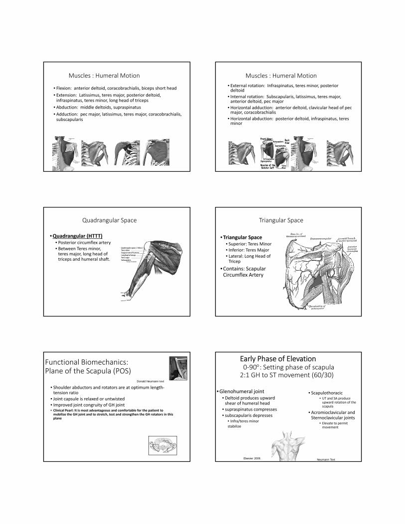
Muscles : Humeral Motion
• Flexion: anterior deltoid, coracobrachialis, biceps short head
• Extension: Latissimus, teres major, posterior deltoid, infraspinatus, teres minor, long head of triceps
• Abduction: middle deltoids, supraspinatus
• Adduction: pec major, latissimus, teres major, coracobrachialis, subscapularis
Muscles : Humeral Motion
• External rotation: Infraspinatus, teres minor, posterior deltoid
• Internal rotation: Subscapularis, latissimus, teres major, anterior deltoid, pec major
• Horizontal adduction: anterior deltoid, clavicular head of pecmajor, coracobrachialis
• Horizontal abduction: posterior deltoid, infraspinatus, teresminor
Quadrangular Space
•Quadrangular (HTTT)• Posterior circumflex artery • Between Teres minor, teres major, long head of triceps and humeral shaft.
Triangular Space
•Triangular Space• Superior: Teres Minor• Inferior: Teres Major
• Lateral: Long Head of Tricep
•Contains: Scapular Circumflex Artery
Functional Biomechanics:Plane of the Scapula (POS)
• Shoulder abductors and rotators are at optimum length‐tension ratio
• Joint capsule is relaxed or untwisted• Improved joint congruity of GH joint• Clinical Pearl: It is most advantageous and comfortable for the patient to mobilize the GH joint and to stretch, test and strengthen the GH rotators in this plane
Donald Neumann text
Early Phase of Elevation0‐90: Setting phase of scapula2:1 GH to ST movement (60/30)
•Glenohumeral joint• Deltoid produces upward shear of humeral head
• supraspinatus compresses• subscapularis depresses
• Infra/teres minor
stabilize
• Scapulothoracic• UT and SA produce upward rotation of the scapula
• Acromioclavicular and Sternoclavicular joints
• Elevate to permit movement
Elsevier 2006 Neumann Text
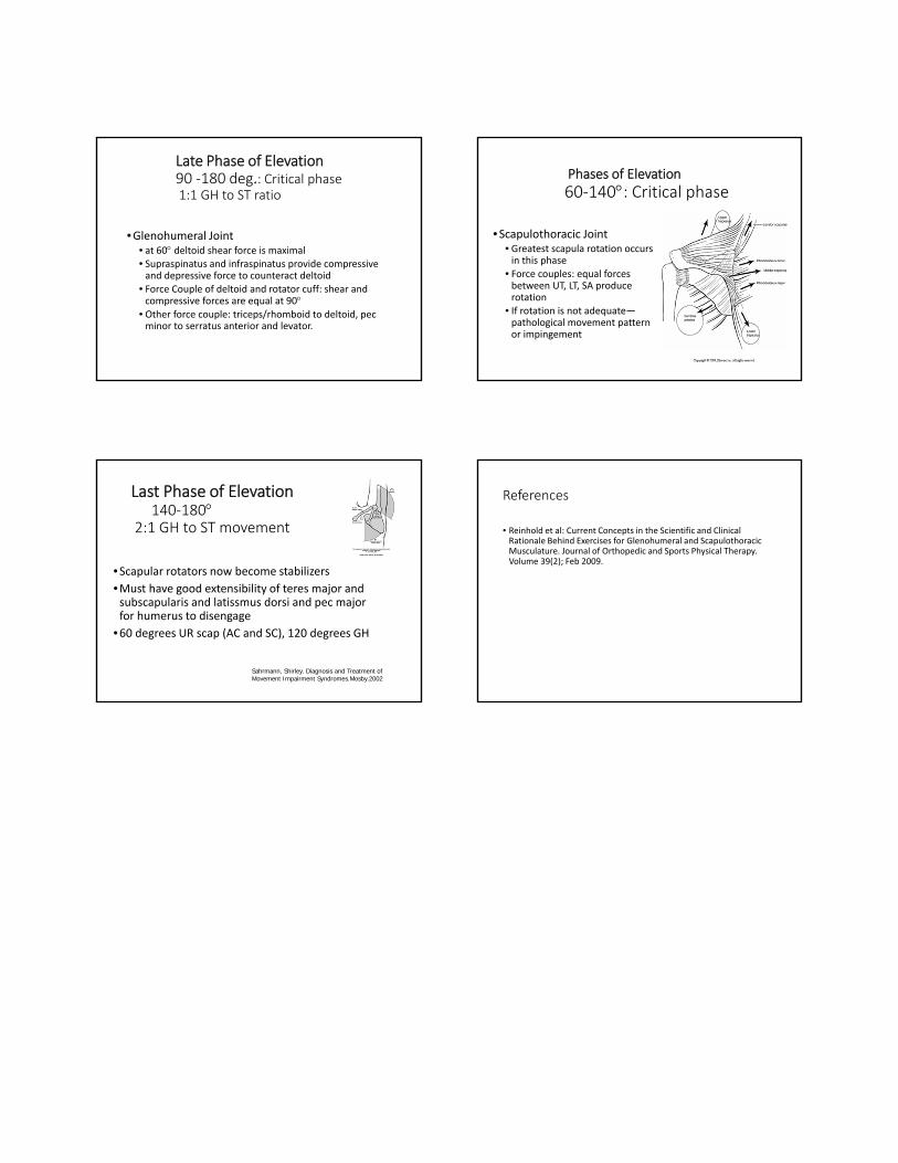
Late Phase of Elevation90 ‐180 deg.: Critical phase1:1 GH to ST ratio
•Glenohumeral Joint• at 60 deltoid shear force is maximal• Supraspinatus and infraspinatus provide compressive and depressive force to counteract deltoid
• Force Couple of deltoid and rotator cuff: shear and compressive forces are equal at 90
• Other force couple: triceps/rhomboid to deltoid, pecminor to serratus anterior and levator.
Phases of Elevation
60‐140: Critical phase
•Scapulothoracic Joint• Greatest scapula rotation occurs in this phase
• Force couples: equal forces between UT, LT, SA produce rotation
• If rotation is not adequate—pathological movement pattern or impingement
Last Phase of Elevation140‐180
2:1 GH to ST movement
•Scapular rotators now become stabilizers
•Must have good extensibility of teres major and subscapularis and latissmus dorsi and pec major for humerus to disengage
•60 degrees UR scap (AC and SC), 120 degrees GH
Sahrmann, Shirley. Diagnosis and Treatment of Movement Impairment Syndromes.Mosby.2002
References
• Reinhold et al: Current Concepts in the Scientific and Clinical Rationale Behind Exercises for Glenohumeral and Scapulothoracic Musculature. Journal of Orthopedic and Sports Physical Therapy. Volume 39(2); Feb 2009.