the sequence of the cyo operon indicates substantial structural
TRANSCRIPT

THE JOURNAL OF BIOLOC~CAL CHEMISTRY 0 1990 by The American Society for Biochemistry and Molecular Biology, Inc.
Vol. 265, No. 19, Issue of July 5, pp. 11185-11192,199O Printed in U.S.A.
The Sequence of the cyo Operon Indicates Substantial Structural Similarities between the Cytochrome o Ubiquinol Oxidase of Escherichia coli and the aas-type Family of Cytochrome c Oxidases*
(Received for publication, December 11, 1989)
Visala Chepuri, Laura Lemieux, Douglas C.-T. Au& and Robert B. Gennisg From the Departments of Biochemistry and Chemistry, University of Illinois, Urbana, Illinois 61801
The cytochrome o complex is one of two ubiquinol oxidases in the aerobic respiratory system of Esche- richia coli. This enzyme catalyzes the two-electron oxidation of ubiquinol-8 which is located in the cyto- plasmic membrane, and the four-electron reduction of molecular oxygen to water. The purified oxidase con- tains at least four subunits by sodium dodecyl sulfate- polyacrylamide gel electrophoresis analysis and has been shown to couple electron flux to the generation of a proton motive force across the membrane. In this paper, the DNA sequence of the cyo operon, containing the structural genes for the oxidase, is reported. This operon is shown to encode five open reading frames, cyoABCDE. The gene products of three of these, cyoA, cyoB, and cyoC, are clearly related to subunits II, I, and III, respectively, of the eukaryotic and prokaryotic aa3-type cytochrome c oxidases. This family of cyto- chrome c oxidases contain heme a and copper as pros- thetic groups, whereas the E. coli enzyme contains heme b (protoheme IX) and copper. The most striking sequence similarities relate the large subunits (I) of both the E. coli quinol oxidase and the cytochrome c oxidases. It is likely that the sequence similarities re- flect a common molecular architecture of the two heme binding sites and of a copper binding site in these enzymes.
In addition, the cyoE open reading frame is closely related to a gene denoted ORFl from Paracoccus den- trificans which is located in between the genes encod- ing subunits II and III of the cytochrome c oxidase of this organism. The function of the ORFl gene product is not known.
These sequence relationships define a superfamily of membrane-bound respiratory oxidases which share structural features but which have different functions. The E. coli cytochrome o complex oxidizes ubiquinol but has no ability to catalyze the oxidation of reduced cytochrome c. Nevertheless, it is clear that the E. coli oxidase and the au&ype cytochrome c oxidases must have very similar structures, at least in the vicinity of the catalytic centers, and they are very likely to have
*This work was supported by Department of Energy Grant DEFGOZ-87ER13716. The costs of publication of this article were defrayed in part by the payment of page charges. This article must therefore be hereby marked “aduertisement” in accordance with 18 USC. Section 1734 solely to indicate this fact.
The nucleotide sequence(s) reported in this paper has been submitted to the GenBankTM/EMBL Data Bank with accession number(s) J05492.
$ Current address: Dept. of Pharmacology, University of Washing- ton, Seattle, WA 98195.
3 To whom correspondence should be addressed: Dept. of Biochem- istry and Chemistry, University of Illinois, 505 S. Mathews Ave., Urbana, IL 61801.
similar mechanisms for bioenergetic coupling (proton pumping),
The aerobic respiratory chain of Escherichia coli is rela- tively simple (for reviews see Refs. l-3). A number of mem- brane-bound dehydrogenases oxidize organic substrates on the inside of the cell and reduce ubiquinone-8 to ubiquinol-8 in the cytoplasmic membrane. An example of such an enzyme is succinate dehydrogenase (Complex II) (4). Ubiquinol-8 can diffuse freely within the cytoplasmic membrane and can be oxidized by either of two respiratory quinol oxidases, the cytochrome o complex or the cytochrome d complex. The cytochrome o complex predominates under growth conditions where the oxygen tension is high, whereas the cytochrome d complex is synthesized and accumulates under low aeration, essentially anaerobic, conditions. In each case, the product of reduction of molecular oxygen is water, not peroxide (5). Note that aerobically grown E. coli does not contain any cytochrome c and there is, therefore, no equivalent of the ubi- quinol:cytochrome c oxidoreductase (bcl complex) or any cy- tochrome c oxidase. Each of the oxidases in E. coli catalyzes the transfer of electrons from quinol to molecular oxygen. The cytochrome d complex contains three heme prosthetic groups (6, 7) and the sequences of the two subunits of this heterodimer shows no relationship to any known proteins, including other respiratory electron transport complexes (8).
The cytochrome o complex (also called cytochrome bo or cytochrome bSG2-o) has been purified in several laboratories and reported to contain four subunits (g-11), although a two- subunit form has also been reported (12). The oxidase con- tains two heme b (protoheme IX) prosthetic groups (9-12) and one or two copper atoms (12). The purified oxidase has been reconstituted in artificial phospholipid vesicles where it utilizes ubiquinol-8 as a substrate and generates a voltage and net proton translocation across the bilayer (13-15). Recent work has shown that in sphaeroplasts of E. coli the enzyme generates a proton motive force by a combination of a scalar mechanism plus a proton pump coupled to the electron trans- port through the enzyme (16). ESR (17, 18) and resonance Raman (19) studies have shown that cytochrome bo contains a low spin (6-coordinate) heme as well as a high spin (5- coordinate) heme and that, furthermore, the high spin heme is electronically coupled to a copper, forming a heme/copper binuclear center (18).
These data suggest similarities to the prokaryotic and eu- karyotic heme u-containing (aas-type) cytochrome c oxidases, insofar as these oxidases also function as proton pumps and the catalytic centers include a 6-coordinate heme plus a heme/ copper binuclear center where oxygen is reduced to water (for reviews see Refs. 20-22). In this paper, it is shown that the
11185
by guest on February 19, 2018http://w
ww
.jbc.org/D
ownloaded from
11186 cyo Operon
deduced sequences of the subunits of the cytochrome o com- plex are very clearly related to subunits (I, II, and III) of the aua-type cytochrome c oxidases. This defines a new superfam- ily of membrane-bound respiratory oxidases which utilize different substrates (ubiquinol or cytochrome c) but share common structural features.
I -- b+--+’ -
cp 96 c& *o cp p*ne H I Ill (?I (?) SDS-PAGE
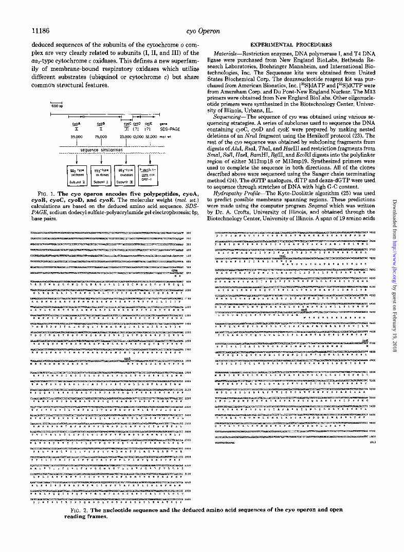
FIG. 1. The cyo operon encodes five polypeptides, cyoA, cyoB, cyoC, cyoD, and cyoE. The molecular weight (mol. wt.) calculations are based on the deduced amino acid sequence. SDS- PAGE, sodium dodecyl sulfate-polyacrylamide gel electrophoresis; bp, base pairs.
FIG. 2. The nucleotide sequence and the deduced amino acid sequences of the cyo operon and open reading frames.
by guest on February 19, 2018http://w
ww
.jbc.org/D
ownloaded from

cyo Operon
FIG. 3. Kyte-Doolittle hydropathy profiles of cyoA, cyoB, cyoC, cyoD, and cyoE. A window of 19 amino acids was used and the urofiles were smoothed twice. The numbers above the peaks indicate potential membrane-spanning a-helical regions.
was used and the resulting profiles were smoothed twice. Sequence Analysis-DNA sequence was analyzed using the pro-
gram DNASTAR. Homology searches were performed on the amino acid sequences of cyoA, cyoB, cyoC, cyoD, and cyoE using the protein data base of DNASTAR.
RESULTS
Fig. 1 shows a schematic of a 5.8-kilobase SalI-SphI DNA fragment that contains the entire cyo operon. Previous studies have shown that expression of the genes within this restriction fragment complement mutations in the cyo locus and can be used to overproduce the cytochrome o complex in the E. coli membrane (26). The DNA sequence reveals five ORFs’ indi- cated as cyoA, cyoB, cyoC, cyoD, and cyoE. Transcriptional studies by Minagawa et al. (27) have identified putative reg- ulatory sequences and a promotor to the left of cyoA. It is likely that cyoABCDE comprises a single transcriptional unit. The complete DNA sequence and the deduced amino acid sequences of the five ORFs are shown in Fig. 2. There are short stretches of DNA between the ORFs (no more than 24 base pairs) except for the junction between cyoB and cyoC. In this case, the genes overlap by 10 base pairs.
The deduced molecular weights of the gene products are 35,000, 75,000, 23,000, 12,000, and 32,000 for cyoA, B, C, D, and E, respectively. The purified cytochrome o complex has four subunits in several preparations (9, 10) with apparent molecular weights of 66,000 (I), 35,000 (II), 22,000 (III), and 17,000 (IV). The two subunits reported in the preparation of Kita et al. (12) have apparent molecular weights of 55,000 (I) and 33,000 (I). Subclones of cyoA and cyoB alone have shown that these gene products correspond to subunit II (Mapp 33,000 or 35,000) and subunit I (Mapp 55,000 or 66,000) (28). Clearly, the migration of subunit I is faster than that predicted for a protein of M, 75,000 and is dependent on the gel system, but this is not unusual for a hydrophobic membrane protein (29).
Protein sequence data have confirmed the identity of the
1 The abbreviations used are: ORFs, open reading frames; DCCD, dicyclohexylcarbodiimide.
gene product of cyoA as subunit II and cyoB as subunit I and, in addition, has shown that cyoC corresponds to subunit III (Mapp 22,000).* There are no data to prove that cyoD and cyoE encode subunits of the cytochrome o complex, although this seems likely. Attempts to obtain amino acid sequence from subunit IV, which might possibly be the cyoD gene product, have been unsuccessful. An sodium dodecyl sulfate-polyacryl- amide gel electrophoresis band (Map,, 26,000) which stains poorly with Coomassie Brilliant Blue has been consistently seen in cytochrome o preparations, and this might correspond to the cyoE gene product. However, there are no data to demonstrate that this is related to cyoE or, indeed, is part of the complex.
Fig. 3 shows the Kyte-Doolittle hydropathy profiles of the five gene products. Based on this analysis, all five subunits are expected to have transmembrane a-helical segments. CyoA (subunit II) has two predicted transmembrane spans, and a large, hydrophilic carboxyl-terminal domain. CyoB (subunit I) has up to 15 transmembrane spans. CyoC (subunit III) has five putative transmembrane spans and cyoD and cyoE have, respectively, three and seven hydrophobic spans. Gene fusion experiments (65) have provided data in support of models of each of these proteins containing the indicated number of transmembrane spans.
Fig. 4, A and B, is the Diagon plots (dot plots) illustrating the amino acid sequence similarities between subunits I and III, respectively, of E. coli cytochrome o and of the cytochrome c oxidase (aas-type) from Paracoccus denitrificarw (30-32). Fig. 4C shows the similarity between the gene products of cyoE (E. coli) and ORFl (P. denitrificans) (30). The most striking similarity is between the large subunits (I), where 37% of the amino acids are identical over a 546-amino acid overlap. Fig. 5 illustrates this further by showing the align- ment of the amino acid sequences of the E. coli cytochrome o subunit I (cyoB) with subunit I from the cytochrome c oxidases from P. denitrificans, beef heart mitochondrion (33), and the thermophilic bacterium PS3 (34). The E. coli subunit is longer
* V. Chepuri and R. B. Gennis, manuscript in preparation.
by guest on February 19, 2018http://w
ww
.jbc.org/D
ownloaded from

11188
A P denifrificons subumt I 100 200 300 400 500
I I 1 I /
cyo Operon
El P denitrificons subumt IU C P denifnficons OR F I
50 100 I50 200 250
t 50-
z ‘\\, e a IOO- ‘, %j. aI E c l50-
-I ‘. s ..\
‘< 200- \.\.
2 j.. ‘.. w ‘\
250
FIG. 4. Comparisons of the amino acid sequences using dot plots to show the similarities between the deduced gene products of cyoA, cyoC, and cyoE with corresponding gene products from P. denitrificana. A, comparison of subunit I (cyoB) for cytochrome o complex of E. coli with subunit I from cytochrome c oxidase of P. denitrificans. B, comparison of subunit III (cyoC) from E. coli oxidase with subunit III fromP. denitrificans cytochrome c oxidase. C, comparison of the deduced amino acid sequences of cyoE from E. coli and ORFl from P. denitrificams.
FIG. 5. Alignment of amino acid sequences from E. coli cytochrome
oxidase complex subunit I ;)CI’OB), PS3 cytochrome c oxidase subunit I (PS3), P. denitrificane cy- tochrome c oxidase subunit I (PDCOI), and beef heart cyto- chrome c oxidase subunit I (BOVCOI). The amino acid residues enclosed in boxes are identical in all four sequences. See text for references. The sequences of the E. coli and P. denitriji- cans subunits are 37% identical over a region of 546 residues.
at both ends than the subunits from these cytochrome c oxidases, but the extent of conservation is remarkable, as shown by the number of residues which are identical in all the sequences shown. Fig. 6 shows one possible model for subunit I of the cytochrome o complex. The hydropathy profile has been interpreted with the aid of results from gene fusion experiments in terms of the 15 proposed transmem- brane cu-helices. The helices numbered I-XII roughly corre- spond to those proposed by Holm et aE. (35) for subunit I of the au&pe cytochrome c oxidases (also numbered I-XII). The model shown for the E. coli oxidase contains one addi- tional proposed transmembrane span at the amino terminus plus two more at the carboxyl terminus that do not correspond to portions of the cytochrome c oxidase subunits. Note that
the 12-span model for subunit I of the cytochrome c oxidases is itself speculative and not universally accepted (36).
The sequence similarity between subunit III (cyoC) of the E. coli oxidase and subunit III from the cytochrome c oxidases is less striking. An alignment between the E. coli subunit and those from I? denitrificms (32) and bovine heart (33) is shown in Fig. 7. The E. coli subunit is considerably shorter, and is missing the portion corresponding to the first 61 amino acid residues of subunit III from the P. denitrificum cytochrome c oxidase. The identical residues are boxed. Also indicated in Fig. 7 is the glutamate (bovine E91) which binds to the inhibitor DCCD in cytochrome c oxidases (37). This is an aspartate in E. coli (D36), but DCCD inhibition or binding have not been demonstrated for the E. coli oxidase.
by guest on February 19, 2018http://w
ww
.jbc.org/D
ownloaded from

FIG. 6. A proposed topological model of subunit I of the E. coti ey- tochrome o terminal oxidase based on the Kyte-Doolittle hydropathy profile and gene fusion experiments (V. Chepuri and R. B. Gennis, un- published data).
cyo Operon 11189
CYOC I F E - L PDC03 T F D P w BO”3 P L N P L
CYOC w L F G A PDCO3 ” I L G ” BO”3 0 ITLG”
--ATYA”---- KNALYPMGPDS HSSL----APT
HGHHDAGGTKIF- 27 GEHTPVVRIGLQY 89
0 GHHTPAVQKGLRY 81
LVNGTAGGPTGKD 62 PIKDGVWPPEGI" 134
ml PELGGCWPPTGIH 122 -
YGMAAIA-MYKNNKSQVISWLALT
0 u
105 “TWAHHAFVLEGDRKTTINGLlVA 179 ITWAHHS-LMEGDRKHMLQALF~T 166
GMGPDRSGFLS AFGLADTVYAG PFTISDGVYGS
161 274 260
FIG. 7. Alignment of amino acid sequences of subunit III of the E. coli cytochrome o complex (CYOC) with subunit III of the P. denitrificans cytochrome c oxidase (PDCO3) and beef heart cytochrome c oxidase (BOW). Boxed amino acid show iden- tical residues in all three sequences. The asterisk indicates the glutamate involved in DCCD binding in the subunits from P. denitr+can.s and beef heart (bovine E91) which is an aspartate in E. cd. Numbers at the right represent amino acid numbers.
Fro 8. Alignment of amino acid sequences from subunits II from the E. coli cytochrome o complex (CYOA), P. denitrificans cyto- chrome c oxidase (PDCOZ), and beef heart cytochrome c oxidase (BOV2) (bottom line). The amino acid residues enclosed in bones are identical in the three sequences. The asterisks indicate the pairs of histidines and cysteines which have been proposed (35) as ligands to CuA in the cytochrome c oxidases. These are not conserved in the sequence of the subunit in E. coli quinol oxidase. Numbers at, the right represent amino acid numbers.
The alignment of the amino acid sequences of subunit II (cyoA) from the cytochrome o and subunit II from two cyto- chrome c oxidases is shown in Fig. 8. There is little sequence identity between the subunit from cytochrome o and those from the cytochrome c oxidase family. This is apparent by comparing the relatively few residues all three subunits have in common, in contrast to the data for both subunits I and III. For comparison, subunit II from the cytochrome c oxidases of P. denitrificans (31) and from bovine heart (33, 38) are 36% identical over 227 residues. Even within primates, it has been pointed out that this subunit is less well conserved than either subunits I and III (39). The asterisks in Fig. 8 indicate the two histidines and two cysteines that have been proposed to be ligands for CuA, the EPR-visible copper center in the
cytochrome c oxidases (see Refs. 20 and 35). None of these four residues is present in the sequence of subunit II from the E. coli cytochrome o complex. All experimental efforts to detect a CuA species in E. coli membrane or in the purified complex have so far proved negative,3 so it seems that the CuA center is absent from the E. coli ubiquinol oxidase. As was mentioned previously, E. coli does not contain cytochrome c and the cytochrome o complex does not utilize cytochrome c as a substrate. With this in mind, it is not surprising that acidic amino acids (bovine Asp-158, Glu-198) which have been implicated in the binding of cytochrome c to the aa&pe oxidases and are conserved within the cytochrome c oxidases
3 N. E. Gabriel, S. I. Chan, K. C. Minghetti, C. D. Georgiou, and R. B. Gennis, manuscript in preparation.
by guest on February 19, 2018http://w
ww
.jbc.org/D
ownloaded from

11190
FIG. 9. Alignment of amino acid sequences for the gene products of cyoE from E. coli (CYOE) and ORFl from P. denitrificans (PDORFI). The boxed residues show identity be- tween the two sequences. Numbers at the right represent amino acid numbers.
(see Ref. 35), are absent in subunit II of the cytochrome o complex. Despite the lack of strong sequence similarity, the hydropathy profile of subunit II from the E. coli oxidase is very similar to that of subunit II from the cytochrome c oxidases (20, 35, 40, 41). Both suggest two transmembrane spans near the amino terminus followed by a large hydrophilic domain.
Fig. 9 shows the alignment of the cyoE gene product with that of ORFI from P. denitrificans (30). Quite clearly, these are closely related proteins, though in neither case has the function of this gene product been demonstrated.
DISCUSSION
The sequence of the E. coli cyo operon shows that there is a dramatically clear structural relationship between three subunits of the E. coli cytochrome o ubiquinol oxidase and subunits of the aa&pe cytochrome c oxidase family. The cytochrome c oxidases have been characterized from a variety of eukaryotic (plant and animal) mitochondria as well as from numerous bacteria (20-22). Those best characterized are from bovine heart mitochondria (20, 22) and from the bacterium P. denitrificans (21, 30-32). Traditionally, these cytochrome c oxidases have been viewed as containing two heme a pros- thetic groups (cytochromes a and as) and two copper atoms (CuA, Cus). More recent work has indicated a third copper (Cu,) plus one zinc and one magnesium as components of the eukaryotic cytochrome c oxidases (42-47). Some bacterial cytochrome c oxidases also contain the third copper (42). In contrast to the aa&pe oxidases, cytochrome o complex con- tains two heme b (protoheme IX) prosthetic groups (9, 11, 12). It should be noted that E. coli does not synthesize heme a, nor does it have any cytochrome c oxidase. Aerobically grown E. coli does not contain any cytochrome c, and the cytochrome o complex has no measurable cytochrome c oxi- dase activity in uitro. The preparation of the enzyme by Kita et al. (12) contains two copper atoms, but other preparations appear to have only one copper.3
The eukaryotic cytochrome c oxidases all contain three mitochondrial-encoded subunits (I, II, and III) plus numerous nuclear-encoded subunits (22). The roles of the nuclear-en- coded subunits are not clear. The three mitochondrial-en- coded subunits are the ones related to subunits of the cyto- chrome o quinol oxidase. Bacterial auS-type cytochrome c oxidases all contain fewer subunits than do the mitochondrial oxidases, and, in all cases, the bacterial cytochrome c oxidases have subunits that are related by sequence to the mitochon- drial-encoded subunits of the eukaryotic enzymes (21). For example, the cytochrome c oxidase from P. denitrificans con- tains three subunits which closely correspond to subunits I, II, and III of the mitochondrial oxidases (42). Prokaryotic cytochrome c oxidases can have fewer than three subunits (21). In some cases, this might be due to loss of the equivalent
FIG. 10. Schematic comparing the cytochrome o quino1 ox- idase with the aar-type cytochrome e oxidases. This superfamily shares at least three subunits which are structurally related. The E. coli cytochrome o complex utilizes ubiquinol-8 as a substrate, contains heme b, but does not contain an EPR-detectable copper. The cyto- chrome c oxidases utilize cytochrome c as a substrate, and contain heme a as well as an EPR-detectable CUA.
of subunit III during the preparation, as was found with the original two-subunit form of the oxidase from P. denitrificans (48), or it might be due to gene fusions, as shown recently for the oxidase from Thermus thermophilus (49). In no case has a prokaryotic oxidase been shown to contain a subunit which is sequence-related to a eukaryotic nuclear-encoded oxidase subunit.
Fig. 10 schematically summarizes the relationship between the aa&pe cytochrome c oxidases and the E. coli cytochrome o ubiquinol oxidase. Recent work has demonstrated that sub- unit I of the aaS-type cytochrome c oxidases very likely con- tains both heme a groups and Cue (Refs. 50 and 51; see Ref. 20). Indeed, this subunit alone appears to be sufficient for in vitro cytochrome c oxidase activity (50,51). Certainly, subunit I provides the catalytic core of the oxidase and contains the binuclear heme/Cua center where molecular oxygen is reduced to water, as well as at least part of an active site where cytochrome c is oxidized.
Nakamura et al. (28) have also shown qualitatively that subunit I in the absence of other subunits of the cytochrome o complex contains heme. However, expression of cyoB (sub- unit I) alone does not result in an active oxidase species (28). The sequence comparison shown in Fig. 5 illustrates the
by guest on February 19, 2018http://w
ww
.jbc.org/D
ownloaded from
cyo Operon 11191
strong sequence conservation in subunit I of the quinol oxi- dase and the cytochrome c oxidases. Also indicated by aster- isks in Fig. 5 are seven conserved histidines which are likely candidates as heme or copper ligands. With the possible exception of His-106 (E. co& equivalent to bovine His-61), these histidines are conserved in 21 sequences of subunit I from both prokaryotes and eukaryotes (20 cytochrome c oxi- dases plus E. coli cytochrome 0). His-106 (E. coli) is absent only in the reported sequence of cytochrome c oxidase from sea urchin (52). The complete set of subunit I sequences reveals no totally conserved cysteine and only one conserved methionine (Met-110, E. coli; Met-65, bovine), which is also a potential copper ligand. One histidine that has previously been postulated as a heme ligand, His-233 (bovine) (35) is an asparagine (Asn-277) in E. coli. Recent protein sequence of the aa&pe oxidase from T. thermophilus shows this to be a glutamine in this organism (49). In brief, there is a sufficient number of candidates among the fully conserved residues in subunit I to serve as ligands for both hemes plus one copper (Cud.
Subunit II of the cytochrome oxidases has been implicated both as a cytochrome c-binding site as well as the site where CuA is located (see Refs. 20 and 22). Although definitive proof is lacking, it is very likely that this subunit provides some, if not all, of the CuA ligands. The electrons from cytochrome c enter at the level of either or both cytochrome a or CuA which, in any event, are in very rapid redox equilibrium (53). Con- sidering the probable role of CuA and subunit II as assisting in the oxidation of cytochrome c it is, perhaps, not surprising to see so little sequence similarity with subunit II of the E. coli oxidase, which exhibits no measurable activity with cy- tochrome c as a reductant. Nevertheless, the hydropathy profile of the E. coli subunit II (Fig. 3) is very similar to those of the corresponding subunit of cytochrome c oxidases (35), suggesting a similar topographic arrangement in the mem- brane.
Subunit III was at one time thought to play a critical role in the proton pumping activity of the cytochrome c oxidases (see Ref. 54 for review). However, subunit III-deficient cyto- chrome c oxidase has been shown to maintain partial proton pumping activity, so certainly subunit III cannot be essential for this function (e.g. Ref. 55). The sequence comparison (Fig. 7) suggests that whatever role this subunit plays in the cyto- chrome c oxidases is likely to be the same in the ubiquinol oxidase.
There are no data as yet demonstrating that either cyoD or cyoE correspond to subunits of the cytochrome o complex, although sodium dodecyl sulfate-polyacrylamide gel electro- phoresis of the purified oxidase contains bands which are reasonable candidates. If these are subunits, then it is possible that the corresponding gene products in some prokaryotic cytochrome c oxidases may also be subunits that are lost during isolation of these enzymes, as was originally the case with subunit III from the oxidase isolated from P. denitrifi- cans. Recent sequence data have revealed that cyoE has a homologue not only in the oxidase operon from P. denitrifi- cans, but also in Bacillus subtilis and PS3.4 Similarly, cyoD appears to have a homologue in the oxidase operons from both of these bacteria.4 In no case are the functions of these gene products known.
Obviously there is a relationship between the E. coli cyto- chrome o complex and the aaa-type cytochrome c oxidases which was previously unsuspected (e.g. see Ref. 56 for a recent review). These enzymes not only share amino acid sequence similarities, but they all contain a similar heme/copper bi-
4 M. Saraste, personal communication.
nuclear center, in addition to a six-coordinate heme. The mechanism of proton pumping as well as the molecular ar- chitecture in the vicinity of the catalytic centers are likely to be the same or very similar. In this regard, it is evident that if the mechanism of proton pumping is assumed to be the same for the ma-type cytochrome c oxidases as for the cyto- chrome o complex, this places some constraints on the gating mechanism. Since the cytochrome o complex does function as a proton pump (16) but does not contain either CuA or heme a, any proposals for a gating mechanism explicitly requiring these prosthetic groups (e.g. Refs. 57 and 58) will need to be modified.
Clearly, there are significant differences between the cyto- chrome o complex and the cytochrome c oxidases, since cy- tochrome o binds heme b and not heme a and, furthermore, acts as a ubiquinol and not a cytochrome c oxidase. The mechanism by which the two-electron oxidation of ubiquinol is accomplished is unexplored, but may involve a protein- stabilized ubisemiquinone to facilitate the oxidation by heme b (cytochrome bse2 component) in two one-electron steps. This has yet to be examined. In any case, it seems evident that much data relating to the structure and mechanism of the cytochrome o complex will be pertinent to the cytochrome c oxidase family of enzymes, and vice versa.
It should be noted that there is a great diversity among bacterial respiratory oxidases (see Refs. 2 and 59). The o-type oxidases, i.e. those that contain heme b as a CO-binding species, include both ubiquinol oxidases, as in E. coli, as well as cytochrome c oxidases. Three o-type ubiquinol oxidases have been characterized from bacteria other than E. coli, and they both appear similar to the cytochrome o complex from E. coli (10, 60-62). Although several o-type (or co-type) cyto- chrome c oxidases have been isolated from bacteria, there are insufficient data to tell whether they belong to the same superfamily as defined by this work, although it would be reasonable to speculate that at least some of these oxidases are variants within this family.
Finally, it is worth pointing out two interesting one-subunit bacterial oxidases that have recently been described. Cyto- chrome baa from T. thermophilus contains one heme b and one heme a as well as the CuA and Cus centers (63). This seems to be a cytochrome c oxidase, although the reported turnover is quite low. In addition, the one-subunit cytochrome aa from Sulfolobus contains two heme a groups plus two copper atoms, but appears to be a quinol rather than a cytochrome c oxidase (64). Amino acid sequence data on these oxidases should be particularly revealing.
In conclusion, the relationship between the E. coli cyto- chrome o ubiquinol oxidase and the aa&pe cytochrome c oxidases defines a new superfamily of structurally related but functionally diverse respiratory oxidases. It is very likely that additional data concerning other bacterial oxidases will bring further order to what has previously appeared to be a very diverse collection of enzymes.
AcknoluledgmenLs-We would like to thank Matti Sara&e and Mauro degli Esposti for many stimulating conversations and assist- ance. We would also like to thank Yasuhiro Anraku for providing independently determined DNA sequence information upstream of cyoA for comparison with our own data.
REFERENCES 1. Anraku, Y., and Gennis, R. B. (1987) Trends Biochem. Sci. 12,
262-266 2. Anraku, Y. (1988) Annu. Reu. Biochem. 57, 101-132 3. Ingledew, W. J. and Poole, R. K. (1984) Microbial. Reu. 48, 222-
271 4. Kita, K., Vibat, C. R. T., Meinhardt, S., Guest, J. R., and Gennis,
by guest on February 19, 2018http://w
ww
.jbc.org/D
ownloaded from
11192 cyo Operon
R. B. (1989) J. Biol. Chem. 264, 2672-2677 5. Minghetti, K. C., and Gennis, R. B. (1988) Biochem. Biophys.
Res. Commun. 155,243-248 6. Meinhardt, S. W., Gennis, R. B., and Ohnishi, T. (1989) Biochim.
Biophys. Acta 975,175-184 7. Rothery, R. A., and Ingledew, W. J. (1989) Biochem. J. 261,437-
443 8. Green, G. N., Fang, H., Lin, R.-J., Newton, G., Mather, M.,
Georgiou, C. D., and Gennis, R. B. (1988) J. Biol. Chem. 263, 13138-13143
9. Matsushita, K., Patel, L., and Kaback, H. R. (1984) Biochemistry 23,4703-4714
10. Georgiou, C. D., Cokic, P., Carter, K., Webster, D. A., and Gennis, R. B. (1988) Biochim. Biophvs. Acta 933,179-183
11. Matsushita, K. and Patel,.L.: Gennis, R. B., and Kaback, H. (1983) Proc. Natl. Acad. Sci. U. S. A. 80.4889-4893
12. Kita, K:, Konishi, K., and Anraku, Y. (1984) J. Biol. Chem. 259, 3368-3374
13. Carter, K., and Gennis, R. B. (1985) J. Biol. Chem. 260,10986- 10990
14. Matsushita, K., and Kaback, H. R. (1986) Biochemistry 25,2321- 2327
15. Kita, K., Kasahara, M., and Anraku, Y. (1982) J. Biol. Chem. 257,7933-7935
16. Puustinen, A., Finel, M., Virkki, M., and WikstrBm, M. (1989) FEBS &tt. 249, i63-i67
17. Hata. A.. Kirino. Y.. Matsuura. K.. Itoh. S.. Hivama. T.. Konishi. K.,’ Kita, K., & Anraku, k. (1985j Biochim. &obhys. Act; 810,62-72
18. Salerno, J. C., Bolgiano, B., and Ingledew, W. J. (1989) FEBS Lett. 247, 101-105
19. Uno, T., Nishimura, Y., Tsuboi, M., Kita, K., and Anraku, Y. (1985) J. Biol. Chem. 260. 6755-6760
20. B&on, R. (1990) in Bioelectrochemistry (Milazzo G., and Blank, M., ed) Vol. III, pp. 3-13, Plenum Publishing Corp.
21. Ludwig, B. (1987) FEMS Microbial. Reu. 46.41-56 22. Kadeniach, B., Kuhn-Nentwig, L., and Bilge, U. (1987) Curr.
Top. Bioenerg. 15,113-161 23. Henikoff, S. (1984) Gene (Amst.) 28,351-359 24. Sanger, F., Nicklen, S., and Coulson, A. R. (1977) Proc. Natl.
Acad. Sci. U. S. A. 74,5463-5467 25. Kyte, J., and Doolittle, R. F. (1982) J. Mol. Biol. 157, 105-132 26. Au, D. C.-T., and Gennis, R. B. (1987) J. Bacterial. 169, 3237-
3244 27. Minagawa, J., Nakamura, H., Yamato, I., Mogi, T., and Anraku,
Y. (1989) J. Biol. Chem. 265,11198-11203 28. Nakamura, H., Yamato, I., Anraku, Y., Lemieux, L., and Gennis,
R. B. (1989) J. Biol. Chem. 265,11193-11197 29. Gennis, R. B. (1989) Biomembranes: Molecular Structure and 59. Poole, R. K. (1983) Biochim. Biophys. Acta 726, 205-243
Function. DD. 91-92. &ringer-Verlaa. New York 60. Schrattenholz, A. S., Nawroth, T., and Dose, K. (1989) EUF. J. ___ 30. Raitio, M., Jalli, T., and Saraste, M. (1987) EMBO J. 6, 2825-
2833 31. Steinriicke, P., Steffens, G. C. M., Panskus, G., Buse, G., and
Ludwig, B. (1987) Eur. J. Biochem. 167,431-439 32. Sara&e, M., Raitio, M., Jalli, T., and Perlmaa, A. (1986) FEBS
Lett. 206,154-156 33. Anderson, J., deBruijn, M. H. L., Coulson, R. A., Epeson, I. C.,
Sanger, F., and Young, I. G. (1982) J. Mol. Biol. 156,683-717 34. Sone, N., Yokoi, F., Fu, T., Ohta, S., Metso, T., Raitio, M., and
35.
36.
37.
38.
39.
40.
41. 42.
43.
44.
45.
46.
47.
48.
49.
50.
51.
52.
53.
54.
55.
56.
57. 58.
61. Matsushita. K.. Shinaeawa. E.. Adachi. 0.. and Amevama. M.
62.
63.
64. 65.
Saraste, M. (1988) J. Biochem. (Tokyo) 103,606-610 Holm, L., Sara&e, M., and Wikstram, M. (1987) EMBO J. 6,
2819-2823 Esposti, M. D., Ghelli, A., Luchetti, R., Crimi, M., and Lenaz, G.
(1989) Ital. J. Biochem. 38, l-22 Prochoska, L. J., Bisson, R., Capaldi, R. A., Steffens, G. C. M.
and Buse, G. (1981) Biochim. Biophys. Acta 637,360-373 Steffens, G. J., and Buse, G. (1979) Hoppe-Seyler’s Z. Physiol.
Chem. 360,613-619 Ramharack, R., and Deeley, R. G. (1987) J. Biol. Chem. 262,
14014-14021 Bisson, R., Steffens, G. C. M., and Buse, G. (1982) J. Biol. Chem.
257,6716-6720 Finel, M. (1988) FEBS Z&t. 236,415-419 Steffens, G. C. M., Biewald, R., and Buse, G. (1987) Eur. J.
Biochem. 164,295-300 Yewev, G. L.. and Cauphev, W. S. (1987) Biochem. Biouhvs. Res.
Coknun. i48,1520-1526 _ -
Moubarak. A.. Pan. L. P.. and Millett. F. (1987) Biochem. Bio- phys. Reb. Cbmmun. 143,1030-1038
Ngqii, A., Powers, L., Lundeen, M., Constantinescu, A., and Chance. B. (1988) J. Biol. Chem. 263.12342-12345
Oblad, hi., Selin, E., Malmstrijm, B., &rid, L., Aasa, R., and Malmstram, B. G. (1989) Biochim. Biophys. Acta 975, 267- 270
Bombelka, E., Richter, F.-W., Stroh, A., and Kadenbach, B. (1986) Biochem. Biophys. Res. Commun. 140, 1007-1014
Ludwig, B., and Shatz, G. (1980) Proc. Natl. Acad. Sci. U. S. A. 77,196-200
Buse, G., Hensel, S., and Fee, J. A. (1989) Eur. J. Biochem. 181, 261-268
Miiller, M., Schliipfer, B., and Azzi, A. (1988) Proc. Natl. Acad. Sci. U. S. A. 85,6647-6651
Miiller, M., Schliipfer, B., and Azzi, A. (1988) Biochemistry 27, 7546-7551
Jacobs, H. T., Elliott, D. J., Math, V. B., and Farquharson, A. (1988) J. Mol. Biol. 202, 185-217
Morgan, J. E., Li, P. M., Jang, D.-J., El-Sayed, M. A., and Chan, S. I. (1989) Biochmistry 28,6975-6983
Prochaska, L. J., and Fink, P. S. (1987) J. Bioenerg. Biomembr. 19,143-165
Finel, M., and Wikstram, M. (1986) Biochim. Biophys. Acta 851, 99-108
Jones, C. W. (1985) The Euolution of Bacterial Respiration, pp. 175-204, Academic Press, New York
Chan, S. I. (1988) Ann. N. Y. Acad. Sci. 550, 207-222 Babcock, G. T., and Callahan, P. M. (1983) Biochemistry 22,
2314-2319
Biochem. 181,689-694
(1987) B&him. BiopYhys. kc& 894,364-312 - Georgiou, C., and Webster, D. A. (1987) Biochemistry 26, 6521-
6526 Zimmermann, B. H., Nitsche, C. I., Fee, J. A., Rusnak, F., and
Miinck, E. (1988) Biochemistry 85,5779-5783 Anemiiller, S., and Sch&fer, G. (1989) FEBS L&t. 244, 451-455 Chepuri, V., and Gennis, R. B. (1990) J. Biol. Chem., in press
by guest on February 19, 2018http://w
ww
.jbc.org/D
ownloaded from

V Chepuri, L Lemieux, D C Au and R B Gennisfamily of cytochrome c oxidases.
between the cytochrome o ubiquinol oxidase of Escherichia coli and the aa3-type The sequence of the cyo operon indicates substantial structural similarities
1990, 265:11185-11192.J. Biol. Chem.
http://www.jbc.org/content/265/19/11185Access the most updated version of this article at
Alerts:
When a correction for this article is posted•
When this article is cited•
to choose from all of JBC's e-mail alertsClick here
http://www.jbc.org/content/265/19/11185.full.html#ref-list-1
This article cites 0 references, 0 of which can be accessed free at
by guest on February 19, 2018http://w
ww
.jbc.org/D
ownloaded from