the secotioid from of lentinus tigrinus: genetics and development
TRANSCRIPT
The Secotioid From of Lentinus tigrinus: Genetics and Development of a FungalMorphological Innovation
David S. Hibbett; Akihiko Tsuneda; Shigeyuki Murakami
American Journal of Botany, Vol. 81, No. 4. (Apr., 1994), pp. 466-478.
Stable URL:http://links.jstor.org/sici?sici=0002-9122%28199404%2981%3A4%3C466%3ATSFOLT%3E2.0.CO%3B2-5
American Journal of Botany is currently published by Botanical Society of America.
Your use of the JSTOR archive indicates your acceptance of JSTOR's Terms and Conditions of Use, available athttp://www.jstor.org/about/terms.html. JSTOR's Terms and Conditions of Use provides, in part, that unless you have obtainedprior permission, you may not download an entire issue of a journal or multiple copies of articles, and you may use content inthe JSTOR archive only for your personal, non-commercial use.
Please contact the publisher regarding any further use of this work. Publisher contact information may be obtained athttp://www.jstor.org/journals/botsam.html.
Each copy of any part of a JSTOR transmission must contain the same copyright notice that appears on the screen or printedpage of such transmission.
The JSTOR Archive is a trusted digital repository providing for long-term preservation and access to leading academicjournals and scholarly literature from around the world. The Archive is supported by libraries, scholarly societies, publishers,and foundations. It is an initiative of JSTOR, a not-for-profit organization with a mission to help the scholarly community takeadvantage of advances in technology. For more information regarding JSTOR, please contact [email protected].
http://www.jstor.orgTue Jan 8 09:57:13 2008

American Journal of Botany 81(4): 466-478. 1994.
GENETICS AND DEVELOPMENT OF A FUNGAL
Tottori Mycological Institute, 21 1 Kokoge, Tottori 689-1 1 Japan
Secotioid fungi resemble gasteromycetes, but are presumably closely related to agaricoid fungi. Lentinus tigrinus is a wood- decaying mushroom that has both a secotioid and an agaricoid form. We examined ontogeny and heritability of the secotioid phenotype in L. tigrlnus with a combination of formal genetic crosses, scanning electron microscopy, and macroscopic observation of cultured sporocarps. For F1 analysis, we crossed single-spore isolates (SSIs) representing four mating types derived from a secotioid dikaryon and four mating types from an agaricoid dikaryon. All F1 sporocarps had typical agaricoid morphology. For F2 analysis, 200 SSIs from one F1 sporocarp and 100 SSIs from another F1 sporocarp were backcrossed to tester SSIs from sporocarps produced by the parental secotioid dikaryon. Ratios of secotioid to agaricoid dikaryons thus produced were 47:49 and 84: 109, which confirms previous reports that the secotioid phenotype is conferred by a recessive allele at a single locus (x2 = 0.0417, P > 0.05, x2 = 3.2383, P > 0.05, respectively). Early ontogeny of the secotioid form is indistinguishable from that of the agaricoid form. Later, the hymenophore is obscured by a weft of hyphae that proliferates from the margins of the developing lamellae. Longevity of the sporocarps and rate and duration of sporocarp growth are approximately equal in the secotioid and agaricoid forms. Developmental evolution of the secotioid form is interpreted as an example of von Baerian differentiation, rather than paedomorphosis, which has been implicated in evolution of other secotioid taxa.
Mushrooms are produced in diverse groups of basid- iomycetes and appear to be elegantly adapted to their sole function of aerial spore dispersal. In some families of predominantly mushroom-forming basidiomycetes there are also gasteromycete-like secotioid species that do not form exposed gills or tubes, but rather produce their spores internally on contorted lamellar plates or in locules. De- spite their macromorphological differences, close rela- tionships between certain secotioid and agaricoid (mush- roomlike) fungi have been proposed on the basis of anatomical features (e.g., Thiers, 1984; Singer, 1986). Polyphyletic derivation of secotioid fungi from diverse hymenomycete ancestors has been argued on the basis of morphological characters and parsimony (Thiers, 1984). Recently, molecular phylogenetic studies have supported the view that certain secotioid taxa have been derived from within the Boletales (Bruns et al., 1989; Baura, Szaro, and Bruns, 1992), Tricholomataceae (Pine and Mueller, 1993), and Coprinaceae (Hopple, 1990). Nevertheless, some authors have maintained that secotioid fungi are a plesiomorphic, paraphyletic group from which various lineages of hymenomycetes have been independently de- rived (e.g., Singer, 195 1, 1986). Regardless of their po- larity, secotioid-agaricoid transformations certainly rep-
I Received for publication 2 1 August 1993; revision accepted 26 Oc- tober 1993.
The authors thank the staff of the Tottori Mycological Institute for assistance in culturing and fruiting Lentinus tigrinus; Scott Redhead and Rytas Vilgalys for providing isolates; the curators of MICH, NY, FLAS, WTU, SFSU, and FH for loaning herbarium materials; and Wilma Lingle, Orson K. Miller, Jr., Elizabeth Kellogg, and Donald Pfister for reviewing the manuscript. This research was supported by a Science and Technology Agency of Japan Postdoctoral Research Fellowship to DSH. Paper number 284 of the Tottori Mycological Institute.
2 Author for correspondence, current address: Harvard University Herbaria, 22 Divinity Avenue, Cambridge, Massachusetts 02138.
resent major innovations in fungal morphology, with potentially profound influences on spore dispersal, breed- ing systems, and ecology. In this paper, we examine the developmental and genetic bases of the secotioid phe- notype in the mushroom, Lentinus tigrinus (Bull.:Fr.) Fr.
Lentinus tigrinus is a common wood-decaying basid- iomycete that typically has toothed, decurrent lamellae, and a white spore print. Lentinus tigrinus has a broad, generally Laurasian distribution, but also extends to the tropics in both the New and Old World (Corner, 198 1; Pegler, 1983; Redhead, 1988). In addition to the typical agaricoid form, there is also a secotioid form that occurs throughout central and eastern North America (Fig. 1). The secotioid form has a membranous weft of hyphae, previously termed a veil (Lyman, 1907), that encloses the hymenophore, which may become contorted to varying degrees. The secotioid and agaricoid forms are otherwise anatomically very similar. Indeed, some mycologists may object to calling the atypical, veiled form of L. tigrinus secotioid on the grounds that it has curved sterigmata and forcible spore discharge, whereas secotioid fungi in the strict sense lack forcible spore discharge. Nevertheless, for lack of a better term we will continue to call this fungus secotioid.
The secotioid form was first noted bv Lea (1849) who identified the unusual collections as L: tigrin'us, dkspite their enclosed, contorted lamellae. Berkeley (1 845, p. 302) examined Lea's and agreed that they represented "a very curious, but monstrous state of our European species." Morgan (1 895, p. 37) considered the secOtioid form to be a ''normal production," rather than a monster, and therefore erected a new genus for the secotioid form which he named Lentodium squamulosum Morgan. Lyman (1 907) and Snell(1923) upheld Morgan's concept of L. squamulosum on the grounds that the se- cotioid form could be regularly produced from spores in

468 AMERICAN OF BOTANYJOURNAL [Vol. 8 1
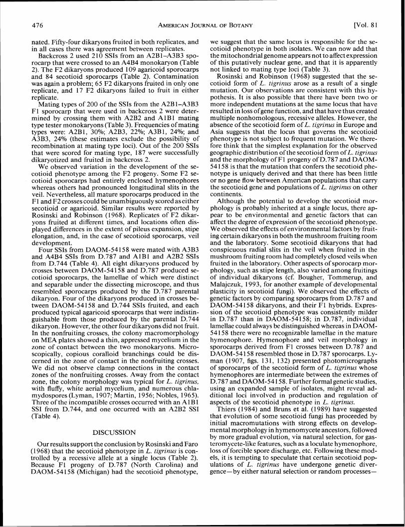
TABLE 1. Wild L. tigrintls dikaryons used in this study.
Isolate number" Hymenophore type Geographic origin
D.787 Secotioid North Carolina D.744 Agaricoid North Carolina DAOM 54158 Secotioid Michigan
a D-isolates were obtained from Dr. Rytas Vilgalys, Department of Botany, Duke University, Durham, North Carolina. DAOM isolate was obtained from Dr. Scott Redhead, Biosystematics Research Centre, Ot- tawa, Ontario. All isolates have been deposited in the culture collection of the Tottori Mycological Institute.
MATERIALS AND METHODS
Culturing and developmental morphology -The three dikaryotic isolates of L. tigrinus used in this study (Table 1) and voucher sporocarps produced in culture are de- posited at the Tottori Mycological Institute (TMI). Iso- lates DAOM-54158 and D.787 were derived from se- cotioid sporocarps, and isolate D.744 was derived from an agaricoid sporocarp. Culturing and fruiting procedures were essentially the same as those that we previously reported for Lentinus (Hibbett, Murakami, and Tsuneda, 1993a). Sporocarps were produced either in a moist cham- ber in the laboratory at ambient light and temperature conditions (approximately 25-30 C), or in a mushroom fruiting room with a 12-hour fluorescent light cycle. Oc- casionally, a second flush was produced by scraping the spawn blocks clean after the first fruiting and replacing them in the fruiting areas.
Scanning electron microscopy (SEM) was used to ob- serve developmental morphology of sporocarps from the three original dikaryotic isolates, following the same pro- tocols for sample preparation, observation, and photog- raphy that we used previously for Lentinus and Panus Fr. (Hibbett, Murakami, and Tsuneda, 1993a, b). Sporocarps produced in the crossing studies were examined macro- scopically. We also examined a total of 71 herbarium collections of the secotioid form of L. tigrinus from the holdings at MICH, NY, FLAS, WTU, SFSU, and FH (collection data are available on request).
Genetic crosses -Lentinus tigrinus has previously been reported to have a tetrapolar mating system (mating abil- ity is determined by two unlinked loci; Nobles, 1965; Rosinski and Robinson, 1968). To obtain monokaryons of all four mating types, up to 25 SSIs from cultured sporocarps of D.787 and D.744 were crossed in all pos- sible intrastrain combinations on MEA (1.25% malt-ex-
TABLE2. Results of F2 backcrosses between isolates D.787 (secotioid) and D.744 (agaricoid).
Backcross 1 Backcross 2
F1 parent dikaryon mating type A2B1-A4B4 A2B1-A3B3 ~ e s i e rmonoka&on mating G e A3B3 A4B4 Total F2 progeny 9 6 193 F2 agaricoid progenya - - . 49/48 109/96.5 F2 secotioid progenya 47/48 84/96.5 x2 0.041 7b 3.2383b
a Obsemed/expected values for 1: 1 segregation. P < 0.05.
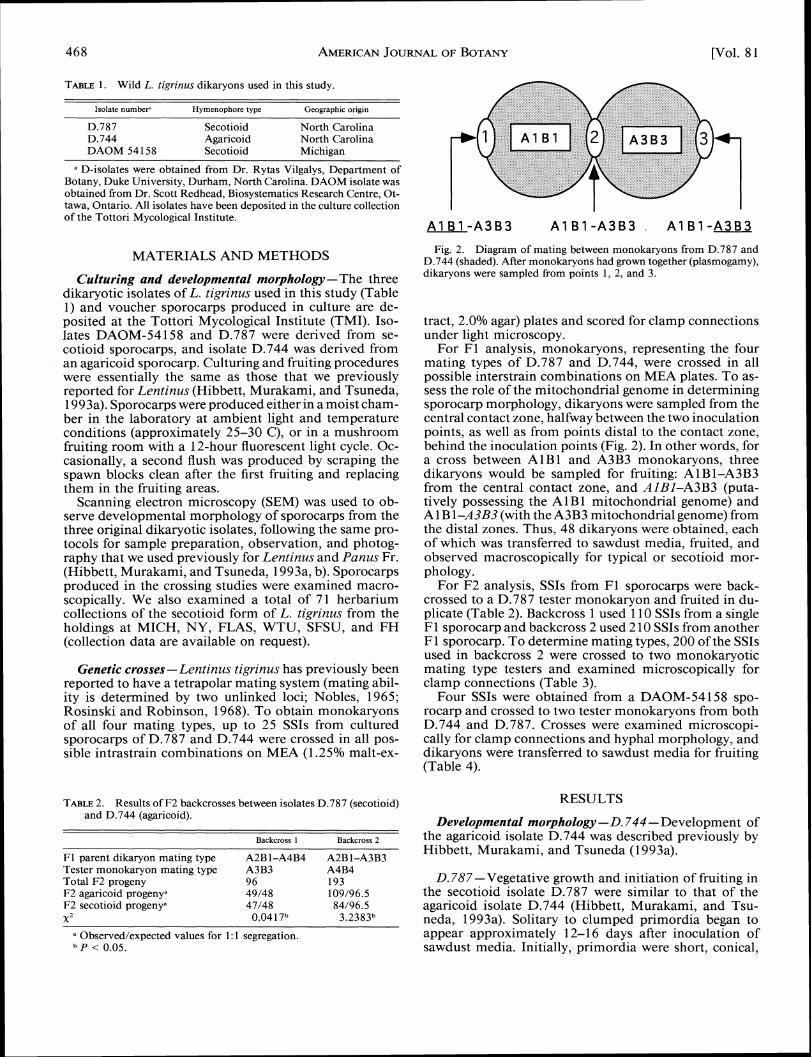
Fig. 2. Diagram of mating between monokaryons from D.787 and D.744 (shaded). After monokaryons had grown together (plasmogamy), dikaryons were sampled from points 1, 2, and 3.
tract, 2.0% agar) plates and scored for clamp connections under light microscopy.
For F1 analysis, monokaryons, representing the four mating types of D.787 and D.744, were crossed in all possible interstrain combinations on MEA plates. To as- sess the role of the mitochondrial genome in determining sporocarp morphology, dikaryons were sampled from the central contact zone, halfway between the two inoculation points, as well as from points distal to the contact zone, behind the inoculation points (Fig. 2). In other words, for a cross between AlBl and A3B3 monokaryons, three dikaryons would be sampled for fruiting: A 1 B 1-A3B3 from the central contact zone, and AIBI-A3B3 (puta-tively possessing the A 1 B 1 mitochondrial genome) and A 1B 1-A3B3 (with the A3B3 mitochondrial genome) from the distal zones. Thus, 48 dikaryons were obtained, each of which was transferred to sawdust media, fruited, and observed macroscopically for typical or secotioid mor- phology.
For F2 analysis, SSIs from F1 sporocarps were back- crossed to a D.787 tester monokaryon and fruited in du- plicate (Table 2). Backcross 1 used 110 SSIs from a single F1 sporocarp and backcross 2 used 2 10 SSIs from another F1 sporocarp. To determine mating types, 200 of the SSIs used in backcross 2 were crossed to two monokaryotic mating type testers and examined microscopically for clamp connections (Table 3).
Four SSIs were obtained from a DAOM-54158 spo- rocarp and crossed to two tester monokaryons from both D.744 and D.787. Crosses were examined microscopi- cally for clamp connections and hyphal morphology, and dikaryons were transferred to sawdust media for fruiting (Table 4).
RESULTS
Developmental morphology -D.744 -Development of the agaricoid isolate D.744 was described previously by Hibbett, Murakami, and Tsuneda (1 993a).
D.787-Vegetative growth and initiation of fruiting in the secotioid isolate D.787 were similar to that of the agaricoid isolate D.744 (Hibbett, Murakami, and Tsu- neda, 1993a). Solitary to clumped primordia began to appear approximately 12-1 6 days after inoculation of sawdust media. Initially, primordia were short, conical,

April 19941 HIBBETT FORM OF LENTZNUS 469ET AL. -SECOTIOID TZGRZNUS
TABLE 3. Analysis of linkage between F2 sporocarp morphology and mating types of SSIs used in backcross 2.
F2 morphology Mating compatibilityb
Mating type" Secotioid Agaricoid A2B2 AlBl
All A2 43/49.5 56/49.5 All A3 36/44 52/44
x2 = 7.6644d A11 B 1 39/5 1 63/5 1 All B3 40/42.5 45/42.5
x2 = 2.41 17*
a Inferred mating genotypes of monokaryotic progeny from F1 spo- rocarp derived from A2B1-A3B3 hybrid. Recombinant mating alleles excluded.
Determined by presence (+) or absence (-) of clamp connections in crosses on MEA.
Under null hypothesis that mating type loci and secotioid locus are unlinked, with independent assortment.
P > 0.05.
and pale grey. As the primordia elongated, they became darkly pigmented at the apex. Pileus initiation generally began on the second or third day after primordium ini- tiation. The hymenophore was first visible on the stipe apex about 1 day after pileus initiation (Fig. 3). Initially, the hymenophore was composed of irregular, crested ridg- es ofhyphaeapproximately 15-30 ym wide oriented more or less parallel to the stipe axis, with occasional anasto- moses and dichotomies (Figs. 3-5). This is identical to the young hymenophore of typical L. tigrinus that we observed previously and is also very similar to the early hymenophore of L. crinitus (which is also agaricoid; Hib- bett, Murakami, and Tsuneda, 1993a).
Differentiation of veil tissue began approximately 3 days after primordium initiation, when individual la- mellae had just become macroscopically visible. The veil developed as hyphae at the margins of the young lamellae began to proliferate and overarch the spaces between the lamellae (Figs. 6-1 0). Soon the veil hyphae formed a con- tinuous covering over the hymenophore (Figs. 6, 7, 1 1, 12). The growth of the veil began near the margin of the pileus and advanced toward the hymenophore at the stipe
TABLE 4. Sporocarp morphologies of F1 hybrids between secotioid isolates DAOM-54158 and D.787 and agaricoid isolate D.744.
DAOM monokaryons and sporocarp morphologies"
D.787 monokaryons A3B3 sec sec sec sec A4B4 sec sec sec sec
D.744 monokaryons
a sec = secotioid sporocarp; aga agaricoid sporocarp; inc =
AlBl aga inc inc inc A2B2 a ~ a aaa aga inc
= incom-patible cross, no sporocarps produced.
apex, which is the first-formed portion of the hymeno- phore (Figs. 6, 7). Often the veil did not completely ob- scure the hymenophore in a narrow zone at the stipe apex, which thus remained exposed throughout the life of the sporocarp.
The lamellae continued to develop in a nearly normal fashion. At maturity, almost all of the hymenophore was enclosed, but individual lamellae were separate and dis- tinct structures (Fig. 12). However, the lamellae were somewhat misshapen with frequent anastomoses or bi- furcations (Fig. 12). Unlike the smooth lamellae of the typical form (Hibbett, Murakami, and Tsuneda, 1993a, figs. 21, 25), the radial surfaces of the lamellae of the secotioid form had numerous venose or platelike out- growths (Figs. 13, 14). The hymenium itself appeared normal and covered the entire surface of the hymeno- phore. At the base of the hymenophore there were tan- gential cross-bridges between the lamellae that delimited more or less rectangular spaces, as in typical L. tigrinus (Figs. 15, 16; Hibbett, Murakami, and Tsuneda, 1993a, figs. 24, 25). Finally, there were numerous hyphal pegs produced on the lamellae (Figs. 12-14) that are indistin- guishable from those produced by the typical form.
The veil was approximately 15-20 ym thick in most areas (Fig. 12). The outer surface of the veil was composed of a loose, irregular weft of smooth generative hyphae with slight apical swellings (Figs. 17, 18). Macroscopically, faint longitudinal striations corresponding to the arrange- ment of lamellae under the veil could usually be observed (Fig. 1). Longitudinal slits sometimes formed in the veil tissue along the striations near the margin of the pileus
Figs. 3-8. Early sporocarp ontogeny in secotioid isolate D.787. 3. Early hymenophore is composed of irregular, crested ridges of hyphae over the stipe apex. At this stage, the hymenophore is identical to that of typical L. tigrinus.There are no veils or other protective tissues. Bar = 400 Fm. 4. Detail of young hymenophore. Bar = 100 Fm. 5. A single young lamella showing apical growth of surface hyphae to form hymenophore. Bar = 20 Fm. 6. Later developmental stage. The veil tissues have formed over the distal portions of the hymenophore. At the stipe apex parts of the hymenophore remain exposed. Note the faint radial striations in the veil that correspond to spaces between the underlying lamellae. Bar = 50 pm. 7. Detail from same sporocarp as in Fig. 6 showing veil advancing over hymenophore toward stipe apex. Bar = 200 Fm. 8. Tangential vertical section through young lamellae near zone of veil development. The lamellae are distinct and separate even to their bases. Bar = 75 pm.
Figs. 9-14. Later hymenophore and veil development in D.787. 9. Tangential section through hymenophore near zone of veil development. Note that hyphae at the margins of the lamellae are proliferating outward. Bar = 20 pm. 10. Tangential section through hymenophore in zone of veil development. The hyphae at the margins of the lamellae have overarched the intralamellar space and have begun to interweave. Bar = 20 Fm. 11. Tangential section through mature hymenophore and veil. The veil has completely enclosed the hymenophore. Bar = 150 Fm. 12. Tangential section through mature hymenophore and veil. There are some anastomoses and other irregularities in the lamellae, but they are clearly recognizable as lamellae (compare to Figs. 27, 28). Hyphal pegs protrude from the lamellae. Bar = 400 Fm. 13. Radial section through hymenophore showing lamellar face with venose outgrowths. Bar = 150 Fm. 14. Detail from Fig. 13 showing hyphal pegs and hymenium covering venose outgrowths from lamella. Bar = 75 pm.


April 19941 HIBBETT FORM OF LENTINUSET AL. -SECOTIOID TIGRINUS

Figs. 15-1 8. Mature hymenophore and veil morphology in D.787. 15. Scalp section through hymenophore showing subporoid arrangement of hymenophore at the base (compare to Fig. 28). Bar = 50 pm. 16. Closer view of regular, rectangular chambers at base of hymenophore. The subporoid structure is also observed in typical L. tigrinus and has been hypothesized to be a vestige of polyporoid ancestry (Pegler, 1983; Hibbett, Murakami, and Tsuneda, 1993a). Bar = 250 pm. 17. Surface view of veil. Bar = 100 pm. 18. Detail from Fig. 17 showing tips of generative hyphae that form veil. Bar = 10 wm.
in older sporocarps (Fig. 1). In a few extreme cases, the hyphal pegs were present (Figs. 11, 12). Lyman (1907) slits extended through the pileus, which then took on a described and illustrated conidia that were formed on lobate aspect. The inner surface of the veil was lined by sporocarps of the secotioid form of L. tigrinus,but we a hymenium that was continuous with and indistinguish- never observed them. able from the hymenium of the lamellae, except that no In terms of overall shape and stature, duration ofgrowth,
Figs. 19-24. Early ontogeny of secotioid isolate DAOM-54158. 19. Primordium. Bar = 125 pm. 20. Early stage of pileus differentiation. There are no hymenophore elements visible. Bar = 200 pm. 21. Later developmental stage. The veil has started to form over the distal portions of the young hymenophore. Bar = 200 pm. 22. Radial section through hymenophore and veil at approximately same stage as in Fig. 2 1. The hymenophore is exposed at the stipe apex only. At this stage, the development of the veil is already more luxuriant than in D.787 sporocarps. Bar = 200 pm. 23. Later developmental stage. The veil has completely covered the hymenophore. Bar = 400 pm. 24. Radial section through veil and hymeno- phore at approximately same stage as Fig. 23. The hymenophore is loculate with thick veil tissue and no discernible lamellae (but see Fig. 25). Bar = 400 pm.

April 19941 HIBBETTET AL. -SECOTIOIDFORM OF LENTINUSTIGRINUS 473

474 AMERICANJOURNALOF BOTANY [Vol. 8 1

475 April 19941 HIBBETTET AL. -SECOTIOIDFORM OF LENTINUSTIGRINUS
and longevity, the D.787 sporocarps were similar to those of the agaricoid form (Fig. 1). Sporocarps usually matured in about 5 days and persisted for up to 10 days.
DAOM-54158 -Vegetative growth, primordium ini- tiation, and early differentiation of the stipe, pileus, and hymenophore were essentially the same as in D.787 and D.744 (Figs. 19, 20). Again, veil tissue began to develop from the distal portions of the hymenophore and grew toward the stipe apex (Figs. 21, 22). However, from the outset the growth ofthe veil was more luxuriant in DAOM- 54 1 5 8 than in D.78 7 (Figs. 22-26). Individual lamellae could just barely be differentiated early in the ontogeny of DAOM-54158 (Figs. 25, 26). Later, the hymenophore became so convoluted that the lamellae could not be re- solved. Instead, the hymenophore became loculate, with irregular hymenium-lined pockets scattered through the confluent mass of veil and tramal tissue (Figs. 27-30). At maturity, these pockets became filled with apparently nor- mal, discharged ballistospores (Figs. 29, 30). Hyphal pegs that had emerged from the hymenium could still be ob- served (Figs. 27, 28).
The overall rate and duration of growth and shape and proportions of the sporocarps in DAOM-54 158 were sim- ilar to those of D.787. However, the veil and hymeno- phore tissue frequently became so thick that the pilei lost their normal form and took on a lumpy, irregular shape with an uplifted, undulate margin. Longitudinal slits and striations like those observed in D.787 were rarely ob- served in DAOM-54 1 5 8 sporocarps.
Herbarium materials-The collections of the secotioid form of L. tigrinus that we examined are distributed from Michigan and southern Ontario south to Mississippi and Texas, and from the Atlantic coast west to Arizona (Fig. 3 1). The Arizona collection (Sycamore Canyon, Santa Cruz Co., Aug. 7, 1980, Bigelow 18 180 [NY]) represents a significant westward range extension from that reported for the species by Redhead (1988). Otherwise, the range of the secotioid form in North America overlaps with that of the agaricoid form (Redhead, 1988). Herbarium label data and published (although sometimes anecdotal) ac- counts indicate that the secotioid form of L. tigrinus oc-curs in pure populations (Morgan, 1895; Lyman, 1907) or in mixtures with the typical form (Peck, 1909; Kauff- man, 19 18; Harper, 1921). Seven of the herbarium col- lections that we examined included mixtures of secotioid and agaricoid sporocarps. We made four collections of the secotioid form and three collections of the agaricoid form on a single day along a 1-mile stretch of the Ipswich River in Massachusetts.
Lentinus tigrinus is noted for its variability in sporocarp size and proportions (Harper, 192 1; Pegler, 1983). We
Fig. 3 1. Distribution ofcollectionsofthe secotioid form of L. tigrinus examined in this study.
observed such variation among collections of the secotioid form, as well as variation in the apparent severity of the secotioid phenotype. There was variation in the devel- opment of striations and slits in the veil, anastomoses and contortions of the lamellae, deviation of the pileus from the typical shape, etc. However, there was no ap- parent correlation between geographic distribution and variation in expression of the secotioid phenotype.
Genetic crosses-As anticipated, monokaryons from both D.744 and D.787 fell into four mating types, which is consistent with previous reports of a tetrapolar mating system in L. tigrinus (Nobles, 1965; Rosinski and Rob- inson, 1968). Mating type alleles from D.744 were des- ignated Al , A2, B1, and B2. Those from D.787 were designated A3, A4, B3, and B4.
F1 dikaryons from interstrain crosses of D.744 and D.787 fully colonized the sawdust media and began to produce primordia within 1 1-1 6 days. All but one (A2B2-A3B4) of the dikaryons fruited, and all of these produced typical, agaricoid sporocarps that were indistinguishable from those produced by the D.744 dikaryon.
For backcross 1, 1 10 SSIs from an A2B 1-A4B4 F1 sporocarp were crossed to an A3B3 monokaryon (Table 2). The F2 dikaryons thus produced were fruited and gave rise to 49 agaricoid sporocarps and 47 secotioid sporo- carps (Table 2). We encountered contamination problems in the mushroom fruiting room; 42 of the F2 dikaryons only fruited in one replicate, and 14 of the F2 dikaryons failed to fruit at all. In almost all cases, the failed fruitings were producing primordia when they became contami-
Figs. 25-30. Immature and mature hymenophore anatomy of secotioid isolate DAOM-54158. 25. Vertical tangential section through hymeno- phore of immature sporocarp at approximately same developmental stage as in Fig. 24. Lamellae were not discernible in radial section in Fig. 24, but here in tangential section there are vertically elongate locules that may be vestiges of lamellar structures. Bar = 200 pm. 26. Detail from Fig. 25 showing hymenium-lined locules of hymenophore and irregular trama. Bar = 40 pm. 27. Radial section through loculate mature hymenophore. Note hyphal pegs emergent from hymenium (and in Fig. 28). Bar = 400 Km. 28. Scalp section through mature hymenophore (compare to Fig. 15). Bar = 250 pm. 29. Detail from Fig. 28 showing locule filled with discharged spores. Bar = 20 pm. 30. Detail from Fig. 29 showing discharged ellipsoid, asymmetric basidiospores. Bar = 7 wm.
nated. Fifty-four dikaryons fruited in both replicates, and in all cases there was agreement between replicates.
Backcross 2 used 2 10 SSIs from an A2B 1-A3B3 spo- rocarp that were crossed to an A4B4 monokaryon (Table 2). The F2 dikaryons produced 109 agaricoid sporocarps and 84 secotioid sporocarps (Table 2). Contamination was again a problem; 65 F2 dikaryons fruited in only one replicate, and 17 F2 dikaryons failed to fruit in either replicate.
Mating types of 200 of the SSIs from the A2B 1-A3B3 F1 sporocarp that were used in backcross 2 were deter- mined by crossing them with A2B2 and AlB 1 mating type tester monokaryons (Table 3). Frequencies of mating types were: A2B1, 30%; A2B3, 22%; A3B1, 24%; and A3B3, 24% (these estimates exclude the possibility of recombination at mating type loci). Out of the 200 SSIs that were scored for mating type, 187 were successfully dikaryotized and fruited in backcross 2.
We observed variation in the development of the se- cotioid phenotype among the F2 progeny. Some F2 se- cotioid sporocarps had entirely enclosed hymenophores whereas others had pronounced longitudinal slits in the veil. Nevertheless, all mature sporocarps produced in the F1 and F2 crosses could be unambiguously scored as either secotioid or agaricoid. Similar results were reported by Rosinski and Robinson (1968). Replicates of F2 dikar- yons fruited at different times, and locations often dis- played differences in the extent of pileus expansion, stipe elongation, and, in the case of secotioid sporocarps, veil development.
Four SSIs from DAOM-54158 were mated with A3B3 and A4B4 SSIs from D.787 and AlBl and A2B2 SSIs from D.744 (Table 4). All eight dikaryons produced by crosses between DAOM-54158 and D.787 produced se- cotioid sporocarps, the lamellae of which were distinct and separable under the dissecting microscope, and thus resembled sporocarps produced by the D.787 parental dikaryon. Four of the dikaryons produced in crosses be- tween DAOM-54158 and D.744 SSIs fruited, and each produced typical agaricoid sporocarps that were indistin- guishable from those produced by the parental D.744 dikaryon. However, the other four dikaryons did not fruit. In the nonfruiting crosses, the colony macromorphology on MEA plates showed a thin, appressed mycelium in the zone of contact between the two monokaryons. Micro- scopically, copious coralloid branchings could be dis- cerned in the zone of contact in the nonfruiting crosses. We did not observe clamp connections in the contact zones of the nonfruiting crosses. Away from the contact zone, the colony morphology was typical for L. tigrinus, with fluffy, white aerial mycelium, and numerous chla- mydospores (Lyman, 1907; Martin, 1956; Nobles, 1965). Three of the incompatible crosses occurred with an A 1B 1 SSI from D.744, and one occurred with an A2B2 SSI (Table 4).
DISCUSSION
Our results support the conclusionby Rosinski andFaro (1 968) that the secotioid phenotype in L. tigrinus is con- trolled by a recessive allele at a single locus (Table 2). Because F1 progeny of D.787 (North Carolina) and DAOM-54158 (Michigan) had the secotioid phenotype,
we suggest that the same locus is responsible for the se- cotioid phenotype in both isolates. We can now add that the mitochondria1 genome appears not to affect expression of this putatively nuclear gene, and that it is apparently not linked to mating type loci (Table 3).
Rosinski and Robinson (1968) suggested that the se- cotioid form of L. tigrinus arose as a result of a single mutation. Our observations are consistent with this hy- pothesis. It is also possible that there have been two or more independent mutations at the same locus that have resulted in loss ofgene function, and that have thus created multiple nonhomologous, recessive alleles. However, the absence of the secotioid form of L. tigrinus in Europe and Asia suggests that the locus that governs the secotioid phenotype is not subject to frequent mutation. We there- fore think that the simplest explanation for the observed geographic distribution of the secotioid form ofL. tigrinus and the morphology of F l progeny of D.787 and DAOM- 54 158 is that the mutation that confers the secotioid phe- notype is uniquely derived and that there has been little or no gene flow between American populations that carry the secotioid gene and populations of L. tigrinus on other continents.
Although the potential to develop the secotioid mor- phology is probably inherited at a single locus, there ap- pear to be environmental and genetic factors that can affect the degree of expression of the secotioid phenotype. We observed the effects of environmental factors by fruit- ing certain dikaryons in both the mushroom fruiting room and the laboratory. Some secotioid dikaryons that had conspicuous radial slits in the veil when fruited in the mushroom fruiting room had completely closed veils when fruited in the laboratory. Other aspects of sporocarp mor- phology, such as stipe length, also varied among fruitings of individual dikaryons (cf. Bougher, Tommerup, and Malajczuk, 1993, for another example of developmental plasticity in secotioid fungi). We observed the effects of genetic factors by comparing sporocarps from D.787 and DAOM-54158 dikaryons, and their F1 hybrids. Expres- sion of the secotioid phenotype was consistently milder in D.787 than in DAOM-54158; in D.787, individual lamellae could always be distinguished whereas in DAOM- 541 58 there were no recognizable lamellae in the mature hymenophore. Hymenophore and veil morphology in sporocarps derived from F1 crosses between D.787 and DAOM-54158 resembled those in D.787 sporocarps. Ly- man (1 907, figs. 13 1, 132) presented photomicrographs of sporocarps of the secotioid form of L. tigrinus whose hymenophores are intermediate between the extremes of D.787 and DAOM-54 158. Further formal genetic studies, using an expanded sample of isolates, might reveal ad- ditional loci involved in production and regulation of aspects of the secotioid phenotype in L. tigrinus.
Thiers (1984) and Bruns et al. (1989) have suggested that evolution of some secotioid fungi has proceeded by initial macromutations with strong effects on develop- mental morphology in hymenomycete ancestors, followed by more gradual evolution, via natural selection, for gas- teromycete-like features, such as a loculate hymenophore, loss of forcible spore discharge, etc. Following these mod- els, it is tempting to speculate that certain secotioid pop- ulations of L. tigrinus have undergone genetic diver- gence-by either natural selection or random processes-

April 19941 HIBBETTET AL. -SECOTIOID TZGRINUS 477FORM OF LENTINUS
toward a gasteroid morphology, while other populations have retained a more plesiomorphic, agaricoid mor-phology. However, to evaluate this hypothesis it will be necessary to achieve a greater understanding of devel- opmental genetics, population and breeding biology, and intraspecific genealogy in L. tigrinus. We mention this here only to encourage future research, which we feel might provide insights into processes of morphological diversification in fungi.
The switch from the agaricoid to the secotioid phe- notype could have profound influences on ecology and evolution of L. tigrinus populations. Intuitively, it seems likely that the secotioid phenotype would lower the fitness of individuals of L. tigrinus by reducing the capacity for aerial spore dispersal and perhaps by reducing outcross- ing. However, the effect of the secotioid phenotype on fitness is unknown; it could actually be neutral or positive. As in many basidiomycetes, the relative importance of basidiospores and asexual propagules for establishing and maintaining L. tigrinus populations is unknown. Recent studies on giant Armillaria clones (Smith, Bruhn, and Anderson, 1992) have demonstrated clearly that some basidiomycetes can spread extensively by purely vege- tative growth. Lentinus tigrinus does not form rhizo- morphs like those of Armillaria, but its dikaryotic and monokaryotic mycelia produce chlamydospores (Lyman, 1907; Tsuneda, Thorn, and Hibbett, 1993) whose role in dispersal and colonization remains to be determined.
Previous authors (e.g., Thiers, 1984) have suggested that secotioid fungi are well adapted to arid environments, in which they are often found. In L. tigrinus, the presence of the veil reduces the ratio of surface area to volume and might therefore increase desiccation tolerance. However, L. tigrinus is typically found not in dry habitats, but on logs adjacent to or emergent from water (Redhead, 1988, and others). Noting this, Rosinski and Robinson (1968) suggested that the secotioid sporocarps might be well adapted to dispersal of spores by water. In other secotioid fungi, mycophagy by rodents has been suggested as a mechanism for spore dispersal (Thiers, 1984; Bruns et al., 1989), but this has not been observed in L. tigrinus. Snell (1 923) reported that spores from below the veil ofa 5-year- old herbarium collection germinated at high frequency and suggested that the protection provided by the veil increases spore longevity (there are no comparable data on longevity of spores from agaricoid sporocarps). What- ever its functional attributes, the secotioid form of L. tigrinus has been frequently collected since the late nine- teenth century. If the allele that confers the secotioid form did arise by a single mutational event, then it has been able not only to persist, but to spread across much of North America (Fig. 3 1). Collectively, this evidence sug- gests that the secotioid phenotype in L. tigrinus is an evolutionarily successful innovation (cf. Martin, 1956). This stands in contrast to the idea that secotioid fungi are necessarily poorly adapted aberrations that must evolve into more refined gasteromycete-like forms or else be doomed to extinction (e.g., Baura et al., 1992). Although such may often be the case, the occurrence of stipitate, epigeous, secotioid taxa in diverse families of basidio- mycetes (e.g., Podaxis Desv. and Montagnea Fr. in Co- prinaceae, Thaxterogaster Singer in Cortinariaceae, Lon-gula Zeller in Agaricaceae, etc.) suggests that evolution
of secotioid forms has been a common and successful mode of sporocarp morphological evolution (cf. Thiers, 1984; Miller and Miller, 1988).
Evolution of certain secotioid fungi has previously been interpreted as involving paedomorphosis, resulting from an arrest of an ancestral ontogeny (Gould, 1977; Thiers, 1984; Bruns et al., 1989; Baura, Szaro, and Bruns, 1992). Indeed, the commonly applied definition of secotioid fun- gi as resembling unopened agarics (e.g., Miller and Miller, 1988) has an inherent implication of paedomorphosis. We do not contest that paedomorphosis has been im- portant in evolution of some secotioid fungi, but it has not been involved in evolution of the secotioid form of L. tigrinus. At no point in the ontogeny of agaricoid L. tigrinus is there a structure like the veil of the secotioid form (Bobbitt and Crang, 1975; Hibbett, Murakami, and Tsuneda, 1993a). Typical L. tigrinusis gymnocarpic (Bob- bitt and Crang, 1975; Hibbett, Murakami, and Tsuneda, 1993a), and therefore a hymenophore that is enclosed at maturity could not be derived by paedomorphosis. Only hemiangiocarpy, in which an initially enclosed hymeno- phore becomes exposed at maturity (Watling, 1985), can lead from an exposed to an enclosed mature hymenophore strictly by paedomorphosis. Finally, we note that the se- cotioid form of L. tigrinus grows to the same size and proportions as the typical form, in about the same amount of time. For these reasons, we feel that the developmental modifications that are involved in derivation of the se- cotioid form from the typical agaricoid form are best interpreted as a realization of von Baer's law of differ- entiation (Gould, 1977) which predicts that derived on- togenies parallel ancestral ontogenies in early stages, and then deviate to produce novel features in later stages. Secotioid fungi are doubtless a highly polyphyletic group. Other secotioid taxa have probably been derived by dif- ferent classes of developmental changes, possibly includ- ing paedomorphosis.
We do not know the reason for the incompatibility reactions between certain SSIs ofDAOM-54 158 and D.744 (Table 4). For the incompatibility to be due to shared mating type alleles, it would be necessary for the DAOM- 541 58 and D.744 dikaryons to have at least two mating type alleles in common (incompatibility of certain DAOM- 54158 SSIs with either AlBl or A2B2 mating type SSIs requires presence of one A 1 or B 1 allele and one A2 or B2 allele in the DAOM-54 158 dikaryon). Given the geo- graphic separation of the isolates (Michigan and North Carolina), this seems unlikely. An alternate explanation is that there is partial vegetative incompatibility between certain North American populations of L. tigrinus, but more extensive crossing studies will be needed to resolve this issue.
Agaricoid-secotioid transformations provide fascinat- ing demonstrations of morphological and ecological evo- lution in fungi. Among secotioid fungi, the secotioid form ofL. tigrinus is unique because it is still mating compatible with the typical agaricoid form, and because a specific locus has been identified that confers the secotioid phe- notype. Developmental mutants have already provided insights into mechanisms of morphogenesis in culture studies of Agaricus bisporus, Coprinus cinereus, Schizo- phyllum commune, and other basidiomycetes (see papers in Moore et al., 1985; Reijnders, 1991, and references

--
478 AMERICANJOURNALOF BOTANY [Vol. 81
therein). Lentinus tigrinus also fruits well in culture and could therefore be an excellent system for understanding the molecular-developmental basis for evolution of se- cotioid fungi.
LITERATURE CITED
BAURA, G., T. M. SZARO, AND T. D. BRUNS. 1992. Gastrosuillus larici- nus is a recent derivative of Suillus grevillei: molecular evidence. Mycologia 84: 592-597.
BERKELEY, 1845. Australian and North American fungi. LondonM. J. Journal ofBotany 4: 298-3 15.
BOBBITT,T. F., AND R. E. CRANG. 1974. Light effects on fruiting in Panus tigrinus var. tigrinus. Canadian Journal of Botany 52: 255- 257.
,AND- . 1975. Basidiocarp development of the two va- rieties of Panus tigrinus and their light-induced abnormal forms. Mycologia 67: 182-1 87.
BOUGHER,N. L., I. C. TOMMERUP, AND N. MALAJCZUK. 1993. Broad variation in developmental and mature basidiome morphology of the ectomycorrhizal fungus Hydnangium sublamellatum sp. nov. bridges morphologically based generic concepts of Hydnangium, Podohydnangiumand Laccaria. MycologicalResearch 97: 6 13-6 19.
BRUNS, T. R., R. FOGEL, T. J. WHITE, AND J. W. TAYLOR. 1989. AC- celerated evolution of a false truffle from a mushroom ancestor. Nature 339: 140-142.
CORNER,E. J. H. 1981. The agaric genera Lentinus, Panus, and Pleu-rotus. Beihefte zur Nova Hedwigia 69: 1-1 89.
FARO, S. 1972. Physiological aspects of pigment production in relation to morphogenesis in Panus tigrinus. Mycologia 64: 375-387.
COULD, S. J. 1977. Ontogeny and phylogeny. Belknap Press, Cam- bridge, MA.
HARPER, E. T. 192 1. Species of Lentinus in the region of the Great Lakes. Transactions of the Wisconsin Academy of Sciences, Arts, and Letters 20: 365-387.
HIBBETT,D. S., S. MURAKAMI, AND A. TSUNEDA. 1993a. Hymenophore development and evolution in Lentinus. Mycologia 85: 428-443.
, A N D - . 1993b. Sporocarp ontogeny in Panus (Basidiomycotina): evolution and classification. American Journal of Botany 80: 1336-1348.
, A N DR. VILGALYS. 199 1. Evolutionary relationships ofLentinus to the Polyporaceae: evidence from restriction analysis of enzy- matically amplified ribosomal DNA. Mycologia 83: 425-439.
,AND- . 1993. Phylogenetic relationships of Lentinus (Ba-sidiomycotina) inferred from molecular and morphological char- acters. Systematic Botany 18: 409-433.
HOPPLE,J. S., JR. 1990. Phylogenetic relationships within the genus Coprinus based on molecular and morphological evidence. In A. Reisinger and A. Bresinsky [eds.], Fourth International Mycological Congress Abstracts, 25. Botanical Institute, University of Regens- burg, Regensburg, Germany.
KAUFFMAN,C. H. 19 18. The Agaricaceae of Michigan, vol. 1. Michigan Geological and Biological Survey, Publication 26: Biological Series 5. Wynkoop, Hallenback, and Crawford, Lansing, MI.
K~~HNER,1925. Le dkveloppement du Lentinus tigrinus Bull. ComptesR.
Rendus Hebdomaires des Sgances de L 'Acadgmie des Sciences 18 1: 137-1 39.
LEA, T. G. 1849. Catalogue of the plants of Cincinnati. T. K. and P. G. Collins, Philadelphia, PA.
LYMAN, G. R. 1907. Culture studies on polymorphism of hymeno- mycetes. Proceedings of the Boston Society of Natural History 33: 125-209.
MARTIN, G. W. 1956. On Lentod~um squamulosum. Proceedings of the Iowa Academy of Science 63: 280-286.
MILLER,0.K., JR., AND H. H. MILLER. 1988. Gasteromycetes: mor- phological and developmental features with keys to the orders, families, and genera. Mad River Press, Eureka, CA.
MOORE, D., L. A. CASSELTON, D. A. WOOD, ANDJ. C. FRANKLAND [eds.]. 1985. Developmental biology of higher fungi. Cambridge Uni- versity Press, Cambridge.
MORGAN, A. P. 1895. New North American fungi. Journal of the Cincinnati Society of Natural History 18: 36-45.
NOBLES, M. K. 1965. Identification of cultures of wood-inhabiting hymenomycetes. Canadian Journal ofBotany 43: 1097-1 139.
PECK, C. H. 1909. Report of the State Botanist 1908. New York State Museum Bulletin 13 1 : 5-202.
PEGLER,D. M. 1983. The genus Lentinus-a world monograph. Kew Bulletin 10: 1-28 1 (Additional series).
PINE, E. M., AND G. M. MUELLER. 1993. Clarifying evolutionary re- lationships between and within two major groups of basidiomy- cetous fungi (mushrooms and false-truffles) by means of rDNA sequencing. Inoculum 43: 30 (Abstract).
REDHEAD,S. A. 1988. A biogeographical overview of the Canadian mushroom flora. Canadian Journal of Botany 67: 3003-3062.
REUNDERS,A. F. M. 199 1. Differentiation in agaric basidiomata and phylogenetic problems. Mycological Research 95: 1249-1252.
R o s m s ~ ~ ,M. A., AND S. FARO. 1968. The genetic basis ofhymenophore morphology in Panus tigrinus (Bull. ex Fr.) Singer. Amerzcan Journal ofBotany 55: 720 (Abstract).
,AND A. D. ROBINSON. 1968. Hybridization of Panus tigrinus and Lentodium squamulosum. Amer~can Journal ofBotany 55: 242- 246.
,AND- . 1969. Secotioid divergence in Panus tigrinus: an emended discussion. Mycologia 61: 830-832.
SINGER,R. 195 1. Thaxterogaster-a new link between gasteromycetes and Agaricales. Mycologia 43: 21 5-228.
. 1986. The Agaricales in modern taxonomy, 4th ed. Koeltz Scientific Books, Koenigstein, Germany.
SMITH, M. L., J. N. BRUHN, AND J. B. ANDERSON. 1992. The fungus Armillaria bulbosa is among the largest and oldest living organisms. Nature 356: 428-43 1.
SNELL, W. H. 1923. Occurrence and identity of cotton mill fungi. Mycologia 15: 153-165.
THIERS, H. D. 1984. The secotioid syndrome. Mycologia 76: 1-8. TSUNEDA,A,, R. G. THORN, AND D. S. HIBBETT. 1993. Lentinus ti-
grinus: chlamydospores and interaction with Pseudomonas flu- orescens. Reports of the Tottori Mycological Institute 30: 1-1 2.
WATLING,R. 1985. Developmental characters ofagarics. In D. Moore, L. A. Casselton, D. A. Wood, and J. C. Frankland [eds.], Devel- opmental biology of higher fungi, 28 1-3 10. Cambridge University Press, Cambridge.