the rxlr motif of the host targeting effector avr3a of ... report the rxlr motif of the host...
TRANSCRIPT
BREAKTHROUGH REPORT
The RxLR Motif of the Host Targeting Effector AVR3a ofPhytophthora infestans Is Cleaved before SecretionOPEN
Stephan Wawra,a,1,2 Franziska Trusch,a,1 Anja Matena,b Kostis Apostolakis,a Uwe Linne,c Igor Zhukov,d,e
Jan Stanek,f Wiktor Kozminski,f Ian Davidson,g Chris J. Secombes,h Peter Bayer,b and Pieter van Westa,3
a Aberdeen Oomycete Laboratory, Institute of Medical Sciences, University of Aberdeen, Foresterhill, Aberdeen AB25 2ZD, UnitedKingdombStructural and Medicinal Biochemistry, Centre of Medicinal Biotechnology, University of Duisburg-Essen, 45141 Essen, GermanycCore Facility for Mass Spectrometry and Chemistry, Philipps-Universität Marburg, D-35032 Marburg, Germanyd Institute of Biochemistry and Biophysics, Polish Academy of Sciences, 02-106 Warsaw, PolandeNanoBioMedical Centre, Adam Mickiewicz University, 61-614 Poznan, Polandf Biological and Chemical Research Centre (CENT III), Faculty of Chemistry, University of Warsaw, 02-089 Warsaw, PolandgProteomics Facility, Institute of Medical Sciences, University of Aberdeen, Foresterhill, Aberdeen AB25 2ZD, United KingdomhScottish Fish Immunology Research Centre, Institute of Biological and Environmental Sciences, University of Aberdeen, AberdeenAB24 2TZ, United Kingdom
ORCID IDs: 0000-0003-0608-202X (F.T.); 0000-0002-9912-1018 (I.Z.); 0000-0003-1660-168X (J.S.); 0000-0003-2319-4525 (W.K.);0000-0001-8781-176X (C.J.S.); 0000-0003-0435-7202 (P.B.); 0000-0002-0767-6017 (P.v.W.)
When plant-pathogenic oomycetes infect their hosts, they employ a large arsenal of effector proteins to establisha successful infection. Some effector proteins are secreted and are destined to be translocated and function inside host cells.The largest group of translocated proteins from oomycetes is the RxLR effectors, defined by their conserved N-terminal Arg-Xaa-Leu-Arg (RxLR) motif. However, the precise role of this motif in the host cell translocation process is unclear. Here,detailed biochemical studies of the RxLR effector AVR3a from the potato pathogen Phytophthora infestans are presented.Mass spectrometric analysis revealed that the RxLR sequence of native AVR3a is cleaved off prior to secretion by thepathogen and the N terminus of the mature effector was found likely to be acetylated. High-resolution NMR structure analysisof AVR3a indicates that the RxLR motif is well accessible to potential processing enzymes. Processing and modification ofAVR3a is to some extent similar to events occurring with the export element (PEXEL) found in malaria effector proteins fromPlasmodium falciparum. These findings imply a role for the RxLR motif in the secretion of AVR3a by the pathogen, rather thana direct role in the host cell entry process itself.
INTRODUCTION
Many eukaryotic pathogens including plant pathogenic fungi andoomycetes use effector proteins to successfully infect their hosts.Effectors are secreted molecules that help both the invasion andthe propagation of the pathogen by suppressing host defenseresponses as well as adapting host metabolism (Wawra et al.,2012a).
The phylogenetically related malaria parasites (Plasmodiumspp) and the plant pathogenic water molds (oomycetes) havenumerous effector proteins, which are characterized by con-served N-terminal amino acid sequences (Hiller et al., 2004; Marti
et al., 2004) that play a role in the host cell targeting process(Whissonetal., 2007;Boddeyetal., 2009). Inmalariaparasites, thePlasmodium export element (PEXEL) consists of Arg-Xaa-Leu-Xaa-Glu/Asp/Gln (withXaabeinganyaminoacid) positionedcloseafter the signal peptide cleavage site (Hiller et al., 2004;Marti et al.,2004). This sequence element is cleaved by endoplasmic re-ticulum-located proteases and the N terminus becomes acety-lated (Ac-Xaa-Glu/Asp/Gln) (Chang et al., 2008; Boddey et al.,2009, 2010; Russo et al., 2010). The conserved spatial position ofthe PEXEL motif and its very rapid proteolytic modification arenecessary for the export of effector proteins and seem to work asan internal signal for a specialized effector export pathway (Martiand Spielmann, 2013; Boddey et al., 2016). Recently, a similarmotif (TEXEL)was found in effector proteins of Toxoplasmagondiiand reported to play a crucial role in effector maturation andsorting during the infection process (Hammoudi et al., 2015;Coffey et al., 2015; Curt-Varesano et al., 2016).Numerous effector proteins from plant pathogenic oomycetes
carry a similar highly conserved motif: the Arg-Xaa-Leu-Arg(RxLR) motif. This sequence is located within ;40 residuesdownstream of the secretion signal cleavage site and is often
1 These authors contributed equally to this work.2 Current address: Botanical Institute, Genetical Institute, University ofCologne; Zulpicher Strasse 47a, 50674 Cologne, Germany.3 Address correspondence to [email protected] author responsible for distribution of materials integral to the findingspresented in this article in accordance with the policy described in theInstructions for Authors (www.plantcell.org) is: Pieter van West([email protected]).OPENArticles can be viewed without a subscription.www.plantcell.org/cgi/doi/10.1105/tpc.16.00552
The Plant Cell, Vol. 29: 1184–1195, June 2017, www.plantcell.org ã 2017 ASPB.
followed by another second conserved motif Ser/Asp-Glu-Glu-Arg (s/dEER; reviewed in Wawra et al., 2012a). Effector secretion,and presumably entry into the host cell, happens at the hausto-rium, an infection-specific interface between pathogen and hostthat forms after the pathogen has colonized the host (Whissonet al., 2007; Rafiqi et al., 2010). Haustoria are always surroundedby a host plant-derived membrane (Lu et al., 2012). Evidenceexists that RxLR effectors translocate after secretion from theextrahaustorial space into plant cells (Whisson et al., 2007; vanPoppel et al., 2008; Lokossou et al., 2010;Chouet al., 2011;Gilroyet al., 2011; Saunders et al., 2012). RxLR and RxLR-like effectorsmay also be present in fungi (summarized in Kale and Tyler, 2011),and different models for the translocation process have beenproposed, but the precise mechanism is unclear (Petre andKamoun, 2014).
Some reports suggest that the RxLR sequences are directlyinvolved in host cell entry. For theRxLReffector avirulence protein3a (AVR3a) from the potato blight pathogen Phytophthora in-festans, it was shown that the RxLR motif is required for hostcell translocation based on a loss-of-function infection assay(Whisson et al., 2007). In addition, oomycete and fungal RxLR andRxLR-like effectors have been reported to translocate into hostcells in the absence of the pathogen (Plett et al., 2011; Gu et al.,2011; summarized in Kale and Tyler, 2011). The autonomoustranslocation was proposed to occur after binding of the RxLRmotif to lipids via phospholipid-mediated endocytosis (Kale et al.,2010). However, this was not observed uniformly for all RxLReffectors and excludes phospholipid binding as a general hostentrymechanism (Ganetal., 2010;Yaenoetal., 2011;Wawraetal.,2012b). Furthermore, a recent study indicates that cell-basedreentry assays do not support models of pathogen-independenttranslocation of effectors into plant cells and detected cytosolicamounts of proteins are presumably based on accumulation dueto the overexpression of effector proteins (Petre at al., 2016; Naet al., 2013).
The oomycete RxLR motif might have a function similar to thePEXELmotif as a potential internal sorting signal. A fusion proteincontaining the effector core domain of AVR3a from P. infestanscoupled to the PEXEL sequence of HRPII (histidine rich protein II)from Plasmodium falciparum is delivered into the plant byP. infestans (Grouffaud et al., 2008). The equivalent experiment inPlasmodium, the delivery of HRPII with the RxLR leader of AVR3a,was also reported to be successful (Bhattacharjee et al., 2006),although this observation was not reproducible in a recent study(Boddey et al., 2016). Nevertheless, RxLR motifs reside withinflexible protein regions (Yaeno et al., 2011; Wawra et al., 2012b;Sun et al., 2013) and could be easily accessed by endopeptidasesand modified by acetyltransferases. In addition, P. infestans alsopossesses a putative protease similar to the protease responsiblefor the PEXEL cleavage in Plasmodium (Boddey et al., 2010; Kayet al., 2011).
Here, we investigated the biochemical integrity of the nativeRxLR effector AVR3a secreted into culture filtrate of axenicallygrown P. infestans. In combination with the structural analysis ofrecombinant AVR3a by NMR spectroscopy, the obtained datasupport a model comprising the processing of the effector RxLRsequence before secretion.
RESULTS
To investigate the potential processing of the RxLRmotif of RxLReffector proteins from P. infestans, ideally the proteins should beobtained from the interhaustorial space of infected plant material.However, as the quantities of effector produced during infectionarenegligible it is not feasiblewith thecurrent available technologyto isolate and purify suitable amounts of secreted effectors forbiophysical studies. Therefore, an RxLR effector protein fromculture filtrate was purified. The only validated effector candidatethat is secreted in reasonable quantities into the culture filtrate ofP. infestans is the RxLR effector AVR3a (Torto et al., 2003). Sincetagging and/or modification of the sequence can impair thebiophysical properties of the effector (Wawra et al., 2012b), wefocusedonnativeAVR3asecretedduringaxenicgrowthof invitro-cultivated P. infestans. The enrichment of the naturally occurringvariant AVR3aEM [AVR3a(S19)E80M103] from the supernatant ofan axenic culture was optimized and native AVR3a was purified(Figure 1A). The corresponding bands on SDS-PAGE were ana-lyzed by liquid chromatography-tandemmass spectrometry (LC-MS/MS), and AVR3aEM (gi|289470504) was identified through thedetection of several peptides with significant thresholds usingtwo different mass spectrometers (Figure 1B). Interestingly, nopeptides were detected N-terminally of the RxLR motif, buta “KNEENEETSEER” peptide starting immediately after the RxLRmotif was identified (Figure 1C; ion score, 54; e-value, 3.8e-01).The weak MS/MS fragmentation of the “KNEENEETSEER” pep-tide is likely causedby the acidic character (63Glu), which resultsin elution very early in the LC run and insufficient ionizationfor mass spectrometry. A control spectrum of a 10-fold amountof recombinant AVR3a22-147 protein containing the residuesN-terminal to the RxLR motif resulted in a similar weak spectrumfor the same peptide (ion score, 21; e-value, 1.8e-01). The massspectrometric data also support the prediction of the N-terminalacetylation of native AVR3a (e-value, 1.7e+02), which would bepossible only if the RxLR is cleaved before secretion of AVR3a,rendering theN terminusof thecorresponding lysineaccessible toacetyltransferases, which are exclusively intracellular.To further test for the potential cleavage of the RxLR motif in
AVR3a, native AVR3a was separated by reverse-phase (RP)chromatography and the main constituent of the sample wassubsequently analyzed by MALDI-TOF to determine its absolutemolecular mass (Figure 1D). Several peaks were observed, butmost interestingly in the mass range of 11.5 to 13 kD, peaksshowed a Gaussian distribution with a separation of D162 D. Thispattern is typical for protein glycosylation (Mirgorodskaya et al.,2000); therefore, the remaining fraction was lyophilized and en-zymatically deglycosylated. Trichloroacetic acid (TCA) pre-cipitation and subsequent SDS-PAGE analysis revealed a bandshift for AVR3a corresponding to a lower molecular mass; sucha shift was not observed for recombinant AVR3a48-147 (Figure 1E).The shifted band was analyzed via LC-MS/MS and also identifiedas AVR3aEM through the detection of the same peptides as for theglycosylated form (Figure1B). Inaddition, thebandcorrespondingto the deglycosylated AVR3a was reanalyzed byMALDI-TOF andinstead of the Gaussian distributed peak group, this time only onepeak for AVR3a was detected. The exact molecular mass of11,621Dwas confirmed after internal calibrationwith cytochrome
RxLR of Secreted AVR3a Is Processed 1185
Figure 1. Purification of Native Processed AVR3a from P. infestans Culture Filtrate and MALDI-TOF Analysis.
(A) SDS-PAGE of native AVR3a purified from axenic growth culture of P. infestans. All bands on the gel were identified by LC-MS/MS as AVR3aEM, whichseems to form SDS-resistant multimers (asterisks).(B) Amino acid sequence of AVR3aEM from P. infestans isolate 88069. Peptides are highlighted according to their detection by LC-MS/MS (green),nanoRSLC-HPLC (orange), or both methods (blue). The RxLR motif is underlined, and the cleavage site is highlighted by an asterisk.
c (Figure 1F). The occurrence of a single peak indicates that therewas complete deglycosylation of the up to nine hexose units innative AVR3a and thereby excluded the presence of multiplecleaved polypeptides. The closest mass match of AVR3a to themeasured 11,621 D is a polypeptide including amino acids 48 to147 (11,577 D), especially considering that processing from the Cterminus is unlikely due to the physiological importance of Tyr147of AVR3a (Bos et al., 2010).
However, the observed +44 D difference between the theo-reticalmassandtheexperimentallydeterminedmassofAVR3a48-147is too large to be considered an error. Indeed, a polypeptidemassdifference of +44 D would correspond exactly to an acetylmodification (with an additional H+) of AVR3a48-147, which is inperfect agreement with the data obtained from the initial LC-MS/MS analysis (Figure 1B). To validate the LC-MS/MS data thatindicated an N-acetylation at amino acid 48 (RLLR*K), Edmandegradation was performed. Edman degradation results in chemicalcleavage of the N-terminal peptide bond of a polypeptide chain onlywhen the amino group is sterically accessible and not modified inanyway (Edman,1950).To reducepotential side reactions,Edmandegradation was performed with deglycosylated AVR3a and,indeed, no significant amino acid sequence could be detecteddespite employingasufficient quantity ofAVR3a (datanot shown).Consequently, the experimentally obtained mass difference of+44 D between the measured and the theoretical expected massfor AVR3a48-147 and the N-terminal blockage observed by Edmansequencing indicates that native AVR3a secreted by an axenicP. infestans culture into the medium is cleaved at R47 after theRxLRmotif and probably isN-acetylated at Lys-48, the first aminoacid downstream of the RxLR.
Since Kay et al. (2011) described a phylogenetic homologybetween thePEXEL-cleavingplasmepsinV fromP. falciparumandPiAP12 of P. infestans, we overexpressed the protease domainof PiAP12 (79–432 amino acids) in Escherichia coli and purifiedthe soluble fraction (Figure 2A, left). We incubated the PiAP12-containing fractionswith 5mg AVR3a22-147 for 1 h at 37°C. However,no differences in the band patterns were observed compared withthe control, even after reduction of the pH (Figure 2A, right). Sinceimpurities might disturb the activity of the protease, we performedanother purification of the elution fractions under denaturingconditions. After refolding, PiAP12 remained unable to processrecombinant AVR3a (Figure 2B). Hence, we cloned the other10 aspartic proteases of P. infestans. To avoid any precipitationand inactivation of the proteases during the purification pro-cedure, we cotransformed the catalytic domain of each protease
with AVR3a22-147 into E. coli. We reasoned that the simultaneousexpression of both proteins should increase our chances of de-tecting any potential proteolytic activity. Although each of theaspartic protease domains could be detected in the solublefraction without significant degradation, no additional bands forAVR3a22-147 could be observed, indicating a lack of cleavage(Figure 2C).Another interesting biochemical feature of native AVR3aEM
seems to be the formation of stable multimers that can be ob-served after SDS-PAGE (asterisks in Figure 1A). To explore thismultimer formation further, 15N-labeled AVR3a48-147 was over-expressed (Figure 3A), the backbone atoms assigned, and therelaxation times measured by NMR spectroscopy. From the re-laxation times, we estimated the rotation correlation time (tc),which correlates to the size of the protein (Figure 3B). The T1/T2
constant ratiowas30.562.7msandequal to amolecularmassof24 kD, confirming the dimerization of recombinant AVR3a48-147.By contrast, AVR3a60-147 was shown to behave like amonomer(Wawra et al., 2012b), which is in line with our relaxation data(T1/T2 = 6.96 0.3 ms) and an estimated molecular mass of 11 kD.For a detailed understanding of the dimerization interface,
several 2D-, 3D-, and 4D-NMR spectra of AVR3a60-147 weremeasured, and thebackboneaswell assidechainatomsassignedbecause the monomeric state is more suitable for NMR studies.The NMR solution structure of AVR3a60-147 adopted the typical4 a-helix bundle in the core domain with a flexible, disordered Nterminus until Glu-70 (Figure 3C, Table 1) that was also observedfor thePhytophthoracapsicihomologAVR3a4 (Yaenoetal., 2011).The alignment of the AVR3a60-147 structure with the structuresof AVR3a4 and AVR3a11 from P. capsici showed high similarity,with RMSD values of 3.1 and 3.3 Å, respectively (Figure 3D).Furthermore, the electrostatic potential of the molecular surfaceof AVR3a60-147 was calculated with Pymol using the PoissonBoltzmann Equation (Baker et al., 2001). As already speculatedfrom homologymodels of AVR3a60-147, a positively charged patchon the surface of the core domain was formed by Lys-85, Lys-86,Lys-89, Arg-124, Lys-125, and Lys-130 (Figure 3E). The formationof a positively charged patch seems to be restricted toAVR3a-likeeffectors since no patches could be found in effector proteins likePexRD2 or ATR1 although their folds are highly similar (data notshown; Win et al., 2012).Basedonourabove results,weconcluded that (1)AVR3a60-147 is
a monomer, while AVR3a48-147 forms a homodimer, and (2) resi-dues 48 to 59 (KNEENEETSEER) are essential for the multi-merization of native as well as recombinant AVR3a as already
Figure 1. (continued).
(C)MS/MS fragmentation of the peptide KNEENEESTEER of native AVR3a originating from the main band in (A) (left; HCT ultra PTM Discovery System;Bruker) and recombinant AVR3a22-147 6x His-tag (right; Q Exactive Hybrid Quadrupole-Orbitrap; Bruker).(D) Left: MALDI-TOF spectrum of native AVR3a (main constitution from reverse-phase liquid chromatography), recordedwith a 2,5-dihydroxybenzoic acidmatrix. Right: Detail of spectrumof nativeAVR3a (massarea: 11.4–13.2 kD), recordedwith aa-cyano-4-hydroxycinnamic acidmatrix. Arrowheadshighlightpeaks with a mass difference of ;162 D.(E) SDS-PAGE of purified native AVR3a (left) and recombinant AVR3a48-147 (right) before and after incubation with an enzymatic deglycosylation mix.Additional bands originate from the enzyme mix. n, native conditions; d, denaturing conditions.(F)Amolecularmass for AVR3aEMof 11,621Dwas obtained after deglycosylation (square) and internal calibrationwith cytochrome c. The samemasswasalso found to a lesser extent in the untreated sample (filled arrowhead in [D]).
RxLR of Secreted AVR3a Is Processed 1187

postulatedbyWawraetal. (2012b).Toevaluate the influenceof thepositively charged poly-E linker on the multimerization of AVR3a,the charge of the N terminus (K48-R59) was changed bymutatingall six glutamates to glutamines. In line with the hypothesis of anelectrostatic-mediated dimerization, the mutant was monomericaccording to the time constant ratio of 10.1 6 0.6 ms, which in-dicated a mass of 13 kD similar to that of AVR3a60-147 (Figure 3B).The tendency for the multimerization of wild-type AVR3a48-147 incontrast to the monomeric state of the poly-E/Q mutant was alsoobserved by size-exclusion chromatography (SEC) (Figure 3F).Although the flexible N terminus of AVR3a caused a bigger hy-drodynamic radius that leads to shorter running times in SECexperiments, the appearance of one peak for the mutant and twopeaks for the wild type clearly pointed toward a multimerizationphenomenon mediated by the poly-E stretch.
DISCUSSION
This study shows that native AVR3aEM is cleaved and probablyN-acetylateddownstreamof theRxLRmotif byP. infestansbeforesecretion into culture filtrate and that it forms stable multimers.N-acetylation ismediated byN-terminal acetyltransferases. SinceN-terminal acetyltransferases are exclusively intracellular en-zymes, N-acetylation of proteins after secretion is not possible(Starheim et al., 2012). Hence, the intracellular cleavage of AVR3ais a prerequisite for the potential acetylation of theN-terminal Lys-48 and cleavage by proteases of P. infestans cosecreted into theculture filtrate can be excluded.
The cleavage site directly after the RxLR sequence and the highconservationamongothereffectorproteinssuggest that theRxLRmotifmight play a crucial role in the intracellular processing beforesecretion. Notably, the RxLR effector Avh241 from Phytophthorasojae contains a myristoylation motif (GAAKAK) after its RxLRmotif (Yuetal., 2012). Ingeneral,myristoylation sitesare locatedatthe N terminus of a protein. Based on our model invoking RxLRprocessing, this myristoylation modification site in Avh241 would
no longer lie within the sequence but instead would be present atthe very N terminus after removal of the RxLR motif (RWLR).We have not directly shown that processed AVR3a is able to
translocate into plant cells; thus, we cannot completely rule outthat an alternative pathway might be involved during infection.However, the model whereby the RxLR motif is involved in se-cretion rather than working as an uptake motif is strongly sup-ported by the fact that an RxLR deletion construct of AVR3a stillshowed translocation activity (Kemen et al., 2011). Based on ourstudy, mutations or deletions of RxLR leaders could result notonly in miscleavage and concomitant missorting, but also pos-sibly protein destabilization. Hence, loss-of-function phenotypescan even occur when the effector translocation process is notimpaired. Accordingly, the loss-of-recognition phenotype ofP. infestans-delivered AVR3a mutants (Whisson et al., 2007) orP. sojae-delivered AVR1b mutants (Dou et al., 2008) could becaused by defects of effector sorting and/or stabilization. Fur-thermore, results of studies in which chimeric proteins containingthe effector domain of AVR3a and N-terminal sequences of othereffectors, especially in model systems without the natural host-pathogen interaction, should be revisited (Anderson et al., 2012;Stassen et al., 2013). Negative results might be due to the lack ofthe correct processing of the N terminus and concomitant se-cretion and ultimately not caused by the loss of their translocationabilities into the host.In general, the current models for pathogen-independent host
cell uptake and endocytosis of effector proteins via RxLR bindingto plant cell surface phospholipids should probably be revisited(Kale et al., 2010; Ellis and Dodds, 2011;Wawra et al., 2012b). Ourstudy also strengthens the model whereby the lipid binding ofeffector proteins ismainlymediated by a positively charged lysinepatch on the surface of the core domains rather than the RxLRmotif in theN terminus (Sun et al., 2013; Yaeno andShirasu, 2013;Lu et al., 2013). Since the lipid binding and themultimerization aremediated by the same patch, masking of the lysine patch by the Nterminus of AVR3a might also have a protective function to avoidunspecific interactions during the translocation process.
Figure 2. Recombinant AVR3a22-147 Is Not Cleaved by Catalytic Domains of Aspartic Proteases in Vitro.
(A) SDS-PAGE and immunoblot of the elution fraction containing the PiAP12 domain and coincubation with recombinant AVR3a22-147 containing theN-terminal RxLR sequence for 1 h at 37°C at pH values as indicated. No additional bands for AVR3a appeared.(B) SDS-PAGE and immunoblot of the elution fraction with refolded PiAP12 after purification under denaturing conditions and coincubation with re-combinant AVR3a22-147 for 1 h at 37°C. Again, no additional bands for AVR3a were observed.(C) Soluble fractions of Escherichia coli coexpressing AVR3a22-147 6xHis and each protease domain as indicated (c, recombinant AVR3a22-147 63His;dashed boxes indicate bands less soluble proteases; upper blot iswith extended exposure time). No additional bandsof AVR3awere observed under theseconditions that indicate proteolytic activity of the protease domains.
1188 The Plant Cell
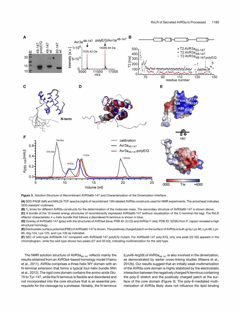
The NMR solution structure of AVR3a60-147 reflects mainly theresults obtained from an AVR3a4-based homology model (Yaenoet al., 2011). AVR3a comprises a three-helix WY domain with anN-terminal extension that forms a typical four-helix bundle (Winet al., 2012). The rigid core domain contains the amino acids Glu-70 to Tyr-147, while the N terminus is flexible and disordered andnot incorporated into the core structure that is an essential pre-requisite for the cleavage by a protease. Notably, the N terminus
(Lys48-Arg59) of AVR3a48-147 is also involved in the dimerization,as demonstrated by earlier cross-linking studies (Wawra et al.,2012b). Our results suggest that an initially weak multimerizationof the AVR3a core domain is highly stabilized by the electrostaticinteraction between the negatively chargedN terminus containingthe poly-E stretch and the positively charged patch at the sur-face of the core domain (Figure 3). The poly-E-mediated multi-merization of AVR3a likely does not influence the lipid binding
Figure 3. Solution Structure of Recombinant AVR3a60-147 and Characterization of the Dimerization Interface.
(A) SDS-PAGE (left) and MALDI-TOF spectra (right) of recombinant 15N-labeled AVR3a constructs used for NMR experiments. The arrowhead indicatesSDS-resistant multimers.(B) T2 times for different AVR3a constructs for the determination of the molecular mass. The secondary structure of AVR3a60-147 is shown above.(C) A bundle of the 10 lowest energy structures of recombinantly expressed AVR3a60-147 (without visualization of the C-terminal His-tag). The RxLReffector characteristic 4 a-helix bundle that follows a disordered N terminus is shown in blue.(D) Overlay of AVR3a60-147 (gray) with the structures of AVR3a4 (blue; PDB-ID: 2LC2) and AVR3a11 (red; PDB-ID: 3ZGK) from P. capsici revealed a highstructural homology.(E)Electrostatic surface potential (PBE) of AVR3a60-147 is shown. The positively chargedpatch on the surface of AVR3a is built up by Lys-85, Lys-86, Lys-89, Arg-124, Lys-125, and Lys-130 as indicated.(F) SEC of wild-type AVR3a48-147 compared with AVR3a48-147 polyE/Q mutant. For AVR3a48-147 poly-E/Q, only one peak (22 kD) appears in thechromatogram, while the wild type shows two peaks (27 and 50 kD), indicating multimerization for the wild type.
RxLR of Secreted AVR3a Is Processed 1189

properties of AVR3a because mutation of the RxLR motif of theeffector AVR1d (RxLR/EEEE) does not have an effect on lipidbinding (Naetal., 2013). Thus,asmentionedabove, theNterminusserves tomask the positively charged patch of the core domain to
avoid unspecific interaction during secretion and translocationuntil the point of membrane-mediated uptake into the host cell.Multimerization of another effector protein, PexRD2, is essentialfor its function in planta (King et al., 2014). Hence, the dimerizationof AVR3a might be essential for its interaction with potato R3asince thedifferentR3a recognition sites (K/E80and I/M103),whichare important for the hypersensitive response in planta, do notinterfere with the multimerization (Bos et al., 2006).The processing of native AVR3a secreted from P. infestans is
indeed remarkably similar to the cleavage observed for the PEXELmotif of effectors from P. falciparum (Chang et al., 2008; Boddeyet al., 2009; Russo et al., 2010) (Figure 4) and also for the TEXELmotif of effectors fromT.gondii (Coffeyet al., 2015;Curt-Varesanoet al., 2016). However, the RxLR motif is not recognized andcleavedby theER-located plasmepsin VproteaseofP. falciparum(Boddey et al., 2016). Furthermore, despite the phylogenetic re-lationship between plasmepsin V from P. falciparum and PiAP12(Kay et al., 2011), we could not detect any proteolytic activityagainst recombinant AVR3a22-147 for PiAP12 nor for any of theother 10 aspartic protease domains (Figure 2C). An analogoussystemmightexist inP. infestanswithasimilarmodeofactionas inP. falciparumbut havingevolved fromadifferent origin. In contrastto thePEXELmotif,which ispartly retainedafterprocessing (Ac-X-E/D/Q) (Chang et al., 2008), the RxLR sequence of AVR3aEM iscleaved off completely. The cleavedPEXELmotif is involved in thebindingof the respective effector to thePTEX redbloodcell importtranslocon (Boddey et al., 2009; Russo et al., 2010). It is intriguingto speculate what role might be played by the retained s/dEERsequence that is conserved inmany RxLR effectors. Although theprotein export and host cell translocation mechanisms betweenthe pathogens are not interchangeable, probably due to their
Table 1. Statistics for the Solution Structure Calculation of AVR3a60-147
Number of distance and dihedral angle constraintsTotal NOEs 1287Intraresidual NOEs 269Sequential NOEs (i to i + 1) 351Medium-range NOEs (i to i + 2,3,4) 311Long-range NOEs 356Dihedral angle restraints (w + c) 61 + 61RMSD to the mean structure (Å)RMSD for backbone atoms (T80-G144) 0.30 6 0.09RMSD for heavy chain atoms (T80-G144) 0.98 6 0.17Average target function 1.39 6 0.07Restraint violationsAverage of upper distance (Å) 0.006 6 0.001Average of maximum upper distance (Å) 0.22 6 0.07Average of sum of van der Waals (Å) 6.2 6 0.2Average of torsion angle (°) 0.464 6 0.034Average of maximum torsion angle (°) 2.43 6 0.35Ramachandran analysis (%)Residues in most favored regions 75.2Residues in additionally allowed regions 24.6Residues in the generously allowed regions 0.0Residues in the disallowed regions 0.2
Data are derived from a set of the 10 lowest-energy structures calculatedby CYANA 2.1 using automated NOE (nuclear Overhauser effect) assign-ment without a homology model. RMSD, root mean square deviation.
Figure 4. Comparison of the Processing of the PEXEL Motif in P. falciparum and the RxLR Motif of AVR3a in P. infestans.
The processing of AVR3a is strikingly similar to the stepwise modification observed for the PEXEL effectors from P. falciparum. However, the PEXELmotif(RxLxE/D/Q) is partially retained, while the RxLR motif is cleaved off completely, although both are acetylated (Ac). The corresponding RxLR-modifyingenzymes remain to be investigated. NAT, N-terminal acetyltransferase; PEP, signal peptide peptidase; PMV, plasmepsin V; PTEX, Plasmodium transloconof exported proteins; SP, signal peptide.
1190 The Plant Cell

different infection structures (intracellular parasitophorous va-cuoles for P. falciparum/T. gondii versus extracellular haustoriafor oomycetes), the stepwise processing of the RxLR motif inoomycetescouldactasasignal that facilitates the transport toandsecretion of effectors from the haustorial infection structuresimilarly to PEXEL and TEXEL.
METHODS
Purification of Native AVR3aEM
To establish a purification protocol for native AVR3a, initially the optimi-zation was done with Phytophthora infestans transformants carryinga translational AVR3a-mRFP fusion protein (AVR3aKI:mRFP) as describedpreviously (Whisson et al., 2007). For the purification of native AVR3aEM,6 liters of a liquid culture of P. infestans (isolate 88069) in pea broth wasgrown in several Petri dishes (each75mL) for 22d to confluence.Myceliumwas removed by paper filtration and the supernatant cleared by centri-fugation (15 min, 15,000g) and an additional filtration step (0.45 mm, ni-trocellulose). The supernatant was supplemented with 2 mM EDTA andaprotease inhibitormix (two tablets/3 liters;Roche).Afterwards, theproteinsuspension was loaded onto a SO3
2 column (Fractogel-EMD-SO32;Merck) for initial concentration. The columnwas washed once with 25mMNaPi buffer, pH 7.3, and oncemore with the same buffer containing 80mMNaCl. Subsequently, bound proteins were eluted with the same buffersupplementedwith 1MNaCl and theprotease inhibitorAEBSF. Theelutionfraction was ammonium sulfate precipitated (80%) for 30 min at roomtemperature. Pellets obtained from precipitation after centrifugation(20 min, 9000g) were resuspended in 20 mL of 50 mMNaPi buffer, pH 7.3,and additionally dialyzed (1 3 3 liters for 3 h and 1 3 3 liters overnight)against NaPi buffer supplemented with 15 mM MgSO4. The dialyzedprotein fractions were resubjected to the SO32 column to remove re-maining contaminants and washed twice with 25 mM NaPi buffer, pH 7.3(the second onewith additional 100mMNaCl), followed by elutionwith thesame buffer supplemented with 200 mM NaCl. An aliquot was TCA pre-cipitated and prominent bands on SDS-PAGE were analyzed by LC-MS/MS. The results in this study arose from several independent preparationsof AVR3a from in total 70 liters of P. infestans culture.
TCA Precipitation
For 300- to 500-mL sample, 1 mL of 40% TCA was used for protein pre-cipitation overnight at 4°C. Subsequently, samples were centrifuged for30min at 16,000g (4°C) and the supernatantwas discarded. The remainingpellet was washed with 1 mL acetone and centrifuged again for 20 min at16,000g (4°C). This step was repeated twice before drying the pellet.
LC-MS/MS Analysis
Proteins of interest from SDS-PAGE were trypsin digested, dried, andresuspended in 0.1% formic acid. Dissolved peptideswere analyzed usinganHCTultraPTMDiscoverySystemwithan electrospray source fittedwitha low-flow nebulizer (Bruker) coupled to an UltiMate 3000 LC system(Thermo Fisher Scientific). Peptides were separated on a PepSwiftmonolithic PS-DVB capillary column (200 mm i.d. 3 5 cm; Thermo FisherScientific) at a flow rate of 2.0 mL/min. Eluent A (3% acetonitrile/0.05%formic acid) and Eluent B (80% acetonitrile/0.04% formic acid) were usedfor a gradient (3% to 45% B, 12 min), followed by a column wash (90% B,1min) and an equilibration step (3%B, 5min). MS/MSdata, averaged fromtwo spectra, with a scan range of 100 to 2200m/z, were acquired in data-dependentAutoMS(2)mode.Up to threeprecursor ionswereselected fromthe MS scan (range 300 to 1500 m/z, averages 3) in each cycle. Singlycharged ions and precursors were actively excluded after selection twice
within a 1.0-min window. Peptide peaks were detected and deconvolutedautomatically using DataAnalysis software (Bruker). Mass lists were cre-atedautomaticallyandusedas the input forMascotMS/MS ionsearchesofthe NCBInr database (www.matrixscience.com). The Mascot significancethreshold was set to <0.05. Default search parameters were enzyme =trypsin; maximum missed cleavages = 1; fixed modifications = carbami-domethyl (C); variable modifications = oxidation (M); acetyl (N-term) anddeamination (NQ); peptide tolerance =61.5 D; MS/MS tolerance6 0.5 D;peptide charge = 2+ and 3+; instrument = ESI-TRAP.
NanoRSLC-HPLC Analysis
Mass spectrometric analysis of the digested peptides was performed usingan Orbitrap Velos Pro mass spectrometer (Thermo Scientific). An UltimatenanoRSLC-HPLC system (Thermo Scientific) equipped with a nanoC18 RPcolumn was connected online to the mass spectrometer through a nano-spray ion source. The tryptic digest (6 mL) was injected onto a C18 pre-concentration column. Automated trapping and desalting of the samplewere performed at a flow rate of 6 mL/min with 0.05% formic acid. Eluent A(0.045% formic acid) and Eluent B (80% acetonitrile/0.05% formic acid)were used. Separationwas achievedwith a gradient (4% to45%B, 30min)at a flow rate of 300 nL/min, followedby a columnwash (95%B, 5min). Thecolumnwas connected to a stainless steel nanoemitter (ThermoScientific)and the eluent sprayed directly toward the heated capillary of the massspectrometer using apotential of 2300V. A survey scanwith a resolution of60,000 within the Orbitrapmass analyzer was combined with at least threedata-dependentMS/MSscanswithdynamicexclusion for 30seither usingCID with the linear ion trap or using HCD and Orbitrap detection ata resolution of 7500. Data analysis was performed using Proteome Dis-coverer (Thermo Scientific) with SEQUEST and MASCOT (version 2.2;Matrix Science) search.
Reverse-Phase Liquid Chromatography
TCA precipitated pellets were dissolved in 50 mL of 0.1% trifluoroaceticacid (TFA) and subjected to reverse-phase separation. Reverse-phaseliquid chromatography was carried out using an UltiMate3000 system(Thermo Fisher Scientific) equipped with a YMC-Pack Protein-RP column(YMC Europe; 100 3 2.1 mm S-5 mm). Eluent A (0.1% TFA) and Eluent B(80% acetonitrile, 0.085% TFA) were used. The flow rate was set to100 mL/min with a gradient of 0 to 70% B for 25 min. Fractions of 100 mLwere collected.
MALDI-TOF
All spectra were acquired on an ultrafleXtreme MALDI-ToF/ToF massspectrometer (Bruker) equipped with a 1-kHz Bruker Smartbeam-II solid-state laser. A 2,5-dihydroxybenzoic acid or a a-cyano-4-hydroxycinnamicacid carrier matrix was used as indicated. Linear mode acquisition wasperformed with a high resolution in a 3000 to 20,000 m/z range, and thesample rate was 1.00 GS/s with Bruker Protein Calibration I Standard P/N206355. Between 1000 and 10,000 shotswere accumulatedmanually withSmartbeam laser focus raster width set at 50 mm. Spectra were processedin Compass 1.3 software incorporating Flex Control version 3.3 and FlexAnalysis version 3.3 using “centroid” peak picking (80%), smoothingperformedusingSavitzky-Golay0.2m/z foronecycle,andTopHatbaselinesubtraction for reproducible peak annotation on nonresolved isotopedistributions prior to calibration.
Enzymatic Deglycosylation
Enzymatic deglycosylationwasperformedoneither lyophilized samplesorprotein pellets obtained after TCA precipitation. The denaturing reactionconditions protocol provided with the New England Biolabs Protein
RxLR of Secreted AVR3a Is Processed 1191

Deglycosylation Mix (P6039S) was chosen. Deglycosylation was carriedovernight at 37°C.
Cloning and Mutagenesis
The sequences encoding AVR3a48-147 and AVR3a48-147 poly-E/Q (E50Q,E51Q, E53Q, E54Q, E57Q, and E58Q) were PCR amplified with oligonu-cleotides as indicated in Supplemental Table 1 using AVR3a22-147 asa template (Wawra et al., 2012b). The resulting fragments containing therestriction sites for NdeI and EcoR1 (NEB) were cloned into a pET21bvector. Constructs were confirmed by Sanger sequencing (OneSourceBioscience).
Sequences encoding the aspartic proteases were PCR amplified witholigonucleotides as indicated in Supplemental Table 1 using cDNA fromP.infestans (isolate 88069) as a template. The resulting fragments containingthe restriction sites for NdeI and EcoR1 or NdeI and EcoR1 (NEB) for PiAPwere cloned into a pET21b vector. Constructs were confirmed by Sangersequencing (OneSource Bioscience).
AVR3a22-147 containing the N-terminal RxLR motif was cloned intopHisTEV (kanamycin resistance, C-terminal 63 His-tag) with NdeI/XhoIusing AVR3a22-147 as a template (Wawra et al., 2012b). Constructs wereconfirmed by Sanger sequencing (OneSource Bioscience).
Expression and Purification of Recombinant AVR3a Constructs
Recombinant AVR3a protein containing a C-terminal 6x His-tag wasobtained as described elsewhere (Wawra et al., 2012b). Briefly, for iso-topically labeled protein, cells from 0.5 liters LB culture (OD600 of 0.8) weretransferred to 2 liters M9 minimal medium supplemented with 1 g/L [15N]NH4Cl and/or 0.4% (w/v) [U-13C]glucose and further grown to an OD600 of0.8. Subsequently, protein expression was induced with 0.2 mM IPTGovernight at 25°C.Cellswere collected, lysedbysonication (60% intensity,4 3 45 s, 1 min interval at 4°C; Sonoplus; Bandelin), and ultracentrifuged(Beckman Coulter; 98,000g, 70 min, 4°C). Subsequently, the supernatantwas applied to aQ-Sepharose column (equilibratedwith 50mMKPi buffer,pH 6.5). The flow-through containing AVR3a was applied to a Ni-NTAcolumnequilibratedwith50mMKPibuffer, pH6.5,500mMKCl, and20mMimidazole. After awashing step, proteinswere elutedwith 300mM imidazole.Fractions containing the AVR3a proteins were pooled and the imidazole re-movedbydialysis forNMRspectroscopy.The integrity of each constructwasconfirmed by SDS-PAGE and MALDI-TOF mass spectrometry.
Expression and Purification of Recombinant PiAP12
The domain of PiAP12 containing a C-terminal 63 His tag was obtainedfrom Escherichia coli [BL21(DE3)T1r]. Briefly, cells were incubated in 2xYTat37°C toOD600=0.8, andproteinexpressionwas inducedwith1mMIPTGfor 6 h at 30°C. Cells were collected and lysed by sonication and thesupernatant after ultracentrifugation applied to Fractogel EMD TMAE (M)resin (equilibratedwith20mMTris-HCl,pH6.6). Theelution fraction (20mMTris-HCl and 500mMNaCl, pH6.6) containingPiAP12wasdilutedwithHisequilibrationbuffer (50mMNaH2PO4, 300mMNaCl, and10mM imidazole,pH8.0) andapplied toanequilibratedNi-NTAcolumn.After awashingstep,the protein was eluted with 300 mM imidazole. The buffer of the elutionfractions with PiAP12was changed to 50mMTris-HCl, pH 8.0. In addition,impurities in the elution fraction of the supernatant were removed usingdenaturing conditions. For this, PiAP12 was diluted in urea buffer (50 mMNaH2PO4,300mMNaCl, 10mMimidazole, and8Murea,pH8.0), applied toNi-NTA beads, and incubated for 1 h at room temperature under rotation.After awashing step, PiAP12was elutedwith 500mM imidazole. Refoldingwas performed by a gradual buffer exchange over 60 h to 20 mM HEPES,100 mM NaCl, and 0.2 mM DTT (pH 7.2), which resulted in semistableprotein (Hodder et al., 2015).
In Vitro Cleavage Assays with AVR3a
PurifiedPiAP12 (180mg)was incubatedwith5mg recombinantAVR3a22-1476xHis containing the N-terminal RxLR motif for 1 h at 37°C. The reactionwas stopped with SDS sample buffer and subsequent heating up to 90°Cfor 10 min. Samples were analyzed by SDS-PAGE and immunoblotting(anti-His antibody, 1:10,000; Qiagen; no. 34460).
For another assay, domains of all aspartic proteases from P. infestanswere cloned (Kay et al., 2011). AVR3a and each domain were co-transformed into BL21(DE3)T1r. Cells containing both constructs wereincubated inLBmediumat37°C toOD600=0.8, andproteinexpressionwasinduced with 1 mM IPTG for 6 h at 30°C. Cells were collected (15 min,4500g, 4°C) and lysedby sonication. The supernatant after a centrifugationstep (30 min, 16,100g, 4°C) was analyzed by immunoblot with an a-5xHisantibody (1:10,000; Qiagen; no. 34460).
NMR Spectroscopy and Resonance Assignment
The 2D and 3D spectra were recorded at 25°C on a 700-MHz UltrashieldNMR spectrometer (Bruker) equipped with a triple cryoprobe head (BrukerBiospin). The 3D and 4D spectra were recorded with a Varian VNMRS700 NMR spectrometer equipped with four RF channels, a Performa-XYZPFG gradient unit, and a room temperature 1H/13C/15N triple-resonanceprobehead.AVR3a60-147 (300mMin50mMKPibuffer,pH6.5)waspreparedwith 10% D2O and 50 mM DSS as calibration standard. All spectra wereanalyzed with Sparky 3.113. For assignment, all 4D nonuniform sampled(NUS) NMR data sets were used. The process of sequentially specificassignment employing 4D NUS NMR data sets was performed with a 3DHNCO experiment based on NUS (1000 data points, maximal evolutiontimes: 30 ms for 13CO and 28 ms for 15N, acquisition time: 6 h), a 4DHNCOCANMRspectrum (Zawadzka-Kazimierczuketal., 2010)correlatinginterresidual HN(i)-N(i)-CO(i-1)-Ca(i-1) resonances (1400 data points,maximum evolution times of 30ms for 13CO, 10ms for 13Ca, and 28ms for15N, acquisition time: 18 h), the complementary 4D HNCACO (Zawadzka-Kazimierczuk et al., 2010), correlating both intraresidual HN(i)-N(i)-Ca(i)-CO(i) and interresidual HN(i)-N(i)-Ca(i-1)-CO(i-1) resonances (2500 datapoints, maximum evolution times of 20 ms for 13CO, 10 ms for 13Ca, and28 ms for 15N, acquisition time: 31 h), a 4D HBHACBCANH spectrumcorrelating 1H, 13C, and 15N signals for intraresidual [HN(i)-N(i)-Cab(i)-Hab(i)] and interresidual [HN(i)-N(i)-Cab(i-1)-Hab(i-1)] resonances (2500data points, maximum evolution times of 10 ms for 13C, 7 ms for 1H, and28 ms for 15N, acquisition time: 50 h). Additionally, aliphatic side chain 1Hand 13C chemical shifts were assigned with HCCH-COSY, HCCH-TOCSY,HBHAHN, and HBHA(CO)NH spectra and aromatic side chain resonanceswere assigned from two-dimensional COSY, TOCSY, and NOESY spectra.Distance constraints were obtained from two 1H-homonuclear NOESYspectra, one recorded in water, the other recorded in D2O buffer, fromthe 13C-NOESY-HSQC and the 15N-NOESY-HSQC. The 3D 13C-editedNOESY-HSQC spectra were acquired with a mixing time of 150 ms foraliphatic and aromatic regions on Varian VNMRS 800 NMR spectrometerequippedwith four RF channels, a Performa IV PFGmodule, and cryogenic1H/13C/15N triple resonance probe head. Processing and evaluation ofspectra were performed with Topspin 3.0 (Bruker) or NMRPipe (Delaglioet al., 1995); assignment was done with Sparky.
Structure Calculation
15N and 13C resonances were calibrated unidirectionally following theIUPAC-IUB recommended chemical shift referencing ratios (Wishart et al.,1995;Markley et al., 1998). NOE restraints were identified and transformedinto distance constraints using the automated standard protocol of Cyana(Güntert et al., 1991). Dihedral angles were calculated using Talos+ (Shenet al., 2009). The structure of AVR3a60-147 was calculated using Cyana 2.1(López-Méndez and Güntert, 2006) without using a homolog as a guide
1192 The Plant Cell
structure. Energy minimization was performed with YASARA 11.12.31(Krieger and Vriend, 2002).
Molecular Weight Estimation by Measuring Relaxation Constants
For estimation of the molecular weight of the AVR3a protein constructs,different spectra with varying relaxation times t were recorded (20, 100,150, 200, 300, 500, 750, 1000, and 1400 ms for T1 and 10, 30, 50, 90, 130,170, 210, and 250 ms for T2). With the different signal intensities, the timeconstants T1 and T2 were calculated for each amino acid as described byKay et al. (1992). For analysis, only resonances from amino acids locatedin secondary structure elements without contribution of chemical ex-change processes were chosen (Blackledge et al., 1998). Subsequently,the rotation correlation time tc and the standard curve for the estimationof the molecular weight were calculated as described elsewhere (Rossiet al., 2010).
SEC
SECexperiments for AVR3a48-147wild type and the poly-E/Qmutantwereperformed in50mMNaPi, 500mMNaCl, pH7.0,withSuperdex7510/300on a Bio-Rad system at a flow rate of 0.5 mL/min. The column wascalibrated under the same conditions with thyroglobulin (670 kD),g-globuline (158 kD), ovalbumin (44 kD),myoglobulin (17 kD), and vitaminB12 (1.35 kD); R2 was 0.9199 after linear regression of the calibrationstandards.
Accession Numbers
Sequence data from this article can be found in the Arabidopsis GenomeInitiative or GenBank/EMBL databases under the following accessionnumbers: AVR3a (AVR3aEM,ADC96691.1;AVR3aKI, ADC96694.1), PiAP01(HM588685, corrected;Kayetal., 2011),PiAP02 (XP_002900284.1),PiAP04(XP_002998816.1), PiAP05 (XP_002902303.1), PiAP06 (XP_002905662.1),PiAP07 (XP_002905687.1), PiAP08 (HM588686, corrected;Kayet al., 2011),PiAP09 (XP_002904877),PiAP10 (XP_002907533), PiAP11 (XP_002907534),andPiAP12(XP_002901082).Structural coordinatesandNMRshiftdataweredeposited in theRCSBdatabank (entry ID: 2NAR) and in theBMRBdatabank(entry ID: rcsb104633), respectively. Structures alreadypublished, but used inthis study are as follows: AVR3a4 (PDB-ID: 2LC2) and AVR3a11 (PDB-ID:3ZGK).
Supplemental Data
Supplemental Table 1. Primers and restriction sites used for cloning ofthe constructs used in this study.
ACKNOWLEDGMENTS
Our work is supported by the BBSRC (S.W., C.J.S., and P.v.W.), NERC(P.v.W.), and the University of Aberdeen (C.J.S., P.v.W., and I.D.). This workwas supported by EU East-NMR FP7 Project (Contract 228461) and PolishNational Centre for Research and Development under research grantnumber 178479 (contract number PBS1/A9/13/2012) (for I.Z.). We thankKevin MacKenzie of the core microscopy facility of the University ofAberdeen forhelpful suggestionsandRegineKahmann forcritical readingof the manuscript.
AUTHOR CONTRIBUTIONS
S.W., F.T., A.M., K.A., C.J.S., P.B., and P.v.W. designed research. S.W.,F.T., A.M., K.A., U.L., I.Z., W.K., J.S., and I.D. performed research andanalyzed data. S.W., F.T., C.J.S., and P.v.W. wrote the article. All authorshave given approval to the final version of the manuscript.
Received July 8, 2016; revised April 4, 2017; accepted May 10, 2017;published May 18, 2017.
REFERENCES
Anderson, R.G., Casady, M.S., Fee, R.A., Vaughan, M.M., Deb, D.,Fedkenheuer, K., Huffaker, A., Schmelz, E.A., Tyler, B.M., andMcDowell, J.M. (2012). Homologous RXLR effectors from Hyalo-peronospora arabidopsidis and Phytophthora sojae suppress im-munity in distantly related plants. Plant J. 72: 882–893.
Baker, N.A., Sept, D., Joseph, S., Holst, M.J., and McCammon, J.A.(2001). Electrostatics of nanosystems: application to microtubulesand the ribosome. Proc. Natl. Acad. Sci. USA 98: 10037–10041.
Bhattacharjee, S., Hiller, N.L., Liolios, K., Win, J., Kanneganti, T.D.,Young, C., Kamoun, S., and Haldar, K. (2006). The malarial host-targeting signal is conserved in the Irish potato famine pathogen.PLoS Pathog. 2: e50.
Blackledge, M., Cordier, F., Dosset, P., and Marion, D. (1998).Precision and uncertainty in the characterization of rotational dif-fusion from heteronuclear relaxation data. J. Am. Chem. Soc. 120:4538–4539.
Boddey, J.A., Moritz, R.L., Simpson, R.J., and Cowman, A.F.(2009). Role of the Plasmodium export element in trafficking para-site proteins to the infected erythrocyte. Traffic 10: 285–299.
Boddey, J.A., Hodder, A.N., Günther, S., Gilson, P.R., Patsiouras,H., Kapp, E.A., Pearce, J.A., de Koning-Ward, T.F., Simpson,R.J., Crabb, B.S., and Cowman, A.F. (2010). An aspartyl proteasedirects malaria effector proteins to the host cell. Nature 463: 627–631.
Boddey, J.A., et al. (2016). Export of malaria proteins requiresco-translational processing of the PEXEL motif independent ofphosphatidylinositol-3-phosphate binding. Nat. Commun. 7: 10470.
Bos, J.I., et al. (2010). Phytophthora infestans effector AVR3a is es-sential for virulence and manipulates plant immunity by stabilizinghost E3 ligase CMPG1. Proc. Natl. Acad. Sci. USA 107: 9909–9914.
Bos, J.I., Kanneganti, T.D., Young, C., Cakir, C., Huitema, E., Win,J., Armstrong, M.R., Birch, P.R., and Kamoun, S. (2006). TheC-terminal half of Phytophthora infestans RXLR effector AVR3a issufficient to trigger R3a-mediated hypersensitivity and suppressINF1-induced cell death in Nicotiana benthamiana. Plant J. 48: 165–176.
Chang, H.H., Falick, A.M., Carlton, P.M., Sedat, J.W., DeRisi, J.L.,and Marletta, M.A. (2008). N-terminal processing of proteins ex-ported by malaria parasites. Mol. Biochem. Parasitol. 160: 107–115.
Chou, S., Krasileva, K.V., Holton, J.M., Steinbrenner, A.D., Alber,T., and Staskawicz, B.J. (2011). Hyaloperonospora arabidopsidisATR1 effector is a repeat protein with distributed recognition sur-faces. Proc. Natl. Acad. Sci. USA 108: 13323–13328.
Coffey, M.J., et al. (2015). An aspartyl protease defines a novelpathway for export of Toxoplasma proteins into the host cell. eLife4: e10809.
Curt-Varesano, A., Braun, L., Ranquet, C., Hakimi, M.A., andBougdour, A. (2016). The aspartyl protease TgASP5 mediates theexport of the Toxoplasma GRA16 and GRA24 effectors into hostcells. Cell. Microbiol. 18: 151–167.
Delaglio, F., Grzesiek, S., Vuister, G.W., Zhu, G., Pfeifer, J., andBax, A. (1995). NMRPipe: a multidimensional spectral processingsystem based on UNIX pipes. J. Biomol. NMR 6: 277–293.
Dou, D., Kale, S.D., Wang, X., Jiang, R.H., Bruce, N.A., Arredondo,F.D., Zhang, X., and Tyler, B.M. (2008). RXLR-mediated entry ofPhytophthora sojae effector Avr1b into soybean cells does not re-quire pathogen-encoded machinery. Plant Cell 20: 1930–1947.
RxLR of Secreted AVR3a Is Processed 1193
Edman, P.H. (1950). Method for determination of the amino acid se-quence in peptides. Acta Chem. Scand. 4: 283–293.
Ellis, J.G., and Dodds, P.N. (2011). Showdown at the RXLR motif:Serious differences of opinion in how effector proteins from fila-mentous eukaryotic pathogens enter plant cells. Proc. Natl. Acad.Sci. USA 108: 14381–14382.
Gan, P.H., Rafiqi, M., Ellis, J.G., Jones, D.A., Hardham, A.R., andDodds, P.N. (2010). Lipid binding activities of flax rust AvrM andAvrL567 effectors. Plant Signal. Behav. 5: 1272–1275.
Gilroy, E.M., Taylor, R.M., Hein, I., Boevink, P., Sadanandom, A.,and Birch, P.R. (2011). CMPG1-dependent cell death follows per-ception of diverse pathogen elicitors at the host plasma membraneand is suppressed by Phytophthora infestans RXLR effector AVR3a.New Phytol. 190: 653–666.
Grouffaud, S., van West, P., Avrova, A.O., Birch, P.R., and Whisson,S.C. (2008). Plasmodium falciparum and Hyaloperonospora para-sitica effector translocation motifs are functional in Phytophthorainfestans. Microbiology 154: 3743–3751.
Gu, B., Kale, S.D., Wang, Q., Wang, D., Pan, Q., Cao, H., Meng, Y.,Kang, Z., Tyler, B.M., and Shan, W. (2011). Rust secreted proteinPs87 is conserved in diverse fungal pathogens and containsa RXLR-like motif sufficient for translocation into plant cells. PLoSOne 6: e27217.
Güntert, P., Braun, W., and Wüthrich, K. (1991). Efficient computa-tion of three-dimensional protein structures in solution from nuclearmagnetic resonance data using the program DIANA and the supportingprograms CALIBA, HABAS and GLOMSA. J. Mol. Biol. 217: 517–530.
Hammoudi, P.M., et al. (2015). Fundamental roles of the Golgi-associated Toxoplasma aspartyl protease, ASP5, at the host-parasite interface. PLoS Pathog. 11: e1005211.
Hiller, N.L., Bhattacharjee, S., van Ooij, C., Liolios, K., Harrison, T.,Lopez-Estraño, C., and Haldar, K. (2004). A host-targeting signalin virulence proteins reveals a secretome in malarial infection. Sci-ence 306: 1934–1937.
Hodder, A.N., et al. (2015). Structural basis for plasmepsin V in-hibition that blocks export of malaria proteins to human eryth-rocytes. Nat. Struct. Mol. Biol. 22: 590–596.
King, S.R., McLellan, H., Boevink, P.C., Armstrong, M.R., Bukharova, T.,Sukarta, O., Win, J., Kamoun, S., Birch, P.R., and Banfield, M.J.(2014). Phytophthora infestans RXLR effector PexRD2 interacts withhost MAPKKK e to suppress plant immune signaling. Plant Cell 26:1345–1359.
Kale, S.D., et al. (2010). External lipid PI3P mediates entry of eu-karyotic pathogen effectors into plant and animal host cells. Cell142: 284–295.
Kale, S.D., and Tyler, B.M. (2011). Entry of oomycete and fungal ef-fectors into plant and animal host cells. Cell. Microbiol. 13: 1839–1848.
Kay, J., Meijer, H.J., ten Have, A., and van Kan, J.A. (2011). Theaspartic proteinase family of three Phytophthora species. BMCGenomics 12: 254.
Kay, L.E., Nicholson, L.K., Delaglio, F., Bax, A., and Torchia, D.A.(1992). Pulse sequences for removal of the effects of cross-correlationbetween dipolar and chemical-shift anisotropy relaxation mechanism onthe measurement of heteronuclear T1 and T2 values in proteins. J. Magn.Reson. 97: 359–375.
Kemen, E., Gardiner, A., Schultz-Larsen, T., Kemen, A.C., Balmuth,A.L., Robert-Seilaniantz, A., Bailey, K., Holub, E., Studholme,D.J., Maclean, D., and Jones, J.D. (2011). Gene gain and lossduring evolution of obligate parasitism in the white rust pathogen ofArabidopsis thaliana. PLoS Biol. 9: e1001094.
Krieger, E., and Vriend, G. (2002). Models@Home: distributed com-puting in bioinformatics using a screensaver based approach. Bio-informatics 18: 315–318.
Lokossou, A.A., Rietman, H., Wang, M., Krenek, P., van derSchoot, H., Henken, B., Hoekstra, R., Vleeshouwers, V.G., vander Vossen, E.A., Visser, R.G., Jacobsen, E., and Vosman, B.(2010). Diversity, distribution, and evolution of Solanum bulbocas-tanum late blight resistance genes. Mol. Plant Microbe Interact. 23:1206–1216.
López-Méndez, B., and Güntert, P. (2006). Automated proteinstructure determination from NMR spectra. J. Am. Chem. Soc. 128:13112–13122.
Lu, S., Chen, L., Tao, K., Sun, N., Wu, Y., Lu, X., Wang, Y., and Dou,D. (2013). Intracellular and extracellular phosphatidylinositol3-phosphate produced by Phytophthora species is important forinfection. Mol. Plant 6: 1592–1604.
Lu, Y.J., Schornack, S., Spallek, T., Geldner, N., Chory, J.,Schellmann, S., Schumacher, K., Kamoun, S., and Robatzek,S. (2012). Patterns of plant subcellular responses to successfuloomycete infections reveal differences in host cell reprogrammingand endocytic trafficking. Cell. Microbiol. 14: 682–697.
Markley, J.L., Bax, A., Arata, Y., Hilbers, C.W., Kaptein, R., Sykes,B.D., Wright, P.E., and Wüthrich, K. (1998). Recommendations forthe presentation of NMR structures of proteins and nucleic acids.Pure Appl. Chem. 70: 117–142.
Marti, M., Good, R.T., Rug, M., Knuepfer, E., and Cowman, A.F.(2004). Targeting malaria virulence and remodeling proteins to thehost erythrocyte. Science 306: 1930–1933.
Marti, M., and Spielmann, T. (2013). Protein export in malaria para-sites: many membranes to cross. Curr. Opin. Microbiol. 16: 445–451.
Mirgorodskaya, E.N., Krogh, T., and Roepstorff, P. (2000). Char-acterization of protein glycosylation by MALDI-TOFMS. In MassSpectrometry of Proteins and Peptides, J. Chapman, ed (Totowa,NJ: Humana Press), pp. 273–292.
Na, R., Yu, D., Qutob, D., Zhao, J., and Gijzen, M. (2013). Deletion ofthe Phytophthora sojae avirulence gene Avr1d causes gain of vir-ulence on Rps1d. Mol. Plant Microbe Interact. 26: 969–976.
Petre, B., and Kamoun, S. (2014). How do filamentous pathogensdeliver effector proteins into plant cells? PLoS Biol. 12: e1001801.
Petre, B., Kopischke, M., Evrard, A., Robatzek, S., and Kamoun, S.(2016). Cell re-entry assays do not support models of pathogen-independent translocation of AvrM and AVR3a effectors into plantcells. bioRxiv doi/10.1101/038232.
Plett, J.M., Kemppainen, M., Kale, S.D., Kohler, A., Legué, V., Brun,A., Tyler, B.M., Pardo, A.G., and Martin, F. (2011). A secretedeffector protein of Laccaria bicolor is required for symbiosis de-velopment. Curr. Biol. 21: 1197–1203.
Rafiqi, M., Gan, P.H., Ravensdale, M., Lawrence, G.J., Ellis, J.G.,Jones, D.A., Hardham, A.R., and Dodds, P.N. (2010). Internaliza-tion of flax rust avirulence proteins into flax and tobacco cellscan occur in the absence of the pathogen. Plant Cell 22: 2017–2032.
Rossi, P., Swapna, G.V., Huang, Y.J., Aramini, J.M., Anklin, C.,Conover, K., Hamilton, K., Xiao, R., Acton, T.B., Ertekin, A.,Everett, J.K., and Montelione, G.T. (2010). A microscale proteinNMR sample screening pipeline. J. Biomol. NMR 46: 11–22.
Russo, I., Babbitt, S., Muralidharan, V., Butler, T., Oksman, A., andGoldberg, D.E. (2010). Plasmepsin V licenses Plasmodium proteinsfor export into the host erythrocyte. Nature 463: 632–636.
Saunders, D.G., Breen, S., Win, J., Schornack, S., Hein, I., Bozkurt,T.O., Champouret, N., Vleeshouwers, V.G., Birch, P.R., Gilroy,E.M., and Kamoun, S. (2012). Host protein BSL1 associates withPhytophthora infestans RXLR effector AVR2 and the Solanum de-missum Immune receptor R2 to mediate disease resistance. PlantCell 24: 3420–3434.
1194 The Plant Cell

Shen, Y., Delaglio, F., Cornilescu, G., and Bax, A. (2009). TALOS+:a hybrid method for predicting protein backbone torsion anglesfrom NMR chemical shifts. J. Biomol. NMR 44: 213–223.
Starheim, K.K., Gevaert, K., and Arnesen, T. (2012). ProteinN-terminal acetyltransferases: when the start matters. Trends Bio-chem. Sci. 37: 152–161.
Stassen, J.H., den Boer, E., Vergeer, P.W., Andel, A., Ellendorff, U.,Pelgrom, K., Pel, M., Schut, J., Zonneveld, O., Jeuken, M.J., andVan den Ackerveken, G. (2013). Specific in planta recognition oftwo GKLR proteins of the downy mildew Bremia lactucae revealedin a large effector screen in lettuce. Mol. Plant Microbe Interact. 26:1259–1270.
Sun, F., Kale, S.D., Azurmendi, H.F., Li, D., Tyler, B.M., andCapelluto, D.G. (2013). Structural basis for interactions of thePhytophthora sojae RxLR effector Avh5 with phosphatidylinositol3-phosphate and for host cell entry. Mol. Plant Microbe Interact. 26:330–344.
Torto, T.A., Li, S., Styer, A., Huitema, E., Testa, A., Gow, N.A., vanWest, P., and Kamoun, S. (2003). EST mining and functional ex-pression assays identify extracellular effector proteins from theplant pathogen Phytophthora. Genome Res. 13: 1675–1685.
van Poppel, P.M., Guo, J., van de Vondervoort, P.J., Jung, M.W.,Birch, P.R., Whisson, S.C., and Govers, F. (2008). The Phytoph-thora infestans avirulence gene Avr4 encodes an RXLR-dEER ef-fector. Mol. Plant Microbe Interact. 21: 1460–1470.
Wawra, S., Belmonte, R., Löbach, L., Saraiva, M., Willems, A., andvan West, P. (2012a). Secretion, delivery and function of oomyceteeffector proteins. Curr. Opin. Microbiol. 15: 685–691.
Wawra, S., Agacan, M., Boddey, J.A., Davidson, I., Gachon, C.M.,Zanda, M., Grouffaud, S., Whisson, S.C., Birch, P.R., Porter, A.J.,and van West, P. (2012b). Avirulence protein 3a (AVR3a) from the
potato pathogen Phytophthora infestans forms homodimers throughits predicted translocation region and does not specifically bindphospholipids. J. Biol. Chem. 287: 38101–38109.
Whisson, S.C., et al. (2007). A translocation signal for delivery ofoomycete effector proteins into host plant cells. Nature 450: 115–118.
Win, J., Krasileva, K.V., Kamoun, S., Shirasu, K., Staskawicz, B.J.,and Banfield, M.J. (2012). Sequence divergent RXLR effectorsshare a structural fold conserved across plant pathogenic oomy-cete species. PLoS Pathog. 8: e1002400.
Wishart, D.S., Bigam, C.G., Yao, J., Abildgaard, F., Dyson, H.J.,Oldfield, E., Markley, J.L., and Sykes, B.D. (1995). 1H, 13C and15N chemical shift referencing in biomolecular NMR. J. Biomol.NMR 6: 135–140.
Yaeno, T., Li, H., Chaparro-Garcia, A., Schornack, S., Koshiba, S.,Watanabe, S., Kigawa, T., Kamoun, S., and Shirasu, K. (2011).Phosphatidylinositol monophosphate-binding interface in the oo-mycete RXLR effector AVR3a is required for its stability in host cellsto modulate plant immunity. Proc. Natl. Acad. Sci. USA 108: 14682–14687.
Yaeno, T., and Shirasu, K. (2013). The RXLR motif of oomycete ef-fectors is not a sufficient element for binding to phosphatidylinositolmonophosphates. Plant Signal. Behav. 8: e23865.
Yu, X., Tang, J., Wang, Q., Ye, W., Tao, K., Duan, S., Lu, C., Yang,X., Dong, S., Zheng, X., and Wang, Y. (2012). The RxLR effectorAvh241 from Phytophthora sojae requires plasma membrane lo-calization to induce plant cell death. New Phytol. 196: 247–260.
Zawadzka-Kazimierczuk, A., Kazimierczuk, K., and Kozminski, W.(2010). A set of 4D NMR experiments of enhanced resolution foreasy resonance assignment in proteins. J. Magn. Reson. 202: 109–116.
RxLR of Secreted AVR3a Is Processed 1195
DOI 10.1105/tpc.16.00552; originally published online May 18, 2017; 2017;29;1184-1195Plant Cell
Stanek, Wiktor Kozminski, Ian Davidson, Chris J. Secombes, Peter Bayer and Pieter van WestStephan Wawra, Franziska Trusch, Anja Matena, Kostis Apostolakis, Uwe Linne, Igor Zhukov, Jan
before Secretion Is CleavedPhytophthora infestansThe RxLR Motif of the Host Targeting Effector AVR3a of
This information is current as of June 17, 2018
Supplemental Data /content/suppl/2017/07/29/tpc.16.00552.DC2.html /content/suppl/2017/05/19/tpc.16.00552.DC1.html
References /content/29/6/1184.full.html#ref-list-1
This article cites 61 articles, 13 of which can be accessed free at:
Permissions https://www.copyright.com/ccc/openurl.do?sid=pd_hw1532298X&issn=1532298X&WT.mc_id=pd_hw1532298X
eTOCs http://www.plantcell.org/cgi/alerts/ctmain
Sign up for eTOCs at:
CiteTrack Alerts http://www.plantcell.org/cgi/alerts/ctmain
Sign up for CiteTrack Alerts at:
Subscription Information http://www.aspb.org/publications/subscriptions.cfm
is available at:Plant Physiology and The Plant CellSubscription Information for
ADVANCING THE SCIENCE OF PLANT BIOLOGY © American Society of Plant Biologists