the roles of gsk-3ß and apc in cytoplasmic dynein regulation
TRANSCRIPT
University of South CarolinaScholar Commons
Theses and Dissertations
12-15-2014
The Roles of GSK-3ß and APC in CytoplasmicDynein RegulationFeng GaoUniversity of South Carolina - Columbia
Follow this and additional works at: https://scholarcommons.sc.edu/etd
Part of the Biology Commons
This Open Access Dissertation is brought to you by Scholar Commons. It has been accepted for inclusion in Theses and Dissertations by an authorizedadministrator of Scholar Commons. For more information, please contact [email protected].
Recommended CitationGao, F.(2014). The Roles of GSK-3ß and APC in Cytoplasmic Dynein Regulation. (Doctoral dissertation). Retrieved fromhttps://scholarcommons.sc.edu/etd/2947
THE ROLES OF GSK-3β AND APC IN CYTOPLASMIC DYNEIN REGULATION
by
Feng Gao
Bachelor of Science Zhejiang University, 2008
Submitted in Partial Fulfillment of the Requirements
For the Degree of Doctor of Philosophy in
Biological Sciences
College of Arts and Sciences
University of South Carolina
2014
Accepted by:
Deanna Smith, Major Professor
Vicki Vance, Chairman, Examining Committee
James R. Fadel, Committee Member
Michael Felder, Committee Member
Lydia Matesic, Committee Member
Lacy Ford, Vice Provost and Dean of Graduate Studies

ii
© Copyright by Feng Gao, 2014 All Rights Reserved.

iii
DEDICATION
I dedicate this work to my parents and my wife. I am really grateful for all
your support. Thank you for the sacrifices you have made. I love you guys so
much!

iv
ACKNOWLEGEMENTS
I am very grateful that I have the opportunity to conduct research under
the mentorship of Dr. Deanna Smith. Your support is meant a lot to me.
I appreciate the help and suggestions from my dissertation committee. I
enjoy every minute with you in my committee meetings, which help my
development in scientific area.
I enjoyed the time with all my colleagues in the Smith Lab. Dr. Sachin
Hebbar, thank you for all guidance and suggestions, which means a lot to a new
graduate student. Dr. Jai Pandey, thank you for helping me be familiar with the
lab and the department. Dr. Liang Shi, Thank you for teaching me how to use
mass spectrometry. Ms. Xu Gao, Thank you for your all suggestions. Mr. Tim
Hines, Thank you for your positive energy.
I also want to say thank you to all my friends. You all make my life more
exciting.
v
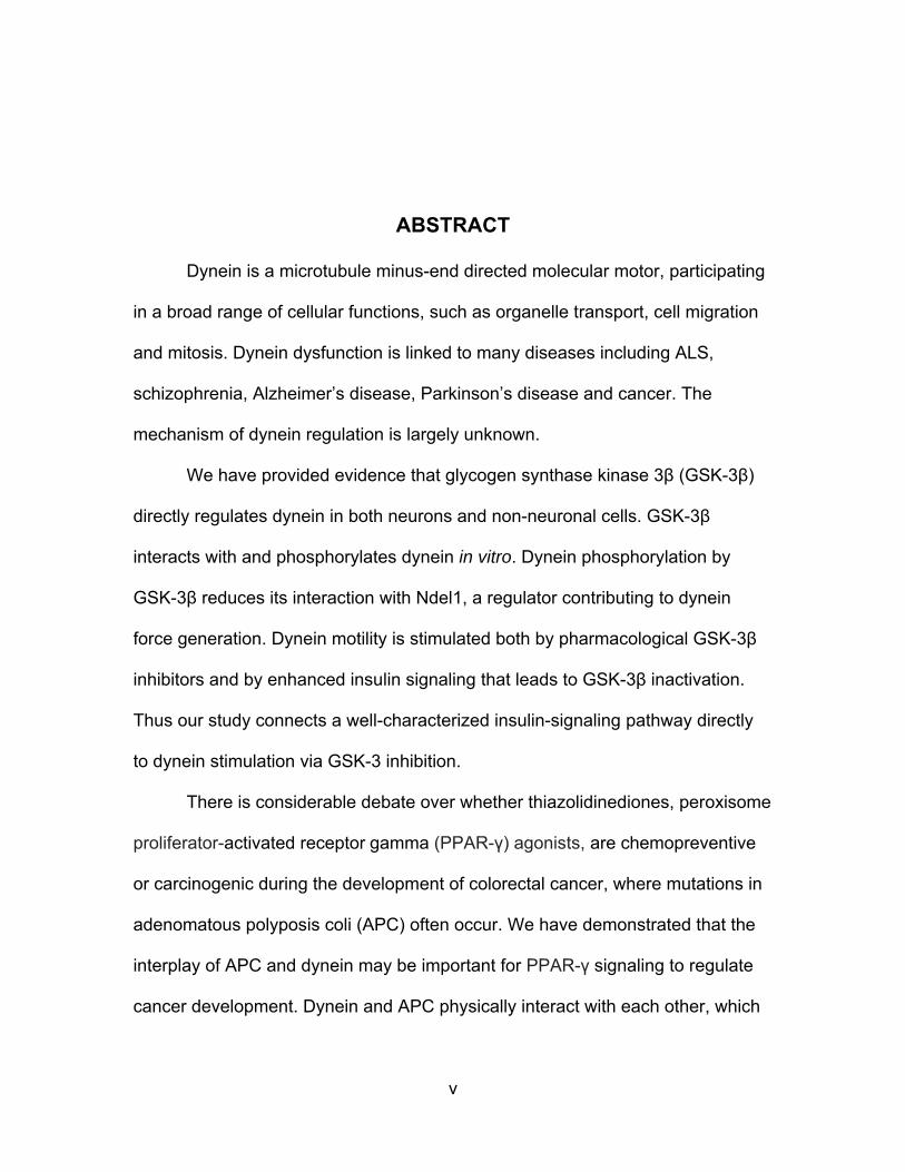
ABSTRACT
Dynein is a microtubule minus-end directed molecular motor, participating
in a broad range of cellular functions, such as organelle transport, cell migration
and mitosis. Dynein dysfunction is linked to many diseases including ALS,
schizophrenia, Alzheimer’s disease, Parkinson’s disease and cancer. The
mechanism of dynein regulation is largely unknown.
We have provided evidence that glycogen synthase kinase 3β (GSK-3β)
directly regulates dynein in both neurons and non-neuronal cells. GSK-3β
interacts with and phosphorylates dynein in vitro. Dynein phosphorylation by
GSK-3β reduces its interaction with Ndel1, a regulator contributing to dynein
force generation. Dynein motility is stimulated both by pharmacological GSK-3β
inhibitors and by enhanced insulin signaling that leads to GSK-3β inactivation.
Thus our study connects a well-characterized insulin-signaling pathway directly
to dynein stimulation via GSK-3 inhibition.
There is considerable debate over whether thiazolidinediones, peroxisome
proliferator-activated receptor gamma (PPAR-γ) agonists, are chemopreventive
or carcinogenic during the development of colorectal cancer, where mutations in
adenomatous polyposis coli (APC) often occur. We have demonstrated that the
interplay of APC and dynein may be important for PPAR-γ signaling to regulate
cancer development. Dynein and APC physically interact with each other, which
vi
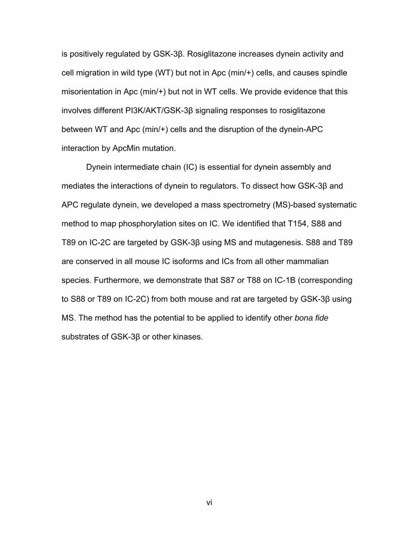
is positively regulated by GSK-3β. Rosiglitazone increases dynein activity and
cell migration in wild type (WT) but not in Apc (min/+) cells, and causes spindle
misorientation in Apc (min/+) but not in WT cells. We provide evidence that this
involves different PI3K/AKT/GSK-3β signaling responses to rosiglitazone
between WT and Apc (min/+) cells and the disruption of the dynein-APC
interaction by ApcMin mutation.
Dynein intermediate chain (IC) is essential for dynein assembly and
mediates the interactions of dynein to regulators. To dissect how GSK-3β and
APC regulate dynein, we developed a mass spectrometry (MS)-based systematic
method to map phosphorylation sites on IC. We identified that T154, S88 and
T89 on IC-2C are targeted by GSK-3β using MS and mutagenesis. S88 and T89
are conserved in all mouse IC isoforms and ICs from all other mammalian
species. Furthermore, we demonstrate that S87 or T88 on IC-1B (corresponding
to S88 or T89 on IC-2C) from both mouse and rat are targeted by GSK-3β using
MS. The method has the potential to be applied to identify other bona fide
substrates of GSK-3β or other kinases.

vii
TABLE OF CONTENTS
DEDICATION ....................................................................................................... iii
ACKNOWLEDGEMENTS .................................................................................... iv
ABSTRACT ........................................................................................................... v
LIST OF FIGURES .............................................................................................. viii
LIST OF ABBREVIATIONS ................................................................................... x
GENERAL INTRODUCTION ................................................................................ 1 CHAPTER 1: GSK-3β phosphorylates cytoplasmic dynein and inhibits dynein motility .................................................................................................................. 6 CHAPTER 2: Adenomatous polyposis coli (APC) is a novel multifaceted dynein regulator: the concerted interplay is vital for the effect of thiazolidinediones ...... 47 CHAPTER 3: A mass spectrometry-based systematic method to map GSK-3β phosphorylation sites on dynein intermediate chain ............................................ 79 CONCLUSIONS ................................................................................................ 112 REFERENCES ................................................................................................. 114
viii
LIST OF FIGURES
Figure 1.1 Inhibition of GSK-3β stimulates retrograde transport in adult rat DRG neurons ............................................................................................................... 32
Figure 1.2 Direct phosphorylation of dynein by GSK-3β ..................................... 34
Figure 1.3 Dynein phosphorylation by GSK-3β impacts Ndel1 interaction .......... 36
Figure 1.4 Pharmacological inhibition of GSK-3β causes dynein to accumulate at centrosomes in colon cell lines ........................................................................... 37
Figure 1.5. GSK-3β inhibition is responsible for the dynein accumulation at centrosomes ....................................................................................................... 38
Figure 1.6 Dynein is released from peripheral/cortical sites in response to GSK-3β inhibition ......................................................................................................... 40
Figure 1.7 An insulin-sensitizing drug, rosiglitazone, causes centrosomal dynein accumulation via GSK-3β inactivation ................................................................ 42
Figure 1.8 Rosiglitazone induced dynein redistribution requires PPAR-γ and new transcription, but expression of dynein and related proteins is not increased ..... 43
Figure 1.9 Dynein accumulation at centrosomes in response to ROZ is due to increased motor transport activity ....................................................................... 44
Figure 1.10 A model for GSK-3β-dependent regulation of dynein dependent retrograde organelle transport ............................................................................ 46
Figure 2.1 Dynein, APC and GSK-3β physically interact with each other in mouse brain .................................................................................................................... 69
Figure 2.2 GSK-3β positively regulates the interaction of dynein to APC ........... 70
Figure 2.3 Characterize APC status and PPAR-γ expression in YAMC and IMCE ................................................................................................................... 71
Figure 2.4 ApcMin mutation prevents dynein activation in response to ROZ ...... 72
ix
Figure 2.5 ApcMin mutation alters spindle orientation in response to ROZ ........ 73
Figure 2.6 ApcMin mutation prevents ROZ induced cell migration shown in wound healing and enterocyte motility assay ...................................................... 74
Figure 2.7 ApcMin mutation alters PI3K/AKT/GSK-3β signaling, which is essential for ROZ response ................................................................................ 76
Figure 2.8 GSK-3β inhibition significantly rescues dynein activation in ApcMin mutation cells, and AKT inhibition prevents ROZ induced cell migration in WT cells ..................................................................................................................... 77
Figure 2.9 ApcMin mutation disrupts the interaction between dynein and WT APC .................................................................................................................... 78
Figure 3.1 A systematic workflow of identifying and confirming kinase-specific phosphorylation sites based on tandem mass spectrometry and mutagenesis .. 96
Figure 3.2 GSK-3β phosphorylates N-terminal of IC-2C without priming phosphorylation................................................................................................... 97
Figure 3.3 Identification of T154 phosphorylation on IC-2C by mass spectrometry ....................................................................................................... 99
Figure 3.4 Identification of S88, T89 phosphorylation sites on IC-2C by mass spectrometry ..................................................................................................... 101
Figure 3.5 Quantification of T154, S88 or T89 phosphorylation on IC-2C by mass spectrometry ..................................................................................................... 103
Figure 3.6 Confirmation of T154, S88 or T89 phosphorylation on IC-2C by mutagenesis ..................................................................................................... 105
Figure 3.7 Location of and conservation of T154, S88 or T89 on IC ................. 107
Figure 3.8 Identification of S87 or T88 on Rat IC-1B targeted by GSK-3β ........ 108
Figure 3.9 Identification of S87 or T88 on mouse IC-1B targeted by GSK-3β ... 110
x
LIST OF ABBREVATIONS
AD ........................................................................................... Alzheimer’s disease
AKT ............................................................................................... protein kinase B
APC .......................................................................... Adenomatous Polyposis Coli
ATP ................................................................................... Adenosine triphosphate
ALS ............................................................................ amyotrophic lateral sclerosis
C-APC ............................................................................ c-fragment of APC protein
CDK5RAP2 ..................................... CDK5 regulatory subunit-associated protein 2
CDK .................................................................................. cyclin dependent kinase
DMSO ........................................................................................ dimethyl sulfoxide
DRG .........................................................................................dorsal root ganglion
ERK ............................................................. extracellular signal-regulated kinases
FL ............................................................................................................. full length
GSK-3 ......................................................................... glycogen synthase kinase 3
GST .................................................................................. glutatione S-transferase
EGFP ............................................................. enhanced Green fluorescent protein
HC ............................................................................................ dynein heavy chain
IRS .................................................................................. insulin receptor substrate
IC ................................................................................... dynein intermediate chain
IC-1B .......................................................................... dynein intermediate chain1B
IC-2C ........................................................................ dynein intermediate chain 2C
xi
IP ............................................................................................ immunoprecipitation
IPTG ............................................................................ isopropyl-d-thiogalactoside
ITS ........................................................................... Insulin, Transferrin, Selenium
LIC .......................................................................... dynein light intermediate chain
LiCl .................................................................................................. lithium chloride
LC ............................................................................................... dynein light chain
LC-MS/MS ........................................................ HPLC-tandem mass spectrometry
Lis1 ............................... platelet-activating factor acetylhydrolase IB subunit alpha
MBO ...................................................................... membrane-bounded organelles
MDCK .......................................................................... Madin-Darby canine kidney
MT ........................................................................................................ microtubule
Min .............................................................................. multiple intestinal neoplasia
MS ............................................................................................ mass spectrometry
N106 ....................................... N-terminal fragment of IC-2C with 106 amino acids
N237 ....................................... N-terminal fragment of IC-2C with 237 amino acids
Nde1 .......................................................................... nuclear distribution protein E
Ndel1 ......................................................... nuclear distribution protein nudE-like 1
NOC ...................................................................................................... nocodazole
P150 ................................................................... p150-glued, a Dynactin subunit 1
P50 ............................................................................................ dynactin subunit 2
PBS ................................................................................ phosphate buffered saline
PI3K ............................................................................... phosphoinositide 3 kinase
PIP3 ..................................................... phosphatidylinositol (3, 4, 5)-trisphosphate

xii
PLCγ ................................................................ phosphoinositide phospholipase C
PPAR-γ .................................... peroxisome proliferator-activated receptor gamma
PTM ......................................................................... post-translational modification
ROZ .................................................................................................... rosiglitazone
RT .............................................................................................. room temperature
S/T ................................................................................................ serine/threonine
SGG .................................................... Shaggy, a homolog of GSK-3 in Drosophila
WT ............................................................................................................ wild type
YAMC ............................................................................. young adult mouse colon

1
GENERAL INTRODUCTION
Transport is essential for life: as we all know transport is essential for
our life, we transport different cargo by different transportation system and tools
to different destinations in order to support our life. There is also an essential
transport system happening inside us all the time, which is cellular transport.
Cells need to exchange items with outside in order to support itself. It uptakes
cargo, such molecules and vesicles, from outside though endocytosis (Marsh
and McMahon, 1999). Endocytotic vesicles could be transported by intracellular
transport system to different locations and utilized (Vale, 2003). Cell also
produces cargo by its organelles such as nucleus and mitochondria. These cargo
are transported by intracellular transport system to the different location inside
cells, and some of them could be transported to cell periphery followed by
exocytosis. Microtubule is the main system responsible for the intracellular
transport (Vale, 2003). Microtubules (MT) are polymerized of alpha and beta
tubulins and have a distinct polarity. The minus end of MT is anchored at the
microtubule organization center (MTOC), which typically locates at the
centrosome, while the plus end is continuing growing to the cell periphery
(Brinkley, 1985). The polarity of microtubules is very important for intracellular
transport, because there are two distinct types of molecular motors, cytoplasmic
dynein and kinesin (Vale, 2003). Dynein prefers to moving cargo from the plus
end of MT to the minus end of MT, while kinesin moves the cargo from the minus
2
end of MT to the plus end of MT. I am particularly interested in dynein dependent
organelles transport, because it is a very important cellular process and dynein is
not as well studied as kinesin.
Functions and regulation of cytoplasmic dynein: As a motor, dynein
uses ATP as power source (Gibbons, 1988). Dynein is an ATPase that
hydrolyzes ATP into ADP. In order to efficiently use ATP and transport cargo on
the microtubules, dynein needs a sophisticate structure (Schiavo et al., 2013).
Dynein heavy chain (HC) has 4626 amino acids. C-terminus of HC is the motor
domain, and N-terminus is stem domain. With the help from other dynein
subunits, intermediate chains (IC) and light intermediate chains (LIC), HCs form
a dimer (Allan, 2011). Dynein has many functions that could be divided into two
main categories: interphase and mitosis. During the interphase, dynein plays
important roles in intracellular transport and cell migration; during the mitosis,
dynein plays essential roles in spindle formation, spindle position, chromosome
alignment and separation. Loss of both copy of Dync1h1 is embryonic lethal
(Harada et al., 1998). Dysregulation of dynein is involved in many
neurodegenerative diseases such as Alzheimer's disease, Parkinson's disease,
Huntington’s disease and ALS, which strongly affect patient’s life quality
(Eschbach and Dupuis, 2011). The common feature of diseases is the
accumulation of toxic aggregation-prone proteins in related neurons (Blokhuis et
al., 2013; Rubinsztein, 2006; Tanzi and Bertram, 2005). Therefore, if we are able
to remove these toxic proteins by transporting them into proper subcellular
locations to degrade them, such as lysosome, these diseases may be cured.
3
Dynein may play important roles in cancer development because of regulating
spindle formation and chromosome separation (Raaijmakers et al., 2013). In
order to cure those diseases, we need to understand how dynein is regulated. In
order to properly carry out its functions, dynein needs help from other regulators
such as dynactin, Bicaudal D, Lis1, Nde1/Ndel1 and more (Kardon and Vale,
2009). Dynactin is able to increase dynein processivity on microtubules
(McKenney et al., 2014). The processivity of dynein on microtubules is essential,
because dynein only move 8 nm/s and axons of some neurons are over 1 m.
Lis1 and Nde1/Ndel1 induces a persistent force generation by dynein (McKenney
et al., 2010). The ability of dynein to generate the persistent force is also very
important, because many of dynein cargo are very heavy, such as nucleus and
mitochondria, and need to be transported for a long distance to reach
destinations. If everything works properly, dynein is a very reliable and powerful
motor. Dynein is able to move cargo at 1 µm/s, which is 125 steps/s. We are
impressed by Bolt for his 100 m world record, which he finished with 41 steps in
9.58 s. However, it is still less than 5 steps/s. Lifting capacity of dynein is even
more impressive: the molecular weight of dynein is 2.49X10⁻⁹ pg and dynein is
able to lift over 1 pN or 100 pg, so dynein is able to lift over 4.01X10¹⁰ times its
body weight. Because dynein is so powerful, it really needs precise regulation.
Signaling that controls dynein to start, pause or stop at the proper time and area
is needed. The signaling passing on dynein could be post-translational
modifications, and phosphorylation is the one of the most important.
4
Phosphorylation regulates interactions and turnover of proteins, therefore, dynein
regulation by kinases can be a very important mechanism.
The functions and regulation of glycogen synthase kinases-3 (GSK-
3): GSK-3 is a serine/threonine protein kinase. It is a fundamental enzyme
involved in many cellular processes. It has over 100 substrates and many of
them are very essential, such as glycogen synthase, Tau and β-catenin
(Sutherland, 2011). In mammals there are two GSK-3 genes, GSK-3α and GSK-
3β (Woodgett, 1991), but their catalytic domains are highly conserved (Castano
et al., 2010; Mukai et al., 2002; Soutar et al., 2010; Wood-Kaczmar et al., 2009).
GSK-3β usually phosphorylates and inhibits its substrates. Dysregulation of
GSK-3β is involved in many diseases including type-2 diabetes,
neurodegenerative diseases and cancer. Therefore, GSK-3β activity also
requires precise regulation. GSK-3β is highly active in resting cells because of an
activating auto-phosphorylation at Tyr 216 (Cole et al., 2004). However,
upstream kinases including AKT/PKB, protein kinase A, and protein kinase C
phosphorylate GSK-3β at serine 9 (S9) and inhibit it (Cross et al., 1995; Fang et
al., 2002; Fang et al., 2000). Inhibition is a main tool to regulate GSK-3β in the
cell. Many pharmacological inhibitors of GSK-3β are being studied in order to
treat diseases such as Alzheimer’s disease and diabetes (Eldar-Finkelman and
Martinez, 2011; Meijer et al., 2004).
The functions and regulation of Adenomatous Polyposis Coli (APC):
APC is well known as a scaffolding protein in Wnt signaling, where it regulates
phosphorylation, ubiquitination and turnover of β-catenin (Espada et al., 2009).
5
Besides that, APC is also known as a MT plus end-binding protein and plays
important roles in cell migration, spindle assembly, cell adhesion and
chromosome segregation (Hanson and Miller, 2005). The Apc (min/+) mouse,
which harbors a truncating APC mutation, is a prominent animal model to study
human colorectal cancer (Taketo and Edelmann, 2009; Yamada and Mori, 2007).
There is considerable debate over whether thiazolidinediones, peroxisome
proliferator-activated receptor gamma (PPAR-γ) agonists, are chemopreventive
or carcinogenic during the development of colorectal cancer, where mutations in
APC often occur.
In chapter 1, I will provide evidence that GSK-3β directly phosphorylates
and regulates dynein in both neurons and non-neuronal cells. In chapter 2, I will
show that APC is a novel multifaceted regulator of dynein and the interplay of
APC and dynein may be important for PPAR-γ signaling to regulate development
of cancer and other diseases. In chapter 3, I will demonstrate a mass
spectrometry (MS)-based systematic method to map GSK-3β dependent
phosphorylation sites on dynein intermediate chains.

6
CHAPTER 1
GSK-3β phosphorylates cytoplasmic dynein and inhibits dynein motility1
ABSTRACT
Glycogen synthase kinase 3 (GSK-3) has been linked to regulation of
kinesin-dependent axonal transport in squid and flies, and to indirect regulation of
cytoplasmic dynein. We have now found evidence for direct regulation of dynein
by mammalian GSK-3β in neurons and in non-neuronal cells. GSK-3β
coprecipitates with mouse brain dynein and phosphorylates purified dynein in
vitro. Dynein phosphorylation by GSK-3β reduces its interaction with Ndel1, a
protein that contributes to dynein force generation. Mammalian dynein motility is
stimulated both by pharmacological GSK-3β inhibitors and by an insulin response
pathway that leads to GSK-3β inactivation. Thus our study connects a well-
characterized insulin-signaling pathway directly to dynein stimulation via GSK-3
inhibition.
INTRODUCTION
Microtubule motors are important force-generating proteins found in all
eukaryotic cells. Two types of processive motors move along microtubules in
animal cells. The kinesins are mainly plus end directed, and cytoplasmic dynein
is the primary motor for minus end directed transport. Both classes of motors
Feng J. Gao1, Sachin Hebbar2, Xu A. Gao1, Michael Alexander1, Jai P. Pandey3, Stephen J. King4, Deanna S. Smith1*. Submitted to Traffic Journal, 10/13/2014.

7
carry out axonal and dendritic transport and defects in both classes have been
linked to late- neurological disorders (Franker and Hoogenraad, 2013; Hirokawa
et al., 2010). Defective microtubule-based transport can also contribute to other
diseases, including cancer (Liu et al., 2013) and diabetes (Baptista et al., 2014).
Two focus areas have dominated the study of motor proteins over the last
decade – studies aimed at elucidating how ATP hydrolysis translates into
processivity, and studies aimed at elucidating how motors “select” specific cargo.
Structural analyses of motor components have begun to answer the first question
(Gennerich and Vale, 2009), and there is a large body of work addressing cargo
selectivity through interaction of accessory subunits, scaffolds, and cargo
adaptors (Fu and Holzbaur, 2014). The sheer diversity of cargos, coupled with
the diversity of potential subcellular destinations, suggest that mechanisms must
be in place to regulate the timing and whereabouts of motor activation by
extracellular cues. There is growing evidence that movement of cargo, especially
membrane-bounded organelles and vesicles, is an effort of teamwork among
motors rather than individual motors functioning in isolation (Rai et al., 2013).
Motors activity may be influenced by regulatory binding proteins, by changes in
the cytoskeleton or cell membranes, by signaling pathways or by a combination
of all of the above (Dobrowolski and De Robertis, 2012).
Our particular interest is in how cytoplasmic dynein is regulated by
extracellular signals. It was recently shown that the presence of cargo adaptor
components was sufficient to induce in vitro processivity of mammalian dynein
motors (Schlager et al., 2014). Yeast dynein is processive in the absence of both
8
cargo or cargo adaptors, so there may be some species variability in activation
mechanisms among different organisms or cell types (Huang et al., 2012). Less
is known about the signals regulating dynein’s association with cargo adaptors or
the signals regulating its cooperation with other motors. It is thought that dynein
is ferried towards microtubule plus ends by kinesin (Roberts et al., 2014; Yamada
et al., 2008), where it can become attached to the membrane (Kardon and Vale,
2009; Markus et al., 2009). At this point motors are poised to become activated.
In neurons a well-supported hypothesis proposes that neurotrophins acting on
neurotrophin receptors in growth cones or synapses induce the transport of
signaling endosomes carrying survival signals to the cell body (Wu et al., 2009).
These signaling endosomes contain the neurotrophins and receptor, as well as
downstream effectors including components of the Ras/ERK pathway, PI3K, and
PLCγ. In zebra fish, stimulation with a growth factor, IGF-1, caused the rapid
dissociation of GSK-3β from signaling endosomes following the transient
association of AKT with the endosomes (Schenck et al., 2008). However, there is
little known about the mechanisms activating retrograde transport motors to carry
the signaling endosomes.
Kinases have been directly or indirectly linked to motor regulation (Mitchell
et al., 2012; Yano et al., 2001). Relevant to this study are three reports linking
glycogen synthase kinase 3 (GSK-3) to axonal transport. The Drosophila
homolog of GSK-3 (SHAGGY, SGG) was found to regulate axonal transport of a
kinesin-1 cargo (amyloid precursor protein, APP) but not a kinesin-3 cargo
(synaptic vesicle precursor, SVP) (Weaver et al., 2013). Because retrograde
9
movements of the kinesin-1 cargo were also affected, the authors speculated
that dynein motors were indirectly affected by SGG regulation of kinesin-1,
pointing to growing evidence that motors of one class can impact the other class
(Mallik et al., 2013). Another group found that expressing a constitutively active
form of SGG resulted in more kinesin, dynein, and activated SGG co-
fractionating with a light membrane fraction from larval brains (Dolma et al.,
2014). They observed bidirectional SGG dependent changes in speeds, run
lengths, and pause frequencies of synaptobrevin vesicles and mitochondria
moving in larval segmental nerves. An earlier study demonstrated that rat kinesin
light chains were substrates for purified GSK-3 (Morfini et al., 2002). However, in
squid axoplasm, active GSK-3 only inhibited plus end (but not minus end)
transport of membrane-bounded organelles (MBOs) (Morfini et al., 2002). Taken
together, these data suggest that it is not clear whether GSK-3 directly regulates
dynein, which may be species and cargo dependent.
Our study now adds significantly to the understanding of how GSK-3
influences dynein-dependent axonal transport in mammalian systems. We find
that direct inhibition of GSK-3 or increased insulin signaling activates dynein
motility. We also show that GSK-3 can directly phosphorylate dynein, which
negatively impacts its interaction with a well-characterized regulatory protein,
Ndel1.
RESULTS
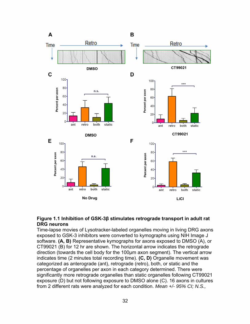
Inhibition of GSK-3β stimulates retrograde movement in neuronal
cells: To ascertain whether dynein-dependent transport is influenced by GSK-3
10
in mammalian axons, we examined organelle transport in axons of cultured adult
dorsal root ganglion (DRG) neurons, which can extend many hundreds of
microns in culture. We used Lysotracker dye to label acidic organelles. In a
previous study kymographs were used to determine the percentage of organelles
that moved anterogradely, retrogradely, switched directions, or remained static
during the recording interval (Pandey and Smith, 2011). On average, three times
more organelles moved retrogradely in control axons. Axons extended by these
cells have uniformly polarized microtubules with minus ends towards the cell
body (Baas and Lin, 2011), so retrogradely moving organelles are likely to be
dynein-driven. Indeed, interfering with dynein or its regulators, Lis1 or Ndel1,
interfered with motility in these assays.
In the current study DRG neurons were exposed to the GSK-3 inhibitors
CT90221 or LiCl for 12 hr. Time-lapse movies from 100 µm segments of 11-30
axons for each condition (and relevant controls) were used to generate
kymographs. Figure 1.1 A and B shows representative kymographs from DMSO
and CT99021 treated axons. The absolute number of organelles analyzed for
each condition ranged from 160-429. In our system, blocking GSK-3 by both
CT99021 (Figure 1.1 C, D) and LiCl (Figure 1.1 E, F) caused a shift towards
more retrogradely moving organelles relative to static organelles, and had little if
any effect on anterograde trafficking, possibly because of the choice of organelle
that is being analyzed.
Dynein interacts with GSK-3β in vivo and is phosphorylated by GSK-
3β in vitro: Because the number of retrogradely moving organelles was
11
increased by GSK-3β inhibition, we considered the possibility that dynein might
be a target for GSK-3β phosphorylation. A small but reproducible amount of
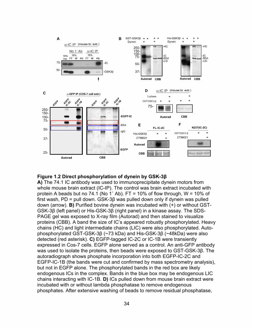
GSK-3β coprecipitated with dynein from adult mouse brain homogenates,
indicating that these proteins may exist in a complex in vivo and supporting the
idea that GSK-3β may be in a position to phosphorylate dynein (Figure 1.2A).
Moreover, several dynein subunits (heavy chains, HC, intermediate chains, IC,
and light intermediate chains, LIC) in a highly purified bovine brain dynein
preparation (Bingham et al., 1998) incorporated γ-32P-ATP when incubated with
purified human GST-GSK-3β in an in vitro kinase assay (Figure 1.2B). Unlike
most kinases, GSK-3β is constitutively active in resting cells because of an
activating auto-phosphorylation at Tyr 216 (Cole et al., 2004). This site is also
known to be phosphorylated in GST-GSK-3β, whose size is similar to IC. There
is a small amount of autophosphorylation apparent in a reaction that did not
include dynein (Figure 1.2B left panels). However, to ensure IC was a target in
vitro, we also used a smaller his-tagged GSK-3β (~48kDa) in vitro kinase assay
(Figure 1.2B right panels).
For future studies we chose to focus our attention on ICs, which interact
directly with several regulatory proteins including Ndel1/Nde1, dynactin, and LCs
(Nyarko et al., 2012). Mammalian ICs are encoded by two genes, DYNC1I1 and
DYNC1I2, that share about 70% protein identity (Kuta et al., 2010). Both are
highly conserved among mammalian species. We will refer to the proteins as IC-
1 and IC-2. To examine whether one or both ICs can be targets of the kinase in
vitro, we expressed EGFP-tagged proteins (rat IC-1B and mouse IC-2C) in Cos-7
12
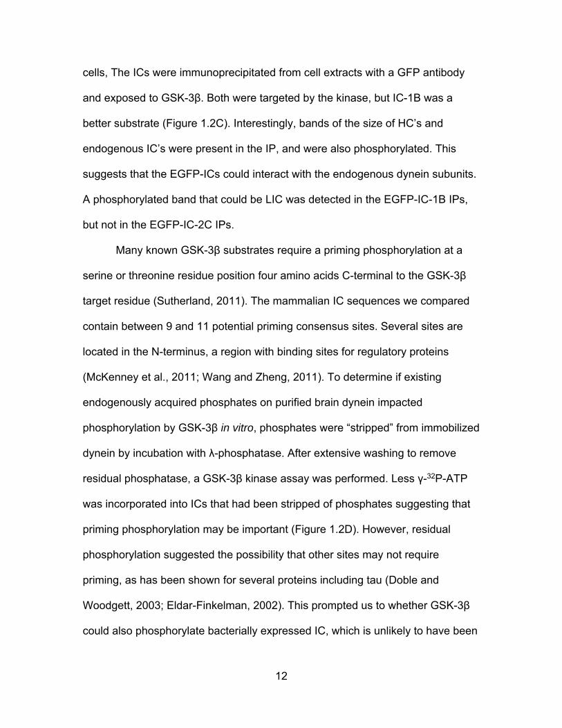
cells, The ICs were immunoprecipitated from cell extracts with a GFP antibody
and exposed to GSK-3β. Both were targeted by the kinase, but IC-1B was a
better substrate (Figure 1.2C). Interestingly, bands of the size of HC’s and
endogenous IC’s were present in the IP, and were also phosphorylated. This
suggests that the EGFP-ICs could interact with the endogenous dynein subunits.
A phosphorylated band that could be LIC was detected in the EGFP-IC-1B IPs,
but not in the EGFP-IC-2C IPs.
Many known GSK-3β substrates require a priming phosphorylation at a
serine or threonine residue position four amino acids C-terminal to the GSK-3β
target residue (Sutherland, 2011). The mammalian IC sequences we compared
contain between 9 and 11 potential priming consensus sites. Several sites are
located in the N-terminus, a region with binding sites for regulatory proteins
(McKenney et al., 2011; Wang and Zheng, 2011). To determine if existing
endogenously acquired phosphates on purified brain dynein impacted
phosphorylation by GSK-3β in vitro, phosphates were “stripped” from immobilized
dynein by incubation with λ-phosphatase. After extensive washing to remove
residual phosphatase, a GSK-3β kinase assay was performed. Less γ-32P-ATP
was incorporated into ICs that had been stripped of phosphates suggesting that
priming phosphorylation may be important (Figure 1.2D). However, residual
phosphorylation suggested the possibility that other sites may not require
priming, as has been shown for several proteins including tau (Doble and
Woodgett, 2003; Eldar-Finkelman, 2002). This prompted us to whether GSK-3β
could also phosphorylate bacterially expressed IC, which is unlikely to have been

13
subjected to priming phosphorylation. Interestingly, two bacterially expressed
recombinant IC-2C proteins, FL-IC-2C and an N-terminal fragment of IC-2C
(N237) (King et al., 2003), were phosphorylated by GSK-3β in vitro (Figure 1.2E,
F). Future studies will be aimed at trying to identify both GSK-3β targeted
consensus and non-consensus sites on ICs.
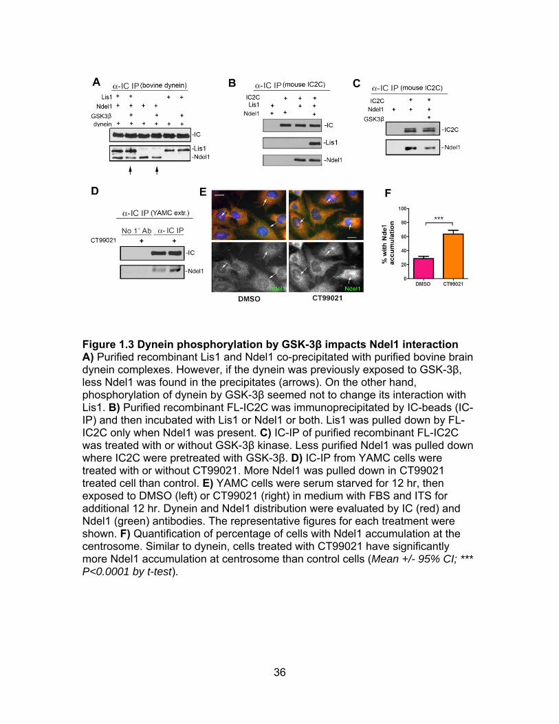
IC phosphorylation by GSK-3β impacts Ndel1 interaction: Lis1 and
Ndel1/Nde1 are dynein-binding proteins that work together to regulate dynein.
Some studies suggest that these proteins regulate force production (McKenney
et al., 2010), while other studies suggest that they regulate dynein plus end
trafficking (Roberts et al., 2014; Yamada et al., 2008) and directing dynein to
cortical sites during mitosis (Li et al., 2005; Moon et al., 2014). We previously
showed that phosphorylation of Ndel1 by cyclin dependent kinases impacted its
interaction with dynein and Lis1 (Hebbar et al., 2008b). To determine if GSK-3β-
dependent phosphorylation of dynein alters its capacity to bind to Lis1 or Ndel1,
immunoprecipitated, purified dynein was incubated with GSK-3β. After extensive
washing to remove kinase, beads were exposed to purified Lis1 and Ndel1.
Ndel1, but not Lis1, was less likely to coprecipitate with dynein if it was pre-
phosphorylated by GSK-3β (Figure 1.3A). McKenney et al (2010) reported that
Nde1, a homolog of Ndel1, stabilized the interaction of LIS1 with purified dynein
motors (McKenney et al., 2010). In support of this we found that purified FL-IC2C
was unable to pull down Lis1 unless Ndel1 was present (Figure 1.3B).
Interestingly, less Ndel1 was pulled down if FL-IC2C was first phosphorylated by
GSK-3β (Figure 1.3C).
14
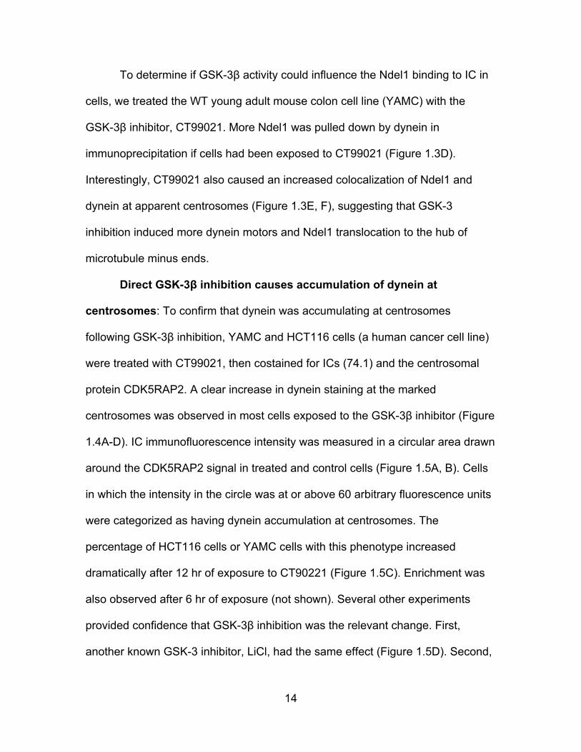
To determine if GSK-3β activity could influence the Ndel1 binding to IC in
cells, we treated the WT young adult mouse colon cell line (YAMC) with the
GSK-3β inhibitor, CT99021. More Ndel1 was pulled down by dynein in
immunoprecipitation if cells had been exposed to CT99021 (Figure 1.3D).
Interestingly, CT99021 also caused an increased colocalization of Ndel1 and
dynein at apparent centrosomes (Figure 1.3E, F), suggesting that GSK-3
inhibition induced more dynein motors and Ndel1 translocation to the hub of
microtubule minus ends.
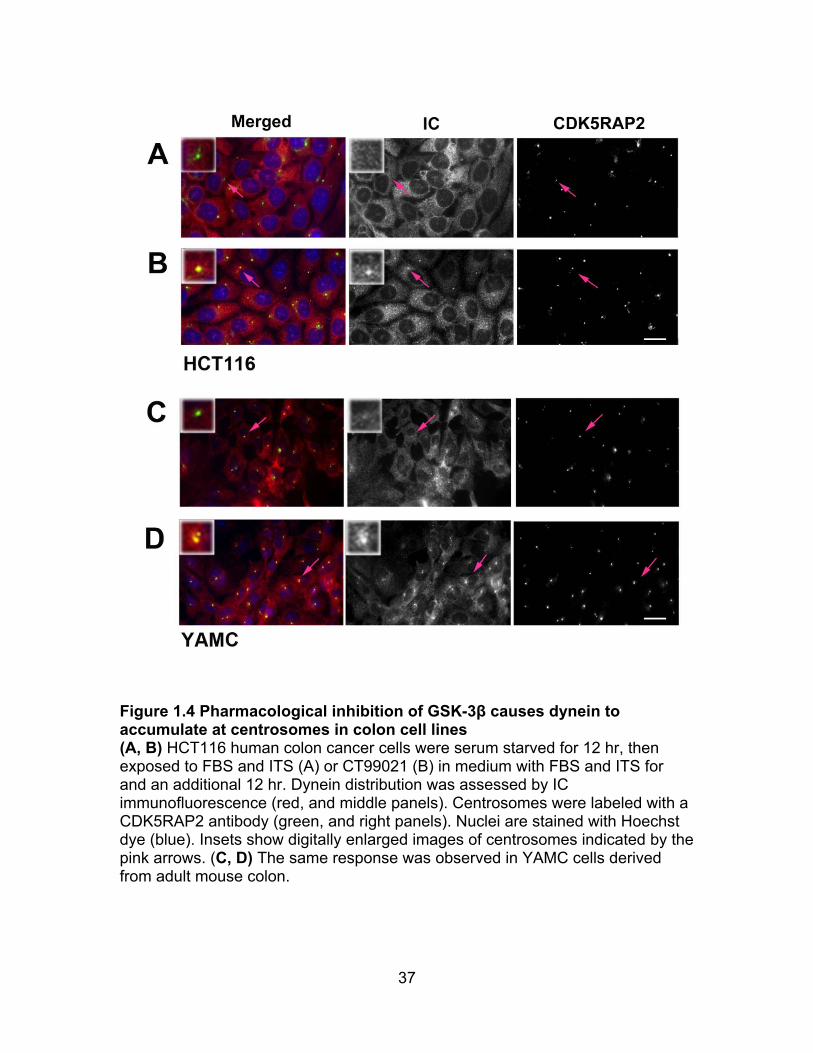
Direct GSK-3β inhibition causes accumulation of dynein at
centrosomes: To confirm that dynein was accumulating at centrosomes
following GSK-3β inhibition, YAMC and HCT116 cells (a human cancer cell line)
were treated with CT99021, then costained for ICs (74.1) and the centrosomal
protein CDK5RAP2. A clear increase in dynein staining at the marked
centrosomes was observed in most cells exposed to the GSK-3β inhibitor (Figure
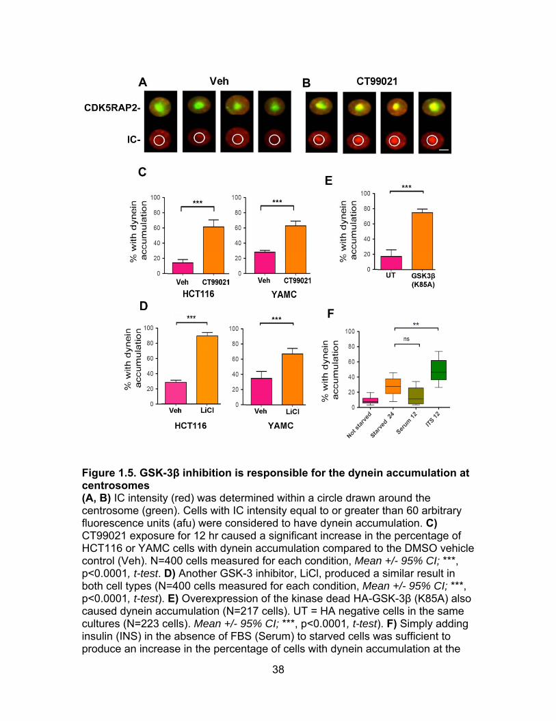
1.4A-D). IC immunofluorescence intensity was measured in a circular area drawn
around the CDK5RAP2 signal in treated and control cells (Figure 1.5A, B). Cells
in which the intensity in the circle was at or above 60 arbitrary fluorescence units
were categorized as having dynein accumulation at centrosomes. The
percentage of HCT116 cells or YAMC cells with this phenotype increased
dramatically after 12 hr of exposure to CT90221 (Figure 1.5C). Enrichment was
also observed after 6 hr of exposure (not shown). Several other experiments
provided confidence that GSK-3β inhibition was the relevant change. First,
another known GSK-3 inhibitor, LiCl, had the same effect (Figure 1.5D). Second,
15
transient expression of a dominant negative GSK-3β construct (GSK-3β K85A)
also caused the accumulation phenotype (Figure 1.5E). Finally, because GSK-3
activity is known to be inhibited by insulin, we also looked for and found the
accumulation phenotype if starved cells were exposed to a supplement
containing high levels of insulin (ITS - Figure 1.5F). Serum alone was not
sufficient to induce the change, possibly because FBS has tenfold lower insulin
concentration than the ITS supplement.
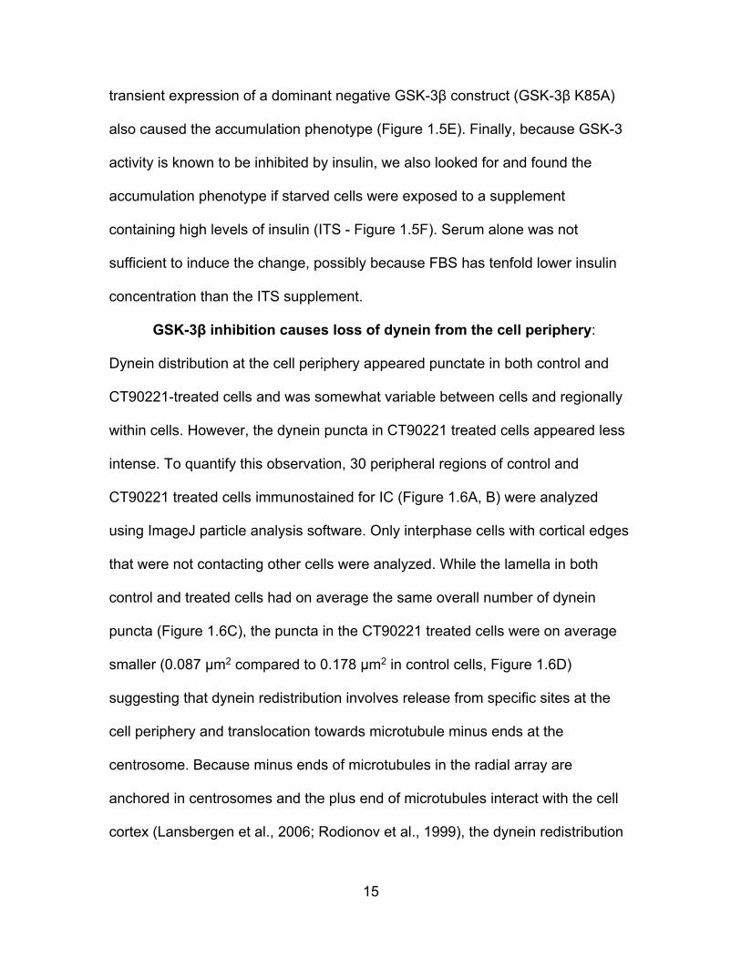
GSK-3β inhibition causes loss of dynein from the cell periphery:
Dynein distribution at the cell periphery appeared punctate in both control and
CT90221-treated cells and was somewhat variable between cells and regionally
within cells. However, the dynein puncta in CT90221 treated cells appeared less
intense. To quantify this observation, 30 peripheral regions of control and
CT90221 treated cells immunostained for IC (Figure 1.6A, B) were analyzed
using ImageJ particle analysis software. Only interphase cells with cortical edges
that were not contacting other cells were analyzed. While the lamella in both
control and treated cells had on average the same overall number of dynein
puncta (Figure 1.6C), the puncta in the CT90221 treated cells were on average
smaller (0.087 µm2 compared to 0.178 µm2 in control cells, Figure 1.6D)
suggesting that dynein redistribution involves release from specific sites at the
cell periphery and translocation towards microtubule minus ends at the
centrosome. Because minus ends of microtubules in the radial array are
anchored in centrosomes and the plus end of microtubules interact with the cell
cortex (Lansbergen et al., 2006; Rodionov et al., 1999), the dynein redistribution
16
may represent increased minus end directed movement of a pool of dynein in
response to GSK-3 inhibition.
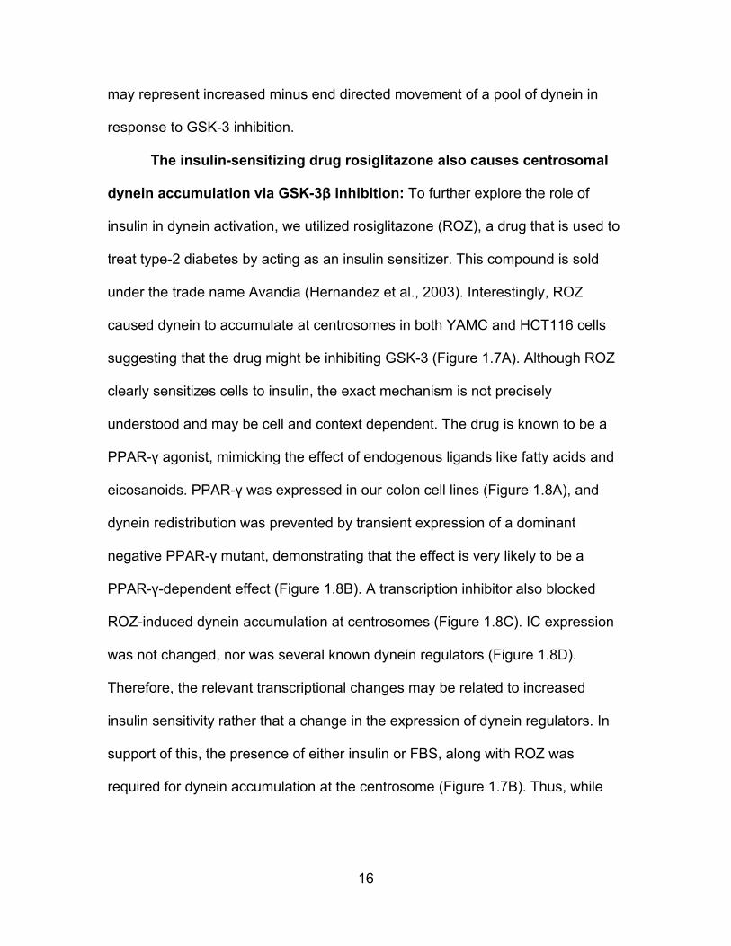
The insulin-sensitizing drug rosiglitazone also causes centrosomal
dynein accumulation via GSK-3β inhibition: To further explore the role of
insulin in dynein activation, we utilized rosiglitazone (ROZ), a drug that is used to
treat type-2 diabetes by acting as an insulin sensitizer. This compound is sold
under the trade name Avandia (Hernandez et al., 2003). Interestingly, ROZ
caused dynein to accumulate at centrosomes in both YAMC and HCT116 cells
suggesting that the drug might be inhibiting GSK-3 (Figure 1.7A). Although ROZ
clearly sensitizes cells to insulin, the exact mechanism is not precisely
understood and may be cell and context dependent. The drug is known to be a
PPAR-γ agonist, mimicking the effect of endogenous ligands like fatty acids and
eicosanoids. PPAR-γ was expressed in our colon cell lines (Figure 1.8A), and
dynein redistribution was prevented by transient expression of a dominant
negative PPAR-γ mutant, demonstrating that the effect is very likely to be a
PPAR-γ-dependent effect (Figure 1.8B). A transcription inhibitor also blocked
ROZ-induced dynein accumulation at centrosomes (Figure 1.8C). IC expression
was not changed, nor was several known dynein regulators (Figure 1.8D).
Therefore, the relevant transcriptional changes may be related to increased
insulin sensitivity rather that a change in the expression of dynein regulators. In
support of this, the presence of either insulin or FBS, along with ROZ was
required for dynein accumulation at the centrosome (Figure 1.7B). Thus, while

17
there was insufficient insulin in FBS to stimulate dynein accumulation in the
absence of ROZ, ROZ was able to sensitize cells to this amount of insulin.
PI3K/AKT signaling is involved in the dynein response to ROZ: An
early response to insulin signaling is activation of PI3K (phosphoinositide 3
kinase) (Saltiel and Pessin, 2002). Auto-phosphorylation of insulin receptors
promotes binding and phosphorylation of IRS (insulin receptor substrate family).
This leads to activation of PI3K and production of PIP3 (phosphatidylinositol (3,
4, 5)-trisphosphate) on the cytoplasmic side of the plasma membrane. PIP3
recruits AKT/PKB (protein kinase B), which is then is stimulated by other kinases
at the plasma membrane, where it phosphorylates and inactivates GSK-3β on
serine 9 (S9). This pathway is likely involved in the response to rosiglitazone
because pharmacological inhibition of PI3K by either LY294002 or Wortmannin
blocked dynein redistribution (Figure 1.7C). Also, phospho-AKT (Thr308) became
prominently localized at the plasma membrane in starved cells exposed to ROZ
in the presence of insulin and FBS (Figure 1.7D). The percentage of cells with
this staining pattern increased from ~10% to over 30% by 12 hr (Mean +/- 95%
CI; ***P<0.0001, t-test). Total phospho-AKT levels had increased in cells by 6 hr
after ROZ exposure, but were reduced by 12 hr (Figure 1.7E). ROZ also induced
S9 phosphorylation to the same extent as LiCl, further supporting the
involvement of this well-known signaling pathway (Figure 1.7F). Moreover,
centrosome accumulation of dynein was blocked by overexpression of a
constitutively active GSK-3β isoform (GSK-3β S9A), demonstrating that the
rosiglitazone response involves inactivation of GSK-3β (Figure 1.7G).
18
Interestingly, the baseline of dynein accumulation in control cells was also
significantly reduced by expression of this construct.
Dynein redistribution in response to ROZ requires its motor activity:
Because dynein normally accumulates at centrosomes as cells enter the S/G2
phase in cycling cells (Quintyne and Schroer, 2002), we were interested in
determining whether drug treatments were causing a S/G2 arrest. However, we
did not observe that rosiglitazone exposure increased the number of cells with
nuclear CENPF (not shown), which becomes detectable in late S/G2 and is
degraded after mitosis (Landberg et al., 1996; Liao et al., 1995). Also, most of the
cells with enriched centrosomal dynein were not CENPF positive (Figure 1.9A),
so S/G2 arrest was unlikely to be the underlying cause of dynein accumulation at
centrosomes. Rather, changes in dynein motility seem to be involved, as two
different manipulations known to reduce dynein-dependent transport, Lis1 RNAi
or dynactin p50 overexpression, prevented dynein accumulation (Figure 1.9B, C)
(Lam et al., 2010; McKenney et al., 2010; Mesngon et al., 2006; Smith et al.,
2000). Dynactin subunits also became enriched at the centrosome in response to
the insulin sensitizer suggesting that the dynein that moved towards centrosomes
was coupled with dynactin (Figure 1.9D, E). In support of increased dynein
motility, acidic organelles labeled with Lysotracker (late endosomes and
lysosomes) moved more rapidly inward and clustered near the nucleus (Figure
1.9F-H). Finally, ROZ increased retrograde transport of Lysotracker-labeled
organelles in adult rat DRG axons (Figure 1.9I, J).
19
DISCUSSION
A novel and direct link between GSK-3 activity and dynein motors:
This study is, to our knowledge, the first showing a direct interaction between
cytoplasmic dynein and GSK-3. We show that brain dynein and GSK-3 can exist
in a complex, and that the purified kinase can phosphorylate multiple dynein
subunits in vitro. We also demonstrate that phosphorylation impacts the
interaction of dynein with Ndel1, and show that GSK-3 activity alters dynein and
Ndel1 distribution in cells as well as retrograde transport of acidic organelles in
axons and in non-neuronal cells.
Our work adds significantly to previous studies of effects of GSK-3 on
transport. The study of squid axoplasm only reported an impact on speeds of
anterograde MBOs (Morfini et al., 2002). We used a different organelle pool
(acidic organelles) and saw primarily an impact on retrograde, presumably
dynein-driven, transport. One of the Drosophila studies did observe changes in
both retrograde and anterograde movement of a kinesin-1 cargo (APP-
associated vesicles) in larval segmental nerves but only found a small change in
anterograde movements of a kinesin-3 cargo, SVPs (Weaver et al., 2013). The
study also showed that GSK-3 influences the number of active kinesin motors
per vesicle, not the total number of motors. The authors speculated that the
change in retrograde movements of APP vesicles was caused indirectly through
kinesin-1 regulation of dynein, as has been suggested by many studies (Mallik et
al., 2013). The second study in Drosophila found that SHAGGY alterations
influenced bidirectional transport of other cargos and that expression of active
20
SHAGGY increased binding of both dynein and kinesin to light membranes
(Dolma et al., 2014). However, neither study determined which motor protein
subunits or regulatory factors were being targeted by the kinase. Thus, our
finding that GSK-3β directly phosphorylates dynein represents a significant
advance in our understanding of how this kinase could influences axon transport.
It is likely that kinesin was also being affected in our manipulations in neurons
and non-neuronal cell lines. This could contribute to the changes in dynein
distribution, but is less likely to be responsible for the changes we observed in
long-distance retrograde transport of acidic organelles in axons.
GSK-3β phosphorylation of dynein reduces its interaction with
Ndel1: In mammals there are two GSK-3 genes, GSK-3α and GSK-3β. Many of
our studies used GSK-3β specific tools. However, it is possible that GSK-3α also
interacts with dynein motors – this remains to be determined. Nonetheless, when
dynein intermediate chains are phosphorylated by GSK-3β they are less able to
interact with Ndel1, an important dynein regulator. We have reported that Ndel1
and its partner protein, Lis1, are critical for dynein dependent axon transport of
acidic organelles (Hebbar et al., 2008b; Pandey and Smith, 2011). Moreover, a
homolog of Ndel1, Nde1, modulates dynein force production based on single
molecule studies in vitro (McKenney et al., 2010). In a different study, Lis1 was
found to be important for maintaining processive movement of dynein along
microtubules (Moughamian et al., 2013). However, other studies indicate that
Lis1 and Ndel1/Nde1 are important for plus end directed transport of dynein
motors by kinesin and recruitment of dynein to plus ends (Lansbergen et al.,
21
2006; Markus et al., 2009; Moughamian et al., 2013; Roberts et al., 2014;
Yamada et al., 2008; Zhang et al.). Our current finding supports an activating role
of Ndel1 with respect to dynein, but does not rule out other functions. Moreover,
phosphorylation of Ndel1 itself is likely to contribute to distinct modes of motor
regulation (Bradshaw et al., 2013).
Mapping target sites in ICs: Both IC-1 and IC-2 can be phosphorylated
by GSK-3β. The majority of known or suspected GSK-3β substrates share a
consensus target sequence, S/TXXXSp/Tp, in which priming phosphorylation of
the downstream serine or threonine dramatically increases the capacity for
phosphorylation by GSK-3β at an S/T four residues upstream of the priming site
(Doble and Woodgett, 2003; Sutherland, 2011). There are multiple GSK-3β
consensus sequences in both ICs (9-11, depending on the isoform and the
species). Based on kinase assays, we suspect that one or more of these are
targeted by GSK-3β. Mass spectrometry of purified bovine brain ICs and ICs
precipitated from mouse brain extracts supports this, as phosphates were
detected at both priming and GSK-3β target sites in both IC isoforms (not
shown). Analysis of an in vitro phosphorylated N-terminal fragment of IC-2
expressed in bacteria suggests that a non-consensus residue may also be
targeted by the kinase. Studies are under way to determine which of these
candidate sites is bona fide and biologically relevant.
Insulin and insulin sensitizing drugs stimulate dynein: Another novel
aspect of our study is that it directly links a known insulin-signaling pathway to
dynein regulation. Insulin is a hormone that acts through insulin receptors and
22
insulin-like receptors in many tissues. In adipocytes, rosiglitazone stimulates
trafficking of a glucose transporter, GLUT4 (Velebit et al., 2011). Interestingly,
dynein-driven translocation of GLUT4 to perinuclear membranes appears to be
required for optimal GLUT 4 regulation by insulin (Huang et al., 2001). It will be
interesting to determine if GSK-3 inhibition contributes to trafficking of this
transmembrane protein, and if insulin regulates trafficking of other receptor types.
Although there are many remaining questions, our data allow us to advance a
model in which GSK-3 inhibition serves as an activating switch for dynein motors
in response to extracellular cues (Figure 1.10). In an unstimulated cell,
constitutively active GSK-3β phosphorylates dynein motors, helping to maintain a
steady-state equilibrium between phosphorylated and unphosphorylated dynein.
The phosphorylated pool of dynein is prevented from interacting with Ndel1, and
remains paused on microtubules or docked at the cell cortex (Figure 1.10A).
When the cell is stimulated by insulin, PI3K/AKT signaling shuts down GSK-3β at
the plasma membrane. This shifts the equilibrium between phosphorylated and
dephosphorylated dynein, so that Ndel1/Lis1 can "jump start” this pool of dynein
motors (Figure 9B). When cells are treated with GSK-3β inhibitors such as
CT99021 or LiCl, it is likely that GSK-3 is inhibited throughout the cell, and the
equilibrium shifts more towards active dynein motors (Figure 1.10C). This latter
may explain why we observed a greater increase in retrograde transport in axons
when GSK-3 was inhibited directly (compare Figure 1.1 to Figure 1.9I, J).
Insulin, GSK-3, dynein and human neurological diseases: There is
increasing evidence that type 2 Diabetes causes age-related dementia, and this
23
has in some cases been linked to alterations in insulin signaling (Sato and
Morishita, 2014). In fact, neurodegenerative diseases have frequently been
associated with the onset of insulin resistance – and many of these are also
associated with altered dynein-dependent transport (Eschbach and Dupuis,
2011). Our finding that the insulin sensitizing drug rosiglitazone stimulates
retrograde axonal transport in adult neurons may provide a framework from
which to understand how insulin resistance might negatively impact neuronal
function. It will be interesting to determine if dynein-dependent alterations in
receptor trafficking are also triggered in neurons by increased insulin sensitivity.
The GSK-3β inhibitor LiCl has been widely used to treat mood disorders, but
whether this involves alterations in dynein remains to be determined (Marmol,
2008). PPAR-γ agonists like rosiglitazone are being considered for therapeutic
treatments in AD primarily because of their known anti-inflammatory
characteristics (Landreth et al., 2008). Our findings concerning the impact of this
class of drugs on dynein dependent trafficking will be of interest for those and
similar studies.
GSK-3 regulation of dynein in other cell types: Organization of the
microtubule network within specific cells could play a role in where and when
cells use this GSK-3β regulatory mechanism to control cargo trafficking. In our
cultured colon cell lines microtubule plus ends tend to be positioned near the cell
periphery so that dynein activation results cargo being transported towards minus
ends near the nucleus. Other cell types may have different microtubule arrays
and cargo could be transported to other cellular locations by dynein. Also, dynein

24
functions in mitotic events that require force generation including nuclear
envelope breakdown, spindle orientation, and chromosome segregation (Barton
and Goldstein, 1996; Hebbar et al., 2008b; Kotak and Gonczy, 2013;
Raaijmakers and Medema, 2014). GSK-3 activity has been linked to many of the
same processes (Acevedo et al., 2007; Bobinnec et al., 2006; Fumoto et al.,
2008; Harwood et al., 2013; Izumi et al., 2008; Tighe et al., 2007; Wakefield et
al., 2003; Wojcik, 2008), and it remains to be determined whether GSK-3
regulation of dynein is relevant in these events. Cells in the mammalian colon
express PPAR-γ protein, so our studies indicate that rosiglitazone (and insulin)
have the potential to regulate dynein in the colon (Lefebvre et al., 1998; Su et al.,
2007). Moreover, rosiglitazone and similar drugs have been reported to be both
chemopreventive and carcinogenic in rodent models, which has garnered
substantial interest in these compounds (Lefebvre et al., 1998; Su et al., 2007).
Interestingly, there is a higher incidence of colorectal cancer among diabetics
(Larsson et al., 2005). Our studies should guide experiments that examine the
effect of these drugs on mitotic regulation and will be useful in interpreting
outcomes of pre-clinical and clinical trials in non-nervous tissues.
How are dynein regulatory mechanisms coordinated? Our studies
have begun to define a mechanism that can regulate dynein motors in response
to an increase in insulin signaling. They also raise a new set of questions. For
example, we also detected phosphate incorporation in HC and LIC subunits –
how are these events coordinated? Do phosphatases play a role in dynein
regulation? Do other post-translational modifications of dynein and its interacting

25
proteins function coordinately with GSK-3β, or are they utilized in different
cellular processes that require distinct modes of dynein regulation?
Phosphorylation of dynein by other kinases has been identified (Mitchell et al.,
2012; Pullikuth et al., 2013) and Ndel1/Nde1 phosphorylation by multiple kinases
has been reported (Bradshaw et al., 2013). For example, we found that
phosphorylation of Ndel1 by both CDK5 and CDK1, but not either alone, inhibited
Ndel1 binding to dynein and increased its interaction with Lis1 (Hebbar et al.,
2008b). How are all of these events coordinated in the cell? Finally, can other
ligands known to induce GSK-3 inhibition also stimulate dynein? How does
dynein regulation alter kinesin behavior and vice versa? Future studies from our
group and others should be able to resolve these interesting questions and shed
light on the complex regulatory mechanisms controlling these vital motor
proteins.
MATERIALS AND METHODS
Cells: Adult rat DRG neurons were prepared as described in (Pandey and
Smith, 2011) and maintained in Hamm’s F12 medium supplemented with 10%
horse serum. The human colon cancer cell line, HCT-116, was maintained in
DMEM supplemented with glutamine (2mM), 10% FBS, penicillin (100 U/ml) and
streptomycin (100 µg/ml). The murine YAMC epithelial cell line was derived from
the colonic mucosa of a transgenic mouse generated by the introduction of a
temperature sensitive, interferon inducible, SV40 T Ag, tsA58, the Immortomouse
(Whitehead et al., 1993). YAMC cells were maintained at the permissive
temperature (33°C) in RPMI 1640 media supplemented with glutamine (2mM),
26
10% FBS, penicillin (100 U/ml), streptomycin (100 µg/ml), murine gamma
interferon (5 U/ml), and 1% ITS.
Pharmaceutical reagents: The following pharmaceutical reagents were
used: GSK-3 inhibitors CT99021 (3µM, Selleck) and LiCl (10mM, Sigma-Aldrich),
The PPAR-γ agonist and insulin sensitizer, rosiglitazone (10µM, Biomol), the
transcription inhibitor, 5, 6-dichloro-1-β-D-ribofuranosylbenzimidazole DRB
(80µM, Fisher, Inc.), and the PI3K inhibitors LY294002 (10µM, Cell Signaling)
and Wortmannin (0.5µm, Biomol). Cultures exposed to vehicle alone (DMSO or
H2O) served as controls. For drug treatments, YAMC or HCT116 cells were
serum starved for 12 hr prior to exposure to drugs in full medium for an additional
12 hr (or as indicated). The starvation was designed to increase insulin receptor
trafficking to the cell surface and/or to lower exposure to natural ligands of
PPAR-γ to increase sensitivity to rosiglitazone. DRG neurons did not undergo
serum starvation, but were maintained in culture for 24 hr with 10% horse serum
prior to addition of drugs.
Expression Vectors: EGFP-C2 IC2C, PRSET-A IC2C and PRSET-A
N237 expression vectors were described previously (King et al., 2003). The
EGFP-IC1B vector was provided by K.Pfister (Univ. VA). HA-GSK-3β K85A and
HA-GSK-3β S9A expression vectors were from Addgene (plasmid ID 14755 and
14754). The p50-EGFP plasmid was provided by T.A. Schroer (Johns Hopkins).
Complementary hairpin sequences for Lis1 (1,062–1,080 bp; GAGTTGTGC-
TGATGACAAG) were synthesized and cloned into pSilencer under the control of
the U6 promoter (version 2.0; Ambion) (Pandey and Smith, 2011). The flag-
27
tagged, dominant-negative human PPAR-γ expression vector (dnPPAR-γ) was
provided by V.K. Chatterjee (Oxford Univ). This mutant retains ligand and DNA
binding, but exhibits markedly reduced transactivation and impaired corepressor
interaction, which is thought to produce the dominant negative effect (Gurnell et
al., 2000). Cells were transfected using Lipofectamine 2000 reagent (Invitrogen).
Protein purification: Bovine brain cytoplasmic dynein was purified as
described previously (Bingham et al., 1998; Culver-Hanlon et al., 2006).
Recombinant dynein and Ndel1 proteins were expressed in BL-21 cells. Cells
were grown at 37°C to an OD600 of 0.4, and then 0.1 mM isopropyl-d-
thiogalactoside (IPTG) was added to induce protein expression. Bacteria were
lysed in his-tagged protein purification binding buffer (Invitrogen) with protease
inhibitors. The cells were sonicated and pelleted by centrifugation at 10,000 g at
4°C for 30 min. Ni-NTA beads (Invitrogen) were added into the cell supernatant
and incubated at 4°C for 1 hr. The protein was washed three times and then
eluted from beads. His-tagged recombinant Lis1 was expressed in Sf9 insect
cells using a baculovirus kindly provided by A. Musacchio. His-tagged Lis1 was
purified using Ni-NTA beads.
Cell and brain extract preparation: For preparation of cell extract, cells
at 90% confluency were lysed in buffer containing 50 mM Tris (pH 7.5), 0.1% NP-
40, 100mM NaCl, 1 mM MgCl2, 5 mM EDTA, protease inhibitor cocktail (Fisher)
and Halt phosphatase inhibitor cocktail (Fisher) on ice for 30 minutes. Cell
lysates were sonicated for 10 pulses at level 1 with 10% output 3 times. The
lysates were incubated on ice for another 10 min and then centrifuged at 17,000
28
g for 20 minutes at 4°C. For preparation of mouse brain extract, brains were
quickly dissected and dounce-homogenized in the above lysis buffer. The lysates
were incubated on ice for 30 min and then centrifuged at 17,000 g for 30 minutes
at 4°C. Concentrations of extracts were determined by a BCA protein assay
(Pierce).
Protein kinase assays: The GST-GSK-3β Kinase Enzyme System and
SignalChem GST-GSK-3β or His-GSK-3β purified kinases (Promega) were used
for all kinase assays. Lambda protein phosphatase was purchased from New
England BioLabs. For some assays, purified dynein was first immobilized on 74.1
mouse monoclonal IC antibody conjugated agarose (Santa Cruz Biotechnology,
Inc). Potential substrates were incubated with 50ng GSK-3β and 0.03 µCi/µl
32P-ATP for 30 minutes at 37 °C. The reaction was stopped by the addition of
sample buffer. In one experiment brain dynein was pre-incubated with Lambda
protein phosphatase (1000 U) to remove preexisting phosphates. Some
reactions also included 3µM of the GSK-3 inhibitor CT99021. Proteins were
separated by SDS-PAGE and the wet min-gel was sealed in saran wrap and
exposed to X-ray film overnight at -80°C. After exposure gels were stained with
Coomassie brilliant blue to visualize proteins.
Immunoprecipitation and Western blot: For IPs from cell or brain
extracts, 1 µg 74.1 IC antibodies were first incubated with 30 µl Protein-A
dynabeads (Invitrogen) for 2 hr at room temperature and washed with lysis buffer
twice. The antibody conjugated dynabeads were incubated with 1 mg extracts at
4°C overnight. Dynabeads were subject to two washes of lysis buffer and then
29
two washes of PBS-T (phosphate buffered saline with 0.1% Tween 20) at 4°C.
For IPs from purified bovine brain dynein or IC2C, proteins were first incubated
with 74.1 mouse monoclonal IC antibody conjugated agarose (Santa Cruz
Biotechnology, Inc) in PBS-T with protease and phosphatase inhibitors overnight.
Beads were spun down and washed with PBS-T twice and then PHM-T buffer
(60 mM PIPES, 25 mM HEPES, 4 mM MgSO4, 0.1% Tween 20, pH 6.9). Beads
then were incubated with Lis1 or Ndel1 or both in PHM-T buffer with protease
and phosphatase inhibitors for 1 hr at RT. Beads were washed 3 times with
PHM-T buffer and eluted in 60 µl PBS plus 20 µl 6X sample buffer and boiled for
3 min before samples were loaded onto SDS-PAGE gels. For western blots,
samples were transferred to PVDF or nitrocellulose membranes and subjected to
standard protocols to identify proteins.
Antibodies: The 74.1 dynein IC mouse mAb, H100 PPAR-γ1 rabbit
polyclonal Ab, H-3 His-probe rabbit polyclonal Ab, and IRβ mouse mAb were
from Santa Cruz Biotechnology. The 3D10 GSK-3β mouse mAb, 5B3 S9
Phospho-GSK-3β rabbit mAb, 11E7 AKT rabbit mAb, D25E6 T308 pAKT rabbit
mAb and D9E S473 p-AKT rabbit mAb were from Cell Signaling, Inc. The EB1,
p150, p50, and -catenin mouse mAbs were from BD Biosciences. The
CDK5RAP2 rabbit polyclonal Ab was from Millipore. The α-tubulin mouse mAb
was from Sigma-Aldrich. The rabbit polyclonal CENPF Ab was from Novus
Biologicals. Lis1 and Ndel1 rabbit polyclonal antibodies were described
previously (Hebbar et al., 2008b; Pandey and Smith, 2011).

30
Immunofluorescence: For IC or pericentrin, cells were fixed in 100% ice-
cold methanol for 2 min. For p50, p150glued and pAKT immunofluorescence,
cells were fixed in 3% paraformaldehyde followed by permeabilization with 0.2%
Triton X-100 for 10 minutes. Nuclei were visualized using Hoechst dye (33258;
Sigma-Aldrich). Coverslips were mounted on glass slides using ProLong Gold
Antifade (Invitrogen). Cells were visualized with an Axiovert 200 inverted
microscope (Carl Zeiss, Inc.) using Plan-Neo 100×/1.30 or Plan-Apo 63×/1.40 oil-
immersion objectives (Immersol 518F; Carl Zeiss, Inc.). Optical sections were
deconvolved using AxioVision's combined iterative algorithm to obtain confocal
images if necessary. The accumulation of dynein at centrosomes was
determined by measuring the mean pixel intensity of immunofluorescence
(arbitrary fluorescence units – afu) in a fixed circular area (0.008 inches²)
encompassing the centrosome visualized by CDK5Rap2. Intensities were
determined using ImageJ software. For most experiments, dynein enrichment
was considered positive if it was greater than or equal to 60 afu (this was
typically 3 times higher than randomly selected regions of the cell). The analysis
of dynein puncta at cell periphery is described in the legend for Figure 1.6.
Analysis of acidic organelle movement in living cells: Cells were
incubated with 100 nmol Lysotracker Red (Invitrogen) for 30 min prior to imaging.
Coverslips were transferred into fresh medium containing 25 mM HEPES, pH 7.4
and OxyFluor (Oxyrase Inc.) in a water-heated custom-built microscope stage
warmed to 37°C. Cells expressing a relatively low level of Lysotracker Red were
selected for imaging. Fluorescent images were acquired every 2s for 4 min
31
(YAMC) or 2 min (DRG axons) using a Plan-Apo 63×/1.2 W/0.17 water objective.
YAMC cells: Velocities and run lengths of retrograde, minus end-directed
organelle movement (towards the nucleus) were measured using the “particle
tracking” plugin for ImageJ software. DRG axons: kymographs were generated
from time-lapse movies using NIH ImageJ software. Images were acquired in 2s
intervals for 2 minutes. The kymographs were generated such that the direction
toward the cell body was always to the right, so lines that sloped toward the right
at any point with a net displacement of >5 µm were categorized as retrograde
organelles. Lines that sloped toward the left >5 µm at any time during the
recording interval were considered anterograde organelles. Lines that zigzagged
were categorized as bidirectional, and lines that showed <5 µm lateral
displacement in any direction during the recording interval were categorized as
static.
Statistics: All analyses were carried out using GraphPad prism. In all
figures, error bars represent +/-95% CI (confidence interval). One-way ANOVA
with Tukey’s multiple comparison test or an unpaired, two-tailed student’s t-test
was used and described in each figure legend
32
Figure 1.1 Inhibition of GSK-3β stimulates retrograde transport in adult rat DRG neurons Time-lapse movies of Lysotracker-labeled organelles moving in living DRG axons exposed to GSK-3 inhibitors were converted to kymographs using NIH Image J software. (A, B) Representative kymographs for axons exposed to DMSO (A), or CT99021 (B) for 12 hr are shown. The horizontal arrow indicates the retrograde direction (towards the cell body for the 100µm axon segment). The vertical arrow indicates time (2 minutes total recording time). (C, D) Organelle movement was categorized as anterograde (ant), retrograde (retro), both, or static and the percentage of organelles per axon in each category determined. There were significantly more retrograde organelles than static organelles following CT99021 exposure (D) but not following exposure to DMSO alone (C). 16 axons in cultures from 2 different rats were analyzed for each condition. Mean +/- 95% CI; N.S.,

33
P>0.05, *** P<0.0001,one-way ANOVA). (E, F) Similar results were obtained with LiCl (Mean +/- 95% CI; N.S., P>0.05, *** P<0.0001, one-way ANOVA). (Xu Gao performed the experiments in figure 1.1 for the manuscript.)
34
Figure 1.2 Direct phosphorylation of dynein by GSK-3β A) The 74.1 IC antibody was used to immunoprecipitate dynein motors from whole mouse brain extract (IC-IP). The control was brain extract incubated with protein A beads but no 74.1 (No 1˚ Ab). FT = 10% of flow through, W = 10% of first wash, PD = pull down. GSK-3β was pulled down only if dynein was pulled down (arrow). B) Purified bovine dynein was incubated with (+) or without GST-GSK-3β (left panel) or His-GSK-3β (right panel) in a kinase assay. The SDS-PAGE gel was exposed to X-ray film (Autorad) and then stained to visualize proteins (CBB). A band the size of IC’s appeared robustly phosphorylated. Heavy chains (HC) and light intermediate chains (LIC) were also phosphorylated. Auto-phosphorylated GST-GSK-3β (~73 kDa) and His-GSK-3β (~48kDa) were also detected (red asterisk). C) EGFP-tagged IC-2C or IC-1B were transiently expressed in Cos-7 cells. EGFP alone served as a control. An anti-GFP antibody was used to isolate the proteins, then beads were exposed to GST-GSK-3β. The autoradiograph shows phosphate incorporation into both EGFP-IC-2C and EGFP-IC-1B (the bands were cut and confirmed by mass spectrometry analysis), but not in EGFP alone. The phosphorylated bands in the red box are likely endogenous ICs in the complex. Bands in the blue box may be endogenous LIC chains interacting with IC-1B. D) ICs pulled down from mouse brain extract were incubated with or without lambda phosphatase to remove endogenous phosphates. After extensive washing of beads to remove residual phosphatase,

35
immobilized dynein was subjected to the GST-GSK-3β kinase assay (Autorad). “Stripped” dynein incorporated less phosphate suggesting priming may be important. E) Full length his-tagged IC-2C (FL-IC-2C) and an N-terminal fragment (N237 IC-2C) were expressed and purified from bacteria. Both proteins were phosphorylated by GSK-3β in vitro and phosphorylation was blocked by the specific GSK-3β inhibitor, CT90221, suggesting that some sites may not require priming phosphorylation.
36
Figure 1.3 Dynein phosphorylation by GSK-3β impacts Ndel1 interaction A) Purified recombinant Lis1 and Ndel1 co-precipitated with purified bovine brain dynein complexes. However, if the dynein was previously exposed to GSK-3β, less Ndel1 was found in the precipitates (arrows). On the other hand, phosphorylation of dynein by GSK-3β seemed not to change its interaction with Lis1. B) Purified recombinant FL-IC2C was immunoprecipitated by IC-beads (IC-IP) and then incubated with Lis1 or Ndel1 or both. Lis1 was pulled down by FL-IC2C only when Ndel1 was present. C) IC-IP of purified recombinant FL-IC2C was treated with or without GSK-3β kinase. Less purified Ndel1 was pulled down where IC2C were pretreated with GSK-3β. D) IC-IP from YAMC cells were treated with or without CT99021. More Ndel1 was pulled down in CT99021 treated cell than control. E) YAMC cells were serum starved for 12 hr, then exposed to DMSO (left) or CT99021 (right) in medium with FBS and ITS for additional 12 hr. Dynein and Ndel1 distribution were evaluated by IC (red) and Ndel1 (green) antibodies. The representative figures for each treatment were shown. F) Quantification of percentage of cells with Ndel1 accumulation at the centrosome. Similar to dynein, cells treated with CT99021 have significantly more Ndel1 accumulation at centrosome than control cells (Mean +/- 95% CI; *** P<0.0001 by t-test).
37
Figure 1.4 Pharmacological inhibition of GSK-3β causes dynein to accumulate at centrosomes in colon cell lines (A, B) HCT116 human colon cancer cells were serum starved for 12 hr, then exposed to FBS and ITS (A) or CT99021 (B) in medium with FBS and ITS for and an additional 12 hr. Dynein distribution was assessed by IC immunofluorescence (red, and middle panels). Centrosomes were labeled with a CDK5RAP2 antibody (green, and right panels). Nuclei are stained with Hoechst dye (blue). Insets show digitally enlarged images of centrosomes indicated by the pink arrows. (C, D) The same response was observed in YAMC cells derived from adult mouse colon.
38
Figure 1.5. GSK-3β inhibition is responsible for the dynein accumulation at centrosomes (A, B) IC intensity (red) was determined within a circle drawn around the centrosome (green). Cells with IC intensity equal to or greater than 60 arbitrary fluorescence units (afu) were considered to have dynein accumulation. C) CT99021 exposure for 12 hr caused a significant increase in the percentage of HCT116 or YAMC cells with dynein accumulation compared to the DMSO vehicle control (Veh). N=400 cells measured for each condition, Mean +/- 95% CI; ***, p<0.0001, t-test. D) Another GSK-3 inhibitor, LiCl, produced a similar result in both cell types (N=400 cells measured for each condition, Mean +/- 95% CI; ***, p<0.0001, t-test). E) Overexpression of the kinase dead HA-GSK-3β (K85A) also caused dynein accumulation (N=217 cells). UT = HA negative cells in the same cultures (N=223 cells). Mean +/- 95% CI; ***, p<0.0001, t-test). F) Simply adding insulin (INS) in the absence of FBS (Serum) to starved cells was sufficient to produce an increase in the percentage of cells with dynein accumulation at the

39
centrosome. FBS alone did not induce accumulation. (N=100 cells measured for each condition **, p<0.001, t-test). (Xu Gao performed the experiments in figure 1.5F for the manuscript.)

40
Figure 1.6 Dynein is released from peripheral/cortical sites in response to GSK-3β inhibition A) YAMC cells were treated with or without CT99021 for 12 hr after serum starvation. Cells were fixed and immunostained with IC antibodies (Red) and Hoechst dye to label nuclei. B) 30 images of lamella of DMSO control and CT99021 treated interphase cells were acquired for each condition using the same exposure times. All of the images were adjusted together in image J using the image J threshold controls to select pixels between 150 and 200 afu, a good fit for the data. Image J was used to quantify the number and size of dynein

41
puncta. C) The average number of dynein puncta did not change (~60 per lamella). D) The average size of dynein puncta in CT99021 treated cells was significantly reduced compared to control cells (Mean +/- 95% CI; ***, P<0.0001, t-test). A total of 1852 puncta were measured in the control images and 1861 puncta in the CT99021 treated images.
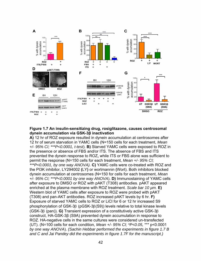
42
Figure 1.7 An insulin-sensitizing drug, rosiglitazone, causes centrosomal dynein accumulation via GSK-3β inactivation A) 12 hr of ROZ exposure resulted in dynein accumulation at centrosomes after 12 hr of serum starvation in YAMC cells (N=150 cells for each treatment, Mean +/- 95% CI; ***P<0.0001, t-test). B) Starved YAMC cells were exposed to ROZ in the presence or absence of FBS and/or ITS. The absence of FBS and ITS prevented the dynein response to ROZ, while ITS or FBS alone was sufficient to permit the response (N=150 cells for each treatment, Mean +/- 95% CI; ***P<0.0001, by one way ANOVA). C) YAMC cells were co-treated with ROZ and the PI3K inhibitor, LY294002 (LY) or wortmannin (Wort). Both inhibitors blocked dynein accumulation at centrosomes (N=150 for cells for each treatment, Mean +/- 95% CI; ***P<0.0001 by one way ANOVA). D) Immunostaining of YAMC cells after exposure to DMSO or ROZ with pAKT (T308) antibodies. pAKT appeared enriched at the plasma membrane with ROZ treatment. Scale bar 10 µm. E) Western blot of YAMC cells after exposure to ROZ were probed with pAKT (T308) and pan-AKT antibodies. ROZ increased pAKT levels by 6 hr. F) Exposure of starved YAMC cells to ROZ or LiCl for 6 or 12 hr increased S9 phosphorylation of GSK-3 (pGSK-3(S9)) levels relative to total kinase levels (GSK-3 (pan)). G) Transient expression of a constitutively active GSK-3 construct, HA-GSK-3β (S9A) prevented dynein accumulation in response to ROZ. HA-negative cells in the same cultures were considered un-transfected (UT). (N=100 cells for each condition, Mean +/- 95% CI; *P<0.05, *** p<0.0001 by one way ANOVA). (Sachin Hebbar performed the experiments in figure 1.7 B and C and Jai Pandey did the experiments in figure 1.7F for the manuscript.)
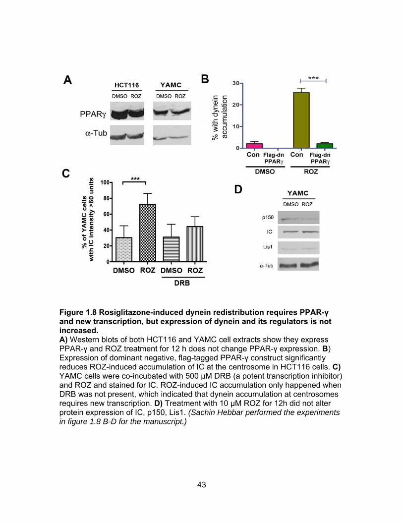
43
Figure 1.8 Rosiglitazone-induced dynein redistribution requires PPAR-γ and new transcription, but expression of dynein and its regulators is not increased. A) Western blots of both HCT116 and YAMC cell extracts show they express PPAR-γ and ROZ treatment for 12 h does not change PPAR-γ expression. B) Expression of dominant negative, flag-tagged PPAR-γ construct significantly reduces ROZ-induced accumulation of IC at the centrosome in HCT116 cells. C) YAMC cells were co-incubated with 500 μM DRB (a potent transcription inhibitor) and ROZ and stained for IC. ROZ-induced IC accumulation only happened when DRB was not present, which indicated that dynein accumulation at centrosomes requires new transcription. D) Treatment with 10 μM ROZ for 12h did not alter protein expression of IC, p150, Lis1. (Sachin Hebbar performed the experiments in figure 1.8 B-D for the manuscript.)
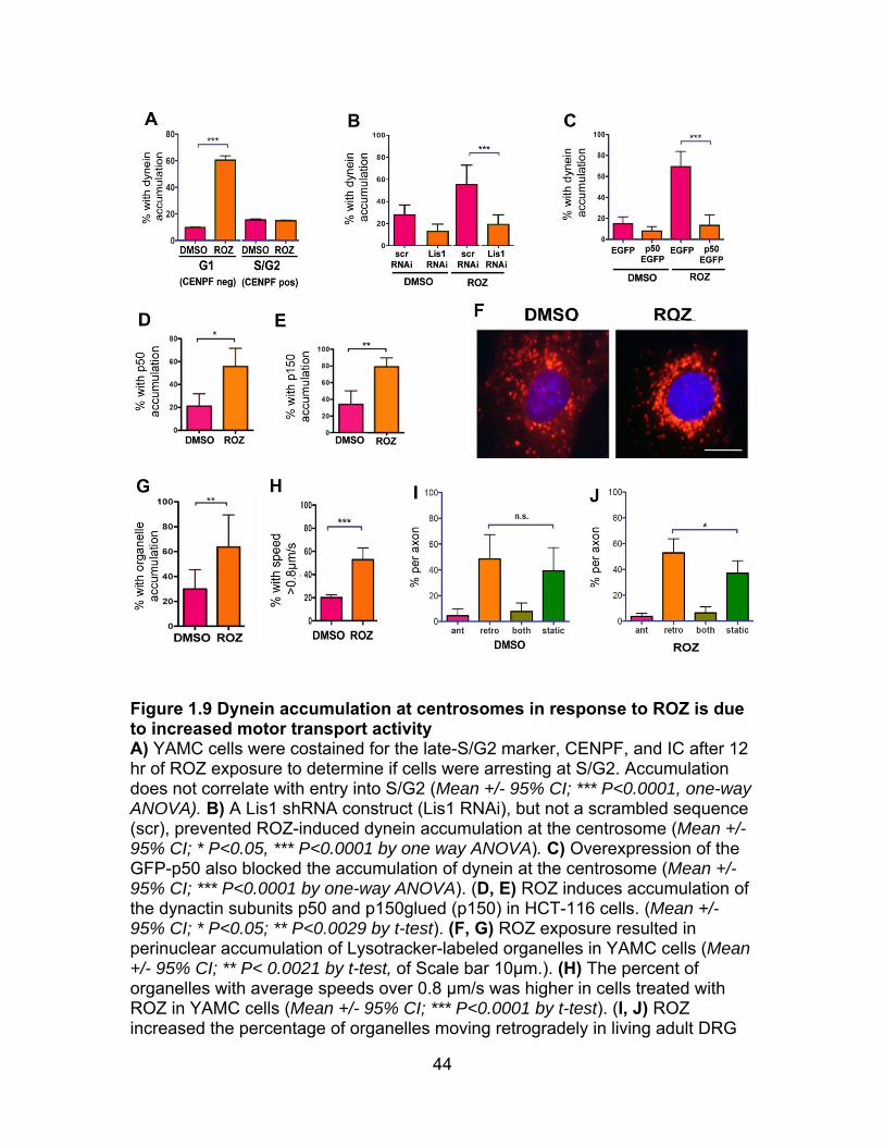
44
Figure 1.9 Dynein accumulation at centrosomes in response to ROZ is due to increased motor transport activity A) YAMC cells were costained for the late-S/G2 marker, CENPF, and IC after 12 hr of ROZ exposure to determine if cells were arresting at S/G2. Accumulation does not correlate with entry into S/G2 (Mean +/- 95% CI; *** P<0.0001, one-way ANOVA). B) A Lis1 shRNA construct (Lis1 RNAi), but not a scrambled sequence (scr), prevented ROZ-induced dynein accumulation at the centrosome (Mean +/- 95% CI; * P<0.05, *** P<0.0001 by one way ANOVA). C) Overexpression of the GFP-p50 also blocked the accumulation of dynein at the centrosome (Mean +/- 95% CI; *** P<0.0001 by one-way ANOVA). (D, E) ROZ induces accumulation of the dynactin subunits p50 and p150glued (p150) in HCT-116 cells. (Mean +/- 95% CI; * P<0.05; ** P<0.0029 by t-test). (F, G) ROZ exposure resulted in perinuclear accumulation of Lysotracker-labeled organelles in YAMC cells (Mean +/- 95% CI; ** P< 0.0021 by t-test, of Scale bar 10µm.). (H) The percent of organelles with average speeds over 0.8 µm/s was higher in cells treated with ROZ in YAMC cells (Mean +/- 95% CI; *** P<0.0001 by t-test). (I, J) ROZ increased the percentage of organelles moving retrogradely in living adult DRG

45
axons. The neurons were not serum starved prior to drug exposure (Mean +/-
95% CI; N.S., P>0.05, * P<0.05,one-way ANOVA).(Sachin Hebbar performed the experiments in figure 1.9 B-E, Jai Pandey performed the experiments in figure 1.9 F-H, and Xu Gao performed experiments in figure 1.9 A,I, and J for the manuscript.)

46
Figure 1.10 A model for GSK-3β- dependent regulation of dynein dependent retrograde organelle transport A) In an unstimulated "resting" cell, GSK-3β is active and phosphorylates dynein. Phosphorylated dynein is not motile, and remains at microtubule plus ends and/or the cell cortex. B) When the cell is stimulated by insulin, PI3K/AKT signaling is initiated and GSK-3β is shut off. As cortical dynein loses phosphate at GSK-3 sites, or is replaced with unphosphorylated dynein, it interacts more efficiently with Ndel1/Lis1. Ndel1/Lis1 can then "jump start" dynein, leading to increased retrograde transport of cargo. C) Inhibition of GSK-3β by pharmacological inhibitors such as CT99021 (CT) or LiCl stimulates dynein-dependent retrograde organelle transport, bypassing the need for the signal transduction pathway.
47
CHAPTER 2
Adenomatous polyposis coli (APC) is a novel multifaceted dynein regulator: the concerted interplay is vital for the effect of thiazolidinediones
ABSTRACT
There is considerable debate over whether thiazolidinediones, PPAR-γ
agonists, are chemopreventive or carcinogenic during the development of
colorectal cancer, where mutations in adenomatous polyposis coli (APC) often
occur. We find that dynein and APC physically interact with each other by
reciprocal co-immunoprecipitation assays. The interaction is positively regulated
by GSK-3β, which can be inhibited by rosiglitazone through activation of
PI3K/AKT pathway as shown in the chapter 1. In both cell lines and mouse
models, we demonstrate that rosiglitazone increases dynein activity and cell
migration in wild type (WT) cells but not in Apc (min/+) cells, and that
rosiglitazone induces spindle misorientation in Apc (min/+) cells but not in WT
cells. We provide evidence that this involves different PI3K/AKT/GSK-3β
signaling responses to rosiglitazone between WT and Apc (min/+) cells, and that
ApcMin mutation negatively regulates the interaction of dynein to WT APC. Our
discovery of the interplay of APC and dynein provides a new insight into how
PPAR-γ signaling regulates cancer development and other cellular events.2
Feng J. Gao1, Sachin Hebbar2, Deanna S. Smith1*. To be submitted to Journal of Cell Biology (Feng J Gao and Sachin Hebbar are co-first authors)
48
INTRODUCTION
Adenomatous Polyposis Coli (APC) is well known for playing a central role
in Wnt signaling, where a “destruction complex” including APC, Axin, GSK-3β,
and casein kinase 1 (CK1) and the E3-ubiquitin ligase β-TrCP is formed (Polakis,
2002; Stamos and Weis, 2013). The complex carries out the phosphorylation and
ubiquitination on β-catenin, which is subsequently degraded by the proteasome.
Loss function of APC via mutation and aberrant Wnt signaling result in the
deregulation of β-catenin (Kongkanuntn et al., 1999; Phelps et al., 2009; Sansom
et al., 2004), which is the critical event in carcinogenesis (Espada et al., 2009).
Although there is increasing evidence supporting APC as a tumor suppressor,
the mechanism is still incomplete. Moreover, a large body of research implicates
non-traditional roles of APC including cell migration, spindle assembly, cell
adhesion, chromosome segregation, and neuronal differentiation (Hanson and
Miller, 2005).
The Apc (min/+) mouse, which harbors a truncating APC mutation, is a
prominent animal model to study human colorectal cancer (Taketo and
Edelmann, 2009; Yamada and Mori, 2007). Many studies indicate that
rosiglitazone, a peroxisome proliferator-activated receptor gamma (PPAR-γ)
agonist, prevents colon carcinogenesis (Burgermeister et al., 2003; Chang et al.,
2012; Friedrich et al., 2013; Marin et al., 2006). However, several reports indicate
that the drug is carcinogenic in Apc (min/+) mice (Lefebvre et al., 1998; Peters et
al., 2012; Saez et al., 1998). It is not clear how PPAR-γ signaling affects
carcinogenesis in the presence of truncating APC mutations, and most studies
49
have focused on its role in β-catenin regulation. However, an interesting
relationship between APC and the microtubule (MT) cytoskeleton has been
emerging over the last decade (Etienne-Manneville, 2010). Our early report
showed a link between APC and Lis1, a dynein regulator (Hebbar et al., 2008a).
More recently, we demonstrated that rosiglitazone induced dynein activity though
the activation of PI3K/AKT/GSK-3β as shown in the chapter 1. Both dynein and
APC are substrates of GSK-3β (Ferrarese et al., 2007) and well known for
participating in fundamental cellular processes including cell migration, mitosis,
and differentiation. Here, we characterized the interaction of dynein with APC
and how the PPAR-γ signaling regulates the interaction in both WT and Apc
(min/+) cells.
Spindle orientation is important in normal intestinal turnover as stem cells
dividing in intestinal crypts replenish enterocytes that migrate to the tips of villi
before being shed into the intestinal lumen(Reya and Clevers, 2005). The
ApcMin mutation causes a modest defect in spindle orientation in mouse
intestinal stem cells, which is further exacerbated in the tumors that developed in
these mice (Fleming et al., 2009). Defective spindle orientation was also reported
in precancerous tissue heterozygous for Apc in mice (Quyn et al., 2010). Dynein
and its regulators also have a profound impact on spindle orientation (Dujardin
and Vallee, 2002; Yingling et al., 2008). It has been reported that the interactions
of “MT plus end proteins” including APC, dynein and their associate proteins may
be important to stabilize spindle orientation (Mimori-Kiyosue and Tsukita, 2003).
Therefore, if rosiglitazone alters these interactions, different spindle orientation
50
responses could occur between WT and Apc (min/+) cells, which could lead to
different cell fate decisions.
Cell migration is a complicated and highly integrated process that
participates in many biological functions including embryogenesis, immune
responses, wound healing and cancer development (Lauffenburger and Horwitz,
1996; Ridley et al., 2003). A change in the movement of cells away from crypts
could affect cell fate decisions and increase or reduce the time that enterocytes
are exposed to mutagenic substances in the gut (Nathke, 2004). Precise
regulation of signaling events and protein-protein interactions is required for cell
migration. The PI3K/AKT pathway is essential for cell migration (Seo et al.,
2014). APC/MT/EB1 interactions has been linked to cell migration in a range of
cell types (Etienne-Manneville, 2009). Dynein and its regulators are critical for
cell migration, controlling nucleokinesis and the interaction between the nucleus
and the MT-organizing center (MTOC) (Ayala et al., 2007; Tsai et al., 2007; Tsai
and Gleeson, 2005). Here, we tested if the effect of rosiglitazone on cell
migration in both WT and Apc (min/+) cells.
Our findings provide, for the first time, evidence that APC is a novel
multifaceted dynein regulator. Specifically, we discovered that dynein, APC and
GSK3β physically interact with each other, and the interaction of dynein to APC
is positively regulated by GSK3β; we also demonstrated that rosiglitazone
impacts dynein activity, spindle orientation, and cell migration in an APC
dependent manner; we report that ApcMin mutation affect PI3K/AKT/GSK3β

51
signaling in response to rosiglitazone and negatively regulates the interaction
between dynein and WT APC.
RESULTS
Dynein, APC and GSK-3β physically interact with each other: we
recently showed that GSK-3β directly phosphorylated and regulated dynein
activity (chapter 1). APC is a known substrate of GSK-3β and forms complex with
GSK-3β (Ikeda et al., 2000). In order to study the potential interplay of dynein
and APC, we first tested if dynein, APC and GSK-3β physically interacted with
each other. APC is a large protein with 2843 amino acids (aa) (Figure 2.1A).
ApcMin, a truncation mutant, is due to an early termination at amino acid 850 of
Apc (Su et al., 1992). A large disordered c-terminal region of APC (over 2000 aa)
which is not stable and carries many missense mutations, is responsible for the
interaction with many other proteins(Minde et al., 2011). We used APC-M2 pAb
raised against the region of 15-aa repeats on APC to detect full length APC
protein (FL-APC) (Wang et al., 2009). The antibody recognized FL-APC in both
WT and Apc (min/+) mouse brain extracts. The level of FL-APC detected in WT
brain was approximately two-fold greater than that in Apc (min/+) brain, while
dynein intermediate chain (IC) expression was about the same in both (Figure
2.1B). Immunoprecipitation (IP) from WT mouse brain extract by an IC antibody
showed that both FL-APC and GSK-3β were pulled down by dynein (Figure
2.1C). Reciprocal IP using anti-APC antibody corroborated these findings with
both dynein and GSK-3β pulling down with APC (Figure 2.1D). Taken together,

52
these results support the hypothesis that APC, dynein and GSK-3β may be part
of the same complex in mouse brain.
GSK-3β positively regulates the interaction of dynein and APC: APC
is transported along MT by kinesin and accumulates at plus end of MT, and c-
terminal of APC is important for this event (Jimbo et al., 2002; Mimori-Kiyosue et
al., 2000; Mogensen et al., 2002). The Intracellular distribution of APC and
dynein are important for their proper functions including MT-dependent organelle
transport, spindle orientation and cell migration. Therefore, the regulation of the
interaction between dynein and APC could be essential. We found that
phosphorylation of purified dynein by GSK-3β increased the interaction of dynein
with bacterial expressed c-fragment of APC protein in vitro (Figure 2.2A). YAMC
cells were derived from WT mice and IMCE cells were derived from Apc (min/+)
mice with the same genetic background (Whitehead and Joseph, 1994;
Whitehead et al., 1993). Only IMCE cells carry the ApcMin mutation (Figure
2.3A). Similar to what we had observed in tissues, the APC-M2 pAb detected FL-
APC protein in both YAMC and IMCE cells (Figure 2.2B). YAMC cells treated
with CT99021, a potent GSK-3β inhibitor, showed reduced interaction between
dynein and FL-APC by co-IP compared to DMSO (Figure 2.2C). Inhibition of
GSK-3β by CT99021 also significantly deceased the colocolization of dynein and
APC at the cell periphery by immunostaining (Figure 2.2D, E). These findings
suggest that GSK-3β could regulate APC and dynein functions by regulating their
interactions with one another.
53
The ApcMin mutation prevents dynein activation in response to
rosiglitazone: Our recent study showed that rosiglitazone induced dynein
activity through activation of PI3K/AKT/GSK-3β signaling. In order to study the
interplay of PPAR-γ signaling and the dynein/APC complex, we first examined
dynein activity in response to rosiglitazone in both WT and Apc (min/+) cells,
since both YAMC and IMCE cells express PPAR-γ receptors (Figure 2.3B).
Interestingly, unlike the effect on dynein distribution in YAMC cells, rosiglitazone
was not able induce dynein accumulation at the centrosome in IMCE cells
(Figure 2.4A, B). Moreover, transient expression of a truncated N-terminal APC
fragment (APCN746) completely blocked the dynein response to rosiglitazone in
HCT116 cells, indicating that the truncated isoform can act in a dominant
negative capacity (Figure 2.4C). MT minus-ends are enriched at the apical
surface of intestinal epithelial cells (Sansom et al., 2004). If dynein motility
towards minus ends was stimulated by rosiglitazone, one might expect to see
dynein accumulation in this region. This was in fact observed in sections from
WT mice, and the phenotype was most pronounced in intestinal crypts (Figure
2.4D). This change in dynein distribution was not observed in sections from Apc
(min/+) mice (Figure 2.4D). Together, the findings suggest that acute exposure to
rosiglitazone exerts similar effects on dynein activity in both cultured cells and
mouse intestine, and that APC status plays an important role in how the dynein
responds to the drug.
The ApcMin mutation alters spindle orientation in response to
rosiglitazone: To further characterize how PPAR-γ signaling may regulate the
54
APC-dynein complex, we examined the effect of rosiglitazone on spindle
orientation in both WT and Apc (min/+) cells. To examine spindle formation,
YAMC and IMCE cells were exposed to rosiglitazone and immunostained with an
α-tubulin antibody. In control YAMC and IMCE cells, the majority of spindles were
oriented parallel to the coverslip, with a small percentage oriented in a non-
parallel fashion that only allowed a single spindle pole to be observed in a given
focal plane. Examples of parallel and nonparallel spindles are shown in Figure
2.5A. In YAMC cells the percentage of parallel spindles was not significantly
altered by rosiglitazone treatment (Figure 2.5B). However, rosiglitazone
significantly increased the percentage of non-parallel spindles in IMCE cells
(Figure 2.5B).
We next determined whether rosiglitazone also has a similar effect on the
spindle orientation of intestinal crypt stem cells in Apc (min/+) mice. Specifically,
WT and Apc (min/+) mice were administered rosiglitazone or DMSO via oral
gavage for six days. Spindles in individual crypt sections were positioned with
varying angles with respect to the apical surface (Figure 2.5C). Spindle angles
were determined as shown in Figure 2.5D. Only spindles in which both spindle
poles were visible were analyzed for spindle angle (the percentage with only one
spindle pole visible was not significantly different between the WT and Apc
(min/+) mice, and was not significantly altered by drug exposure). In WT crypts,
the spindle was often nearly parallel to the apical surface. In WT crypts, fewer
than 15% were categorized as near perpendicular (Figure 2.5E). Apc (min/+)
crypts had significantly more near perpendicular spindles. Exposure to
55
rosiglitazone essentially randomized spindle orientation so that ~50% were
categorized as “near perpendicular”. This is consistent with our cell culture data,
suggesting that inappropriate PPAR-γ signaling in the presence of the ApcMin
mutation causes defects in spindle dynamics.
The ApcMin mutation prevents rosiglitazone-induced cell migration:
We next examined to see if the impact of rosiglitazone on cell migration is APC
dependent. Changes in the migratory capacity of YAMC and IMCE cells treated
with rosiglitazone were evaluated by wound-healing assay. Confluent cells were
exposed to the drug for 12 hr prior to wounding. The average distance cells
migrated into the wound was determined 24 hr later (Figure 2.6A). Treatment of
YAMC cells with rosiglitazone significantly increased the distance migrated
compared to controls (Figure 2.6B). In contrast, rosiglitazone did not stimulate
migration of IMCE cells.
The movement of enterocytes towards the intestinal lumen is a form of
sheet migration that causes complete turnover of the intestinal epithelium in a
matter of days (Reya and Clevers, 2005). We examined enterocyte migration by
monitoring the position of BrdU positive cells 24 hr after BrdU injection in WT and
Apc (min/+) mice (Figure 2.6C-F). Rosiglitazone induced a significant shift in the
position of BrdU positive cells towards the intestinal lumen in WT animals (Figure
2.6E). However, this was not the case for Apc (min/+) animals (Figure 2.6F),
which is consistent with the wound healing assay in cell lines.
Together these data indicate that stimulating PPAR-γ by rosiglitazone
promotes migration of WT cells, which is prevented by the ApcMin mutation.
56
The ApcMin mutation alters PI3K/AKT/GSK-3β signaling, which is
essential for the rosiglitazone response: In order to determine why
rosiglitazone impacts dynein activity, spindle orientation, and cell migration in an
APC-dependent manner, we examined the effect of the drug on PI3K/AKT/GSK-
3β signaling in both WT and APC (min/+) cells. GSK-3β activity is important for
the regulation of both APC and dynein. As we showed previously, treatment of
YAMC cells for 12 hr with rosiglitazone significantly inhibited GSK-3β as reflected
by increased S9 phosphorylation (Figure 2.7A, B). Surprisingly, rosiglitazone did
not increase S9 phosphorylation of GSK-3β in IMCE cells. We further tested
activities of upstream kinases in response to rosiglitazone. YAMC and IMCE cells
were exposed to rosiglitazone for 0, 0.5, 2, 6 or 12 hr after serum starvation.
There were significant differences in PI3K/AKT/GSK-3β signaling between the
two cell lines when cells were treated with rosiglitazone for 6 and 12 hr (Figure
2.7C, D). Rosiglitazone did not affect P110α expression in YAMC, but
significantly reduced P110α in IMCE at 6 and 12 hr. YAMC but not IMCE cells
continued keeping high level of AKT phosphorylation after the serum return for 6
or 12 hr. Rosiglitazone increased pAKT (T308) in YAMC at 6 hr, while it reduced
pAKT (T308) in IMCE at 6 hr. Rosiglitazone significantly increased S9
phosphorylation of GSK-3β in YAMC and slightly reduced that in IMCE at 6 or 12
hr.
GSK-3β inhibition significantly rescues dynein activation in ApcMin
mutation, and AKT inhibition prevents rosiglitazone-induced cell migration:
To test if the failure of signal transduction from rosiglitazone to PI3K/AKT/ GSK-
57
3β is the reason for different dynein and cell migration responses to rosiglitazone
in cells with ApcMin mutation, we first examined if direct inhibition of GSK-3β
increased dynein accumulation at the centrosome in IMCE. Unlike rosiglitazone,
CT99021 significantly increased dynein accumulation at the centrosomes in both
YAMC and IMCE (Figure 2.8A, B). We previously showed that the inhibition of
PI3K prevented dynein activation in response to rosiglitazone in YAMC. Here, we
tested if PI3K inhibition also prevented rosiglitazone-induced cell migration in
YAMC. YAMC and IMCE cells were exposed to both ROZ and an LY294004, a
potent AKT inhibitor. Cell migration was analyzed by wound healing assay. The
rosiglitazone-induced cell migration was totally vanished by LY294004 in YAMC
(Figure 2.8C). Moreover, the direct inhibition of PI3K reduced cell migration in
both cell types (Figure 2.8C, D).
The ApcMin mutation disrupts the interaction between dynein and
WT APC: When cells are treated with CT99021, almost all of GSK-3β are
inhibited. However, IMCE still have less dynein accumulation at the centrosome
than YAMC in the treatment of CT99021 (Figure 2.8B). Therefore, other than
changing PI3K/ AKT/ GSK-3β signaling, there could be other ways by which
ApcMin mutation affects dynein activity. One of them could be through changing
the interaction of dynein and WT APC as GSK-3β does in Figure 2.2. Co-IP
experiments showed that much less FL-APC was pulled down by dynein in IMCE
cells than that in YAMC (Figure 2.9A). Similarly, less FL-APC and dynein were
pulled down by each other in APC (min/+) than that in WT mouse brain shown in
co-IP (Figure 2.9B). One obvious explanation is that there are less FL-APC in
58
Apc (Min/+) cells than that in WT cells (Figure 2.2B). However, ApcMin may also
have a dominant negative effect on the interaction of dynein with WT APC. To
test the hypothesis, we quantified the FL-APC pulled down ratio of WT to Apc
(min/+) from mouse brain extract using IC-IP (Figure 2.9C, D). The mean of the
ratio was 7, while FL-APC in WT brain is only about 2 times higher than that in
min/+ brain as shown in figure 2.1B.
DISCUSSION
The dynein and APC interaction: we report here for the first time that
dynein and APC physically interact with each other as accessed by co-IP. We
also demonstrate the interaction is positively regulated by GSK-3β and negatively
affected by the ApcMin mutation. First, this interaction could be very important for
the intracellular distribution of APC and dynein. Their special functions make their
distribution even more important. As a molecular motor, dynein could transport
APC to different cellular locations, while APC, as a MT associate protein, may act
as an anchor for dynein controlling its motility on MT. Second, the interaction
may be important for the post-translational modifications of dynein and APC.
Dynein is able to transport retrograde signaling (Heerssen et al., 2004; Zweifel et
al., 2005), which may induce APC post-translational modifications (PTMs). On
the other hand, APC may act as a scaffold protein to recruit other signaling
molecules capable of modifying dynein subunits and its regulators such as
dynactin, Lis1 or Ndel1. The discovery of the role of GSK-3β in the regulation of
the dynein and APC interaction is a good example of these events. In the future,
we will study if dynein is able to change the intracellular distribution of APC, and
59
if the interaction of dynein with APC could affect their PTMs both in vitro and in
vivo. The result that the ApcMin mutation disrupts the dynein-APC interaction
provides a new angle to study the mechanism of carcinogenesis. It will be
interesting to map the dynein and APC interaction in tissues from different stages
of colon cancer.
The dance of rosiglitazone with the APC/dynein complex in cellular
events: here, we also report that rosiglitazone impacts dynein activity, spindle
orientation, and cell migration in an APC-dependent manner. Our previous study
showed that rosiglitazone induced dynein activity, and one of the phenotypes
was that it increased dynein accumulation at the centrosome. Here, we find that
acute exposure to rosiglitazone induces a significant increase in the percentage
of cells with dynein accumulation at the centrosome in both cultured cells and
mouse intestine, and that the ApcMin mutation blocks dynein response to the
drug.
The effect of rosiglitazone on dynein activity is likely to be important for the
increased rate of migration observed in both cultured cells and intestinal
enterocytes in response to the drug. Dynein has been implicated in cell migration
in developing nervous system and in many cultured cell lines (Schmoranzer et
al., 2009; Vallee et al., 2009). Intriguingly, the presence of truncated APC
prevents rosiglitazone-induced cell migration rates in both cultures cells and the
intestine. Another interesting finding is that rosiglitazone influences the spindle
orientation in cultured cells and in the mouse intestine only when the ApcMin is
present. Even in the absence of the drug, Apc (min/+) cells have more

60
abnormally oriented spindles. Both cell migration and spindle orientation play
essential roles in cell-fate decision. The underlying molecular explanation for this
is not clear, but our studies open up the possibility that the proper interaction
between APC and dynein is necessary for maintaining the proper dynein activity,
cell migration and spindle formation in response to signaling events.
Different rosiglitazone signaling in WT and Apc (min/+) cells: We
previously reported that rosiglitazone-induced dynein activation requires the
intact PI3K/AKT/GSK-3β signaling. To answer the question of why rosiglitazone
affects dynein activity, spindle orientation, and cell migration in an APC status
dependent manner, we compared PI3K/AKT/GSK-3β signaling in WT and Apc
(min/+) cells. There are significant differences in PI3K/AKT/GSK-3β signaling
between the two cell lines in response to rosiglitazone.
In PI3K/AKT pathway, rosiglitazone has no visible effect on PI3K
expression in YAMC cells, but significantly reduces PI3K in IMCE cells after 6 hr.
IMCE cells also display significantly lower levels of phospho-AKT as compared to
YAMC cells after 6 hr return of serum, and rosiglitazone increases pAKT levels in
YAMC cells but not in IMCE cells at 6 hr. As active AKT promotes cell migration
(Kim et al., 2001), which may explain why we found that rosiglitazone does not
induce cell migration in Apc (min/+) cells. We further support this by showing that
the inhibition of PI3K prevented rosiglitazone-induced cell migration in WT cells.
PI3K/AKT is enhanced in many human cancer types and regarded as an
attractive therapeutic target (Carnero and Paramio, 2014; Pal and Mandal, 2012).
Our report that rosiglitazone reduces PI3K/AKT activity in IMCE supports the
61
theory that it may act as a chemopreventive drug in patients with truncating APC
mutations, though this will require further evaluation.
We also found that rosiglitazone only inhibits GSK-3β in YAMC and not in
IMCE cells as accessed by western blot. As the inhibition of GSK-3β increased
dynein dependent transport and accumulation at the centrosome, the failure of
rosiglitazone-induced GSK-3β inhibition could be a reason why the ApcMin
mutation prevents dynein activation by rosiglitazone. We further support this by
showing that CT99021, a potent GSK-3β inhibitor, significantly rescued dynein
activation in IMCE cells. Our study shows that the PI3K/AKT/GSK-3β response to
rosiglitazone is a key difference between WT cells and cells with ApcMin
mutation, and also provides a linkage of dynein activation to cell migration.
Future study is needed to determine if rosiglitazone has a similar effect on
PI3K/AKT/GSK-3β in other APC mutation models.
Thiazolidinediones: chemopreventive or carcinogenic? This question
cannot have a simple answer. Our study shows that rosiglitazone misorients the
spindle in Apc (min/+) cells, which is generally regarded as carcinogenic
property. However, we also demonstrate that rosiglitazone reduces PI3K/AKT
activity in Apc (min/+) cells, which can be viewed as chemopreventive. Therefore,
we think that whether it is chemopreventive or carcinogenic in cells with ApcMin
mutation is cell type dependent. For those cell types undergoing mitosis and
differentiation, rosiglitazone may be carcinogenic by changing their fates. For
undivided or undifferentiated cells, the rosiglitazone may be chemopreventive by
inhibiting PI3K/AKT pathway. Even for the same type of cells, in different stages

62
of development rosiglitazone may have different effects, depending on who has a
dominant effect, spindle orientation or PI3K/AKT signaling. In the future, it will be
interesting to characterize the effect of rosiglitazone in other Apc (min/+) cell
types.
MATERIALS AND METHODS
Cell lines: The murine YAMC epithelial cell line was derived from the
colonic mucosa of a transgenic mouse generated by the introduction of a
temperature sensitive, interferon inducible, SV40 T Ag, tsA58 (Immortomouse)
(Whitehead et al., 1993). The murine IMCE colon epithelial cell line was derived
from the progeny of a cross between the Immortomouse strain and the Apc
(min/+) mouse strain (Whitehead and Joseph, 1994). YAMC and IMCE cells were
maintained at the permissive temperature (33°C) in RPMI 1640 media
supplemented with glutamine (2mM), 10% FBS, penicillin (0.5 U/ml) and
streptomycin (100 μg/ml), murine gamma interferon (5 U/ml), and 1% ITS
(insulin, transferrin and selenium; Cellgro, Inc). The human colon cancer cell line,
HCT-116, and Cos-7 cells were maintained in DMEM supplemented with
glutamine (2mM), 10% FBS, penicillin (0.5U/ml) and streptomycin (100 μg/ml).
Animals and genotyping: C57BL6/J Apc (min/+) mice, obtained from
Jackson Labs, were maintained in the Mouse Core Facility of the Center for
Colon Cancer Research at the University of South Carolina. These mice carry a
mutation at codon 850 in one allele of the Apc gene that encodes a truncated
version of APC (Pellman, 2001). Apc (min/+) males were bred with C57BL6/J
63
wild type females and heterozygous progeny were genotyped by PCR analysis of
tail genomic DNA using allele specific primers (Murphy et al., 2004).
Pharmacological treatments: Cultured cells: Cells were serum starved
for 12h prior to treatment with 10 μM rosiglitazone (Biomol) for 12 hr (or another
specified period of time as indicated) in full media. In some experiments cells
were coincubated with 10μm of the PI3K inhibitor, LY294009 (Biomol). In other
experiments, cells were arrested in mitotic phase by incubating with nocodazole
(2μM) for 16h.To study the effect of the direct GSK-3 inhibition in IMCE and
YAMC cells, the starved cells were treated with 3 μM CT99021 for 12hr.
Mice: 4 week old male WT and Apc (min/+) mice were used in the
experiment. Three WT and Apc (min/+) mice were orally gavage daily for 6 days
with rosiglitazone at a dose of 10mg/kg body weight, while three other mice
received an equal volume of vehicle (DMSO).
Constructs and transfection: Cells were transfected using
Lipofectamine 2000 reagent (Invitrogen) according to the manufacturer's
directions. Transfected cells were detected by immunofluorescence or by
cotransfecting with a GFP vector. The full-length APC mammalian expression
vector FL-APC EGFP originally described by Dr. J. Victor-Small (Langford et al.,
2006) was provided by M. Bienz. APC N-746 was generated by cloning a BspEI
and HindIII fragment of FL-APC EGFP into a pEGFP C1 vector. A 747-base-pair
fragment of the 3’-end of APC (cAPC) was generated by PCR amplification from
a full-length human APC construct and cloned into a pET 30 EK/LIC vector
(Novagen) for expression in E coli.
64
Antibodies: APC-M2 pAb raised against the region of 15 aa repeats on
APC was described previously (Wang et al., 2009). The Lis1 antibody has been
described previously (Smith et al., 2000). The following antibodies were from
Santa Cruz Biotechnology: Anti-IC mouse mAb (74.1), His-probe (H-3) and rabbit
polyclonal anti-PPAR-γ1 (H100). GSK-3β (3D10) Mouse mAb, Phospho-GSK-3β
(Ser9) (5B3) Rabbit mAb, AKT (pan) (11E7) Rabbit mAb, Phospho- AKT
(Thr308) (D25E6) Rabbit mAb and Phospho- AKT (Ser473) (D9E) Rabbit mAb,
PI3 Kinase p110α (C73F8) Rabbit mAb and anti-BrdU mouse mAb (Bu20a) were
from Cell signaling Inc. Anti-α-tubulin monoclonal mouse Ab was from Sigma-
Aldrich. Anti-CDK5RAP2 rabbit polyclonal Antibody was purchased from
Millipore.
Imaging of fixed cells: Cells were plated onto 12-mm glass coverslips in
24-well plates prior to drug treatment or transfection. For IC
immunofluorescence, cells were fixed in 100% ice cold methanol for 2 min. For
other immunofluorescence, cells were fixed in 3% paraformaldehyde followed by
permeabilization with 0.2% Triton X-100 for 10 minutes. Cells were processed for
immunofluorescence according to routine staining methods. Nuclei were
visualized using Hoechst dye (33258; Sigma-Aldrich). Coverslips were mounted
on glass slides using ProLong Gold Antifade (Invitrogen). Cells were visualized
with an Axiovert 200 inverted microscope (Carl Zeiss, Inc.) using Plan-Neo
100×/1.30 or Plan-Apo 63×/1.40 oil-immersion objectives (Immersol 518F; Carl
Zeiss, Inc.). Some animal sections were visualized with Zeiss AxioImager M2
using EC Plan-Neofluor 40X/1.30 oil-immersion objectives. Digital images were
65
acquired with a CCD camera linked to AxioVision software (Carl Zeiss, Inc.). In
some cases, optical sections were deconvolved using AxioVision's combined
iterative algorithm to obtain confocal images. The accumulation of dynein at the
centrosome was determined by measuring the mean pixel intensity of dynein
immunofluorescence in a fixed area using ImageJ software.
Cell and brain extract preparation: For preparation of cell extract, cells
at 90% confluency were lysed in buffer containing 50 mM Tris (pH 7.5), 0.1% NP-
40, 100 mM NaCl, 1 mM MgCl2, 5 mM EDTA, protease inhibitor cocktail (Fisher)
and Halt phosphatase inhibitor cocktail (Fisher) on ice for 30 minutes. Cell
lysates were sonicated for 10 pulses at level 1 with 10% output 3 times. The
lysates were incubated on ice for another 10 min and then centrifuged at 17,000
g for 20 minutes at 4°C. For preparation of mouse brain extract, brains were
dissected with clean tools as quickly as possible to prevent degradation. Tissues
were dounce-homogenized in lysis buffer as above. The lysates were incubated
on ice for 30 min and then centrifuged at 17,000 g for 30 minutes at 4°C.
Concentration of extracts was determined by BCA protein assay (Pierce).
Expression and purification of proteins: His-tagged c-APC: Bacteria
were transformed with the relevant constructs, then exposed to isopropyl-D-
thiogalactopyranoside for 3 h and lysed in B-PER buffer (Pierce) containing
lysozyme and a protease inhibitor cocktail. The extract was used in dynein co-
IPs. Cytoplasmic dynein was purified from bovine brain as described previously
(Bingham et al., 1998). The dynein contains all subunits in the motor

66
holoenzyme, and the prep contains no detectable Lis1, p150glued, Ndel1, EB1 or
tubulin (Mesngon et al., 2006).
Immunoprecipitation and Western blot: For IPs from cell or brain
extracts, 1 µg 74.1 IC or APC-M2 antibodies were first incubated with 30 µl
Protein-A dynabeads (Invitrogen) for 2 hr at room temperature and washed with
lysis buffer twice. The antibody conjugated dynabeads were incubated with 1 mg
of extracts at 4°C overnight. Dynabeads were subject to two washes of lysis
buffer and then two washes of PBS-T (phosphate buffered saline with 0.1%
Tween 20) at 4°C, followed by resuspension in 60 µl PBS plus 20 µl 6X sample
buffer. The mixtures were boiled and supernatants were analyzed by western
blot.
For IPs from purified proteins, purified bovine brain dynein was first
incubated with 74.1 mouse monoclonal IC antibody conjugated agarose beads
(IC-Beads, Santa Cruz Biotechnology, Inc) in PBS-T with protease and
phosphatase inhibitors overnight. Beads were washed twice with PBS-T and
once with 1X kinase buffer. Beads were incubated in kinase assay reaction with
or without GSK-3β at 37°C for 1 hr. Beads were spun down and washed with
PBS-T twice and then PHM-T buffer (60 mM PIPES, 25 mM HEPES, 4 mM
MgSO4, 0.1% Tween 20, pH 6.9). Beads then were incubated with 500 µl of the
c-APC lysate. Beads were washed 3 times with PHM-T buffer and eluted in 60 µl
PBS plus 20 µl 6X sample buffer.
For western blot, lysate or IP elution were separated by SDS-PAGE and
transferred to PVDF or Nitrocellulose membranes. Blots were incubated with

67
primary antibodies for 1 hr. at RT or overnight at 4°C, and then exposed to the
appropriate HRP-conjugated secondary antibodies. Labeled proteins were
detected using a Western Lightning ECL reagent (Millipore). The blots were
exposed to autoradiography films (Denville) for a proper time to observe and
minimize the saturation of bands.
In vivo bromodeoxyuridine (BrdU) enterocyte migration analysis:
Intraperitoneal injection (i.p.) was used in animals with 30 mg/kg BrdU to label
proliferating cells. After 24 hr, animals were euthanized and the intestinal tracts
were excised from the duodenum to the caecum, flushed with cold PBS and
divided into three equal sections, duodenum (proximal), jejunum (intermediate)
and ileum/colon (distal), and opened longitudinally. The duodenal segments were
fixed in 70% ethanol for the BrdU migration analysis, because the villi are the
longest in the region of the intestine. Jejunal sections were fixed in 3%
paraformaldehyde to stain for microtubules. Tissues were “jelly-rolled”,
dehydrated, embedded in paraffin and sectioned to obtain 5 μm thick sections.
Sections were deparaffinized in xylene and rehydrated through an alcohol series.
Endogenous peroxidase activity was blocked by incubating the slides in 3% H2O2
for 15 min at room temperature. DNA was then denatured by immersing the
slides in 2 N HCl for 90 min, followed by neutralization in 0.1 M borate buffer, pH
8.5, for 10 min at room temperature. Serum block using 5% horse serum was
then applied to the slides for 20 min at room temperature. Specimens were then
incubated for 2 h with anti-BrdU antibody. Specimens were incubated with
Vectastain anti-mouse HRP for 30 min at room temperature, followed by color
68
development in DAB for 5 min. Specimens were then dehydrated in a graded
ethanol series and coverslipped.
Immunofluorescence and immunohistochemistry of intestinal
sections: Jejunal segments were fixed in 3% paraformaldehyde and embedded
in paraffin. Sections were processed for tubulin and IC immunofluorescence.
Slides were heated in 10 mM citrate buffer, pH 6.0, for 10 min to perform antigen
retrieval. Slides were then incubated in anti-tubulin or anti-IC (74.1) for 2 h at
room temperature. Slides were then washed in PBST, 3 times for 5 min each and
incubated with AlexaFluor 568 conjugated goat anti-mouse secondary antibody
(Molecular Probes) for 1 h. Slides were washed and stained with Hoechst dye
and mounted in ProLong Gold Anti-fade.
69
Figure 2.1 Dynein, APC and GSK-3β physically interact with each other in mouse brain A) Schematic of APC protein showing domains, functions and truncated isoforms used in this study. MIN: An 850 aa N-terminal fragment present in Apc (min/+) mice and IMCE cells; APCN746: A 746 aa N-terminal fragment used in HCT116 cell transfections; cAPC: A 271 aa C-terminal fragment used in immunoprecipitation. B) APC-M2 antibody test in WT and Apc (min/+) mice brain by western blot. APC-M2 is able to recognize FL-APC in the brain extract of both mouse types, but Min/+ mice has about two times less FL-APC than WT. There is no noticeable change in the IC expression. C) IC-IP from mouse brain extract showed that both FL-APC and GSK-3β were pulled down. D) APC-M2-IP from mouse brain extract showed that both dynein and GSK-3β were pulled down.
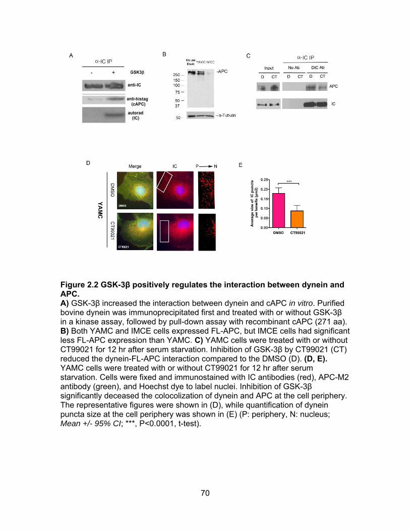
70
Figure 2.2 GSK-3β positively regulates the interaction between dynein and APC. A) GSK-3β increased the interaction between dynein and cAPC in vitro. Purified bovine dynein was immunoprecipitated first and treated with or without GSK-3β in a kinase assay, followed by pull-down assay with recombinant cAPC (271 aa). B) Both YAMC and IMCE cells expressed FL-APC, but IMCE cells had significant less FL-APC expression than YAMC. C) YAMC cells were treated with or without CT99021 for 12 hr after serum starvation. Inhibition of GSK-3β by CT99021 (CT) reduced the dynein-FL-APC interaction compared to the DMSO (D). (D, E). YAMC cells were treated with or without CT99021 for 12 hr after serum starvation. Cells were fixed and immunostained with IC antibodies (red), APC-M2 antibody (green), and Hoechst dye to label nuclei. Inhibition of GSK-3β significantly deceased the colocolization of dynein and APC at the cell periphery. The representative figures were shown in (D), while quantification of dynein puncta size at the cell periphery was shown in (E) (P: periphery, N: nucleus; Mean +/- 95% CI; ***, P<0.0001, t-test).
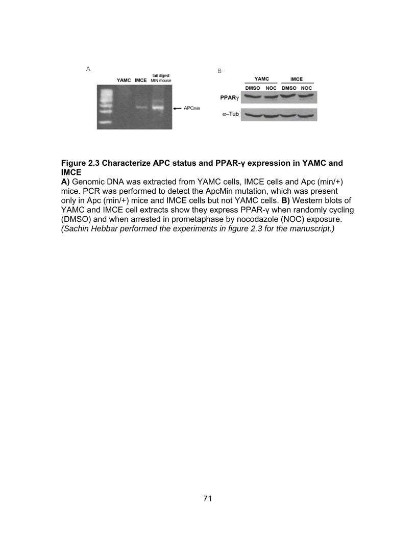
71
Figure 2.3 Characterize APC status and PPAR-γ expression in YAMC and IMCE A) Genomic DNA was extracted from YAMC cells, IMCE cells and Apc (min/+) mice. PCR was performed to detect the ApcMin mutation, which was present only in Apc (min/+) mice and IMCE cells but not YAMC cells. B) Western blots of YAMC and IMCE cell extracts show they express PPAR-γ when randomly cycling (DMSO) and when arrested in prometaphase by nocodazole (NOC) exposure. (Sachin Hebbar performed the experiments in figure 2.3 for the manuscript.)
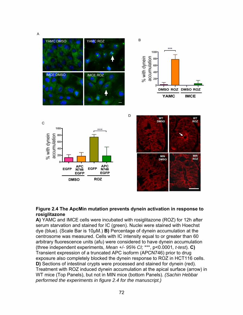
72
Figure 2.4 The ApcMin mutation prevents dynein activation in response to rosiglitazone A) YAMC and IMCE cells were incubated with rosiglitazone (ROZ) for 12h after serum starvation and stained for IC (green). Nuclei were stained with Hoechst dye (blue). (Scale Bar is 10μM.) B) Percentage of dynein accumulation at the centrosome was measured. Cells with IC intensity equal to or greater than 60 arbitrary fluorescence units (afu) were considered to have dynein accumulation (three independent experiments, Mean +/- 95% CI; ***, p<0.0001, t-test). C) Transient expression of a truncated APC isoform (APCN746) prior to drug exposure also completely blocked the dynein response to ROZ in HCT116 cells. D) Sections of intestinal crypts were processed and stained for dynein (red). Treatment with ROZ induced dynein accumulation at the apical surface (arrow) in WT mice (Top Panels), but not in MIN mice (bottom Panels). (Sachin Hebbar performed the experiments in figure 2.4 for the manuscript.)
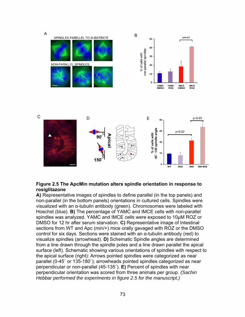
73
Figure 2.5 The ApcMin mutation alters spindle orientation in response to rosiglitazone A) Representative images of spindles to define parallel (in the top panels) and non-parallel (in the bottom panels) orientations in cultured cells. Spindles were visualized with an α-tubulin antibody (green). Chromosomes were labeled with Hoechst (blue). B) The percentage of YAMC and IMCE cells with non-parallel spindles was analyzed. YAMC and IMCE cells were exposed to 10μM ROZ or DMSO for 12 hr after serum starvation. C) Representative image of Intestinal sections from WT and Apc (min/+) mice orally gavaged with ROZ or the DMSO control for six days. Sections were stained with an α-tubulin antibody (red) to visualize spindles (arrowhead). D) Schematic Spindle angles are determined from a line drawn through the spindle poles and a line drawn parallel the apical surface (left). Schematic showing various orientations of spindles with respect to the apical surface (right): Arrows pointed spindles were categorized as near parallel (0-45˚ or 135-180˚); arrowheads pointed spindles categorized as near perpendicular or non-parallel (45-135˚). E) Percent of spindles with near perpendicular orientation was scored from three animals per group. (Sachin Hebbar performed the experiments in figure 2.5 for the manuscript.)

74
Figure 2.6 The ApcMin mutation prevents rosiglitazone-induced cell migration by both wound healing assay and enterocyte motility assay A) Representative images of wound healing response to ROZ in YAMC or IMCE cells. After a 12h exposure to ROZ or DMSO, a scratch wound was applied to confluent YAMC and IMCE cells. Wound edges (yellow lines) and typical gap widths (white lines) are shown 24 hr after wounding. B) Quantification of YAMC and IMCE wound healing assay. Average gap widths (AGW) at 0 and 24 hr after wounding were used to calculate mean distance migrated: (AGW (0hr) - AGW (24hr)) / 2. C) Representative images of enterocyte motility in response to ROZ in WT or Apc (min/+) mice. Mice were fed with ROZ or DMSO for 6 days and then
75
i.p injected with BrdU. After 12 hr. intestines were processed for BrdU immunoperoxidase activity. ROZ altered the distribution of labeled cells in WT mice (left panel), but not in Apc (min/+) mice (right panel). D) Schematic of epithelial cells lining the crypt and villus were numbered from the base of the crypt. (E, F). Quantification of BrdU positive cells at different positions of the crypt-villus axis. ROZ induced a significant shift of BrdU positive cells from crypt towards villus in WT animals (E) but not in Apc (min/+) animals (F). 25 crypt-villus units were analyzed in each treatment. (Sachin Hebbar performed the experiments in figure 2.6 for the manuscript.)
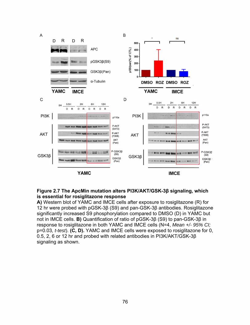
76
Figure 2.7 The ApcMin mutation alters PI3K/AKT/GSK-3β signaling, which is essential for rosiglitazone response A) Western blot of YAMC and IMCE cells after exposure to rosiglitazone (R) for 12 hr were probed with pGSK-3β (S9) and pan-GSK-3β antibodies. Rosiglitazone significantly increased S9 phosphorylation compared to DMSO (D) in YAMC but not in IMCE cells. B) Quantification of ratio of pGSK-3β (S9) to pan-GSK-3β in response to rosiglitazone in both YAMC and IMCE cells (N=4, Mean +/- 95% CI; p=0.03, t-test). (C, D). YAMC and IMCE cells were exposed to rosiglitazone for 0, 0.5, 2, 6 or 12 hr and probed with related antibodies in PI3K/AKT/GSK-3β signaling as shown.
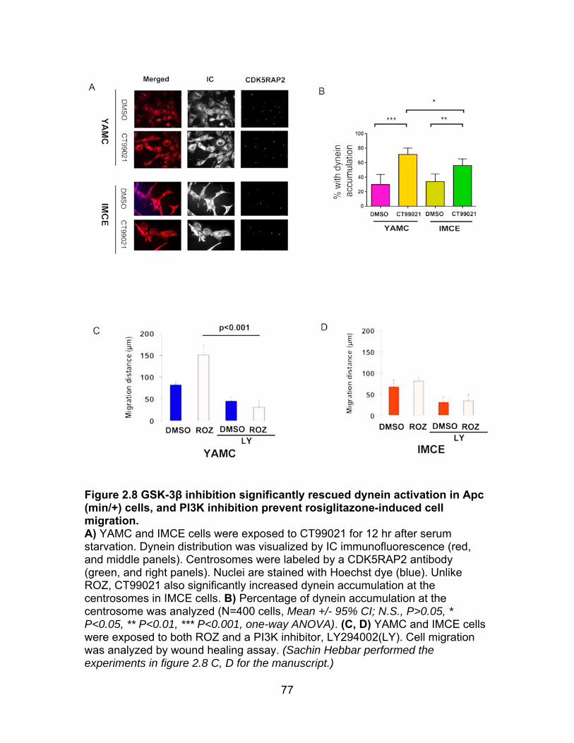
77
Figure 2.8 GSK-3β inhibition significantly rescued dynein activation in Apc (min/+) cells, and PI3K inhibition prevent rosiglitazone-induced cell migration. A) YAMC and IMCE cells were exposed to CT99021 for 12 hr after serum starvation. Dynein distribution was visualized by IC immunofluorescence (red, and middle panels). Centrosomes were labeled by a CDK5RAP2 antibody (green, and right panels). Nuclei are stained with Hoechst dye (blue). Unlike ROZ, CT99021 also significantly increased dynein accumulation at the centrosomes in IMCE cells. B) Percentage of dynein accumulation at the centrosome was analyzed (N=400 cells, Mean +/- 95% CI; N.S., P>0.05, * P<0.05, ** P<0.01, *** P<0.001, one-way ANOVA). (C, D) YAMC and IMCE cells were exposed to both ROZ and a PI3K inhibitor, LY294002(LY). Cell migration was analyzed by wound healing assay. (Sachin Hebbar performed the experiments in figure 2.8 C, D for the manuscript.)
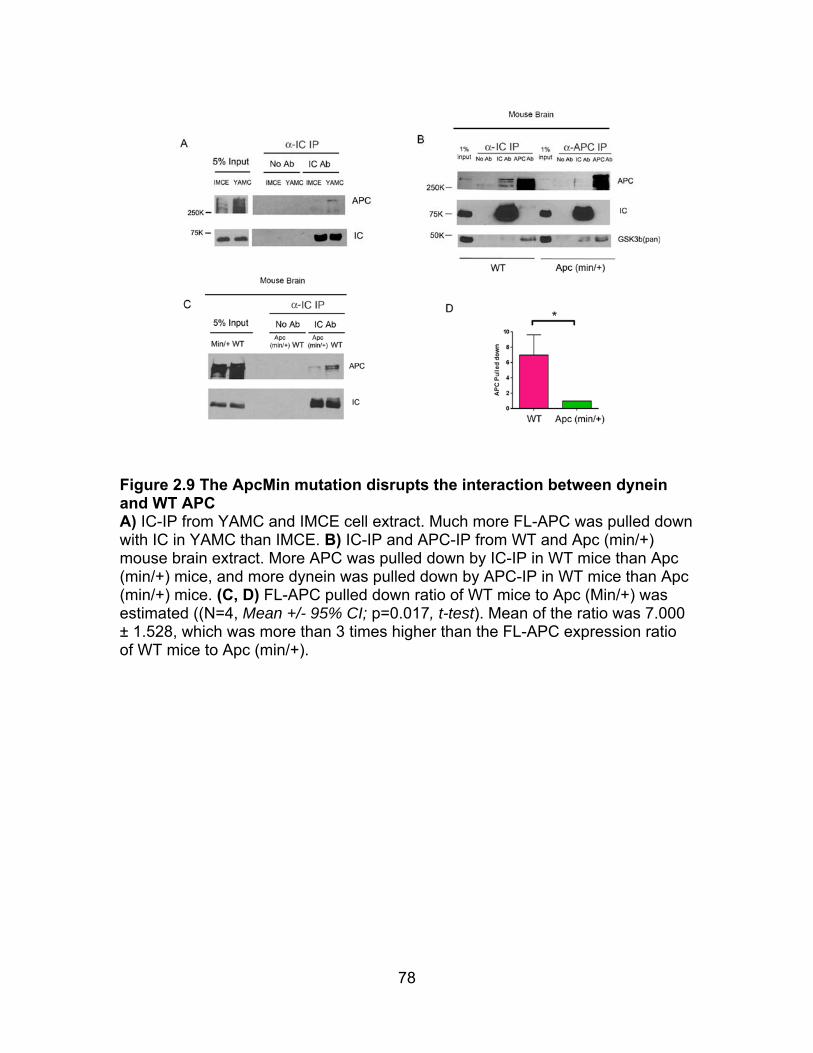
78
Figure 2.9 The ApcMin mutation disrupts the interaction between dynein and WT APC A) IC-IP from YAMC and IMCE cell extract. Much more FL-APC was pulled down with IC in YAMC than IMCE. B) IC-IP and APC-IP from WT and Apc (min/+) mouse brain extract. More APC was pulled down by IC-IP in WT mice than Apc (min/+) mice, and more dynein was pulled down by APC-IP in WT mice than Apc (min/+) mice. (C, D) FL-APC pulled down ratio of WT mice to Apc (Min/+) was estimated ((N=4, Mean +/- 95% CI; p=0.017, t-test). Mean of the ratio was 7.000 ± 1.528, which was more than 3 times higher than the FL-APC expression ratio of WT mice to Apc (min/+).
79
CHAPTER 3
A mass spectrometry-based systematic method to map GSK-3β phosphorylation sites on dynein intermediate chain
ABSTRACT
Dynein intermediate chain (IC) is essential for dynein complex assembly
and mediates the interactions of dynein with other regulators including dynactin,
Ndel1/ Nde1, ZW10. However, the mechanism of how IC regulates these
interactions is largely unknown. We previously demonstrated that GSK-3β
regulates many dynein-dependent cellular functions and is able to directly
phosphorylate IC in vitro. To dissect how GSK-3 regulates dynein, we develop a
systematic method to map phosphorylation sites on IC, which integrates
recombinant protein purification, in vitro kinase assay, tandem mass
spectrometry (MS), mutagenesis, sequence conservation analysis and immuno-
purification. We identify T154, S88 and T89 on IC-2C are targeted by GSK-3β
using MS and mutagenesis. We find that T154 is conserved in all mouse IC-2
isoforms but not in IC-1, and it exists in bovine and horse IC-2 but not in rat or
human IC-2. However, S88 and T89 are conserved in all mouse IC isoforms as
well as in ICs from all other mammalian species examined. Furthermore, we
demonstrate that S87 or T88 on IC-1B from both mouse and rat (corresponding
to 3S88 or T89 on IC-2C) is targeted by GSK-3β using mass spectrometry.
Feng J. Gao1,Deanna S. Smith1*. To be submitted to J of Biological Chemistry
80
Together, the method has been tested to map authentic GSK-3β-dependent
phosphorylation sites on ICs, and has the potential to be applied to identify other
bona fide substrates of GSK-3β or other kinases.
INTRODUCTION
Cytoplasmic dynein is a microtubule-based retrograde molecular motor. It
is a multisubunit complex with molecular weight of over 1.5 MDa, consisting of
heavy chains (HCs), intermediate chains(ICs), light intermediate chains(LICs),
and light chains(LCs) (Pfister et al., 2006). Dynein participates in a broad range
of cellular functions, such as mitosis, cell migration, and organelle trafficking
(Kardon and Vale, 2009). Dynein regulators such as dynactin, Ndel1/Nde1 and
Lis1 appear to regulate the motor’s functions by recruiting it to the appropriate
sites or by modulating its mechanochemical properties (Kardon and Vale, 2009).
Dynactin is a large multisubunit complex of ~1 MDa (Schroer, 2004), which plays
many roles in dynein regulation, such as linking dynein to cargo, targeting dynein
to specific subcellular locations , and increasing dynein’s processivity (Culver-
Hanlon et al., 2006). Ndel1/Nde1 function to recruit dynein to cargo as well as to
recruit Lis1 to dynein (McKenney et al., 2010). Lis1 and Ndel1/Nde1 are crucial
for many dynein functions including spindle orientation, cell migration, axon
transport and more (Hebbar et al., 2008b; McKenney et al., 2010; Mesngon et
al., 2006; Pandey and Smith, 2011; Wang et al., 2013). However, the mechanism
of dynein regulation is still largely unknown.
The ICs are encoded by two genes, and both of them have several
splicing isoforms (Allan, 2011; Kuta et al., 2010; Pfister et al., 2006; Wickstead
81
and Gull, 2007). ICs form a dimer during dynein complex assembly, and can
dimerize with all combinations of isoforms (Lo et al., 2006). ICs act as a dynein
regulatory center, because they directly interact with other dynein subunits as
well as many dynein regulators and cargo. The C-terminus of ICs binds directly
to HCs, and the N-terminus has three LC-binding domains (King et al., 2003;
McKenney et al., 2011). Dynactin binds to the N-terminus of ICs via its subunit
p150Glued (Karki and Holzbaur, 1995; Vaughan and Vallee, 1995), and
Ndel1/Nde1 also interact with ICs directly (Stehman et al., 2007). Ndel1/Nde1
have the same binding sites on IC as p150Glued, which causes a clear competition
between them for binding to dynein (McKenney et al., 2011). ICs interact with
zw10, which is a kinetochore protein playing a role in mitotic checkpoint signaling
(Kardon and Vale, 2009). ICs are essential for dynein cargo selection (Kardon
and Vale, 2009; Kuta et al., 2010): ICs interact with dynactin to modulate cargo
and microtubule interactions (Schroer, 2004; Vaughan and Vallee, 1995); ICs
directly interact with cargo, including -catenin, casein kinase, neurofilaments,
kinesin light chains, and huntingtin, adenovirus particles and lysosomes (Allan,
2011; Kuta et al., 2010). Therefore, precise regulation of ICs is needed by the
cell.
The IC obviously is essential for dynein activity in response to various
signaling, but there are few studies out there that is able to form a link between
kinases, phosphorylation sites on IC and relevant functions. The phosphorylation
of T89 on IC-2C acts as a switch of regulating the interaction of dynein with zw10
and p150 Glued during mitosis (Whyte et al., 2008): phosphorylation of T89
82
increases the interaction of dynein and zw10 and recruits dynein to kinetochores,
while the dephosphorylation of T89 increases the dynein-dynactin interaction and
leads to poleward movement of kinetochore components (Vaughan et al., 2001;
Whyte et al., 2008). Later, it was shown that Polo-like Kinase1 (PLK1) may
phosphorylate T89 on the N-terminus of IC-2C (1-284 AA) in vitro (Bader et al.,
2011). Recently, two groups have shown that the activation of extracellular-signal
regulated kinase (ERK) signaling increases S80 phosphorylation on IC-2C, which
helps dynein bind to certain signaling endosomes and subsequent axon transport
(Mitchell et al., 2012; Pullikuth et al., 2013). Another kinase, casein kinase 1
(CK1) stimulates dynein motor activity by phosphorylation of ICs, but the target
sites are not known (Ikeda et al., 2011).
Glycogen Synthase kinase (GSK-3), a serine/threonine protein kinase,
was described to phosphorylate glycogen synthase in rabbit skeletal muscle
(Embi et al., 1980). Later, it was found that GSK-3 plays a major role in
phosphorylation of tau and regulating its association with microtubules (Ishiguro
et al., 1993). To date, GSK-3 turns out to be a fundamental enzyme involved in
almost all cellular processes, including embryonic development, cell
differentiation, apoptosis, and insulin signaling (Sutherland, 2011). In mammals
there are two GSK-3 genes, GSK-3α and GSK-3β (Woodgett, 1991), but the
catalytic domain is highly conserved (Castano et al., 2010; Mukai et al., 2002;
Soutar et al., 2010; Wood-Kaczmar et al., 2009). Unlike most other kinases,
GSK-3β is highly active in resting cells because of an activating auto-
phosphorylation at Tyr 216. However, multiple kinases including AKT/PKB,
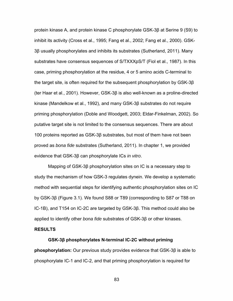
83
protein kinase A, and protein kinase C phosphorylate GSK-3β at Serine 9 (S9) to
inhibit its activity (Cross et al., 1995; Fang et al., 2002; Fang et al., 2000). GSK-
3β usually phosphorylates and inhibits its substrates (Sutherland, 2011). Many
substrates have consensus sequences of S/TXXXpS/T (Fiol et al., 1987). In this
case, priming phosphorylation at the residue, 4 or 5 amino acids C-terminal to
the target site, is often required for the subsequent phosphorylation by GSK-3β
(ter Haar et al., 2001). However, GSK-3β is also well-known as a proline-directed
kinase (Mandelkow et al., 1992), and many GSK-3β substrates do not require
priming phosphorylation (Doble and Woodgett, 2003; Eldar-Finkelman, 2002). So
putative target site is not limited to the consensus sequences. There are about
100 proteins reported as GSK-3β substrates, but most of them have not been
proved as bona fide substrates (Sutherland, 2011). In chapter 1, we provided
evidence that GSK-3β can phosphorylate ICs in vitro.
Mapping of GSK-3β phosphorylation sites on IC is a necessary step to
study the mechanism of how GSK-3 regulates dynein. We develop a systematic
method with sequential steps for identifying authentic phosphorylation sites on IC
by GSK-3β (Figure 3.1). We found S88 or T89 (corresponding to S87 or T88 on
IC-1B), and T154 on IC-2C are targeted by GSK-3β. This method could also be
applied to identify other bona fide substrates of GSK-3β or other kinases.
RESULTS
GSK-3β phosphorylates N-terminal IC-2C without priming
phosphorylation: Our previous study provides evidence that GSK-3β is able to
phosphorylate IC-1 and IC-2, and that priming phosphorylation is required for

84
some but not all target sites. Here, we map GSK-3β phosphorylation sites on ICs,
which do not require priming phosphorylation. There is high homology between
IC isoforms, and IC-2C is regarded as the only ubiquitously expressed isoform
(Brill and Pfister, 2000; Ha et al., 2008; King et al., 2003; Vaughan and Vallee,
1995).
IC-2C has 612 amino acids (aa). N-terminal coiled-coil and nearby regions
of IC-2C are responsible for interacting with Ndel1/Nde1 and p150Glued, followed
by three domains for binding to different types of LCs. The dimerization domain
and 7 WD-40 repeats are essential for the assembly of dynein complex (Figure
3.2A). IC-2C sequences contain nine consensus sites and nine proline-directed
serine/threonine sites (Figure 3.2B). Several of them are located at the N-
terminus of IC-2C. To get reliable mass spectrometry analysis of IC-2C, we
characterized and optimized N-terminal 237aa of IC-2C (N237) phosphorylation
by GSK-3β using purified N237 protein (Figure 3.2C, D). It showed that the
intensity of N237 phosphorylation was positively correlated with the amount of
GSK-3β (Figure 3.2C), and that there was more N237 phosphorylation with
longer incubation in a GSK-3β kinase assay (Figure 3.2D). We tested
phosphorylation of the N-terminal 106 aa of IC-2C (N106) by GSK-3β and found
that N106 was less phosphorylated compared to N237. It indicates that certain
sites in N106 as well as in the region between positions 106 and 237 on IC-2C
are phosphorylated by GSK-3β.
Identification of phosphorylation sites on IC-2C by mass
spectrometry: To determine the functional significance of the specific sites
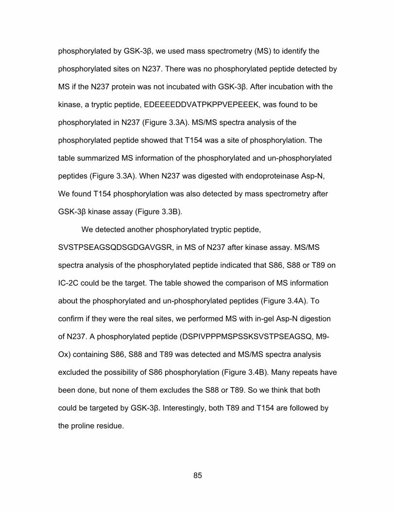
85
phosphorylated by GSK-3β, we used mass spectrometry (MS) to identify the
phosphorylated sites on N237. There was no phosphorylated peptide detected by
MS if the N237 protein was not incubated with GSK-3β. After incubation with the
kinase, a tryptic peptide, EDEEEEDDVATPKPPVEPEEEK, was found to be
phosphorylated in N237 (Figure 3.3A). MS/MS spectra analysis of the
phosphorylated peptide showed that T154 was a site of phosphorylation. The
table summarized MS information of the phosphorylated and un-phosphorylated
peptides (Figure 3.3A). When N237 was digested with endoproteinase Asp-N,
We found T154 phosphorylation was also detected by mass spectrometry after
GSK-3β kinase assay (Figure 3.3B).
We detected another phosphorylated tryptic peptide,
SVSTPSEAGSQDSGDGAVGSR, in MS of N237 after kinase assay. MS/MS
spectra analysis of the phosphorylated peptide indicated that S86, S88 or T89 on
IC-2C could be the target. The table showed the comparison of MS information
about the phosphorylated and un-phosphorylated peptides (Figure 3.4A). To
confirm if they were the real sites, we performed MS with in-gel Asp-N digestion
of N237. A phosphorylated peptide (DSPIVPPPMSPSSKSVSTPSEAGSQ, M9-
Ox) containing S86, S88 and T89 was detected and MS/MS spectra analysis
excluded the possibility of S86 phosphorylation (Figure 3.4B). Many repeats have
been done, but none of them excludes the S88 or T89. So we think that both
could be targeted by GSK-3β. Interestingly, both T89 and T154 are followed by
the proline residue.
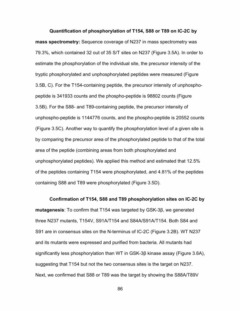
86
Quantification of phosphorylation of T154, S88 or T89 on IC-2C by
mass spectrometry: Sequence coverage of N237 in mass spectrometry was
79.3%, which contained 32 out of 35 S/T sites on N237 (Figure 3.5A). In order to
estimate the phosphorylation of the individual site, the precursor intensity of the
tryptic phosphorylated and unphosphorylated peptides were measured (Figure
3.5B, C). For the T154-containing peptide, the precursor intensity of unphospho-
peptide is 341933 counts and the phospho-peptide is 98802 counts (Figure
3.5B). For the S88- and T89-containing peptide, the precursor intensity of
unphospho-peptide is 1144776 counts, and the phospho-peptide is 20552 counts
(Figure 3.5C). Another way to quantify the phosphorylation level of a given site is
by comparing the precursor area of the phosphorylated peptide to that of the total
area of the peptide (combining areas from both phosphorylated and
unphosphorylated peptides). We applied this method and estimated that 12.5%
of the peptides containing T154 were phosphorylated, and 4.81% of the peptides
containing S88 and T89 were phosphorylated (Figure 3.5D).
Confirmation of T154, S88 and T89 phosphorylation sites on IC-2C by
mutagenesis: To confirm that T154 was targeted by GSK-3β, we generated
three N237 mutants, T154V, S91A/T154 and S84A/S91A/T154. Both S84 and
S91 are in consensus sites on the N-terminus of IC-2C (Figure 3.2B). WT N237
and its mutants were expressed and purified from bacteria. All mutants had
significantly less phosphorylation than WT in GSK-3β kinase assay (Figure 3.6A),
suggesting that T154 but not the two consensus sites is the target on N237.
Next, we confirmed that S88 or T89 was the target by showing the S88A/T89V
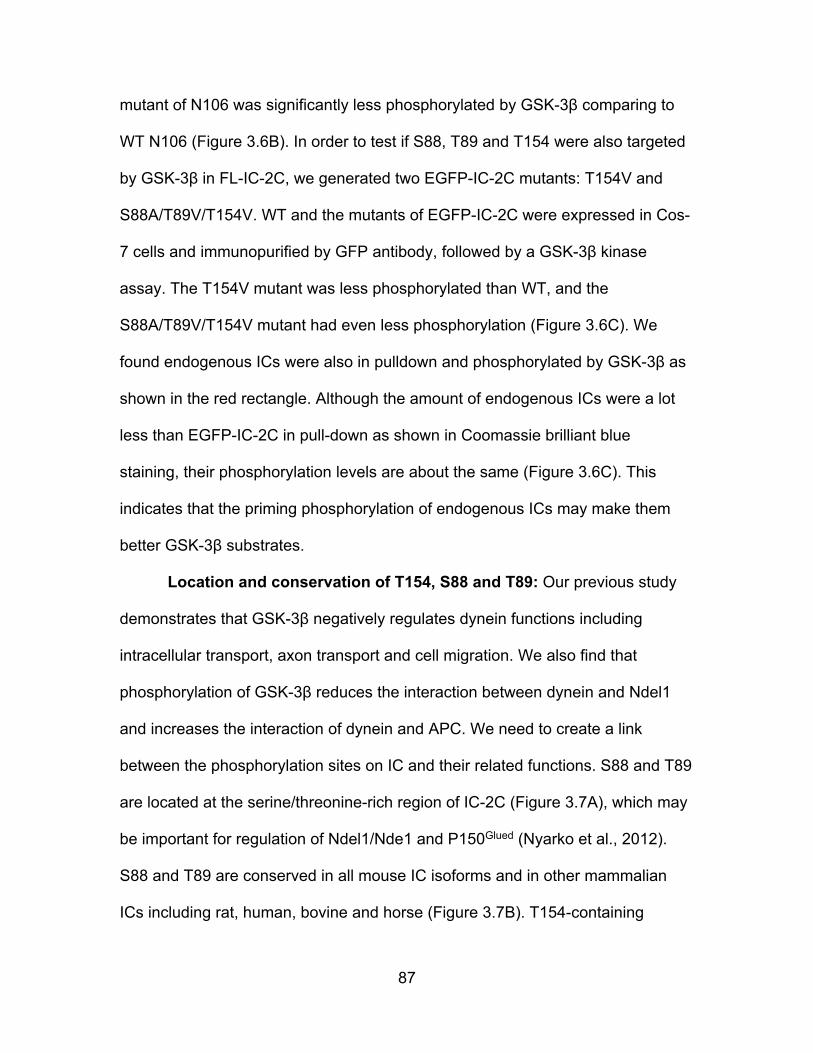
87
mutant of N106 was significantly less phosphorylated by GSK-3β comparing to
WT N106 (Figure 3.6B). In order to test if S88, T89 and T154 were also targeted
by GSK-3β in FL-IC-2C, we generated two EGFP-IC-2C mutants: T154V and
S88A/T89V/T154V. WT and the mutants of EGFP-IC-2C were expressed in Cos-
7 cells and immunopurified by GFP antibody, followed by a GSK-3β kinase
assay. The T154V mutant was less phosphorylated than WT, and the
S88A/T89V/T154V mutant had even less phosphorylation (Figure 3.6C). We
found endogenous ICs were also in pulldown and phosphorylated by GSK-3β as
shown in the red rectangle. Although the amount of endogenous ICs were a lot
less than EGFP-IC-2C in pull-down as shown in Coomassie brilliant blue
staining, their phosphorylation levels are about the same (Figure 3.6C). This
indicates that the priming phosphorylation of endogenous ICs may make them
better GSK-3β substrates.
Location and conservation of T154, S88 and T89: Our previous study
demonstrates that GSK-3β negatively regulates dynein functions including
intracellular transport, axon transport and cell migration. We also find that
phosphorylation of GSK-3β reduces the interaction between dynein and Ndel1
and increases the interaction of dynein and APC. We need to create a link
between the phosphorylation sites on IC and their related functions. S88 and T89
are located at the serine/threonine-rich region of IC-2C (Figure 3.7A), which may
be important for regulation of Ndel1/Nde1 and P150Glued (Nyarko et al., 2012).
S88 and T89 are conserved in all mouse IC isoforms and in other mammalian
ICs including rat, human, bovine and horse (Figure 3.7B). T154-containing
88
phospho-peptide is near the dimerization domain and LC7 binding domain
(Figure 3.7A), which may affect dynein assembly. In figure 3.6C, the
phosphorylation of endogenous ICs pulled down by WT EGFP-IC-2C was higher
than that from the mutants, which suggests that T154 may regulate the
dimerization of ICs. T154 exists in all mouse IC-2 isoforms but not in IC-1, and
T154 is conserved in bovine and horse IC-2 but not in rat or human IC-2 (Figure
3.7B).
Confirmation of GSK-3β-dependent phosphorylation of S87 and T88
on rat IC-1B by MS: IC-1B is a neuron specific isoform of ICs (Ha et al., 2008),
which has S87 and T88 sites corresponding to S88 and T89 on mouse IC-2C. It
does not have a corresponding site for T154. Previously, we found that rat
EGFP-IC-1B was significantly phosphorylated by GSK-3β. EGFP-IC-1B were
expressed in Cos-7 cells and immunopurified by GFP antibody, and GSK-3β
kinase assay was performed for 1hr. We analyzed the GSK-3β dependent
phosphorylation of EGFP-IC-1B by mass spectrometry with tryptic digestion. The
sequence coverage of IC-1B is 69.65%, and a phospho-peptide
(SVSTPSEAGSQDDLGPLTR) containing S87 and T88 was detected (Figure
3.8A). MS/MS spectra analysis of the peptide showed that S87 or T88 was the
site of phosphorylation (Figure 3.8B, C). The precursor intensity (ion counts) of
unphospho-peptide is 121891, and 57484 for the phospho-peptide (Figure 3.8D).
When we used the precursor area to quantify phosphorylation of the peptide,
there was ~ 4.91% of the peptides were phosphorylated (Figure 3.8E).
89
Confirmation of GSK-3β-dependent phosphorylation of S87 and T88
on mouse IC-1B by MS: ICs were immunopurified by IC antibody-conjugated
beads from mouse brain extract, and GSK-3β kinase assay was performed for
1hr. Mouse IC-1B was analyzed by mass spectrometry with tryptic digestion. The
sequence coverage of mouse IC-1B was 64.17%, and a phospho-peptide
(SVSTPSDAGSQDSGDLGPLTR) containing S87 and T88 was detected (Figure
3.9A). MS/MS spectrum showed S87 or T88 was the site of phosphorylation,
which did not exist before GSK-3β kinase assay (Figure 3.9B, C). The precursor
intensity (ion counts) of unphospho-peptide is 254328, and 124687 for the
phospho-peptide (Figure 3.9D). We estimated that ~ 6.69% of the peptides were
phosphorylated by GSK-3β using MS precursor area quantification (Figure 3.9E).
Together, these results show that GSK-3β is able to target S87 or T88 on
IC-1B from both mouse and rat, which is consistent with the prediction by the
sequence conservation analysis. We feel more confident to predict that GSK-3β
phosphorylation of ICs is a real cellular event, which is happening in many
species.
90
DISCUSSION
The efficiency of GSK-3β phosphorylation of IC in vitro is biological
significance? In GSK-3β kinase assay, we found that more kinase increased
phosphorylation of N237, and longer incubation also increased phosphorylation.
After optimizing the conditions of the kinase assay, we consistently detect that
GSK-3β is able to phosphorylate ~5% of S88 or T89 and ~15% of T154 on IC-2C
using MS precursor area quantification (Figure 5). In IC-1B phosphorylation
experiments, GSK-3β is able to phosphorylate 5-7% of S87 or T88 on IC-1B after
1 hr (Figure 3.8 and Figure 3.9). People may think that if you incubate the kinase
and substrate together for enough time, the percentage of phosphorylation could
be close to 100%. However, this is not the case for GSK-3β. Besides activating
auto-phosphorylation of Y216, purified GSK-3β also auto-phosphorylates at S9
and inhibits itself during in vitro kinase assay (Ilouz et al., 2008; Wang et al.,
1994). We detect ~50% of GSK-3β with Y216 phosphorylation and ~50% of
GSK-3β with S9 phosphorylation after kinase assay by mass spectrometry.
Therefore, auto-inhibition property may prevent GSK-3β achieving higher
phosphorylation efficacy of ICs in vitro, and the phosphorylation level of T154,
S88 or T89 we get is reasonable to believe they are real target sites.
Biological functions of T154, S88 or T89: We show that GSK-3β is able
to phosphorylate S88 or T89 on IC-2C (S87 or T88 on IC-1B). S88 and T89 are
on S/T rich region of IC where is thought to be important for Ndel1 and dynactin
regulation (Nyarko et al., 2012). Moreover, phosphorylation of T89 has been
reported to reduce the dynein-dynactin interaction but increase the dynein-zw10

91
interaction (Whyte et al., 2008). Previously, we showed that dynein or IC-2C
phosphorylation by GSK-3β reduces its interaction with Ndel1. Therefore, we
think that S88 or T89 phosphorylation may have an important role in regulating
the interaction of dynein to Ndel1, dynactin, and ZW10. These interactions are
important for mitosis, and intracellular transport. Consistent with this, our
previous study demonstrates that GSK-3β negatively regulates dynein functions
including axon transport and cell migration.
We also provide evidence that GSK-3β phosphorylates T154 on IC-2C
and that the site may be important for dynein assembly. There are two reasons
for us to come to the hypothesis: first, T154 is located near the dimerization and
LC-7 binding domain; second, when we transfected T154V IC-2C and
S88A/T89V/T154V mutants into Cos-7 cells, it seemed that less endogenous IC
was pulled down by the mutants than WT. The site does not exist in IC-1 or
human IC-2, but the relevant function of the site should not be underestimated
because of this.
In future, we will generate phospho-specific antibodies to detect whether
GSK-3β phosphorylate ICs in vitro and in vivo. With the antibodies, we will
explore how GSK-3β regulates ICs phosphorylation, dynein activity and the
interaction of dynein to other regulators.
Applications of the method: The method is designed to quickly and
accurately detect a novel kinase-substrate link, especially useful for detecting the
site that does not require priming phosphorylation as demonstrated here.
However, if a kinase targeting its substrate requires priming phosphorylation, the
92
method could be applied to screen priming kinases. For example, GSK-3β
targets some sites on ICs requiring priming phosphorylation. First, we will
determine the priming kinase candidates, which could be reached by combining
information from literature and mass spectrometry of kinases pulldown by IC-
immunoprecipitation from mouse brain extract. Second, we will apply the method
to determine if it is the priming kinase, which is based on three criteria: 1,
whether it phosphorylates purified IC; 2, whether the target site is at the priming
site of consensus sites; 3, whether the phosphorylation of IC by this kinase
significantly increases GSK-3β dependent phosphorylation.
MATERIALS AND METHODS
Constructs: EGFP-IC2C, PRSET-A IC2C, PRSET-A N237 and PRSET-A
N106 expression vectors were described previously (King et al., 2003). The
EGFP-IC1B vector was provided by K. Pfister (Univ. VA). PRSET-A point
mutants (T154V IC-2C, S88A/T89V/T154V IC-2C; T154V N237, S91A/T154V
N237, S84A/S91/T154V N237; S88A/T89V N106) were be generated from their
related WT vector using the QuickChange site-directed mutagenesis kit. T154V
EGFP-IC2C and S88A/T89V/T154V EGFP-IC2C were subcloned from PRSET-A
IC-2C mutants.
Expression and purification of proteins: PRSET-A N106 and N237
constructs were expressed in BL-21 cells. Cells were grown at 37°C to an OD600
of 0.4, and 0.1 mM isopropyl-d-thiogalactoside (IPTG) was added to induce
protein expression for 3 hr at 37 °C. Cells were lysed in his-tag protein
purification binding buffer (Invitrogen) with protease inhibitors. Ni-NTA beads
93
(Invitrogen) were added into the cell supernatant and incubating at RT for 1 hr.
The beads were washed 3 times and then the protein was eluted.
EGFP-IC2C and EGFP-IC1B constructs were transfected into Cos7 cells
using Lipofectamine 2000 reagent (Invitrogen). Cells were collected after 30 hr.
For immuno-purification of EGFP-ICs from cell extract, 1µg EGFP antibody was
first incubated with 30 µl Protein-A dynabeads (Invitrogen) for 2 hr at room
temperature and washed with lysis buffer twice. The antibody conjugated
dynabeads were incubated with 1 mg extracts at 4°C overnight. For
immunopurification of ICs from mouse brain extract, 15 µl 74.1 mouse
monoclonal IC antibody conjugated agarose beads (IC-beads) were incubated
with 1mg extract at 4°C overnight (Santa Cruz Biotechnology, Inc.). Dynabeads
or IC-beads were subject to three washes with lysis buffer and then two washes
with PBS-T at 4 °C. Beads were collected for kinase assay.
Cell and brain extract preparation: For preparation of cell extract, cells
at 90% confluency were lysed in buffer containing 50 mM Tris (pH 7.5), 0.1% NP-
40, 100mM NaCl, 1 mM MgCl2, 5 mM EDTA, protease inhibitor cocktail (Fisher)
and Halt phosphatase inhibitor cocktail (Fisher) on ice for 30 minutes. Cell
lysates were sonicated for 10 pulses at level 1 with 10% output 3 times. The
lysates were incubated on ice for another 10 min and then centrifuged at 17,000
g for 20 minutes at 4 °C. For preparation of mouse brain extract, brains were
dissected with clean tools as quickly as possible to prevent degradation. Tissues
were dounce-homogenized in lysis buffer as above. The lysate would be
94
incubated on ice for 30 min and then centrifuged at 17,000 g for 30 minutes at 4
°C. Concentration of extracts were determined by BCA protein assay (Pierce).
Protein kinase assay: The GSK-3β Kinase Enzyme System which
consists of GSK-3β Kinase, the substrate, reaction buffer and DTT was
purchased from Promega. Purified proteins (IC-2C, N237 or N106) or
immunopurified ICs (EGFP-IC2C or EGFP-IC1B or mouse brain ICs) were
incubated with GSK-3β and 0.03 µCi/µl 32P-ATP at 37°C for a specific amount
of time. The reaction was stopped by the addition of sample buffer. Proteins were
separated by SDS-PAGE, followed by Coomassie brilliant blue staining. The wet
min-gel was sealed in saran wrap and exposed to X-ray film overnight at -80 °C.
Mass spectrometry analysis of IC: Gel bands were excised and
subjected to in-gel digestion (Trypsin/Lys-c Mix or Asp-N from Promega)
according to UCSF Protocol (2002.04.08 version). Sample introduction is via a
Dionex Ultimate 3000 RSLC nano liquid chromatograph (nanospray ionization),
and the Orbitrap Velos Pro (Thermo Fisher Scientific), a hybrid ion trap-orbitrap
mass spectrometer, was used. This spectrometer is used for high resolution LC-
MS/MS, which has complementary fragmentation modes including CID, HCD,
and optional ETD. The raw data were searched by SEQUEST in Thermo
Scientific Proteome Discoverer software (1.4). Cytoplasmic dynein 1 intermediate
chains database was used, which was generated from uniprot database. Search
parameters were set: variable modifications of oxidation (at methionine residues)
and phosphorylation (at serine, threonine, and tyrosine); static Modification:
Carbamidomethyl; proper enzyme selected; the number of allowed missed

95
cleavage sites at 2; fragment match tolerance at 0.8 Da; precursor Mass
Tolerance: 10 ppm.
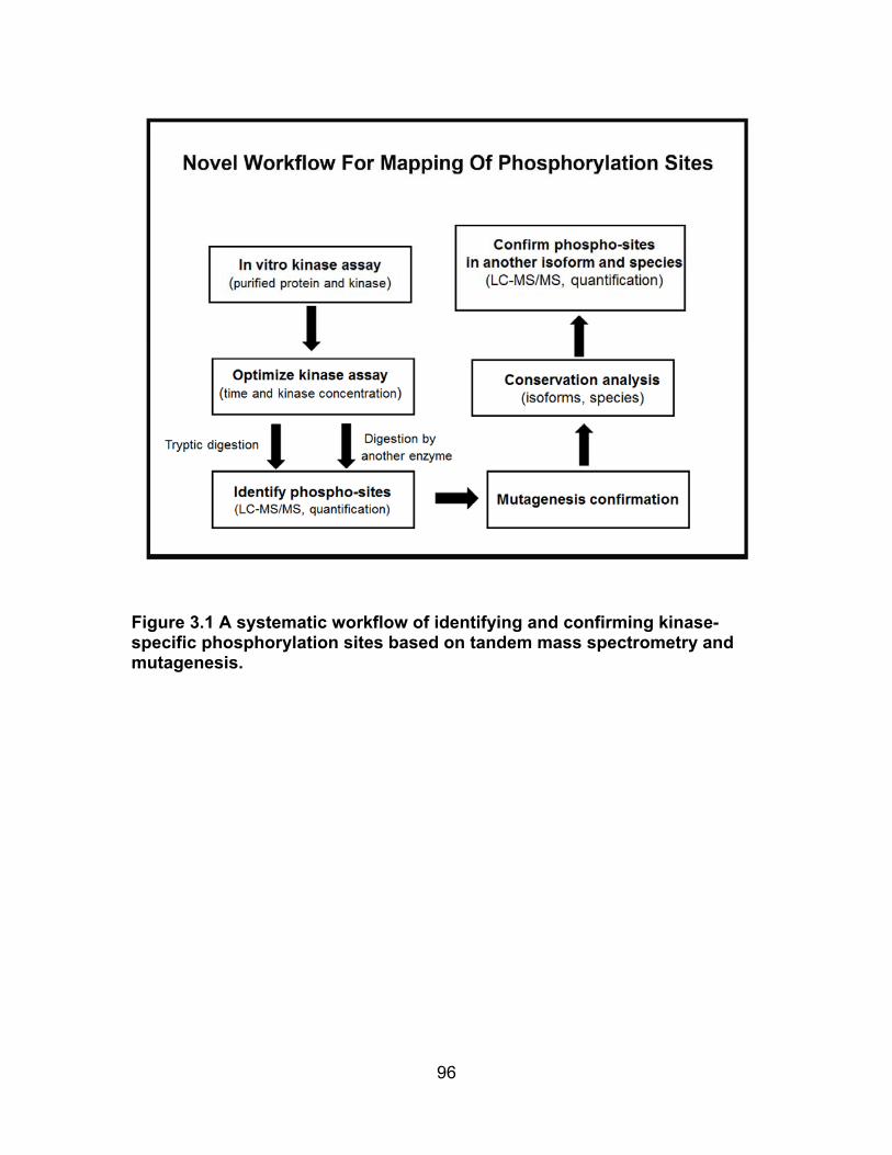
96
Figure 3.1 A systematic workflow of identifying and confirming kinase-specific phosphorylation sites based on tandem mass spectrometry and mutagenesis.
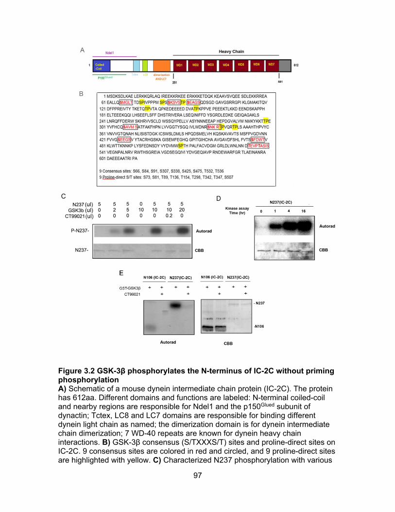
97
Figure 3.2 GSK-3β phosphorylates the N-terminus of IC-2C without priming phosphorylation A) Schematic of a mouse dynein intermediate chain protein (IC-2C). The protein has 612aa. Different domains and functions are labeled: N-terminal coiled-coil and nearby regions are responsible for Ndel1 and the p150Glued subunit of dynactin; Tctex, LC8 and LC7 domains are responsible for binding different dynein light chain as named; the dimerization domain is for dynein intermediate chain dimerization; 7 WD-40 repeats are known for dynein heavy chain interactions. B) GSK-3β consensus (S/TXXXS/T) sites and proline-direct sites on IC-2C. 9 consensus sites are colored in red and circled, and 9 proline-direct sites are highlighted with yellow. C) Characterized N237 phosphorylation with various
98
amount of GSK-3β in kinase assay. 5 µl purified protein was incubated with 0, 2, 5, 10, or 20 µl of purified GST-GSK-3β for 1hr, and autoradiograph (Autorad) showed that the N237 phosphorylation was positively correlated with the amount of the kinase added. If there was no N237 or added with CT99021, the phosphorylation signal was gone. Coomassie brilliant blue staining (CBB) showed the level of N237 protein in the kinase assay. D) Characterized N237 phosphorylation with various incubation times in kinase assay. The same amount of purified N237 protein and GSK-3β were incubated for 0, 1, 4 and 16 hr. It showed that the longer of the incubation time the more of the N237 phosphorylation. E) Compared the GSK-3β phosphorylation of two N-terminal fragments of IC-2C, N106 and N237. The same amount of the kinase was incubated with purified N106 and N237 protein for 1 hr. N106 was less phosphorylated by GSK-3β comparing to N237.
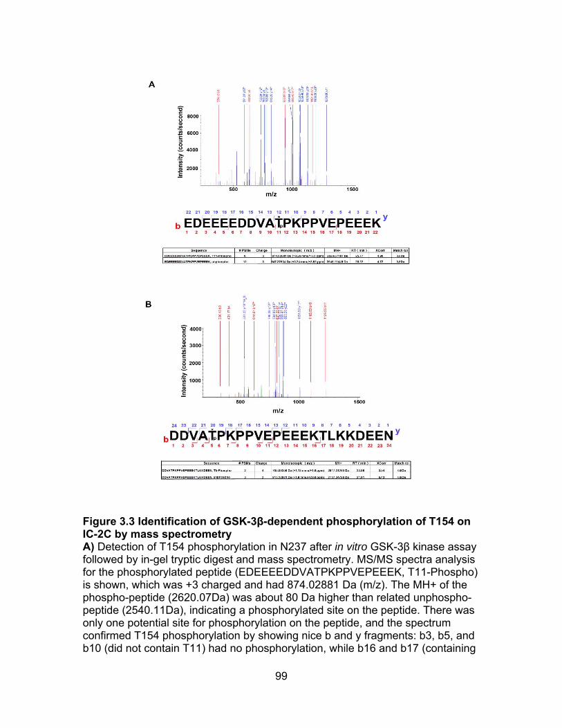
99
Figure 3.3 Identification of GSK-3β-dependent phosphorylation of T154 on IC-2C by mass spectrometry A) Detection of T154 phosphorylation in N237 after in vitro GSK-3β kinase assay followed by in-gel tryptic digest and mass spectrometry. MS/MS spectra analysis for the phosphorylated peptide (EDEEEEDDVATPKPPVEPEEEK, T11-Phospho) is shown, which was +3 charged and had 874.02881 Da (m/z). The MH+ of the phospho-peptide (2620.07Da) was about 80 Da higher than related unphospho-peptide (2540.11Da), indicating a phosphorylated site on the peptide. There was only one potential site for phosphorylation on the peptide, and the spectrum confirmed T154 phosphorylation by showing nice b and y fragments: b3, b5, and b10 (did not contain T11) had no phosphorylation, while b16 and b17 (containing
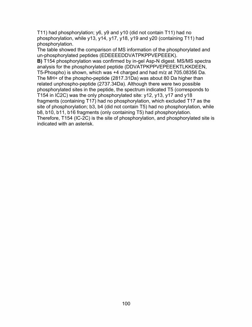
100
T11) had phosphorylation; y6, y9 and y10 (did not contain T11) had no phosphorylation, while y13, y14, y17, y18, y19 and y20 (containing T11) had phosphorylation. The table showed the comparison of MS information of the phosphorylated and un-phosphorylated peptides (EDEEEEDDVATPKPPVEPEEEK). B) T154 phosphorylation was confirmed by in-gel Asp-N digest. MS/MS spectra analysis for the phosphorylated peptide (DDVATPKPPVEPEEEKTLKKDEEN, T5-Phospho) is shown, which was +4 charged and had m/z at 705.08356 Da. The MH+ of the phospho-peptide (2817.31Da) was about 80 Da higher than related unphospho-peptide (2737.34Da). Although there were two possible phosphorylated sites in the peptide, the spectrum indicated T5 (corresponds to T154 in IC2C) was the only phosphorylated site: y12, y13, y17 and y18 fragments (containing T17) had no phosphorylation, which excluded T17 as the site of phosphorylation; b3, b4 (did not contain T5) had no phosphorylation, while b8, b10, b11, b16 fragments (only containing T5) had phosphorylation. Therefore, T154 (IC-2C) is the site of phosphorylation, and phosphorylated site is indicated with an asterisk.

101
Figure 3.4 Identification of GSK-3β-dependent phosphorylation of S88 and T89 on IC-2C by mass spectrometry A) Detection of S88 or T89 phosphorylation on N237 after in vitro GSK-3β kinase assay followed by in-gel tryptic digest and mass spectrometry. MS/MS spectra analysis for the phosphorylated peptide (SVSTPSEAGSQDSGDGAVGSR, S1, S3 orT4-Phospho) is shown, which was +2 charged and had 1015.91937 Da (m/z). The MH+ of the phospho-peptide (2030.83) was about 80 Da higher than related unphospho-peptide (1950.86Da), indicating a phosphorylated site in the peptide. Although there was 7 possible phosphorylation sites on the peptide, the spectra excluded 4 C-terminal amino acids as the site of modification at by showing y4, y5, y6, y7, y8, y9, y10, y11, y12, y13, y14, y15, y16, y17 fragments had no phosphorylation. The b4 fragment indicated there was a phosphorylation
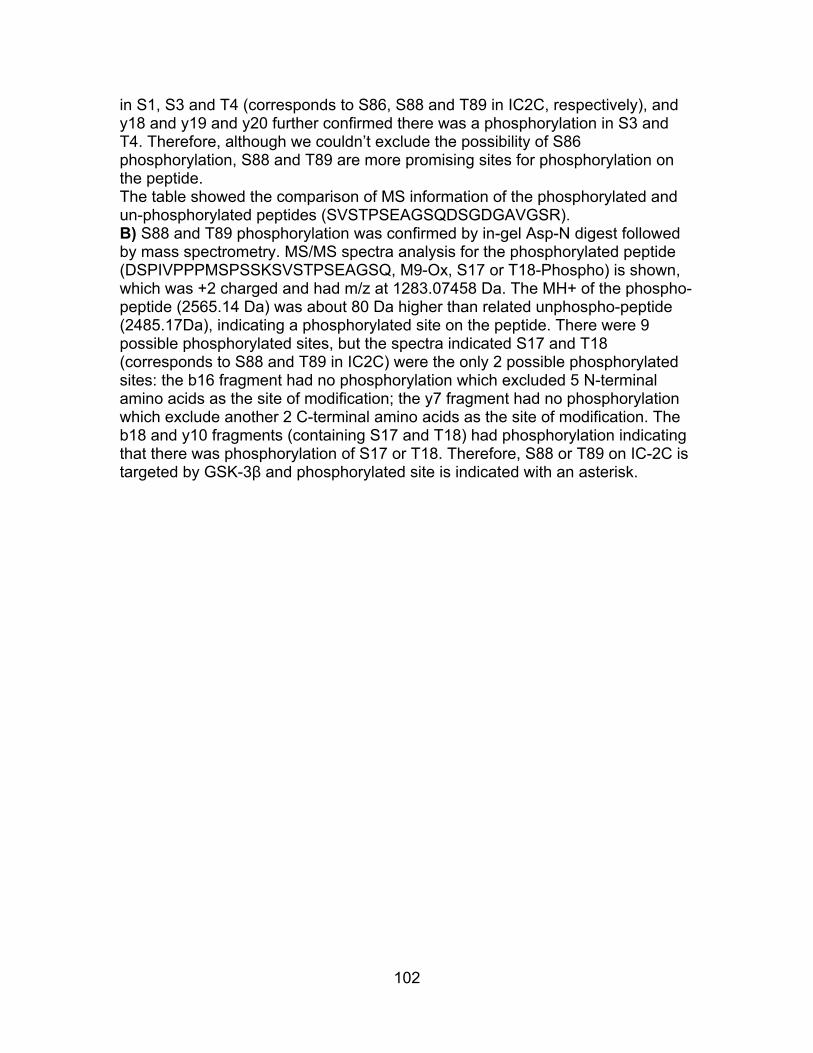
102
in S1, S3 and T4 (corresponds to S86, S88 and T89 in IC2C, respectively), and y18 and y19 and y20 further confirmed there was a phosphorylation in S3 and T4. Therefore, although we couldn’t exclude the possibility of S86 phosphorylation, S88 and T89 are more promising sites for phosphorylation on the peptide. The table showed the comparison of MS information of the phosphorylated and un-phosphorylated peptides (SVSTPSEAGSQDSGDGAVGSR). B) S88 and T89 phosphorylation was confirmed by in-gel Asp-N digest followed by mass spectrometry. MS/MS spectra analysis for the phosphorylated peptide (DSPIVPPPMSPSSKSVSTPSEAGSQ, M9-Ox, S17 or T18-Phospho) is shown, which was +2 charged and had m/z at 1283.07458 Da. The MH+ of the phospho-peptide (2565.14 Da) was about 80 Da higher than related unphospho-peptide (2485.17Da), indicating a phosphorylated site on the peptide. There were 9 possible phosphorylated sites, but the spectra indicated S17 and T18 (corresponds to S88 and T89 in IC2C) were the only 2 possible phosphorylated sites: the b16 fragment had no phosphorylation which excluded 5 N-terminal amino acids as the site of modification; the y7 fragment had no phosphorylation which exclude another 2 C-terminal amino acids as the site of modification. The b18 and y10 fragments (containing S17 and T18) had phosphorylation indicating that there was phosphorylation of S17 or T18. Therefore, S88 or T89 on IC-2C is targeted by GSK-3β and phosphorylated site is indicated with an asterisk.
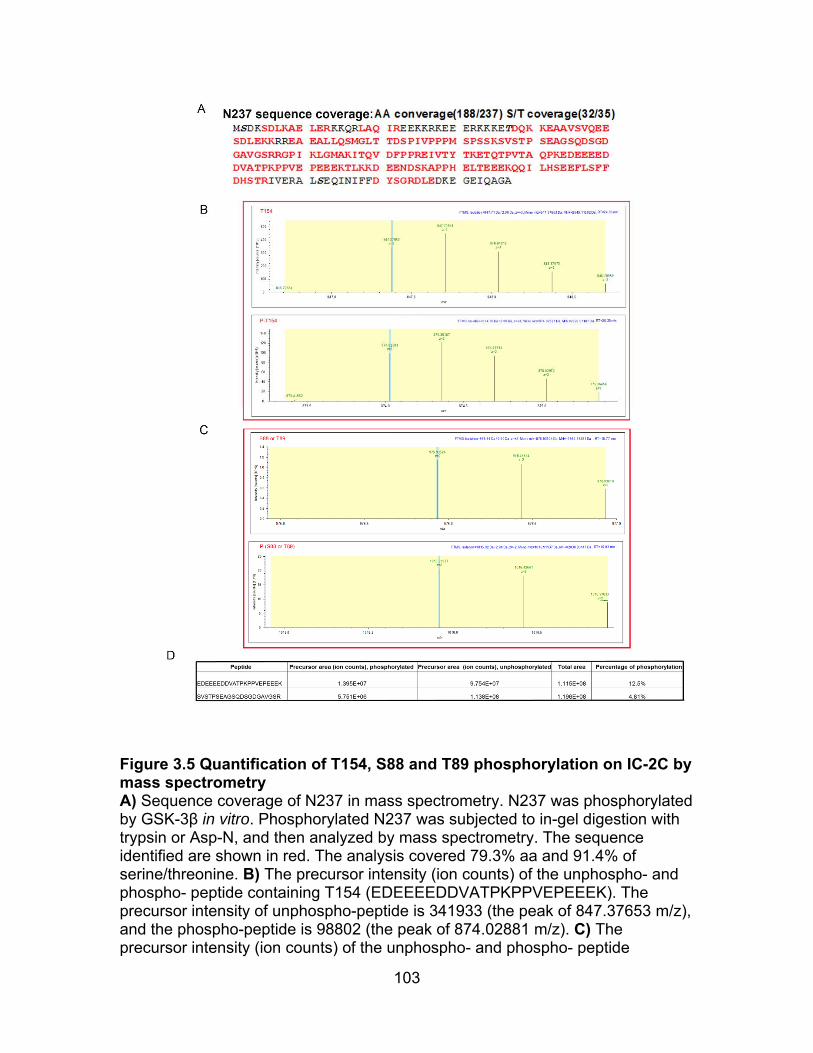
103
Figure 3.5 Quantification of T154, S88 and T89 phosphorylation on IC-2C by mass spectrometry A) Sequence coverage of N237 in mass spectrometry. N237 was phosphorylated by GSK-3β in vitro. Phosphorylated N237 was subjected to in-gel digestion with trypsin or Asp-N, and then analyzed by mass spectrometry. The sequence identified are shown in red. The analysis covered 79.3% aa and 91.4% of serine/threonine. B) The precursor intensity (ion counts) of the unphospho- and phospho- peptide containing T154 (EDEEEEDDVATPKPPVEPEEEK). The precursor intensity of unphospho-peptide is 341933 (the peak of 847.37653 m/z), and the phospho-peptide is 98802 (the peak of 874.02881 m/z). C) The precursor intensity (ion counts) of the unphospho- and phospho- peptide

104
containing S88 or T89 (SVSTPSEAGSQDSGDGAVGSR). The precursor intensity of unphospho-peptide is 1144776 (the peak of 975.93524 m/z), and the phospho-peptide is 20552 (the peak of 1015.91937 m/z). D) Quantification of phosphorylation of T154, S88 or T89 by precursor area. The precursor area of phosphorylated and un-phosphorylated peptides were calculated, and the total area ware calculated by adding them together. Percentage of phosphorylation = area of phosphorylated peptide/ total area. Estimated phosphorylation efficiency of T154 is 12.5%, while S88 and T89 is 4.81%.
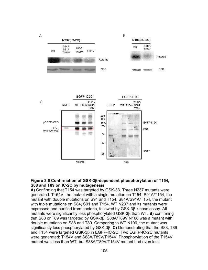
105
Figure 3.6 Confirmation of GSK-3β-dependent phosphorylation of T154, S88 and T89 on IC-2C by mutagenesis A) Confirming that T154 was targeted by GSK-3β. Three N237 mutants were generated: T154V, the mutant with a single mutation on T154; S91A/T154, the mutant with double mutations on S91 and T154; S84A/S91A/T154, the mutant with triple mutations on S84, S91 and T154. WT N237 and its mutants were expressed and purified from bacteria, followed by GSK-3β kinase assay. All mutants were significantly less phosphorylated GSK-3β than WT. B) confirming that S88 or T89 was targeted by GSK-3β. S88A/T89V N106 was a mutant with double mutations on S88 and T89. Comparing to WT N106, the mutant was significantly less phosphorylated by GSK-3β. C) Demonstrating that the S88, T89 and T154 were targeted GSK-3β in EGFP-IC-2C. Two EGFP-IC-2C mutants were generated: T154V and S88A/T89V/T154V. Phosphorylation of the T154V mutant was less than WT, but S88A/T89V/T154V mutant had even less

106
phosphorylation by GSK-3β. The phosphorylation signal in red rectangle was probably mainly from endogenous ICs pulled down by EGFP-IC-2C. Endogenous ICs were much less than EGFP-IC-2C in pulldown, but their phosphorylation signals are about the same.
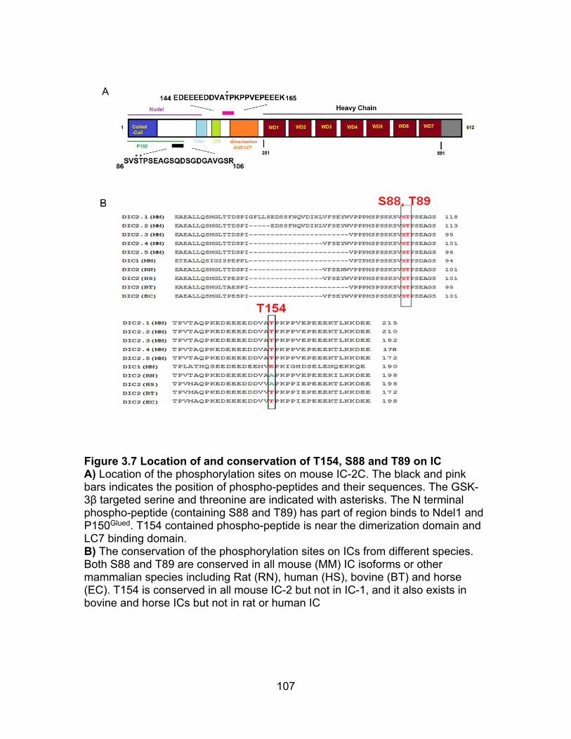
107
Figure 3.7 Location of and conservation of T154, S88 and T89 on IC A) Location of the phosphorylation sites on mouse IC-2C. The black and pink bars indicates the position of phospho-peptides and their sequences. The GSK-3β targeted serine and threonine are indicated with asterisks. The N terminal phospho-peptide (containing S88 and T89) has part of region binds to Ndel1 and P150Glued. T154 contained phospho-peptide is near the dimerization domain and LC7 binding domain. B) The conservation of the phosphorylation sites on ICs from different species. Both S88 and T89 are conserved in all mouse (MM) IC isoforms or other mammalian species including Rat (RN), human (HS), bovine (BT) and horse (EC). T154 is conserved in all mouse IC-2 but not in IC-1, and it also exists in bovine and horse ICs but not in rat or human IC
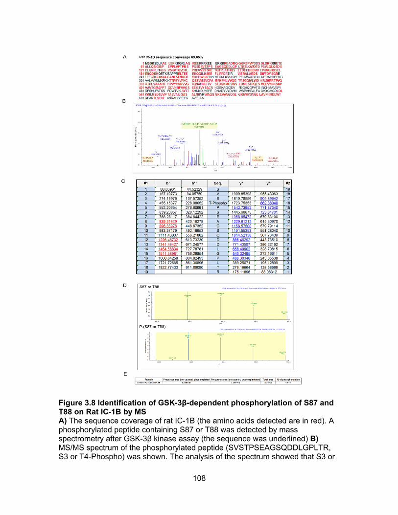
108
Figure 3.8 Identification of GSK-3β-dependent phosphorylation of S87 and T88 on Rat IC-1B by MS A) The sequence coverage of rat IC-1B (the amino acids detected are in red). A phosphorylated peptide containing S87 or T88 was detected by mass spectrometry after GSK-3β kinase assay (the sequence was underlined) B) MS/MS spectrum of the phosphorylated peptide (SVSTPSEAGSQDDLGPLTR, S3 or T4-Phospho) was shown. The analysis of the spectrum showed that S3 or

109
T4 (corresponding to S87 or T88 on IC-1B) was phosphorylated. C) The theoretical m/z values for MS/MS fragment ions of the phospho-peptide containing S87 or T88 was shown, and detected fragment ions were in red and underlined. Only the fragment ions without any losses were shown. D) The precursor intensity of the unphospho- and phospho- peptide containing S87 and T88. E) Quantification of phosphorylation efficiency of S87 or T88 on IC-1B by precursor area.

110
Figure 3.9 Identification of GSK-3β-dependent phosphorylation of S87 and T88 on mouse IC-1B by MS A) The sequence coverage of mouse rat IC-1B. A phosphorylated peptide containing S87 or T88 was detected by mass spectrometry after GSK-3β kinase

111
assay (the sequence was underlined) B) MS/MS spectrum of the phospho-peptide (SVSTPSDAGSQDSGDLGPLTR) containing S87 and T88 was detected. C) The theoretical m/z values for MS/MS fragment ions of the phospho-peptide containing S87 or T88 was shown, and detected fragment ions were in red and underlined. Only the fragment ions without any losses were shown. D) The precursor intensity of the unphospho- and phospho- peptide (SVSTPSDAGSQDSGDLGPLTR). E) Quantification of phosphorylation efficiency of S87 or T88 on IC-1B by precursor area.
112
CONCLUSIONS
In this study, we have provided evidence that GSK-3β directly regulates
dynein in both neurons and non-neuronal cells. GSK-3β interacts and
phosphorylates dynein in vitro. Dynein phosphorylation by GSK-3β reduces its
interaction with Ndel1, a regulator contributing to dynein force generation. Dynein
motility is stimulated both by pharmacological GSK-3β inhibitors and by
enhanced insulin signaling that leads to GSK-3β inactivation. Thus our study
connects a well-characterized insulin-signaling pathway, PI3K/AKT/GSK-3,
directly to dynein stimulation.
We find that dynein and APC physically interact with each other, which is
positively regulated by GSK-3β. In both cell lines and mouse models, we
demonstrate that rosiglitazone increases dynein activity and cell migration in WT
cells but not in Apc (min/+) cells, and rosiglitazone induces spindle misorientation
in Apc (min/+) cells but not in WT cells. We provide evidence that this involves
different PI3K/AKT/GSK-3β signaling responses to rosiglitazone between WT
and Apc (min/+) cells, and that ApcMin mutation negatively regulates the
interaction of dynein with WT APC. Our discovery of the interplay of APC and
dynein provides a new insight into how PPAR-γ signaling regulates cancer
development and other cellular events.
To dissect how GSK-3β and APC regulate dynein, we have demonstrated
a mass spectrometry (MS)-based systematic method to map phosphorylation

113
sites on IC. We identified that T154, S88 and T89 on IC-2C are targeted by GSK-
3β by using both MS and mutagenesis. S88 and T89 are conserved in all mouse
IC isoforms as well as in ICs from all other mammalian species examined.
Furthermore, we demonstrate that S87 or T88 on IC-1B (corresponding to S88 or
T89 on IC-2C) from both mouse and rat are targeted by GSK-3β using MS
analysis. The method has the potential to be applied to identify other bona fide
substrates of GSK-3β or other kinases.

114
REFERENCES
Acevedo, N., X. Wang, R.L. Dunn, and G.D. Smith. 2007. Glycogen synthase kinase-3 regulation of chromatin segregation and cytokinesis in mouse preimplantation embryos. Molecular reproduction and development. 74:178-188.
Allan, V.J. 2011. Cytoplasmic dynein. Biochemical Society transactions. 39:1169-1178.
Ayala, R., T. Shu, and L.H. Tsai. 2007. Trekking across the brain: the journey of neuronal migration. Cell. 128:29-43.
Baas, P.W., and S. Lin. 2011. Hooks and comets: The story of microtubule polarity orientation in the neuron. Dev Neurobiol. 71:403-418.
Bader, J.R., J.M. Kasuboski, M. Winding, P.S. Vaughan, E.H. Hinchcliffe, and K.T. Vaughan. 2011. Polo-like kinase1 is required for recruitment of dynein to kinetochores during mitosis. J Biol Chem. 286:20769-20777.
Baptista, F.I., M.J. Pinto, F. Elvas, T. Martins, R.D. Almeida, and A.F. Ambrosio. 2014. Diabetes induces changes in KIF1A, KIF5B and dynein distribution in the rat retina: Implications for axonal transport. Experimental eye research. 127:91-103.
Barton, N.R., and L.S. Goldstein. 1996. Going mobile: microtubule motors and chromosome segregation. Proceedings of the National Academy of Sciences of the United States of America. 93:1735-1742.
Bingham, J.B., S.J. King, and T.A. Schroer. 1998. Purification of dynactin and dynein from brain tissue. Methods in enzymology. 298:171-184.
Blokhuis, A.M., E.J. Groen, M. Koppers, L.H. van den Berg, and R.J. Pasterkamp. 2013. Protein aggregation in amyotrophic lateral sclerosis. Acta neuropathologica. 125:777-794.
Bobinnec, Y., X. Morin, and A. Debec. 2006. Shaggy/GSK-3beta kinase localizes to the centrosome and to specialized cytoskeletal structures in Drosophila. Cell motility and the cytoskeleton. 63:313-320.
Bradshaw, N.J., W. Hennah, and D.C. Soares. 2013. NDE1 and NDEL1: twin neurodevelopmental proteins with similar 'nature' but different 'nurture'. Biomolecular concepts. 4:447-464.
Brill, L.B., 2nd, and K.K. Pfister. 2000. Biochemical and molecular analysis of the mammalian cytoplasmic dynein intermediate chain. Methods. 22:307-316.
Brinkley, B.R. 1985. Microtubule organizing centers. Annual review of cell biology. 1:145-172.
Burgermeister, E., L. Tencer, and M. Liscovitch. 2003. Peroxisome proliferator-activated receptor-gamma upregulates caveolin-1 and caveolin-2 expression in human carcinoma cells. Oncogene. 22:3888-3900.
115
Carnero, A., and J.M. Paramio. 2014. The PTEN/PI3K/AKT Pathway in vivo, Cancer Mouse Models. Frontiers in oncology. 4:252.
Castano, Z., P.R. Gordon-Weeks, and R.M. Kypta. 2010. The neuron-specific isoform of glycogen synthase kinase-3beta is required for axon growth. J Neurochem. 113:117-130.
Chang, C.H., J.W. Lin, L.C. Wu, M.S. Lai, L.M. Chuang, and K.A. Chan. 2012. Association of thiazolidinediones with liver cancer and colorectal cancer in type 2 diabetes mellitus. Hepatology. 55:1462-1472.
Cole, A., S. Frame, and P. Cohen. 2004. Further evidence that the tyrosine phosphorylation of glycogen synthase kinase-3 (GSK3) in mammalian cells is an autophosphorylation event. The Biochemical journal. 377:249-255.
Cross, D.A., D.R. Alessi, P. Cohen, M. Andjelkovich, and B.A. Hemmings. 1995. Inhibition of glycogen synthase kinase-3 by insulin mediated by protein kinase B. Nature. 378:785-789.
Culver-Hanlon, T.L., S.A. Lex, A.D. Stephens, N.J. Quintyne, and S.J. King. 2006. A microtubule-binding domain in dynactin increases dynein processivity by skating along microtubules. Nat Cell Biol. 8:264-270.
Doble, B.W., and J.R. Woodgett. 2003. GSK-3: tricks of the trade for a multi-tasking kinase. Journal of cell science. 116:1175-1186.
Dobrowolski, R., and E.M. De Robertis. 2012. Endocytic control of growth factor signalling: multivesicular bodies as signalling organelles. Nature reviews. Molecular cell biology. 13:53-60.
Dolma, K., G.J. Iacobucci, K. Hong Zheng, J. Shandilya, E. Toska, J.A. White, 2nd, E. Spina, and S. Gunawardena. 2014. Presenilin influences glycogen synthase kinase-3 beta (GSK-3beta) for kinesin-1 and dynein function during axonal transport. Human molecular genetics. 23:1121-1133.
Dujardin, D.L., and R.B. Vallee. 2002. Dynein at the cortex. Current opinion in cell biology. 14:44-49.
Eldar-Finkelman, H. 2002. Glycogen synthase kinase 3: an emerging therapeutic target. Trends Mol Med. 8:126-132.
Eldar-Finkelman, H., and A. Martinez. 2011. GSK-3 Inhibitors: Preclinical and Clinical Focus on CNS. Frontiers in molecular neuroscience. 4:32.
Embi, N., D.B. Rylatt, and P. Cohen. 1980. Glycogen synthase kinase-3 from rabbit skeletal muscle. Separation from cyclic-AMP-dependent protein kinase and phosphorylase kinase. Eur J Biochem. 107:519-527.
Eschbach, J., and L. Dupuis. 2011. Cytoplasmic dynein in neurodegeneration. Pharmacology & therapeutics. 130:348-363.
Espada, J., M.B. Calvo, S. Diaz-Prado, and V. Medina. 2009. Wnt signalling and cancer stem cells. Clinical & translational oncology : official publication of the Federation of Spanish Oncology Societies and of the National Cancer Institute of Mexico. 11:411-427.
Etienne-Manneville, S. 2009. APC in cell migration. Advances in experimental medicine and biology. 656:30-40.
Etienne-Manneville, S. 2010. From signaling pathways to microtubule dynamics: the key players. Current opinion in cell biology. 22:104-111.

116
Fang, X., S. Yu, J.L. Tanyi, Y. Lu, J.R. Woodgett, and G.B. Mills. 2002. Convergence of multiple signaling cascades at glycogen synthase kinase 3: Edg receptor-mediated phosphorylation and inactivation by lysophosphatidic acid through a protein kinase C-dependent intracellular pathway. Mol Cell Biol. 22:2099-2110.
Fang, X., S.X. Yu, Y. Lu, R.C. Bast, Jr., J.R. Woodgett, and G.B. Mills. 2000. Phosphorylation and inactivation of glycogen synthase kinase 3 by protein kinase A. Proc Natl Acad Sci U S A. 97:11960-11965.
Ferrarese, A., O. Marin, V.H. Bustos, A. Venerando, M. Antonelli, J.E. Allende, and L.A. Pinna. 2007. Chemical dissection of the APC Repeat 3 multistep phosphorylation by the concerted action of protein kinases CK1 and GSK3. Biochemistry. 46:11902-11910.
Fiol, C.J., A.M. Mahrenholz, Y. Wang, R.W. Roeske, and P.J. Roach. 1987. Formation of protein kinase recognition sites by covalent modification of the substrate. Molecular mechanism for the synergistic action of casein kinase II and glycogen synthase kinase 3. J Biol Chem. 262:14042-14048.
Fleming, E.S., M. Temchin, Q. Wu, L. Maggio-Price, and J.S. Tirnauer. 2009. Spindle misorientation in tumors from APC(min/+) mice. Molecular carcinogenesis. 48:592-598.
Franker, M.A., and C.C. Hoogenraad. 2013. Microtubule-based transport - basic mechanisms, traffic rules and role in neurological pathogenesis. Journal of cell science. 126:2319-2329.
Friedrich, T., B. Richter, T. Gaiser, C. Weiss, K.P. Janssen, H. Einwachter, R.M. Schmid, M.P. Ebert, and E. Burgermeister. 2013. Deficiency of caveolin-1 in Apc(min/+) mice promotes colorectal tumorigenesis. Carcinogenesis. 34:2109-2118.
Fu, M.M., and E.L. Holzbaur. 2014. Integrated regulation of motor-driven organelle transport by scaffolding proteins. Trends in cell biology. 24:564-574.
Fumoto, K., P.C. Lee, H. Saya, and A. Kikuchi. 2008. AIP regulates stability of Aurora-A at early mitotic phase coordinately with GSK-3beta. Oncogene. 27:4478-4487.
Gennerich, A., and R.D. Vale. 2009. Walking the walk: how kinesin and dynein coordinate their steps. Current opinion in cell biology. 21:59-67.
Gibbons, I.R. 1988. Dynein ATPases as microtubule motors. The Journal of biological chemistry. 263:15837-15840.
Gurnell, M., J.M. Wentworth, M. Agostini, M. Adams, T.N. Collingwood, C. Provenzano, P.O. Browne, O. Rajanayagam, T.P. Burris, J.W. Schwabe, M.A. Lazar, and V.K. Chatterjee. 2000. A dominant-negative peroxisome proliferator-activated receptor gamma (PPARgamma) mutant is a constitutive repressor and inhibits PPARgamma-mediated adipogenesis. The Journal of biological chemistry. 275:5754-5759.
Ha, J., K.W. Lo, K.R. Myers, T.M. Carr, M.K. Humsi, B.A. Rasoul, R.A. Segal, and K.K. Pfister. 2008. A neuron-specific cytoplasmic dynein isoform preferentially transports TrkB signaling endosomes. J Cell Biol. 181:1027-1039.
117
Hanson, C.A., and J.R. Miller. 2005. Non-traditional roles for the Adenomatous Polyposis Coli (APC) tumor suppressor protein. Gene. 361:1-12.
Harada, A., Y. Takei, Y. Kanai, Y. Tanaka, S. Nonaka, and N. Hirokawa. 1998. Golgi vesiculation and lysosome dispersion in cells lacking cytoplasmic dynein. The Journal of cell biology. 141:51-59.
Harwood, A.J., J.E. Forde-Thomas, H. Williams, M. Samereier, and A. Muller-Taubenberger. 2013. Aberrant spindle dynamics and cytokinesis in Dictyostelium discoideum cells that lack glycogen synthase kinase 3. European journal of cell biology. 92:222-228.
Hebbar, S., A.M. Guillotte, M.T. Mesngon, Q. Zhou, A. Wynshaw-Boris, and D.S. Smith. 2008a. Genetic enhancement of the Lis1+/- phenotype by a heterozygous mutation in the adenomatous polyposis coli gene. Developmental neuroscience. 30:157-170.
Hebbar, S., M.T. Mesngon, A.M. Guillotte, B. Desai, R. Ayala, and D.S. Smith. 2008b. Lis1 and Ndel1 influence the timing of nuclear envelope breakdown in neural stem cells. J Cell Biol. 182:1063-1071.
Heerssen, H.M., M.F. Pazyra, and R.A. Segal. 2004. Dynein motors transport activated Trks to promote survival of target-dependent neurons. Nature neuroscience. 7:596-604.
Hernandez, R., T. Teruel, and M. Lorenzo. 2003. Rosiglitazone produces insulin sensitisation by increasing expression of the insulin receptor and its tyrosine kinase activity in brown adipocytes. Diabetologia. 46:1618-1628.
Hirokawa, N., S. Niwa, and Y. Tanaka. 2010. Molecular motors in neurons: transport mechanisms and roles in brain function, development, and disease. Neuron. 68:610-638.
Huang, J., T. Imamura, and J.M. Olefsky. 2001. Insulin can regulate GLUT4 internalization by signaling to Rab5 and the motor protein dynein. Proceedings of the National Academy of Sciences of the United States of America. 98:13084-13089.
Huang, J., A.J. Roberts, A.E. Leschziner, and S.L. Reck-Peterson. 2012. Lis1 acts as a "clutch" between the ATPase and microtubule-binding domains of the dynein motor. Cell. 150:975-986.
Ikeda, K., O. Zhapparova, I. Brodsky, I. Semenova, J.S. Tirnauer, I. Zaliapin, and V. Rodionov. 2011. CK1 activates minus-end-directed transport of membrane organelles along microtubules. Mol Biol Cell. 22:1321-1329.
Ikeda, S., M. Kishida, Y. Matsuura, H. Usui, and A. Kikuchi. 2000. GSK-3beta-dependent phosphorylation of adenomatous polyposis coli gene product can be modulated by beta-catenin and protein phosphatase 2A complexed with Axin. Oncogene. 19:537-545.
Ilouz, R., S. Pietrokovski, M. Eisenstein, and H. Eldar-Finkelman. 2008. New insights into the autoinhibition mechanism of glycogen synthase kinase-3beta. J Mol Biol. 383:999-1007.
Ishiguro, K., A. Shiratsuchi, S. Sato, A. Omori, M. Arioka, S. Kobayashi, T. Uchida, and K. Imahori. 1993. Glycogen synthase kinase 3 beta is identical to tau protein kinase I generating several epitopes of paired helical filaments. FEBS Lett. 325:167-172.
118
Izumi, N., K. Fumoto, S. Izumi, and A. Kikuchi. 2008. GSK-3beta regulates proper mitotic spindle formation in cooperation with a component of the gamma-tubulin ring complex, GCP5. The Journal of biological chemistry. 283:12981-12991.
Jimbo, T., Y. Kawasaki, R. Koyama, R. Sato, S. Takada, K. Haraguchi, and T. Akiyama. 2002. Identification of a link between the tumour suppressor APC and the kinesin superfamily. Nature cell biology. 4:323-327.
Kardon, J.R., and R.D. Vale. 2009. Regulators of the cytoplasmic dynein motor. Nature reviews. Molecular cell biology. 10:854-865.
Karki, S., and E.L. Holzbaur. 1995. Affinity chromatography demonstrates a direct binding between cytoplasmic dynein and the dynactin complex. J Biol Chem. 270:28806-28811.
Kim, D., S. Kim, H. Koh, S.O. Yoon, A.S. Chung, K.S. Cho, and J. Chung. 2001. Akt/PKB promotes cancer cell invasion via increased motility and metalloproteinase production. FASEB journal : official publication of the Federation of American Societies for Experimental Biology. 15:1953-1962.
King, S.J., C.L. Brown, K.C. Maier, N.J. Quintyne, and T.A. Schroer. 2003. Analysis of the dynein-dynactin interaction in vitro and in vivo. Mol Biol Cell. 14:5089-5097.
Kongkanuntn, R., V.J. Bubb, O.J. Sansom, A.H. Wyllie, D.J. Harrison, and A.R. Clarke. 1999. Dysregulated expression of beta-catenin marks early neoplastic change in Apc mutant mice, but not all lesions arising in Msh2 deficient mice. Oncogene. 18:7219-7225.
Kotak, S., and P. Gonczy. 2013. Mechanisms of spindle positioning: cortical force generators in the limelight. Current opinion in cell biology. 25:741-748.
Kuta, A., W. Deng, A. Morsi El-Kadi, G.T. Banks, M. Hafezparast, K.K. Pfister, and E.M. Fisher. 2010. Mouse cytoplasmic dynein intermediate chains: identification of new isoforms, alternative splicing and tissue distribution of transcripts. PLoS One. 5:e11682.
Lam, C., M.A. Vergnolle, L. Thorpe, P.G. Woodman, and V.J. Allan. 2010. Functional interplay between LIS1, NDE1 and NDEL1 in dynein-dependent organelle positioning. Journal of cell science. 123:202-212.
Landberg, G., M. Erlanson, G. Roos, E.M. Tan, and C.A. Casiano. 1996. Nuclear autoantigen p330d/CENP-F: a marker for cell proliferation in human malignancies. Cytometry. 25:90-98.
Landreth, G., Q. Jiang, S. Mandrekar, and M. Heneka. 2008. PPARgamma agonists as therapeutics for the treatment of Alzheimer's disease. Neurotherapeutics. 5:481-489.
Lansbergen, G., I. Grigoriev, Y. Mimori-Kiyosue, T. Ohtsuka, S. Higa, I. Kitajima, J. Demmers, N. Galjart, A.B. Houtsmuller, F. Grosveld, and A. Akhmanova. 2006. CLASPs attach microtubule plus ends to the cell cortex through a complex with LL5beta. Dev Cell. 11:21-32.
Larsson, S.C., N. Orsini, and A. Wolk. 2005. Diabetes mellitus and risk of colorectal cancer: a meta-analysis. J Natl Cancer Inst. 97:1679-1687.
Lauffenburger, D.A., and A.F. Horwitz. 1996. Cell migration: a physically integrated molecular process. Cell. 84:359-369.

119
Lefebvre, A.M., I. Chen, P. Desreumaux, J. Najib, J.C. Fruchart, K. Geboes, M. Briggs, R. Heyman, and J. Auwerx. 1998. Activation of the peroxisome proliferator-activated receptor gamma promotes the development of colon tumors in C57BL/6J-APCMin/+ mice. Nature medicine. 4:1053-1057.
Li, J., W.L. Lee, and J.A. Cooper. 2005. NudEL targets dynein to microtubule ends through LIS1. Nat Cell Biol. 7:686-690.
Liao, H., R.J. Winkfein, G. Mack, J.B. Rattner, and T.J. Yen. 1995. CENP-F is a protein of the nuclear matrix that assembles onto kinetochores at late G2 and is rapidly degraded after mitosis. The Journal of cell biology. 130:507-518.
Liu, X., H. Gong, and K. Huang. 2013. Oncogenic role of kinesin proteins and targeting kinesin therapy. Cancer science. 104:651-656.
Lo, K.W., H.M. Kan, and K.K. Pfister. 2006. Identification of a novel region of the cytoplasmic Dynein intermediate chain important for dimerization in the absence of the light chains. J Biol Chem. 281:9552-9559.
Mallik, R., A.K. Rai, P. Barak, A. Rai, and A. Kunwar. 2013. Teamwork in microtubule motors. Trends in cell biology. 23:575-582.
Mandelkow, E.M., G. Drewes, J. Biernat, N. Gustke, J. Van Lint, J.R. Vandenheede, and E. Mandelkow. 1992. Glycogen synthase kinase-3 and the Alzheimer-like state of microtubule-associated protein tau. FEBS Lett. 314:315-321.
Marin, H.E., M.A. Peraza, A.N. Billin, T.M. Willson, J.M. Ward, M.J. Kennett, F.J. Gonzalez, and J.M. Peters. 2006. Ligand activation of peroxisome proliferator-activated receptor beta inhibits colon carcinogenesis. Cancer research. 66:4394-4401.
Markus, S.M., J.J. Punch, and W.L. Lee. 2009. Motor- and tail-dependent targeting of dynein to microtubule plus ends and the cell cortex. Current biology : CB. 19:196-205.
Marmol, F. 2008. Lithium: bipolar disorder and neurodegenerative diseases Possible cellular mechanisms of the therapeutic effects of lithium. Prog Neuropsychopharmacol Biol Psychiatry. 32:1761-1771.
Marsh, M., and H.T. McMahon. 1999. The structural era of endocytosis. Science. 285:215-220.
McKenney, R.J., W. Huynh, M.E. Tanenbaum, G. Bhabha, and R.D. Vale. 2014. Activation of cytoplasmic dynein motility by dynactin-cargo adapter complexes. Science. 345:337-341.
McKenney, R.J., M. Vershinin, A. Kunwar, R.B. Vallee, and S.P. Gross. 2010. LIS1 and NudE induce a persistent dynein force-producing state. Cell. 141:304-314.
McKenney, R.J., S.J. Weil, J. Scherer, and R.B. Vallee. 2011. Mutually Exclusive Cytoplasmic Dynein Regulation by NudE-Lis1 and Dynactin. J Biol Chem. 286:39615-39622.
Meijer, L., M. Flajolet, and P. Greengard. 2004. Pharmacological inhibitors of glycogen synthase kinase 3. Trends Pharmacol Sci. 25:471-480.
Mesngon, M.T., C. Tarricone, S. Hebbar, A.M. Guillotte, E.W. Schmitt, L. Lanier, A. Musacchio, S.J. King, and D.S. Smith. 2006. Regulation of cytoplasmic
120
dynein ATPase by Lis1. The Journal of neuroscience : the official journal of the Society for Neuroscience. 26:2132-2139.
Mimori-Kiyosue, Y., N. Shiina, and S. Tsukita. 2000. Adenomatous polyposis coli (APC) protein moves along microtubules and concentrates at their growing ends in epithelial cells. The Journal of cell biology. 148:505-518.
Mimori-Kiyosue, Y., and S. Tsukita. 2003. "Search-and-capture" of microtubules through plus-end-binding proteins (+TIPs). Journal of biochemistry. 134:321-326.
Minde, D.P., Z. Anvarian, S.G.D. Rudiger, and M.M. Maurice. 2011. Messing up disorder: how do missense mutations in the tumor suppressor protein APC lead to cancer? Mol Cancer. 10.
Mitchell, D.J., K.R. Blasier, E.D. Jeffery, M.W. Ross, A.K. Pullikuth, D. Suo, J. Park, W.R. Smiley, K.W. Lo, J. Shabanowitz, C.D. Deppmann, J.C. Trinidad, D.F. Hunt, A.D. Catling, and K.K. Pfister. 2012. Trk activation of the ERK1/2 kinase pathway stimulates intermediate chain phosphorylation and recruits cytoplasmic dynein to signaling endosomes for retrograde axonal transport. J Neurosci. 32:15495-15510.
Mogensen, M.M., J.B. Tucker, J.B. Mackie, A.R. Prescott, and I.S. Nathke. 2002. The adenomatous polyposis coli protein unambiguously localizes to microtubule plus ends and is involved in establishing parallel arrays of microtubule bundles in highly polarized epithelial cells. The Journal of cell biology. 157:1041-1048.
Moon, H.M., Y.H. Youn, H. Pemble, J. Yingling, T. Wittmann, and A. Wynshaw-Boris. 2014. LIS1 controls mitosis and mitotic spindle organization via the LIS1-NDEL1-dynein complex. Human molecular genetics. 23:449-466.
Morfini, G., G. Szebenyi, R. Elluru, N. Ratner, and S.T. Brady. 2002. Glycogen synthase kinase 3 phosphorylates kinesin light chains and negatively regulates kinesin-based motility. EMBO J. 21:281-293.
Moughamian, A.J., G.E. Osborn, J.E. Lazarus, S. Maday, and E.L. Holzbaur. 2013. Ordered recruitment of dynactin to the microtubule plus-end is required for efficient initiation of retrograde axonal transport. J Neurosci. 33:13190-13203.
Mukai, F., K. Ishiguro, Y. Sano, and S.C. Fujita. 2002. Alternative splicing isoform of tau protein kinase I/glycogen synthase kinase 3beta. J Neurochem. 81:1073-1083.
Murphy, J.T., J.M. Tucker, C. Davis, and F.G. Berger. 2004. Raltitrexed increases tumorigenesis as a single agent yet exhibits anti-tumor synergy with 5-fluorouracil in ApcMin/+ mice. Cancer biology & therapy. 3:1169-1176.
Nathke, I.S. 2004. The adenomatous polyposis coli protein: the Achilles heel of the gut epithelium. Annual review of cell and developmental biology. 20:337-366.
Nyarko, A., Y. Song, and E. Barbar. 2012. Intrinsic disorder in dynein intermediate chain modulates its interactions with NudE and dynactin. J Biol Chem. 287:24884-24893.
121
Pal, I., and M. Mandal. 2012. PI3K and Akt as molecular targets for cancer therapy: current clinical outcomes. Acta pharmacologica Sinica. 33:1441-1458.
Pandey, J.P., and D.S. Smith. 2011. A Cdk5-dependent switch regulates Lis1/Ndel1/dynein-driven organelle transport in adult axons. J Neurosci. 31:17207-17219.
Pellman, D. 2001. Cancer. A CINtillating new job for the APC tumor suppressor. Science. 291:2555-2556.
Peters, J.M., Y.M. Shah, and F.J. Gonzalez. 2012. The role of peroxisome proliferator-activated receptors in carcinogenesis and chemoprevention. Nature reviews. Cancer. 12:181-195.
Pfister, K.K., P.R. Shah, H. Hummerich, A. Russ, J. Cotton, A.A. Annuar, S.M. King, and E.M. Fisher. 2006. Genetic analysis of the cytoplasmic dynein subunit families. PLoS Genet. 2:e1.
Phelps, R.A., S. Chidester, S. Dehghanizadeh, J. Phelps, I.T. Sandoval, K. Rai, T. Broadbent, S. Sarkar, R.W. Burt, and D.A. Jones. 2009. A two-step model for colon adenoma initiation and progression caused by APC loss. Cell. 137:623-634.
Polakis, P. 2002. Casein kinase 1: a Wnt'er of disconnect. Current biology : CB. 12:R499-R501.
Pullikuth, A.K., A. Ozdemir, D. Cardenas, E. Bailey, N.E. Sherman, K.K. Pfister, and A.D. Catling. 2013. Epidermal growth factor stimulates extracellular-signal regulated kinase phosphorylation of a novel site on cytoplasmic Dynein intermediate chain 2. International journal of molecular sciences. 14:3595-3620.
Quintyne, N.J., and T.A. Schroer. 2002. Distinct cell cycle-dependent roles for dynactin and dynein at centrosomes. The Journal of cell biology. 159:245-254.
Quyn, A.J., P.L. Appleton, F.A. Carey, R.J. Steele, N. Barker, H. Clevers, R.A. Ridgway, O.J. Sansom, and I.S. Nathke. 2010. Spindle orientation bias in gut epithelial stem cell compartments is lost in precancerous tissue. Cell stem cell. 6:175-181.
Raaijmakers, J.A., and R.H. Medema. 2014. Function and regulation of dynein in mitotic chromosome segregation. Chromosoma. 123:407-422.
Raaijmakers, J.A., M.E. Tanenbaum, and R.H. Medema. 2013. Systematic dissection of dynein regulators in mitosis. The Journal of cell biology. 201:201-215.
Rai, A.K., A. Rai, A.J. Ramaiya, R. Jha, and R. Mallik. 2013. Molecular adaptations allow dynein to generate large collective forces inside cells. Cell. 152:172-182.
Reya, T., and H. Clevers. 2005. Wnt signalling in stem cells and cancer. Nature. 434:843-850.
Ridley, A.J., M.A. Schwartz, K. Burridge, R.A. Firtel, M.H. Ginsberg, G. Borisy, J.T. Parsons, and A.R. Horwitz. 2003. Cell migration: integrating signals from front to back. Science. 302:1704-1709.
122
Roberts, A.J., B.S. Goodman, and S.L. Reck-Peterson. 2014. Reconstitution of dynein transport to the microtubule plus end by kinesin. eLife. 3:e02641.
Rodionov, V., E. Nadezhdina, and G. Borisy. 1999. Centrosomal control of microtubule dynamics. Proceedings of the National Academy of Sciences of the United States of America. 96:115-120.
Rubinsztein, D.C. 2006. The roles of intracellular protein-degradation pathways in neurodegeneration. Nature. 443:780-786.
Saez, E., P. Tontonoz, M.C. Nelson, J.G. Alvarez, U.T. Ming, S.M. Baird, V.A. Thomazy, and R.M. Evans. 1998. Activators of the nuclear receptor PPARgamma enhance colon polyp formation. Nature medicine. 4:1058-1061.
Saltiel, A.R., and J.E. Pessin. 2002. Insulin signaling pathways in time and space. Trends in cell biology. 12:65-71.
Sansom, O.J., K.R. Reed, A.J. Hayes, H. Ireland, H. Brinkmann, I.P. Newton, E. Batlle, P. Simon-Assmann, H. Clevers, I.S. Nathke, A.R. Clarke, and D.J. Winton. 2004. Loss of Apc in vivo immediately perturbs Wnt signaling, differentiation, and migration. Genes & development. 18:1385-1390.
Sato, N., and R. Morishita. 2014. Brain alterations and clinical symptoms of dementia in diabetes: abeta/tau-dependent and independent mechanisms. Frontiers in endocrinology. 5:143.
Schenck, A., L. Goto-Silva, C. Collinet, M. Rhinn, A. Giner, B. Habermann, M. Brand, and M. Zerial. 2008. The endosomal protein Appl1 mediates Akt substrate specificity and cell survival in vertebrate development. Cell. 133:486-497.
Schiavo, G., L. Greensmith, M. Hafezparast, and E.M. Fisher. 2013. Cytoplasmic dynein heavy chain: the servant of many masters. Trends in neurosciences. 36:641-651.
Schlager, M.A., H.T. Hoang, L. Urnavicius, S.L. Bullock, and A.P. Carter. 2014. In vitro reconstitution of a highly processive recombinant human dynein complex. The EMBO journal. 33:1855-1868.
Schmoranzer, J., J.P. Fawcett, M. Segura, S. Tan, R.B. Vallee, T. Pawson, and G.G. Gundersen. 2009. Par3 and dynein associate to regulate local microtubule dynamics and centrosome orientation during migration. Current biology : CB. 19:1065-1074.
Schroer, T.A. 2004. Dynactin. Annu Rev Cell Dev Biol. 20:759-779. Seo, M., S. Lee, J.H. Kim, W.H. Lee, G. Hu, S.J. Elledge, and K. Suk. 2014.
RNAi-based functional selection identifies novel cell migration determinants dependent on PI3K and AKT pathways. Nature communications. 5:5217.
Smith, D.S., M. Niethammer, R. Ayala, Y. Zhou, M.J. Gambello, A. Wynshaw-Boris, and L.H. Tsai. 2000. Regulation of cytoplasmic dynein behaviour and microtubule organization by mammalian Lis1. Nature cell biology. 2:767-775.
Soutar, M.P., W.Y. Kim, R. Williamson, M. Peggie, C.J. Hastie, H. McLauchlan, W.D. Snider, P.R. Gordon-Weeks, and C. Sutherland. 2010. Evidence that
123
glycogen synthase kinase-3 isoforms have distinct substrate preference in the brain. J Neurochem. 115:974-983.
Stamos, J.L., and W.I. Weis. 2013. The beta-catenin destruction complex. Cold Spring Harbor perspectives in biology. 5:a007898.
Stehman, S.A., Y. Chen, R.J. McKenney, and R.B. Vallee. 2007. NudE and NudEL are required for mitotic progression and are involved in dynein recruitment to kinetochores. J Cell Biol. 178:583-594.
Su, L.K., K.W. Kinzler, B. Vogelstein, A.C. Preisinger, A.R. Moser, C. Luongo, K.A. Gould, and W.F. Dove. 1992. Multiple intestinal neoplasia caused by a mutation in the murine homolog of the APC gene. Science. 256:668-670.
Su, W., C.R. Bush, B.M. Necela, S.R. Calcagno, N.R. Murray, A.P. Fields, and E.A. Thompson. 2007. Differential expression, distribution, and function of PPAR-gamma in the proximal and distal colon. Physiol Genomics. 30:342-353.
Sutherland, C. 2011. What Are the bona fide GSK3 Substrates? Int J Alzheimers Dis. 2011:505607.
Taketo, M.M., and W. Edelmann. 2009. Mouse models of colon cancer. Gastroenterology. 136:780-798.
Tanzi, R.E., and L. Bertram. 2005. Twenty years of the Alzheimer's disease amyloid hypothesis: a genetic perspective. Cell. 120:545-555.
ter Haar, E., J.T. Coll, D.A. Austen, H.M. Hsiao, L. Swenson, and J. Jain. 2001. Structure of GSK3beta reveals a primed phosphorylation mechanism. Nat Struct Biol. 8:593-596.
Tighe, A., A. Ray-Sinha, O.D. Staples, and S.S. Taylor. 2007. GSK-3 inhibitors induce chromosome instability. BMC cell biology. 8:34.
Tsai, J.W., K.H. Bremner, and R.B. Vallee. 2007. Dual subcellular roles for LIS1 and dynein in radial neuronal migration in live brain tissue. Nature neuroscience. 10:970-979.
Tsai, L.H., and J.G. Gleeson. 2005. Nucleokinesis in neuronal migration. Neuron. 46:383-388.
Vale, R.D. 2003. The molecular motor toolbox for intracellular transport. Cell. 112:467-480.
Vallee, R.B., G.E. Seale, and J.W. Tsai. 2009. Emerging roles for myosin II and cytoplasmic dynein in migrating neurons and growth cones. Trends in cell biology. 19:347-355.
Vaughan, K.T., and R.B. Vallee. 1995. Cytoplasmic dynein binds dynactin through a direct interaction between the intermediate chains and p150Glued. J Cell Biol. 131:1507-1516.
Vaughan, P.S., J.D. Leszyk, and K.T. Vaughan. 2001. Cytoplasmic dynein intermediate chain phosphorylation regulates binding to dynactin. J Biol Chem. 276:26171-26179.
Velebit, J., H.H. Chowdhury, M. Kreft, and R. Zorec. 2011. Rosiglitazone balances insulin-induced exo- and endocytosis in single 3T3-L1 adipocytes. Mol Cell Endocrinol. 333:70-77.
124
Wakefield, J.G., D.J. Stephens, and J.M. Tavare. 2003. A role for glycogen synthase kinase-3 in mitotic spindle dynamics and chromosome alignment. Journal of cell science. 116:637-646.
Wang, Q.M., C.J. Fiol, A.A. DePaoli-Roach, and P.J. Roach. 1994. Glycogen synthase kinase-3 beta is a dual specificity kinase differentially regulated by tyrosine and serine/threonine phosphorylation. J Biol Chem. 269:14566-14574.
Wang, S., S.A. Ketcham, A. Schon, B. Goodman, Y. Wang, J. Yates, 3rd, E. Freire, T.A. Schroer, and Y. Zheng. 2013. Nudel/NudE and Lis1 promote dynein and dynactin interaction in the context of spindle morphogenesis. Mol Biol Cell. 24:3522-3533.
Wang, S., and Y. Zheng. 2011. Identification of a novel dynein binding domain in nudel essential for spindle pole organization in Xenopus egg extract. The Journal of biological chemistry. 286:587-593.
Wang, Y., Y. Azuma, D.B. Friedman, R.J. Coffey, and K.L. Neufeld. 2009. Novel association of APC with intermediate filaments identified using a new versatile APC antibody. BMC cell biology. 10:75.
Weaver, C., C. Leidel, L. Szpankowski, N.M. Farley, G.T. Shubeita, and L.S. Goldstein. 2013. Endogenous GSK-3/shaggy regulates bidirectional axonal transport of the amyloid precursor protein. Traffic. 14:295-308.
Whitehead, R.H., and J.L. Joseph. 1994. Derivation of conditionally immortalized cell lines containing the Min mutation from the normal colonic mucosa and other tissues of an "Immortomouse"/Min hybrid. Epithelial cell biology. 3:119-125.
Whitehead, R.H., P.E. VanEeden, M.D. Noble, P. Ataliotis, and P.S. Jat. 1993. Establishment of conditionally immortalized epithelial cell lines from both colon and small intestine of adult H-2Kb-tsA58 transgenic mice. Proceedings of the National Academy of Sciences of the United States of America. 90:587-591.
Whyte, J., J.R. Bader, S.B. Tauhata, M. Raycroft, J. Hornick, K.K. Pfister, W.S. Lane, G.K. Chan, E.H. Hinchcliffe, P.S. Vaughan, and K.T. Vaughan. 2008. Phosphorylation regulates targeting of cytoplasmic dynein to kinetochores during mitosis. J Cell Biol. 183:819-834.
Wickstead, B., and K. Gull. 2007. Dyneins across eukaryotes: a comparative genomic analysis. Traffic. 8:1708-1721.
Wojcik, E.J. 2008. A mitotic role for GSK-3beta kinase in Drosophila. Cell cycle. 7:3699-3708.
Wood-Kaczmar, A., M. Kraus, K. Ishiguro, K.L. Philpott, and P.R. Gordon-Weeks. 2009. An alternatively spliced form of glycogen synthase kinase-3beta is targeted to growing neurites and growth cones. Mol Cell Neurosci. 42:184-194.
Woodgett, J.R. 1991. cDNA cloning and properties of glycogen synthase kinase-3. Methods Enzymol. 200:564-577.
Wu, C., B. Cui, L. He, L. Chen, and W.C. Mobley. 2009. The coming of age of axonal neurotrophin signaling endosomes. Journal of proteomics. 72:46-55.

125
Yamada, M., S. Toba, Y. Yoshida, K. Haratani, D. Mori, Y. Yano, Y. Mimori-Kiyosue, T. Nakamura, K. Itoh, S. Fushiki, M. Setou, A. Wynshaw-Boris, T. Torisawa, Y.Y. Toyoshima, and S. Hirotsune. 2008. LIS1 and NDEL1 coordinate the plus-end-directed transport of cytoplasmic dynein. The EMBO journal. 27:2471-2483.
Yamada, Y., and H. Mori. 2007. Multistep carcinogenesis of the colon in Apc(Min/+) mouse. Cancer science. 98:6-10.
Yano, H., F.S. Lee, H. Kong, J. Chuang, J. Arevalo, P. Perez, C. Sung, and M.V. Chao. 2001. Association of Trk neurotrophin receptors with components of the cytoplasmic dynein motor. J Neurosci. 21:RC125.
Yingling, J., Y.H. Youn, D. Darling, K. Toyo-Oka, T. Pramparo, S. Hirotsune, and A. Wynshaw-Boris. 2008. Neuroepithelial stem cell proliferation requires LIS1 for precise spindle orientation and symmetric division. Cell. 132:474-486.
Zhang, J., L. Zhuang, Y. Lee, J.F. Abenza, M.A. Penalva, and X. Xiang. The microtubule plus-end localization of Aspergillus dynein is important for dynein-early-endosome interaction but not for dynein ATPase activation. Journal of cell science. 123:3596-3604.
Zweifel, L.S., R. Kuruvilla, and D.D. Ginty. 2005. Functions and mechanisms of retrograde neurotrophin signalling. Nature reviews. Neuroscience. 6:615-625.