the roleof mastocytes in the regional fixation of blood-borne particles
TRANSCRIPT

38
THE ROLE OF MASTOCYTES IN THE REGIONAL FIXATIONOF BLOOD-BORNE PARTICLES
H. SELYE, G. GABBIANI AND BEATRIZ TUCHWEBER
From 1'Institut de Medecine et de Chirurgie experimentales,(Tn,iversite de Montre'al, Montreal, Canada
Received fo- publication AMay 28, 1962
THE fact that inflamed tissue can fix and retain blood-borne particulate sub-stances (blood-cells, microbes, chemicals) has long been known. A special formof this inflammatory sequestration is " angiotaxis ", the storage of granules(e.g., of dyes, carbon or microbes) in endothelia that do not belong to the RES-system proper in that they do not phagocytose except wheni they are activate(dby inflammatory irritants. In the course of angiotaxis, the blood-borne particlesfirst become adherent to the iimer surface of the endothelial cells in the minuteterminal vessels; then, after being taken up by these endothelia they are dis-charged into pericytes and other phagocytes of the surroundings. This processhas been referred to merely as the " transit of granules through endothelialcells " (Herzog, 1924 ; Lang, 1926; Stilwell, 1926), " endothelial granulopexis "
(Ovary, 1951), the " phenomenoni of endothelial activation " (Jancso, 19575) OI0" angiotaxis " (Selye, Lemire and Cantin, 1959). The latter term has the ad-vantage of brevity and of being consonant with the terminology generally usedto indicate movement towards a source of attraction (e.g., chemotaxis, geotaxis.heliotaxis, stereotaxis).
We first became interested in angiotaxis in conniectioni with the anaphvlactoidinflammation that manifests itself in the rat by an acute inflammatory swellingand hyperaemia of the snout, ears, paws, and genital region. This response wasfirst observed in rats treated with egg white (Selye, 1937), but it can also beelicited by many other substances (e.g., dextran, globin, 48/80) (Selye, Horavaand Heuser, 1.951-56; Jasmin, 1956). Anaphylactoid inflammation producesa particularly pronounced angiotaxis of intraveniously injected carbon particles(Jancso, 1955; Gozsv and Kato, 1957; Burrows, 1932) concurrently with aregional discharge of mastocyte granules in the target organs that undergo acuteinflammatory swelling (Jasmin, 1956; Selye et al., 1951-56). We felt, therefore,that there might be some causal relationship between the discharge of mastocytegranules and the local attraction of blood-borne particles. If so, the phenomenollcould be of some importance in pathology since angiotaxis and cognate types ofinflammatory sequestration play a prominent role in the localization of variousmorbid processes, e.g., those due to microbes or immune bodies (Burrows, 1932).
The possibility of some connectioin between the mastocytes and angiotaxiswas again brought to our attention in the course of studies on calciphylaxis.Calciphylaxis is a biological reaction form that enables the organism to depositcalcium selectivity in certain areas. Thus, following pretreatment with para-thyroid hormone or dihydrotachysterol, various calciphylactic challengers (e.g..egg white, metals. serotonin) can induce selective calcification in the skin, muscles.

MASTOCYTES AND FIXATION OF BLOOD-BORNE PARTICLES
cardiovascular system, pancreas, salivary glands, or uterus. The phenomenonrepresents an induced hypersensitivity reaction to certain compounds not knownto be antigenic in the usual sense. When a calciphylactic response is elicited bysensitization with an iron compound that at the same time is capable of producingan anaphylactoid reaction (e.g., ferric hydroxide with dextran or dextrin), calci-fication an-d ferrification occur conjointly in the shock organs (particularly thelips and paws). Here, it can be histochemically demonistrated that both theiron and the calcium granules are selectively precipitated round discharged meta-chromatic mastocyte granules, subsequently impregnating the walls of adjacentminute blood vessels in a manner characteristic of angiotaxis (Selye, 1962).
However, some kind of an interaction betweeni mastocytes and angiotaxisappears to occur even in morbid phenomena other than aniaphylactoid inflamma-tionl or calciphylaxis. For example, in rats intravenous injections of ferric oxidesaccharate (Fe-OS) cause acute dilatation and inflammation of the extrahepaticbile ducts and the duodenum, associated with an explosive discharge of mastocytegranules withini the walls of these structures and a concurrent angiotactic irondeposition in the minute regional vessels (Selye, Va'skiu and Gabbiani, 1962).These findings are in agreement with those of Zemplenyi, Fodor and Lojda(1'960), who observed that, following intraperitoneal injection ofprotamine sulphateintravenously- injected colloidal Indian ink or egg yolk particles are selectivelydeposited in the endothelium and perivascular histiocytes of the rat mesenterysimultaneously with a degranulation of the local histiocytes. However, in thecase of local treatment with a drug such as protamine sulphate, it is difficult todraw definite conclusions concerning the nature of the phenomenon, since virtu-ally any irritant can induce local dye accumulation owing to the resulting increasein capillary permeability and the local accumulation of histiocytes.
A good deal of evidence has accumulated in favour of the assumptioni that-inladdition to several other substances-mastocytes can produce histamine (Riley,1959), and earlier investigations intimated that, irrespective of its possible source.histamine stimulates angiotaxis (Jancso, 1955).
All these scattered observationis suggested to us that the mastocyte granulesmay play an important role in the induction of angiotaxis, perhaps by furnishingmaterial which increases the adhesiveness of the vascular wall and its surroundings.If so, it might be expected that an especially massive discharge of mastocytegranules would even lead to local intravascular thrombosis and consequent tissuenecrosis. The experiments to be described in this commulnication were designedto test this hypothesis.
MATERIALS AND METHODS
Three experiinental series were performed on a total of 280 female albino rats of theHoltzman strain, maintained exclusively on Purina Laboratory Chow (Purina Co. of Canada)and tap water.
As a mastocyte discharger we used polymyxin-B sulphate (Burroughs Welleome andCo., Montreal, Canada) in aqueous solution.
For the study of phagoeytosis in general and angiotaxis in partictular, we employedcolloidal carbon in the form of " Higgins American India Ink ", 1 part of the commercialsolution diluted with 2 parts of distilled water.
Ferric oxide saccharate, or Proferring, " Fe-0O " (Merck, Sharp and Dohme, Phila-delphia, U.S.A.) is a stable iron preparation suitable for intravenous use. It producesangiotaxis particularly in the small vessels of the bile duct system (even without any topicalirritation) as well as a cholangitis with dilatation of the choledochus and its extrahepatic
39

H. SELYE. G. GABBIANI AND) BEAI'RIZ TUCHWEBER
brailches. Fe-08 was emiiployed to study both these effects arnd angiotaxis in Cutalleoussites at whiclh a mastocyte discharge is elicited by polymyxin.
For histological study specimens of the affected tissues were fixed in alcohol-fornmol (4parts of absolute alcohol and 1 part of ]0 per cent formiialin), embedded in paraffin andstainedl wvith haenmatoxylin-eosin (general struieture), thie PAS procedure (hyaline), the Prus-sian-blue techniquie (iron), methylene blue (inetachrornasia), and s-on Kossa's AgNO3 tech-nique (calcifie deposits).
Trhe intensity of the organ lesions was expressedl in an arbitrary scale of 0 to + + + asoutlined elsewhlere (Selye, 1962), and the means sunuinnarized in tabular form.
Additional technical details concerning in(li-idual experiments will be mentioned with thelescription of the latter.
RESULTS
Effect of polymyxin upon the distribution of intravenously injected India inkOne hundred and sixty rats with a mean initial body weight of 100 g. (range
90 -110 g.) were subdivided into 16 equal groups. 8 of which received polymyxin(1 mg. in 0O-5 ml. water) subcutaneously and the remaining 8 the same amountof polvmyxin iiitraperitoneally, at various time intervals before and after theadmin-listrationi of I ml. of the India ink solution into the jugular vein. All animalswere killed 48 hr after the ink injection. Since the systemic effects of subcutane-ously and intraperitoneally administered polymyxin were essentially the same, welist the results of the two experimental series conjointly in Table I to facilitatecomparisonis as regards their topical effects.
TABLE I. Effect of Polymiyxin upon the Distribution of Intrarenously InjectedIndia Ink
Deposition of carbon particles at various sites
Cutaneltousinijection
site Omientuin Anaphv-Tinile of (ioly- (poly- lactoid Mortality
polvrnvxinl mnyxin, inyxin, shock (per(Grz1 p) treatnent * S.c.) i.p.) organis lung Liver Spleeu ceent)
None 01. (I 0o(i 004- -4-+ 0-' 12 hr. (( 0 0 0 -4- 4---3 -5.(h r (0 .-+---- ++- , 0
4 . 2 hr. . (1 + + +-- 1- ++-.-
,hr. 0 --- 1 .(.6030 Inill. 0- + 2-- +-4- +' 10
5 30mi. 0 0 0 0-
* In additioni tio tIe treatiments imentioned in this coluini, the rats of all groups receive(d Indiainik (as described inl tle text) at the time defined as 0 iminlutes.
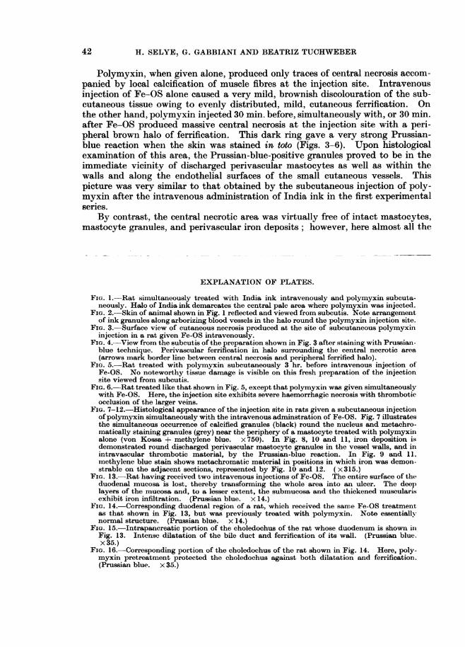
Intense staining of the inijection site (skin. peritoneum) occurred only whenpolymyxin was giveni simultaneously with India ink at the time defined as 0 min.After subcutaneous injection, the centre of the injected area remained virtuallyfree of the dye. but it was surrounded by an intensely stained halo, which could beseen through the shaved skin as a grey ring, even in the living animal. Uponireflection of the skin and inspection from the subcutis, the arrangement of the inkgranules along arborizing blood vessels was clearly visible (Figs. 1 and 2).
Histological study revealed that the ink particles are invariably located rounddischarged mastocvte granules in the immediate vicinitv of the minute skinl vessels.
40

MASTOCYTES AND FIXATION OF BLOOD-BORNE PARTICLES
The carboin particles gradually infiltrate the vessel wall, eventually penietratinig tothe endothelium. In the unstained central region, mastocytes, their dischargedgranules, and ink particles were virtually absent. However, in rare iinstanices wefound an occasional blood vessel containiing a thrombus-like aggregate of carbonlparticles in this central area.
Following intraperitoneal polymyxini treatment, a similar ilntense mastocvtedegranulation and ink imbibitioni was observed round the vessels of the omentum,whereas the mesenteric vessels remained virtually unaffected. It is noteworthythat iintraperitoneally administered polymyxin produced a slight degree ofomental staining even when the mastocvte discharger was given 30 min. to 2 hr.prior to the intravenious inik injection.
As regards their distant effects, subcutanieous anid intraperitoneal polymyxininijections elicited essentially identical changes. Blackening of the anaphylactoidshock organs. particularly the snout and the paws. occurred (both after subcutane-ous anid after initraperitoneal polymyxini injection) only when the mastocytedischarger was administered simultaneously with the India ink. Conversely.intense carbon staining of the lungs was seen only in the animals that receivedpolymyxin shortly prior to India ink. This carbon deposition was accompaniedby lung oedema and a high mortality rate. Histological examination showed thatin the lung India ink tended to precipitate. forming microthrombi in the alveolarcapillaries.
The liver anid spleeni, which contain the largest accumulations of reticulo-endothelial cells, were intensely stained in all groups except those in which thelung had attracted much of the India ink.
Evidently, polymyxin attracts carbon particles to the site of injection mosteffectively when it is given simultaneously with India ink; on the other hand. itcauses carbon deviation from the reticulo-endothelial system to the lung onlvwheni it is given shortly before the India ink.
E'ffect of topically applied polymyxin upon the cutaneous response to intravenouslyinjected Fe-OS
The second experimenital series was performed on 70 rats subdivided into7 equal groups. Fe-OS (I ml. 20 mg. of metallic iron) was injected intra-venously at the time defined as 0 min, and polymyxin (1-5 mg. in 0 5 ml.) sub-cutaneously at the times indicated in Table II. The experiment was terminatedon the 6th dav by killing all survivors with chloroform.
TA BLE II.- Efect of Topically Applied Polymnyxin upon the (Cutaneous Responseto Jntravenously Injected Fe-OS
Injectioin site
Central Peripheral MIortality(Group Treatinent* necros is forrificationi (per cenit)
I Polynmyxin Ilill.. 4 0 02 Fe-OS 0 rnimi. ()3 Fe-OS -)olmyxin -48 hr. . 0 114 .. ., -30 inimi. 6()
0 it ,in. - - 40,, - 30 min. .--. -4 60)
7 ,. ., L48 hr. 000* The time of Fe-OS injection is defined as 0 minutes.
41
H. SELYE, G. GABBIANI AND BEATRIZ TUCHWEBER
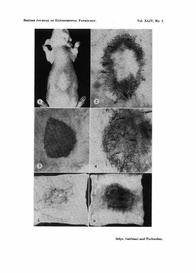
Polymyxin, when given alone, produced only traces of central necrosis accom-panied by local calcification of muscle fibres at the injection site. Intravenousinjection of Fe-OS alone caused a very mild, brownish discolouration of the sub-cutaneous tissue owing to evenly distributed, mild, cutaneous ferrification. Onthe other hand, polymyxin injected 30 min. before, simultaneously with, or 30 min.after Fe-OS produced massive central necrosis at the injection site with a peri-pheral brown halo of ferrification. This dark ring gave a very strong Prussian-blue reaction when the skin was stained in toto (Figs. 3-6). Upon histologicalexamination of this area, the Prussian-blue-positive granules proved to be in theimmediate vicinity of discharged perivascular mastocytes as well as within thewalls and along the endothelial surfaces of the small cutaneous vessels. Thispicture was very similar to that obtained by the subcutaneous injection of poly-myxin after the intravenous administration of India ink in the first experimentalseries.
By contrast, the central necrotic area was virtually free of intact mastocytes,mastocyte granules, and perivascular iron deposits; however, here almost all the
EXPLANATION OF PLATES.
FIG. 1.-Rat simultaneously treated with India ink intravenously and polymyxin subcuta-neously. Halo of India ink demarcates the central pale area where polymyxin was injected.
FIG. 2.-Skin of animal shown in Fig. 1 reflected and viewed from subcutis. Note arrangementof ink granules along arborizing blood vessels in the halo round the polymyxin injection site.
FIG. 3.-Surface view of cutaneous necrosis produced at the site of subcutaneous polymyxininjection in a rat given Fe-OS intravenously.
FIG. 4.-View from the subcutis of the preparation shown in Fig. 3 after staining with Prussian-blue technique. Perivascular ferrification in halo surrounding the central necrotic area(arrows mark border line between central necrosis and peripheral ferrified halo).
FIG. 5.-Rat treated with polymyxin subcutaneously 3 hr. before intravenous injection ofFe-OS. No noteworthy tissue damage is visible on this fresh preparation of the injectionsite viewed from subcutis.
FIG. 6.-Rat treated like that shown in Fig. 5, except that polymyxin was given simultaneouslywith Fe-OS. Here, the injection site exhibits severe haemorrhagic necrosis with thromboticocclusion of the larger veins.
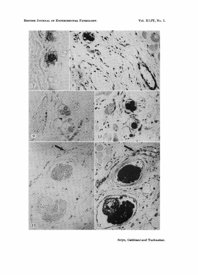
FIG. 7-12.-Histological appearance of the injection site in rats given a subcutaneous injectionofpolymyxin simultaneously with the intravenous adminstration of Fe-OS. Fig. 7 illustratesthe simultaneous occurrence of calcified granules (black) round the nucleus and metachro-matically staining granules (grey) near the periphery of a mastocyte treated with polymyxinalone (von Kossa + methylene blue. x 750). In Fig. 8, 10 and 11, iron deposition isdemonstrated round discharged perivascular mastocyte granules in the vessel walls, and inintravascular thrombotic material, by the Prussian-blue reaction. In Fig. 9 and 11.methylene blue stain shows metachromatic material in positions in which iron was demon-strable on the adjacent sections, represented by Fig. 10 and 12. (x315.)
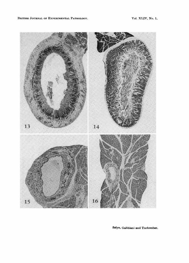
FIG. 13.-Rat having received two intravenous injections of Fe-OS. The entire surface of theduodenal mucosa is lost, thereby transforming the whole area into an ulcer. The deeplayers of the mucosa and, to a lesser extent, the submucosa and the thickened muscularisexhibit iron infiltration. (Prussian blue. x 14.)
FIG. 14.-Corresponding duodenal region of a rat, which received the same Fe-OS treatmentas that shown in Fig. 13, but was previously treated with polymyxin. Note essentiallynormal structure. (Prussian blue. x 14.)
FIG. 15.-Intrapancreatic portion of the choledochus of the rat whose duodenum is shown inFig. 13. Intense dilatation of the bile duct and ferrification of its wall. (Prussian blue.x35.)
FIG. 16.-Corresponding portion of the choledochus of the rat shown in Fig. 14. Here, poly-myxin pretreatment protected the choledochus against both dilatation and ferrification.(Prussian blue. x 3,5.)
42
BRITISH JOURNAL OF EXPERIMENTAL PATHOLOGY.
Selye, Gabbiani and Tuchweber.
Z.
VOl. XLIV, NO. 1.
BRITISH JOURNAL OF EXPERIMENTAL PATHOLOGY.
V I
AtI "..:
..
' '.
*'.
I'I.
Selye, Gabbiani and Tuchweber.
VOl. XLIV, NO. 1.
BRITISH JOURNAL OF EXPERIMENTAL PATHOLOGY.
16
Selye, Gabbiani and Tuchweber.
VOl. XLIV, NO. 1.
MASTOCYTES AND) FIXATION OF BLO(O)D)-BHORNE PARTICLES,
arterioles. venules. and even the larger veinis were occluded by thrombotic materialcontaining large amounts of iron. Along the borderline between the centralniecrosis and the peripheral halo of ferrificationi, it was sometimes possible toidentifyr mastocytes which simultaneously contained calcified granules (voI1Kossa reaction) rounid the nucleus and metachromatically staining mastocytegranules near the periphery (Fig. 7). In this zone. we also found extensive areasin which the larger vessels were occluded by iron-conitaining thrombi, whereastheir smaller branches showed angiotaxis of iron particles and mastocyte granule'salong their walls (Figs. 8-12). Combined treatmenit with Fe-OS and polymyxinwas lethal only to rats that received the two compounds simultaneously or atleast in rapid successioni. Here, the mortality was associated with, and pre-sumably the consequence of, haemorrhagic lung oedema resulting from the forma-tion of multiple iron precipitates in the pulmonary capillaries and diffuse ferri-ficatioin of the pulmonary stroma.
In this experiment we used comparatively younig rats because the thiin skiin ofthese is particularly subject to the inductioin of cutaneous necroses. However.as our previous observations had shown. Fe-OS produces severe biliary duct aild(luodenal lesions only in older rats, particularly if repeated injections are givet(Selye et al., 1962). Hence, another experimental series had to be performed onlarger animals in order to see whether the induction of widespread angiotaxis bypolymyxin could protect the choledochus and its branches against the inductioinof lesions by repeated injections of Fe-OS. We felt that a prophylaxis of thistvpe might be induced as a consequence of iron dispersion by widespread angio-taxis, throughout the organism, thereby deviating the excess iroin from the hepaticducts, in which it normally produces damage.
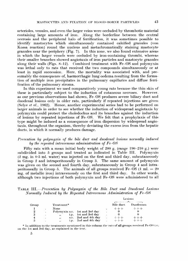
Prevention by polywnyxin of the bile duct and duodenal lesions normally inducedby the repeated intravenous administration of Fe-OS
Fifty rats with a mean initial body weight of 200 g. (ranige 190-210 g.) weresubdivided into 5 groups and treated as indicated in Table III. Polymyxiii(2 mg. in 0 5 ml. water) was injected on the first and third day, subcutaneouslvin Group 2 and intraperitoneally in Group 3. The same amounit of polymyxiinwas giveni on the second and fourth day, subcutaneously in Group 4 and initra-peritoneally in Group 5. The animals of all groups received Fe-OS (1 ml. 2(mg. of metallic iron) intravenously on the first and third day. In other words.although two injectioins of both polymvxin and Fe-OS were admiinistered to all
TABLE III. Prevention by Polymyxin of the Bile, Duct and D)uodenal Lesion7sNormally Induced by the Repeated Intravenous Administration of Fe-OS
Lesionis
Group Treatment* Bile duct Duodenum1 None . +±++±++2 Polymyxin s.c. 1st arid 3rd day . 0 03 ,, i.p. lst and 3rd day 0 04 ,, s.c. 2nd and 4th day + + + + + +5 , i.p. 2nd and 4th day + + + + +
* It addition to the treatmenits imienitioined in this colunn the rats of all groups received Fe-OS i.% .
on the 1st and 3rd day, as, explained in the text.
5
43

H. SELYE, G. GABBIANI AND BEATRIZ TUCHWEBER
the rats of Groups 2 to 5, the two compounds were given simultaneously in Groups2 and 3 only; Groups 4 and 5, though otherwise similarly treated, received poly-myxin always one day after each Fe-OS injection.
The dilatation of the bile ducts with angiotaxis of iron in their vessels and theferrification of the duodenal stroma with ulceration of the mucosa, both ofwhich are characteristic of Fe-OS treatment, were evident only in the animalsthat received Fe-OS alone or one day before each polymyxin injection. By con-trast, the rats simultaneously treated with Fe-OS and polymyxin showed no note-worthy lesions in either the bile ducts or the duodenum ; presumably polymyxingiven concurrently with Fe-OS produces widespread angiotaxis of iron throughoutthe organism, thereby deviating the metal from the particularly sensitive targetareas in which it normally tends to accumulate (Figs. 13-16). Pulmonary oedemaand mortality were not observed in these large animals under any of the experi-mential conditions tested.
DISCUSSION
It has been previously noted that under certain conditions India ink ad-ministered intravenously to rats can accumulate in the lung and cause death as aconsequence of pulmonary oedema. For example, an amount of intravenouslvinjected India ink that is normally well tolerated and merely deposited in thereticulo-endothelial cells (particularly those of the liver and spleen) can cause fatalpulmonary oedema as a consequence of its massive deposition in the lungs whenthe animals are simultaneously exposed to the stress of cold, spinal cord transectionor exhausting forced muscular exercise (Timiras and Selye, 1949; Selye, 1950Timiras, 1953). Stress is known to produce mastocyte discharge; hence, it isnot unlikely, in the light of the present experiments with polymyxin, that thedeposition of blood-borne India ink in the lung depends, at least in part, upol amastocyte discharge.
Sudden flooding of the circulation with unusually large amounts of India inkcan in itself cause precipitation of the carbon particles within the pulmonarvcapillaries (Halpern, Benacerraf and Biozzi, 1953). This response resembles thatwhich we have obtained with normally well-tolerated amounts of India ink whenthese were given in conjunction with a severe stressor agent or with a mastocvtedischarger, such as polymyxin. It has also been found that topical injury tocertain vascular territories can cause intravascular precipitation of blood-borneparticulate substances under certain conditions. For example, following damageto the myocardium, the intravenous injection of Fe-OS causes thrombosis of thecardiac vessels, often accompanied by infarct-like necroses (Grasso and Selve.1962 ; Selye and Grasso, 1961).
The observations reported in this communication suggest that blood-borneparticles of carbon and iron tend to precipitate in contact with discharged masto-cyte granules. If this discharge is of moderate intensity for example, in theshock organs during the anaphylactoid reaction, or in the periphery of a sub-cutaneous polymyxin-injection site-the result is angiotaxis. Presumably. thedischarged mastocyte granules contain substances that increase the adhesivenessof the adjacent tissue, including the vascular walls, and thereby fix the blood-borne foreign particles. (Sometimes this fixation is also quite evident round dis-charged mastocyte granules at some distance from the vessels. Here it may be
44

'MASTOCYTES AND FIXATION OF BLOOD-BORNE PARTICLES
more correct to speak of " mastocytopexis ". sinice we are dealing with a blindiiigpexis) to mastocyte debris and the vessels do not participate in the process
of fixation.)On the other hanid, if the mastocyte discharge is massive-as for example in
the centre of a subcutaneous polymyxin-injection site so much mastocytematerial may enter the blood that occlusive aggregates of foreign particles areformed and these lead to local thrombosis and necrosis. Finallv, if the mastocv-tedischarge, though of moderate intensity, is very generalized, iinnumerable inasto-cyte granules enter the circulation throughout the body and are carried to thepulmonarv capillaries, in which they cause aggregation of foreign particles withsecondary thrombus formation.
The assumption that a mastocvte discharge is responsible for angiotaxis isalso consonant with the fact that angiotaxis occurs wh-lerever there is a mastocytedischarge, for example, in the shock organs during the aniaphylactoid reactionl olin the subcutis following local administrationi of a mastocyte discharger, such asp)olymyxin.
It is noteworthy that polymyxiii can either produce or prevent the develop-menit of morbid lesions, depending upon circumstances. The cutanieous blood-vessel thrombosis with conisequent necrosis induced by polymyxin in colnjulctiowith the intravenous iinjection of Fe-OS is somewhat reminiscenlt of the localShwartzman-Sanarelli pheniomenion and may help to elucidate the mechanismof such topical hypersensitivity reactions. (Converselv. the preventioni by poly-mnyxin of the biliary and duodenal lesions normally produced by the intravenonsadministration of Fe-OS mav be ain illustration of the defenisive value of angio-taxis. Presumably, here the protection is due to the deviationi of circulatingironi from the most sensitive targets by widespread fixationi of the iron particlesin the walls of small vessels throughout the body.
Finally, it should be emphasized that this interpretationi is still fal from lprovell,hut at least it appears to be compatible with all our observationis.
SUMMARY
Experimenits on rats suggest that discharged mastocyte material can cal)tureblood-borne particulate substances anid thereby participate in the localization ofdlisease.
A subcutaneous inijection of the mastocyte discharger polymyxin. given simul-taiieously with ain intravenous injectioin of India ink, causes localization of carbonparticles particularly in a halo round the polymyxini injectionl site. Here, theInidia ink anid discharged mastocyte granules are found in close association withinthe connective tissue and in the walls of small blood vessels perhaps because masto-cyte material can fix blood-borne particles bv increasing the adhesiveiless oftissues.
If a similar polymyxini injection is giveni somewhat prior to the intravelnolusadminiistrationi of India ink, there is Ino local fixationi of carboll particles in the.subcutis, but numerous pulmonary capillaries are occluded by thrombi, whichconitain aggregates of Inidia ink. Here, the discharged mastocyte granules mayhave already been carried from the site of polymyxin injection to the pulmonaryVcirculation by the time the India ink is introduced inito the circulation, and it isperhaps for this reasoni that the carboni particles are fixed in the lung.
45,

46 H. SELYE. G. GABBIANI AN) IBEATRIZ TUCHWEBER
Essentially similar observations are made when ferric oxide saccharate (Fe--OS) is administered intravenously instead of Inidia ink. However, since iron ismore toxic to tissues than India ink, the local precipitationi of Fe-OS, especiallyin the centre of the polymyxin injectioni site, may cause inecrosis similar to thatseen in the local Shwartzman-Sanarelli phenomenoni.
Repeated intravenous administration of Fe-OS to older rats produces heavyselective iron precipitation in the choledochus and duodenum; this results in anextreme dilatationi of the bile ducts and development of duodenal ulcers withniecrosis. Here again, the iron particles precipitate predominantly in the imme-(iate vicinity of the mastocytes, which are particularly inumerous round theextrahepatic bile ducts and the duodenum of the rat. In order to test the hypo-thesis of a causal relationship between mastocyte granules and iron precipitatioll.a mastocyte discharge was produced prior to the administration of Fe-OS. Itwas found that after depletion of the mastocytes, the bile duct and choledochuslesions normallv elicited by the iron preparation are preveilted.
This work was supported by Grants from The Gustavus and Louise PfeifferResearch Foundation and The Medical Research Couincil of C'anada.
REFERENCESBURROWS, H. (1932) Some Factors in the Localization of Disease in the Bodv.
London (Bailli'ere, Tindall and Cox).Grozsy. B. AND KATO, L.-(1957) ' Studies on Phagocytic Stimulation'. Institute of
Microbiology and Hygiene, University of Montreal.GRASSO, S. AND SELYE, H.-(1962) J. Path. Bact., 83, 495.HALPERN, B. N., BENACERRAF, B. AND Biozzi, G. (1953) Brit. J. exp. Path., 34, 426.HERZOG, F.-(1924) Z. exp. Med., 43, 79.JANCSO N.-(1955) ' Speicherung, Stoffanreicherung in Rektulo-Endothel und in der
Niere'. Budapest (Akademiai Kiado).JASMIN, G.-(1956) Rev. canad. Biol., 15, 107.LJANG, F. J.-(1926) Arch. Path.. 1, 41.OVARY, Z.-(1951) Boll. Soc. ital. Biol. sper., 27, 387.RILEY, J. F.-(1959) ' The Mast Cells'. Edinburgh and London (Livingstone).SELYE, H. (1937) Endocrinology, 21, 169.-(1950) Stress'. Montreal (Acta Inc.. Med.
Publ.).-(1962) 'Calciphylaxis'. The University of Chicago Press.Jdem AND GRASSO, S.-(1961) Brit. J. exp. Path., 42, 564.Idem HORAVA, A. AND HEUSER, G.-(1951-56) Annual Reports on Stress . Montreal
(Acta Inc., Med. Publ.).Idem, LEMIRE, Y. AND CANTIN, M.-(1959) Med. Exper., 1, 11.Idem, VASKUT, J. AND GABBIANI, G.-(1962) Amer. J. Gastroenterol., 38, 450.STILWELL, F.-(1926) Folia haemat., 33, 81.TIMIRAS, P. S.-(1953) Acta endocrinol., 12, 13.Idem AND SELYE, H.-(1949) Science, 110, 560.ZEMPL1ENYI, T., FODOR, J. AND LOJDA, Z.-(1960) Quart. J. exp. Biol., 45, 50.