the role of the sympathetic nervous system in...
TRANSCRIPT

THE ROLE OF THE SYMPATHETIC NERVOUS SYSTEM IN THE ACUTE HYPOTHERMIC EFFECT OF D-FENFLURAMINE
by
Srividya Subramanian
B.Pharm, Medical University of Madras, 1995
Submitted to the Graduate Faculty of
School of Pharmacy in partial fulfillment
of the requirements for the degree of
Doctor of Philosophy
University of Pittsburgh
2002

ii
THE ROLE OF THE SYMPATHETIC NERVOUS SYSTEM IN THE ACUTE
HYPOTHERMIC EFFECT OF D-FENFLURAMINE
Srividya Subramanian, PhD,
University of Pittsburgh
Experiments in this dissertation were conducted to characterize the effects of d-fenfluramine
on body temperature and the mechanisms by which d-fenfluramine alter body temperature.
The experiments were conducted in conscious male Sprague-Dawley rats. Body temperature
was measured in all animals using telemetry. The results of the experiments indicated that d-
fenfluramine altered body temperature in animals kept 28, 22, 16 and 4oC. D-fenfluramine
produced hyperthermia in animals kept at 28oC and varying degrees hypothermia at normal
and cooler ambient temperatures. Further experiments were conducted to explore the effects
of d-fenfluramine on brown adipose tissue (BAT) thermogenesis, cutaneous vascular tone
and whole body oxygen consumption. In animals kept at 22 and 4oC, we found that d-
fenfluramine activated BAT, as indicated by a decrease in BAT norepinephrine content, to
the same magnitude. Thus, the hypothermia seen at normal and cooler ambient temperature
was not due to lack of BAT activation. Also, activation of BAT by d-fenfluramine was
mediated through the sympathetic nervous system and through release of central serotonin,
since ganglionic blocker pentolinium and serotonin reuptake inhibitor fluoxetine blocked d-
fenfluramine-mediated BAT activation. In animals kept at 16oC, d-fenfluramine increased
tail-skin temperature (Tsk), an index of cutaneous vascular tone, indicating that d-
fenfluramine produced cutaneous vasodilation. d-fenfluramine-induced increase in Tsk was
mediated through withdrawal of the sympathetic vasoconstrictor tone to the tail, since

iii
pentolinium blocks this effect. In animals kept at 28oC, d-fenfluramine produced a decrease
in Tsk, indicating vasoconstriction. The effects of d-fenfluramine on the Tsk were mediated
through release of serotonin, since fluoxetine blocked these effects. D-fenfluramine increased
whole body oxygen consumption, an index of metabolic activity and the increase was due to
BAT activation, since pentolinium prevented the increase. Thus, although d-fenfluramine
increased metabolic activity through BAT activation, the increase was insufficient to make
up for the heat loss produced by cutaneous vasodilation and thus produces hypothermia. The
hyperthermia seen at 28oC is due to activation of BAT and the subsequent inability of the
animal to lose the excess heat due to cutaneous vasoconstriction produced by d-fenfluramine
at 28oC.

iv
FOREWORD
I wish to express my sincere gratitude to my advisor, Dr. Regis R. Vollmer for his
constant advice, support and encouragement during the conduct of the research and the
preparation of this document. I also wish to thank Dr. David J. Edwards for all his help and
advice with the assays. Sincere gratitude is also extended to other members of my thesis
committee, Drs. Janet Amico, Balwant Dixit and Subhash Vyas for their valuable comments
and suggestions during the course of my research.
I wish to thank Judith J. Balcita, Camille Price, Micheal Tortorici, Joseph Karam for
their technical assistance and their friendships; fellow graduate students and friends for their
physical and moral support and the excellent secretarial staff who took care of all my paper
work.
Finally, I wish to express my special appreciation to my husband, Atif, my parents,
my siblings and my uncle and aunt for their unfailing support, love and encouragement.

v
TABLE OF CONTENTS
FOREWORD .................................................................................................................... iv
TABLE OF CONTENTS.................................................................................................. v
LIST OF FIGURES ......................................................................................................... ix
I. Overview ................................................................................................................... 1
A. Introduction ................................................................................................. 1
B. Literature Survey......................................................................................... 5
1. The role of the sympathetic nervous system in thermoregulation .......................................................................... 5
a. Sympathetic nervous system control of BAT
thermogenesis...................................................................... 6 b. Sympathetic nervous system control of
cutaneous vascular responses .............................................. 9
2. Effects of fenfluramine on body temperature regulation.................................................................................... 12
a. Effects of fenfluramine on BAT thermogenesis ............... 13 b. Effects of fenfluramine on the cutaneous
vasculature......................................................................... 14
c. Effects of fenfluramine on metabolic rate......................... 17
3. The role of serotonin in thermoregulation.................................. 17
C. Specific Objectives of Research................................................................ 20
II. Methods ................................................................................................................... 23
A. Care and Housing of Animals ................................................................... 23
B. Measurement of Body Temperature by Temperature Telemetry and Computerized Data Acquisition System ............................................ 23

vi
C. Measurement of Tail Skin Temperature Using Thermocouples in a Controlled Temperature Environment................................................ 25
D. Measurement of Oxygen Consumption Using Open Circuit
Calorimetry................................................................................................ 27
E. Collection of Tissue Samples for Biochemical Analyses ......................... 27
F. Measurement of Catecholamines by High Performance Liquid Chromatography........................................................................................ 30
G. Statistics .................................................................................................... 31
III. Pharmacologic Assessment of the Contribution of the Sympathetic Nervous System to the Maintenance of Body Temperature in Conscious Rats........................................................................................................ 32
A. Protocols.................................................................................................... 34
B. Results ....................................................................................................... 35
1. Effects of saline, 1ml/kg, i.p., on body temperature, locomotor activity, adrenal and BAT catecholamine content ........................................................................................ 35
2. Effects of phentolamine, 2 mg/kg, i.p., on body
temperature, locomotor activity, adrenal and BAT catecholamine content................................................................ 36
3. Effects of propranolol, 3 mg/kg, i.p., on body
temperature, locomotor activity, adrenal and BAT catecholamine content................................................................ 36
4. Effects of phentolamine, 2 mg/kg, i.p., and
propranolol, 3 mg/kg, i.p., on body temperature, locomotor activity, adrenal and BAT catecholamine content ........................................................................................ 41
5. Effects of chlorisondamine, 3 mg/kg, i.p., on body
temperature, locomotor activity, adrenal and BAT catecholamine content................................................................ 42
C. Discussion ................................................................................................. 47

vii
IV. Evaluation of the Effects of Fenfluramine on Sympathetic Nervous System Regulation of Body Temperature ............................................................ 54
A. Protocols.................................................................................................... 57
B. Results ....................................................................................................... 60
1. Effects of dl- fenfluramine, 10 mg/kg, i.p., on body temperature and BAT NE content.............................................. 60
2. Effects of d-fenfluramine, 3 and10 mg/kg, i.p., on
body temperature and catecholamine content of sympathetically innervated tissues............................................. 61
3. Effects of pentolinium, 10 mg/kg, i.p., and fluoxetine,
10 mg/kg, i.p., pretreatment on d-fenfluramine-induced BAT NE depletion........................................................ 64
C. Discussion ................................................................................................. 68
V. Evaluation of the Effects of d-Fenfluramine on the Cutaneous Vasculature and Total Metabolic Heat Production ............................................ 75
A. Protocols.................................................................................................... 78
B. Results ....................................................................................................... 81
1. Effects of d-fenfluramine, 10 mg/kg, i.p., on tail skin and body temperature of rats kept at 160C................................. 81
2. Effects of pentolinium pretreatment on d-
fenfluramine-induced increases in tail skin temperature and hypothermia..................................................... 82
3. Effects of d-fenfluramine, 10 mg/kg, i.p., on tail skin
and body temperature of rats kept at 280C (thermoneutrality) ...................................................................... 83
4. Effects of fluoxetine pretreatment on d-fenfluramine
induced increases in tail skin temperature and hypothermia................................................................................ 83
5. Effects of d-fenfluramine and pentolinium on whole
body oxygen consumption ......................................................... 87
viii
C. Discussion ................................................................................................. 97
VI. Conclusions ........................................................................................................... 104 VII. Bibliography ......................................................................................................... 109

ix
LIST OF FIGURES Figure 1. Sympathetic pathways involved in thermoregulation ................................. 8 Figure 2. The chemical structures of fenfluramine and amphetamine ..................... 11 Figure 3. The sites of actions of fenfluramine.......................................................... 16 Figure 4. Effect of chlorisondamine, 3mg/kg, i.p., on body
temperature of rats 24-hours after surgery to implant temperature transmitter ............................................................................. 26
Figure 5. Effect of chlorisondamine, 3 mg/kg, i.p., on body
temperature of rats after five days of recovery following surgery to implant temperature transmitter ............................................... 26
Figure 6. Measurement of body temperature using temperature
telemetry and computerized data acquisition system and tail skin temperature using thermocouples...................................................... 28
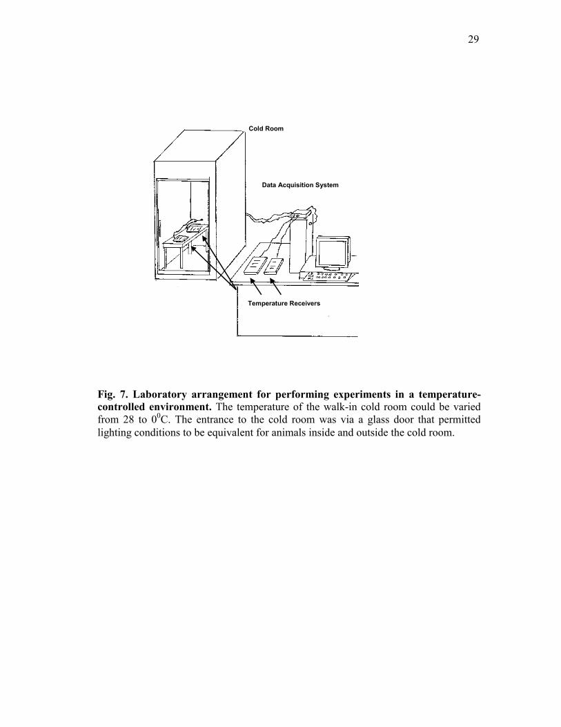
Figure 7. Laboratory arrangement for performing experiments in a
temperature-controlled environment ......................................................... 29 Figure 8. Effects of saline, 1ml/kg, i.p., on body temperature and
locomotor activity of rats kept at 220C (n=7) and 40C (n=7).................... 37
Figure 9. Effects of saline, 1ml/kg, i.p., on adrenal epinephrine and norepinephrine (NE) content and brown adipose tissue (BAT) NE content in rats kept at 220C (n=7) and 40C (n=7).................... 38
Figure 10. Effects of phentolamine, 2mg/kg, i.p., on body temperature
and locomotor activity of rats kept at 220C (n=7) and 40C (n=7) .......................................................................................................... 39
Figure 11. Effects of phentolamine, 2mg/kg, i.p., on adrenal
epinephrine and norepinephrine (NE) content and brown adipose tissue (BAT) NE content in rats kept at 220C (n=7) and 40C (n=7) ............................................................................................ 40
Figure 12. Effects of propranolol, 3 mg/kg, i.p., on body temperature
and locomotor activity of rats kept at 220C (n=7) and 40C (n=7) .......................................................................................................... 43
Figure 13. Effects of propranolol, 3 mg/kg, i.p., on adrenal
epinephrine and norepinephrine (NE) content and brown

x
adipose tissue (BAT) NE content in rats kept at 220C (n=7) and 40C (n=7) ............................................................................................ 44
Figure 14. Effects of phentolamine, 2 mg/kg, i.p. and propranolol, 3
mg/kg, i.p., on body temperature and locomotor activity of rats kept at 220C (n=7) and 40C (n=7)....................................................... 45
Figure 15. Effects of phentolamine, 2 mg/kg, i.p. and propranolol, 3
mg/kg, i.p., on adrenal epinephrine and norepinephrine (NE) content and brown adipose tissue (BAT) NE content in rats kept at 220C (n=7) and 40C (n=7)................................................... 46
Figure 16. Effects of chlorisondamine, 3 mg/kg, i.p., on body
temperature and locomotor activity of rats kept at 220C (n=7) and 40C (n=7) .................................................................................. 48
Figure 17. Effects of chlorisondamine, 3mg/kg, i.p., on adrenal
epinephrine and norepinephrine content (NE) and brown adipose tissue (BAT) NE content in rats kept at 220C (n=7) and 40C (n=7) ............................................................................................ 49
Figure 18. Effects of saline or dl-fenfluramine , 10 mg/kg, i.p., on
body temperature of rats kept at 22 and 40C (n=7/group)......................... 62 Figure 19. Effects of saline or dl-fenfluramine, 10 mg/kg, i.p., on
brown adipose tissue (BAT) norepinephrine (NE) content of rats kept at 22 and 40C (n=7/group)...................................................... 63
Figure 20. Effects of saline or d-fenfluramine, 3, 10 mg/kg, i.p., on
body temperature of rats kept at 22 and 40C (n=7/group)......................... 65 Figure 21. Effects of saline or d-fenfluramine, 3 and 10 mg/kg, i.p., on
brown adipose tissue (BAT) and white adipose tissue (WAT) norepinephrine (NE) content of rats kept at 220C and 40C (n=7/group).................................................................................. 66
Figure 22. Effects of saline (Sal) or pentolinium (Pent), 10mg/kg, i.p.
pretreatment on d-fenfluramine (FEN), 10 mg/kg, i.p. induced changes in brown adipose tissue (BAT) norepinephrine (NE) content (n=7/group) ................................................ 69
Figure 23. Effects of saline (Sal) or fluoxetine (Fluo), 10mg/kg, i.p.
pretreatment on d-fenfluramine (FEN), 10 mg/kg, i.p. induced changes in brown adipose tissue (BAT) norepinephrine (NE) content (n=7/group) ................................................ 70

xi
Figure 24. Effects of saline or d-fenfluramine, 10 mg/kg, i.p., on tail skin and body temperature of rats kept at 160C (n=7/group) .................... 84
Figure 25. Effects of saline or pentolinium, 10 mg/kg, i.p., on tail skin
and body temperature of rats kept at 160C (n=7/group)............................ 85 Figure 26. Effects of pentolinium, 10 mg/kg, i.p., pretreatment on d-
fenfluramine, 10 mg/kg, induced increases in tail skin temperature and hypothermia (n= 7/group) .............................................. 86
Figure 27. Effects of increasing ambient temperature on body and tail
skin temperature of rats (n=7) ................................................................... 88 Figure 28. Effects of saline or d-fenfluramine, 10 mg/kg, i.p., on tail
skin and body temperature of rats kept at 280C (n=7/group) .................... 89 Figure 29. Effects of saline or fluoxetine, 10 mg/kg, i.p., on tail skin
and body temperature of rats kept at 160C (n=7/group)............................ 90 Figure 30. Effects of fluoxetine, 10 mg/kg, i.p., pretreatment on d-
fenfluramine, 10 mg/kg, induced increases in tail skin temperature and hypothermia (n= 7/group) .............................................. 91
Figure 31. Effects of saline or d-fenfluramine, 10 mg/kg, i.p., on
whole body oxygen consumption (VO2) and body temperature of rats kept at 160C (n=7/group) ........................................... 93
Figure 32. Effects of saline or pentolinium, 10 mg/kg, i.p., on whole
body oxygen consumption (VO2) and body temperature of rats kept at 160C (n=7/group).................................................................... 94
Figure 33. Effects of d-fenfluramine (Fen), 10 mg/kg, i.p.,
pretreatment on pentolinium-induced increases in whole body oxygen consumption (VO2), measured 30 minutes after treatment, in rats kept at 160C (n=7/group) ...................................... 95
Figure 34. Effects of pentolinium (Pent), 10 mg/kg, i.p., pretreatment
on d-fenfluramine-induced increases in whole body oxygen consumption (VO2), measured one hour after treatment, in rats kept at 160C (n=7/group).................................................................... 96
Figure 35. Summary of the effects of d-fenfluramine, 10 mg/kg, i.p.,
or saline on body temperature, one hour post-treatment, of rats kept at different ambient temperatures ............................................. 106

xii
Figure 36. Summary of the mechanisms by which d-fenfluramine produces hypothermia ............................................................................. 108

1
Chapter I. Overview
A. Introduction
Obesity is a complex, chronic disease requiring long-term weight management.
Annual healthcare costs in the U.S. associated with obesity have reached nearly $100 billion
(Weiser et al., 1997). The past 30-40 years have seen an increased emphasis on the need to
recognize obesity as a chronic condition, driven by evidence that obesity has a causal role in
cardiovascular diseases, diabetes mellitus and osteoarthritis (Gordon et al., 1976; Colditz et
al., 1995). The strong relationship between obesity and various disease states has led to a
heavy demand for strategies that result in long-term weight loss. Obesity is due to an
imbalance between caloric intake and caloric expenditure resulting in storage of excess
calories as fat. Thus, interventions to treat obesity have focused on reducing caloric intake or
increasing caloric expenditure. The standard nonpharmacologic interventions are dietary
restriction and increased physical activity (exercise). Unfortunately, nonpharmacologic
treatments such as dieting and exercise have low efficacy due to patient noncompliance
(Wooley et al., 1991). Consequently, their replacement or supplementation by pharmacologic
agents that reduce body weight have received much attention.
Drugs that have been investigated for the management of obesity act by two general
mechanisms, they either decrease caloric intake and/or increase caloric expenditure. Drugs
that reduce caloric intake are commonly called anorexic or hypophagic agents. The
hypophagic agents comprise two pharmacologic classes: drugs that affect catecholaminergic
system (the amphetamines, phentermine, mazindol, phenylproponalamine) and those that
affect the serotonergic system (fenfluramine, fluoxetine, sertraline) (Weiser et al., 1997). One

2
of the most recently developed antiobesity agent, sibutramine affects both catecholaminergic
and serotonergic neuronal systems (Connoley et al., 1995).
Drugs that increase caloric expenditure are called thermogenic drugs. Thermogenic
agents increase metabolic rate. Drugs that increase metabolic rate include ephedrine
(Pasquali et al., 1993) and experimental agents such as BRL 26830A, a beta-adrenoceptor
agonist (Connacher et al., 1992). None of the thermogenic agents is currently approved by
the FDA for weight control. Some of the appetite suppressive agents such as fenfluramine
and sibutramine are also known to increase caloric expenditure (Connoley et al., 1999).
The most commonly prescribed appetite suppressant weight-loss medication in the
1950s and 1960s was amphetamine. However, the use of amphetamine had serious
limitations due to the psychomotor stimulant properties and a high abuse potential (Bray,
1993). The other sympathomimetic agents (mazindol, phenylpropanolamine and
phentermine) were approved by the FDA only for short-term treatment of obesity since
tolerance to the anorexic agents develops during long-term therapy (Weintraub, 1992).
In an effort to overcome the limitations of amphetamine, yet retain the very potent
antiobesity effect, fenfluramine, an amphetamine analog, was developed in the 1960s as an
appetite suppressive agent. Fenfluramine was found to be devoid of the psychomotor
stimulant and abuse potential of amphetamine (Rowland et al., 1986). The potency of
fenfluramine coupled with the lack of psychostimulant side effects made fenfluramine one of
the most widely prescribed antiobesity agents. In over 40 years of therapeutic use, more than
70 million patients worldwide have been treated by fenfluramine (Curzon et al., 1997).
Reports of serious cardiovascular side effects, pulmonary hypertension and cardiac
valvular lesions (Fishman, 1999; Teramae et al., 2000) following fenfluramine treatment led

3
to the withdrawal of fenfluramine from the U.S. markets in 1997. But fenfluramine still
serves as a prototype against which new agents are compared. Newer agents have been
developed with the hope of producing a drug with the antiobesity effects of fenfluramine but
without the toxic side effects.
In order to achieve the objective of developing antiobesity agents as potent and as
efficacious as fenfluramine, it is important that the mechanisms by which fenfluramine
produces weight loss be understood. However, the pharmacologic mechanisms by which
fenfluramine reduces body weight are not completely elucidated. There are two general
mechanisms by which a drug can produce weight loss, appetite suppression and thermogenic
activity. Thus, research has focused on the possibility that fenfluramine may possess appetite
suppressive and thermogenic effects.
Fenfluramine has been shown to be a potent appetite suppressive agent (Jackson et
al., 1997; Curzon et al., 1997; Gibson et al., 1993; Oluyomi et al., 1994). However, appetite
suppression alone does not account for the lowering of body weight since the appetite
suppressive effect wanes after initial therapy, yet the weight loss persists. In rats,
fenfluramine suppresses appetite during the first two weeks of treatment and then the effect
diminishes and caloric intake returns to pretreatment levels. Despite normal caloric intake,
weight loss persists (Arase et al., 1988). And, if fenfluramine treatment was terminated, the
animals gained weight without increasing their food intake. This observation has led
investigators to the conclusion that in addition to appetite suppression, fenfluramine must
also affect energy expenditure.
In fact, it has been shown that fenfluramine increases metabolic rate in rats by
increasing the activity of brown adipose tissue (BAT) (Rothwell et al., 1992; Rothwell et al.,

4
1987; Levitsky et al., 1986; Levitsky et al., 1992; Preston et al., 1990). BAT is a major organ
of thermogenesis (Lupien et al., 1985; Lupien et al., 1986) in rodents. In human infants, BAT
is important for body temperature maintenance (Himms-Hagan, 1990). However, in human
adults, the function of BAT is not known. Nevertheless, there is evidence that part of the
antiobesity effect of fenfluramine in humans might be related to increased metabolic rate.
Fenfluramine appears to potentiate the expenditure of calories, whenever caloric expenditure
increases (Levitsky et al., 1992).
Despite the fact that fenfluramine increases caloric expenditure, there is evidence in
the literature that fenfluramine treatment results in a hypothermia (Cryan et al., 2000;
Malberg et al., 1997). However, the mechanisms of fenfluramine-mediated hypothermia have
not been characterized. Because body temperature regulation is intrinsically interrelated to
caloric utilization (Carlisle et al., 1993; Klaus et al., 1998; Trayhurn et al., 1995),
investigating the underlying mechanisms by which fenfluramine lowers body temperature
could lead to an understanding of the mechanisms by which fenfluramine produces weight
loss. Thus, the specific focus of the experiments in this dissertation was to investigate the
mechanisms by which fenfluramine lowers body temperature.
The sympathetic nervous system plays a major role in the maintenance of body
temperature. A major hypothesis of the thesis was that the hypothermia produced by
fenfluramine could be due to dysregulation of the sympathetic nervous system mechanisms
of thermoregulation. Thus, the hypothermia to fenfluramine could be due to impairment of
sympathetic regulation of heat generation and/or heat conservation.

5
B. Literature Survey
The following survey of the literature is divided into three sections. In part one, the
role of sympathetic nervous system in thermoregulation is described. In part two, the effects
of fenfluramine on thermoregulation are discussed. In part three, the role of serotonin in
thermoregulation is described since fenfluramine is a serotonin agonist and mimics serotonin
in its pharmacologic actions.
1. The role of sympathetic nervous system in thermoregulation
The sympathetic nervous system plays a critical role in the maintenance of body core
temperature (Landsberg et al., 1984; Maickel et al., 1967). Sympathetic neurons, via the
release of norepinephrine (NE) and the consequent stimulation of alpha-adrenoceptors,
promote heat conservation through cutaneous vascular constriction and piloerection (Kent et
al., 1991; Kent et al., 1990). The sympathetic neurons also promote heat loss by cutaneous
vasodilation (O'leary et al., 1985). NE stimulation of beta-adrenoceptors results in heat
generation in BAT (Arch, 1989; Hsieh et al., 1957; Tsukazaki et al., 1995) and stimulation of
the heart (Barney et al., 1980; Fregly et al., 1989; Sun et al., 1997) to allow for appropriate
adjustments in cardiac output resulting from changes in metabolic rate and cutaneous blood
flow. These actions of NE are augmented by the adrenal medullary catecholamines (Himms-
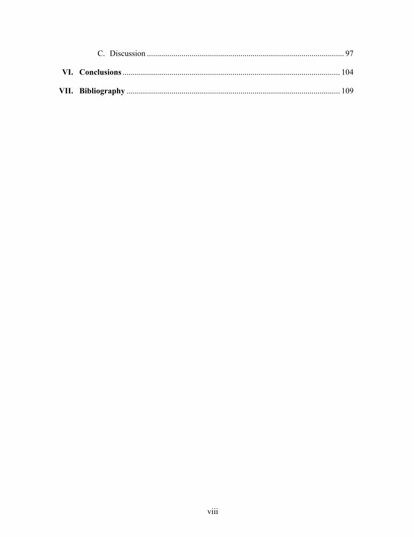
Hagan, 1975), Fig. 1.
The role of the sympathetic nervous system in the maintenance of body temperature
is clearly demonstrated when animals are exposed to a cold environment (Maickel et al.,
1967). The sympathetic nervous system is activated when
animals are exposed to a cold environment. When animals were maintained at an ambient
temperature of 40C, the plasma NE levels were increased indicating that the sympathetic

6
neurons were activated (Leduc, 1961; Vollmer, 1996; Picotti et al., 1980). In fact, direct
sympathetic nerve recordings have indicated that sympathetic nerve impulse traffic to BAT
was increased during cold exposure (Banet et al., 1978). Also, when animals are exposed to a
cold environment NE turnover rates in the BAT, white adipose tissue and heart, increase
(Young et al., 1982; Garafalo et al., 1996; Landsberg et al., 1984).
Blockade of the sympathetic nervous system and adrenal medullary mechanisms of
thermoregulatory responses to cold exposure impairs the ability of an animal to maintain
body temperature (Maickel et al., 1967; Ramey et al., 1957; Taylor, 1960; Wekstein, 1964).
Chemical sympathectomy by treating animals with reserpine, blockade of the autonomic
ganglia by ganglionic blocking agents chlorisondamine and pentolinium (Maickel et al.,
1967; Picotti et al., 1980) and blockade of alpha-receptor mediated vasoconstrictor response
by phentolamine results in hypothermia (Redfern et al., 1995). Mutant mice that lack NE and
EPI are cold intolerant because they have impaired peripheral vasoconstriction and are
unable to induce BAT thermogenesis (Thomas et al., 1999).
The next two sections discuss the sympathetic nervous system control of metabolic
heat production by BAT and heat conservation by constriction of the cutaneous vasculature.
a. Sympathetic nervous system control of BAT thermogenesis
BAT is a major site of nonshivering thermogenesis in rodents (Himms-Hagan, 1995).
The thermogenesis in BAT can be stimulated by both exposure to cold (nonshivering
thermogenesis, NST) and by overeating (diet-induced thermogenesis, DIT) (Himms-Hagan,
1990), thus, BAT contributes to both thermal and energy balance respectively. Destruction of
BAT causes obesity and cold intolerance (Klaus et al., 1998). Studies have demonstrated that
the BAT thermogenesis is regulated by the sympathetic nervous system, via direct

7
noradrenergic innervation of the BAT cells (Smith et al., 1969). Sympathetic control of BAT
thermogenesis is also evidenced by increased NE turnover rate in BAT during cold exposure
and by induction of thermogenesis by NE infusions in BAT (McDonald et al., 1993). NE
released from the sympathetic neurons act on beta-adrenoceptors, particularly of the beta-3
subtype and activates heat production (Lowell et al., 1997; Tanaka et al., 1995). Beta-3
agonists and nonspecific beta agonists increase heat production in BAT and beta-antagonists
like propranolol block thermogenesis in BAT (Carlisle et al., 1992; Benzi et al., 1988).
Heat production in BAT involves a unique mitochondrial protein called the
uncoupling protein (UCP), which forms a proton conductance pathway allowing cells to
oxidize substrates without the oxidation process linked to ATP synthesis. The UCP in the
mitochondrial membrane binds to GDP and other purine nucleotides. The level of GDP
binding to BAT mitochondria has been a reliable measure of the activity of the proton
conductance pathway and hence thermogenic activity of BAT. Cold exposure increases GDP
binding to BAT by at least two-fold as revealed by studies conducted using the intrascapular
BAT (IBAT), which is the most widely studied BAT depot (henceforth, IBAT will be
mentioned as BAT since almost all the studies have examined this BAT depot) (Cageao et
al., 1995; Trayhurn et al., 1987). Also, blood flow and glucose uptake by BAT increases
during cold exposure due to increased metabolic activity of BAT (Foster et al., 1977; Ma et
al., 1991). The thermogenic effect of BAT to cold exposure can be prevented by local
denervation of the sympathetic nerves entering intrascapular BAT (Minokoshi et al., 1986b;
Minokoshi et al., 1986a; Foster et al., 1981).
8
Fig. 1. Sympathetic pathways involved in thermoregulation. The sympathetic nervous system regulates heat conservation through cutaneous vasoconstriction and piloerection and heat generation through BAT activation. The sympathetic nervous system stimulates heart to adjust for changes in cardiac output resulting from changes in metabolic rate and cutaneous circulation. The sympathetic nervous system also controls adrenal release of catecholamines.
Heart
Cutaneous vasculature
BAT (brown adipose tissue)
Piloerector muscles
Hypothalamic Preoptic Area senses blood temperature and receives input from somatic and visceral thermoreceptors
Spinal Cord
Descending Pathways
NE
EPI EPI
EPIEPI
Adrenal Medulla

9
Brain centers that are involved in the control of BAT thermogenesis include the
ventromedial hypothalamus, lateral hypothalamus, preoptic area and caudal raphe
nucleus (Bamshad et al., 1999). Electrical stimulation of these areas activates BAT
thermogenesis and this activation is blocked by local denervation of the sympathetic
nerves to BAT (Amir, 1990; Minokoshi et al., 1986a).
b. Sympathetic nervous system control of cutaneous vascular
responses
The cutaneous vasculature is an important thermoregulatory system. Cutaneous
vasoconstriction prevents heat-loss and vasodilation promotes heat loss. The sympathetic
nervous system regulates cutaneous vascular responses. The sympathetic neurons release
NE, which interacts with the vascular smooth muscle alpha-adrenoreceptors to produce
constriction. Withdrawal of sympathetic tone to the cutaneous vasculature leads to
dilation (Redfern et al., 1995). Interference with sympathetic control of vascular tone
leads to impairment of body temperature regulation. For example, treatment with alpha-
receptor antagonists phentolamine and delequamine results in cutaneous vasodilation and
hypothermia in rats (Kent et al., 1991; Redfern et al., 1995).
Rats are particularly suited for the study of cutaneous vascular responses because
of the important role of the tail vasculature. The tail of the rat has frequently been used to
study cutaneous vascular responses since it is a major thermoregulatory organ in the rat.
About 20% of the heat loss in a rat occurs by sympathetically mediated increases in blood
flow in the skin of the tail (O'leary et al., 1985; Redfern et al., 1995). The tail is well
suited for dissipating heat since it lacks fur, is highly vascularized and has a relatively

10
large surface area to volume ratio. Thus the dilation of the tail is important for heat loss
when the environmental temperature is elevated and conversely constriction of this
circulation is crucial for heat conservation when the ambient temperature is reduced.
Many studies have measured tail surface temperature as an index of tail blood flow (Lin,
1978; Redfern et al., 1995). Blood flow to the tail and hence tail temperature is minimal
at ambient temperatures below 250C. At about 28-300C, there is an abrupt increase in the
blood flow suggestive of an on-off sequence of vasoconstriction and vasodilation within a
very short period of time (Young et al., 1982).
The tail artery is thought to be controlled by a single class of sympathetic
vasoconstrictive fibres. Pseudo-rabies virus tracing studies have revealed that the central
cell groups that project to this sympathetic outflow lie in the ventral medulla, the preoptic
area and the paraventricular nucleus of the hypothalamus. These cell groups project to the
spinal cord where preganglionic fibres that innervate the tail vasculature originate (Smith
et al., 1998b; Smith et al., 1998a). The postganglionic sympathetic fibres release NE,
which activates alpha receptors, particularly alpha-2 receptors, mediating tonic
sympathetic vasoconstriction (Redfern et al., 1995).
The foot of the rat is also an important site that regulates heat loss. However, it is
difficult to obtain foot skin temperature of freely moving rats. The foot and tail have been
shown to vasodilate simultaneously in response to various drugs (Lin, 1978; Lin et al.,
1979). Like tail-skin temperature, foot temperature also rises abruptly at ambient
temperatures above thermoneutrality (Gordon, 1990).

11
CH3
CHCH2 N
C2H5
H
CF3
CH3
CHCH2 N
H
H
Fenfluramine Amphetamine
CH3
CHCH2 N
C2H5
H
CF3
CH3
CHCH2 N
H
H
Fenfluramine Amphetamine
Fig. 2. The chemical structures of fenfluramine and amphetamine. Fenfluramine is an amphetamine analog. However, fenfluramine lacks the psychostimulant and abuse potential of amphetamine. Fenfluramine affects serotonergic neurons whereas amphetamine affects noradrenergic and dopaminergic neurons as well.

12
2. Effects of fenfluramine on body temperature regulation
Fenfluramine is a derivative of amphetamine, Fig. 2. Fenfluramine acts as a
serotonin agonist by stimulating serotonin release, blocking serotonin reuptake in to the
nerve terminals and by directly acting on serotonergic receptors (Garattini et al., 1975;
Raiteri et al., 1995; Berger et al., 1992), Fig. 3.
Antiobesity agents generally produce weight loss by decreasing appetite and/or
increasing energy expenditure. There exists evidence that fenfluramine possesses both
actions to produce weight loss. Fenfluramine acutely decreases caloric intake in both rats
and humans (Jackson et al., 1997; Gibson et al., 1993; Oluyomi et al., 1994; Weiser et al.,
1997). However, on prolonged treatment, the food intake returns to pre-drug levels, yet,
the weight loss is maintained (Arase et al., 1989) (Lupien et al., 1985). These studies
imply that fenfluramine also increases energy expenditure, i.e., it is thermogenic.
Fenfluramine treatment could sustain long-term weight loss by increasing metabolic heat
production.
Fenfluramine increases the metabolic rate acutely in rats as revealed by an
increase in whole body oxygen consumption (Rothwell et al., 1992). In rodents, the
thermogenic effect of fenfluramine is accounted for by its ability to increase the
sympathetic neural drive to BAT (Rothwell et al., 1984; Preston et al., 1990). Despite the
fact that fenfluramine is thermogenic, acute fenfluramine treatment has been shown to
produce hypothermia (Cryan et al., 2000; Malberg et al., 1997). The expression of
hypothermia following fenfluramine treatment depends on the ambient temperature at
which the animals are maintained. At normal room temperature of 220C, some studies

13
have reported that fenfluramine had no effect on body temperature (Sugrue, 1984)
whereas other studies have shown that fenfluramine produces hypothermia at 220C
(Cryan et al., 2000). Though the effect of fenfluramine on body temperature in animals
kept at 220C remains controversial, at 40C, a distinct hypothermia to fenfluramine
treatment has been reported (Malberg et al., 1997; Preston et al., 1990).
The hypothermic effect of fenfluramine appears to be mediated by serotonin since
the effect was attenuated by pretreatment with serotonin reuptake blocker sertraline,
which blocks fenfluramine-mediated serotonin release (Cryan et al., 2000). The serotonin
receptor antagonist metergoline also attenuates the hypothermia to fenfluramine
(MacLeod et al., 1992; MacLeod et al., 1993). Serotonin-1A receptor antagonist
cyclohexane carboxamide (WAY 100635) and serotonin-2C receptor antagonist
benzofuran-2-carboxamidine (RO 43-0440) were also able to attenuate the hypothermia
to fenfluramine (Cryan et al., 2000).
However, the mechanism by which fenfluramine produces hypothermia remains
to be elucidated. The hypothermia resulting from fenfluramine treatment could be due to
decreased heat production and/or increased heat loss. The effects of fenfluramine on BAT
thermogenesis, cutaneous vasodilation and total metabolic rate are reviewed in the
following three sections.
a. Effects of fenfluramine on BAT thermogenesis
Fenfluramine increases metabolic rate acutely, in animal models as shown by
increases in resting oxygen consumption, which are not associated with increases in
physical activity (MacLeod et al., 1992; Preston et al., 1990; Rothwell et al., 1992). The
increase in oxygen consumption produced by fenfluramine in rats is accounted for by its
14
ability to activate BAT. If fenfluramine-induced BAT activation is blocked, then
fenfluramine does not increase oxygen consumption (Rothwell et al., 1987).
Fenfluramine stimulates BAT thermogenesis in rats. Fenfluramine increases the
sympathetic neural drive to BAT as indicated by increased firing rates of sympathetic
efferent neurons to BAT (Arase et al., 1988). Fenfluramine-induced activation of BAT is
also indicated by increased blood flow to BAT (Ma et al., 1991; Foster et al., 1978) and
increased GDP binding to BAT (Trayhurn et al., 1987) following fenfluramine
administration. Central injections of fenfluramine in anesthetized rats also elicit an
increase in intrascapular BAT temperature (Amir et al., 1991) indicating the thermogenic
effect of fenfluramine on BAT. All these experiments were conducted at an ambient
temperature of 220C or at thermoneutrality (280C). The effects of fenfluramine on BAT
thermogenesis at cooler ambient temperatures are not known. It is not known whether the
exacerbated hypothermia to fenfluramine treatment at cooler ambient temperatures is due
to impairment of BAT thermogenesis at those temperatures.
Fenfluramine does not stimulate BAT thermogenesis directly, but via central
sympathetic activation, since fenfluramine-mediated BAT activation was blocked when
the sympathetic nerves to BAT were severed or when animals were pretreated with
ganglionic blocker hexamethonium, which blocks sympathetic ganglionic transmission to
BAT (Rothwell et al., 1984) . It is not known whether the activation of BAT by
fenfluramine is mediated via serotonin.
b. Effects of fenfluramine on the cutaneous vasculature
Fenfluramine could produce hypothermia by increasing heat loss. Fenfluramine
administration has been shown to produce heat loss in domestic fowls (MacLeod et al.,

15
1992). Although the effects of fenfluramine on heat loss pathways in rodents have not
been studied, there are substantial reports that have shown that serotonin and certain
serotonin agonists elicit hypothermia in rodents, associated with dilation of the cutaneous
vasculature (Key et al., 1992)(Lin, 1978). Fluoxetine, a serotonin reuptake inhibitor, is
known to increase tail and foot skin temperature of rats implying the involvement of
serotonin in producing heat loss in rats (Lin, 1978). Also, serotonin 1A-receptor agonist
8-hydroxy-2-(di-n-propylamino)tetralin (8-OH DPAT) has been shown to induce
cutaneous vasodilation (Oerther, 2000).

16
Fig. 3. The sites of action of fenfluramine. 1. Fenfluramine inhibits the reuptake of synaptic serotonin by blocking the reuptake transporter. 2. Fenfluramine disrupts serotonin storage in the vesicles and the unstored serotonin is released into the synapse through an outward carrier mediated transport. 3. Fenfluramine is also known to act on post-synaptic 5-HT receptors.
Serotonin reuptaketransporter
Serotoninpostsynapticreceptor
Postsynaptic Membrane
Serotonergic neuron
FenfluramineFluoxetine
-
Presynaptic Membrane
Synaptic Cleft
Fenfluramine
+
Fenfluramine-
Serotonin Vesicle
1
2 3

17
c. Effects of fenfluramine on metabolic rate
Fenfluramine is known to increase oxygen consumption, which is a measure of
metabolic rate, in rats (Rothwell et al., 1992; Preston et al., 1990) kept at 22 and 280C. The
effect of fenfluramine on metabolic rate at cooler ambient temperatures has not been
evaluated.
The increase in metabolic rate could be due to shivering thermogenesis or
nonshivering thermogenesis through activation of BAT. The effect of fenfluramine on the
metabolic rate in rats is clearly accounted for by its ability to increase BAT thermogenesis
i.e., nonshivering thermogenesis, because blockade of fenfluramine-induced BAT activation
by ganglionic blocker hexamethonium, also antagonizes the increase in oxygen consumption
produced by fenfluramine (Rothwell et al., 1984). Furthermore, the thermogenic effect of
fenfluramine clearly involves serotonergic systems since the increase in oxygen consumption
following treatment with fenfluramine could be blocked by serotonin antagonists metergoline
and methysergide (Rothwell et al., 1987).
3. The role of serotonin in thermoregulation
Numerous studies have revealed that serotonin is intimately involved in the regulation
of body temperature (Sugimoto.Y et al., 1990) (Clark et al., 1986; Myers et al., 1978).
Central injections of serotonin have been found to produce hypothermia (Brittain et al., 1967;
Yamada et al., 1988) (Feldberg et al., 1967). Also, several agonists of serotonin, like
selective serotonin reuptake inhibitor fluoxetine (Lin, 1978), which increases synaptic
serotonin levels, serotonin-1A receptor agonist 8-OH DPAT (Lin et al., 1998), serotonin

18
releaser and reuptake inhibitor, fenfluramine and 3,4-methylene-dioxy-methamphetamine
(MDMA) (Malberg et al., 1998) are known to produce hypothermia.
The hypothermic response to serotonin is thought to be mediated through activation
of serotonin-1A receptors. The serotonin-induced hypothermia was attenuated by serotonin-
1A receptor antagonist, pindolol (Yamada et al., 1988). And agonists at this receptor subtype,
like, 8-OH-DPAT and ipsapirone, produce a dose-dependent hypothermia (Hjorth.S, 1985;
Hillegaart, 1991). Also, as mentioned in the previous section, the hypothermia produced by
fenfluramine was blocked by pretreating the animals with serotonin-1A receptor antagonist
WAY-100635. The serotonin-2A/2C receptors have also been implicated in the hypothermic
response to serotonin. The serotonin-2A/2C receptor agonist, 1-(2,5-dimethoxy-4-
iodophenyl)-2-aminopropane (DOI) also produces a dose dependent hypothermia (Salmi et
al., 1998) and the antagonist RO 43-0440 attenuates the hypothermia to fenfluramine (Cryan
et al., 2000).
The hypothermia resulting from treatment with serotonin and certain serotonin
agonists is associated with the dilation of the cutaneous vasculature. Peripheral
administration of fluoxetine produced an increase in temperature of the rat-tail skin and foot
sole implying that fluoxetine produces cutaneous vasodilation (Lin, 1978). Serotonin-1A
agonist-induced hypothermia was also associated with cutaneous vasodilation (Oerther,
2000). Also, the serotonin antagonist methysergide is known to block heat loss produced by
fenfluramine administration, in the domestic fowl (MacLeod et al., 1992). These
observations suggest that serotonin produces heat loss. In addition, the brain centers like the
ventral medulla and raphe pallidus that control tail vascular responses contain serotonin
immunoreactivity (Smith et al., 1998a).

19
The hypothermia to serotonin could also be due to a decrease in metabolic heat
production. However, numerous studies have demonstrated that serotonin is thermogenic.
Central serotonin injections in rats have increased the resting metabolic rates by 20%
(Rothwell et al., 1987). These increases in metabolic rate are associated with elevated BAT
activity and blood flow to BAT (Yoshimura et al., 1969). The effect of serotonin on BAT is
mediated via the increased sympathetic stimulation of the tissue. Acute injections of
serotonin in the paraventricular nucleus and the ventromedial hypothalamus of anesthetized
rats were reported to increase the firing rate of sympathetic nerves to BAT (Sakaguchi et al.,
1989). Also, the increase in oxygen consumption by serotonin was blocked by the ganglionic
blocker hexamethonium (Rothwell et al., 1987). Anatomically, serotonergic neurons are
associated with the central neural pathways, which includes the hypothalamus, raphe nuclei
and medullary centers that project to BAT. Also, it has recently been shown that the
premotor neurons to BAT, located in the raphe pallidus, are under the tonic inhibitory
influence of the GABAergic neurons. Pharmacologic blockade of the GABA receptors has
been shown to produce a significant increase in the postganglionic sympathetic firing in
neurons innervating BAT. The increased neuronal firing was attenuated by the serotonin-1A
receptor agonist 8-OH-DPAT, which inhibits the release of serotonin by acting on the
presynaptic autoreceptors (Morrison et al., 1999). This observation concurs with the theory
that increased central serotonin levels leads to activation of premotor neurons to BAT.
In summary, from the background information it is clear that fenfluramine produces
hypothermia. However, the mechanisms by which fenfluramine produce hypothermia are not
understood. The overall objective of this dissertation was to characterize the mechanism by
which fenfluramine lowers body temperature. Therefore, experiments were designed first, to

20
assess the role of the sympathetic nervous system to body temperature maintenance and
second, to determine if fenfluramine-induced hypothermia was due to dysregulation of the
sympathetic nervous system control of BAT thermogenesis and/or cutaneous
vasoconstriction. The specific objectives of the research are discussed in the following
section.
C. Specific Objectives of the Research
The overall objective of this research was to characterize the mechanisms that
mediate the hypothermic effect of fenfluramine. The effects of fenfluramine on body
temperature, BAT thermogenesis, cutaneous vascular tone and total metabolic rate were
studied in order to elucidate the hypothermic response to fenfluramine.
The specific objectives of the dissertation are
1. To Assess the Contribution of the Sympathetic Nervous System to the Maintenance
of Body Temperature in Conscious Rats.
a. To determine the effects of pharmacologic blockade of heat conservation and heat
generation mechanisms on body temperature. This was achieved by treating animals
with alpha-adrenoceptor blocker phentolamine, which blocks sympathetic nervous
system-mediated cutaneous vasoconstriction, beta-adrenoceptor blocker propranolol,
which blocks sympathetic nervous system-mediated BAT thermogenesis, alpha+beta-
adrenoceptor blockers phentolamine + propranolol and the blockade of the autonomic
ganglia by chlorisondamine.
b. To determine if catecholamine contents of the adrenal glands and BAT could serve as
an index of sympathetic activation. This was achieved by treating animals with

21
phentolamine, propranolol, phentolamine + propranolol, chlorisondamine and
measuring adrenal and BAT catecholamine content.
2. To Evaluate the Effects of Fenfluramine on Sympathetic Nervous System
Regulation of Body Temperature.
a. To determine the effects of fenfluramine on body temperature and BAT NE content.
This was accomplished by treating animals with fenfluramine and measuring body
temperature and BAT NE content.
b. To determine if fenfluramine-induced BAT thermogenesis was mediated through
activation of the sympathetic nervous system. This was accomplished by comparing
the effects of fenfluramine on BAT NE content in animals treated with and without
the ganglionic blocker, pentolinium.
c. To determine if fenfluramine-induced BAT thermogenesis was mediated through
release of serotonin. This was accomplished by comparing the effects of fenfluramine
on BAT NE content in animals treated with and without serotonin reuptake inhibitor
fluoxetine, which blocks fenfluramine-induced serotonin release.
3. To Evaluate the Effects of Fenfluramine on the Cutaneous Vasculature and Total
Metabolic Heat Production.
a. To determine the effects of fenfluramine on the cutaneous vasculature. This was
achieved by treating animals with fenfluramine and measuring rat-tail skin
temperature.
b. To determine if the vasodilatory effect of fenfluramine was due to withdrawal of
sympathetic vasoconstrictor tone. This was achieved by comparing the effects of
fenfluramine on tail-skin temperature in animals treated with and without the

22
ganglionic blocker, pentolinium, which removes the sympathetic vascular tone to the
tail. The effect of fenfluramine on tail dilation was also studied at 280C, a temperature
at which the sympathetic constrictor tone to the tail is absent.
c. To determine if the vasodilatory effect of fenfluramine was due to fenfluramine-
induced serotonin release. This was achieved by comparing the effects of
fenfluramine on tail-skin temperature in animals treated with and without fluoxetine,
which blocks fenfluramine-mediated serotonin release.
d. To determine the effects of fenfluramine on total metabolic rate. This was achieved
by treating animals with fenfluramine and measuring whole body consumption,
which is an index of total metabolic rate.
e. To compare increases in metabolic rate due to BAT thermogenesis and non-BAT
thermogenesis. This was achieved by treating animals with fenfluramine (BAT
thermogenesis) and pentolinium (non-BAT thermogenesis) and measuring whole
body oxygen consumption.

23
Chapter II. METHODS
All procedures for animal experimentation in this dissertation conform to the ethical
standards set forth in the "Guide for the Care and Use of Laboratory Animals" and the "PHS
Policy on Humane Care and Use of Laboratory Animals". The protocols were approved by
the Animal Care and Use Committee of the University of Pittsburgh.
A. Care and Housing of Animals
Male Sprague-Dawley rats (Hilltop Laboratories, Scottsdale, PA) weighing 225-250g
upon arrival were housed in pairs in a room maintained at 22 + 10C with a 12:12 hour light-
dark cycle. Food and water were available ad libitum. A one-week acclimatization period
was allowed before the rats were used for any surgical procedure or experiment. Animals in
which surgery was performed, were housed individually and were allowed to recover for at
least five days before being used for any experiment.
B. Measurement of Body Core Temperature by Temperature Telemetry and
Computerized Data Acquisition System
Implantable temperature transmitters (Model #100-0001, VM-FH, Minimitters,
Sunriver, OR) were used in these experiments. The transmitters weighed approximately 2
grams and had a sensitivity of 0.10C. The transmitter transmits signal in the form of a series
of short bursts of radio frequency energy to a receiver, which is connected to a computerized
data acquisition system (Datacol, Minimitters, OR).

24
Prior to implantation, the transmitters were coated with warmed paraffin
(Minimitters, Sunriver, OR) to prevent entry of fluid into the transmitter and to enhance
biocompatibility.
Surgery was performed under aseptic conditions. Rats were anesthetized with
pentobarbital sodium, 60 mg/kg, i.p., and temperature transmitters were implanted into the
abdominal cavity via a midline incision. The incision was closed using surgical sutures.
Following surgery, the animals were housed individually and a minimum of five days was
allowed for recovery before experiments were performed.
When body temperature was measured immediately after surgery, the animals were
hypothermic, an observation consistent with literature reports (Shimizu et al., 1991).
However, 24 hours post-surgery, body temperature returned to normal levels. Therefore it
was considered that the animals had recovered from the surgery and could maintain body
temperature. In order to test this we conducted a preliminary study. The animals were treated
with a ganglionic blocker, chlorisondamine, 3 mg/kg, i.p., which has been shown by others
not to have affected body temperature maintenance in spite of blocking the sympathetic
nervous system inputs to body temperature homeostasis, at normal ambient temperatures,
220C (Maickel et al., 1967).
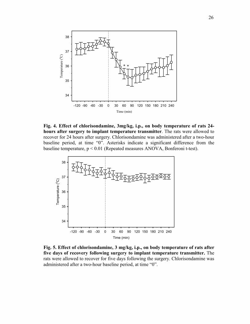
The animals were studied a day after the transmitter implantation surgery. They were
treated with ganglionic blocker, chlorisondamine. The animals became hypothermic and the
body temperature remained lower than control values throughout the four-hour observation
period (Fig. 4). However, subsequently, when animals were allowed to recover for five days
and then treated with chlorisondamine, the animals were able to maintain their body core

25
temperature (Fig. 5). Therefore, in all subsequent experiments, the animals were studied after
a minimum of five days after the surgery.
On the day of the experiment, the animals were transferred to plastic observation
boxes, which were placed on temperature receivers. Throughout the experimental period, the
temperature was recorded at one-minute intervals via a computerized data acquisition system.
Experiments involving cold exposure were conducted in a walk-in cold room whose
temperature could be controlled. The cold room is within the same laboratory and
immediately adjacent to the area where other animals were kept. Entry to the cold room is via
a glass door, which permits the lighting conditions to be equivalent for all animals (Fig. 7).
C. Measurement of Tail Skin Temperature Using Thermocouples in a Controlled
Temperature Environment
Male Sprague-Dawley rats weighing 225-250 g upon arrival were housed in pairs for
a week. Temperature transmitters were surgically implanted in the abdominal cavity and the
animals were allowed a minimum of five days to recover. On the day of the experiment, the
animals were transferred to plastic observation boxes and were attached to a tethering system
(Harvard Apparatus, MA) that allowed free movement. A thermocouple (Omega, CT) was
attached to the dorsal skin of the tail, approximately 7 cm from the tip, using surgical tape as
described by Redfern et al (Fig. 6)(Redfern et al., 1995). The electrical leads were then
passed to the back of the neck and through the wire spring of the tethering device. The
animals were transferred to the
26
Time (min)
-120 -90 -60 -30 0 30 60 90 120 150 180 210 240
Tem
pera
ture
(0 C)
34
35
36
37
38
Fig. 4. Effect of chlorisondamine, 3mg/kg, i.p., on body temperature of rats 24-hours after surgery to implant temperature transmitter. The rats were allowed to recover for 24 hours after surgery. Chlorisondamine was administered after a two-hour baseline period, at time “0”. Asterisks indicate a significant difference from thebaseline temperature, p < 0.01 (Repeated measures ANOVA, Bonferoni t-test).
Fig. 5. Effect of chlorisondamine, 3 mg/kg, i.p., on body temperature of rats afterfive days of recovery following surgery to implant temperature transmitter. The rats were allowed to recover for five days following the surgery. Chlorisondamine wasadministered after a two-hour baseline period, at time “0”.
Time (min)
-120 -90 -60 -30 0 30 60 90 120 150 180 210 240
Tem
pera
ture
(0 C)
34
35
36
37
38
* *

27
walk-in cold room maintained at an ambient temperature of 16-180C and they were then
placed on the temperature receiver to obtain a continuous record of the body core
temperature. The tail temperature was recorded every minute in a hand held thermocouple
thermometer (Omega, CT).
D. Measurement of Oxygen Consumption Using Open Circuit Calorimetry
Oxygen consumption (VO2) was determined in an open circuit calorimeter (Oxymax,
Columbus, OH). The calorimeter was placed in the walk-in cold room maintained at an
ambient temperature of 16-170C. Male rats with in-dwelling temperature transmitters were
placed in the plastic metabolic test chamber through which a known flow of air, was passed.
The system was calibrated with a known concentration of oxygen and carbon dioxide before
each experiment. The system monitors oxygen and carbon dioxide gas fractions at both the
inlet and output port of the test chamber. The gas fraction and flow measurements are used to
compute oxygen consumption and carbon dioxide production. The oxygen consumption was
measured every minute using a computerized data acquisition system.
E. Collection of Tissue Samples for Biochemical Analysis
Tissues (adrenal glands, intrascapular brown adipose tissue, white adipose tissue,
atria and ventricles) were excised and frozen (-700C) for later analysis of catecholamines.
Tissues were homogenized in 0.1 N perchloric acid containing EDTA and sodium
metabisulfite. The volume of homogenization for each tissue was as follows: adrenal glands,
2 ml/gland, atria, 1.5 ml/tissue, ventricles, intrascapular brown adipose tissue and

28
38.6 38.538.6 38.537.9 38.537.9 38.5
Receiver
MinimitterTemperatureTransmitter (Intraperitoneal)
DataAcquisition
Thermocouple
Data Acquisition38.6 38.538.6 38.537.9 38.537.9 38.5
Receiver
MinimitterTemperatureTransmitter (Intraperitoneal)
DataAcquisition
Thermocouple
Data Acquisition
Fig. 6. Measurement of body temperature using temperature telemetry and acomputerized data acquisition system and tail skin temperature usingthermocouples. Body temperature was measured using temperature transmitters (Minimitters, OR) that were surgically implanted in the abdominal cavity of the rats.The data was collected using a computerized system. The tail skin temperature wasmeasured by attaching thermocouples to the surface of the tail. The data was collected using a hand held thermometer.
29
Cold Room
Data Acquisition System
Temperature Receivers
Fig. 7. Laboratory arrangement for performing experiments in a temperature-controlled environment. The temperature of the walk-in cold room could be varied from 28 to 00C. The entrance to the cold room was via a glass door that permitted lighting conditions to be equivalent for animals inside and outside the cold room.

30
white adipose tissue, 200 mg tissue/ml. Homogenization was performed on ice for 30
seconds with a polytron homogenizer (Brinkmann Inst., Westbury, NY). Homogenates were
centrifuged (12,000 x g for 15 minutes at 40C) and the resultant supernatants collected and
stored at -700C for further biochemical analysis.
F. Measurement of Catecholamines by High Performance Liquid Chromatography
(HPLC)
Catecholamines were assayed using high performance liquid chromatography
(HPLC) with electrochemical detection (Waters, Marlborough, MA) by modification of
existing methods (Weicker et al., 1984).
Extraction of catecholamines from tissue homogenates involved adsorption onto
aluminium oxide (Woelm Neutral activity Grade 6). Sample aliquots (100-200µl, depending
on expected concentrations) were placed in 1.5 ml capped propylene tubes along with 10 mg
of alumina. The internal standard, 3,4-dihydroxybenzylamine, was added to each sample, and
the pH was adjusted to 8.6 with 1M tris(hydroxymethyl)aminomethane (Tris). The tubes
were capped and shaken vigorously in a mechanical shaker for 10 minutes and centrifuged
(12,000 RPM) for 2 minutes. The resulting pellet was washed 3 times with 600 µl of Tris (pH
7); catecholamines were desorbed with 100 µl of 0.1N perchloric acid through gentle mixing
in a microtube shaker (TOMY, Peninsula Labs., CA) for 30 seconds. An appropriate volume
of the eluate was immediately injected on to the HPLC system. Supernatants from adrenal
glands did not require extraction, and were directly injected on to the HPLC system
following dilution with perchloric acid (1:100).

31
Extracted catecholamines were separated on a 5µm, 3.9 x 150 mm C18 reverse-phase
column (Waters, Marlborough, MA). The mobile phase consisted of sodium acetate (50
mM), citric acid monohydrate (20 mM), sodium-1-octane-sulfonate (2 mM), di-n-butylamine
(1.0 mM), disodium EDTA (0.1 mM), and methanol (4%). Catecholamines were oxidized
during exposure to a glassy carbon electrode set at a potential of 0.6 V versus Ag/AgCl.
Data were acquired using ChromPerfect software (Justice Innovations, Mountain View, CA)
and calculations were based on peak areas. The limit of quantitation for NE and EPI was 50
pg/ml. The recovery of catecholamines from alumina extraction was 65-70 %.
G. Statistics
Results are presented as mean + S.E. The presence of statistically significant
differences was determined using Sigma Stat Statistical Software (Jandel Scientific, San
Rafael, CA). Normality test was first performed to verify the nature of the distribution. The
difference between multiple groups of means was assessed using analysis of variance
(ANOVA). If ANOVA revealed a statistically significant difference, post hoc pairwise
comparisons of means were done using the Bonferoni t-test. A repeated measures ANOVA
was conducted for contrasts of groups in which multiple measurements were made over time.
Post hoc contrasts were done using the Bonferoni t-test. A statistically significant effect was
accepted when p < 0.05.
The size of the experimental groups was 7 rats/group. The group size is based on the
variance estimates and power function calculations using preliminary data. The power of the
test was calculated by the statistical software whenever data was analyzed.

32
Chapter III. Pharmacologic Assessment of the Contribution of the Sympathetic Nervous System to the Maintenance of Body Temperature in
Conscious Rats
The overall goal of this thesis was to characterize the hypothermia to fenfluramine
treatment. We hypothesized that fenfluramine might influence the sympathetic nervous
system in such a way that the temperature homeostasis would be modified. Thus, the
hypothermia to fenfluramine could be due to interference with sympathetically mediated heat
generation or heat conservation. Despite the general acceptance that the sympathetic nervous
system mediated responses are important to thermoregulation, there is not much evidence in
the literature that equivocally demonstrates that disruption of sympathetic mechanisms of
heat generation or conservation would lead to deficits in body temperature. Therefore,
experiments in this chapter were designed to quantitatively assess the contribution of the
sympathetic nervous system to the maintenance of body temperature.
The sympathetic nervous system is involved in the control of homeostatic
adjustments that are initiated to maintain body temperature as discussed in detail in chapter I.
In order to evaluate the contribution of the sympathetic nervous system mechanisms of heat
generation and heat conservation to maintain body temperature, it was determined if
pharmacologic blockade of the thermoregulatory actions of the sympathetic nervous system
would result in a measurable decrease in body temperature. Therefore, the effects of the
following pharmacological agents on body temperature maintenance were determined:
phentolamine, an alpha-adrenoceptor blocking agent, was used to prevent sympathetic
nervous system mediated cutaneous vasoconstriction. Propranolol, a beta-adrenoceptor
blocking agent, was used to prevent sympathetically mediated thermogenesis, phentolamine
plus propranolol to block both sympathetically mediated heat conservation and generation

33
respectively and chlorisondamine, a ganglionic blocker, was used to block sympathetic
control of heat generation and conservation.
Body core temperature was recorded continuously, since body temperature is a direct
measure of thermoregulation. The animals were conscious and temperature artifacts due to
handling were prevented by using temperature telemetry as discussed in detail in chapter II.
Measurement of body temperature using indwelling temperature transmitters is an
improvement over prior studies because previous studies have measured body temperature
using rectal thermometers, the use of which could lead to stress related increases in body
temperature. Another advantage of using the temperature transmitters was that it also
provided a measure of locomotor activity. Locomotor activity was measured in our
experiments in order to determine if the animals increased physical activity to compensate
for blockade of other thermoregulatory mechanisms.
Tissue catecholamine contents of the adrenal glands and BAT were measured in our
experiments as an index of sympathetic activity. A previous study in our laboratory had
indicated that adrenal catecholamine content was depleted during cold exposure (Vollmer et
al., 1992) and a report from another laboratory had indicated that BAT NE content was also
reduced during cold exposure (Brito et al., 1998). These reports suggested that the tissue NE
content was attenuated due to sympathetic activation during cold exposure.
The measurements were conducted at two different ambient temperatures, 22 and
40C. The experiment was conducted at normal room temperature of 220C. And 40C, a
standard cold ambient temperature was used as a challenge to thermoregulatory mechanisms.
The 40C ambient temperature is known to activate the sympathetic nervous system and so the
effect of the sympathetic nervous system on its effector organs would be more pronounced
34
(Daly et al., 1992; Young et al., 1988). Therefore drugs that affect the sympathetic
mechanisms of body temperature maintenance would be expected to have a greater impact at
40C.
A. Protocols
Male Sprague-Dawley rats with implanted temperature transmitters were used in this
study. Half of the rats were studied at a laboratory room temperature of 220C and the other
half was exposed to a cold environment of 40C. Five treatments groups (n=7/group) were
investigated at 22 and 40C. The treatments were: saline, 1ml/kg, i.p., phentolamine, 2 mg/kg,
i.p., propranolol, 3 mg/kg, i.p., phentolamine, 2 mg/kg, i.p. plus propranolol, 3 mg/kg i.p. and
chlorisondamine, 3 mg/kg, i.p. Doses of propranolol and chlorisondamine were selected
based on previous studies conducted in our laboratory and by other investigators, in which it
was demonstrated that sympathetic neuronal responses were more than 90% abolished for the
time frame in which the current studies were conducted (Bush et al., 1983; Deitchman et al.,
1980). The dose of phentolamine, 2mg/kg, i.p., was shown, by other investigators, to cause
cutaneous vasodilation in rats (Lin et al., 1979).
In all groups, baseline body temperature was recorded for two hours prior to
treatment. After treatment, the animals were monitored for four hours. During the six-hour
experimental period, temperature and locomotor activity data was recorded at one-minute
intervals and averaged every 15 minutes via a computerized data acquisition system. At the
completion of the experiment, the animals were anesthetized with pentobarbital, 60 mg/kg,
i.p. and intrascapular BAT and adrenal glands were removed and frozen (-700C) for later
analysis of catecholamines. Catecholamines were analyzed using HPLC.

35
Statistical Analyses
Results are presented as mean + S.E. The differences in multiple groups of animals
maintained at 22 and 40C for individual parameters such as BAT and adrenal catecholamine
contents and activity data, were assessed using two-way analysis of variance (ANOVA). The
differences in multiple measurements taken over a period of time were assessed using
repeated measures ANOVA. If ANOVA revealed a statistically significant difference, post
hoc pairwise comparisons of groups were done using Bonferoni t-test. A statistically
significant effect was accepted when p < 0.05.
B. Results
1. Effects of saline, 1ml/kg, i.p., on body temperature, locomotor activity,
adrenal and BAT catecholamine content.
During the first two hours of the experimental protocol (baseline period), there was
no difference in the body temperature between the two groups of animals maintained at 220C
and 40C, Fig. 8, left panel. Saline treatment did not affect body temperature maintenance in
animals kept at 22 and 40C, Fig. 8, left panel. Also, locomotor activity was not affected by
saline treatment, Fig. 8, right panel.
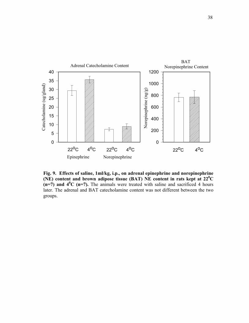
Saline treatment did not affect adrenal norepinephrine (NE) or epinephrine (EPI)
content of rats kept at 40C when compared to rats kept at 220C, Fig. 9, left panel. Also, saline
treatment did not affect BAT norepinephrine (NE) content of rats kept at 40C when compared
to rats kept at 220C, Fig. 9, right panel.

36
2. Effects of phentolamine, 2 mg/kg, i.p., on body temperature,
locomotor activity, adrenal and BAT catecholamine content.
During the first two hours of the experimental protocol (baseline period), there was
no difference in the body temperature between the two groups of animals maintained at 220C
and 40C, Fig. 10, left panel. The body temperature of animals treated with phentolamine at 22
and 40C were not different, Fig. 10, left panel. Phentolamine treatment at 220C increased
locomotor activity when compared to saline treated controls, p < 0.001 (Student t-test).
However, when the activity of all the groups of animals were analyzed using ANOVA, there
was no statistical difference, Fig. 10, right panel.
Phentolamine treatment did not affect adrenal NE or EPI content, Fig. 11, left panel.
However, BAT NE content was significantly decreased (-57 %) in the cold exposed
phentolamine treated animals, p < 0.001 (Two-way ANOVA). BAT NE content of the group
of animals treated with phentolamine and kept at 220C was significantly different from the
group of animals treated with phentolamine and kept at 40C (Bonferoni t-test), Fig. 11, right
panel.
3. Effects of propranolol, 3 mg/kg, i.p., on body temperature, locomotor
activity, adrenal and BAT catecholamine content.
During the first two hours of the experimental protocol (baseline period), there was
no difference in the body temperature between the two groups of animals maintained at 220C
and 40C, Fig. 12, left panel. Propranolol treatment did not affect body temperature
maintenance in animals kept at 220C. However, treatment of animals with

37
Fig. 8. Effects of saline, 1ml/kg, i.p., on body temperature and locomotor activity of rats kept at 220C (n=7) and 40C (n=7). Saline was administered at time “0”. Saline treatment did not affect body temperature maintenance and locomotor activity.
Time (hr)-2 -1 0 1 2 3 4
Tem
pera
ture
(o C)
34
35
36
37
38
39
22o C4oC
22oC 4oC
Body Temperature Activity
Act
ivity
(cou
nts/
hr)
0
20
40
60
80
38
Fig. 9. Effects of saline, 1ml/kg, i.p., on adrenal epinephrine and norepinephrine (NE) content and brown adipose tissue (BAT) NE content in rats kept at 220C (n=7) and 40C (n=7). The animals were treated with saline and sacrificed 4 hourslater. The adrenal and BAT catecholamine content was not different between the two groups.
Nor
epin
ephr
ine
(ng/
g)0
200
400
600
800
1000
1200
BAT Norepinephrine Content
22oC 4oC
Adrenal Catecholamine Content
Cat
echo
lam
ine
(ug/
glan
d)
0
5
10
15
20
25
30
35
40
22oC 4oCEpinephrine
22oC 4oCNorepinephrine

39
Fig. 10. Effects of phentolamine, 2mg/kg, i.p., on body temperature andlocomotor activity of rats kept at 220C (n=7) and 40C (n=7). Phentolamine was administered at time “0”. Body temperature remained constant throughout the observation period. The locomotor activity between the two groups was notsignificantly different.
Time (hr)-2 -1 0 1 2 3 4
Tem
pera
ture
(o C)
34
35
36
37
38
39
22o C4oC
22oC 4oC
Body Temperature Activity
Act
ivity
(cou
nts/
hr)
0
20
40
60
80

40
Fig. 11. Effects of phentolamine, 2mg/kg, i.p., on adrenal epinephrine andnorepinephrine (NE) content and brown adipose tissue (BAT) NE content in rats kept at 220C (n=7) and 40C (n=7). The animals were treated with phentolamine and sacrificed 4 hours later. The adrenal catecholamine content between the two groupswas not different. However, BAT of the cold exposed animals showed a significant depletion of its NE content. Two way ANOVA indicated that the groups aresignificantly different, p < 0.001. An asterisks indicate that there is a significantdifference in BAT NE content between the phentolamine treated group kept at 220C and the phentolamine treated group kept at 40C, Bonferoni t-test.
Nor
epin
ephr
ine
(ng/
g)
0
200
400
600
800
1000
1200
BAT Norepinephrine Content
22oC 4oC
Adrenal Catecholamine Content
Cat
echo
lam
ine
(ug/
glan
d)
0
5
10
15
20
25
30
35
40
22oC 4oCEpinephrine
22oC 4oCNorepinephrine
*
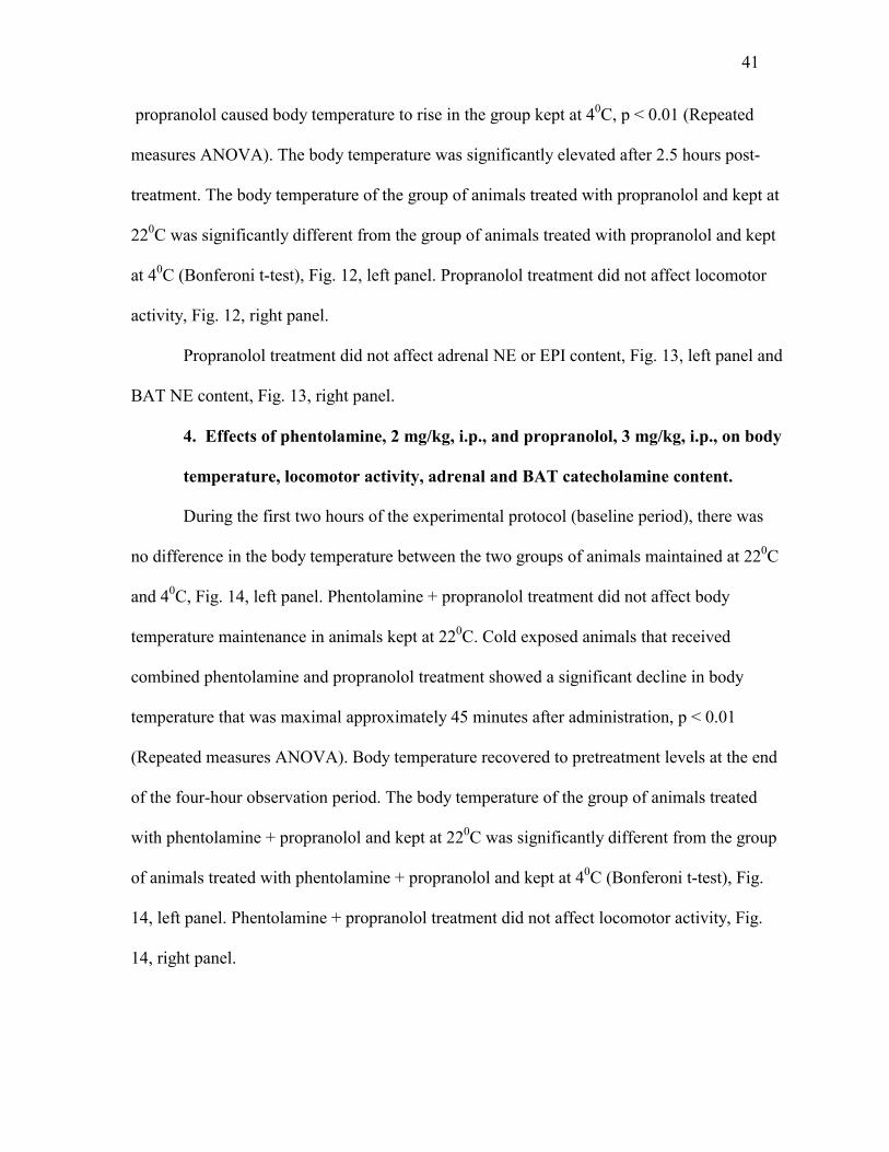
41
propranolol caused body temperature to rise in the group kept at 40C, p < 0.01 (Repeated
measures ANOVA). The body temperature was significantly elevated after 2.5 hours post-
treatment. The body temperature of the group of animals treated with propranolol and kept at
220C was significantly different from the group of animals treated with propranolol and kept
at 40C (Bonferoni t-test), Fig. 12, left panel. Propranolol treatment did not affect locomotor
activity, Fig. 12, right panel.
Propranolol treatment did not affect adrenal NE or EPI content, Fig. 13, left panel and
BAT NE content, Fig. 13, right panel.
4. Effects of phentolamine, 2 mg/kg, i.p., and propranolol, 3 mg/kg, i.p., on body
temperature, locomotor activity, adrenal and BAT catecholamine content.
During the first two hours of the experimental protocol (baseline period), there was
no difference in the body temperature between the two groups of animals maintained at 220C
and 40C, Fig. 14, left panel. Phentolamine + propranolol treatment did not affect body
temperature maintenance in animals kept at 220C. Cold exposed animals that received
combined phentolamine and propranolol treatment showed a significant decline in body
temperature that was maximal approximately 45 minutes after administration, p < 0.01
(Repeated measures ANOVA). Body temperature recovered to pretreatment levels at the end
of the four-hour observation period. The body temperature of the group of animals treated
with phentolamine + propranolol and kept at 220C was significantly different from the group
of animals treated with phentolamine + propranolol and kept at 40C (Bonferoni t-test), Fig.
14, left panel. Phentolamine + propranolol treatment did not affect locomotor activity, Fig.
14, right panel.

42
Phentolamine + propranolol treatment did not affect adrenal NE or EPI content of rats
kept at 40C when compared to rats kept at 220C, Fig. 15, left panel. Cold exposed animals
that received the combined treatment also showed a very substantial reduction in BAT NE
content (-97%), p < 0.001 (Two-way ANOVA). The BAT NE content of the group of
animals treated with phentolamine + propranolol and kept at 220C was significantly different
from the group of animals treated with phentolamine + propranolol and kept at 40C
(Bonferoni t-test), Fig. 15, right panel.
5. Effects of chlorisondamine, 3 mg/kg, i.p., on body temperature, locomotor
activity, adrenal and BAT catecholamine content.
During the first two hours of the experimental protocol (baseline period), there was
no difference in the body temperature between the two groups of animals maintained at 220C
and 40C, Fig. 16, left panel. Chlorisondamine treatment did not affect body temperature
maintenance in animals kept at 220C. The group of cold exposed animals treated with
chlorisondamine showed a significant decline in body temperature, p < 0.01 (Repeated
measures ANOVA). The peak decline in core temperature occurred one hour after
administration and temperature recovered to pretreatment levels by the end of the
experimental period. The body temperature of the group of animals treated with
chlorisondamine and kept at 220C was significantly different from the group of animals
treated with chlorisondamine and kept at 40C (Bonferoni t-test), Fig. 16, left panel.
Chlorisondamine treatment did not affect locomotor activity, Fig. 16, right panel.
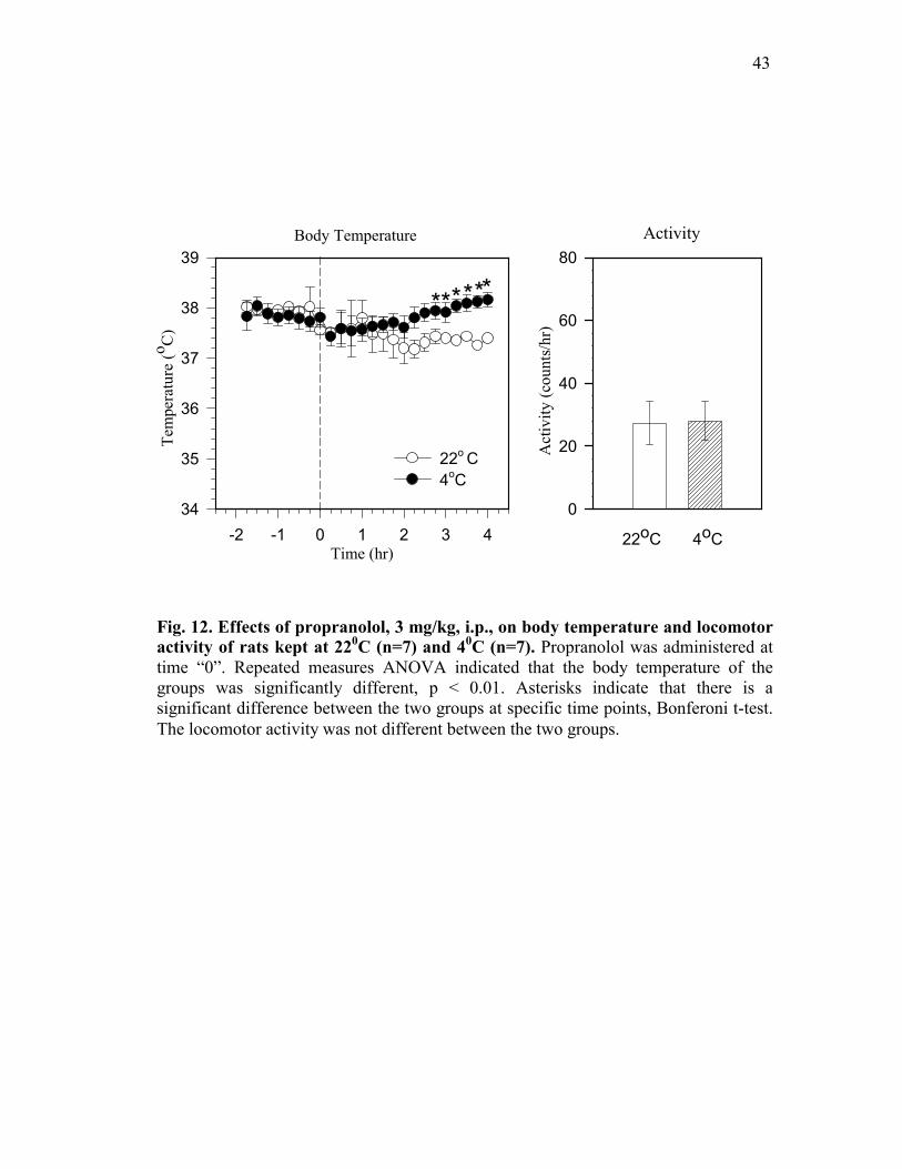
43
Fig. 12. Effects of propranolol, 3 mg/kg, i.p., on body temperature and locomotoractivity of rats kept at 220C (n=7) and 40C (n=7). Propranolol was administered at time “0”. Repeated measures ANOVA indicated that the body temperature of thegroups was significantly different, p < 0.01. Asterisks indicate that there is asignificant difference between the two groups at specific time points, Bonferoni t-test. The locomotor activity was not different between the two groups.
Time (hr)-2 -1 0 1 2 3 4
Tem
pera
ture
(o C)
34
35
36
37
38
39
22o C4oC
22oC 4oC
Body Temperature Activity
Act
ivity
(cou
nts/
hr)
0
20
40
60
80
*** ***

44
Fig. 13. Effects of propranolol, 3 mg/kg, i.p., on adrenal epinephrine andnorepinephrine (NE) content and brown adipose tissue (BAT) NE content in ratskept at 220C (n=7) and 40C (n=7). The animals were treated with propranolol and sacrificed 4 hours later. The adrenal and BAT catecholamine content was not differentbetween the two groups.
Nor
epin
ephr
ine
(ng/
g)0
200
400
600
800
1000
1200
BAT Norepinephrine Content
22oC 4oC
Adrenal Catecholamine Content
Cat
echo
lam
ine
(ug/
glan
d)
0
5
10
15
20
25
30
35
40
22oC 4oCEpinephrine
22oC 4oCNorepinephrine

45
Fig. 14. Effects of phentolamine, 2 mg/kg, i.p. and propranolol, 3 mg/kg, i.p., onbody temperature and locomotor activity of rats kept at 220C (n=7) and 40C (n=7). The drugs were administered at time “0”. Repeated measures ANOVA indicated that the body temperature of the groups was significantly different, p < 0.01.Asterisks indicate that there is a difference between the two groups at specific timepoints, Bonferoni t-test. The locomotor activity was not different between the two groups.
Time (hr)-2 -1 0 1 2 3 4
Tem
pera
ture
(o C)
34
35
36
37
38
39
22o C4oC
22oC 4oC
Body Temperature Activity
Act
ivity
(cou
nts/
hr)
0
20
40
60
80
**
** *

46
Fig. 15. Effects of phentolamine, 2 mg/kg, i.p. and propranolol, 3 mg/kg, i.p., onadrenal epinephrine and norepinephrine (NE) content and brown adipose tissue(BAT) NE content in rats kept at 220C (n=7) and 40C (n=7). The animals were treated with the drugs and sacrificed 4 hours later. The adrenal catecholamine contentwas not different between the two groups. However, BAT of the cold exposed animalsshowed a significant depletion of its NE content. Two way ANOVA indicated that the groups are significantly different, p < 0.001. An asterisks indicate that there is asignificant difference between the phentolamine + propranolol treated group kept at220C and the phentolamine + propranolol treated group kept at 40C, Bonferoni t-test.
Nor
epin
ephr
ine
(ng/
g)
0
200
400
600
800
1000
1200
BAT Norepinephrine Content
22oC 4oC
Adrenal Catecholamine Content
Cat
echo
lam
ine
(ug/
glan
d)
0
5
10
15
20
25
30
35
40
22oC 4oCEpinephrine
22oC 4oCNorepinephrine
*

47
Chlorisondamine treatment did not affect adrenal NE or EPI content, Fig. 17, left
panel. However, BAT NE content was significantly less (-42%) in the cold exposed
chlorisondamine treated animals, p < 0.001 (Two-way ANOVA). The BAT NE content of
the group of animals treated with chlorisondamine and kept at 220C was significantly
different from the group of animals treated with chlorisondamine and kept at 40C (Bonferoni
t-test), Fig. 17, right panel.
C. Discussion
The experiments provide quantitative evidence that the sympathetic nervous system
mechanisms of heat generation and conservation are essential to the maintenance of body
temperature. The pharmacologic blockade of alpha and beta adrenoceptor mediated
mechanisms of thermoregulation at 40C resulted in a deficit in body temperature
maintenance.
The animals exposed to a cold (40C) ambient temperature for the six-hour observation
period could maintain body temperature. Thus, cold exposure alone does not affect body
temperature maintenance. In order to determine if the sympathetic nervous system was
activated during cold exposure, we measured BAT NE content as an index of sympathetic
activity. No change in BAT NE content was observed in animals kept at 40C. While it has
been shown by other investigators that BAT is neurally activated during acute cold exposure
as evidenced by increased blood flow to BAT (Foster et al., 1978), increased GDP binding to
mitochondria of BAT (Trayhurn et al., 1987) and increased BAT NE turnover rate
(McDonald et al., 1993), it was found that an increase in neural activity to BAT was not
sufficient to change its NE content. However, there are reports
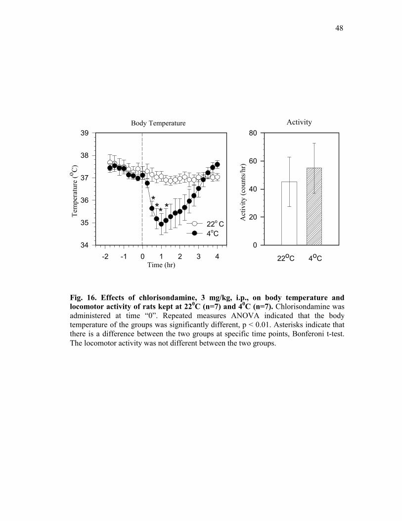
48
Fig. 16. Effects of chlorisondamine, 3 mg/kg, i.p., on body temperature andlocomotor activity of rats kept at 220C (n=7) and 40C (n=7). Chlorisondamine was administered at time “0”. Repeated measures ANOVA indicated that the body temperature of the groups was significantly different, p < 0.01. Asterisks indicate thatthere is a difference between the two groups at specific time points, Bonferoni t-test. The locomotor activity was not different between the two groups.
Time (hr)-2 -1 0 1 2 3 4
Tem
pera
ture
(o C)
34
35
36
37
38
39
22o C4oC
22oC 4oC
Body Temperature Activity
Act
ivity
(cou
nts/
hr)
0
20
40
60
80
*** *

49
Fig. 17. Effects of chlorisondamine, 3mg/kg, i.p., on adrenal epinephrine andnorepinephrine content (NE) and brown adipose tissue (BAT) NE content in ratskept at 220C (n=7) and 40C (n=7). The animals were treated with chlorisondamine and sacrificed 4 hours later. The adrenal catecholamine content between the twogroups was not different. However, BAT of the cold exposed animals showed asignificant depletion of its NE content. Two way ANOVA indicated that the groups are significantly different, p < 0.001. An asterisks indicate that there is a significantdifference between the chlorisondamine treated group kept at 220C and the chlorisondamine treated group kept at 40C, Bonferoni t-test.
Nor
epin
ephr
ine
(ng/
g)0
200
400
600
800
1000
1200
BAT Norepinephrine Content
22oC 4oC
Adrenal Catecholamine Content
Cat
echo
lam
ine
(ug/
glan
d)
0
5
10
15
20
25
30
35
40
22oC 4oCEpinephrine
22oC 4oCNorepinephrine
*

50
that cold exposure alone can cause depletion of BAT NE (King et al., 1999; Brito et al.,
1998). The disparity in experimental results is probably best explained by differences in the
experimental design of each study, which include variability in the duration of cold exposure
and the handling of animals during temperature measurements.
However, the ability to maintain NE content in the presence of increased sympathetic
drive to BAT appears to be limited because blockade of alpha-receptors with phentolamine in
animals exposed to cold resulted in a decline in BAT NE (-57%). This finding can be
interpreted to indicate that phentolamine interfered with alpha-receptor-mediated heat
conserving mechanisms, piloerection and cutaneous vasoconstriction, resulting in an
augmented activation of sympathetic outflow to BAT. It appears that the activation of BAT
offset the effects of phentolamine because core temperature was maintained at normal levels.
The prevention of piloerection by phentolamine was visually verified in these studies and the
dose of phentolamine used was shown to produce cutaneous vasodilation by others (Lin et
al., 1979). Phentolamine did not have an effect on BAT innervation independent of cold
exposure because BAT NE content was not diminished in animals kept at 220C.
When beta-receptors were blocked in addition to alpha-receptors, body core
temperature significantly declined in cold exposed animals. Thus combined blockade of
adrenoceptors presents a greater challenge to thermoregulation than if the alpha- and beta-
receptors are independently blocked. This was evidenced by an intense activation of BAT as
noted by an almost total disappearance of NE from BAT (–97%).
In rats, there is significant stimulation of heart rate and cardiac output in response to
cold exposure (Sun et al., 1997) and the dose of propranolol used in our experiment is
sufficient to block these cardiovascular responses mediated via beta-1 receptors (Young et

51
al., 1992). The extent to which this dose of propranolol would interrupt sympathetic
activation of BAT is uncertain because there is substantial evidence to suggest that the
functional receptor in BAT is the beta-3 receptor subtype (Zhao et al., 1998). While
propranolol is a potent blocker of the beta-1 and beta-2 receptors, higher doses are required to
block beta-3 receptor mediated thermogenesis. In one report (Benzi et al., 1988), the dose of
propranolol used in the present investigation was shown to block BAT thermogenesis but
another report suggests that higher doses are required (Shimizu et al., 1991). Also it must be
considered that the endogenous ligand, NE has less affinity for the beta-3 receptor than the
beta-1 or beta-2 (Chaudhry et al., 1992; Granneman, 1992; Granneman, 1990). Nevertheless
beta-receptor blockade in conjunction with alpha-receptor blockade produced an almost total
disappearance of NE from BAT.
Interestingly, the body temperature of animals treated with propranolol while in the
cold began to rise and became significantly higher than the animals kept at 220C. One
potential explanation for this unexpected finding is that propranolol may interfere with a
beta-2 receptor mediated cutaneous vasodilatation that may have been present prior to
propranolol administration (Carlisle et al., 1992).
The adrenal content of EPI and NE was measured in these experiments as a potential
indicator of adrenal activation but no change in adrenal catecholamines was observed in any
of the groups. The absence of any change in content does not rule out the activation of the
adrenal since there might be an accelerated synthesis of catecholamines to compensate for
the increased secretion. In fact, there is evidence that the adrenals are activated during cold
exposure (Himms-Hagan, 1975; Leduc, 1961). One previous study has shown that inhibition
of catecholamine synthesis by prior treatment with α-methyl-p-tyrosine resulted in a
52
decreased adrenal NE content when animals were exposed to cold for three hours (Vollmer et
al., 1992). In the same study, it was found that a very robust stimulation of the adrenal gland
is required before content is measurably reduced. Prolonged cold exposure resulted in a
selective depletion of adrenal medullary NE, indicating that the adrenal gland became more
active as the duration of cold exposure increased. Another factor that differed from the
present investigation was that in the previous study the animals were shaved, thereby
increasing the severity of the cold exposure.
The somatic nervous system contributes to thermoregulation through two major
actions, shivering induced thermogenesis and through heat generated by increased locomotor
activity. The temperature transmitters provided a record of locomotor activity and no
increase in voluntary motor activity was detected during cold exposure. Shivering induced
heat generation was not measured directly but we visually observed shivering only in the
chlorisondamine treated group exposed to cold. This shivering occurred without an increase
in locomotor activity. The chlorisondamine group also showed the greatest hypothermic
effect during cold exposure. In addition, it was found that the dose of chlorisondamine did
not produce a complete blockade of ganglionic transmission because there was a significant
depletion of BAT NE content (-42%).
Thus, the experiments in this chapter demonstrate that the integrity of sympathetic
input to thermoregulatory effector systems, which include, the adrenals, BAT, cutaneous
blood vessels and piloerector smooth muscle is required to maintain body temperature during
cold exposure. Blockade of sympathetic neural influences pharmacologically, through the use
of alpha- and beta-receptor blockers, demonstrated that both alpha- and beta- adrenoceptor-
mediated mechanisms contribute quantitatively to the maintenance of core temperature
53
during cold exposure. However, due to the overlapping compensations, both alpha- and beta-
receptor mechanisms had to be interrupted before a significant deficit in body temperature
was detected.
Catecholamine content of BAT and the adrenal gland were measured to determine if
the neurotransmitter stores would be reduced proportionately to the intensity of sympathetic
nervous system activation. The major finding was that BAT NE content, but not adrenal
content, was decreased during cold exposure combined with adrenoceptor blockade. The
relationship was consistent with the conclusion that BAT NE content declined when
sympathetic neural activity increased to compensate for blockade of adrenoceptor-mediated
heat conservation or heat generation mechanisms.
Moreover, this study provided a model to evaluate the effects of fenfluramine on
body temperature and BAT activation. This model was used to characterize the effects of
fenfluramine on body temperature and sympathetically mediated mechanisms of heat
generation and conservation in the following chapters.

54
Chapter IV. Evaluation of the Effects of Fenfluramine on Sympathetic Nervous System Regulation of Body Temperature
Experiments presented in this chapter were designed to study the effects of
fenfluramine treatment on sympathetic nervous system control of thermoregulation using the
model developed in chapter III. The results of the experiments in chapter III demonstrated
that the sympathetic nervous system plays an important role in the maintenance of body
temperature. Pharmacologic blockade of sympathetic nervous system mediated mechanisms
of cutaneous vasoconstriction and thermogenesis led to disruption of body temperature
homeostasis. In this chapter, experiments were designed to assess the effects of fenfluramine
treatment on body temperature maintenance and activation of sympathetic effector systems
involved in the maintenance of body temperature that include the BAT, adrenal gland, white
adipose tissue and the heart.
As discussed in the literature survey (Chapter I), the effects of fenfluramine on
thermoregulation are not fully understood. Although, hypothermia is frequently reported
following fenfluramine administration (Cryan et al., 2000; Preston et al., 1990), the effect of
fenfluramine treatment on body temperature regulation appears to depend on the
environmental temperature at which the animal is maintained at the time of fenfluramine
administration. At normal laboratory temperatures of around 220C, some studies report that
fenfluramine treatment produced a hypothermic response (Cryan et al., 2000) and others
report that there was no change in body temperature of experimental animals to fenfluramine
treatment (Sugrue, 1984). The lack of consistency in the results of these studies are due to
differences in methodology used to measure body temperature, differences in ambient
temperature (20-240C) and the use of different doses of fenfluramine. In contrast, at lower

55
ambient temperatures, fenfluramine treatment is consistently associated with varying degrees
of hypothermia (Malberg et al., 1997; Preston et al., 1990).
Therefore, the first objective of this study was to study the relationship between
environmental temperature and the effect of fenfluramine on body temperature. Animals
were studied at two ambient temperatures, 22 and 40C. Also, the relationship between the
dose of fenfluramine and hypothermia was studied. The doses of fenfluramine were selected
based on studies conducted by others in which these doses produced an antiobesity effect
(Levitsky et al., 1992). The fact that all animals to be used in this study would have
implanted temperature transmitters provided an improvement over prior studies (Sugrue,
1984; Preston et al., 1990) in which animals had to be handled to record core temperature
using rectal thermometers, which could lead to handling related stress and increase body
temperature (Poole et al., 1977).
The results of the experiments described in chapter III demonstrated that sympathetic
activation was important to the homeostatic regulation of body core temperature during cold
exposure. Therefore the second objective of these experiments was to determine if a
perturbation in sympathetic activation of BAT and other sympathetic effector systems, that
include the adrenal glands, heart and white adipose tissue, could account for the hypothermia
that has been reported to occur following the administration of fenfluramine. Based on the
observation from the experiments presented in the previous chapter and the work of others
(Brito et al., 1998; King et al., 1999), BAT NE content was used as an indicator of BAT
activation. Previous studies, conducted in our laboratory and others’, demonstrated that NE
content of the adrenal gland, white adipose tissue and heart was depleted during cold
exposure, when the sympathetic nervous system is known to be activated (Vollmer et al.,

56
1992; Garafalo et al., 1996; Himms-Hagan, 1975). Therefore, NE content of BAT, adrenal
glands, heart and white adipose tissue were measured as an index of sympathetic activation.
To test the sensitivity of BAT NE content as an index of sympathetic activity, a
preliminary study was conducted. Animals were administered dl-fenfluramine. BAT NE
content was found to be profoundly depleted (Experiment 1) in the fenfluramine treated
animals. Additional experiments were designed based on the preliminary study. The
objectives of the experiments were: First, to determine if the BAT NE depletion to
fenfluramine treatment is dose related. Second, to characterize the mechanisms by which
fenfluramine produced a depletion of BAT NE content. We determined if fenfluramine-
induced BAT NE depletion is mediated indirectly, through activation of the sympathetic
nervous system versus a direct effect on the NE stores of BAT (a reserpine-like action). This
objective was accomplished by studying the effect of fenfluramine on BAT activation in
animals pretreated with and without the ganglionic blocking agent pentolinium, which blocks
the sympathetic ganglionic transmission to BAT. We also determined if fenfluramine-
induced BAT NE depletion is mediated through release of serotonin. This objective was
based on literature reports that fenfluramine mediates most of its pharmacologic effects via
serotonin release (Cryan et al., 2000; Lin, 1978). This objective was accomplished by
studying the effect of fenfluramine on BAT activation in animals pretreated with and without
fluoxetine, which blocks fenfluramine-mediated serotonin release.

57
A. Protocols
1. Experiment 1. Effects of dl-fenfluramine on body temperature and BAT NE
content.
This was a preliminary experiment conducted to determine if fenfluramine produced
a change in body temperature and BAT NE content. All animals used in this study were
implanted with temperature transmitters. Two groups of animals (n=7/group) were studied at
an ambient temperature of 220C and two groups (n=7/group) were studied at an ambient
temperature of 40C. Baseline temperature measurements were made for two hours. The
animals were then treated with dl-fenfluramine, 10 mg/kg, i.p. or saline and observed for four
hours. At the end of the observation period, the animals were anesthetized with pentobarbital,
60 mg/kg, i.p. and tissues were removed for catecholamine content analysis. Intrascapular
BAT was removed and frozen for later biochemical analysis. The catecholamine content of
BAT was analyzed using HPLC as described in chapter II.
The racemic mixture, dl-fenfluramine was used in this protocol. The dose, 10 mg/kg,
of dl-fenfluramine was selected based on studies conducted by other investigators in which
the antiobesity effect of fenfluramine was demonstrated at this dose (Preston et al., 1990).
2. Experiment 2. Effects of d-fenfluramine on body temperature and
catecholamine content of sympathetically innervated tissues.
This experiment was conducted to determine if the hypothermia and BAT NE
depletion produced by fenfluramine was dose-related and if the effect depends on the
ambient temperature at which the animals receive fenfluramine. Also, the effects of
fenfluramine on the NE content of other sympathetically innervated organs including the
adrenal gland, heart and white adipose tissue was determined. The d-isomer of fenfluramine

58
was used in this study since it affects only the serotonergic neurons whereas the racemic
mixture has been shown to affect catecholaminergic neurons as well (Invernizzi et al., 1986).
Also, d-fenfluramine is known to be more potent than the racemic mixture. Henceforth, all
experiments were conducted using the d-isomer. All animals used in this study were
implanted with temperature transmitters. Three groups (n=7/group) were studied at 220C and
three groups (n=7/group) at 40C. Three treatments were investigated at each temperature,
saline, d-fenfluramine, 3 or 10 mg/kg, i.p.
As described in Experiment 1, the experiments were conducted over a six-hour
period. Two hours of baseline body temperature measurements were obtained. The animals
received treatments and were observed for another four hours. At the end of the observation
period, the animals were anesthetized with pentobarbital, 60 mg/kg, i.p. and intrascapular
BAT, adrenal glands, heart and perirenal WAT were removed for catecholamine content
analysis using HPLC.
The doses of d-fenfluramine used in this experiment were selected based on studies
conducted by other investigators in which it was demonstrated that these doses of d-
fenfluramine had appetite suppressive and BAT stimulant thermogenic activity (Rothwell et
al., 1992; Jackson et al., 1997).
3. Experiment 3. Effects of pentolinium and fluoxetine pretreatment on d-
fenfluramine-induced BAT NE depletion.
This experiment was conducted to determine if BAT NE depletion produced by d-
fenfluramine was mediated via activation of the sympathetic nervous system and through
release of serotonin. This was accomplished by studying the effect of d-fenfluramine on BAT
NE content in animals pretreated with and without ganglionic blocker pentolinium and

59
selective serotonin reuptake inhibitor fluoxetine. The experiments were conducted at 220C
since the extent of d-fenfluramine-induced BAT NE depletion was similar in animals
maintained at 22 and 40C.
The effects of pretreatment with saline, pentolinium, 10mg/kg, i.p. or fluoxetine, 10
mg/kg, i.p., on d-fenfluramine, 10 mg/kg, i.p.,-induced BAT NE depletion were assessed.
The pretreatments were administered after animals were acclimated to the lab conditions for
a period of two hours. Thirty minutes after pretreatment, d-fenfluramine, 10 mg/kg, i.p. or
saline was administered. After four hours of observation, the animals were anesthetized with
pentobarbital, 60 mg/kg, i.p. and intrascapular BAT was removed for NE content analysis. A
total of six groups of animals (n=7/group) were studied. Group 1 served as controls and
received saline pretreatment followed by a second treatment with saline. Group 2 received
saline pretreatment followed by a second treatment with fenfluramine. Group 3 received
pentolinium pretreatment followed by a second treatment with saline. Group 4 received
pentolinium pretreatment followed by a second treatment with fenfluramine. Group 5
received fluoxetine pretreatment followed by a second treatment with saline. Group 6
received fluoxetine pretreatment followed by a second treatment with fenfluramine.
The dose of d-fenfluramine, 10 mg/kg, i.p., was used since it produced the greatest
depletion of BAT NE content in our experiments. The dose of pentolinium used, 10 mg/kg,
i.p., was shown to have blocked sympathetic ganglionic transmission in rats (Redfern et al.,
1995). The dose of fluoxetine, 10mg/kg, was shown to have blocked fenfluramine-induced
release of serotonin in rat brain microdialysis studies (Gundlah et al., 1997).

60
Statistical Analyses
Results are presented as mean + S.E. The differences between groups of animals
treated with fenfluramine and saline and maintained at 22 and 40C, for individual parameters
(BAT, adrenal, WAT, heart NE content) were assessed using two-way analysis of variance
(ANOVA). The differences in BAT NE content between fenfluramine-treated groups
pretreated with pentolinium, fluoxetine or saline were assessed using one-way ANOVA. If
ANOVA revealed a statistically significant difference, post hoc pairwise comparisons of
groups were done using Bonferoni t-test.
The differences between multiple groups (saline, fenfluramine treated groups at 22 and 40C)
and between multiple measurements taken over a period of time (-120 to 240 minutes.) were
assessed using repeated measures ANOVA. Post-hoc pairwise comparisons were done using
Bonferoni t-test. A statistically significant effect was accepted when p < 0.05.
B. Results.
1. Experiment 1. Effects of dl- fenfluramine, 10 mg/kg, i.p., on body
temperature and BAT NE content.
During the first two hours of the experimental protocol (baseline period), there was
no difference in the body temperature between the groups of animals maintained at 220C and
40C. There was no difference in body temperature between the two groups of animals treated
with saline and kept at 22 and 40C, Fig. 18.
There was no difference in body temperature between the group of animals treated
with dl -fenfluramine and the group of animals treated with saline and kept at 220C.
However animals that were treated with dl-fenfluramine at 40C, became significantly
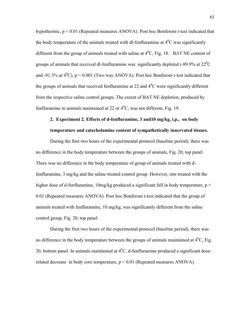
61
hypothermic, p < 0.01 (Repeated measures ANOVA). Post hoc Bonferoni t-test indicated that
the body temperature of the animals treated with dl-fenfluramine at 40C was significantly
different from the group of animals treated with saline at 40C, Fig. 18. BAT NE content of
groups of animals that received dl-fenfluramine was significantly depleted (-89.9% at 220C
and -91.3% at 40C), p < 0.001 (Two way ANOVA). Post hoc Bonferoni t-test indicated that
the groups of animals that received fenfluramine at 22 and 40C were significantly different
from the respective saline control groups. The extent of BAT NE depletion, produced by
fenfluramine in animals maintained at 22 or 40C, was not different, Fig. 19.
2. Experiment 2. Effects of d-fenfluramine, 3 and10 mg/kg, i.p., on body
temperature and catecholamine content of sympathetically innervated tissues.
During the first two hours of the experimental protocol (baseline period), there was
no difference in the body temperature between the groups of animals, Fig. 20, top panel.
There was no difference in the body temperature of group of animals treated with d-
fenfluramine, 3 mg/kg and the saline-treated control group. However, rats treated with the
higher dose of d-fenfluramine, 10mg/kg produced a significant fall in body temperature, p <
0.01 (Repeated measures ANOVA). Post hoc Bonferoni t-test indicated that the group of
animals treated with fenfluramine, 10 mg/kg, was significantly different from the saline
control group, Fig. 20, top panel.
During the first two hours of the experimental protocol (baseline period), there was
no difference in the body temperature between the groups of animals maintained at 40C, Fig.
20, bottom panel. In animals maintained at 40C, d-fenfluramine produced a significant dose-
related decrease in body core temperature, p < 0.01 (Repeated measures ANOVA).

62
Fig. 18. Effects of saline or dl-fenfluramine , 10 mg/kg, i.p., on body temperatureof rats kept at 22 and 40C (n=7/group). Animals with implanted temperature transmitters were maintained at 22 and 40C for six hours. Two hours of baseline body temperature data was collected and fenfluramine or saline were administered at time“0”. In animals maintained at 40C, fenfluramine produced a significant hypothermia.Repeated measures ANOVA indicated that the groups are significantly different, p < 0.01. Asterisks indicate a significant difference between the fenfluramine and thesaline control group maintained at 40C at specific time points, Bonferoni t-test.
Time (hr)-2 -1 0 1 2 3 4
Tem
pera
ture
(0 C)
33
34
35
36
37
38
** * ***
****
220C-Fenfluramine
40C-Fenfluramine
220C-Saline40C-Saline

63
Fig. 19. Effects of saline or dl-fenfluramine, 10 mg/kg, i.p., on brown adiposetissue (BAT) norepinephrine (NE) content of rats kept at 22 and 40C (n=7/group). The animals were treated with saline or fenfluramine and sacrificed four hours later.Fenfluramine produced a significant depletion of BAT NE content in rats kept at 22and 40C. The extent of depletion was similar at 22 and 40C. Two way ANOVA indicated that the groups are significantly different, p < 0.001. Asterisks indicate asignificant difference between the fenfluramine and the respective saline controlgroups, Bonferoni t-test.
BA
T N
orep
inep
hrin
e C
onte
nt (n
g/g)
200
400
600
800
1000220C40C
Saline Fenfluramine
* *

64
Post hoc Bonferoni t-test indicated that the groups of animals treated with d-
fenfluramine, 3 and 10 mg/kg were significantly different from the saline control group, Fig.
20, bottom panel.
d-Fenfluramine produced a dose-related decrease in BAT NE content (-57.4% at 3
mg/kg and -75.9% at 10 mg/kg dose), p < 0.001 (Two-way ANOVA). The BAT NE content
was significantly reduced at 3 and 10 mg/kg doses. Similar decrements in BAT NE content
were produced by d-fenfluramine at 22 and 40C. Post hoc Bonferoni t-test indicated that the
BAT NE content of groups of animals treated with d-fenfluramine, 3 and 10 mg/kg, were
significantly different from the respective saline control groups. Also, the BAT NE content
of the group of animals treated with d-fenfluramine, 3 mg/kg, was significantly different
from the group of animals treated with d-fenfluramine, 10 mg/kg, Fig. 21, top panel.
There was no difference in the WAT NE content between the d-fenfluramine and
saline treated groups at 22 and 40C, Fig. 21, bottom panel. Also, there were no differences in
the adrenal NE and EPI, heart NE contents between the d-fenfluramine and saline treated
groups at 22 and 40C, Table 1.
3. Experiment 3. Effects of pentolinium, 10 mg/kg, i.p., and fluoxetine, 10
mg/kg, i.p., pretreatment on d-fenfluramine-induced BAT NE depletion.
d-fenfluramine produced a significant decrease in BAT NE content, p < 0.001 (One-
way ANOVA). Post hoc Bonferoni t-test indicated that the BAT NE content of the group of
animals treated with fenfluramine-saline was different from the saline-saline control group.
In the group of animals that were pretreated with pentolinium, d-fenfluramine did not
produce BAT NE depletion when compared to the group of animals treated with saline and
d-fenfluramine. Also, in the group that was pretreated with pentolinium and given saline

65
Fig. 20. Effects of saline or d-fenfluramine, 3, 10 mg/kg, i.p., on body temperatureof rats kept at 22 and 40C (n=7/group). Fenfluramine or saline were administered at time “0”. At 220C, fenfluramine, 10 mg/kg, produced hypothermia whereas the lower dose, 3 mg/kg, had no effect on body temperature. Repeated measures ANOVAindicated that the groups are significantly different, p < 0.01. Asterisks indicate asignificant difference between the fenfluramine, 10mg/kg and the saline group at specific time-points, Bonferoni t-test. At 40C, fenfluramine, 3 and 10 mg/kg, produced hypothermia, the higher dose producing a greater hypothermia. Repeated measuresANOVA indicated that the groups are significantly different, p < 0.01. Asterisksindicate a significant difference between the fenfluramine, 3 and10 mg/kg and thesaline group at specific time-points.
Tem
pera
ture
(0 C)
34
35
36
37
38
SalineFenfluramine, 3 mg/kgFenfluramine, 10 mg/kg
*
Time (hr)-2 -1 0 1 2 3 4
Tem
pera
ture
(0 C)
34
35
36
37
38
*
*
* *
* *
**
** * **
220C
40C
66
Nor
epin
ephr
ine
Con
tent
(ng/
g)
500
1000
1500
200022oC4oC
* * * *# #
Nor
epin
ephr
ine
Con
tent
(ng/
g)
20
40
60
80
Saline Fenfluramine(3 mg/kg)
Fenfluramine(10 mg/kg)
White Adipose Tissue
Brown Adipose Tissue
Fig. 21. Effects of saline or d-fenfluramine, 3 and 10 mg/kg, i.p., on brown adipose tissue (BAT) and white adipose tissue (WAT) norepinephrine (NE)content of rats kept at 220C and 40C (n=7/group). The animals were treated with fenfluramine or saline and sacrificed four hours later. Fenfluramine, 3 and 10 mg/kg, produced a significant depletion of BAT NE content, the higher dose producing agreater depletion. The extent of depletion was similar at 22 and 40C. Two way ANOVA indicated that the groups are significantly different, p < 0.001. Asterisksindicates a significant difference between the fenfluramine, 3, 10 mg/kg and the salinegroup, the # sign indicates that the fenfluramine, 3 mg/kg, is different from thefenfluramine, 10 mg/kg, group, Bonferoni t-test. Fenfluramine treatment did not have an effect on WAT NE content.

67
Table 1. Effects of d-fenfluramine (FEN) on Adrenal and Heart Catecholamine
Contents in Rats Kept at 22 and 40C
Tissue Catecholamine
Ambient
Tempera
ture (0C)
Saline
Treatment
d-FEN
(3 mg/kg)
d-FEN
(10mg/kg)
Adrenal Norepinephrine
(µg/gland)
22 6.2 + 0.5 5.9 + 0.3 5.2 + 0.5
4 5.5 + 0.6 5.2 + 0.5 5.2 + 0.6
Epinephrine
(µg/gland)
22 25 + 1.3 22.7 + 1.5 22 + 1.4
4 24.2 + 1.3 21.6 + 2.3 21.1 + 2.9
Atria Norepinephrine
(µg/g of tissue)
22 1.4 + 0.4 1.4 + 0.2 1.4 + 0.3
4 1.3 + 0.3 1.3 + 0.1 1.4 + 0.3
Ventricle 22 0.7 + 0.1 0.7 + 0.1 0.7 + 0.1
4 0.7 + 0.1 0.7 + 0.1 0.7 + 0.1
68
instead of d-fenfluramine, BAT NE content was not different from the saline-saline control
group, Fig. 22.Thus, Pentolinium appeared to produce a more complete ganglionic blockade
to BAT compared to chlorisondamine since, in our experiments, chlorisondamine produced a
significant depletion in BAT NE content. Therefore, henceforth pentolinium will be used in
our studies to produce ganglionic blockade.
In the group of animals that was pretreated with fluoxetine, subsequent fenfluramine
treatment did not produce BAT NE depletion when compared to the group of animals treated
with saline and d-fenfluramine, Bonferoni t-test. Also, in the group that was pretreated with
fluoxetine and given saline instead of d-fenfluramine, BAT NE content was not different
from the saline-saline control group, Fig. 23.
C. Discussion
The results of the experiments indicate that fenfluramine administration resulted in
hypothermia. The magnitude of hypothermia was related to the ambient temperature at which
the animals received fenfluramine. In addition, the magnitude of hypothermia was also
directly related to the dose of fenfluramine. Fenfluramine treatment led to a profound
depletion of BAT NE content. The depletion of BAT NE by fenfluramine was related to the
dose of fenfluramine. However, the depletion was not dependent on the ambient temperature
at which the animals received fenfluramine.
dl-fenfluramine, 10 mg/kg, did not affect body temperature maintenance when
administered to animals kept at 220C, whereas in animals kept at 40C, the same dose of dl-
fenfluramine produced a significant hypothermia. dl-fenfluramine produced a significant
depletion of BAT NE content in animals kept at both 22 and 40C.

69
Fig. 22. Effects of saline (Sal) or pentolinium (Pent), 10mg/kg, i.p. pretreatmenton d-fenfluramine (FEN), 10 mg/kg, i.p. induced changes in brown adipose tissue(BAT) norepinephrine (NE) content (n=7/group). Two groups of animals were treated with saline and two groups with pentolinium and after 30 minutes, theyreceived saline or fenfluramine. The animals were sacrificed four hours later.Fenfluramine produced a significant depletion of BAT NE content. Petoliniumpretreatment prevented fenfluramine-induced BAT NE depletion. Pentolinium treatment did not have any effect on BAT NE content. One way ANOVA indicatedthat the groups are significantly different, p < 0.001. An asterisk indicates that theBAT NE content of the Sal-FEN group is different from that of the Sal-Sal control group. The # sign indicates that the BAT NE content of the Pent-FEN group is different from the Sal-FEN group, Bonferoni t-test.
BA
T N
orep
inep
hrin
e C
onte
nt (n
g/g)
200
400
600
800
1000
1200
1400
Sal-Sal
Sal-FEN
Pent-S
al
Pent-F
EN
*
#
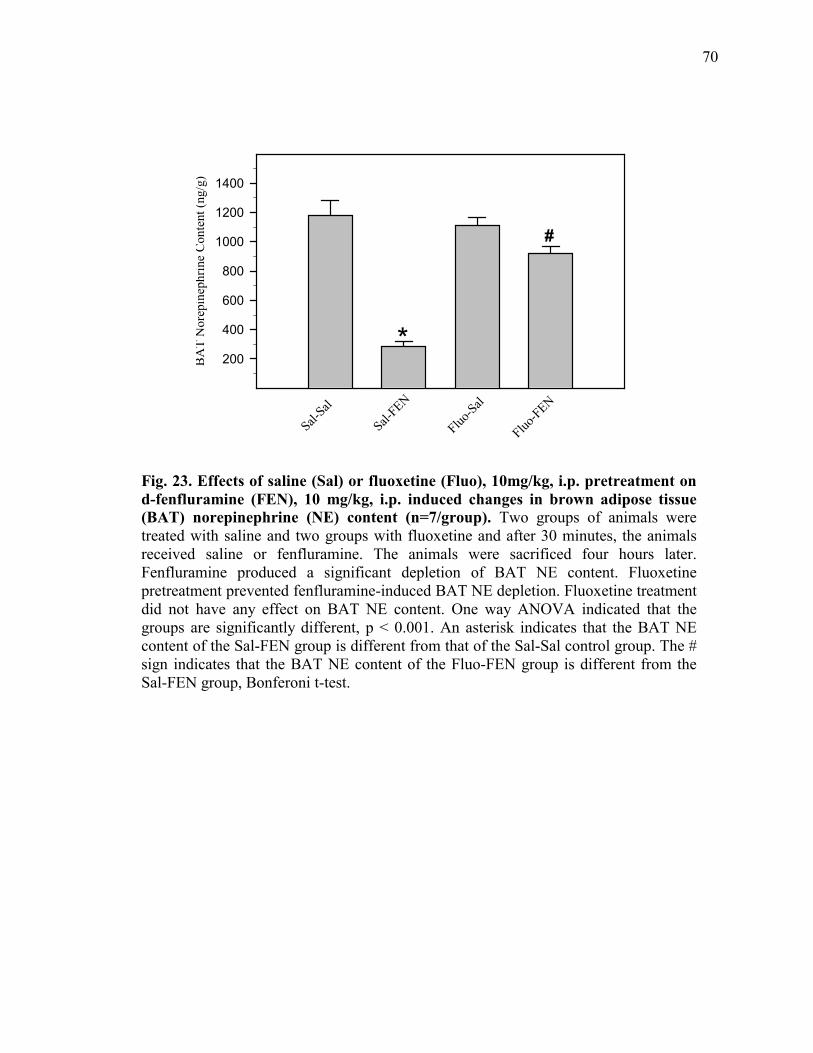
70
Fig. 23. Effects of saline (Sal) or fluoxetine (Fluo), 10mg/kg, i.p. pretreatment ond-fenfluramine (FEN), 10 mg/kg, i.p. induced changes in brown adipose tissue(BAT) norepinephrine (NE) content (n=7/group). Two groups of animals were treated with saline and two groups with fluoxetine and after 30 minutes, the animalsreceived saline or fenfluramine. The animals were sacrificed four hours later.Fenfluramine produced a significant depletion of BAT NE content. Fluoxetine pretreatment prevented fenfluramine-induced BAT NE depletion. Fluoxetine treatment did not have any effect on BAT NE content. One way ANOVA indicated that thegroups are significantly different, p < 0.001. An asterisk indicates that the BAT NE content of the Sal-FEN group is different from that of the Sal-Sal control group. The # sign indicates that the BAT NE content of the Fluo-FEN group is different from the Sal-FEN group, Bonferoni t-test.
Col 2
Fluo-FEN
Fluo-Sal
BA
T N
orep
inep
hrin
e C
onte
nt (n
g/g)
200
400
600
800
1000
1200
1400
Sal-FEN
Sal-Sal
*
#

71
Thus, it appears that although the effect of fenfluramine on body temperature depends on the
ambient temperature at which the animals are kept, the effect of fenfluramine on BAT does
not depend on the ambient temperature at which the animals are maintained.
Similarly, when animals kept at 220C were treated with d-fenfluramine, 3 mg/kg, the
body temperature was not affected. However, the same dose of d-fenfluramine produced a
significant hypothermia in animals kept at 40C. Also, d-fenfluramine, 10 mg/kg, produced a
greater hypothermia in animals kept at 40C compared to animals kept at 220C. The
hypothermia to fenfluramine was exacerbated when animals were exposed to a cold
environment of 40C. d-fenfluramine, 3 and 10 mg/kg doses also produced a significant dose-
related decrease in BAT NE content in animals kept at 22 and 40C. However, the extent of
BAT NE depletion produced by d-fenfluramine was similar in animals kept at 22 and 40C.
Since fenfluramine produced a distinct hypothermia, one might assume that
fenfluramine might have interfered with the sympathetic activation of BAT thermogenesis.
However, fenfluramine treatment resulted in a significant depletion of BAT NE content
suggesting that fenfluramine activated BAT. That BAT is activated by fenfluramine, has
been reported by others (Arase et al., 1988; Lupien et al., 1986). The effect of fenfluramine
on BAT has been evaluated at 22 or 280C. The exaggerated hypothermic response to
fenfluramine at 40C, led to the suggestion that perhaps fenfluramine did not activate BAT as
environmental temperature was reduced (Ma et al., 1991). However, our results demonstrate
that BAT NE content was depleted by fenfluramine at 40C.
Interestingly, the racemic mixture dl- fenfluramine, at a dose of 10 mg/kg, produced a
greater depletion of BAT NE than 10 mg/kg of the d-isomer. Perhaps this is due to the fact

72
that the l-isomer is reported to have significant effects on central noradrenergic neurons as
well as serotonergic neurons (Invernizzi et al., 1986).
Metabolic production of heat by BAT (non-shivering thermogenesis) is controlled by
brain thermoregulatory centers via a sympathetic neuronal pathway (Flaim et al., 1976). BAT
thermogenesis is particularly important in rodents as a means of maintaining core body
temperature. In cold-exposed animals, BAT activation has been measured by such indices as
increased GDP binding to mitochondria (Trayhurn et al., 1987), increased firing of
sympathetic neurons (Banet et al., 1978) and increased blood flow to BAT (Foster et al.,
1977). Local denervation of the sympathetic neurons entering BAT prevents its activation
during cold exposure (Foster et al., 1981).
Like cold exposure, fenfluramine produces marked sympathetic activation of BAT as
indicated by increased GDP binding (Lupien et al., 1985) and increased blood flow to BAT
(Ma et al., 1991). And like cold-induced BAT activation, the effects of fenfluramine were
blocked by surgical denervation (Rothwell et al., 1984) of BAT. The depletion of BAT NE
content was used as the indicator of sympathetic activity to BAT and it was confirmed that
fenfluramine-induced activation of BAT is mediated via sympathetic neurons. This
conclusion is supported by the finding that d-fenfluramine-induced depletion of BAT NE
content could be completely prevented by pretreatment with the ganglionic blocker
pentolinium.
Noradrenergic neurons innervating BAT seem to be quite sensitive to depletion
following an increase in sympathetic discharge. After fenfluramine treatment, the depletion
occurred over a rather short time period of four hours. However, NE content of other
sympathetically innervated tissues, white fat, the adrenals and the heart, were not affected by

73
d-fenfluramine. Thus, it is unlikely that fenfluramine depleted NE by a direct effect on the
nerve terminals, i.e. a reserpine like action, because all noradrenergically innervated tissue
should have been similarly affected.
There are limits to the sensitivity of BAT NE content to detect an activation of BAT.
For example, cold exposure alone, which probably increased sympathetic outflow to BAT
did not produce a measurable depletion. As mentioned previously, other investigators have
shown that GDP binding and blood flow to BAT were increased by cold exposure.
The major pharmacologic effect of fenfluramine is its ability to release serotonin from
nerve terminals (Raiteri et al., 1995). Fluoxetine is a selective serotonin reuptake inhibitor
(SSRI) that has been shown to block fenfluramine-induced release of serotonin (Gundlah et
al., 1997). The release of serotonin by d-fenfluramine appears to be a key step in mediating
the increase in sympathetic activation of BAT, since the depletion of NE in BAT was totally
abolished by fluoxetine. Interestingly, a recent study has demonstrated that the hypothermia
to fenfluramine could also be blocked by pretreating animals with sertraline, another SSRI
which prevents fenfluramine-induced serotonin release (Cryan et al., 2000).
Fluoxetine treatment alone produced no change in BAT NE content. This is
consistent with other studies in which fluoxetine and other SSRIs were shown not to possess
thermogenic activity (Connoley et al., 1999). While fluoxetine would be expected to initially
increase the synaptic concentration of endogenously released serotonin in a manner directly
proportional to the ongoing activity of serotonin neurons, it appears that the serotonin
concentration did not rise high enough to elicit a measurable effect on BAT NE content. In
contrast, after fenfluramine treatment, BAT appears to be in a state of constant stimulation
and seems to be no longer regulated appropriate to the environmental temperature at which

74
the animals are maintained. Consistent with this concept is the well documented finding that
BAT is activated even in animals maintained at a warm thermoneutral environment of 280C
(Preston et al., 1990). Also, as evidenced by BAT NE depletion in the present study, BAT
was activated to a marked extent at a normal laboratory temperature of 220C. And there was
no evidence of further activation by fenfluramine when the animals were kept in a cold
environment because the depletion of BAT NE content was the same at 22 and 40C.
Thus, despite the evidence of marked activation of BAT at 22 and 40C, by
fenfluramine, animals became hypothermic. This confirms the findings of other investigators
that fenfluramine causes hypothermia at ambient temperatures of 22 and 40C (Cryan et al.,
2000; Ma et al., 1991). Also, the hypothermic effect was shown to be dose dependent and
exacerbated by lowering the environmental temperature to 40C. However, this exaggerated
hypothermic response to fenfluramine at 40C is not due to a reduction in the magnitude of
BAT activation. Thus, the hypothermia to fenfluramine at 22 and 40C is not due to the lack of
activation of BAT thermogenesis. Also, the results demonstrate that d-fenfluramine-induced
BAT NE depletion was mediated through sympathetic activation and through release of
serotonin.

75
Chapter V. Evaluation of the Effects of d-Fenfluramine on the Cutaneous Vasculature and Total Metabolic Heat Production
Experiments presented in this chapter were designed to investigate the possible
mechanisms involved in the hypothermia produced by fenfluramine treatment. The results of
the previous chapter, chapter IV, clearly demonstrate that fenfluramine produced dose-related
decreases in body temperature and the decreases were exacerbated at 40C. Moreover, the
results indicated that the hypothermia occurred despite stimulation of heat generation through
BAT activation. Thus, the mechanisms that account for the hypothermia to fenfluramine
treatment remain undefined. Body temperature maintenance depends on the balance between
heat generation and heat loss. Therefore, experiments were designed to determine if
fenfluramine-induced hypothermia was due to either accelerated heat loss or insufficient heat
generation.
As discussed in Chapter I, the cutaneous vasculature is an important
thermoregulatory system, when constricted it prevents heat loss and when dilated, it
promotes heat loss. The primary mechanism for control of heat loss is the sympathetic
nervous system control of cutaneous vascular tone. At normal ambient temperatures, the
cutaneous vasculature is constricted and the constriction is sustained as environmental
temperature is reduced. Previous studies have shown that agents that interfere with the
sympathetic tone to the cutaneous vasculature produce a decline in body temperature. For
example, drugs that block alpha-adrenoceptor mediated vasoconstriction such as
phentolamine and delequamine and ganglionic blocking agents such as pentolinium and
chlorisondamine produce hypothermia (Kobayashi et al., 1998; Redfern et al., 1995; Lin et
al., 1979; Brittain et al., 1967). Therefore, we hypothesized that fenfluramine could interfere

76
with sympathetic nervous system control of cutaneous vascular tone and prevent cutaneous
vasoconstriction and produce heat loss and consequently the body temperature falls. To our
knowledge, no studies have been conducted to explore the possibility that fenfluramine
interferes with the sympathetic nervous system tone to the cutaneous vasculature and
produces heat loss.
It is most likely, based on observations discussed in chapter I (“Role of serotonin in
thermoregulation”), that fenfluramine influences sympathetic activity through its effects on
the serotonergic neuronal systems since serotonin and certain serotonin agonists have been
shown to produce heat loss. Serotonin, when administered intracerebroventricularly,
produces heat loss in rabbits and rats (Bligh et al., 1971; Key et al., 1992). Additional proof
that serotonergic systems are involved in producing heat loss through cutaneous vasodilation
is that fluoxetine, which increases synaptic serotonin levels and serotonin-1A receptor
agonist 8-OHDPAT produce heat loss as indicated by increases in rat tail skin temperature
(Lin, 1978; Oerther, 2000).
Rats are particularly suited for the study of cutaneous thermoregulation because of
the important role of the tail vasculature in thermoregulation. The tail of the rat has been
commonly used to study peripheral vascular tone (Dawson et al., 1979; Rand et al., 1965).
Tail surface temperature is generally measured in order to obtain an indication of changes in
tail blood flow. Increases in tail surface temperature were interpreted as increased blood flow
to the tail due to cutaneous vascular dilation and decreases in tail skin temperature were
interpreted as decreased blood flow to the tail due to cutaneous vascular constriction.
The experiments were conducted at a cool ambient temperature of 16-170C. The
selection of the ambient temperature was based on two criteria. First, at 160C, the magnitude

77
of d-fenfluramine-induced hypothermia was equivalent to that observed at 40C. Second, other
investigators have demonstrated that robust cutaneous vasodilation can be observed at this
temperature with agents that interfere with sympathetic vasoconstrictor tone (Redfern et al.,
1995).
The first experiment was conducted to determine the effect of d-fenfluramine on tail
skin temperature. The results indicated that d-fenfluramine administration increased tail skin
temperature (Experiment 1) supporting the supposition that d-fenfluramine caused cutaneous
vasodilation. This observation led to the development of two additional experiments. The
second experiment was designed to determine if d-fenfluramine altered tail skin temperature
by producing a withdrawal of the sympathetic vasoconstrictor tone. To test this possibility,
the effect of d-fenfluramine on tail skin temperature was studied in animals treated with and
without the ganglionic blocker pentolinium, which blocks the sympathetic tone to the
cutaneous vasculature. Another approach was also used to determine if d-fenfluramine
caused changes in cutaneous blood flow that were independent of sympathetic tone. In this
third experiment, animals were placed in the test chamber that had been warmed to 280C,
ambient temperature at which the cutaneous vasculature of the rat would be dilated due to
withdrawal of sympathetic tone (Hellstorm, 1975a). Thus, once the tail vasculature is dilated,
subsequently administered d-fenfluramine should not be able to affect cutaneous blood
vessels by removing sympathetic tone. The fourth experiment was designed to determine if
d-fenfluramine-induced increase in tail skin temperature was secondary to release of
serotonin. This was achieved by evaluating the effect of d-fenfluramine on tail skin
temperature in animals treated with and without fluoxetine, a selective serotonin reuptake

78
inhibitor, which blocks d-fenfluramine-induced serotonin release (Berger et al., 1992; Sabol
et al., 1992; Cheetam et al., 2000).
The fifth experiment of this chapter was designed to assess the effect of d-
fenfluramine on total metabolic heat production. As stated earlier, the hypothermia to
fenfluramine could be due to insufficient heat generation. It was considered that fenfluramine
could produce a fall in body core temperature by reducing total metabolic heat production.
The total heat generated by an animal is the sum of shivering and nonshivering
thermogenesis. Although fenfluramine activates nonshivering thermogenesis (BAT), it might
interfere with shivering thermogenesis and thus reduce total heat production. Therefore the
effect of d-fenfluramine on total heat generation would indicate if d-fenfluramine produces
hypothermia by interfering with heat generation by non-BAT thermogenesis. Whole body
oxygen consumption (VO2) was measured as an index of total metabolic heat generation.
This experiment was also conducted at an ambient temperature of 16-170C.
A. Protocols
1. Experiment 1. Effects of d-fenfluramine on tail skin and body
temperature of rats kept at 160C.
This experiment was conducted to determine if d-fenfluramine treatment would affect
tail skin temperature. All animals were implanted with temperature transmitters. On the day
of the experiment, a thermocouple was attached to the surface of the tail. The animals were
then moved to the walk-in cold room maintained at 160C and baseline tail and body
temperature were measured for an hour. The animals were then treated with d-fenfluramine,
10 mg/kg, i.p., or saline (n=7/group) and observed for another 90 minutes. The dose of d-
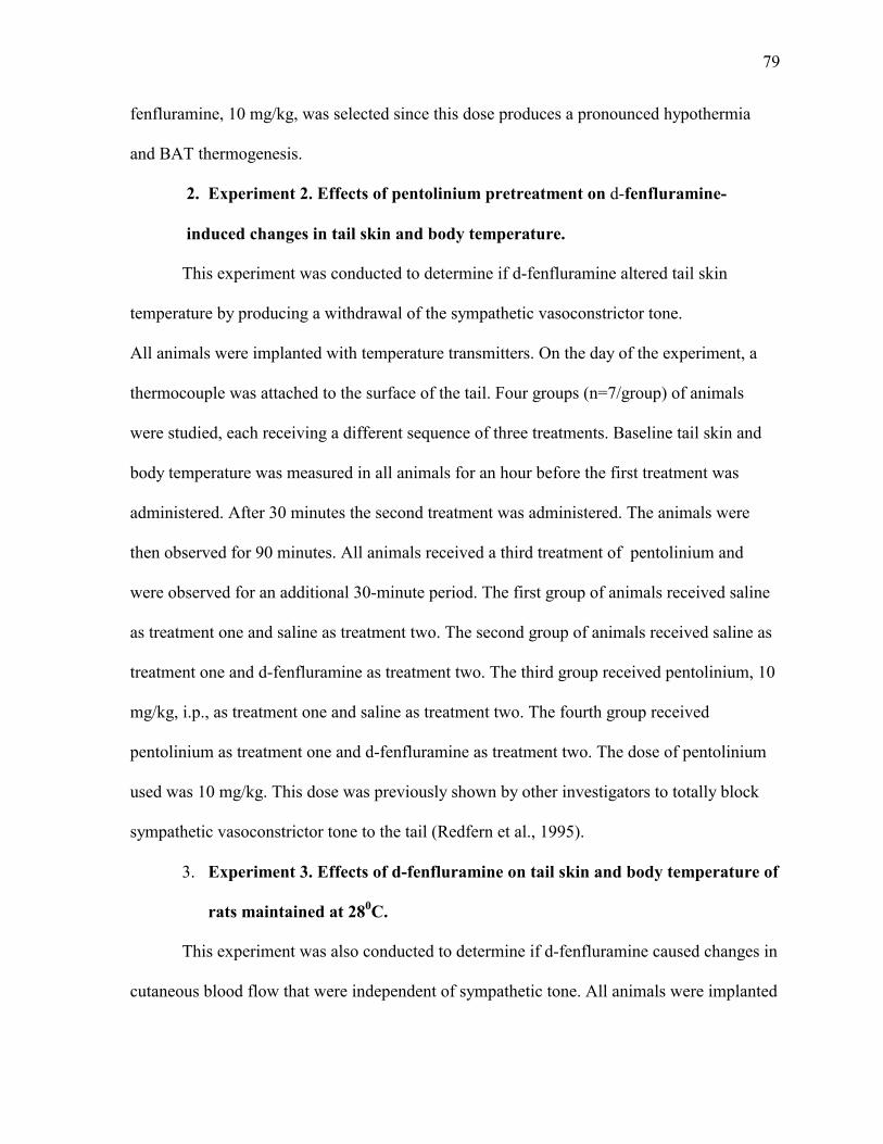
79
fenfluramine, 10 mg/kg, was selected since this dose produces a pronounced hypothermia
and BAT thermogenesis.
2. Experiment 2. Effects of pentolinium pretreatment on d-fenfluramine-
induced changes in tail skin and body temperature.
This experiment was conducted to determine if d-fenfluramine altered tail skin
temperature by producing a withdrawal of the sympathetic vasoconstrictor tone.
All animals were implanted with temperature transmitters. On the day of the experiment, a
thermocouple was attached to the surface of the tail. Four groups (n=7/group) of animals
were studied, each receiving a different sequence of three treatments. Baseline tail skin and
body temperature was measured in all animals for an hour before the first treatment was
administered. After 30 minutes the second treatment was administered. The animals were
then observed for 90 minutes. All animals received a third treatment of pentolinium and
were observed for an additional 30-minute period. The first group of animals received saline
as treatment one and saline as treatment two. The second group of animals received saline as
treatment one and d-fenfluramine as treatment two. The third group received pentolinium, 10
mg/kg, i.p., as treatment one and saline as treatment two. The fourth group received
pentolinium as treatment one and d-fenfluramine as treatment two. The dose of pentolinium
used was 10 mg/kg. This dose was previously shown by other investigators to totally block
sympathetic vasoconstrictor tone to the tail (Redfern et al., 1995).
3. Experiment 3. Effects of d-fenfluramine on tail skin and body temperature of
rats maintained at 280C.
This experiment was also conducted to determine if d-fenfluramine caused changes in
cutaneous blood flow that were independent of sympathetic tone. All animals were implanted

80
with temperature transmitters. On the day of the experiment, a thermocouple was attached to
the surface of the tail. Two groups (n=7/group) of animals with indwelling temperature
transmitters and tail thermocouples were transferred to the test chamber maintained at 220C
and the temperature was slowly increased to 280C. Baseline tail skin and body temperature
measurements were then made for 30 minutes. d-Fenfluramine, 10 mg/kg, i.p., or saline was
then administered and the animals were observed for another 90 minutes.
4. Experiment 4. Effects of fluoxetine pretreatment on d-fenfluramine-induced
changes in tail skin and body temperature.
This experiment was conducted to determine if d-fenfluramine-induced changes in
tail skin and body temperature were mediated via release of serotonin. All animals were
implanted with temperature transmitters. On the day of the experiment, a thermocouple was
attached to the surface of the tail. Two groups (n=7/group) of animals were studied. Baseline
tail skin and body temperature measurements were made for 30 minutes. Fluoxetine, 10
mg/kg, i.p., was then administered to both groups. After 40 minutes, one group received d-
fenfluramine, 10 mg/kg, i.p. and the other group received saline. The animals were observed
for 90 minutes. At the end of the 90-minute observation period, the two groups received
pentolinium and were observed for an additional 30 minutes. The dose of fluoxetine, 10
mg/kg, has been shown by others to have blocked fenfluramine-induced serotonin release
(Gundlah et al., 1997).
5. Experiment 5. Effects of d-fenfluramine and pentolinium on whole body
oxygen consumption (VO2).
This experiment was conducted to assess the effects of d-fenfluramine and
pentolinium on total metabolic heat generation. All animals were implanted with temperature

81
transmitters. On the day of the experiment, the animals were transferred to a metabolic
chamber in the walk-in cold room maintained at 160C. Baseline body temperature and
oxygen consumption measurements were made for a period of one hour. The animals were
then separated in to three treatment groups (n=7/group). Group one received d-fenfluramine,
10 mg/kg, i.p., group two received pentolinium, 10 mg/kg, i.p. and group three received
saline. The animals were observed for 90 minutes. At the end of the 90-minute observation
period the d-fenfluramine treated group was treated with pentolinium and observed for an
additional hour. The group initially treated with pentolinium was treated with d-fenfluramine
at the end of the 90-minute period and observed for another hour.
Statistical Analyses.
Results are presented as mean + S.E. The differences in tail skin and body
temperature and whole body oxygen consumption between multiple groups and between
multiple measurements taken over a period of time were assessed using repeated measures
analysis of variance. Post hoc pairwise comparisons were done using Bonferoni t-test. A
statistically significant effect was accepted when p < 0.05.
B. Results.
1. Experiment 1. Effects of d-fenfluramine, 10 mg/kg, i.p., on tail skin and body
temperature of rats kept at 160C.
d-Fenfluramine produced a rapid increase in tail skin temperature when compared to
the saline-treated controls, p < 0.01 (Repeated measures ANOVA), Fig. 24, top panel. The
tail skin temperature reached a maximum approximately 10 minutes after injection, but

82
returned to pretreatment levels by the end of the 90-minute observation period (Bonferoni t-
test).
d-Fenfluramine treatment produced a significant decrease in body temperature when
compared to the saline-treated controls, p < 0.01 (Repeated measures ANOVA), Fig. 24,
bottom panel.
Note that the magnitude of hypothermia to d-fenfluramine at 160C is similar to the magnitude
of hypothermia to d-fenfluramine treatment at 40C.
2. Experiment 2. Effects of pentolinium pretreatment on d-fenfluramine-
induced increases in tail skin temperature and hypothermia.
Pentolinium treatment produced a significant increase in tail skin temperature, p <
0.01 (Repeated measures ANOVA), Fig. 25, top panel. The tail skin temperature did not
return to control levels by the end of the 90-minute observation period. In the group of
animals pretreated with pentolinium, subsequent d-fenfluramine treatment decreased tail skin
temperature when compared to the pentolinium-pretreated group that received saline
treatment and the tail skin temperature returned to control levels by the end of the
observation period (Bonferoni t-test), Fig. 26, top panel. Pentolinium administered at the end
of the 90-minute observation period did not affect tail skin temperature, Fig. 26, top panel.
Pentolinium treatment produced a decrease in body temperature when compared to
the saline controls, p < 0.01(Repeated measures ANOVA), Fig. 25, bottom panel. However,
the body temperature returned to control levels by the end of the observation period. In the
group of animals pretreated with pentolinium, subsequent administration of d-fenfluramine
produced an additional, more pronounced hypothermia (Bonferoni t-test), Fig. 26, bottom
panel.

83
3. Experiment 3. Effects of d-fenfluramine, 10 mg/kg, i.p., on tail skin and
body temperature of rats kept at 280C (thermoneutrality).
In the range of ambient temperatures from 21.90 to 27.70C, tail temperature increased
parallel to ambient temperature. However when the ambient temperature reached 27.9 +
0.10C, tail temperature rose more rapidly than the ambient temperature, Fig. 27.
d-Fenfluramine was administered when tail ambient temperature was above the
temperature point at which the tail surface temperature was no longer changing, i.e. 280C. d-
Fenfluramine administration at 280C produced a significant decrease in tail skin temperature
when compared to the saline controls, p < 0.01 (Repeated measures ANOVA), Fig. 28, top
panel. The decrease in tail skin temperature became maximal approximately 20 minutes after
treatment (Bonferoni t-test).
d-Fenfluramine produced a significant hyperthermic response when compared to the
saline treated controls, p < 0.01 (Repeated measures ANOVA), Fig. 28, bottom panel. The
hyperthermia lasted the entire 90-minute observation period (Bonferoni t-test).
4. Experiment 4. Effects of fluoxetine pretreatment on d-fenfluramine induced
increases in tail skin temperature and hypothermia.
Fluoxetine treatment resulted in increases in tail skin temperature when compared to
the saline controls, p < 0.01 (Repeated measures ANOVA), Fig. 29, top panel. However, the
tail skin temperature returned to baseline levels at the end of 45 minutes (Bonferoni t-test). In
the group of animals pretreated with fluoxetine, subsequent d-fenfluramine treatment did not
produce an increase in tail temperature (Bonferoni t-test), Fig. 30, top panel.
Pentolinium administered to the fluoxetine-saline group at the end of the 90 -
minute observation period caused a significant increase in tail temperature (Bonferoni t-test).

84
-15 0 15 30 45 60 75 90
Bod
y Te
mpe
ratu
re (0 C
)
32
34
36
38
40
Time (min)
Tail
Tem
pera
ture
(0 C)
16
18
20
22
24
26
28
30
32d-FenfluramineSaline
* *
*
* * * ** * *
**
** ****
*
Fig. 24. Effects of saline or d-fenfluramine, 10 mg/kg, i.p., on tail skin and bodytemperature of rats kept at 160C (n=7/group). D-Fenfluramine or saline was administered at time “0”. The tail skin temperature responses of the d-fenfluramine and saline treated groups were significantly different, p < 0.01 (Repeated measures ANOVA). The body temperature responses of the d-fenfluramine and saline treated groups were significantly different, p < 0.01 (Repeated measures ANOVA). Asterisks indicate that there is a significant difference in tail skin and body temperature between the two groups at specific time points,Bonferoni t-test.

85
-30 -15 0 15 30 45 60 75 90
32
34
36
38
40
Bod
y Te
mpe
ratu
re (0 C
)
Time (mins)
Tail
Tem
pera
ture
(0 C)
16
18
20
22
24
26
28
30
32 SalinePentolinium
** * * *
*
**
**
***
***
*****
Fig. 25. Effects of saline or pentolinium, 10 mg/kg, i.p., on tail skin and bodytemperature of rats kept at 160C (n=7/group). Pentolinium or saline was administered at time “0”and saline was administered to both groups after 30 minutes. Pentolinium produced a significant increase in tail skin temperature. The tail skin temperature responses of the pentolinium and saline treated groups were significantly different, p < 0.01 (Repeatedmeasures ANOVA). Pentolinium also produced a mild hypothermia. The body temperature responses of the pentolinium and saline treated groups were significantly different, p < 0.01(Repeated measures ANOVA). Asterisks indicate that there is a significant difference in tailskin and body temperature between the two groups at specific time points, Bonferoni t-test.

86
Bod
y Te
mpe
ratu
re (0 C
)
-30 -15 0 15 30 45 60 75 90 105 120
32
34
36
38
40
Time (min)
16
18
20
22
24
26
28
30
32
Pentolinium-Saline-PentoliniumPentolinium-d-Fenfluramine-Pentolinium
Tail
Tem
pera
ture
(0 C)
* * *** **
*** * **
Fig. 26. Effects of pentolinium, 10 mg/kg, i.p., pretreatment on d-fenfluramine, 10 mg/kg, induced increases in tail skin temperature and hypothermia (n= 7/group). Pentolinium was administered to both groups at time “0” and after 30 minutes, one group received salineand the other fenfluramine. After 90 minutes, pentolinium was administered to both groups.Pentolinium produced a significant increase in tail skin temperature, subsequent fenfluraminetreatment did not produce any additional increase, instead produced a decrease in tail skintemperature. Pentolinium administration at the end of 90 minutes did not affect tail skintemperature. Pentolinium produced a mild hypothermia. Subsequent d-fenfluramine treatment produced an additional more severe hypothermia. The tail skin and body temperatureresponses of the groups are significantly different, p < 0.01 (Repeated measures ANOVA). Asterisks indicate that there is a significant difference between the two groups at specific timepoints, Bonferoni t-test.

87
Also, pentolinium caused an increase in tail temperature in the fluoxetine-fenfluramine
group, Fig. 30, top panel.
Fluoxetine treatment produced a significant hypothermia, p < 0.01 (Repeated
measures ANOVA) when compared to the saline controls, Fig. 29, bottom panel. Core
temperature recovered to baseline by the end of the 90-minute observation period (Bonferoni
t-test). In the group of animals pretreated with fluoxetine, subsequent d-fenfluramine
treatment also produced a hypothermia, p < 0.01 (Repeated measures ANOVA), Fig. 30,
bottom panel. But the hypothermia to d-fenfluramine was attenuated.
5. Experiment 5. Effects of d-fenfluramine and pentolinium on whole body
oxygen consumption.
d-fenfluramine increased whole body oxygen consumption as compared to the saline
controls, p < 0.01 (Repeated measures ANOVA). The increase in oxygen consumption
develops slowly, Fig. 31, top panel. d-fenfluramine treatment produced a significant
hypothermia as compared to the saline controls, p < 0.01 (Repeated measures ANOVA), Fig.
31, bottom panel. The increase in oxygen consumption occurs while the body temperature
was still declining.
Pentolinium treatment also increases oxygen consumption when compared to the
saline controls, p < 0.01 (Repeated measures ANOVA), Fig. 32, top panel. The increase
peaks at approximately 30 minutes after pentolinium administration. Pentolinium treatment
also produces a mild hypothermia when compared to the saline controls, p < 0.01 (Repeated
measures ANOVA), Fig. 32, bottom panel. The hypothermia to d-fenfluramine was greater
than the hypothermia to pentolinium treatment, (Bonferoni t-test).

88
Fig. 27. Effects of increasing ambient temperature on body and tail skin temperature ofrats (n=7). The rats were placed in an observation chamber initially maintained at 220C. Theambient temperature was gradually increased to 280C. There was an abrupt increase in tail skin temperature at 280C.
Time (min)0 10 20 30 40 50 60 70
Tem
pera
ture
(o C)
202224262830323436384042
Body Temp.Tail Temp.Ambient Temp.

89
Tail
Tem
pera
ture
(0 C)
22
24
26
28
30
32
34
36
d-FenfluramineSaline
******
Time (mins)
-15 0 15 30 45 60 75 90
Bod
y Te
mpe
ratu
re (0 C
)
36
38
40
42*
** * * ***
Fig. 28. Effects of saline or d-fenfluramine, 10 mg/kg, i.p., on tail skin and bodytemperature of rats kept at 280C (n=7/group). D-Fenfluramine or saline was administered at time “0”. D-Fenfluramine produced a significant decrease in tail skin temperature. D-Fenfluramine also produced a significant hyperthermia. The tail skin and body temperature responses of the d-fenfluramine and saline groups are significantly different, p < 0.01 (Repeated measures ANOVA). Asterisks indicate that there is a significant difference in tailskin and body temperature between the two groups at specific time points, Bonferoni t-test.

90
-30 -15 0 15 30 45 60 75 90
32
34
36
38
40
Bod
y Te
mpe
ratu
re (0 C
)
Time (mins)
Tail
Tem
pera
ture
(0 C)
16
18
20
22
24
26
28
30
32
SalineFluoxetine
** * *
*
** *
Fig. 29. Effects of saline or fluoxetine, 10 mg/kg, i.p., on tail skin and body temperatureof rats kept at 160C (n=7/group). Fluoxetine or saline was administered at time “0” andsaline was administered to both groups after 30 minutes. Fluoxetine produced an increase in tail skin temperature. Fluoxetine also produced a mild hypothermia. The tail skin and body temperature responses of the groups are significantly different, p < 0.01 (Repeated measuresANOVA). Asterisks indicate that there is a significant difference in tail skin and body temperature between the two groups at specific time points, Bonferoni t-test.
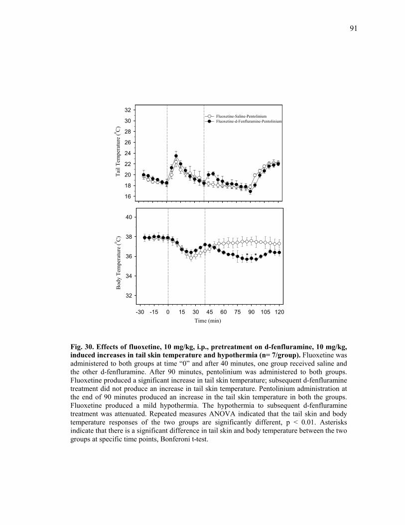
91
Fig. 30. Effects of fluoxetine, 10 mg/kg, i.p., pretreatment on d-fenfluramine, 10 mg/kg, induced increases in tail skin temperature and hypothermia (n= 7/group). Fluoxetine was administered to both groups at time “0” and after 40 minutes, one group received saline andthe other d-fenfluramine. After 90 minutes, pentolinium was administered to both groups.Fluoxetine produced a significant increase in tail skin temperature; subsequent d-fenfluramine treatment did not produce an increase in tail skin temperature. Pentolinium administration atthe end of 90 minutes produced an increase in the tail skin temperature in both the groups. Fluoxetine produced a mild hypothermia. The hypothermia to subsequent d-fenfluramine treatment was attenuated. Repeated measures ANOVA indicated that the tail skin and body temperature responses of the two groups are significantly different, p < 0.01. Asterisks indicate that there is a significant difference in tail skin and body temperature between the two groups at specific time points, Bonferoni t-test.
Bod
y Te
mpe
ratu
re (0 C
)
-30 -15 0 15 30 45 60 75 90 105 120
32
34
36
38
40
Time (min)
16
18
20
22
24
26
28
30
32Fluoxetine-Saline-PentoliniumFluoxetine-d-Fenfluramine-Pentolinium
Tail
Tem
pera
ture
(0 C)
* *

92
The increase in oxygen consumption produced by pentolinium was greater than the oxygen
consumption to d-fenfluramine treatment, (Bonferoni t-test).
Pentolinium treatment at the end of 90 minutes, in the d-fenfluramine-treated group
did not produce any increase in oxygen consumption, p < 0.01 (One way ANOVA,
Bonferoni t-test), Fig. 33. Similarly, d-fenfluramine treatment at the end of 90 minutes, in the
pentolinium treated group did not increase oxygen consumption, p < 0.01 (One way
ANOVA, Bonferoni t-test), Fig. 34.

93
Time (mins)-15 0 15 30 45 60 75 90
Bod
y Te
mpe
ratu
re (0 C
)
34
35
36
37
38
39
-15 0 15 30 45 60 75 90
Oxy
gen
Con
sum
ptio
n (m
l/hr/k
g)
1000
1200
1400
1600
1800
2000d-fenfluramineSaline
**
*
*
** *
*
Fig. 31. Effects of saline or d-fenfluramine, 10 mg/kg, i.p., on whole body oxygenconsumption (VO2) and body temperature of rats kept at 160C (n=7/group). D-Fenfluramine or saline was administered at time "0". D-Fenfluramine produced a significant increase in VO2. Repeated measures ANOVA indicated that the groups are significantlydifferent, p < 0.01. Asterisks indicate a significant difference between the d-fenfluramine and the saline group at specific time-points, Bonferoni t-test. However, d-fenfluramine produced a significant hypothermia. Repeated measures ANOVA indicated that the groups aresignificantly different, p < 0.01. Asterisks indicate a significant difference between the d-fenfluramine-treated group and the saline control group at specific time-points, Bonferoni t-test.

94
Fig. 32. Effects of saline or pentolinium, 10 mg/kg, i.p., on whole body oxygenconsumption (VO2) and body temperature of rats kept at 160C (n=7/group). Pentolinium or saline was administered at time "0". Pentolinium produced a significant increase in VO2. Repeated measures ANOVA indicated that the groups are significantly different, p < 0.01.Asterisks indicate a significant difference between the pentolinium and the saline group atspecific time-points, Bonferoni t-test. However, pentolinium produced a significant hypothermia. Repeated measures ANOVA indicated that the groups are significantly different,p < 0.01. Asterisks indicate a significant difference between the pentolinium-treated group and the saline control group at specific time-points, Bonferoni t-test.
Time (mins)-15 0 15 30 45 60 75 90
Bod
y Te
mpe
ratu
re (0 C
)
34
35
36
37
38
39
-15 0 15 30 45 60 75 90
Oxy
gen
Con
sum
ptio
n (m
l/hr/k
g)
1000
1200
1400
1600
1800
2000 PentoliniumSaline
**
*
* * * *
*

95
Fig. 33. Effect of d-fenfluramine (Fen), 10 mg/kg, i.p., pretreatment on pentolinium-induced increases in whole body oxygen consumption (VO2), measured 30 minutes after treatment, in rats kept at 160C (n=7/group). Pentolinium (Pent) increased VO2significantly, 30 minutes post-treatment. Fen, when administered 90 minutes before Pent,blocked Pent-induced increases in VO2. One-way ANOVA indicated that the groups are significantly different, p < 0.01. Asterisks indicate that the VO2 to Pent treatment is significantly different from the VO2 to the saline (Sal) treated control group. The # signindicates that the VO2 to Fen-Pent treatment is different from the VO2 to Pent treatment, Bonferoni t-test.
Oxy
gen
Con
sum
ptio
n (m
l/hr/k
g)
(3
0 m
ins.
Post
-Tre
atm
ent)
500
1000
1500
2000 *
Sal Pent Fen-Pent
#

96
Fig. 34. Effect of pentolinium (Pent), 10 mg/kg, i.p., pretreatment on d-fenfluramine-induced increases in whole body oxygen consumption (VO2), measured one hour after treatment, in rats kept at 160C (n=7/group). D-Fenfluramine (Fen) increased VO2significantly, one hour post-treatment. Pent, when administered 90 minutes before Fen,blocked Fen-induced increases in VO2. One-way ANOVA indicated that the groups are significantly different, p < 0.01. Asterisks indicate that the VO2 to Fen treatment is significantly different from the VO2 to the saline (Sal) treated control group. The # signindicates that the VO2 to Pent-Fen treatment is different from the VO2 to Fen treatment, Bonferoni t-test.
Oxy
gen
Con
sum
ptio
n (m
l/hr/k
g)
(1
hr.
Post
-Tre
atm
ent)
500
1000
1500
Sal Fen Pent-Fen
* #

97
C. Discussion
The results of the experiments indicate that when animals were maintained at a cool
ambient temperature of 160C, d-fenfluramine treatment produced an increase in tail skin
temperature. This observation suggests that d-fenfluramine produced a dilation of the tail
cutaneous vasculature and provides support for the conclusion that d-fenfluramine produces
heat loss that may contribute to its hypothermic effect. Furthermore, the results support the
conclusion that d-fenfluramine produces a withdrawal of the sympathetic vasoconstrictor
tone due to central release of serotonin. Also, the increase in metabolic heat production to d-
fenfluramine treatment was insufficient to overcome its hypothermic effect.
An important finding of these experiments is that d-fenfluramine produces heat loss
by dilating the cutaneous vasculature in animals maintained at 160C. At 160C, dilation of the
cutaneous vasculature is physiologically inappropriate. d-fenfluramine produced a significant
increase in tail skin temperature within minutes of intraperitoneal injection. The increase
lasted for about 30 minutes and the tail skin temperature returned to control levels by the end
of the 90-minute observation period. Also, d-fenfluramine produced a significant drop in
body temperature consistent with the results of the experiments in chapter IV.
The importance of the presence of sympathetic vasoconstrictor tone to the expression
of d-fenfluramine-induced increases in tail skin temperature was explored by contrasting the
effects of d-fenfluramine on tail skin temperature in animals treated with and without
ganglionic blocker pentolinium. Pentolinium treatment resulted in a marked increase in tail
skin temperature. The effect lasted the full duration of the 90-minute observation period. This
observation is consistent with the reports of others that pentolinium produces an increase in
tail skin temperature (Redfern et al., 1995). Disruption of sympathetic cutaneous

98
vasoconstrictor tone by pentolinium leads to vasodilation as indicated by increases in tail
skin temperature. Although there was some recovery of tail temperature towards control
levels at the end of the 90-minute observation period, the recovery could not be explained by
a waning of the ganglionic blocking activity of pentolinium, since an additional dose of
pentolinium had no effect on tail temperature. Thus the dose of pentolinium we selected for
the study produced a complete blockade of sympathetic vasoconstrictor tone for the duration
of the experiments.
In animals pretreated with pentolinium, subsequent d-fenfluramine treatment did not
result in additional increases in tail skin temperature, instead the tail skin temperature
decreased and returned to control levels by the end of the 90-minute observation period. This
observation indicates that d-fenfluramine cannot cause vasodilation if the sympathetic tone is
withdrawn and also that d-fenfluramine may have a vasoconstrictive effect. The fact that d-
fenfluramine may have a direct vasoconstrictive effect is also demonstrated by administering
pentolinium at the end of the 90 minute observation period. The additional dose of
pentolinium did not result in an increase in tail skin temperature. The reduction in tail
temperature in the presence of sympathetic blockade suggests that d-fenfluramine produced
vasoconstriction of the cutaneous vasculature that was completely independent of
sympathetic innervation.
Pentolinium treatment also produced a distinct hypothermia. However, the body
temperature returned to control levels by the end of the observation period. In pentolinium
pretreated animals, although d-fenfluramine did not produce additional cutaneous
vasodilation, d-fenfluramine treatment produced an additional more severe hypothermia.

99
In addition to the pharmacologic blockade of sympathetic tone, d-fenfluramine was
also administered to animals maintained at a warm environmental temperature of 280C, an
ambient temperature at which the sympathetic tone to the cutaneous vasculature is absent
(Hellstorm, 1975a). To assure that the cutaneous vasculature was dilated, animals were
placed in the test chamber that was initially at 220C. The chamber was gradually warmed
toward thermoneutrality and an abrupt dilation of the tail that occurred at approximately 27-
280C was observed. This confirms previous reports that the physiologic removal of
sympathetic tone is an abrupt on/off response (Young et al., 1982; Hellstorm, 1975b). In
these animals the withdrawal of sympathetic tone was sustained when the chamber was kept
at 280C. Tonic sympathetic activity normally keeps the vasculature constricted until high
ambient temperatures are reached. This is due to the fact that the animal is able to maintain a
normal body core temperature throughout a wide range of temperatures before the ambient
temperature reaches the point at which vasodilation is needed to help dissipate heat. The
results are consistent with this concept, since no evidence of cutaneous dilation was found
until the ambient temperature was increased to about 280C. At that temperature there was a
dramatic increase in tail temperature that is consistent with vasodilation. Administration of d-
fenfluramine in the warm environment did not cause vasodilation, a finding that supports the
concept that d-fenfluramine causes vasodilation by withdrawing sympathetic tone. Thus,
when sympathetic tone is removed physiologically in a warm environment or
pharmacologically through administration of pentolinium, d-fenfluramine does not produce
additional vasodilation. Also in the absence of sympathetic tone at 280C, d-fenfluramine
produced a vasoconstriction of the cutaneous vasculature, which is physiologically
inappropriate at this temperature. The vasoconstriction was similar in time course and

100
magnitude to the vasoconstriction observed in animals pretreated with pentolinium at an
environmental temperature of 160C.
d-fenfluramine treatment of animals kept at a warm environmental temperature, 280C,
produced an increase in body core temperature. While the finding that d-fenfluramine
produced hyperthermia at 280C confirms previous reports that the environmental temperature
influences the effects of d-fenfluramine on body temperature (Malberg et al., 1997; Preston
et al., 1990), this study provides new insights that relate the changes in body temperature to
the effect of d-fenfluramine on the tone of the cutaneous vasculature.
The dilation of the cutaneous vasculature by d-fenfluramine also appears to depend
on d-fenfluramine-mediated release of serotonin. This point was demonstrated by pretreating
animals with the serotonin reuptake inhibitor fluoxetine, which blocks d-fenfluramine-
induced serotonin release. In fluoxetine treated animals, d-fenfluramine produced no
vasodilation. This observation suggests that d-fenfluramine must release serotonin in order
for its vasodilator action to be observed. Also, in fluoxetine treated animals, d-fenfluramine
produced no vasoconstriction. This observation suggests that d-fenfluramine must release
serotonin to produce vasoconstriction. From the other experiments conducted using
pentolinium, we know that the vasoconstrictor effect of fenfluramine is not mediated via the
sympathetic nervous system but appears to be a direct effect. Therefore it appears that
fenfluramine releases serotonin, possibly from the platelets and produces vasoconstriction.
Central administration of serotonin to urethane anesthetized rats produced tail
vasodilation as determined by measurement of tail surface temperature (Key et al., 1992).
Interestingly, our results indicate that d-fenfluramine administration releases sufficient
serotonin centrally to produce an increase in tail skin temperature. And also, fluoxetine,

101
which increases synaptic serotonin concentrations, produces tail dilation as shown by others
(Lin, 1978) and us. However, the effect lasted only for about 30 minutes.
Fluoxetine treatment also produced a mild hypothermia, which lasted for about 30
minutes. Fluoxetine-mediated hypothermia could be attributable to its effects on tail vascular
tone. The hypothermia to d-fenfluramine also appears to depend on its ability to release
serotonin because blockade of d-fenfluramine-mediated serotonin release by fluoxetine
attenuates the hypothermic response to d-fenfluramine. A previous study has also
demonstrated that serotonin reuptake inhibitor sertraline-pretreatment attenuates the
hypothermia to fenfluramine (Cryan et al., 2000).
Body temperature of the animals started to fall after d-fenfluramine treatment. The
hypothermia lasted the entire 90-minute observation period. In fact, the hypothermia to d-
fenfluramine at 160C was similar in magnitude to the hypothermia to d-fenfluramine at 40C.
The vasodilation and consequent heat loss to d-fenfluramine treatment may contribute to the
hypothermia that was observed. However cutaneous heat loss cannot be the full explanation
for the fall in core temperature after d-fenfluramine because pentolinium produced an even
greater dilation of the cutaneous vasculature but the fall in core temperature to pentolinium
was significantly less than the fall in core temperature to d-fenfluramine. The animals treated
with pentolinium recovered from the hypothermia and their body temperature returned to
control levels by the end of the 90-minute observation period. Also, despite the fact that d-
fenfluramine did not produce additional cutaneous dilation after pentolinium pretreatment,
the treatment resulted in an additional more severe hypothermia.
Thus, cutaneous vasodilation and consequent heat loss could account, in part, for the
hypothermia to d-fenfluramine. However, for reasons mentioned in the above paragraph,

102
cutaneous vasodilation alone could not account for the hypothermia to d-fenfluramine
treatment. The other possible mechanism that could account for the hypothermia was
insufficient heat generation. The second major objective of this study was to evaluate the
effect of d-fenfluramine on total metabolic heat production. Whole body oxygen
consumption was measured as an index of metabolic rate.
The results of the experiments clearly demonstrate that d-fenfluramine increases
whole body oxygen consumption in animals maintained at 160C. However, the increment in
oxygen consumption was not sufficient to offset the fall in body temperature as indicated by
the marked hypothermia. Interestingly, pentolinium increased oxygen consumption to a
much greater extent than the increase produced by d-fenfluramine treatment. Taken with the
results of the other experiments in this chapter, which demonstrated the tremendous
cutaneous vasodilation produced by pentolinium, it appears that the heat generated by
increased metabolic activity offset the heat loss to pentolinium treatment and explains why
pentolinium treated animals became only slightly hypothermic when compared to d-
fenfluramine treated animals.
Clearly, pentolinium produces a marked increase in oxygen consumption in order to
offset the heat being lost due to cutaneous vasodilation. The increased metabolic activity is
likely not explainable by increased activity of the sympathetic or parasympathetic nervous
systems, since pentolinium is a ganglionic blocker. Of particular importance is that BAT
thermogenesis produced by sympathetic stimulation would be blocked by pentolinium.
Therefore, the increase in metabolic activity would have to be mediated by the nonautonomic
system. Nonautonomic mechanisms involved in heat generation have classically been
referred to as shivering thermogenesis whereas BAT thermogenesis has been referred to as

103
nonshivering-thermogenesis. Thus despite the fact that pentolinium cannot activate
sympathetically mediated thermogenesis, pentolinium mediated increases in whole body
oxygen consumption is greater than that of d-fenfluramine. One potential explanation for this
could be the fact that pentolinium must activate other nonsympathetic thermogenic
mechanisms like shivering to generate heat to compensate for the heat loss mediated through
cutaneous vasodilation. Alternatively, d-fenfluramine, activates BAT thermogenesis, yet
produces a lesser increase in oxygen consumption probably due to the impairment of
nonsympathetically mediated mechanisms of heat generation i.e., shivering thermogenesis.
This finding is also supported by the fact that d-fenfluramine pretreatment blocks the
increase in oxygen consumption to pentolinium. This indicates that d-fenfluramine blocks
shivering mechanisms of heat generation and thereby blocks the increase in oxygen
consumption to subsequent pentolinium treatment. Similarly, pentolinium pretreatment
blocks d-fenfluramine-induced increases in oxygen consumption implying that pentolinium
blocks nonshivering thermogenesis (which was also indicated by the fact that pentolinium
blocks d-fenfluramine-induced BAT NE depletion) and therefore subsequent d-fenfluramine
treatment cannot produce an increase in oxygen consumption.
Thus, the results of the experiments indicate that even though d-fenfluramine
increases metabolic heat production through BAT thermogenesis, the increase is insufficient
to offset the heat loss it produces and the animal becomes hypothermic.

104
Chapter VI. Conclusions
The experimental findings presented in this thesis provide insights into the
mechanisms by which d-fenfluramine affects body temperature regulation. In the course of
conduct of the experiments in this thesis, we found that d-fenfluramine treatment altered
body temperature regulation over a range of ambient temperatures as shown in Fig. 35. From
the figure, it is clear that the effect of d-fenfluramine on body temperature is dependent upon
the ambient temperature at which the animal is maintained, at warm ambient temperatures, d-
fenfluramine produces hyperthermia whereas at normal and cooler ambient temperatures, d-
fenfluramine produces varying degrees of hypothermia. It is important to note that animals
not treated (saline treated) with d-fenfluramine were able to maintain body temperature
through the different ambient temperatures (Fig. 35). The effects of d-fenfluramine on body
temperature regulation at each of the ambient temperatures shown in Fig. 35 can be
explained by the findings of the present study.
As shown in Fig. 35, at an ambient temperature of 280C, d-fenfluramine treatment
resulted in a distinct hyperthermia. We found that the hyperthermia to d-fenfluramine
treatment is due to increased heat generation through BAT activation and the subsequent
inability of the animal to lose the excess heat, since d-fenfluramine produced a constriction
of the cutaneous vasculature at 280C. Although, others have reported that d-fenfluramine
increased body temperature at 280C, they attributed the hyperthermia to only the activation of
BAT thermogenesis. Thus, an important finding of our study is that d-fenfluramine produces
cutaneous vasoconstriction at 280C. The vasoconstrictive effect is independent of the ambient
temperature at which the animal is maintained because d-fenfluramine produced
vasoconstriction at 160C also. Similarly, the activation of BAT thermogenesis by d-

105
fenfluramine also occurs at all ambient temperatures. d-fenfluramine mediated BAT
activation appears to depend upon fenfluramine-induced serotonin release since fluoxetine
blocks this effect. Thus, d-fenfluramine stimulates thermogenesis in brown fat and caused
cutaneous vasoconstriction, both actions are inappropriate at the environmental temperature,
280C, at which the animal is maintained and consequently produces hyperthermia.
As shown in Fig. 35, our results indicate that d-fenfluramine treatment resulted in a
significant hypothermia at 220C. However, some reports in the literature have indicated that
d-fenfluramine produced no change in body temperature at 220C. The fact that we observed a
significant hypothermia is because we measured body temperature by telemetry, which
eliminated temperature artifacts due to handling related stress involved in using rectal
thermometers, which leads to an increase in body temperature. At 160C, d-fenfluramine
produced a marked hypothermia. In fact, the magnitude of the hypothermia at 160C was no
greater than the magnitude of hypothermia at 40C. Our results indicate that at 160C, d-
fenfluramine treatment resulted in a dilation of the cutaneous vasculature. The effects of d-
fenfluramine on the cutaneous vasculature, vasodilation and vasoconstriction are mediated
via fenfluramine-induced release of serotonin, since fluoxetine blocked both these effects.
However, the vasodilation and the consequent heat loss cannot completely account for the
hypothermia to d-fenfluramine because pentolinium produced a greater cutaneous dilation,
which lasted for a longer duration of time, yet produced a lesser hypothermia. Therefore, we
speculated that d-fenfluramine might interfere with non-BAT heat generating mechanisms
that include increased locomotor activity and shivering.
106
Ambient Temperature (0C)
46810121416182022242628
Bod
y Te
mpe
ratu
re (0 C
)
33
34
35
36
37
38
39
40
Fenfluramine
Saline
*
*
*
*
Fig. 35. Summary of the effects of d-fenfluramine, 10 mg/kg, i.p., or saline on body temperature, one hour post-treatment, of rats kept at different ambient temperatures. The data presented in this figure is consolidated from the results ofchapters IV and V. At 280C, d-fenfluramine treatment resulted in hyperthermia.However, at 22, 16 and 40C, d-fenfluramine treatment resulted in varying degrees of hypothermia. The body temperature responses of d-fenfluramine treated animals are significantly different from the corresponding saline treated controls (as determinedpreviously by Repeated measures ANOVA).

107
d-fenfluramine treatment did not affect locomotor activity. However, we visually
determined that d-fenfluramine treated animals did not shiver. On the other hand, animals
treated with pentolinium were observed to be markedly shivering and the heat generated
through shivering appears to compensate for the heat loss that occurs following pentolinium
treatment since the animals recovered from the hypothermia. Thus, we concluded that d-
fenfluramine interfered with shivering thermogenesis and the heat generated through BAT
activation was not sufficient to offset the heat loss and thus d-fenfluramine produces
hypothermia.
In support of our conclusion, we found that the pentolinium treatment increased total
metabolic heat production to a much greater magnitude compared to d-fenfluramine
treatment. And pentolinium, being a ganglionic blocker, cannot activate sympathetically
mediated BAT thermogenesis. Even though, d-fenfluramine increased total metabolic heat
production through BAT activation, the heat generated could not compensate for the heat lost
and thus produces hypothermia.
In summary, the experiments in this thesis have provided insights into the multiple
actions of d-fenfluramine that compromise the maintenance of body temperature as indicated
in Fig. 36.

108
Fenfluramine
Serotonin
Sympathetic Nervous System
CutaneousVasoconstriction
BAT Thermogenesis
Body Temperature
Fluoxetine
Phentolamine PropranololChlorisondaminePentolinium
+
+- --
- -
-
Fenfluramine
Serotonin
Sympathetic Nervous System
CutaneousVasoconstriction
BAT Thermogenesis
Body Temperature
Fluoxetine
Phentolamine PropranololChlorisondaminePentolinium
+
+- --
- -
-
Figure 36. Summary of the mechanisms by which d-fenfluramine produces hypothermia. The results of the experiments in this dissertation indicate that even though d-fenfluramine activates brown adipose tissue (BAT) thermogenesis, the heat generated could not compensate for the heat loss produced due to cutaneous vasodilation and thus produces hypothermia. The effects of d-fenfluramine on BAT and the cutaneous vasculature were produced via release of central serotonin and through subsequent modulation of the sympathetic nervous system.

109
VI. Bibliography
Amir S . Intra-ventromedial hypothalamic injection of glutamate stimulates brown adipose tissue thermogenesis in the rat. Brain Res.1990;511:341-344.
Amir S, De Blasio E. Activation of brown adipose tissue thermogenesis by chemical stimulation of the hypothalamic supraoptic nucleus. Brain Res.1991;563:349-352.
Arase K, York DA, Shargrill NS, Bray GA. Interaction of adrenalectomy and fenfluramine treatment on body weight, food intake and brown adipose tissue. Physiol.Behav.1989;45:557-564.
Arase K, Sakaguchi T, Bray GA. Effect of fenfluramine on sympathetic firing rate. Pharmcol.Biochem.Behav.1988;29:675-680.
Arch JRS . The brown adipocyte β-adrenoceptor. Proc.Nutr.Soc.1989;48:215-223.
Bamshad M, Song CK, Bartness TJ. CNS origins of the sympathetic nervous system outflow to brown adipose tissue. Am.J.Physiol.1999;276:R1569-R1578.
Banet, M, Hensel, H, and Liebermann, H. The central control of shivering and non-shivering thermogenesis in the rat. J. Physiol.1978;383, 569-584.
Barney CC, Katovich MJ, Fregly MJ, Tyler PE. Changes in beta-adrenergic responsiveness of rats during chronic cold exposure. J.Appl.Physiol.1980;49:923-929.
Benzi RH, Shibata M, Seydoux J, Girardier L. Prepontine knife cut-induced hyperthermia in the rat. Effect of chemical sympathectomy and surgical denervation of brown adipose tissue. Eur.J.Physiol.1988;411:593-599.
Berger UV, Gu XF, Azmitia EC. The substituted amphetamine 3, 4-methylenedioxymethamphetamine, methamphetamine, p-chloroamphetamine and fenfluramine induce 5-hydroxytrytamine release via a common mechanism blocked by fluoxetine and cocaine. Eur.J.Pharmacol.1992;215:153-160.
Bligh J, Cottle WH, Maskrey M. Influence of ambient temperature on the thermoregulatory responses to 5-hydroxytryptamine, noradrenaline and acetylcholine injected into the lateral cerebral ventricles of sheep, goat and rabbits. J.Physiol.1971;212:377-392.
Bray GA . Use and abuse of appetite-suppressant drugs in the treatment of obesity. Ann.Intern.Med.1993;119:707-713.
Brito MN, Brito NA, Garofalo MA, Kettelhut IC, Migliorini RH. Sympathetic activity in brown adipose tissue from rats adapted to a high protein, carbohydrate-free diet. J.Auton.Nerv.Sys.1998;69:1-5.

110
Brittain RT, Handley SL. Temperature changes produced by the injection of catecholamines and 5-hydroxytryptamine into the cerebral ventricles of the conscious mouse. J.Physiol.1967;192:805-813.
Bush EN, Vollmer RR. Effects of dietary sodium restriction on heart rate control in rats. Am.J.Physiol.1983;244:R264-R272.
Cageao LF, Noli MI, Mignone JR, Farber M, Ricci CR, Hagmuller K, Zaninovich AA. Relative roles of the thyroid hormones and noradrenaline on the thermogenic activity of brown adipose tissue in the rat. J.Endocrinol.1995;145(3):579-584.
Carlisle HJ, Dubuc PU, Stock MJ. Effects of epinephrine on thermoregulatory behavior in lean and obese Zucker rats in the cold. Pharmacol.Biochem.Behav.1993;51:255-261.
Carlisle HJ, Stock MJ. Potentiation of thermoregulatory responses to isoproterenol by β-adrenergic antagonists. Am.J.Physiol.1992;263:R915-R923.
Chaudhry A, Lahners KN, Granneman JG. Perinatal changes in the coupling of beta1 and beta3 adrenergic receptors to brown fat adenylyl cyclase. J.Pharmacol.Exp.Ther.1992;261:633-637.
Cheetam SC, Viggers JA, Slater NA, Heal DJ. Sibutramine does not decrease the number of 5-HT re-uptake sites in rat brain and, like fluoxetine, protects against the deficits produced by dexfenfluramine. Neuropharmacol. 2000;39:2028-2035.
Clark, J. W and Lipton, J. M. Changes in body temperature after administration of adrenergic and serotonergic agents and related drugs including antidepressants. Neurosci.Biobehav.Rev.1986;10:153-220.
Colditz GA, Willet WC, Rotnitzky A, Menson JE. Weight gain as a risk factor for clinical diabetes mellitus in women. Ann.Intern.Med.1995;122:481-488.
Connacher AA, Bennet WM, Jung RT. Clinical studies with beta-adrenoceptor agonist BRL 26830A. Am.J.Clin.Nutr.1992;55:258S-261S.
Connoley IP, Heal DJ, Stock MJ. A study in rats of the effects of sibutramine on food intake and thermogenesis. Br.J.Pharmacol.1995;114:388P.
Connoley IP, Liu YL, Frost I, Reckless IP, Heal DJ, Stock MJ. Thermogenic effects of sibutramine and its metabolites. Br.J.Pharmacol.1999;126:1487-1495.
Cryan JF, Harkin A, Naughton M, Kelly JP, Leonard BE. Characterization of d-fenfluramine-induced hypothermia: evidence for multiple sites of action. Eur.J.Pharmacol.2000;390:275-285.
Curzon GE, Gibson L, Oluyomi AO. Appetite suppression by commonly used drugs depends on 5-HT receptors but not on 5-HT availability. TiPS. 1997;18:21-25.

111
Daly PA, Young JB, Landsberg L. Effect of cold exposure and nutrient intake on sympathetic nervous system activity in rat kidney. Am.J.Physiol.1992;263:F586-F593.
Dawson NJ, Malcolm JL. Physiology of heat loss from an extremity: the tail of the rat. Clin.Exp.Pharm.Physiol.1979;6:69-80.
Deitchman D, Braselto JP, Hayes DC, Stratman RL. The ganglion-blocked, angiotensin-II-supported rat - a model for demonstrating antihypertensive vasodilator activity. J.Pharmacol.Methods1980;3:311-321.
Feldberg W, Lotti VJ. Temperature response to monoamines and inhibitors of MAO injected into the cerebral ventricles of rats. Br.J.Pharmacol.1967;31:152-161.
Fishman AP . Aminorex to fen/phen: an epidemic foretold. Circulation1999;99:156-161.
Flaim KE, Horwitz BA. Functional and anatomical characteristics of the nerve-brown adipose interaction in the rat. Pflugers Arch.1976;365:9-14.
Foster D, Frydman M. Nonshivering thermogenesis in the rat.II. Measurement of blood flow with microspheres point to brown adipose tissue as the dominant site of calorigenesis induced by noradrenaline. Can.J.Physiol.Pharmacol.1977;56:110-122.
Foster DO, Depocas F, Zabor-Behrens G. Unilaterality of the sympathetic innervation of each pad of rat intrascapular brown adipose tissue. Can.J.Physiol.Pharmacol.1981;60:107-113.
Foster DO, Frydman ML. Tissue distribution of cold-induced thermogenesis in conscious warm- or cold-acclimated rats reevaluated from changes in tissue blood flow; the dominant role of brown adipose tissue in the replacement of shivering by nonshivering thermogenesis. Can.J.Physiol.Pharmacol.1978;57:257-270.
Fregly MJ, Kikta DC, Threstte RM, Torres JL, Barney CC. Development of hypertension in rats during chronic exposure to cold. J.Appl.Physiol.1989;66:741-749.
Garafalo MA, Kettelhut IC, Roselino JE, Migliorini RH. Effect of acute cold exposure on norepinephrine turnover rates in rat white adipose tissue. J.Auton.Nerv.Sys.1996;60:206-208.
Garattini S, Buczko W, Jori A, Samanin R. On the mechanism of action of fenfluramine. Postgrad.Med.J1975;51 suppl.1:27.
Gibson EL, Kennedy AJ, Curzon G. d-fenfluramine and d-norfenfluramine-induced hypophagia: differential mechanisms and involvement of postsynaptic 5-HT receptors. Eur.J.Pharmacol.1993;242:82-88.
Gordon, CJ. Thermal biology of the laboratory rat. Physiol.Behav. 1990;47:963-991.
Gordon T, Kannel WB. Obesity and cardiovascular disease: the Framingham study. Clin.Endocrinol.Metab.1976;5:367-375.

112
Granneman JG . Norepinephrine and BRL 37344 stimulate adenylate cyclase by different receptors in rat brown adipose tissue. J.Pharmacol.Exp.Ther.1990;254:508-513.
Granneman JG . Effects of agonist exposure on the coupling of beta1 and beta3 adrenergic receptors to adenylyl cyclase in isolated adipocytes. J.Pharmacol.Exp.Ther.1992;261:638-642.
Gundlah C, Martin KF, Heal DJ, Auerbach SB. In vivo criteria to differentiate monoamine reuptake inhibitors from releasing agents - sibutramine is a reuptake inhibitor. J.Pharmacol.Exp.Therap.1997;283(2):581-591.
Hellstorm B . Cold vasodilation of the rat tail. Can.J.Physiol.Pharmacol.1975b;53:207-210.
Hellstorm B . Heat vasodilation of the rat tail. Can.J.Physiol.Pharmacol.1975a;53:202-206.
Hillegaart V . Effects of local application of 5-HT and 8-OH-DPAT into the dorsal and median raphe nuclei on core temperature in the rat. Psychopharmacol. 1991;103:291-296.
Himms-Hagan, J. Role of adrenal medulla in adaptation to cold. In: Handbook of Physiology. Endocrinology. Adrenal Gland. Washington, D.C: Am. Physiol. Soc.; 1975:637-665.
Himms-Hagan J . Brown adipose tissue thermogenesis: interdisciplinary studies. FASEB J.1990;4:2890-2898.
Himms-Hagan J . Role of brown adipose tissue thermogenesis in control of thermoregulatory feeding in rats: a new hypothesis that links thermostatic and glucostatic hypotheses for control of food intake. Proceedings of the Society for Experimental Biology & Medicine1995;208(2):159-169.
Hjorth.S . Hypothermia in the rat induced by the serotonergic agent 8-OH-DPAT. J.Neural.Trans.1985;61:131-135.
Hsieh ACL, Carlson LD. Role of the sympathetic nervous system in the control of chemical regulation of heat production. Am.J.Physiol.1957;190:247-251.
Invernizzi R, Berettera C, Garattini S, Samanin R. D- and L- isomers of fenfluramine differ markedly in their interaction with brain serotonin and catecholamines in the rat. Eur.J.Pharmacol.1986;120:9-15.
Jackson HC, Needham AM, Hutchins LJ, Mazurkiewicz SE, Heal DJ. Comparison of the effects of sibutramine and other monoamine reuptake inhibitors on food intake in the rat. Br.J.Pharmacol.1997;121:1758-1762.
Kent S, Hurd M, Satinoff E. Phentolamine and thermoregulation in rats. Pharmacol.Biochem.Behav.1991;40:709-716.
Kent S, Satinoff E. Influence of ambient temperature on sleep and body temperature after phentolamine treatment in rats. Brain Res.1990;511:227-233.

113
Key, B. J and Wigfield, C. C. Changes in the tail surface temperature of the rat following injection of 5-hydroxytryptamine into the ventrolateral medulla. Neuropharmacol.1992;31:717-723.
King VL, Dwoskin LP, Cassis LA. Cold exposure regulates the norepinephrine uptake transporter in rat brown adipose tissue. Am.J.Physiol.1999;276(1 Pt 2):R143-R151.
Klaus S, Munzberg H, Truloff C, Heldmaier G. Physiology of transgenic mice with brown fat ablation: obesity is due to lowered body temperature. Am.J.Physiol.1998;274:R287-R293.
Kobayashi A, Osaka T, Yoshio N, Inoue S, Lee TH, Kimura S. Capsaicin activates heat loss and heat production simultaneously and independently in rats. Am.J.Physiol.1998;275:R92-R98.
Landsberg L, Saville ME, Young JB. Sympathoadrenal system and regulation of thermogenesis. Am.J.Physiol.1984;247:E181-E189.
Leduc J . Catecholamine production and release in exposure and acclimation to cold. Acta Physiol.Scand.Suppl.1961;183:1-101.
Levitsky DA, Schuster JA, Stallone D, Strupp BJ. Modulation of the thermic effect of food by fenfluramine. Int.J.Obesity1986;10:169-173.
Levitsky DA, Troiano R. Metabolic consequences of fenfluramine for the control of body weight. Am.J.Clin.Nutr.1992;55:162S-172S.
Lin MT . Effects of specific inhibitors of 5-hydroxytryptamine uptake on thermoregulation in rats. J.Physiol.1978;284:147-154.
Lin MT, Chandra A, Fung TC. Effects of phentolamine on thermoregulatory functions of rats to different ambient temperatures. Can.J.Physiol.Pharmacol.1979;57:1401-1406.
Lin MT, Tsay HJ, Su WH, Chueh FY. Changes in extracellular serotonin in rat hypothalamus affect thermoregulatory function. Am.J.Physiol.1998;274:R1260-R1267.
Lowell B, Flier J. Brown adipose tissue, β3-adrenergic receptors and obesity. Annu.Rev.Med.1997;48:307-316.
Lupien JR, Bray GA. Effect of fenfluramine on GDP-binding to brown adipose tissue mitochondria. Pharmacol.Biochem.Behav.1985;23:509-511.
Lupien JR, Tolunaga K, kemnitz JW. Lateral hypothalamic lesions and fenfluramine increase thermogenesis in brown adipose tissue. Physiol.Behav.1986;38:15-20.
Ma S, Preston E. Disparate effects of fenfluramine on thermogenesis in brown adipose tissue in the rat. Can.J.Physiol.Pharmacol.1991;70:214-218.
MacLeod MG, Watson A. Thermogenic, thermoregulatory and activity effects of fenfluramine, a 5 - hydroxytryptamine agonist, in immature domestic fowl (Gallus domesticus). Comp.Biochem.Physiol.1993;104:365-371.

114
MacLeod MG, Watson A, Sonada T. Thermogenic, thermolytic and body temperature effects of fenfluramine, a 5-hydroxytryptamine agonist, in the domestic fowl (Gallus domesticus). Comp.Biochem.Physiol.1992;101:213-220.
Maickel RP, Matussek N, Stern DN, Brodie BB. The sympathetic nervous system as a homeostatic mechanism. I. Absolute need for sympathetic nervous function in body temperature maintenance of cold-exposed rats. J.Pharmacol.Exp.Ther.1967;157:103-110.
Malberg JE, Seiden LS. Small changes in ambient temperature cause large changes in 3,4-methylenedioxymethamphetamine (MDMA)- induced serotonin neurotoxicity and core body temperature in the rat. J.Nerosci.1998;18:5086-5094.
Malberg JE, Seiden LS. Administration of fenfluramine at different ambient temperatures produces different core temperature and 5-HT neurotoxicity profiles. Brain Res.1997;765:101-107.
McDonald RB, Hamilton JS, Horwitz BA. Influence of age and gender on brown adipose tissue norepinephrine turnover. Proc.Soc.Expt.Biol.Med.1993;204(1):117-121.
Minokoshi Y, Saito M, Shimazu T. Metabolic and morphological alterations of brown adipose tissue after sympathetic denervation in rats. J.Auton.Nerv.Sys.1986b;15:197-204.
Minokoshi Y, Saito M, Shimazu T. Sympathetic denervation impairs responses of brown adipose tissue to VMH stimulation. Am.J.Physiol.1986a;251:R1005-R1008.
Morrison SF, Sved AF, Passerin AM. GABA-mediated inhibition of raphe pallidus neurons regulates sympathetic outflow to brown adipose tissue. Am.J.Physiol.1999;276:R290-R297.
Myers, R. D and Waller, M. B. Thermoregulation and serotonin. Essman. Serotonin in health and disease , 1976;1-67.
O'leary DS, Johnson JM, Taylor F. Mode of neural control mediating rat tail vasodilation during heating. J.Appl.Physiol.1985;59:1533-1538.
Oerther S . Temperature set-point changes induced by DA D2/3 and 5-HT1A receptor agonists in the rat. Neuroreport2000;11:3949-3951.
Oluyomi AO, Gibson EL, Barnfield MC, Curzon G. d-Fenfluramine and d-norfenfluramine hypophagias do not require increased hypothalamic 5-hydroxytryptamine release. Eur.J.Pharmacol.1994;264:111-115.
Pasquali R, Cassimirri F. Clinical aspects of ephedrine in the treatment of obesity. Int.J.Obesity1993;17:S65-S68.
Picotti, G. B., Carruba, M. O, Ravazzani, C., Cesura, A. M, Galva, M. D, and Prada, M. Plasma catecholamines in rats exposed to cold: effects of ganglionic and adrenoceptor blockade. Eur.J. Pharmacol. 1980;69:321-329.
115
Poole S, Stephenson JD. Body temperature maintenance and thermo-neutrality in rats. Q.J.Exp.Physiol.1977;62:143-149.
Poole S, Stephenson JD. Core temperature: Some shortcomings of rectal temperature measurements. Physiol.Behav.1977;18:203-205.
Preston E, Ma S, Haas N. Ambient temperature modulation of fenfluramine-induced thermogenesis in the rat. Neuropharmacol.1990;29:277-283.
Raiteri M, Vallebuona F. In Vitro and in Vivo effects of d-fenfluramine: No apparent relation between 5-Hydroxytryptamine release and hypophagia. J.Pharmacol.Exp.Ther.1995;273:643-649.
Ramey ER, Goldstein MS. The adrenal cortex and the sympathetic nervous system. Physiol.Rev.1957;37:155-195.
Rand MJ, Burton AC, Ing T. The tail of the rat, in temperature regulation and acclimatization. Can.J.Physiol.Pharmacol.1965;43:257-267.
Redfern WS, MacLean MR, Clague RU, McGrath JC. The role of alpha 2-adrenoceptors in the vasculature of the rat tail. Br.J.Pharmacol.1995;114:1724-1730.
Rothwell NJ, LeFeuvre RA. Thermogenesis, brown adipose tissue and dexfenfluramine in animal studies. Int.J.Obesity1992;S67-71.
Rothwell NJ, Stock MJ. Effects of denervating brown adipose tissue on the responses to cold, hyperphagia and noradrenaline treatment in the rat. J.Physiol.1984;355:457-463.
Rothwell NJ, Stock MJ. Effect of diet and fenfluramine on thermogenesis in the rat: possible involvement of serotonergic mechanisms. Int.J.Obesity1987;11:319-324.
Rowland NE, Carlton J. Neurobiology of an anorectic drug: fenfluramine. Prog.Neurobiol.1986;27:13-62.
Sabol KE, Richards JB, Seiden LS. Fluoxetine attenuates the d, l-fenfluramine-induced increase in extracellular serotonin as measured by in vivo dialysis. Brain Res.1992;585:421-424.
Sakaguchi T, Bray GA. Effect of norepinephrine, serotonin and tryptophan on the firing rate of sympathetic nerves. Brain Res.1989;492:271-280.
Salmi P, Ahlenius S. Evidence for functional interactions between 5-HT1A and 5-HT2A receptors in rat thermoregulatory mechanisms. Pharmacol. Toxicol.1998;82:122-127.
Shimizu Y, Saito H. Activation of brown adipose tissue thermogenesis in recovery from anesthetic hypothermia in rats. Am.J.Physiol.1991;261:R301-R304.
Smith JE, Gilbey MP. Segmental origin of sympathetic preganglionic neurons regulating the tail circulation in the rat. J.Auton.Nerv.Sys.1998b;68:109-114.

116
Smith JE, Jansen ASP, Gilbey MP, Loewy AD. CNS cell groups projecting to sympathetic outflow of tail artery: neural circuits involved in heat loss in the rat. Brain Res.1998a;786:153-164.
Smith RE, Horwitz BA. Brown Fat and Thermogenesis. Physiol.Rev.1969;49:330-389.
Sugimoto.Y, Yamada J, Horisaka K. Activation of peripheral serotonin2 receptors induces hypothermia in mice. Life Sci.1990;48:419-423.
Sugrue MF . Antagonism of fenfluramine-induced hyperthermia in rats by some, but not all, selective inhibitors of 5 - hydroxytryptamine uptake. Br.J.Pharmacol.1984;81:651-657.
Sun Z, Cade JR, Fregly MJ, Rowland NE. Effect of chronic treatment with propranolol on the cardiovascular responses to chronic cold exposure. Physiol.Behav.1997;62:379-384.
Tanaka E, Yamakawa A, Yamamura M, Borai N, Ito K, Nakano S, Nakazawa S. Regulation of heat production of brown adipocytes via typical and atypical β-adrenoceptors in rats. Japan.J.Physiol.1995;45:1043-1051.
Taylor PM . Oxygen consumption in new-born rats. J.Physiol.1960;154:153-168.
Teramae CV, Connolly HM, Miller G. Diet drug-related cardiac valve disease: the Mayo Clinic echocardiographic laboratory experience. Mayo Clinic Proceedings2000;75:456-461.
Thomas SA, Palmiter RD. Thermoregulatory and metabolic phenotypes of mice lacking noradrenaline and adrenaline. Nature1999;387:94-97.
Trayhurn P, Ashwell M, Jennings G, Richard D, Stirling DM. Effect of warm or cold exposure on GDP binding and uncoupling protein in rat brown fat. Am.J.Physiol.1987;252:E237-E243.
Trayhurn P, Duncan JS, Rayner DV. Acute cold-induced suppression of ob (obese) gene expression in white adipose tissue of mice:mediation by the sympathetic system. Biochem.J.1995;311:729-733.
Tsukazaki K, Nikami H, Shimizu N, Kawada T, Yoshida T, Saito M. Chronic administration of β-adrenergic agonists can mimic the stimulative effect of cold exposure on protein synthesis in rat brown adipose tissue. J.Biochem.1995;117:96-100.
Vollmer RR . Selective neural regulation of epinephrine and norepinephrine cells in the adrenal medulla - cardiovascular implications. Clin.Exp.Hypertens.1996;18:731-751.
Vollmer RR, Baruchin A, Kolibal-Pegher SS, Corey SP, Stricker EM, Kaplan BB. Selective activation of norepinephrine- and epinephrine- secreting chromaffin cells in rat adrenal medulla. Am.J.Physiol.1992;263:R716-R721.

117
Weicker H, Feraudi M, Hagele H, Pluto R. Electrochemical detection of catecholamines in urine and plasma after separation with HPLC. Clin. Chemica Acta 1984;141:17-25.
Weintraub M . Long-term weight control: the National Heart, Lung and Blood Institute funded multi-modal intervention study. Clin.Pharmacol.Ther.1992;51:581-585.
Weiser M, Frishman WH, Michealson M, Abdeen MA. The pharmacologic approach to the treatment of obesity. J.Clin.Pharmacol.1997;37:453-473.
Wekstein DR . Sympathetic function and development of temperature regulation. Am.J.Physiol.1964;206:823-826.
Wooley SC, Garner DM. Obesity treatment: the high cost of false hope. J.Am.Diet.Assoc.1991;91:1248-1251.
Yamada J, Sugimoto.Y, Wakita H, Horisaka K. The involvement of serotonergic and dopaminergic systems in hypothermia induced in mice by intracerebroventricular injection of serotonin. Japan.J.Physiol.1988;48:145-148.
Yoshimura K, Hiroshige T, Itoh S. Role of serotonin in the thermogenesis in rat brown adipose tissue. Japan.J.Physiol.1969;19:791-800.
Young, A. A and Dawson, N. J. Evidence for on-off control of heat dissipation from the tail of the rat. Can.J.Physiol.Pharmacol. 1982;60:392-398.
Young JB, Saville E, Rothwell NJ, Stock MJ, Landsberg L. Effect of diet and cold exposure on norepinephrine turnover in brown adipose tissue of the rat. J.Clin.Invest.1982;69:1061-1071.
Young JB, Landsberg L. Sympathetic nervous system responses to cold exposure and diet in rat skeletal muscle. Am.J.Physiol.1988;255:E180-E188.
Young MS, Lin KW, Lin MT. Effects of adrenoceptor agonists and antagonists on cardiovascular functional parameters in rats. Pharmacol.1992;44:225-236.
Zhao J, Cannon B, Nedergaard J. Thermogenesis is β3 - but not β1-adrenergically mediated in rat brown fat cells, even after cold acclimation. Am.J.Physiol.1998;275(6 Pt 2):R2002-R2011.