the role of the gynecologic oncologist in ovarian cancer … role of the gynecologic... ·...
TRANSCRIPT

The Role of the Gynecologic Oncologist in Ovarian Cancer Care
Frederick R. Ueland, MD Professor and Director of Gynecologic Oncology University of Kentucky

Disclosure
Member of the Vermillion’s Speakers Bureau
• OVA1 blood test for ovarian cancer
2

Objectives
1. Understand the importance of specialists in the care of women with ovarian cancer
2. Understand the role of imaging tests, biomarkers, and algorithms in the evaluation of an ovarian tumor
3. Educate providers on contemporary referral strategies


Presentation Outline
1. Background and Relevance 2. Tools for Referral Decisions
Imaging Biomarkers Algorithms
3. UK Investigator Clinical Trials 4. Conclusions
5

Background and Relevance Cancer Mortality Rates NIH Consensus Endorsement of Specialists NCCN guidelines

Cancer Mortality Rates
Source: US Mortality Public Use Data Tapes 196-2003, US Mortality Volumes 1930-1959 National Center for health Statistics, Center for Disease Control and Prevention, 2006
CA125
Platinum
Surgical renaissance
SGO
7

Ovarian Cancer Survival
8

“Adequate and complete surgical intervention is mandatory primary therapy for ovarian carcinoma, permitting precise staging, accurate diagnosis, and optimal cytoreduction. The procedure is best conducted by a qualified gynecologic oncologist when there is a high probability of ovarian carcinoma.”
NIH Consensus Conference
JAMA 273: 491-7, 1995
9

• Meta-analysis (18 studies) concluded survival benefit with gynecologic oncologist
‒ Complete surgical staging with early stage disease ‒ Optimal cytoreductive surgery with advanced disease ‒ Improved median and overall survival
• Organizations supporting GO involvement ‒ ACOG & SGO ‒ GOC ‒ London Medical Advisory statement ‒ NIH consensus statement ‒ NCCN guidelines
Value of Specialists
Giede K. et al. Gynecologic Oncology 99:447– 461, 2005
10

11
– 2M women in state of Kentucky – ~400 ovarian cancers each year – 213.5 active physicians per 100,000 population
(ranked 32 of 50 states) – 20 gynecologic oncologists
Ovarian Cancer
Surgeries
Gynecologic Oncologist Gynecologist General
Surgeon
N=30671 33% 45% 22%
Are Women Receiving Optimal Surgery?
1Earle et al. J Natl Cancer Inst. 98(3):172-180, 2006

Ovarian Cancer in the United States
12
Incidence 11 per 100,000 Deaths 8 per 100,000
Cancer Incidence Death Rate

NCCN Clinical Practice Guidelines in Oncology: Ovarian Cancer. Version 2.2012. March 2012
2012 NCCN Guidelines: Ovarian Cancer
• Consider FH evaluation • Abdominal/pelvic exam • GI evaluation if clinically
indicated • Ultrasound and/or CT of
abdomen/pelvis • Chest imaging • CA-125 or other tumor
markers • CBC • Chemistry profile with
LFTs
• Laparotomy with comprehensive staging
• USO (clinical stage 1A or 1C, all grades) with comprehensive staging if patient desires fertility
• Cytoreductive surgery if clinical stage II, III, or IV
• Consider neoadjuvant chemotherapy with interval cytoreduction for very advanced disease or poor surgical candidates
• Suspicious/palpable pelvic mass detected on abdominal/pelvic exam
• Ascites • Abdominal distention • Symptoms w/o other
sources of malignancy • Bloating • Pelvic or abdominal pain • Difficulty eating or feeling
full quickly • Urinary symptoms
Clinical Presentation Workup Primary Treatment
13

• California Cancer Registry, 1999-2006 • N=13,321 • Recommended surgical procedures and
chemotherapy • 37% received NCCN-adherent care
High volume hospitals (20 or more per year) High volume surgeons (10 or more per year)
Adherence to Treatment Guidelines for Ovarian Cancer as a Measure of Quality Care
14
Bristow et al. Obstet Gynecol 121:1226–34, 2013

15

16
Imaging Ultrasound CT scan MR imaging

Bimanual examination Ultrasound P value
Age ≥ 55 yr 0.30 0.74 <0.001
Patient wt ≥ 200 lb 0.09 0.73 <0.001
Uterine wt ≥ 200 g 0.16 0.80 <0.001
Ovarian Detection
Ueland et al. Gynecol Oncol 99: 400-403, 2005
18

Kentucky Morphology Index
19
Ascites
Ueland et al. Gynecol Oncol. 91: 46-50, 2003

0102030405060708090
100
5 6 7 8 9 10
% Benign% Malignant
Ueland et al. Gynecol Oncol. 91(1):46-50, 2003
N=442 ovarian tumors
Kentucky Morphology Index
MI ≥ 5
20
Sensitivity 0.981 Specificity 0.807 PPV 0.409 NPV 0.997 Accuracy 0.828

Serial Ultrasound
21
Annual Meeting on Women’s Cancer, Los Angeles CA. March, 2013
UK Morphology Index

Management of Adnexal Mass- AHRQ
• Evidence Report/Technology Assessment, Number 130 – 530 pages – Prepared for the Agency for Healthcare Research and Quality – Prepared by Duke Evidence-based Practice Center
• “All diagnostic modalities showed trade-offs between sensitivity and specificity, but the literature does not provide sufficient detail to allow confident estimation of alternative diagnostic strategies”
22
Myers et al. Management of adnexal mass. Rockville (MD): U.S. Department of Health and Human Services, 2006

Sensitivity Specificity Cost ($)
Bimanual exam 0.45 0.90 $
CA-125 0.78 0.78 $
US morphology 0.86 0.91 $$$
CT scan of pelvis 0.90 0.78 $$$$$$$$$$$
MRI of pelvis 0.91 0.88 $$$$$$$$$$$$$$$
Predicting Malignancy- pooled analysis
Myers et al. Management of adnexal mass. Rockville (MD): U.S. Department of Health and Human Services, 2006
23
$=$200

Biomarkers General biomarkers CA125 HE4 OVA1

General Biomarkers
• CEA ‒ Mucinous neoplasms
• CA19-9 ‒ Gastrointestinal (pancreatic)
• LDH* ‒ Germ cell tumors
(Dysgerminoma)
• β-hCG* ‒ Pregnancy ‒ Trophoblastic disease ‒ Germ cell tumors
(choriocarcinoma)
• AFP* ‒ Hepatic neoplasms ‒ Germ cell tumors (endodermal
sinus tumors)
*Most beneficial in young women with solid tumors
25

• Antigen derived from:
‒ Coelomic epithelium (pericardium, pleura, peritoneum) ‒ Mullerian epithelium (tubal, endometrial, endocervical)
• Two different assays ‒ CA125 ≤ 35 U/mL ‒ CA125-II < 20 U/mL
• Expressed by 80% advanced ovarian cancers
‒ Rarely expressed in mucinous, clear cell, undifferentiated, sarcomatoid ovarian malignancies
• Expressed by only 50% of early stage ovarian cancers
CA125
Bast et al. N Engl J Med 309:883-887, 1983; Jacobs, Bast Jr. Human Reproduction vol. 4, No. 1:1-12, 1989
26

• FDA-cleared in 2008 to monitor cancer treatment • Antigen derived from human epididymis protein • Individual performance
– 93% serous; 100% endometrioid; 50% clear cell – Not for use monitoring mucinous CA or germ cell tumors
• Better than CA125 (Moore et al Gynecol Oncol 2008)
• No better than CA125 (Allard J et al Clinical Oncol 2009; Karlsen et al Gynecol Oncol 2012)
HE4
Quest Diagnostics Website www.questdiagnostics.com HE4 Product Insert, Fujirebio Diagnostics, Inc.
27

• FDA-cleared September, 2009 • Multivariate Index Assay ‒ Β2 microglobulin, CA125-II, Apolipoprotein A1, Prealbumin, Transferrin
• First preoperative test FDA-clearance to evaluate malignant risk of an ovarian tumor when combined with clinical assessment ‒ Not to be used to determine if surgery is indicated
OVA1
OVA1 Instructions for Use, Vermillion, Inc. Austin TX
28

Combined Algorithms ACOG referral guidelines RMI ROMA OVA1 with clinical assessment OVA1 with ACOG Kentucky recommendations

ACOG Referral Guidelines
Premenopausal (<50 yrs) • Very Elevated CA125 • Ascites • Evidence of
abdominal/distant mets (by exam/imaging study)
• First degree family history of breast/ovarian cancer
Postmenopausal (>50 yrs) • Elevated CA125 • Ascites • Evidence of abdominal/distant
mets (by exam/imaging study • First degree family history of
breast/ovarian cancer • Nodular/fixed pelvic mass
ACOG Committee Opinion No. 477. Obstet Gynecol 117: 742-6, 2011
30

ACOG Validation
• Im et al. Obstet Gynecol, 2005 – Retrospective chart review 1035 patients, 7 tertiary centers – Imaging- 95%, CA125- 68%, both- 24% – “SGO and ACOG referral guidelines effectively separate women with
pelvic masses into two risk categories for malignancy” • Dearking et al. Obstet Gynecol, 2007
– Prospective, 837 patients, single-institution – ACOG guidelines performed well in predicting advanced-stage
disease, but “poorly” in early-stage disease, premenopausal women – “Need a more sensitive biomarker” – Recommended modifications:
• CA-125 >67 U/mL (pre) • Exclude FH of breast, ovarian cancer
31

Risk of Malignancy Index U x M x CA125
US Meno Size CA125 HR
Score
RMI 1 Jacobs 1990 0, 1, 3 1, 3 NA U/mL >200
RMI 2 Tingulstad 1996 1, 4 1, 4 NA U/mL >125
RMI 3 Tingulstad 1999 1, 3 1, 3 NA U/mL >200
RMI 4 Yamamoto 2006 1, 4 1, 4 1, 2* U/mL >450
*<7 cm or ≥7 cm
32

Risk of Malignancy Index
• N=402, retrospective, RMI 2 • Denmark, referral to centralized system • Sensitivity 71%, specificity 88% • Conclusions
– A valuable, reliable, and applicable method in the primary evaluation of patients with pelvic masses
– Has significant limitations in borderline ovarian tumors, stage I invasive cancers, and non-epithelial tumors
– Other methods should be evaluated to increase diagnostic accuracy
33
Andersen et al. Gynecol Oncol 90;109-112, 2003

Risk of Ovarian Malignancy Algorithm
• FDA-cleared September, 2011
• Indicated for women with a pelvic mass who are planned for surgery
• ROMA combines results of HE4, CA125 II, menopausal status into numerical risk score 75% sensitivity for early-stage EOC (I and II) 100% sensitivity for late-stage EOC (III and IV) 68.4% sensitivity for LMP tumors and others
Fujirebio Diagnostics Press Release, Sept 6, 2011.ROMA is a trademark of Fujirebio Diagnostics, Inc.
34

Risk of Ovarian Malignancy Algorithm
• ROMA superior to individual markers (prospective, NR) – Nolen, Gynecol Oncol 2010 – Moore, Obstet Gynecol 2011 – Karlesen, Gynecol Oncol 2012 (Danish pelvic mass study) – Kadija, Int J Gynecol Oncol 2012
• ROMA not superior to markers (prospective, NR) – Van Gorp, Br J Cancer 2011 – Jacob, Gynecol Oncol 2011 – Montagnana Clinic Lab Med 2011 – Partheen J Gynecol Oncol 2011 – Molina, Tumour Biol 2011
35

“Effectiveness of a Multivariate Index Assay in the Preoperative Assessment of Ovarian Tumors”
Subjects OVA1 CA125-II
All cancers (n=161) 92.5% 68.9%
All epithelial ovarian cancers (n=96) 99.0% 82.3%
Early stage EOC (n=41) 97.6% 65.9%
Premenopausal women w/ early stage EOC (n=14) 92.9% 35.7%
Performance of OVA1 with clinical impression • OVA1 detected 76% of malignancies missed by CA125 • 70% (GYN) and 95% (GYO) missed by physician assessment
OVA1 and Clinical Impression
Ueland, FR, DeSimone CP, Seamon LG et al. Obstet Gynecol 117(6):1289-97, 2011.

OVA500 Study Results
Bristow, RE, Smith A, Zhang Z et al. Gynecol Oncol 128:252-259, 2013.
Performance summary of OVA1 with clinical impression • 94% sensitivity in premenopausal women • 91% sensitivity for early-stage ovarian cancer • Identified 71% of cancers missed by CA125; 83% missed by clinical impression
Effectiveness of OVA1 in identifying ovarian malignancy by non-gynecologic oncology providers
n=494 Clinical Impression CA125-II OVA1 OVA1 + CI
Sensitivity % 73.9 73.9 92.4 95.7
Specificity % 92.5 94.5 53.5 50.7
PPV % 69.4 75.6 31.3 30.8
NPV % 93.9 94.1 96.8 98.1 Rate of cancers missed (%) 26.1 26.1 7.6 4.3
37

Premenopausal (n=235) Postmenopausal (n=281)
ACOG ACOG w/ OVA1 ACOG ACOG w/
OVA1 Sensitivity % 58 91 84 95 Specificity % 77 43 56 26 PPV % 38 28 58 47 NPV % 89 95 84 88
• Replacing CA125 with OVA1 identified 90% of EOC including 80% of malignancies missed by the current guidelines
• For early stage cancers, ACOG w/OVA1 had higher sensitivity than ACOG ‒ Premenopausal (88% vs 47%) ‒ Postmenopausal (100% vs 88%)
Miller R, et al. Obstet Gynecol 117: 1-9, 2011.
OVA1 and the College Guidelines
38
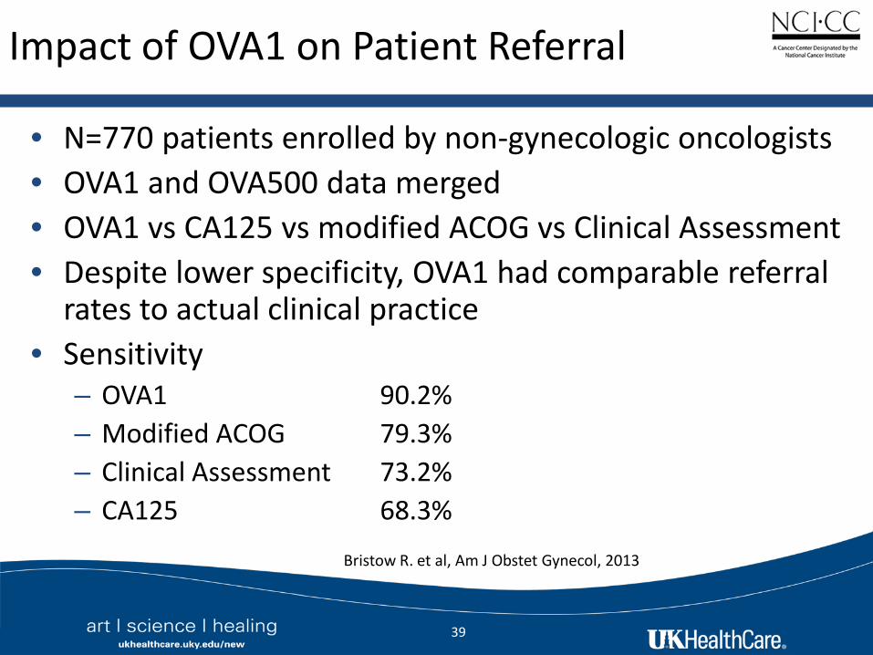
• N=770 patients enrolled by non-gynecologic oncologists • OVA1 and OVA500 data merged • OVA1 vs CA125 vs modified ACOG vs Clinical Assessment • Despite lower specificity, OVA1 had comparable referral
rates to actual clinical practice • Sensitivity
– OVA1 90.2% – Modified ACOG 79.3% – Clinical Assessment 73.2% – CA125 68.3%
Bristow R. et al, Am J Obstet Gynecol, 2013
Impact of OVA1 on Patient Referral
39
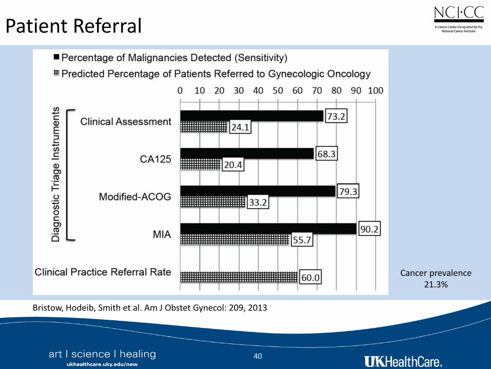
Patient Referral
Bristow, Hodeib, Smith et al. Am J Obstet Gynecol: 209, 2013
40
Cancer prevalence 21.3%

Kentucky Recommendations
41
Unilocular/septate Complex morphology1
Serial US every 6 months
Biomarker (CA125, OVA1, RMI, ROMA, ACOG guidelines)
Surgery with gynecologist Referral to gynecologic oncologist
Serial US every 12 months
low risk high risk2
*Perform tumor morphology indexing (MI) 1Complex morphology: solid or papillary areas, ascites, metastases, or MI ≥ 5 2High risk: CA125 > 200 U/mL (pre), >35 U/mL (post), high risk OVA1, RMI > 200, high risk ROMA, or ACOG guidelines
Ovarian tumor ultrasound*
complex1
low risk

UK Ovarian Cancer Investigator Trials Ovarian Cancer Screening Program Neoadjuvant chemotherapy Ovarian tumor evaluation using Morphology Index Trials in development

• Ovarian Cancer Screening Program (88-00219) – 25 years (1987-current) – 41,500 women, 241,000 visits – 85 cancers detected
• Neoadjuvant chemotherapy (11-GYN-098) • Evaluating the Performance of Morphology Index in
Surgical Decision-Making for Ovarian Tumors (14-GYN-200) – Prospective ultrasound-based decision making – Biomarker discovery
• Trials in development
43
Clinical Trials at the University of Kentucky

Conclusions
1. Ovarian cancer survival has not improved in 50 yrs 2. Most do not have surgery with a gynecologic
oncologist despite better survival 3. Algorithms reliably identify late but not early stage
cancers, and often fail in premenopausal women 4. Educating providers on contemporary referral
strategies is a moral imperative
Conclusions
45

Markey Cancer Center Affiliate Network 2014
*
Cynthiana
Mt. Vernon Hazard
Morehead Louisville
Frankfort Georgetown
Ashland
Harlan
South Williamson Elizabethtown
Lexington
Potential Affiliates Henderson Methodist Hospital Winchester Clark Regional Medical Center Cincinnati OH The Christ Hospital Health Network Huntington WV St. Mary’s Medical Center
Current Affiliates Ashland Our Lady of Bellefonte Hospital Cynthiana Harrison Memorial Hospital Elizabethtown Hardin Memorial Hospital Frankfort Frankfort Regional Medical Center Georgetown Georgetown Community Hospital Harlan Appalachian Regional Healthcare Hazard Appalachian Regional Healthcare Louisville Norton Cancer Institute Morehead St. Claire Regional Medical Center Mt. Vernon Rockcastle Regional Hospital S. Williamson Appalachian Regional Healthcare
Cincinnati, OH
Winchester
Huntington, WV
Henderson
