the role of the calf microbiota on performance and health · the role of the calf microbiota on...
TRANSCRIPT
78
The Role of the Calf Microbiota on Performance and Health
Rodrigo Carvalho Bicalho1 Cornell University
Introduction Human population expansion, higher living standards in many countries, and
economic growth have resulted in increased consumption of animal protein globally (Boland et al., 2013). For example, in Asia, animal protein consumption rose by 225% from 1961 to 2007 (FOASTAT). By 2050, protein consumption is expected to increase by a further 29% over the current level (Herrevo, 2013). Yet, approximately 843 million people worldwide continue to experience chronic hunger and nutrient deficiencies (Food and Agriculture Organization), and almost 1 billion receive insufficient amounts of protein (United Nations; Grover et al., 2009; Ghosh et al., 2012). Therefore, there is a pressing need to increase animal numbers, animal productivity and ultimately animal protein production without intensifying the environmental footprint of agriculture.
Antibiotic Resistance
In animal agriculture, antimicrobial compounds are widely administered (Chee-
Sanford et al., 2001, Smith et al., 2004, Sawant et al., 2007, McKinney et al., 2010). In 2012 alone, >32.2 million pounds of antimicrobial drug active ingredients were sold for animal use (Center for Veterinary Medicine, Food and Drug Administration, 2015). Antibiotics are used in food animals to treat disease, promote growth, and prevent disease by metaphylaxis (McEwen and Fedorka-Cray, 2002, Viola and DeVincent, 2006). In dairy cows, they are used to treat diseases such as mastitis, diarrhea and respiratory infection as well as to improve feed efficiency of pre-weaned calves. Sub-therapeutic levels of antimicrobials (e.g. tetracycline and neomycin) are routinely added to milk or milk replacer for disease prophylaxis and growth promotion (McEwen and Fedorka-Cray, 2002). Because pre-weaned dairy calves are prone to disease, there are advantages to adding antibiotics to calf feed (Morrill et al., 1995)), including higher feed consumption, average daily gain, and phagocytic efficiency, and lower mortality, incidence of scours, and protein requirements (Morrill, 1977).
However, ever-increasing numbers of antibiotic-resistant pathogens are
emerging; for example, 44% of coagulase-negative Staphylococci isolated from intra-mammary infections in dairy cows were found to be resistant to one or more antibiotics (Rajala-Schultz et al., 2004). Addition of antibiotics to livestock feed for growth stimulation and other non-therapeutic applications is believed to be the principal route by which antibiotic-resistant strains arise in food animals (McEwen and Fedorka-Cray, 2002). However, the rise in antimicrobial resistance is not necessarily evident among the pathogenic bacteria for which the antibiotics were applied, but is more likely to occur
1Contact at: Department of Population Medicine and Diagnostic Science, 618 Tower Rd., Ithaca, NY
14853; Tel: (607) 253-3148; E-mail: [email protected].

79
in the ‘innocent’ bystanders, i.e. bacteria making up the normal microbial flora of the intestinal tract (Mollenkopf et al., 2012), the respiratory tract, and possibly the mammary gland.
The emergence of antibiotic-resistant bacteria is a mounting concern, since many
of the antibiotics used in animal agriculture can also be used in human treatment, including tetracyclines, penicillins and sulfonamides (Silbergeld et al., 2008). There is potential for antimicrobial resistance to spread to humans (Smith, 2015) through food products/animal protein (Price et al., 2005), the environment (Graham et al., 2009), and by direct contact via agricultural workers (Smith et al., 2013). Moreover, commensal bacteria in livestock may potentially serve as hosts for resistance genes; in turn, these strains could be transmitted to people via fresh meat products, ultimately leading to transfer of the resistance genes to pathogenic organisms in humans (Diarrassouba et al., 2007, Mena et al., 2008). Disease associated with antimicrobial-resistant pathogens is expensive to treat, time consuming, and results in increased morbidity and mortality rates (Cosgrove, 2006, Maragakis et al., 2008, Mauldin et al., 2010).
Due to the emergence of antibiotic resistant microbes, the use of antibiotics as
growth promoters is now banned in the European Union (EU) and limited in the United States. The use of select antibiotics in animal feed was first sanctioned in Sweden in 1986 (Castanon, 2007), since then all antibiotics used as growth promoters or for metaphylaxis have been prohibited by EU countries (European Parliament and Council Regulation EC No. 1831/2003). Consequently, a reduction in animal productivity and a rise in animal infection, morbidity and mortality occurred (Cheng et al., 2014). In 2015, the US Food and Drug Administration published new directives for the use of antimicrobials in the feed of livestock in the United States, creating a new category of products called veterinary feed directive (VFD) drugs. Under this directive, a VFD drug must be used under the professional supervision of a licensed veterinarian (FDA, 2015). Treatment of animal diseases such as diarrhea with antibiotics and subsequent supportive care is expensive and leads to increased antimicrobial resistance; therefore, development of new strategies to prevent diarrhea and other diseases will improve overall productivity, animal welfare, profitability, and mitigate the emergence of resistance to antibiotics. Considering this, there is a growing demand for a substitute to antibiotic use (Seal et al., 2013); potential alternatives include antibacterial vaccines, immunomodulatory agents as well as prebiotics and probiotics (Millet and Maertens, 2011).
Alternative to Antibiotics
The World Health Organization (WHO) and the Food and Agriculture
Organization of the United Nations (FAO) define probiotics as “microorganisms that when administered live and in adequate amounts, confer a benefit to the health of the host.” A common application of probiotics is to improve gastrointestinal health, presumably by producing a gut environment that disfavors pathogenic microbes. Lactic acid bacteria and bifidobacteria are the most widely administered probiotic microbes (Didari et al., 2014). The biological impact of probiotics depends on the strain type;

80
therefore, strain identification and molecular analysis is critical (Azais-Braesco et al., 2010).
The gut microbiota has an essential role in determining many aspects of
postnatal life, such as contributing to the development of the immune system (Round and Mazmanian, 2009, Peterson and Cardona, 2010) and influencing host physiology, including energy balance by affecting energy expenditure and storage (Ridaura et al., 2013). Gut microbes serve their host by functioning as a key interface with the environment; for example, they can protect the host organism from pathogens that cause infectious diarrhea (O'Hara and Shanahan, 2006). In several studies of food animals, treatment with probiotics reduced the need for antibiotics, thereby potentially reducing human exposure to antimicrobial compounds and lowering the incidence of multidrug resistant microbes (Pedroso et al., 2013, Liang et al., 2014, Punaro et al., 2014). Whereas probiotics are a promising alternative to improve food-animal productivity and health, scientific evidence supporting the use of specific microbes to benefit animal health and performance is limited.
Altering The Gut Microbiome of Neonates Have Lasting Metabolic Consequences
Acquisition of the intestinal microbiota begins at birth, and a stable microbial
community develops from a succession of key organisms. Disruption of the microbiota during this important maturation can alter host metabolism and adiposity. Early life is a critical period for metabolic development (Dietz, 1994, Cunningham et al., 2014) and microbiota disruption during this window could change weight gain and body composition. In humans, early-life microbiota disruption, either due to delivery by Caesarian section (Dominguez-Bello et al., 2010) or antibiotics, is associated with increased risk of overweight status later in childhood (Ajslev et al., 2011). For decades, farmers have been making use of sub-therapeutic doses of antibiotics to promote the growth of farm animals. A landmark study by Cox et al. (2014), in which an in depth investigation of the mechanisms of action of growth promoting antibiotics using a mice model was performed, showed strong evidence that early antibiotic use disrupted the gut microbiome leading to increased weight gain, fat mass, and dramatic changes in liver metabolism. (Cox et al., 2014). Interestingly, it was reported that microbial communities recovered after the cessation of antibiotics, yet the metabolic phenotypes persisted, highlighting the importance of early-life microbiota in growth and development.
Faecalibacterium prausnitzii
Faecalibacterium prausnitzii (FP) is part of the normal intestinal microbiota of
many animal species and one of the most abundant bacteria present in human feces, comprising 2-20% of the human gut microbiota (Suau et al., 2001, Hold et al., 2003, Eckburg et al., 2005, Schwiertz et al., 2010, Arumugam et al., 2011, Walker et al., 2011). FP is also found in the feces of healthy non-human animals such as swine (Haenen et al., 2013), mice (Nava and Stappenbeck, 2011), and poultry (Lund et al., 2010). It is a strict anaerobe, a member of the phylum Firmicutes (Duncan et al., 2002),

81
and can produce large quantities of butyrate, D-lactate and formate through fermentation and utilization of acetate.
A lower abundance of Firmicutes, especially FP, characterizes the fecal and
mucosa-associated microbiota of Crohn’s disease patients (Sokol et al., 2008, Swidsinski et al., 2008, Sokol et al., 2009, Willing et al., 2009, Lopez-Siles et al., 2014). Moreover, lower levels of ileal mucosa-associated FP were correlated with postoperative relapse of ileal Crohn’s disease 6 months after surgical resection (Sokol et al., 2008). Similarly, reduced levels of Firmicutes were detected in ulcerative colitis (Frank et al., 2007, Nagalingam and Lynch, 2012, Machiels et al., 2014), and low levels of FP specifically were identified in association with alternating-type irritable bowel syndrome (Rajilic-Stojanovic et al., 2011, Miquel et al., 2013), colorectal cancer (Balamurugan et al., 2008), ulcerative colitis (Machiels et al., 2014), and even type II diabetes (Qin et al., 2012, Karlsson et al., 2013). These studies highlight the beneficial role of FP in the human gut. However, higher levels of fecal FP have been linked to obesity in children (Balamurugan et al., 2010), suggesting that FP is related to the energy-harvesting capacity of the intestinal microbiota.
Faecalibacterium prausnitzii as an Anti-Inflammatory Microbe
A large body of scientific evidence exists to support the beneficial effects of FP on the prevention and treatment of inflammatory disorders. A lower abundance of Firmicutes, especially FP, characterizes the fecal and mucosa-associated microbiota of patients with Crohn’s disease (Sokol et al., 2008, Swidsinski et al., 2008, Sokol et al., 2009, Willing et al., 2009, Lopez-Siles et al., 2014), ulcerative colitis (Frank et al., 2007, Nagalingam and Lynch, 2012, Machiels et al., 2014), irritable bowel syndrome (Rajilic-Stojanovic et al., 2011, Miquel et al., 2013), colorectal cancer (Balamurugan et al., 2008), and more importantly type-2 diabetes (Qin et al., 2012, Karlsson et al., 2013). The anti-inflammatory properties of FP have been well described. In a pivotal study by Sokol et al (2008), both in vitro and in vivo studies were performed to evaluate the immunomodulatory properties of FP (Sokol et al., 2008). It was demonstrated that the FP supernatant abolished interleukin (IL) 1β-induced nuclear factor (NF) κB activity using Caco-2 cells and led to significantly lower secretion of the pro-inflammatory cytokines such as tumor necrosis factor (TNF) α and IL-12, and higher secretion of IL-10 by peripheral blood mononuclear cells. More importantly, oral administration of either live FP or its supernatant markedly reduced the severity of experimentally induced colitis and tended to correct the dysbiosis associated with it (Sokol et al., 2008). Others have reported that both FP and its supernatant can also inhibit IL-8 secretion (Quevrain et al., 2016a, Quevrain et al., 2016b, Martin et al., 2017). The ability of FP to induce IL-10 expression by dendritic cells was well described by Rossi et al. (2015), and the anti-inflammatory characteristic of FP has been demonstrated by several others (Quevrain et al., 2016a, Rossi et al., 2016, Martin et al., 2017, Munukka et al., 2017). The mechanism of action associated with the anti-inflammatory characteristics of FP are not fully understood but involves the synthesis of butyrate and other factors. Butyrate regulates proliferation, differentiation, and apoptosis of gastrointestinal tract cells while stimulating the production of mucus and decreasing cell permeability, hence preventing leakage of bacterial endotoxins and inflammation (von

82
Engelhardt et al., 1998, Augenlicht et al., 2002). Additionally, butyrate is also known to directly inhibit NF-κB activity (Inan et al., 2000, Segain et al., 2000, Yin et al., 2001, Luhrs et al., 2002a, Luhrs et al., 2002b). Quévrain et al (2016), identified a novel protein from an FP isolate, which they named microbial anti-inflammatory molecule (MAM). Cloning and overexpression of the MAM in human intestinal epithelial cells revealed an inhibitory effect on NF-κB activity. Importantly, oral administration of MAM-expressing recombinant Lactococcus lactis but not the wild-type strain reduced severity of experimentally induced colitis. Animals that received MAM-expressing L. lactis showed improved histopathology, less severe weight loss, and reduced interferon (IFN) γ and IL-17 expression compared with controls (Quevrain et al., 2016b).
Oral administration of FP reduced the incidence of severe diarrhea and related
mortality rate and increased weight gain in pre-weaned dairy Heifers (Foditsch et al., 2015). This research evaluated the effect of administering a live culture of FP to newborn dairy calves on their subsequent growth, health, and fecal microbiome. Faecalibacterium prausnitzii was cultured in VTR2RF medium as previously described (Foditsch et al., 2014). Initially, a safety trial was conducted using 30 newborn bull calves to assess potential adverse effects of oral and rectal administration of live FP to neonatal calves, compared to controls. No adverse reactions (e.g. increased body temperature or heart and respiratory rates) were observed. All bull calves survived the experimental period, and there was no difference in attitude, appetite, dehydration or fecal consistency score between the treatment and control groups. The rectal route was not practical, whereas the oral route ensured that the full dose could be administered to the treated calves.
Subsequently, a randomized field trial was completed in a commercial farm with
pre-weaned calves. In total, 554 Holstein heifers were assigned to one of two treatment groups: treated calves (FPTRT) and non-treated calves (control). Treated calves received two oral doses of live culture of FP, the first dose at treatment assignment (1st week) and the second a week later. The FPTRT group experienced a significantly lower incidence of severe diarrhea (3.1%) compared to the control group (6.8%) (Figure 1C). Treated calves also had a lower mortality rate associated with severe diarrhea (1.5%) compared to control calves (4.4%) (Figure 1A and B). Furthermore, FPTRT calves gained significantly more weight (4.4 kg) over the pre-weaning period than controls calves. The relative abundance of FP in the fecal microbiota was significantly higher in the 3rd and 5th weeks of life of FPTRT calves compared to control calves, as revealed by sequencing of the 16S rRNA gene. These findings demonstrated that oral administration of live culture of FP improves gastrointestinal health and growth of pre-weaned calves, supporting its use as a potential probiotic.
Faecalibacterium prausnitzii and Insulin Sensitivity
Recent studies highlight the importance of the gut microbiota as an environmental factor linked to type-2 diabetes (Furet et al., 2010, Qin et al., 2012, Karlsson et al., 2013). A consistent finding from these studies was a decrease in the relative abundance of butyrate producing bacteria such as FP and Roseburia spp. in

83
individuals with type-2 diabetes. Furthermore, butyrate producers, and more specifically FP, have been linked to improved insulin sensitivity and diabetes amelioration in studies of the human fecal microbiota (Furet et al., 2010, Vrieze et al., 2012).
There is a great body of knowledge both supporting the strong anti-inflammatory
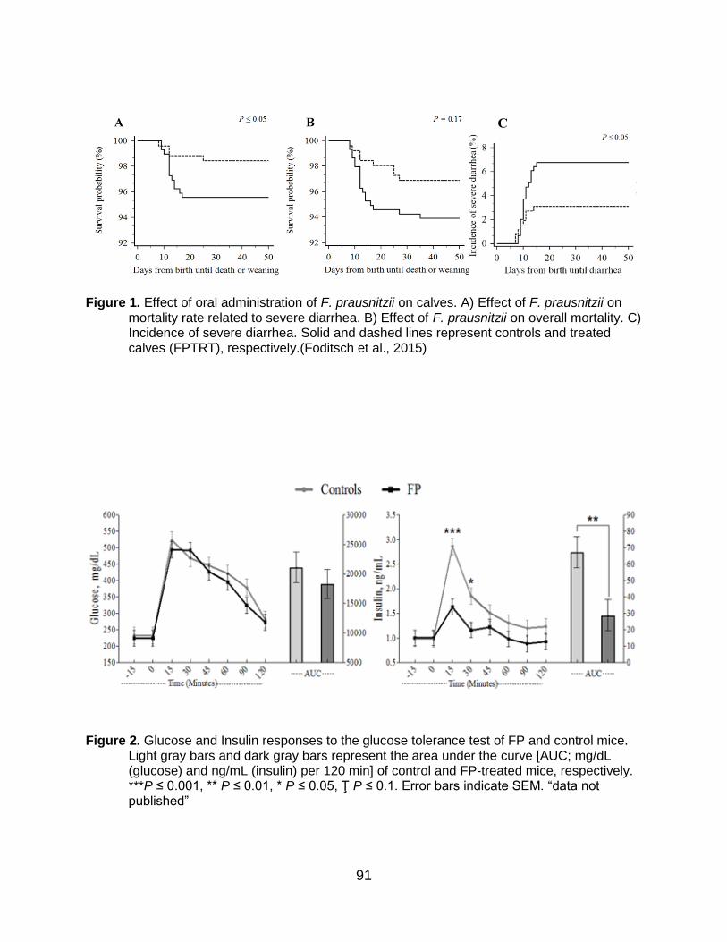
characteristic of FP and the link between inflammation and type-2 diabetes, but ironically, to the best of our knowledge, no published studies have evaluated the direct effect of FP treatment on glucose metabolism. Recently, our group demonstrated that treatment of pre-diabetic, obese mice [10-week-old Male DIO C57BL/6J mice (strain 380050)] with viable FP culture cells and supernatant resulted in a dramatic improvement of insulin sensitivity (Figure 2). Briefly, C57BL/6J wild type mice were fed a high fat diet and treated daily for 20 days with a cocktail of four high butyrate producing FP strains (isolated from bovine and porcine (Foditsch et al., 2014)) or placebo; treatment was performed by oral gavage. Before sacrifice an oral glucose tolerance test was performed, and blood samples collected for insulin serum concentrations. Daily administration of FP significantly increased insulin sensitivity, showing its utility in protecting against insulin resistance in diet induced obese mice (Figure 2).
The work by Munukka et al. (2017), provides further support that oral FP
treatment improved insulin sensitivity in high-fat-fed mice (Munukka et al., 2017). FP-treated mice had increased insulin receptor β, increased hormone-sensitive lipase phosphorylation in in the adipose tissue, decreased leukocyte infiltration into adipose tissue, lower hepatic fat content, and improved liver function compared with control mice. They concluded that FP treatment improved insulin sensitivity, decreased adipose tissue inflammation, and improved hepatic health (Munukka et al., 2017). Interestingly, FP-treatment increases weight gain because of improved insulin sensitivity, FP-treated mice significantly increased muscle mass and subcutaneous fat when compared with placebo treated mice (Munukka et al., 2017).
References Ajslev, T. A., C. S. Andersen, M. Gamborg, T. I. Sorensen, and T. Jess. 2011.
Childhood overweight after establishment of the gut microbiota: the role of delivery mode, pre-pregnancy weight and early administration of antibiotics. Int. J. Obes. (Lond) 35:522-529.
Arumugam, M., J. Raes, E. Pelletier, D. Le Paslier, T. Yamada, D. R. Mende, G. R. Fernandes, J. Tap, T. Bruls, J. M. Batto, M. Bertalan, N. Borruel, F. Casellas, L. Fernandez, L. Gautier, T. Hansen, M. Hattori, T. Hayashi, M. Kleerebezem, K. Kurokawa, M. Leclerc, F. Levenez, C. Manichanh, H. B. Nielsen, T. Nielsen, N. Pons, J. Poulain, J. Qin, T. Sicheritz-Ponten, S. Tims, D. Torrents, E. Ugarte, E. G. Zoetendal, J. Wang, F. Guarner, O. Pedersen, W. M. de Vos, S. Brunak, J. Dore, H. I. T. C. Meta, M. Antolin, F. Artiguenave, H. M. Blottiere, M. Almeida, C. Brechot, C. Cara, C. Chervaux, A. Cultrone, C. Delorme, G. Denariaz, R. Dervyn, K. U. Foerstner, C. Friss, M. van de Guchte, E. Guedon, F. Haimet, W. Huber, J. van Hylckama-Vlieg, A. Jamet, C. Juste, G. Kaci, J. Knol, O. Lakhdari, S. Layec,

84
K. Le Roux, E. Maguin, A. Merieux, R. Melo Minardi, C. M'Rini, J. Muller, R. Oozeer, J. Parkhill, P. Renault, M. Rescigno, N. Sanchez, S. Sunagawa, A. Torrejon, K. Turner, G. Vandemeulebrouck, E. Varela, Y. Winogradsky, G. Zeller, J. Weissenbach, S. D. Ehrlich, and P. Bork. 2011. Enterotypes of the human gut microbiome. Nature 473(7346):174-180.
Augenlicht, L. H., J. M. Mariadason, A. Wilson, D. Arango, W. Yang, B. G. Heerdt, and A. Velcich. 2002. Short chain fatty acids and colon cancer. J. Nutr. 132:3804S-3808S.
Azais-Braesco, V., J. L. Bresson, F. Guarner, and G. Corthier. 2010. Not all lactic acid bacteria are probiotics, ...but some are. Br. J. Nutr. 103:1079-1081.
Balamurugan, R., G. George, J. Kabeerdoss, J. Hepsiba, A. M. Chandragunasekaran, and B. S. Ramakrishna. 2010. Quantitative differences in intestinal Faecalibacterium prausnitzii in obese Indian children. Br. J. Nutr. 103:335-338.
Balamurugan, R., E. Rajendiran, S. George, G. V. Samuel, and B. S. Ramakrishna. 2008. Real-time polymerase chain reaction quantification of specific butyrate-producing bacteria, Desulfovibrio and Enterococcus faecalis in the feces of patients with colorectal cancer. J. Gastroenterol. Hepatol. 23(8 Pt 1):1298-1303.
Castanon, J. I. 2007. History of the use of antibiotic as growth promoters in European poultry feeds. Poult. Sci. 86:2466-2471.
Chee-Sanford, J. C., R. I. Aminov, I. J. Krapac, N. Garrigues-Jeanjean, and R. I. Mackie. 2001. Occurrence and diversity of tetracycline resistance genes in lagoons and groundwater underlying two swine production facilities. Appl. Environ. Microbiol. 67:1494-1502.
Cheng, G., H. Hao, S. Xie, X. Wang, M. Dai, L. Huang, and Z. Yuan. 2014. Antibiotic alternatives: the substitution of antibiotics in animal husbandry? Front. Microbiol. 5:217.
Cosgrove, S. E. 2006. The relationship between antimicrobial resistance and patient outcomes: mortality, length of hospital stay, and health care costs. Clin. Infect. Dis. 42 Suppl 2:S82-89.
Cox, L. M., S. Yamanishi, J. Sohn, A. V. Alekseyenko, J. M. Leung, I. Cho, S. G. Kim, H. Li, Z. Gao, D. Mahana, J. G. Zarate Rodriguez, A. B. Rogers, N. Robine, P. Loke, and M. J. Blaser. 2014. Altering the intestinal microbiota during a critical developmental window has lasting metabolic consequences. Cell 158:705-721.
Cunningham, S. A., M. R. Kramer, and K. M. Narayan. 2014. Incidence of childhood obesity in the United States. N. Engl. J. Med. 370:1660-1661.
Diarrassouba, F., M. S. Diarra, S. Bach, P. Delaquis, J. Pritchard, E. Topp, and B. J. Skura. 2007. Antibiotic resistance and virulence genes in commensal Escherichia coli and Salmonella isolates from commercial broiler chicken farms. J. Food Prot. 70:1316-1327.
Didari, T., S. Solki, S. Mozaffari, S. Nikfar, and M. Abdollahi. 2014. A systematic review of the safety of probiotics. Expert Opin. Drug Saf. 13:227-239.

85
Dietz, W. H. 1994. Critical periods in childhood for the development of obesity. Am. J. Clin. Nutr. 59:955-959.
Dominguez-Bello, M. G., E. K. Costello, M. Contreras, M. Magris, G. Hidalgo, N. Fierer, and R. Knight. 2010. Delivery mode shapes the acquisition and structure of the initial microbiota across multiple body habitats in newborns. Proc. Natl. Acad. Sci. U. S. A. 107:11971-11975.
Duncan, S. H., G. L. Hold, H. J. Harmsen, C. S. Stewart, and H. J. Flint. 2002. Growth requirements and fermentation products of Fusobacterium prausnitzii, and a proposal to reclassify it as Faecalibacterium prausnitzii gen. nov., comb. nov. Int. J. Syst. Evol. Microbiol. 52(Pt 6):2141-2146.
Eckburg, P. B., E. M. Bik, C. N. Bernstein, E. Purdom, L. Dethlefsen, M. Sargent, S. R. Gill, K. E. Nelson, and D. A. Relman. 2005. Diversity of the human intestinal microbial flora. Science 308(5728):1635-1638.
Foditsch, C., T. M. Santos, A. G. Teixeira, R. V. Pereira, J. M. Dias, N. Gaeta, and R. C. Bicalho. 2014. Isolation and characterization of Faecalibacterium prausnitzii from calves and piglets. PLoS One 9(12):e116465.
Frank, D. N., A. L. St Amand, R. A. Feldman, E. C. Boedeker, N. Harpaz, and N. R. Pace. 2007. Molecular-phylogenetic characterization of microbial community imbalances in human inflammatory bowel diseases. Proc. Natl. Acad. Sci. U. S. A. 104(34):13780-13785.
Furet, J. P., L. C. Kong, J. Tap, C. Poitou, A. Basdevant, J. L. Bouillot, D. Mariat, G. Corthier, J. Dore, C. Henegar, S. Rizkalla, and K. Clement. 2010. Differential adaptation of human gut microbiota to bariatric surgery-induced weight loss: links with metabolic and low-grade inflammation markers. Diabetes 59:3049-3057.
Graham, J. P., S. L. Evans, L. B. Price, and E. K. Silbergeld. 2009. Fate of antimicrobial-resistant enterococci and staphylococci and resistance determinants in stored poultry litter. Environ. Res. 109:682-689.
Haenen, D., J. Zhang, C. Souza da Silva, G. Bosch, I. M. van der Meer, J. van Arkel, J. J. van den Borne, O. Perez Gutierrez, H. Smidt, B. Kemp, M. Muller, and G. J. Hooiveld. 2013. A diet high in resistant starch modulates microbiota composition, SCFA concentrations, and gene expression in pig intestine. J. Nutr. 143:274-283.
Hold, G. L., A. Schwiertz, R. I. Aminov, M. Blaut, and H. J. Flint. 2003. Oligonucleotide probes that detect quantitatively significant groups of butyrate-producing bacteria in human feces. Appl. Environ. Microbiol. 69:4320-4324.
Inan, M. S., R. J. Rasoulpour, L. Yin, A. K. Hubbard, D. W. Rosenberg, and C. Giardina. 2000. The luminal short-chain fatty acid butyrate modulates NF-kappaB activity in a human colonic epithelial cell line. Gastroenterology 118:724-734.
Karlsson, F. H., V. Tremaroli, I. Nookaew, G. Bergstrom, C. J. Behre, B. Fagerberg, J. Nielsen, and F. Backhed. 2013. Gut metagenome in European women with normal, impaired and diabetic glucose control. Nature 498(7452):99-103.

86
Liang, S., T. Webb, and Z. Li. 2014. Probiotic antigens stimulate hepatic natural killer T cells. Immunology 141:203-210.
Lopez-Siles, M., M. Martinez-Medina, D. Busquets, M. Sabat-Mir, S. H. Duncan, H. J. Flint, X. Aldeguer, and L. J. Garcia-Gil. 2014. Mucosa-associated Faecalibacterium prausnitzii and Escherichia coli co-abundance can distinguish Irritable Bowel Syndrome and Inflammatory Bowel Disease phenotypes. Int. J. Med. Microbiol. 304:464-475.
Luhrs, H., T. Gerke, J. G. Muller, R. Melcher, J. Schauber, F. Boxberge, W. Scheppach, and T. Menzel. 2002a. Butyrate inhibits NF-kappaB activation in lamina propria macrophages of patients with ulcerative colitis. Scand J Gastroenterol 37:458-466.
Luhrs, H., T. Kudlich, M. Neumann, J. Schauber, R. Melcher, A. Gostner, W. Scheppach, and T. P. Menzel. 2002b. Butyrate-enhanced TNFalpha-induced apoptosis is associated with inhibition of NF-kappaB. Anticancer Res. 22:1561-1568.
Lund, M., L. Bjerrum, and K. Pedersen. 2010. Quantification of Faecalibacterium prausnitzii- and Subdoligranulum variabile-like bacteria in the cecum of chickens by real-time PCR. Poult. Sci. 89:1217-1224.
Machiels, K., M. Joossens, J. Sabino, V. De Preter, I. Arijs, V. Eeckhaut, V. Ballet, K. Claes, F. Van Immerseel, K. Verbeke, M. Ferrante, J. Verhaegen, P. Rutgeerts, and S. Vermeire. 2014. A decrease of the butyrate-producing species Roseburia hominis and Faecalibacterium prausnitzii defines dysbiosis in patients with ulcerative colitis. Gut 63:1275-1283.
Maragakis, L. L., E. N. Perencevich, and S. E. Cosgrove. 2008. Clinical and economic burden of antimicrobial resistance. Expert. Rev. Anti. Infect. Ther. 6:751-763.
Martin, R., S. Miquel, L. Benevides, C. Bridonneau, V. Robert, S. Hudault, F. Chain, O. Berteau, V. Azevedo, J. M. Chatel, H. Sokol, L. G. Bermudez-Humaran, M. Thomas, and P. Langella. 2017. Functional characterization of novel Faecalibacterium prausnitzii strains Isolated from healthy volunteers: a step forward in the use of F. prausnitzii as a next-generation probiotic. Front. Microbiol. 8:1226.
Mauldin, P. D., C. D. Salgado, I. S. Hansen, D. T. Durup, and J. A. Bosso. 2010. Attributable hospital cost and length of stay associated with health care-associated infections caused by antibiotic-resistant gram-negative bacteria. Antimicrob. Agents Chemother. 54:109-115.
McEwen, S. A. and P. J. Fedorka-Cray. 2002. Antimicrobial use and resistance in animals. Clin. Infect. Dis. 34 Suppl 3:S93-S106.
McKinney, C. W., K. A. Loftin, M. T. Meyer, J. G. Davis, and A. Pruden. 2010. tet and sul antibiotic resistance genes in livestock lagoons of various operation type, configuration, and antibiotic occurrence. Environ. Sci. Technol. 44:6102-6109.
Mena, C., D. Rodrigues, J. Silva, P. Gibbs, and P. Teixeira. 2008. Occurrence, identification, and characterization of Campylobacter species isolated from
87
portuguese poultry samples collected from retail establishments. Poult. Sci. 87:187-190.
Millet, S. and L. Maertens. 2011. The European ban on antibiotic growth promoters in animal feed: from challenges to opportunities. Vet. J. 187:143-144.
Miquel, S., R. Martin, O. Rossi, L. G. Bermudez-Humaran, J. M. Chatel, H. Sokol, M. Thomas, J. M. Wells, and P. Langella. 2013. Faecalibacterium prausnitzii and human intestinal health. Curr. Opin. Microbiol. 16:255-261.
Mollenkopf, D. F., M. F. Weeman, J. B. Daniels, M. J. Abley, J. L. Mathews, W. A. Gebreyes, and T. E. Wittum. 2012. Variable within- and between-herd diversity of CTX-M cephalosporinase-bearing Escherichia coli isolates from dairy cattle. Appl. Environ. Microbiol. 78:4552-4560.
Morrill, J. L., J. M. Morrill, A. M. Feyerherm, and J. F. Laster. 1995. Plasma proteins and a probiotic as ingredients in milk replacer. J. Dairy Sci. 78:902-907.
Munukka, E., A. Rintala, R. Toivonen, M. Nylund, B. Yang, A. Takanen, A. Hanninen, J. Vuopio, P. Huovinen, S. Jalkanen, and S. Pekkala. 2017. Faecalibacterium prausnitzii treatment improves hepatic health and reduces adipose tissue inflammation in high-fat fed mice. ISME J. 11:1667-1679.
Nagalingam, N. A. and S. V. Lynch. 2012. Role of the microbiota in inflammatory bowel diseases. Inflamm Bowel Dis 18(5):968-984.
Nava, G. M. and T. S. Stappenbeck. 2011. Diversity of the autochthonous colonic microbiota. Gut Microbes 2:99-104.
O'Hara, A. M. and F. Shanahan. 2006. The gut flora as a forgotten organ. EMBO Rep. 7:688-693.
Pedroso, A. A., A. L. Hurley-Bacon, A. S. Zedek, T. W. Kwan, A. P. Jordan, G. Avellaneda, C. L. Hofacre, B. B. Oakley, S. R. Collett, J. J. Maurer, and M. D. Lee. 2013. Can probiotics improve the environmental microbiome and resistome of commercial poultry production? Int. J. Environ. Res. Public Health 10:4534-4559.
Peterson, D. A. and R. A. Cardona. 2010. Specificity of the adaptive immune response to the gut microbiota. Adv. Immunol. 107:71-107.
Price, L. B., E. Johnson, R. Vailes, and E. Silbergeld. 2005. Fluoroquinolone-resistant Campylobacter isolates from conventional and antibiotic-free chicken products. Environ. Health Perspect. 113:557-560.
Punaro, G. R., F. R. Maciel, A. M. Rodrigues, M. M. Rogero, C. S. Bogsan, M. N. Oliveira, S. S. Ihara, S. R. Araujo, T. R. Sanches, L. C. Andrade, and E. M. Higa. 2014. Kefir administration reduced progression of renal injury in STZ-diabetic rats by lowering oxidative stress. Nitric Oxide 37:53-60.
Qin, J., Y. Li, Z. Cai, S. Li, J. Zhu, F. Zhang, S. Liang, W. Zhang, Y. Guan, D. Shen, Y. Peng, D. Zhang, Z. Jie, W. Wu, Y. Qin, W. Xue, J. Li, L. Han, D. Lu, P. Wu, Y. Dai, X. Sun, Z. Li, A. Tang, S. Zhong, X. Li, W. Chen, R. Xu, M. Wang, Q. Feng, M. Gong, J. Yu, Y. Zhang, M. Zhang, T. Hansen, G. Sanchez, J. Raes, G.

88
Falony, S. Okuda, M. Almeida, E. LeChatelier, P. Renault, N. Pons, J. M. Batto, Z. Zhang, H. Chen, R. Yang, W. Zheng, S. Li, H. Yang, J. Wang, S. D. Ehrlich, R. Nielsen, O. Pedersen, K. Kristiansen, and J. Wang. 2012. A metagenome-wide association study of gut microbiota in type 2 diabetes. Nature 490(7418):55-60.
Quevrain, E., M. A. Maubert, C. Michon, F. Chain, R. Marquant, J. Tailhades, S. Miquel, L. Carlier, L. G. Bermudez-Humaran, B. Pigneur, O. Lequin, P. Kharrat, G. Thomas, D. Rainteau, C. Aubry, N. Breyner, C. Afonso, S. Lavielle, J. P. Grill, G. Chassaing, J. M. Chatel, G. Trugnan, R. Xavier, P. Langella, H. Sokol, and P. Seksik. 2016a. Identification of an anti-inflammatory protein from Faecalibacterium prausnitzii, a commensal bacterium deficient in Crohn's disease. Gut 65:415-425.
Quevrain, E., M. A. Maubert, H. Sokol, B. Devreese, and P. Seksik. 2016b. The presence of the anti-inflammatory protein MAM, from Faecalibacterium prausnitzii, in the intestinal ecosystem. Gut 65:882.
Rajala-Schultz, P. J., K. L. Smith, J. S. Hogan, and B. C. Love. 2004. Antimicrobial susceptibility of mastitis pathogens from first lactation and older cows. Vet. Microbiol. 102:33-42.
Rajilic-Stojanovic, M., E. Biagi, H. G. Heilig, K. Kajander, R. A. Kekkonen, S. Tims, and W. M. de Vos. 2011. Global and deep molecular analysis of microbiota signatures in fecal samples from patients with irritable bowel syndrome. Gastroenterology 141:1792-1801.
Ridaura, V. K., J. J. Faith, F. E. Rey, J. Cheng, A. E. Duncan, A. L. Kau, N. W. Griffin, V. Lombard, B. Henrissat, J. R. Bain, M. J. Muehlbauer, O. Ilkayeva, C. F. Semenkovich, K. Funai, D. K. Hayashi, B. J. Lyle, M. C. Martini, L. K. Ursell, J. C. Clemente, W. Van Treuren, W. A. Walters, R. Knight, C. B. Newgard, A. C. Heath, and J. I. Gordon. 2013. Gut microbiota from twins discordant for obesity modulate metabolism in mice. Science 341(6150):1241214.
Rossi, O., L. A. van Berkel, F. Chain, M. Tanweer Khan, N. Taverne, H. Sokol, S. H. Duncan, H. J. Flint, H. J. Harmsen, P. Langella, J. N. Samsom, and J. M. Wells. 2016. Faecalibacterium prausnitzii A2-165 has a high capacity to induce IL-10 in human and murine dendritic cells and modulates T cell responses. Sci. Rep. 6:18507.
Round, J. L. and S. K. Mazmanian. 2009. The gut microbiota shapes intestinal immune responses during health and disease. Nat Rev Immunol 9(5):313-323.
Sawant, A. A., N. V. Hegde, B. A. Straley, S. C. Donaldson, B. C. Love, S. J. Knabel, and B. M. Jayarao. 2007. Antimicrobial-resistant enteric bacteria from dairy cattle. Appl. Environ. Microbiol. 73:156-163.
Schwiertz, A., M. Jacobi, J. S. Frick, M. Richter, K. Rusch, and H. Kohler. 2010. Microbiota in pediatric inflammatory bowel disease. J Pediatr 157(2):240-244 e241.
89
Seal, B. S., H. S. Lillehoj, D. M. Donovan, and C. G. Gay. 2013. Alternatives to antibiotics: a symposium on the challenges and solutions for animal production. Anim. Health Res. Rev. 14:78-87.
Segain, J. P., D. Raingeard de la Bletiere, A. Bourreille, V. Leray, N. Gervois, C. Rosales, L. Ferrier, C. Bonnet, H. M. Blottiere, and J. P. Galmiche. 2000. Butyrate inhibits inflammatory responses through NFkappaB inhibition: implications for Crohn's disease. Gut 47:397-403.
Silbergeld, E. K., J. Graham, and L. B. Price. 2008. Industrial food animal production, antimicrobial resistance, and human health. Annu Rev Public Health 29:151-169.
Smith, M. S., R. K. Yang, C. W. Knapp, Y. Niu, N. Peak, M. M. Hanfelt, J. C. Galland, and D. W. Graham. 2004. Quantification of tetracycline resistance genes in feedlot lagoons by real-time PCR. Appl. Environ. Microbiol. 70:7372-7377.
Smith, T. C. 2015. Livestock-associated Staphylococcus aureus: the United States experience. PLoS Pathog 11(2):e1004564.
Smith, T. C., W. A. Gebreyes, M. J. Abley, A. L. Harper, B. M. Forshey, M. J. Male, H. W. Martin, B. Z. Molla, S. Sreevatsan, S. Thakur, M. Thiruvengadam, and P. R. Davies. 2013. Methicillin-resistant Staphylococcus aureus in pigs and farm workers on conventional and antibiotic-free swine farms in the USA. PLoS One 8(5):e63704.
Sokol, H., B. Pigneur, L. Watterlot, O. Lakhdari, L. G. Bermudez-Humaran, J. J. Gratadoux, S. Blugeon, C. Bridonneau, J. P. Furet, G. Corthier, C. Grangette, N. Vasquez, P. Pochart, G. Trugnan, G. Thomas, H. M. Blottiere, J. Dore, P. Marteau, P. Seksik, and P. Langella. 2008. Faecalibacterium prausnitzii is an anti-inflammatory commensal bacterium identified by gut microbiota analysis of Crohn disease patients. Proc. Natl. Acad. Sci. U. S. A. 105:16731-16736.
Sokol, H., P. Seksik, J. P. Furet, O. Firmesse, I. Nion-Larmurier, L. Beaugerie, J. Cosnes, G. Corthier, P. Marteau, and J. Dore. 2009. Low counts of Faecalibacterium prausnitzii in colitis microbiota. Inflamm. Bowel Dis. 15:1183-1189.
Suau, A., V. Rochet, A. Sghir, G. Gramet, S. Brewaeys, M. Sutren, L. Rigottier-Gois, and J. Dore. 2001. Fusobacterium prausnitzii and related species represent a dominant group within the human fecal flora. Syst. Appl. Microbiol. 24:139-145.
Swidsinski, A., V. Loening-Baucke, M. Vaneechoutte, and Y. Doerffel. 2008. Active Crohn's disease and ulcerative colitis can be specifically diagnosed and monitored based on the biostructure of the fecal flora. Inflamm. Bowel Dis. 14:147-161.
Viola, C. and S. J. DeVincent. 2006. Overview of issues pertaining to the manufacture, distribution, and use of antimicrobials in animals and other information relevant to animal antimicrobial use data collection in the United States. Prev. Vet. Med. 73:111-131.
90
von Engelhardt, W., J. Bartels, S. Kirschberger, H. D. Meyer zu Duttingdorf, and R. Busche. 1998. Role of short-chain fatty acids in the hind gut. Vet. Q. 20 Suppl 3:S52-59.
Vrieze, A., E. Van Nood, F. Holleman, J. Salojarvi, R. S. Kootte, J. F. Bartelsman, G. M. Dallinga-Thie, M. T. Ackermans, M. J. Serlie, R. Oozeer, M. Derrien, A. Druesne, J. E. Van Hylckama Vlieg, V. W. Bloks, A. K. Groen, H. G. Heilig, E. G. Zoetendal, E. S. Stroes, W. M. de Vos, J. B. Hoekstra, and M. Nieuwdorp. 2012. Transfer of intestinal microbiota from lean donors increases insulin sensitivity in individuals with metabolic syndrome. Gastroenterology 143:913-916 e917.
Walker, A. W., J. Ince, S. H. Duncan, L. M. Webster, G. Holtrop, X. Ze, D. Brown, M. D. Stares, P. Scott, A. Bergerat, P. Louis, F. McIntosh, A. M. Johnstone, G. E. Lobley, J. Parkhill, and H. J. Flint. 2011. Dominant and diet-responsive groups of bacteria within the human colonic microbiota. ISME J. 5:220-230.
Willing, B., J. Halfvarson, J. Dicksved, M. Rosenquist, G. Jarnerot, L. Engstrand, C. Tysk, and J. K. Jansson. 2009. Twin studies reveal specific imbalances in the mucosa-associated microbiota of patients with ileal Crohn's disease. Inflamm. Bowel Dis. 15:653-660.
Yin, L., G. Laevsky, and C. Giardina. 2001. Butyrate suppression of colonocyte NF-kappa B activation and cellular proteasome activity. J. Biol. Chem. 276:44641-44646.
91
Figure 1. Effect of oral administration of F. prausnitzii on calves. A) Effect of F. prausnitzii on
mortality rate related to severe diarrhea. B) Effect of F. prausnitzii on overall mortality. C) Incidence of severe diarrhea. Solid and dashed lines represent controls and treated calves (FPTRT), respectively.(Foditsch et al., 2015)
Figure 2. Glucose and Insulin responses to the glucose tolerance test of FP and control mice. Light gray bars and dark gray bars represent the area under the curve [AUC; mg/dL (glucose) and ng/mL (insulin) per 120 min] of control and FP-treated mice, respectively. ***P ≤ 0.001, ** P ≤ 0.01, * P ≤ 0.05, Ţ P ≤ 0.1. Error bars indicate SEM. “data not published”
C

92
SESSION NOTES