the role of monocularly visible regions in depth and surface perception
TRANSCRIPT

Vision Research 49 (2009) 2666–2685
Contents lists available at ScienceDirect
Vision Research
journal homepage: www.elsevier .com/locate /v isres
The role of monocularly visible regions in depth and surface perception
Julie M. Harris a,*, Laurie M. Wilcox b
a School of Psychology, University of St. Andrews, South St., St. Andrews, KY16 9JP, Scotland, United Kingdomb Department of Psychology, Centre for Vision Research, 4700 Keele St., North York, Ontario, Canada M3J 1P3
a r t i c l e i n f o a b s t r a c t
Article history:Received 18 December 2008Received in revised form 22 June 2009
Keywords:StereopsisBinocular visionda Vinci StereopsisMonocular regionsHalf-occlusionMonocular
0042-6989/$ - see front matter � 2009 Elsevier Ltd. Adoi:10.1016/j.visres.2009.06.021
* Corresponding author.E-mail address: [email protected] (J.M
The mainstream of binocular vision research has long been focused on understanding how binocular dis-parity is used for depth perception. In recent years, researchers have begun to explore how monocularregions in binocularly viewed scenes contribute to our perception of the three-dimensional world. Herewe review the field as it currently stands, with a focus on understanding the extent to which the role ofmonocular regions in depth perception can be understood using extant theories of binocular vision.
� 2009 Elsevier Ltd. All rights reserved.
1. Introduction
1.1. The problem: how do we get a 3-D view when the eyes seedifferent things?
Because we have two forward-facing eyes that are separated inthe head, our visual system continuously receives two slightly dif-ferent views of the world. For many decades scientists have beentrying to understand how the visual system deals with the slightdifferences in the images that result from the lateral separationof the two eyes (e.g. Wheatstone, 1838). Some have argued thatthe differences are so small that they are irrelevant. Instead, theysuggest that the primary reason for having two eyes is for binocu-lar concordance: that is, increasing visual efficiency (reducingnoise) by having effectively ‘a second go’ at viewing each scene(e.g. Jones & Lee, 1982). This approach makes sense when oneviews a distant scene, as the two views are very similar.
However, much of the time we function in environments inwhich there are multiple objects at close range, and for suchscenes, there are substantial differences in the retinal images.Rather than emphasising concordance, a successful approach hasbeen to consider the extent to which the visual system can exploitdiscordance by measuring the tiny differences between the twoeyes’ views (binocular disparity), and by using disparity to repre-sent the three-dimensional (3-D) structure of the world. A rich lit-erature has shown that binocular disparity is used by the visualsystem, and that we are exquisitely sensitive to it (e.g. Howard &
ll rights reserved.
. Harris).
Rogers, 2002). Measuring the differences between locations inthe two eye’s views first requires a solution to the binocular corre-spondence problem. That is, which point on one retina matcheswith a given point on the other retina. This is a potentially compli-cated operation given the complex 3-D structure of the naturalenvironment, but it is made even more difficult by the fact thatnot all image points in one eye have a partner in the other. Forexample, Fig. 1a shows a top–down view of a scene viewedthrough an occluding foreground ‘fence’. We refer to this sceneas the background because it is the furthest thing visible in the dis-play. Fig. 1b shows the left and right eye views. Notice that thereare features of the background scene that are present in one eye’sview, that are not present in the other eye’s view, and vice versa (inthis extreme example none of the background is visible to botheyes). We will refer to these as monocular regions.
For many years, researchers studying stereopsis treated thesemonocular regions as ‘noise’: a potential source of false matchesand ambiguity, and therefore an obstacle to binocular disparityprocessing. Since the work of Gillam and Borsting (1988), andnow many others, the prevailing view has changed. We have cometo realize that these monocular regions (sometimes called half-occlusions) are useful, and play a potentially important role in bin-ocular depth perception.
This review focuses on the utility of monocular regions fordepth perception, discussing the relatively scant literature thathas explored how they are processed by the visual system. Afterbriefly reviewing the history of our understanding of the phenom-enon, and the importance of monocular regions, several key topicswill be explored. Our main aim in this review is to consider howinformation from monocular regions may be used for depth and

(a)
(b)
left eye image
right eye image
left eye image
Fig. 1. (a) A top–down view of a background scene occluded by a grey foreground ‘picket fence’. This example shows geometry for the situation where each portion ofbackground is only seen by one eye. (b) The right and middle images show the half-images delivered to each eye for the geometry in (a). Notice how each eye views differentregions of the background behind the occluder. When these half-images are cross-fused, the background is perceived behind the picket fence. Cross-fusing the left and middlehalf-images results in an unstable percept: this arrangement is not consistent with a real 3-D scene.
J.M. Harris, L.M. Wilcox / Vision Research 49 (2009) 2666–2685 2667
surface perception. Many examples show that there appears to bemore going on in depth perception than traditional theories of bin-ocular stereopsis can account for. We will discuss many of thoseexamples here and show that only a few provide major challengesto our understanding of binocular stereopsis. Second, we considerhow monocular and binocular information are brought together toform a stable representation of the world. We go on to work show-ing that monocular regions have a role in the perception of sur-faces. Finally, we consider some recent biologically inspiredmodels of the utility of monocular regions in depth perception.Our aim throughout is to explore the extent to which the monoc-ular regions deliver useful information for depth and surface per-ception, and whether our perceptions can be accounted for usingsome common binocular visual mechanisms.
1.2. History
The study of vision has a long history, stretching back to theancient Greeks and Arabs. Howard (2002) gives an excellent intro-duction to the history of vision in general, and discusses binocularvision and occlusion in particular. He describes how Euclid, around300 BC, first outlined the geometry of binocular vision and the factthat the two eyes see different parts of a sphere. Some 500 yearslater, Galen noted that when a foreground object is viewed, partsof objects lying behind that object are only seen by one eye. Thispoint was later developed visually in a series of drawings from Leo-nardo da Vinci (Richter, 1977; Wade, Ono, & Lillakas, 2001) whowas the first to note that depth perception can arise when eacheye sees different parts of an object. Some of Leonardo’s drawingsalso illustrate that when looking through a hole, there are regionsof the background scene that are only visible to one eye (seeStrong, 1979).
Many of the beautiful and challenging examples of monocularregions that deliver depth perception in binocular vision, wereanticipated and demonstrated by von Szily (1921, translated by
Ehrenstein & Gillam, 1998). Fig. 2 shows two examples of his dem-onstrations. Lawson and Gulick (1967) first demonstrated experi-mentally that monocular regions of a scene can deliver aperception of depth akin to that from stereopsis. Many importantissues in this field were discussed in a PhD thesis (otherwiseunpublished) by Barrand (1979). Kaye (1978) was the first to showthat a sensation of depth can be obtained from viewing an isolatedpoint monocularly, and that its perceived depth depends on itslocation in on the retina.
The first experimental evidence that monocular regions couldspecifically aid depth perception from binocular disparity camefrom Gillam’s lab in the 1980s. She hypothesised that monocularregions could be used to identify the location of depth edges. Thishypothesis was supported by evidence that perception of a depthedge occurs faster when explicitly textured monocular regionsare present, than when they are left blank and the same colouras the surround (Gillam & Borsting, 1988). Perhaps the most wellknown study in this area is that which coined the term ‘da Vincistereopsis’ to refer to the use of monocular regions in depth per-ception (Nakayama & Shimojo, 1990). In this study of depth frommonocular regions, the authors used very simple stimuli, in whichone eye’s view contained a bar that was not visible to the other eye(Fig. 3). When a stimulus is set-up so that both eyes view the rect-angle, and the right eye views an additional vertical bar to the rightof the rectangle, the observer perceives the bar to lie behind therectangle. This is consistent with viewing geometry as shown inFig. 3a, where the bar is occluded by the closer rectangle, in the lefteye’s view.
Though known of for many years, monocular regions havebeen by-passed by much of the binocular vision community. In-stead, there has been a focus on the significant computationalproblem of extracting the correct binocular disparity informationfrom a pair of disparate images. This problem has perhaps mostfamously been set out by Marr, who used it as an exemplar ofhow a computational approach could yield a richer understanding
(a)
(b)
(c)
(d)
left eye image
right eye image
left eye image
left eye image
right eye image
left eye image
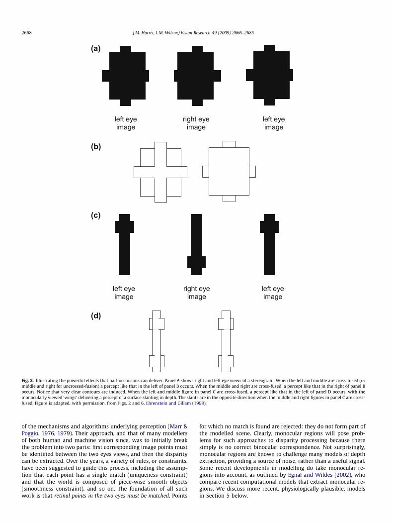
Fig. 2. Illustrating the powerful effects that half-occlusions can deliver. Panel A shows right and left eye views of a stereogram. When the left and middle are cross-fused (ormiddle and right for uncrossed-fusion) a percept like that in the left of panel B occurs. When the middle and right are cross-fused, a percept like that in the right of panel Boccurs. Notice that very clear contours are induced. When the left and middle figure in panel C are cross-fused, a percept like that in the left of panel D occurs, with themonocularly viewed ‘wings’ delivering a percept of a surface slanting in depth. The slants are in the opposite direction when the middle and right figures in panel C are cross-fused. Figure is adapted, with permission, from Figs. 2 and 6, Ehrenstein and Gillam (1998).
2668 J.M. Harris, L.M. Wilcox / Vision Research 49 (2009) 2666–2685
of the mechanisms and algorithms underlying perception (Marr &Poggio, 1976, 1979). Their approach, and that of many modellersof both human and machine vision since, was to initially breakthe problem into two parts: first corresponding image points mustbe identified between the two eyes views, and then the disparitycan be extracted. Over the years, a variety of rules, or constraints,have been suggested to guide this process, including the assump-tion that each point has a single match (uniqueness constraint)and that the world is composed of piece-wise smooth objects(smoothness constraint), and so on. The foundation of all suchwork is that retinal points in the two eyes must be matched. Points
for which no match is found are rejected: they do not form part ofthe modelled scene. Clearly, monocular regions will pose prob-lems for such approaches to disparity processing because theresimply is no correct binocular correspondence. Not surprisingly,monocular regions are known to challenge many models of depthextraction, providing a source of noise, rather than a useful signal.Some recent developments in modelling do take monocular re-gions into account, as outlined by Egnal and Wildes (2002), whocompare recent computational models that extract monocular re-gions. We discuss more recent, physiologically plausible, modelsin Section 5 below.

(a) (b)
(c)
neithereye
Leye
Reye
botheyes
Reye
Leye
left eyeimage
right eyeimage
left eyeimage
Fig. 3. The geometry of occlusion. (a) A foreground rectangle (shown as light grey for illustrative purposes only) sits in front of a continuous background (shown by dottedlines because it is not patterned or coloured). Lines of sight from each eye are drawn to illustrate that, in each case, parts of the background are visible to one eye, but not tothe other. A single bar is visible to the right eye only. (b) A background rectangle is viewed through a hole in an invisible foreground object. A single bar is visible to the left eyeonly. (c) The stimuli used by Nakayama and Shimojo (1990). Cross-fusing the right and middle figures (or uncross fusing the left and middle figures) results in their ‘valid’case: the monocular bar appears behind the rectangle, as in (a). Cross-fusing the left and middle figures give their ‘invalid’ case, the monocular bar may appear in front of therectangle, as in (b). Figure is adapted, with permission, from Fig. 1, Nakayama and Shimojo (1990).
J.M. Harris, L.M. Wilcox / Vision Research 49 (2009) 2666–2685 2669
The exclusion of monocular regions from models of binoculardisparity processing might be understandable if such phenomenawere trivially rare. In fact, monocular regions are abundant inscenes containing a cluttered foreground. Fig. 1 shows a fore-ground ‘picket-fence’ occluding a background scene, and illustrateshow parts of the background scene are only visible to one, or theother eye. In this particular example, the background scene is onlyvisible to one eye, or the other, never both. Whilst such an extremeexample will occur rarely, monocular regions exist in real scenes atall vertical object boundaries where there is a significant depth dis-continuity. In fact, such locations are arguably the regions of mostinterest in a scene, for they indicate where one object ends and thenext begins (Anderson & Nakayama, 1994; Gillam & Borsting,1988). Some recent modelling work has made this proposal expli-cit. For instance, Langer (2008) developed an artificial world inwhich square or spherical objects were randomly distributedthrough a volume (emulating natural cluttered scenes, like foliageor tree branches). He noted that occlusion in one eye’s view of a gi-ven point (resulting in a monocular region visible to the other eye)becomes increasingly more likely as its distance from the observerincreases. That is, in a world densely populated with objects, thefurther away a point is from the observer, the more likely it is thatthere will be an occluding object along any particular line of sight.Another very recent paper has taken this idea a step further.Changizi and Shimojo (2008) suggest that the main reason why
forward-facing eyes have evolved is not for stereopsis, but ratherto take advantage of the increased proportion of background ob-jects that can be viewed in cluttered scenes using two eyes, ratherthan via a single view. As we describe below, this hypothesis can-not account for the apparent use of monocular regions in depthperception, so it surely cannot represent the whole story. However,the theoretical position adopted does illustrate the potentialimportance of monocularly visible regions for vision.
1.3. Geometry and classification of monocular regions
In the real world, objects are located at different depths. Whenviewed from certain locations, foreground objects result in partialocclusion of objects that are further away. The resulting monocularregions are different for each eye, because the eyes are laterallyseparated. There are several different ways in which this can occur.Here, we attempt to classify monocular regions into three types,that depend on the arrangement and features of the objects beingviewed, as well as the relative location of the observer and hereyes. All the studies to be described in this review use stimuli thatconform to one of these three types. It should be noted, however,that these categories are not mutually exclusive, some phenomenafit into more than one category. Our aim here was to provide astructure for describing the various phenomena, not a theoreticalframework.

2670 J.M. Harris, L.M. Wilcox / Vision Research 49 (2009) 2666–2685
1.3.1. Type 1: monocular backgroundIf foreground occluders are of a specific size and at a specific
distance from a background scene, there will be regions of theimages where the eyes are delivered completely different patternsand there is no binocular correspondence. Fig. 1 is an example ofthis kind of situation, where the foreground occluders are justthe right width, ensuring that no part of the background is simul-taneously visible to both eyes (see also Figs. 4 and 8). This situationis rare in natural viewing, and although parts of these scenes canappear rivalrous, overall the scenes are perceived as stable andwith a depth difference between foreground and background(Forte, Peirce, & Lennie, 2002; Howard, 1995; Tsai & Victor,2000). Phenomena linked to this configuration will be discussedin Section 3.1.
1.3.2. Type 2: binocularly visible foregroundFig. 3a shows a top–down view of a foreground rectangle and
featureless background (featureless regions are indicated bydashed lines in our figures). Any object positioned in the ‘righteye only’ region will be visible only to the right eye. Fig. 3c showsstimuli that deliver the percept illustrated in Fig. 3a (or Fig. 3b), asused by Nakayama and Shimojo (1990), in their classic study on ‘daVinci stereopsis’. They found that, for this simple configuration, thebar is perceived as lying behind a foreground rectangle. In manyreal-world situations the foreground and background will bothbe visible, and differently textured or coloured. Under such condi-tions monocular regions are seen at the depth of the background(Julesz, 1971; Shimojo & Nakayama, 1990).
1.3.3. Type 3: invisible foregroundFig. 3b shows a situation in which an observer views a back-
ground (featureless except for the binocularly visible rectangle)through a hole in a featureless foreground object. Such a scene isconsistent with switching the two eye’s views in Fig. 3c, so thatthe left eye now views the stimulus containing the monocularbar. This has been called the camouflage configuration (Howard &Rogers, 2002), and can only occur when an object has the same tex-ture and luminance as the background (i.e. is camouflaged) in one
(a)
(b)
left eye image
rightima
Fig. 4. (a) A top–down view showing an example of monocular camouflage. The small forvisible in the right eyes view (b, centre panel). If the middle and right panel of (b) are crosof the background.
eye, but not in the other (first described by Kaye, 1978). Fig. 4ashows another example, where a small grey foreground line iscamouflaged to the left eye (it occludes a grey section of the back-ground and is therefore invisible to that eye) but not to the righteye (from the right eye’s view the background region visible isstriped). Stereo-pairs that simulate this configuration are shownin Fig. 4b. Although cases like this will be rare in the world, becausethey require the coincidence of identically patterned foregroundand background, the literature provides several recent examplesthat do appear to support depth perception from such images(see Section 4.1, Fig. 11; Section 4.3, Fig. 13).
A key issue to notice when considering these three types ofocclusion is that Type 2 is much more common in the world thaneither Types 1 or 3, but that all are consistent with real 3-D scenes,as shown in the figures (this point runs contrary to the originalideas put forward in Nakayama & Shimojo, 1990, and will be takenup in more detail in Section 2.3 below). The extent to which thesethree types of monocular stimulation may be processed via differ-ent mechanisms, and whether depth perception mechanisms needto ‘know’ the geometrical constraints that underlie occlusion, isdiscussed in the sections below.
2. Monocular regions and depth perception
A number of different lines of research have explored depthfrom monocular regions, most of which use distinct stimuli. Thechallenge is to integrate the diverse effects that have been discov-ered to form a coherent understanding. At the heart of our reviewis the question of whether the visual processing of monocular re-gions is distinctly different from classical stereopsis. Below we willexamine some of the studies that have used stimuli containingmonocular regions, where the monocular regions appear to be in-volved in depth perception. Some of these are directly aimed atunderstanding how depth is perceived from monocular regions,while others are related to this field by nature of the stimuli, butnot by intent. We will consider these in separate sections review-ing what is widely accepted, what is controversial and what re-mains a puzzle.
eye ge
left eye image
eground target is camouflaged in the left eye’s view (in b below), and therefore onlys-fused (or left and middle uncross-fused), the small monocular bar appears in front

J.M. Harris, L.M. Wilcox / Vision Research 49 (2009) 2666–2685 2671
2.1. Speed of depth perception
A monocular region is present whenever there is a significantdepth discontinuity. Perhaps the simplest way that a monocularzone could provide information about depth is to signal the loca-tion of a depth discontinuity. Some work has demonstrated thatmonocular regions speed up depth processing. Saye and Frisby(1975) found that, for large disparities, monocular features didspeed up depth detection in some configurations. Gillam andBorsting (1988) found that depth discontinuities were morerapidly detected when the appropriate monocular zone was filledwith the same pattern as the immediate surrounding background,than if it was left blank. Grove and Ono (1999) explored whetherlonger latencies occurred because a monocular region was missing,or because it was different from the background pattern (a possi-ble, but unlikely, real configuration). They found that responselatencies were longer when the monocular region was differentlypatterned from the background, but not when it was missing,apparently contradicting the Gillam and Borsting study. Andersonand Nakayama (1994) provided evidence, and conceptual models,that went one step further: suggesting that monocular regionsnot only signal the location of depth discontinuities, but also helpconstrain the stereoscopic matching process.
A recent study used photographs of real objects in which half-occluded regions of boxes could be present, or absent (Wilcox &Lakra, 2007). Observers were asked to decide whether they wereviewing scenes with correct depth configurations (where disparityinformation was congruent with depth from perspective, texture,etc.) or whether the disparity information had been reversed. Forrichly textured scenes, reaction times were faster when monocularregions were present than when removed, but only for sceneswhere the disparity was congruent with other depth cues. Thissuggests that occlusion geometry must be consistent with othercues to depth for the rapid perception of depth ordering. One pointto note about the stimuli used for this study is that the monocularregions were ‘self-occluded’ regions that were part of the object,rather than part of the flat background wall. Self-occlusions havebeen very rarely studied in detail but see work on contour stereop-sis, e.g. Nefs, 2008). It is not known if monocular regions due toself-occlusion are processed differently from other forms of mon-ocular region.
Overall, it appears that even though latency effects are subjectto large individual differences and may be specific to particularstimulus characteristics, they suggest a facilitatory role for monoc-ular regions in identifying depth discontinuities.
2.2. Minimum requirements for depth from monocular regions
The simplest possible variant of a stimulus that delivers depthperception from stimulation of one eye is that of monoptic depth,where one eye views a point, or line, and the other a blank screen.Whether this phenomenon can be directly linked to depth frommonocular regions is not yet fully understood.
In the first systematic study of depth from monocular elements,Kaye (1978) showed that the perceived depth of a monocular ele-ment depends on its distance from the fovea. This issue has beenstudied in more detail recently, and experiments have shown thatthe phenomenon does not rest on the notion of a simple ‘local sign’.This concept, outlined by Hering (and discussed in detail in How-ard, 2002) asserts that each location on the retina, in each eye, en-codes a particular direction and relative distance. Wilcox, Harris,and McKee (2007) ruled out the local sign account by showing thatthat no depth is perceived if the non-stimulated eye is patched,rather than viewing a blank screen. They also showed that thedepth percept is lost at small disparities, and with eccentric fixa-tion. Taken together their work suggests that the phenomenon is
most likely due to a crude binocular mechanism that matches apoint in one eye with the line of sight, or fovea, in the other eye.
Traditional stereoscopic mechanisms cannot account for mon-optic depth phenomena. However, there is growing evidence forstereoscopic mechanisms that do not conform to our traditionalunderstanding of binocular disparity processing. This topic is re-viewed in more detail in a companion article (Wilcox & Allison,2009). Whilst the conventional stereoscopic mechanism processesfine disparities present in luminance-defined stimuli such as barsand edges, there is at least one other type of disparity mechanismthat is able to abstract over fine detail and provide a depth signalfor the whole of an object regardless of the similarity of theinter-ocular detail. This is commonly known as 2nd-order stereop-sis (but is also referred to as coarse, or envelope, stereopsis).Stereoacuity using the 2nd-order mechanism is much poorer thanfor 1st-order stereopsis, but delivers depth perception for diplopictargets (Wilcox & Hess, 1995) and for patches of uncorrelated noise(Wilcox & Hess, 1996).
Recent experiments by Fukuda, Wilcox, Allison, and Howard(2009) have provided evidence for a linkage between monopticdepth and 2nd-order stereopsis. It has long been known that thereis a large tolerance to vertical misalignment in stimuli containingbinocular disparity (Mitchell, 1969). Fukuda et al. (2009) showedthat the same patterns of perceived depth are obtained for a singlemonoptic target as for targets with large vertical offsets. This workopens up a new possibility, perhaps a very simple mechanismbased on the responses of binocular mechanisms to monocularregions, or even monoptic elements, could account for some typesof depth processing from monocular regions.
2.3. Does occlusion geometry constrain depth perception formonocular regions?
Nakayama and Shimojo (1990) suggested that the brain’sknowledge, or experience, of 3-D occlusion geometry constrainsour perceptions of depth from monocular regions. When a monoc-ular region is adjacent to an unambiguous background and fore-ground (unambiguous due to the presence of shading, texture orcolour differences: a Type 2 region), its depth interpretation isstraightforward: the monocular region is assigned the same depthas the unambiguous background (Anderson & Nakayama, 1994;Collett, 1985; Julesz, 1971; Shimojo & Nakayama, 1990). Further,monocular probe dots are located more reliably at a specific depthwhen they are located in a monocular region, than when they areplaced in a binocularly visible part of a stimulus (Shimojo &Nakayama, 1994). When monocular regions are textured, so thatthey are clearly not part of the background, less perceived depthresults than if they have the same texture as the background(Grove, Gillam, & Ono, 2002).
A more ambiguous example, is one in which the scene is verysparse (Fig. 3a), and the binocular surface is not specified by a pat-tern (also Type 2). In principle, the geometry (as shown in Fig. 3a)dictates that monocular regions should be perceived as lying some-where behind a foreground occluder but precisely where it shouldbe in depth is not specified in the stimulus. Nakayama and Shimojo(1990) defined the depth constraint zone, as shown in Fig. 5(striped region). Any real points or objects lying within that zonewill only be seen by one eye. An example monocular point in theright eye (indicated by the solid line in Fig. 5) could correspondto a real point with a depth anywhere along that eye’s line of sight,within the depth constraint zone. The zone extends back to aneffectively infinite depth for a large foreground object, but coulditself be constrained if the object were smaller, or if a texturedbackground were present.
These geometric constraints raise the issue of whether thedepth from monocular points is qualitative or quantitative in

Fig. 5. An illustration of the depth constraint zone, shown by the striped region.This figure shows that a monocular point or line (the solid line in the right eye) isconsistent with an object at any depth within the zone, along the point’s line ofsight. For example, the two white circles show two possible locations in depth.Figure adapted, with permission, from Fig. 3, Nakayama and Shimojo (1990).
2672 J.M. Harris, L.M. Wilcox / Vision Research 49 (2009) 2666–2685
nature. That is, whether the perceived depth is somewhere withinthe constraint zone, but the location cannot be precisely identified,or at a specific location in depth, where the depth is matchable todepth from binocular disparity. This issue was dealt with in somedetail by Nakayama and Shimojo (1990). They showed that, for aType 2 monocular region, the perceived depth of a monocular pointcan be matched using a stereoscopic probe and that the depthmatches are quantitative in nature: the perceived depth of themonocular point increased with increasing separation from theoccluding edge. For points located close to the occluder (up toaround 30 min arc in their hands, or around 10–15 min in a relatedstudy (Hakkinen & Nyman, 1996) the matched depth followed theforward edge of the depth constraint zone, suggesting that thedepth assigned was the smallest possible that would be consistentwith occlusion geometry. For larger separations the matched depthgradually fell to zero disparity. When the two eye’s views were in-ter-changed (resulting in a Type 3 monocular region, Fig. 3b),Nakayama and Shimojo found that the matched depth was zerodisparity, whatever the separation between binocular rectangleand monocular bar. This suggests that there are fundamental dif-ferences between the ways in which Type 2 and Type 3 monocularregions are processed in visual perception. Nakayama and Shimojodiscussed this difference in terms of viewing geometry, with con-figurations such as those in Fig. 3b dubbed ‘ecologically invalid’,suggesting that they did not correspond to a monocular region thatwould be present in a real scene. As described below, this assertionand the generality of the result itself, was subsequently challenged.
First, as described in the geometry section above, Fig. 3b showsa real scene that could correspond to the so-called ‘ecologically in-valid’ case. Although such real scenes may be rare, they are clearlypossible. Potential real configurations that correspond to the casesdubbed ‘invalid’ have been discussed at some length by Assee andQian (2007). They suggest that the differences in depth perception
found in the Nakayama and Shimojo (1990) studies may have moreto do with whether the monocular region in the stimulus appearsrivalrous (as can occur for these Type 3 configurations and some-times for Type 1) than with the ecological validity of the stimulus.
Second, Hakkinen and Nyman (1996) found no differences indepth perception between configurations very similar toNakayama & Shimojo’s ‘valid’ and ‘invalid’ cases. The main differ-ence between the experiments was that stimuli in the Hakkinen& Nyman study also contained an additional binocularly visibleplane. These authors showed that the relative depths between thatplane and the occluding plane (the one near which the monocularelement was placed) affected the perceived location in depth of themonocular element. As suggested by Assee and Qian (2007), it maybe that, when the stimulus is particularly ambiguous due to thesparseness of a scene (as in Fig. 3a and b), different observers placedifferent interpretations on why a monocular bar is monocular. Ifthe configuration in 3b was perceived as if behind a featureless oc-cluder, then quantitative depth perception may result. If not, thenno depth would be perceived. Adding additional binocularly visibleobjects to the scene, as Hakkinen and Nyman (1996) did, could re-sult in rather different binocular interpretations. Further evidencethat different observers may use different interpretations comesfrom a recent study in which the perceived depth location (nearor far) of a monocular bar could be manipulated by adjusting thepictorial cues of bar size and contrast (Makino & Yano, 2006).Again, scenes were very sparse and observers were idiosyncraticin their responses.
2.4. Can monocular regions be processed by standard disparitymechanisms?
Our understanding of Nakayama and Shimojo’s (1990) elegantresult has recently been called into question in other ways. The is-sue is whether what they dubbed ‘da Vinci stereopsis’ (using asparse Type 2 geometric arrangement) requires a specific novelbrain mechanism, or whether known binocular processes (likethose that may be involved in depth from Panum’s limiting case,or simply a coarse stereoscopic mechanism) can account for thedepth perceived. A range of evidence supports the possibility thatthe depth perceived using their stimulus provides an example ofPanum’s limiting case, and thus could be detected using standarddisparity-processing mechanisms.
Panum’s case arises when one eye views a single vertical lineand the other a pair of horizontally offset vertical lines. If the lefteye views the single line, then the left line appears closer thanthe right. This configuration is consistent with a pair of real lines,at different distances, that fall along the line of sight of the lefteye (thus appearing as a single line in that eye, but two lines inthe right eye). Ono, Shimono, and Shibuta (1992) pointed out thatin a purely geometrical sense, this is an extreme example of occlu-sion in the da Vinci configuration, so the same mechanisms couldgovern the two phenomena.
Gillam, Blackburn, and Cook (1995) showed that for small dis-parities, depth settings in Panum’s case are made as precisely asfor normal stereopsis, and that disparity curvature effects can alsobe revealed when the monocular line is matched to a curved bin-ocular line. One of the proposed explanations for Panum’s limitingcase is that the lone target in one eye is matched to the alreadymatched line in the other eye, a case of ‘double-duty matching’(e.g. McKee, Bravo, Smallman, & Legge, 1995). Gillam, Cook, andBlackburn (2003) noted that the da Vinci configuration used byNakayama and Shimojo (1990) was reminiscent of a Panum’s lim-iting case stimulus, because the monocular bar was similar inwidth and length to the edge of the rectangular occluder. Gillamet al. (2003) went on to demonstrate that when the stimuli arecarefully controlled so that Panum’s-like mechanisms cannot work,

(a)
(b)
left eye image
right eye image
left eye image
Fig. 6. (A) Illustrates the geometrical situation consistent with monocular gap stereopsis (Gillam et al., 1999). The right eye can see a bright background through a gapbetween two dark foreground objects. For the left eye, there is no gap as it is occluded by the foremost object. (B) Stereograms that illustrate depth in this situation. If themiddle and right figure are cross-fused the left side should appear closer than the right. If left and middle are cross-fused, the right side will appear closer. Figure adapted,with permission, from Fig. 1a, Gillam et al. (1999).
J.M. Harris, L.M. Wilcox / Vision Research 49 (2009) 2666–2685 2673
depth from monocular regions is qualitative: a depth direction (infront or behind) is perceived, but not an accurate or precise depthlocation. To rule out any depth perception due to double-dutymatching in this stimulus, they replaced the vertical line with amonocular disc. The occluded disc was always perceived as lyingbehind the occluder, but there was no change in perceived depthas the disc was separated from the binocular bar. The da Vincistereopsis configuration used by Nakayama and Shimojo (1990)therefore appears to be an example of Panum’s limiting case, andstandard stereoscopic mechanisms could be involved in itsprocessing. This is backed up by recent computational modelling(Assee & Qian, 2007), which demonstrated that both Panum’s case,and the da Vinci configuration can be modelled using a variant ofthe disparity energy model (Chen & Qian, 2004).
So far, then it appears that standard stereoscopic mechanismsmay be responsible for the perception of depth in the da Vinciconfiguration. As will become evident below, however, otherstimuli evade such straightforward explanation.
2.5. Do monocular regions provide evidence for a separate,sophisticated depth mechanism?
When one eye views a black bar and the other a black bar with acentral gap (Fig. 6b), the fused percept is of a pair of rectangles dis-placed in depth (another example of a Type 2 occlusion). Fig. 6(top) shows a possible occlusion situation, in which a pair of ob-jects are located side by side at different depths. One eye seesthrough the gap between them to the featureless background. Inthe other eye this gap is occluded by the near object. This effecthas been dubbed monocular gap stereopsis (Gillam, Blackburn, &Nakayama, 1999).1 This is a potentially important stimulus
1 There is also a dynamic version of this effect (Brooks & Gillam, 2006a).
configuration because the depth settings are precise, because theyare consistent with a real 3-D configuration in which the edges ofthe rectangle abut in one eye’s view, and because the perceiveddepth cannot easily be accounted for by mechanisms responsive totraditional binocular disparity. Depth in these stimuli appears tobe mediated by mechanisms specifically sensitive to the width ofthe monocular gap. Gillam et al. (1999) showed that the amount ofdepth perceived increases with the size of the gap, and can bematched to a stimulus containing depth from binocular disparity.This effect is consistent with the visual system interpreting the cen-tral monocular gap as a gap between the objects that is occluded inthe one eye’s view due to a depth difference between the objects.
Depth thresholds for monocular gap stereopsis have been foundto be very similar to those for standard stereopsis, and, impor-tantly, adaptation to stereopsis results in shifts in perceived depthfrom monocular gaps, and vice versa (Pianta & Gillam, 2003a).Adaptation techniques are frequently used to explore whether dif-ferent stimuli are processed by common sensory mechanisms. Thelogic used is that if a stimulus adapts a particular mechanism, forexample a depth mechanism, then perceived depth should be af-fected in other stimuli that are processed by the same mechanism.Pianta and Gillam’s powerful result, the first using adaptation toexplore the perception of depth from monocular regions, suggeststhat the two forms of depth information may be processed by acommon mechanism. However, an alternative interpretation can-not be ruled out: the depth could be processed by different mech-anisms that converge on a later, common mechanism, which canbe adapted. It has also been found that the perceived depth is clos-est to that provided by a disparity depth probe when the gap con-tains visual information consistent with it actually being a gapthrough to the background: if the ‘gap’ is a different colour or tex-ture than the background region which surrounds it, less depth isperceived (Grove et al., 2002). Further, the stimulus configuration

2674 J.M. Harris, L.M. Wilcox / Vision Research 49 (2009) 2666–2685
must be such that the gap and background can form a continuoussurface (i.e. both are textured, or both featureless), or else per-ceived depth is attenuated (Grove, Ben Sachtler, & Gillam, 2006).
If monocular gap stimuli and binocular disparity are processedby a common mechanism, do we need to rethink how disparity it-self is processed, or could traditional disparity-processing mecha-nisms account for both the depth threshold and adaptationresults? Pianta and Gillam (2003b) suggested (and then tested)two forms of depth processing that might occur. In the originalmonocular gap stimuli, the gap in one eye’s view is obtained by‘pulling apart’ the rectangle that forms the other eye’s view: theresulting rectangle-with-gap is wider than the single rectangle inthe other eye, wider by an amount equal to the gap width (seeFig. 6b). One way that depth could be perceived is if the visual sys-tem were to detect the disparity difference between the outeredges of the black rectangles (dubbed outer-edge disparity byPianta & Gillam) and then to use the monocular region to simplylabel the location of the depth edge. Depth attributed to the rectan-gles would somehow need to propagate from the outer edges, aform of depth interpolation.
Monocular-gap stimuli can also be built without outer-edge dis-parity. This can be achieved by showing each eye a rectangle of thesame width then superimposing a ‘gap’ onto the centre of one ofthem. Now, the outer-edge disparity would be zero. Pianta and Gil-lam argued that depth could only be perceived at the gap if the vi-sual system were able to infer an implicit depth signal, inferringthat the lack of a gap in one eye can only be due to a particular geo-metric arrangement, involving occlusion.
By measuring depth thresholds for no-gap stimuli (without agap, the observer sees a narrower rectangle in one eye than theother, resulting in the perception of slant around a vertical axis),for same-width gap stimuli, and gap-with-outer-edge disparitystimuli, Pianta and Gillam (2003b) demonstrated that the depthpercept at the gap is robust when outer-edge disparity is present,and of the expected sign and magnitude. Importantly, depth is alsoperceived at the gap for same-width stimuli, when the outer-edgedisparity is zero, and it still varies monotonically with gap size, butthe depth magnitude is smaller than that found when outer-edgedisparity is present. These results imply that some mechanismother than outer-edge disparity is at work when interpreting thedepth in monocular gap stereopsis.
Another hint that there may be a separate mechanism underly-ing depth from these gap stimuli comes from work on how depthfrom monocular regions is scaled by changes in accommodationand/or viewing distance. Kuroki and Nakamizo (2006) showed thatperceived depth does not scale with distance for monocular gapstereopsis, as it does for other examples of monocular occlusiondepth, and as it does for standard binocular disparity. Recently,models have been developed that use the output of disparity-detectors in ways that could make use of monocular gaps (seemodelling section below, in particular Cao & Grossberg, 2005;Grossberg & Howe, 2003). These models rely on the use of outer-edge disparities and cannot explain the depth perceived whenthe outer-edge disparity is zero.
A further twist to this story is added by use of a stimulus con-taining even fewer clues to the presence of depth, the ‘stereoscopicsliver’ stimulus (Sachtler & Gillam, 2007). Here there is a monocu-lar gap in one eye’s view, but no outer-edge disparity. The gap doesnot cover the full vertical extent of the stimulus but tapers in widthfrom the centre until it disappears near the top and bottom. Thisstimulus is consistent with a torn piece of fabric, with a featurelessbackground visible through the tear in one eye’s view, but the tearoccluded in the other eye. Depth differences between the edges ofthe tear can be reliably discriminated.
One interesting point to note is that observers in these experi-ments did not see the gap itself in depth (Gillam et al., 1999).
We have noticed that some observers perceive the monoculargap to be in depth with respect to the black surfaces, particularlywith careful fixation on the gap itself. This percept is reminiscentof that found by Kumar (1995) who showed both eyes a light rect-angle and one eye a superimposed dark bar at the centre, the othera lighter bar. For some configurations the two halves of the rectan-gle appeared at different depths, for others the central bar ap-peared in depth. The percept may also be similar to that for theperception of monoptic depth (see Section 2.2), where a singlemonocular element can appear in depth. It is also not clearwhether the effect of instructions, of eye movements, or someother stimulus property could account for this alternative interpre-tation, and we currently do not know whether only a small propor-tion of the population achieve this alternative percept. Furtherstudies with larger numbers of naïve observers, could help us tounderstand just how robust these percepts are.
3. Integration of binocular and half-occluded regions in 3-Dscenes
‘Leonardo’s constraint’ (Ono, Wade, & Lillakas, 2002) is the con-straint that two opaque objects cannot be seen in the same visualdirection. An example of this is the situation where a backgroundsurface is visible to one eye, or the other, but not both, when anoccluding object is smaller than the inter-ocular separation.Fig. 1 shows an example of such a scene where many foregroundobjects occlude the background surface such that any one part ofthe background is only seen by a single eye. We defined this aboveas a Type 1 occlusion. In principal, the visual information is avail-able to ‘see behind’ the foreground object, but it is only availablemonocularly. Can the visual system do this?
This question can be addressed in at least three ways. First, wecould ask whether our visual impressions are stable under condi-tions where large regions of the scene are monocular. We explorethis in Section 3.1 below. Second, it is well known that when theeyes are shown different images, binocular rivalry results. Rivalryconsists of the dominance of one eye’s view, which is periodicallyreplaced with the other eye’s view (e.g. see Blake & Logothetis,2002). There is a considerable literature on the spatial and tempo-ral properties of binocular rivalry, though it rarely occurs undernatural viewing conditions. We discuss literature in Section 3.2which demonstrates that depth can be perceived in occlusion situ-ations, despite rivalry. As we will see, some researchers believe thatrivalry itself has an interpretation in terms of occlusion geometry.Third, when some parts of a scene are viewed by a single eye, andsome by both eyes, it is not clear how our phenomenal perceptionof a single fully ‘stitched together’ world, can be obtained. Fig. 7illustrates the problem. Each eye views the world from a differentdirection, because the eyes are laterally separated. Yet we feel thatwe view the world as if from a single point, mid-way between theeyes. If some transformation occurred to deliver a singular repre-sentation from that point, the brain would have to squeeze the re-gion defined by the separation between points a and d (the righteye view in Fig. 7), into a smaller region separating a0 and d0 (theview as if from a single central location). How, or even if, this isdone is still a hotly debated topic and beyond the scope of this re-view, but the interested reader is directed to Erkelens and van Ee(2002) and Ono, Mapp, & Howard (2002) and for reviews see How-ard and Rogers (2002) and Ono, Wade, and Lillakas (in press).
3.1. Scene stability despite large monocularly visible regions
Forte et al. (2002) studied the stability of monocular regionswhen there was no binocularly visible background, but regions ofthe background surface were visible to one eye or the other (e.g.
a' b’ c’ d’a b c d
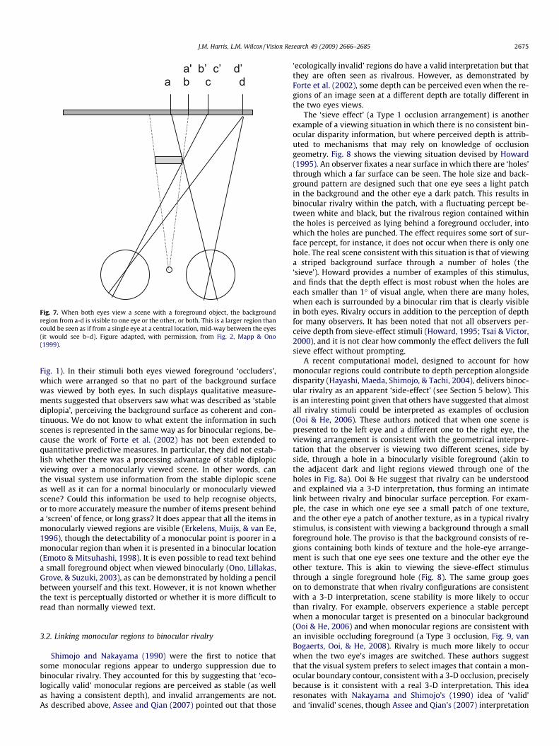
Fig. 7. When both eyes view a scene with a foreground object, the backgroundregion from a-d is visible to one eye or the other, or both. This is a larger region thancould be seen as if from a single eye at a central location, mid-way between the eyes(it would see b–d). Figure adapted, with permission, from Fig. 2, Mapp & Ono(1999).
J.M. Harris, L.M. Wilcox / Vision Research 49 (2009) 2666–2685 2675
Fig. 1). In their stimuli both eyes viewed foreground ‘occluders’,which were arranged so that no part of the background surfacewas viewed by both eyes. In such displays qualitative measure-ments suggested that observers saw what was described as ‘stablediplopia’, perceiving the background surface as coherent and con-tinuous. We do not know to what extent the information in suchscenes is represented in the same way as for binocular regions, be-cause the work of Forte et al. (2002) has not been extended toquantitative predictive measures. In particular, they did not estab-lish whether there was a processing advantage of stable diplopicviewing over a monocularly viewed scene. In other words, canthe visual system use information from the stable diplopic sceneas well as it can for a normal binocularly or monocularly viewedscene? Could this information be used to help recognise objects,or to more accurately measure the number of items present behinda ‘screen’ of fence, or long grass? It does appear that all the items inmonocularly viewed regions are visible (Erkelens, Muijs, & van Ee,1996), though the detectability of a monocular point is poorer in amonocular region than when it is presented in a binocular location(Emoto & Mitsuhashi, 1998). It is even possible to read text behinda small foreground object when viewed binocularly (Ono, Lillakas,Grove, & Suzuki, 2003), as can be demonstrated by holding a pencilbetween yourself and this text. However, it is not known whetherthe text is perceptually distorted or whether it is more difficult toread than normally viewed text.
3.2. Linking monocular regions to binocular rivalry
Shimojo and Nakayama (1990) were the first to notice thatsome monocular regions appear to undergo suppression due tobinocular rivalry. They accounted for this by suggesting that ‘eco-logically valid’ monocular regions are perceived as stable (as wellas having a consistent depth), and invalid arrangements are not.As described above, Assee and Qian (2007) pointed out that those
‘ecologically invalid’ regions do have a valid interpretation but thatthey are often seen as rivalrous. However, as demonstrated byForte et al. (2002), some depth can be perceived even when the re-gions of an image seen at a different depth are totally different inthe two eyes views.
The ‘sieve effect’ (a Type 1 occlusion arrangement) is anotherexample of a viewing situation in which there is no consistent bin-ocular disparity information, but where perceived depth is attrib-uted to mechanisms that may rely on knowledge of occlusiongeometry. Fig. 8 shows the viewing situation devised by Howard(1995). An observer fixates a near surface in which there are ‘holes’through which a far surface can be seen. The hole size and back-ground pattern are designed such that one eye sees a light patchin the background and the other eye a dark patch. This results inbinocular rivalry within the patch, with a fluctuating percept be-tween white and black, but the rivalrous region contained withinthe holes is perceived as lying behind a foreground occluder, intowhich the holes are punched. The effect requires some sort of sur-face percept, for instance, it does not occur when there is only onehole. The real scene consistent with this situation is that of viewinga striped background surface through a number of holes (the‘sieve’). Howard provides a number of examples of this stimulus,and finds that the depth effect is most robust when the holes areeach smaller than 1� of visual angle, when there are many holes,when each is surrounded by a binocular rim that is clearly visiblein both eyes. Rivalry occurs in addition to the perception of depthfor many observers. It has been noted that not all observers per-ceive depth from sieve-effect stimuli (Howard, 1995; Tsai & Victor,2000), and it is not clear how commonly the effect delivers the fullsieve effect without prompting.
A recent computational model, designed to account for howmonocular regions could contribute to depth perception alongsidedisparity (Hayashi, Maeda, Shimojo, & Tachi, 2004), delivers binoc-ular rivalry as an apparent ‘side-effect’ (see Section 5 below). Thisis an interesting point given that others have suggested that almostall rivalry stimuli could be interpreted as examples of occlusion(Ooi & He, 2006). These authors noticed that when one scene ispresented to the left eye and a different one to the right eye, theviewing arrangement is consistent with the geometrical interpre-tation that the observer is viewing two different scenes, side byside, through a hole in a binocularly visible foreground (akin tothe adjacent dark and light regions viewed through one of theholes in Fig. 8a). Ooi & He suggest that rivalry can be understoodand explained via a 3-D interpretation, thus forming an intimatelink between rivalry and binocular surface perception. For exam-ple, the case in which one eye see a small patch of one texture,and the other eye a patch of another texture, as in a typical rivalrystimulus, is consistent with viewing a background through a smallforeground hole. The proviso is that the background consists of re-gions containing both kinds of texture and the hole-eye arrange-ment is such that one eye sees one texture and the other eye theother texture. This is akin to viewing the sieve-effect stimulusthrough a single foreground hole (Fig. 8). The same group goeson to demonstrate that when rivalry configurations are consistentwith a 3-D interpretation, scene stability is more likely to occurthan rivalry. For example, observers experience a stable perceptwhen a monocular target is presented on a binocular background(Ooi & He, 2006) and when monocular regions are consistent withan invisible occluding foreground (a Type 3 occlusion, Fig. 9, vanBogaerts, Ooi, & He, 2008). Rivalry is much more likely to occurwhen the two eye’s images are switched. These authors suggestthat the visual system prefers to select images that contain a mon-ocular boundary contour, consistent with a 3-D occlusion, preciselybecause is it consistent with a real 3-D interpretation. This idearesonates with Nakayama and Shimojo’s (1990) idea of ‘valid’and ‘invalid’ scenes, though Assee and Qian’s (2007) interpretation

(a)
(b)
left eyeimage
right eyeimage
left eyeimage
Fig. 8. (a) Geometry consistent with stimuli used in the sieve effect. The observer views a black and white background through a series of holes in a foreground occluder (thesieve). (b) Stereograms to illustrate the effect. The regions within the circular patch may appear behind the grey rectangle, whichever pair of images are fused. Figure adapted,with permission, from Howard (1995).
2676 J.M. Harris, L.M. Wilcox / Vision Research 49 (2009) 2666–2685
suggests to us that rivalry occurs when the real 3-D scene formedby stimuli containing monocular regions is very unlikely to occur.
The idea that rivalry in general, and the sieve effect in particu-lar, occur because the visual system interprets different inputs tothe left and right eye as due to particular geometrical configura-tions, is compelling. However, work exploring detailed predictionsabout the quantity of depth that should be perceived in the sieveeffect, argues against the idea. Tsai and Victor (2000) discussedseveral predictions of the sieve effect, noting that to be consistentwith occlusion geometry, perceived depth in the sieve effectshould vary with the horizontal size of the viewing apertures. Theyfound that precision of depth perception from sieve-effect stimuliwas poor, around 10 times worse than for standard stereopsis,though the depth within the sieve elements was consistently seenas behind the occluder. This is similar precision to that found forjudging depth from disparity for anti-correlated bars (Cogan,Kontsevich, Lomakin, Halpern, & Blake, 1995), but notice that inthe sieve-effect stimulus there is no disparity applied to theelements, so there must be a different mechanism at work.
In the same study, Tsai and Victor (2000) found other attributesof the sieve effect that were not consistent with an occlusion-basedexplanation. According to such an account, perceived depth shouldvary systematically with element width, but not height. Althoughthe horizontal size of the holes did affect perceived depth a little,
so did the vertical size. In a later study (Tsai & Victor, 2005), therelative locations and luminances of elements were varied to alterthe minimum depth between the occluder and the backgroundsurface that would be consistent with occlusion geometry. Binocu-lar viewing geometry dictates that if two elements are closer thanthe element width, their relative luminance polarity (same or dif-ferent) will determine the perceived separation between occluderand background. The study found no evidence that the visual sys-tem could take account of these geometric constraints. These re-sults cannot be explained by a standard stereoscopic mechanism,or by mechanisms that rely on appropriate occlusion geometry.
Some recent work corroborates this conclusion. Matsumiya,Howard, and Kaneko (2007) measured depth from the sieve effectunder a number of conditions and found it to be maximal whenexclusive rivalry within the elements was also greatest (exclusiverivalry occurs when perception correlates with the view fromone eye, or the other, rather than some intermediate or partial ef-fect, e.g. see Blake & Logothetis, 2002). These authors suggestedthat the same mechanisms might be at work in the processing ofrivalry and depth from the sieve effect, although they did not spec-ulate further. In sum, while there clearly are links between rivalryand depth from monocular regions, the available evidence does notwholeheartedly support the notion that they arise from the sameprocessing mechanism.

(a)
(b)
left eye image
right eye image
left eye image
Fig. 9. (a) A top–down view showing a foreground occluder, with the same pattern as most of the background. A pair of circular regions with a different pattern are each onlypartially visible in one or other eye. (b) Stereo-pairs consistent with the above geometry. If right and middle panels are cross-fused the pattern is stable and a foregroundoccluder perceived. If the other pair is cross-fused there is no such stability and rivalry occurs. After van Bogaerts et al. (2008), with permission.
Fig. 10. Reproduced from Nakayama and Shimojo (1990), with permission from the authors. In the upper stereo-pairs two points are missing from each of the left and righteye views. A clear phantom occluding surface is perceived when the left and middle panels are cross-fused (or middle and right panels divergently fused). In the lower panelsall points are visible in left and right eye views and no occluding contour is seen.
J.M. Harris, L.M. Wilcox / Vision Research 49 (2009) 2666–2685 2677
4. Monocular regions and surfaces
Monocular regions in a scene can generate the percept of an illu-sory surface, and associated illusory contours, consistent with aninvisible foreground occluder. We have defined monocular regionsattributable to this type of occlusion as Type 3, and noted that theywill occur very rarely in natural scenes. That an invisible occludercould be perceived from monocular regions, was first shown usingsparse dot patterns via depth magnitude estimation (Lawson &Gulick, 1967; Lawson & Mount, 1967). An extreme example of the
phenomenon was demonstrated by Nakayama and Shimojo(1990), who devised a stimulus where only four points in a sparserandom dot stereogram are viewed monocularly, yet a clear illusorysurface can be seen in depth (Fig. 10). Vertically oriented monocularregions have also been shown to generate a clear percept of an illu-sory surface (Anderson, 1994, Fig. 11d), and monocular regions pre-sented at the edge of slanting binocular surfaces in a random dotstereogram can increase perceived slant (Gillam & Blackburn,1998). In the sub-sections below we discuss depth perception fromseveral surface-related instances of monocular regions like these.

(a)
(b)
(c)
(d)
left eyeimage
right eyeimage
left eyeimage
Fig. 11. Stereograms illustrating several different stimuli that achieve phantom stereopsis. (a) The central white patch appears in front of the black background when middleand right panels are cross-fused (left and middle for uncrossed-fusion). Figure adapted, with permission, from Fig. 1, Liu et al. (1994). (b) Gillam used these stereo-pairs todemonstrate that stereoscopically matchable features do exist in (a). Figure adapted, with permission, from Fig. 1c, Gillam (1995). (c) Stimulus used to illustrate that phantomstereopsis can occur when matchable features have bene removed. When the middle and right panels are cross-fused a white phantom rectangle is seem in depth. Figureadapted, with permission, from Fig. 2, Gillam and Nakayama (1999). (d) Vertical offsets of bars can result in phantom stereopsis. When the middle and right panel are cross-fused the pattern appears as if behind a phantom window that occludes the pattern at the edges. Figure adapted, with permission, from Fig. 1, Anderson (1994).
2678 J.M. Harris, L.M. Wilcox / Vision Research 49 (2009) 2666–2685
4.1. Phantom stereopsis
Fig. 11a shows the stimulus used by Liu, Stevenson, and Schor(1994) to demonstrate ‘phantom stereopsis’. In this stimulus,depth can be seen in the central white patch despite an apparentlack of traditional stereoscopic matchable features. Liu et al.showed that quantitative depth was obtained from this stimulus,and that it can drive vergence similar to stimuli containing binoc-ular disparity (Liu, Stevenson, & Schor, 1998). This study provides aclassic example of how clever stimuli are invented to deliver depthperception without the involvement of traditional stereopsis, butwhere, later, it is shown that this assumption is not correct. In thiscase, Gillam (1995) argued that if one considers only the horizontalcontours (Fig. 11b), the perception of depth remains, and it is evi-dent that the end points of the lines can be used for standard ste-reoscopic matching. Liu, Stevenson, and Schor (1997) tested asimple model of disparity processing, finding that, although dispar-ity mechanisms would respond differently to phantom-stereopsisstimuli than to stimuli containing standard binocular disparity,there was a disparity signal that could be used to obtain depth signinformation that is consistent with the psychophysical results.These depth signals are required for surface interpolation: the ob-server sees a plane in depth, rather than individual points. Theauthors noted that mechanisms that use some knowledge of occlu-sion geometry to guide disparity selection could achieve the depth
sign consistent with perception, but also that much simpler mech-anisms, perhaps using disparity averaging or other simple heuris-tics, might also work. Clearly, additional careful experimentationis needed to test these ideas more thoroughly.
A start has been made in this direction. Gillam and Nakayama(1999) designed an elegant stimulus composed only of verticallines with gaps in them (Fig. 11c). A central rectangle is perceived,standing out in depth, with strong illusory horizontal contours,dubbed the ‘phantom occluder’ (containing Type 3 monocular re-gions). Observers were able to match the perceived depth of therectangle with a stereoscopic probe target, with perceived depthvarying monotonically with line thickness. This is consistent withthe depth being attributed to an invisible ‘phantom’ rectangle, thatjust covers the lines in one eye’s view. Such geometry constrainsthe minimum depth that could be perceived, but since the invisibleoccluder could stretch beyond the lines by an indeterminateamount, the maximum perceivable depth is not defined. An inter-esting feature of phantom stereopsis is that the apparent depth be-tween the phantom and the background (featureless except for thevertical lines) is slightly greater than the minimum depth con-straint would predict (Gillam & Nakayama, 1999; Grove et al.,2002). In a related study, using visual search in noise defined byeither disparity or half-occlusion elements, Mitsudo, Nakamizo,and Ono (2005) were able to show that depth from phantom stere-opsis appears to be processed at an early stage of visual perception.

J.M. Harris, L.M. Wilcox / Vision Research 49 (2009) 2666–2685 2679
In an earlier study Anderson (1994) showed that vertical offsetsbetween the left and right eye views, consistent with an invisibleoccluder (see Fig. 11d), give rise to perception of a phantomoccluding surface. A more general theory was later developed toaccount for the depth perceived from occlusion junctions (theplaces where objects overlap). Due to occlusion of one object byanother, the occlusion junctions can have both a horizontal andvertical separation between the two eyes’ views. The separationbetween junctions was defined by Malik, Anderson, and Charow-has (1999), as ‘pseudodisparity’ and they demonstrated that forsuch scenes there is a clear quantitative relationship between per-ceived depth and image pseudodisparity. Remarkably, the orienta-tion of the illusory contours can also be precisely judged: a neatdemonstration of the clarity and crispness of such contours. Whileit seems clear that pseuododisparity can be exploited in some in-stances to support depth perception, this is not always the case.More recent studies by van Ee, Anderson, and Farid (2001) haveshown that depth detection near disparity threshold is not im-proved by the presence of pseudodisparities.
Depth perception has been demonstrated in dynamic versionsof the phantom stimulus described above. This was first done byShimojo, Silverman, and Nakayama (1988). They showed thatsequentially stimulating each eye with a moving line that ‘disap-pears’ behind an occluder (so that the depth cue is the differingtime and location of occlusion and reappearance) results in a clearpercept of depth. This depth percept increases as the temporal gapbecomes larger, consistent with the presence of an occluded objectfurther away. Brooks and Gillam (2006b) used a similar stimulusand ruled out the possibility of depth being perceived via inter-ocular delay. They showed that a depth effect was still presentwhen inter-ocular delays far exceeded the range where conven-tional matched stereopsis could provide depth signals, and thatthe perceived depth was not dependent upon the duration of thedelay itself. It is difficult to imagine a way to perceive depth in suchspatial or temporal line stimuli that simply relies on conventionalstereoscopic mechanisms.
Hakkinen and Nyman (2001) have argued that phantom stere-opsis must be closely linked to conventional disparity processing.They showed that a monocularly induced phantom surface caninfluence the depth perceived from stereopsis when that informa-tion is ambiguous. This effect only occurs for specific physicalarrangements, notably when the monocular regions appear to bepart of a continuous surface also defined by an adjacent binocularregion. Grove et al. (2002) explored how monocular regions affectperceived depth for a variety of different stimuli. They found thatless depth was perceived when the monocular texture was dissim-ilar to the background texture. Studies agree (Grove et al., 2002;Hakkinen & Nyman, 2001) that these effects occur most stronglywhen the monocular regions are consistent with there being aforeground invisible occluder. This is an important point becauseit emphasises that, whatever mechanisms are at work, both localprocessing (to account for monocular regions requiring locally con-sistent binocular regions) and larger-scale, or long-distance pro-cessing (to account for the global occlusion geometry) must beinvolved. This point has recently been demonstrated by Mitsudo,Nakamizo, and Ono (2006). They measured contrast sensitivityfor detecting stereo-pairs in noise and found greater sensitivityfor a phantom-stereopsis stimulus (like that in Fig. 11c) than foran equivalent stimulus with the two eye’s views switched round.In the latter case it is possible to contrive a ‘real’ scene arrange-ment that could deliver the left and right eye views, but theywould be rarely encountered in the real world. Sensitivity to theforeground occluder configuration was also greater than for a stim-ulus composed of only the left-most bar in each eye. This work sug-gests that, to obtain and use the phantom surface, information iscombined by large-scale processing mechanisms which process
information across the full extent of the stimulus, rather than rely-ing on individual elements.
4.2. Monocular transparency
Howard and Duke (2003) presented a novel effect that theynamed ‘monocular transparency’, in which perceived depth isattributed to geometrical rules related to transparency, rather thanto occlusion geometry. One of their stimuli is depicted in Fig. 12.One eye views a white rectangle, occluded by a slightly offsettransparent square, and the other the same rectangle, with thesquare aligned. In the configuration shown in Fig. 12, the squareis seen to float in front of the rectangle. When the two eyes viewsare switched, it is seen behind. The depth of the square could bematched to a disparity-defined depth probe and delivered quanti-tative depth percepts. The key point to the design of this display isthat occlusion cannot be required to explain the perception ofdepth because no part of the scene is occluded, and standard dis-parity processing cannot account for the depth because there areno vertical edges of the foreground object in one eye’s view.
Can other explanations account for these findings? Howard andDuke considered the possibility that the vertical contours from theeye containing the target could be matched to vertical contoursabove and below the gap in the other eye. They ruled this out be-cause the contours are of opposite polarity. Grove, Brooks, Ander-son, and Gillam (2006) noted that the extent to which depth canbe seen via opposite contrast edges is controversial. They per-formed experiments showing that such matches can result in per-ceived depth, and suggested that the depth in some configurationscould be obtained via disparity-processing mechanisms which arerobust to local luminance contrast differences. This suggests thatperformance is instead mediated by a disparity mechanism that re-sponds to the overall extent of the stimulus, perhaps akin to the2nd-order mechanism proposed by Hess and Wilcox (1994) and re-ferred to by Cogan et al. (1995). In other configurations, where hor-izontal contours had the same polarity, Grove et al. (2006) foundthat disparity matches were more robust and consistent with stan-dard stereoscopic matching of horizontal contours.
Grove, Brooks et al. went on to study other versions of the trans-parent stimuli used by Howard and Duke and demonstrated thatmany effects do not require transparency. Instead, they appear tobe examples of monocular gap stereopsis (Gillam et al., 1999, Sec-tion 2.4). In sum, it may not be necessary to invoke novel depthprocessing mechanisms to account for depth perceived in this classof visual stimuli.
4.3. Surface intrusion
Cook and Gillam (2004) devised an ‘intrusion stereogram’ (seeFig. 13) in which one eye views a black figure-of-eight and theother eye views that same figure with a white patch removed onone side. When the intrusion is presented on the temporal sideof the stimulus (corresponding to the nasal retina)), all observerssee the intrusion as an object floating in depth in front of a back-ground that consists of the black figure-of-eight object and thewhite surround. This is a camouflage situation similar to thatshown in Fig. 4 (Type 3 region). When the right and left eye imagesare switched, the observer perceives a figure-of-eight shaped holethrough a white foreground, revealing a black surface set back indepth. The intrusion is perceived as lying somewhere betweenthe white foreground and black background surface. Lateral mo-tion of the intrusion has also been shown to result in the percep-tion of motion in depth (Brooks & Gillam, 2007), just as standarddisparity change allows the perception of motion in depth (e.g.see Harris, Nefs, & Grafton, 2008).

left eyeimage
right eyeimage
left eyeimage
Fig. 12. (a) Viewing geometry consistent with monocular transparency. (b) Stereograms used to illustrate this effect. When the middle and right images are cross-fused a greysquare appears in front of the background. When the left and middle images are cross-fused a grey square appears behind the partly transparent white rectangle. Figureadapted from Fig. 5, Howard and Duke (2003).
2680 J.M. Harris, L.M. Wilcox / Vision Research 49 (2009) 2666–2685
For some observers, the depth perceived in both configurationsvaries as a function of the position of the vertical intrusion edge(for others this only works for the temporal stimulus configura-tion), suggesting a quantitative mechanism is at work. The keyquestion, as ever, is whether traditional stereoscopic mechanismscan account for performance. One way to explore this issue wouldbe to exploit the individual differences in precision, noted by Cook& Gillam. This has never been done, though is certainly tractablevia forced choice psychophysical methods, where depth thresholdsfor monocular intrusion stimuli could be compared with those forbinocular occlusion stimuli (where both eyes see an intrusion, butthe intrusion is larger in one eye’s view, providing a traditional bin-ocular disparity).
Cook and Gillam argued that an explanation based on tradi-tional stereopsis is unlikely. First, because the three observerswho could not see consistent depth in the nasal configuration,could do so for the binocular occlusion equivalent. This is intrigu-ing and requires further study. Second, they conducted a controlexperiment in which observers were asked to set a depth probeto the depth seen in a narrow bar, presented at the same locationas the intruding edge in the intrusion displays. Depth did not varyconsistently with bar position in these displays, leading theauthors to conclude that intrusion is a necessary condition forquantitative depth perception. While this argument suggests thatsomething different is occurring for the intrusion displays, it doesnot help clarify what this difference is.
Third, Cook and Gillam note that point-for-point stereo match-ing of each vertical location on the left eye’s edge with that on theright eye’s edge should result in perception of a complex 3-D shapeat the edge, because the cusp-shape in one eye’s view must bematched to a vertical line in the other eye’s view. They demon-strated that this complex depth profile was perceived by three oftheir four observers in a control stimulus where the upper andlower sections of each figure-of-eight were removed (Fig. 13c). In
these stimuli, there is less visual information to suggest occlusion.The inference is that without evidence for occlusion, standard ste-reo matching occurs and produces the complex depth profile. Theauthors concluded that the depths reported in the intrusion stereo-gram can therefore not be explained using traditional stereoscopicmechanisms.
Yet depth perception involves more than the responses of sin-gle-stage disparity detectors that engage in a point-for-pointmatch, as discussed in some of the physiologically inspired modelsdescribed in Section 5 below. For some years now, there has beenthe suggestion that there may be an unconventional stereoscopicmechanism which does not require traditional stereoscopic match-ing between left and right eyes. For example, Mitchell (1969)showed that reliable depth percepts were obtained from stereo-pairs that presented a circle to one eye and a letter x to the other.Similarly, Cogan et al. (1995) showed that correct depth can be ob-tained from half-images of opposite contrast polarities. Othershave shown that this mechanism has different temporal character-istics than standard luminance-based stereopsis (for a review seeWilcox & Allison, 2009). We cannot be certain that these and otherstudies of this ‘envelope-based’ or ‘2nd-order’ stereopsis tap into acommon ‘coarse’ stereoscopic mechanism. However, it is clear thatsuch processing exists and is used to provide depth informationwhen a reliable luminance-based disparity match is unavailable(Wilcox & Hess, 1995, 1996).
Before assuming that depth percepts from the intrusion stimu-lus are due to sophisticated occlusion constraints, it is necessary torule out the possibility that depth is provided via a coarse disparitymechanism. For example, consider the half-images of the intrusionstereogram as delivering a pair of coarse ‘edges’ to a coarse stere-opsis mechanism (Fig. 13c). We will assume the edges to be locatedat, or near, the widest points visible. If those edges were matchedand disparity extracted, the perceived depth could be consistentwith that perceived by Cook and Gillam’s (2004) observers. Further

(a)
(b)
(c)
left eye image
right eye image
left eye image
left eye image
right eye image
left eye image
Fig. 13. (a) Geometry consistent with the intrusion stereogram, where a white rectangle appears to be positioned closer to the observer than the black figure-of-eight. (b)Stereograms used to depict depth from intrusion. If the middle and right images are cross-fused the white rectangle appears in front of the figure-of-eight, as in (a). If the leftand middle images are cross-fused, the black figure-of-eight appears as if set back in depth, and viewed through a white key-hole, with the white rectangle somewherebetween the foreground white key-hole and background black surface. (c) Control stimulus with top and bottom of figure-of-eight removed. Figure adapted, with permission,from Figs. 2 and 5, Cook and Gillam (2004).
J.M. Harris, L.M. Wilcox / Vision Research 49 (2009) 2666–2685 2681
research is required to test this speculation and to fully understandthe depth percepts generated by this intriguing stimulus.
4.4. Occlusion and slant
Monocular occlusion phenomena can interact with slant per-ception, suggesting there is a complex interaction between binoc-ular stereopsis and occlusion. If the left and right eyes view imagesof objects with different horizontal extents, the observer perceivesan object slanted around a vertical axis in 3-D. This interpretationis consistent with the real-world conditions that might generatesuch images in the two eyes (Rogers & Graham, 1983). But differ-ences in horizontal extent can also occur due to monocular occlu-sion. When a foreground object occludes an object further away,one eye’s view will be delivered an image of the background objectthat has a smaller horizontal extent than the image of the back-ground object in the other eye. Yet in this case, observers do notobserve slant, rather they perceive one object as lying front ofthe other. Fig. 2a shows a nice example of this from von Szily’swork. This phenomenon has been quantified by Hakkinen andNyman (1997), who showed that perceived slant of a rectangularregion is much diminished when there is a consistent 3-D
occlusion interpretation provided by a binocularly visible plane.More slant is perceived when the relationship between the binoc-ular and small test plane are consistent with the presence of occlu-sion, than when they are inconsistent. Thus slant processingappears to incorporate both binocular disparity and monocularocclusion information.
Gillam and Grove (2004) have addressed the related issue ofwhat visual information might be used to distinguish between glo-bal occlusion and local slant interpretations, when scenes consistof sets of horizontal lines. Occlusion of a set of horizontal linesby an invisible vertical foreground occluder results in each linebeing shorter by a fixed amount in one eyes view (Fig. 14a). Yet thisstimulus would also be consistent with the lines having differentlocal slants in depth. Gillam and Grove showed that observers per-ceive the former, global, occlusion interpretation, rather than thelocal slant interpretation. However, a local slant interpretation isperceived when the two eyes views are switched, when the stim-ulus is no longer consistent with occlusion by an invisible fore-ground object. Grove, Byrne, and Gillam (2005) have extendedthis work further, showing that for stimuli comprised of a set of ob-lique lines (each with differing orientations, Fig. 14b), perception ofthe occluding contour is stronger than for a similar but horizontal

(a)
(b)
left eyeimage
right eyeimage
left eyeimage
Fig. 14. (a) Line stereograms, after Gillam and Grove (2004), with the lines shorter by a fixed amount in the right eye view. When middle and right stereo-pairs are cross-fused, an apparent foreground occluder is visible on the right of the pattern. (b) Oblique line stereograms, after Grove et al. (2005). As before, the lines are cut, as if by avertical occluder, in the right eye image and the resulting foreground occlusion in the fused image is stronger than that for the horizontal lines in (a).
2682 J.M. Harris, L.M. Wilcox / Vision Research 49 (2009) 2666–2685
line stimulus. The authors suggest that the vertical differences be-tween left and right eye views of the lines (which we know delivera strong perception of occlusion see Anderson, 1994, and Fig. 11d)disambiguate the ambiguous information provided by horizontalbinocular disparity.
5. Models of depth from monocular regions: revealingunderlying mechanisms
When modelling the behavioural results obtained using thevariety of half-occlusion stimuli described in this review, theexperimental approach used must be taken into account. Most ofthe experimental research has concentrated on designing stimulithat do not contain conventional binocular disparity information,yet which contain monocular regions consistent with 3-D occlu-sion geometry. Success in achieving this has been mixed, withsome stimuli harbouring disparity information that is available tonon-standard stereoscopic mechanisms. Those stimuli that have,so far, defied an explanation based on binocular disparity process-ing, have been discussed in terms of the geometrical constraintsthat they fulfil. Such accounts do not, of course, further our under-standing of the neural mechanisms underlying the phenomena.
Tsai and Victor (2000) provide a helpful distinction, between‘purpose’, where the objective is to understand how the world(and its geometry) might constrain perception, and ‘process’,where the aim is to understand what visual brain mechanismsmight actually be at work. They suggest that more work is requiredto try to understand how the brain’s basic binocular disparity-pro-cessing mechanisms (whose understanding is fairly well advancedat the earliest levels, e.g. Ohzawa, DeAngelis, and Freeman (1990),Qian (1994)) might be used to incorporate monocular informationin a relatively simple way. Some efforts in this direction are out-lined below.
A recent suggestion has been directly inspired by the neuro-physiology of stereopsis. Disparity-sensitive simple cells areknown to code for disparity by both phase and position shifts(e.g. Anzai, Ohzawa, & Freeman, 1999; DeAngelis, Ohzawa, &Freeman, 1991; Prince, Pointon, Cumming, & Parker, 2002; Tsao,Conway, & Livingstone, 2003). It has been suggested that someocclusion arrangements generate unusual combinations of phase-and position-shifts in simple cells and that an appropriatecombination of phase- and position-coding could account for some
half-occlusion phenomena (Tsao et al., 2003). This suggestion hasyet to be formalised, but modelling along these lines could gener-ate valuable, testable, predictions.
There have been several computationally implemented modelsof stereopsis that have attempted to deal with the presence of un-paired points, but not necessarily make use of them as a depth cue.We do not have space to review them all here but see Egnal andWildes for a comparison of several models, and Jones and Malik(1992), for a model that takes account of monocular regions.Watanabe and Fukushima (1999) have developed a stereo algo-rithm that combines traditional binocular matching (based onMarr & Poggio’s 1979 cooperative stereoalgorithm, that imple-ments both a smoothness and uniqueness constraint) with monoc-ular detectors that signal the presence of monocular regions. Theyuse a cooperative stage to combine information from binocular dis-parity and neighbouring monocular regions, which relies on a con-straint consistent with binocular occlusion geometry. In this sensethe model is inspired by ‘purpose’: the known properties of theworld are used to direct what depths are signalled. They demon-strate that their model can identify monocular regions that occurdue to partial occlusion of one eye’s view, to aid in finding depthdiscontinuities (see also a similar model by Zitnick & Kanade,2000).
Hayashi et al. (2004) have extended Watanabe & Fukushima’smodel. One of their aims was to use the known properties of earlyvisual neurons to generate a physiologically plausible initialmatching stage, thus linking process with purpose. To do this theystarted with a disparity energy model (Ohzawa et al., 1990) andconstructed a monocular region detector by monitoring the outputof a population of binocular disparity energy neurons, each lookingat the same region of a scene. The signature of a monocular regionis broad activation across the population of disparity detectors,rather than specific activity over a narrow band of disparities, aswould be delivered by strong disparity signals, signalling a partic-ular depth. So these authors have ingeniously used the populationresponse from a purely binocular mechanism to identify locationswhere there is no consistent binocular signal (and hence theremust be a monocular region). Another key feature of the model de-scribes what they call an additional occlusion constraint, that onlyone monocular representation can occur at one instant, in otherwords, that a monocular region is present in one eye, or the other,but not both. To achieve this, right and left eye representations forthe monocular regions inhibit one another. Not only does this

J.M. Harris, L.M. Wilcox / Vision Research 49 (2009) 2666–2685 2683
model account nicely for some of the psychophysical results in par-ticular, it also provides a ‘for-free’ model of binocular rivalry ingeneral. When the two eye’s views are completely different, the in-ter-ocular inhibition between the left- and right-eye monocularrepresentations occurs across the whole scene, resulting in tempo-rally alternating perception of one or the other views. This is thefirst model of stereopsis that can simultaneously accommodateconventional disparity processing, monocular regions as depth sig-nals, and binocular rivalry.
In a recent model, Assee and Qian (2007) critique preceding ap-proaches, noting that in most cases while the front-end of themodel is inspired by neurophysiology, later stages are not. Asseeand Qian develop an algorithm to explain how monocular regionsare perceived at the depth of the background surface, using modelV2 cells (von der Heydt, Zhou, & Friedman, 2000). In their model,the population response from V2 model neurons is used to identifythe location and eye-of-origin of monocular regions, then the geo-metric rules of occlusion are used to assign the same depth as thebackground.
An alternative model also uses simulated binocular neurons andincludes input from monocular regions (Grossberg & McLoughlin,1997; McLoughlin & Grossberg, 1998). Monocular regions are ini-tially assigned all possible depths, then depth is determined basedon higher-level assumptions including filling in. The model hasbeen developed to account for some results from the original daVinci stereopsis study, monocular gap stereopsis (Grossberg &Howe, 2003), and can be elaborated to account for additionalhalf-occlusion effects (Cao & Grossberg, 2005). The model doesnot explicitly require knowledge of occlusion geometry to deliveran output consistent with human perception, but does use a vari-ety of other high-level rules, such as the propagation of depth fromedges into a figure.
What we do not know is how these various models comparewith one another. Egnal and Wildes (2002) compared a variety ofcomputational models designed to detect monocular regions, butthe models were not designed to be physiologically plausible, orto use monocular regions as a source of depth information. Itwould be fascinating to compare the current models directly onsets of the more challenging occlusion stimuli that have been de-scribed in this review.
6. Conclusions
In this article we have attempted to provide a comprehensivereview of the literature related to depth from monocular regions.We have laid out in detail the experimental evidence showinghow these regions can be used as part of a binocular visual repre-sentation. It is clear that monocular regions are important forforming surface representations and for depth perception. Re-search has shown that information from monocular regions isnot simply thrown away by mechanisms dedicated to forming aseamless representation of the world.
At the outset we classified monocular regions into three types,based on the form of the 3-D scene that would be delivered bythe monocular region and its accompanying binocular information.Most of the examples described in this review fit into one of thesecategories. Our classification was not intended to delineate sepa-rate phenomena requiring distinct processing mechanisms, andwe would not pretend to have done so, but some interesting pointsabout each type have emerged.
Type 1 occurs when the geometrical arrangements of fore-ground occluders is such that each portion of a textured or pat-terned background is seen by only one eye. This is interesting,because depth perception from these the arrangements is accom-panied by binocular rivalry, but only sometimes. Stimuli exploring
this type of monocular region have provided tantalising clues forhow binocular vision, rivalry and depth from monocular regions,can be understood together. We are still a long way from this aim.
Type 2 represents the most commonly occurring monocular re-gion, which is hidden by a binocularly visible foreground. As wedescribed, some examples of this type can be explained using stan-dard stereoscopic mechanisms, and modelling efforts are begin-ning to explain how binocular mechanisms can be adapted toobtain depth from the most common case, where the monocularregion is accompanied by binocularly visible foreground and back-ground. However, there remain a few stubborn effects that havenot been amenable to straightforward explanation, in particularmonocular gap stereopsis.
Although Type 3 monocular regions are rare, because they re-quire the binocular foreground to be invisible (camouflaged bybeing the same colour/texture as the surrounding space), they havespawned a whole series of intriguing stimuli, in particular thedepth and phantom surface effects emerging from studying intru-sion stimuli. These will provide challenges to future researchersengaged in forming theories and models of binocular vision.
We have shown that it is now possible to explain some phe-nomena that involve depth from monocular regions, using exten-sions of standard stereoscopic mechanisms. A parsimonious viewwould be that someday, all of these phenomena could be explainedvia elaborations of the binocular mechanisms that we know un-derly standard disparity processing. But this has certainly not yetbeen demonstrated. The key question for the future is the extentto which each of these monocular region phenomena necessitateexplanation via theories and mechanisms that are distinct fromnormal stereopsis and that require inferences about the 3-D geom-etry of the external world. If this is necessary, then surely themechanisms responsible for stereopsis are far more sophisticatedthan we current know.
Acknowledgements
We thank the following individuals for providing commentsand suggestions on early versions of this manuscript: Kevin Brooks,Barbara Gillam, Paul Hibbard, Ken Nakayama, Harold Nefs, HiroshiOno.
References
Anderson, B. L. (1994). The role of partial occlusion in stereopsis. Nature, 367,365–368.
Anderson, B. L., & Nakayama, K. (1994). Towards a general theory of stereopsis:Binocular matching, occluding contours, and fusion. Psychology Review, 101,414–445.
Anzai, A., Ohzawa, I., & Freeman, R. D. (1999). Neural mechanisms for encodingbinocular disparity: Receptive field position versus phase. Journal ofNeurophysiology, 82, 874–890.
Assee, A., & Qian, N. (2007). Solving da Vinci stereopsis with depth-edge-selectiveV2 cells. Vision Research, 47, 2585–2602.
Barrand, A. G. (1979). An ecological approach to binocular perception: the neglectedfacts of occlusion. Cornell University PhD. Michigan: University MicrofilmsInternational.
Blake, R., & Logothetis, N. K. (2002). Visual competition. Nature ReviewsNeuroscience, 3, 13–21.
Brooks, K. R., & Gillam, B. J. (2006a). The swinging doors of perception:Stereomotion without binocular matching. Journal of Vision, 6(7), 685–695. 2.
Brooks, K. R., & Gillam, B. J. (2006b). Quantitative perceived depth from sequentialmonocular decamouflage. Vision Research, 46, 605–613.
Brooks, K. R., & Gillam, B. J. (2007). Stereomotion perception for a monocularlycamouflaged stimulus. Journal of Vision, 7(13), 1–14. 1.
Cao, Y., & Grossberg, S. (2005). A laminar cortical model of stereopsis and 3D surfaceperception: Closure and da Vinci stereopsis. Spatial Vision, 18, 515–578.
Changizi, M. A., & Shimojo, S. (2008). ‘‘X-ray vision” and the evolution of forward-facing eyes. Journal of Theoretical Biology, 254, 756–767.
Chen, Y., & Qian, N. (2004). A coarse-to-fine disparity energy model with bothphase-shift and position-shift receptive field mechanisms. Neural Computation,16, 1545–1577.

2684 J.M. Harris, L.M. Wilcox / Vision Research 49 (2009) 2666–2685
Cogan, A. I., Kontsevich, L. L., Lomakin, A. J., Halpern, D. L., & Blake, R. (1995).Binocular disparity processing with opposite-contrast stimuli. Perception, 24,33–47.
Collett, T. S. (1985). Extrapolating and interpolating surfaces in depth. Proceedings ofthe Royal Society of London Series B – Biological Sciences, 224, 43–56.
Cook, M., & Gillam, B. (2004). Depth of monocular elements in a binocular scene:The conditions for da Vinci stereopsis. Journal of Experimental Psychology.Human Perception and Performance, 30, 92–103.
DeAngelis, G. C., Ohzawa, I., & Freeman, R. D. (1991). Depth is encoded in the visualcortex by a specialized receptive field structure. Nature, 352, 156–159.
Egnal, G., & Wildes, R. P. (2002). Detecting binocular monocular regions: Empiricalcomparisons of five approaches. IEEE Transactions on Pattern Analysis andMachine Intelligence, 24, 1127–1133.
Ehrenstein, W. H., & Gillam, B. J. (1998). Early demonstrations of subjectivecontours, amodal completion, and depth from monocular regions:‘‘Stereoscopic experiments with silhouettes” by Adolf von Szily (1921).Perception, 27, 1407–1416.
Emoto, M., & Mitsuhashi, T. (1998). Interocular suppression of a half-occludedregion of stereoscopic images. Journal of the Optical Society of America A – OpticsImage Science and Vision, 15, 2257–2262.
Erkelens, C. J., Muijs, A. J. M., & van Ee, R. (1996). Binocular alignment in differentdepth planes. Vision Research, 36, 2141–2147.
Erkelens, C. J., & van Ee, R. (2002). The role of the cyclopean eye in vision:Sometimes inappropriate, always irrelevant. Vision Research, 42, 1157–1163.
Forte, J., Peirce, J. W., & Lennie, P. (2002). Binocular integration of partially occludedsurfaces. Vision Research, 42, 1225–1235.
Fukuda, K., Wilcox, L. M., Allison, R. S., & Howard, I. (2009). A reevaluation of thetolerance to vertical misalignment in stereopsis. Journal of Vision, 9(2), 1–8. 1.
Gillam, B. (1995). Matching needed for stereopsis. Nature, 373, 202–203.Gillam, B. J., & Blackburn, S. G. (1998). Surface separation decreases stereoscopic
slant but a monocular aperture increases it. Perception, 27, 1267–1286.Gillam, B., Blackburn, S., & Cook, M. (1995). Panum’s limiting case: Double fusion,
convergence error, or da Vinci stereopsis? Perception, 24, 333–346.Gillam, B., Blackburn, S., & Nakayama, K. (1999). Stereopsis based on monocular
gaps: Metrical encoding of depth and slant without matching contours. VisionResearch, 39, 493–502.
Gillam, B., & Borsting, E. (1988). The role of monocular regions in stereoscopicdisplays. Perception, 17, 603–608.
Gillam, B., Cook, M., & Blackburn, S. (2003). Monocular discs in the occlusion zonesof binocular surfaces do not have quantitative depth – A comparison withPanum’s limiting case. Perception, 32, 1009–1019.
Gillam, B., & Grove, P. M. (2004). Slant or occlusion: Global factors resolvestereoscopic ambiguity in sets of horizontal lines. Vision Research, 44,2359–2366.
Gillam, B., & Nakayama, K. (1999). Quantitative depth for a phantom surface can bebased on cyclopean occlusion cues alone. Vision Research, 39, 109–112.
Grossberg, S., & Howe, P. D. L. (2003). A laminar cortical model of stereopsis andthree-dimensional surface perception. Vision Research, 43, 801–829.
Grossberg, S., & McLoughlin, N. P. (1997). Cortical dynamics of three-dimensionalsurface perception: Binocular and half-occluded scenic images. NeuralNetworks, 10, 1583–1605.
Grove, P. M., Ben Sachtler, W. L., & Gillam, B. J. (2006). Amodal completion withbackground determines depth from monocular gap stereopsis. Vision Research,46, 3771–3774.
Grove, P. M., Brooks, K. R., Anderson, B. L., & Gillam, B. J. (2006). Monoculartransparency and unpaired stereopsis. Vision Research, 46, 3041–3053.
Grove, P. M., Byrne, J. M., & Gillam, B. J. (2005). How configurations of binoculardisparity determine whether stereoscopic slant or stereoscopic occlusion isseen. Perception, 34, 1063–1094.
Grove, P. M., Gillam, B., & Ono, H. (2002). Content and context of monocular regionsdetermine perceived depth in random dot, unpaired background and phantomstereograms. Vision Research, 42, 1859–1870.
Grove, P. M., & Ono, H. (1999). Ecologically invalid monocular texture leads tolonger perceptual latencies in random-dot stereograms. Perception, 28,627–639.
Hakkinen, J., & Nyman, G. (1996). Depth asymmetry in da Vinci stereopsis. VisionResearch, 36, 3815–3819.
Hakkinen, J., & Nyman, G. (1997). Occlusion constraints and stereoscopic slant.Perception, 26, 29–38.
Hakkinen, J., & Nyman, G. (2001). Phantom surface captures stereopsis. VisionResearch, 41(2001), 187–199.
Harris, J. M., Nefs, H. T., & Grafton, C. J. (2008). Binocular vision and motion-in-depth. Spatial Vision, 21, 531–547.
Hayashi, R., Maeda, T., Shimojo, S., & Tachi, S. (2004). An integrative model ofbinocular vision: A stereo model utilizing interocularly unpaired pointsproduces both depth and binocular rivalry. Vision Research, 44, 2367–2380.
Hess, R. F., & Wilcox, L. M. (1994). Linear and nonlinear filtering in stereopsis. VisionResearch, 34, 2431–2438.
Howard, I. P. (1995). Depth from binocular rivalry without spatial disparity.Perception, 24, 67–74.
Howard, I. P. (2002). Seeing in depth. I Porteous Press.Howard, I. P., & Duke, P. A. (2003). Monocular transparency generates quantitative
depth. Vision Research, 43, 2615–2621.Howard, I. P., & Rogers, B. J. (2002). Seeing in depth. II Porteous Press.Jones, R. K., & Lee, D. N. (1982). Why two eyes are better than one: The two views of
binocular vision. JEP:HPP, 7, 30–40.
Jones, D. G., & Malik, J. (1992). Computational framework for determining stereocorrespondence from a set of linear spatial filters. Image and Vision Computing,10, 699–708.
Julesz, B. (1971). Foundations of cyclopean perception. University of Chicago Press.Kaye, M. (1978). Stereopsis without binocular correlation. Vision Research, 18,
1013–1022.Kumar, T. (1995). Stereopsis due to luminance differences in the two eyes. Vision
Research, 35(2), 255–262.Kuroki, D., & Nakamizo, S. (2006). Depth scaling in phantom and monocular gap
stereopsis using absolute distance information. Vision Research, 46, 4206–4216.Langer, M. S., (2008). Surface visibility probabilities in 3D cluttered scenes. In 10th
European Conference on Computer Vision.Lawson, R. B., & Gulick, W. L. (1967). Stereopsis and anomalous contour. Vision
Research, 7, 271–297.Lawson, R. B., & Mount, D. C. (1967). Minimum condition for stereopsis and
anomalous contour. Science, 158, 804–806.Liu, L., Stevenson, S. B., & Schor, C. M. (1994). Quantitative stereoscopic depth
without binocular correspondence. Nature, 367, 66–69.Liu, L., Stevenson, S. B., & Schor, C. M. (1997). Binocular matching of dissimilar
features in phantom stereopsis. Vision Research, 37, 633–644.Liu, L., Stevenson, S. B., & Schor, C. M. (1998). Vergence eye movements illicited by
stimuli without corresponding features. Perception, 27, 7–20.Makino, Y., & Yano, M. (2006). Pictorial cues constrain depth in da Vinci stereopsis.
Vision Research, 46, 91–105.Malik, J., Anderson, B. L., & Charowhas, C. E. (1999). Stereoscopic occlusion
junctions. Nature Neuroscience, 2, 840–843.Mapp, A. P., & Ono, H. (1999). Wondering about the wandering cyclopean eye. Vision
Research, 39, 2381–2386.Marr, D., & Poggio, T. (1976). Cooperative computation of stereo disparity. Science,
194, 283–287.Marr, D., & Poggio, T. (1979). A computational theory of human stereo vision.
Proceedings of the Royal Society of London. Series B – Biological Sciences, 204,301–328.
Matsumiya, K., Howard, I. P., & Kaneko, H. (2007). Perceived depth in the ‘sieveeffect’ and exclusive binocular rivalry. Perception, 36, 990–1002.
McKee, S. P., Bravo, M. J., Smallman, H. S., & Legge, G. E. (1995). The uniquenessconstraint and binocular masking. Perception, 24, 49–65.
McLoughlin, N. P., & Grossberg, S. (1998). Cortical computation of stereo disparity.Vision Research, 38, 91–99.
Mitchell, D. E. (1969). Qualitative depth localization with diplopic images ofdissimilar shape. Vision Research, 9, 991–994.
Mitsudo, H., Nakamizo, S., & Ono, H. (2005). Greater depth seen with phantomstereopsis is coded at the early stages of visual processing. Vision Research, 45,1365–1374.
Mitsudo, H., Nakamizo, S., & Ono, H. (2006). A long-distance stereoscopic detectorfor partially occluding surfaces. Vision Research, 46, 1180–1186.
Nakayama, K., & Shimojo, S. (1990). da Vinci stereopsis: Depth and subjectiveoccluding contours from unpaired image points. Vision Research, 30, 1811–1825.
Nefs, H. T. (2008). Three-dimensional object shape from shading and contourdisparities. Journal of Vision, 8(11), 1–16. 11.
Ohzawa, I., DeAngelis, G. C., & Freeman, R. D. (1990). Stereoscopic depthdiscrimination in the visual cortex: Neurons ideally suited as disparitydetectors. Science, 249, 1037–1041.
Ono, H., Wade, N. J., Lillakas, L. (in press). Binocular vision: Defining the historicaldirections. Perception, doi:10.1068/p6130.
Ono, H., Lillakas, L., Grove, P. M., & Suzuki, M. (2003). Leonardo’s constraint: Twoopaque objects cannot be seen in the same direction. Journal of ExperimentalPsychology-General, 132, 253–265.
Ono, H., Mapp, A. P., & Howard, I. P. (2002a). The cyclopean eye in vision: The newand old data continue to hit you right between the eyes. Vision Research, 42,1307–1324.
Ono, H., Shimono, K., & Shibuta, K. (1992). Occlusion as a depth cue in theWheatstone–Panum limiting case. Perception & Psychophysics, 51, 3–13.
Ono, H., Wade, N. J., & Lillakas, L. (2002b). The pursuit of Leonardo’s constraint.Perception, 31, 83–102.
Ooi, T. L., & He, Z. J. (2006). Binocular rivalry and surface boundary processing.Perception, 35, 581–603.
Pianta, M. J., & Gillam, B. J. (2003a). Paired and unpaired features can be equallyeffective in human depth perception. Vision Research, 43, 1–6.
Pianta, M. J., & Gillam, B. J. (2003b). Monocular gap stereopsis: Manipulation of theouter edge disparity and the shape of the gap. Vision Research, 43, 1937–1950.
Prince, S. J. D., Pointon, A. D., Cumming, B. G., & Parker, A. J. (2002). Quantitativeanalysis of the responses of V1 neurons to horizontal disparity in dynamicrandom-dot stereograms. Journal of Neurophysiology, 87, 191–208.
Qian, N. (1994). Computing stereo disparity and motion with known binocular cellproperties. Neural Computation, 6, 390–404.
Richter (1977). Selections from the notebooks of Leonardo da Vinci. Oxford UniversityPress.
Rogers, B. J., & Graham, M. E. (1983). Anisotropies in the perception of three-dimensional surfaces. Science, 221, 1409–1411.
Sachtler, W. L., & Gillam, B. (2007). The stereoscopic sliver a comparison of durationthresholds for fully stereoscopic and unmatched versions. Perception, 36,135–144.
Saye, A., & Frisby, J. P. (1975). Role of monocularly conspicuous features infacilitating stereopsis from random-dot stereograms. Perception, 4, 159–171.

J.M. Harris, L.M. Wilcox / Vision Research 49 (2009) 2666–2685 2685
Shimojo, S., & Nakayama, K. (1990). Real world occlusion constraints and binocularrivalry. Vision Research, 30, 69–80.
Shimojo, S., & Nakayama, K. (1994). Interocularly unpaired zones escape localbinocular matching. Vision Research, 34, 1875–1881.
Shimojo, S., Silverman, G. H., & Nakayama, K. (1988). An occlusion-relatedmechanism of depth perception based on motion and interocular sequence.Nature, 333, 265–268.
Strong, D. S. (1979). Leonardo on the eye. An English translation and criticalcommentary of MS. D in the Bibliotheque Nationale, Paris, with Studies onLeonardo’s methodology and theories on optics. New York: Garland Publishing.
Tsai, J. J., & Victor, J. D. (2000). Neither occlusion constraint nor binocular disparityaccounts for the perceived depth in the ‘sieve effect’. Vision Research, 40,2265–2276.
Tsai, J. J., & Victor, J. D. (2005). Binocular depth perception from unpaired imagepoints need not depend on scene organization. Vision Research, 45, 527–532.
Tsao, D. Y., Conway, B. R., & Livingstone, M. S. (2003). Receptive fields of disparity-tuned simple cells in macaque V1. Neuron, 38, 103–114.
van Bogaerts, E. A., Ooi, T. L., & He, Z. J. (2008). The monocular-boundary-contourmechanism in binocular surface representation and suppression. Perception, 37,1197–1215.
van Ee, R., Anderson, B. L., & Farid, H. (2001). Occlusion junctions do not improvestereoacuity. Spatial Vision, 15, 45–59.
von der Heydt, R., Zhou, H., & Friedman, H. S. (2000). Representation of stereoscopicedges in monkey visual cortex. Vision Research, 40, 1955–1967.
Wade, N. J., Ono, H., & Lillakas, L. (2001). Leonardo da Vinci’s struggles withrepresentations of reality. Leonardo, 34, 231–235.
Watanabe, O., & Fukushima, K. (1999). Stereo algorithm that extracts a depth cuefrom interocularly unpaired points. Neural Networks, 12, 569–578.
Wheatstone, C. (1838). Contributions to the physiology of vision. Part the first: Onsome remarkable and hitherto unobserved phenomena of binocular vision.Philosophical Transactions of the Royal Society of London Series B – BiologicalSciences, 40, 371–394.
Wilcox, L. M., & Allison, R. (2009). Review of 2nd order stereopsis. Vision Research.Wilcox, L. M., Harris, J. M., & McKee, S. P. (2007). The role of binocular stereopsis in
monoptic depth perception. Vision Research, 47, 2367–2377.Wilcox, L. M., & Hess, R. F. (1995). D-max for stereopsis depends on size, not spatial-
frequency content. Vision Research, 35, 1061–1069.Wilcox, L. M., & Hess, R. F. (1996). Is the site of non-linear filtering in stereopsis
before or after binocular combination? Vision Research, 36, 391–399.Wilcox, L. M., & Lakra, D. C. (2007). Depth from binocular monocular regions in
stereoscopic images of natural scenes. Perception, 36, 830–839.Zitnick, C. L., & Kanade, T. (2000). A cooperative algorithm for stereo matching and
occlusion detection. IEEE Transactions on Pattern Analysis and MachineIntelligence, 22, 675–684.