the role of magnetic resonance imaging (mri) in neurology
TRANSCRIPT
Page 28 - VETcpd - Vol 2 - Issue 4
Peer Reviewed
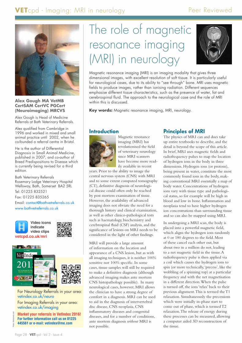
The role of magnetic resonance imaging (MRI) in neurologyMagnetic resonance imaging (MRI) is an imaging modality that gives three dimensional images, with excellent resolution of soft tissue. It is particularly useful for neurological cases, due to its ability to “see through” bone. MRI uses magnetic fields to produce images, rather than ionising radiation. Different sequences emphasise different tissue characteristics, such as the presence of water, fat and cerebrospinal fluid. The approach to the neurological case and the role of MRI within this is discussed.
Key words: Magnetic resonance imaging, MRI, neurology.
IntroductionMagnetic resonance imaging (MRI) has revolutionised the field of veterinary neurology since MRI scanners have become more read-ily available in recent
years. Prior to the ability to image the central nervous system (CNS) with MRI and to some extent computed tomography (CT), definitive diagnosis of neurologi-cal disease could often only be reached by post mortem examination of tissue. However, the availability of advanced imaging does not obviate the need for a thorough history and clinical examination, as well as other clinico-pathological tests such as haematology, biochemistry and cerebrospinal fluid (CSF) analysis, and the significance of lesions on MRI needs to be considered in the light of other findings.
MRI will provide a large amount of information on the location and appearance of a CNS lesion, but as with all imaging techniques, it is neither 100% sensitive nor 100% specific. In some cases, tissue samples will still be required to make a definitive diagnosis (although advanced imaging makes ante mortem CNS histopathology possible). In many neurological cases, however, MRI allows the clinician to have a strong degree of comfort in a diagnosis. MRI can be used to aid in the diagnosis of intervertebral disc disease, CNS neoplasia, CNS inflammatory diseases and congenital diseases, and for a number of conditions, ante mortem diagnosis without MRI is not possible.
Principles of MRIThe physics of MRI can and does take up entire textbooks to describe, and the detail is beyond the scope of this article. In brief, MRI uses magnetic fields and radiofrequency pulses to map the location of hydrogen ions in the body in three dimensions. Hydrogen ions (or protons), being present in water, constitute the most commonly found ions in the body, mak-ing conventional MRI essentially a map of body water. Concentrations of hydrogen ions vary with tissue type and pathologi-cal status, so for example will be high in blood and low in bone. Inflammation and neoplasia tend to have higher hydrogen ion concentrations than surrounding tissue and so can also be mapped using MRI.
In undergoing a MRI scan, the body is placed into a powerful magnetic field, which aligns the hydrogen ions randomly at 0 or 180 degrees to the field. Most of these cancel each other out, but about two in a million do not, leading to a net magnetic field in the tissue. A radiofrequency pulse is then applied via a coil which causes the hydrogen ions to spin (or more technically, ‘precess’, like the wobbling of a spinning top) at a particular frequency and with the net magnetisation in a different direction. When the pulse is turned off, the ions ‘relax’ back to their previous alignment. This is termed the T1 relaxation. Simultaneously the precessions which were initially in-phase start to come out of phase, which is termed T2 relaxation. The release of energy during these processes can be measured, allowing a computer aided 3D reconstruction of the tissue.
VETcpd - Imaging: MRI in neurology
Alex Gough MA VetMB CertSAM CertVC PGCert (Neuroimaging) MRCVSAlex Gough is Head of Medicine Referrals at Bath Veterinary Referrals.
Alex qualified from Cambridge in 1996 and worked in mixed and small animal practice until 2002, when he co-founded a referral centre in Bristol.
He is the author of Differential Diagnosis in Small Animal Medicine, published in 2007, and co-author of Breed Predispositions to Disease which is currently being revised for a third edition.
Bath Veterinary Referrals Rosemary Lodge Veterinary Hospital Wellsway, Bath, Somerset BA2 5RLTel: 01225 832521Fax: 01225 835265Email: [email protected]
Video icons indicate video clips
vetcpd.co.uk/mri
Market your referrals in VetIndex 2016! For further information call us on 01225 445561 or e-mail: [email protected]
For Neurology Referrals in your area: vetindex.co.uk/neuroFor Imaging Referrals in your area: vetindex.co.uk/imaging
Phot
o: iS
tock
phot
o.co
m

Full article available for purchase at www.vetcpd.co.uk/modules/ VETcpd - Vol 2 - Issue 4 - Page 29
VETcpd - Imaging: MRI in neurology
Common MRI sequencesThere are a number of MRI sequences which can be used to examine tissue in different ways. The most common and useful are termed T1 weighted and T2 weighted (or sometimes just T1 and T2).
Features of T1 and T2 weighted scans• T1 weighted scans tend to show up fluid as hypo-intense (i.e. dark) to surrounding tissues (Figure 1).
• T2 weighted scans tend to show fluid up as hyper-intense (i.e. bright) to surrounding tissues (Figure 2).
Short Tau (or T1) Inversion Recovery (STIR) scansHowever, as fat shows up hyperintense (i.e. bright) on T1 and T2 weighted series, it is hard to see pathology which is surrounded by fat. The Short Tau (or T1) Inversion Recovery (STIR) series suppresses the signal from fat, causing it to appear hypointense, and showing up increased fluid content of the tissue as bright, which will often be associated with pathology. This sequence also gives some information on histology, since a mass that shows up hyperintense on T2 weighted scans and dark on STIR scans is likely to be made of fat (e.g. lipoma or liposarcoma).
Fluid Attenuated Inversion Recovery (FLAIR) scansSimilarly, the Fluid Attenuated Inversion Recovery (FLAIR) series suppresses the signal from cerebro-spinal fluid (CSF), and this allows pathology near regions of CSF such as the lateral ventricles to be observed (Figure 3).
Benefits of T1 and T2 scans • Since T2 scans take longer to acquire than T1 weighted scans, T1 series can be repeated more often for the same length of time than T2 series. This increased number of acquisitions allows the software to average out the signal compared to random background noise. This allows an increase in signal to noise ratio. Since increasing the resolution decreases the signal to noise ratio, it is possible to get higher resolution images from T1 weighted scans than T2 weighted scans for a given time of acquisition.
• However, since T2 shows up fluid within tissue better than T1, pathological regions tend to show up better on T2 weighted than T1 weighted scans.
• As a rule of thumb, T1 weighted scans are better at showing anatomy, and T2 weighted scans at showing pathology, and comparing T1 and T2 weighted scans of the same region gives the optimal comparison.
Use of contrastGadolinium is a rare earth metal which has paramagnetic properties. Gadolinium based contrast media show up as hyperintense on T1 weighted scans. Since these contrast agents do not penetrate an intact blood-brain barrier, there should be no increased signal on T1 weighted scans in the undiseased central nervous system. Contrast uptake within the central nervous system suggests a leaky blood-brain barrier, and may indicate pathology such as inflammation or neoplasia. The pattern of contrast uptake can also help to indicate the histological type of a tumour.
Figure 1: T1 weighted transverse MRI of the brain of a normal dog. Note the lateral ventricles are dark.
Figure 3: Transverse FLAIR MRI of the brain of a normal dog. Note the lateral ventricles are dark.
Figure 2: T2 weighted transverse MRI of the brain of a normal dog. Note the lateral ventricles (LV) are bright.
Lateral ventricle (LV)
Masticatory muscles
Skull
Third ventricle
Pituitary gland
Nasopharynx
Airway
LV
LV LV