the role of jnk in the development of hepatocellular...
TRANSCRIPT

The role of JNK in the developmentof hepatocellular carcinoma
Madhumita Das,1 David S. Garlick,2 Dale L. Greiner,1 and Roger J. Davis1,3,4
1Program in Molecular Medicine, University of Massachusetts Medical School, Worcester, Massachusetts 01605, USA;2Department of Cancer Biology, University of Massachusetts Medical School, Worcester, Massachusetts 01605, USA;3Howard Hughes Medical Institute, Worcester, Massachusetts 01605, USA
The cJun NH2-terminal kinase (JNK) signal transduction pathway has been implicated in the growth ofcarcinogen-induced hepatocellular carcinoma. However, the mechanism that accounts for JNK-regulated tumorgrowth is unclear. Here we demonstrate that compound deficiency of the two ubiquitously expressed JNKisoforms (JNK1 and JNK2) in hepatocytes does not prevent hepatocellular carcinoma development. Indeed, JNKdeficiency in hepatocytes increased the tumor burden. In contrast, compound JNK deficiency in hepatocytes andnonparenchymal cells reduced both hepatic inflammation and tumorigenesis. These data indicate that JNK playsa dual role in the development of hepatocellular carcinoma. JNK promotes an inflammatory hepatic environmentthat supports tumor development, but also functions in hepatocytes to reduce tumor development.
[Keywords: JNK; partial hepatectomy; hepatocellular carcinoma]
Supplemental material is available for this article.
Received September 3, 2010; revised version accepted February 2, 2011.
The cJun NH2-terminal kinase (JNK) pathway is impli-cated in tumor development (Davis 2000). Targets ofJNK signaling include members of the activating protein1 (AP1) transcription factor group (e.g., cJun, JunB, JunD,and related proteins). These transcription factors functionwithin a regulatory network that controls multiple as-pects of cellular physiology, including proliferation (Eferland Wagner 2003). Studies of murine embryo fibroblasts(MEFs) demonstrate that loss of JNK causes major defectsin cellular proliferation (Tournier et al. 2000) and AP1-dependent gene expression (Ventura et al. 2003). Theseeffects of JNK are mediated, in part, by the AP1 tran-scription factor. Indeed, cJun phosphorylation is requiredfor normal serum-stimulated cell growth (Behrens et al.1999). The JNK signaling pathway therefore representsan important regulatory mechanism that can controlgrowth. Moreover, dysregulated JNK may contribute totumor development (Davis 2000).
Studies of the role of the JNK and cJun in the liver haveconfirmed the importance of this signaling pathway ingrowth regulation. Thus, cJun-deficient mice (Behrenset al. 2002; Stepniak et al. 2006), JNK1-deficient mice(Hui et al. 2008), and mice treated with a JNK inhibitor(Schwabe et al. 2003) exhibit major defects in liver re-generation following partial hepatectomy (PHx). Further-more, both cJun-deficient mice (Eferl et al. 2003) and
JNK1-deficient mice (Sakurai et al. 2006; Hui et al. 2008)are protected against the development of hepatocellularcarcinoma (HCC) following exposure to the carcinogendiethylnitrosamine (DEN). The critical role of JNK inHCC has been confirmed by pharmacological inhibitionof JNK and studies of human HCC cells (Hui et al. 2008).
The mechanism of JNK and cJun signaling in the liverthat contributes to regeneration and HCC is unclear, butdown-regulation of the proliferation inhibitor p21CIP1 andup-regulation of the growth promoter cMyc appear to becritical factors (Stepniak et al. 2006; Hui et al. 2008). Analternative mechanism for the contribution of JNK toHCC is represented by the role of compensatory pro-liferation in hepatic tumor development (Fausto 1999). Ithas been reported that JNK1 is required for hepatocytedeath in the DEN model of HCC (Sakurai et al. 2006).Reduced hepatocyte death in JNK1-deficient mice resultsin reduced compensatory proliferation and suppressionof HCC (Sakurai et al. 2006). These mechanisms are notmutually exclusive, and it is possible that JNK playsmultiple roles in HCC, including regulation of cell deathand gene expression. Nevertheless, a common themeamong these mechanisms is that JNK plays a critical rolein hepatocytes that is required for HCC development.
The purpose of this study was to examine the role ofJNK in the development of HCC. Specifically, we testedwhether JNK in hepatocytes contributes to tumor forma-tion. Previous studies have focused on an analysis ofJnk1�/� mice (Sakurai et al. 2006; Hui et al. 2008). Herewe report the analysis of mice with tissue-specific de-ficiency of JNK and mice with compound deficiency ofboth JNK1 and JNK2. Our analysis demonstrates that
4Corresponding author.E-MAIL [email protected]; FAX (508) 856-3210.Article is online at http://www.genesdev.org/cgi/doi/10.1101/gad.1989311.Freely available online through the Genes & Development Open Accessoption.
634 GENES & DEVELOPMENT 25:634–645 � 2011 by Cold Spring Harbor Laboratory Press ISSN 0890-9369/11; www.genesdev.org
Cold Spring Harbor Laboratory Press on June 13, 2018 - Published by genesdev.cshlp.orgDownloaded from

compound JNK deficiency in hepatocytes increases thedevelopment of HCC. JNK therefore functions in liverparenchymal cells to reduce tumor development. Weshow that the protumorigenic effects of JNK on HCCare associated with inflammation and require JNK func-tion in nonparenchymal cells.
Results
Compound JNK deficiency in hepatocytesdoes not prevent liver regeneration following PHx
PHx causes JNK activation and a robust regenerationresponse that results in rapid restoration of liver mass(Westwick et al. 1995). It has been reported that Jnk1�/�
mice exhibit a defect in the regeneration response to PHx(Hui et al. 2008). In initial studies, we compared hepaticregeneration in control mice, Jnk1�/� mice, and Jnk2�/�
mice following PHx. This analysis demonstrated similarhepatic regeneration in control and Jnk2�/� mice, buthepatic regeneration was suppressed in Jnk1�/� mice
(Supplemental Fig. S1). These data indicate that JNK1may play an important role in hepatocyte proliferation(Hui et al. 2008).
The Jnk1 and Jnk2 genes are both expressed in hepato-cytes (Davis 2000). Consequently, the reduced hepaticregeneration detected in Jnk1�/�mice (Supplemental Fig.S1) is not caused by loss of total JNK in hepatocytes.These considerations indicated that studies of hepaticregeneration in mice with compound ablation of Jnk1plus Jnk2 are required. We employed a conditional geneablation strategy using Alb-Cre transgenic mice to createanimals with compound deficiency of JNK1 plus JNK2 inhepatocytes (Das et al. 2009). Control HWT mice (Alb-Cre+/�) and JNK-deficient HDJNK mice (Alb-Cre+/�
Jnk1LoxP/LoxP Jnk2�/�) were examined following PHx ora sham surgical procedure (Fig. 1A).
Biochemical analysis of the liver of HWT and HDJNK
mice at 48 h post-PHx demonstrated that JNK deficiencydid not significantly change the expression of cJun, JunB,or Cyclin D1 mRNA, but a modest reduction in JunD
Figure 1. JNK deficiency in hepatocytes does not prevent liver regeneration after PHx. (A) Control mice (HWT mice: Alb-Cre-/+) andJNK-deficient mice (HDJNK mice: Alb-Cre-/+ Jnk1LoxP/LoxP Jnk2�/�) were subjected to two-thirds PHx or a sham procedure. The micewere injected (i.p.) with 100 mg/g BrdU (dissolved in saline) at 2 h prior to euthanasia. Representative sections of liver from mice at 48 hpost-surgery were stained with H&E (top panels) and an antibody to BrdU (bottom panels). (B) The liver/body weight ratio wascalculated and expressed as the mean percentage 6 SD (n = 6 mice) in HWT and HDJNK mice. No statistically significant differencesbetween HWT and HDJNK mice were detected. (C) The percentage of BrdU-positive hepatocytes in HWT and HDJNK mice (mean 6 SD; n =
8 mice) is presented. Statistically significant differences between HWT and HDJNK mice are indicated. (*) P < 0.05; (**) P < 0.001. (D) Thepercentage of hepatocyte mitotic figures detected in H&E-stained sections of liver from HWT and HDJNK mice (mean 6 SD; n = 8 mice) ispresented. Statistically significant differences between HWT and HDJNK mice are indicated. (*) P < 0.05.
JNK and hepatocellular carcinoma
GENES & DEVELOPMENT 635
Cold Spring Harbor Laboratory Press on June 13, 2018 - Published by genesdev.cshlp.orgDownloaded from

mRNA expression was detected in the liver of HDJNK micecompared with HWT mice (Supplemental Fig. S2A). Thesechanges were associated with reduced activation of JNKand reduced phosphorylation of cJun in the liver of HDJNK
mice compared with HWT mice (Supplemental Fig. S2B).These observations are consistent with ablation of theJNK signaling pathway in the hepatocytes of HDJNK mice.We detected no statistically significant changes in acti-vation of AKT or the ERK and p38 MAPKs in the JNK-deficient liver compared with control liver (SupplementalFig. S2B).
Measurement of hepatic mass demonstrated that com-pound deficiency of JNK1 plus JNK2 in hepatocytescaused no significant change in regeneration (Fig. 1B).To confirm this conclusion, we examined hepatocyteproliferation by measuring the incorporation of bromo-deoxyuridine (BrdU) (Fig. 1A). This analysis demonstratedincreased BrdU incorporation at 48 h post-PHx in bothHWT and HDJNK mice, but the amount of BrdU incorpora-tion was reduced in HDJNK mice compared with HWT mice(Fig. 1C). This reduction in BrdU incorporation is consis-tent with the reduced number of mitotic figures detectedin hepatocytes of HDJNK mice compared with HWT miceat 48 h post-PHx (Fig. 1D). However, a time-course analy-sis demonstrated that the overall hepatic proliferation inHWT and HDJNK mice following PHx was similar (Fig.1C,D). These data demonstrate that compound JNK de-ficiency does not prevent hepatic regeneration followingPHx.
The discovery that mice with compound JNK defi-ciency in hepatocytes were capable of hepatic regenera-tion following PHx was unexpected for two reasons. First,hepatic regeneration is strongly reduced in Jnk1�/� mice
(Supplemental Fig. S1; Hui et al. 2008). Second, studies ofJnk1�/� Jnk2�/� primary MEFs demonstrate that theseJNK-deficient cells exhibit severe defects in proliferation,including early senescence (Tournier et al. 2000; Das et al.2007). Compound JNK deficiency was therefore antici-pated to similarly cause growth retardation in other celltypes, including hepatocytes. We therefore sought to con-firm that liver regeneration following PHx is JNK-in-dependent using a different mouse model with compoundhepatic JNK deficiency (Supplemental Fig. S3). Together,these data confirm the conclusion that hepatic regenera-tion following PHx occurs in mice with deficiency of bothJNK1 and JNK2 in the liver (Fig. 1; Supplemental Fig. S3).JNK is therefore not essential for hepatic regeneration.
JNK deficiency in hepatocytes promotes HCC
The conclusion that JNK is not essential for hepaticregeneration led us to question whether JNK is requiredfor hepatocyte proliferation in the context of HCC. Wetherefore tested whether JNK in hepatocytes is requiredfor HCC development by treating HWT and HDJNK micewith the carcinogen DEN. Previous reports indicate thatDEN-induced HCC is markedly reduced in Jnk1�/� mice(Sakurai et al. 2006; Hui et al. 2008). In contrast, we foundthat HDJNK mice developed HCC following exposure toDEN (Fig. 2A; Supplemental Fig. S4). Indeed, the tumorsize in HDJNK mice was significantly greater than inHWT mice (Fig. 2B). However, no significant difference intumor incidence or the number of tumor nodules be-tween HWT and HDJNK mice was detected (Fig. 2C). Thiseffect of JNK deficiency to increase HCC was associatedwith increased hepatic expression of cJun (Supplemental
Figure 2. Effect of hepatocyte-specific JNKdeficiency on HCC. (A) DEN-induced HCCin control mice (HWT mice: Alb-Cre+/�) andJNK-deficient mice (HDJNK mice: Alb-Cre+/�
Jnk1LoxP/LoxP Jnk2�/�) at 38 wk of age. (B)The maximum diameter of individual tu-mor nodules (right panel) and the mean 6
SD (n = 13) width of the tumor nodules (leftpanel) are presented. Statistically signifi-cant differences between HWT and HDJNK
mice are indicated. (*) P < 0.05. (C) Thetumor incidence (left panel) and the numberof tumors per mouse (right panel) wereexamined (n = 14). No statistically signifi-cant differences between HWT and HDJNK
mice were detected. (D) Representative sec-tions of liver from mice treated withoutDEN (control) and with DEN (tumor)stained with H&E are presented. (E) Immu-noblot analysis of tumors isolated fromHWT mice and HDJNK mice was performedby probing with antibodies to JNK and a-Tu-bulin (Tub.). (F) Kaplan-Meier analysis of thesurvival of HWT mice and HDJNK mice (n =
10) treated with DEN is presented. No sta-tistically significant difference between thesurvival or mortality of HWT mice and HDJNK
mice was detected (log-rank test; P = 0.055).
Das et al.
636 GENES & DEVELOPMENT
Cold Spring Harbor Laboratory Press on June 13, 2018 - Published by genesdev.cshlp.orgDownloaded from

Fig. S5A). Histological analysis of hepatic sections dem-onstrated the presence of lesions ranging from adenomato carcinoma in HWT mice, but primarily carcinoma inHDJNK mice (Fig. 2D; Supplemental Figs. S6, S7). Immu-noblot analysis demonstrated that HDJNK tumors wereJNK deficient (Fig. 2E). Together, these data indicate thatJNK in hepatocytes acts as to reduce tumor developmentin this model of HCC. However, Kaplan-Meier analysisdemonstrated that the increased tumor burden in the HDJNK
mice did not cause a statistically significant (P = 0.055)change in survival compared with HWT mice (Fig. 2F).
JNK deficiency can prevent HCC development
The finding that JNK in hepatocytes acts to reduce HCC(Fig. 2) was not anticipated because studies of Jnk1�/�
mice (but not Jnk2�/� mice) have indicated that JNKplays a critical role in HCC development (Sakurai et al.2006; Hui et al. 2008). Together, these findings suggestthat JNK may both inhibit and promote tumor formationduring DEN-induced HCC development. The protumori-genic role of JNK does not require the function of JNK inhepatocytes (Fig. 2), but may involve functions of JNKin nonparenchymal cells. To test this hypothesis, we ex-amined DEN-induced HCC development in mice withcompound JNK deficiency using Mx-1-Cre mice andconditional gene ablation in hepatocytes and nonparen-chymal cells (Das et al. 2009).
Control MxWT mice (Mx-1-Cre+/�) and JNK-deficientMxDJNK mice (Mx-1-Cre+/� Jnk1LoxP/LoxP Jnk2�/�) weretested for HCC development. We found that HCC wasstrongly suppressed in MxDJNK mice compared with MxWT
mice (Fig. 3A; Supplemental Fig. S4). The tumor size inMxDJNK mice was significantly smaller than in MxWT mice(Fig. 3B). Moreover, both the tumor incidence and thenumber of tumor nodules were reduced in MxDJNK micecompared with MxWT mice (Fig. 3C). This effect of JNKdeficiency to suppress HCC was associated with markedlyreduced hepatic expression of cJun (Supplemental Fig.S5B). Histological analysis of hepatic tissue sections dem-onstrated lesions ranging from adenoma to carcinoma inMxWT mice, but primarily only localized adenoma weredetected in MxDJNK mice (Fig. 3D; Supplemental Figs. S7,S8). Immunoblot analysis demonstrated that MxDJNK tu-mors were JNK-deficient (Fig. 3E). Kaplan-Meier analysisdemonstrated that the reduced tumor burden in MxDJNK
mice caused significantly increased survival (P = 0.017)compared with MxWT mice (Fig. 3F). Together, these datademonstrate that JNK can play a protumorigenic role inHCC development. This protumorigenic role does notrequire JNK in hepatocytes (Fig. 2), but does require JNKin nonparenchymal cells (Fig. 3).
JNK regulation of HCC proliferation mediatedby p21CIP1 and cMyc
JNK-mediated transcriptional down-regulation of thegrowth inhibitor p21CIP1 and up-regulation of the growthpromoter cMyc have been implicated as critical stepsduring HCC development (Hui et al. 2008). We thereforeexamined p21CIP1 and cMyc expression in the liver ofmice with compound deficiency of JNK1 plus JNK2 (Fig.4). We found that p21CIP1 expression was significantlyincreased in DEN-treated JNK-deficient mice (HDJNK and
Figure 3. Effect of hepatic JNK deficiencyon HCC. (A) DEN-induced HCC in controlmice (MxWT mice: Mx1-Cre+/�) and JNK-deficient mice (MxDJNK mice: Mx1-Cre+/�
Jnk1LoxP/LoxP Jnk2�/�) at 38 wk of age. TheJNK-deficient mice lack expression ofJNK1 plus JNK2 in both hepatocytes andnonparenchemyal hepatic cells. (B) Themaximum diameter of individual tumornodules (right panel) and the mean 6 SD(n = 13) width of the tumor nodules (left
panel) are presented. Statistically signif-icant differences between MxWT andMxDJNK mice are indicated. (*) P < 0.05;(**) P < 0.01. (C) The tumor incidence (leftpanel) and the number of tumors permouse (right panel) were examined (n =
16). Statistically significant differencesbetween MxWT and MxDJNK mice are in-dicated. (*) P < 0.05. (D) Representativesections of liver from mice treated withoutDEN (control) and with DEN (tumor)stained with H&E are presented. (E) Immu-noblot analysis of tumors isolated fromMxWT mice and MxDJNK mice was per-formed by probing with antibodies to JNKand a-Tubulin (Tub.). (F) Kaplan-Meier anal-
ysis of the survival of MxWT mice and MxDJNK mice (n = 11) treated with DEN is presented. The survival of MxWT mice wassignificantly shorter than MxDJNK mice (log-rank test; P = 0.017).
JNK and hepatocellular carcinoma
GENES & DEVELOPMENT 637
Cold Spring Harbor Laboratory Press on June 13, 2018 - Published by genesdev.cshlp.orgDownloaded from
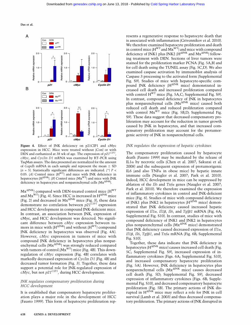
MxDJNK) compared with DEN-treated control mice (HWT
and MxWT) (Fig. 4). Since HCC is increased in HDJNK mice(Fig. 2) and decreased in MxDJNK mice (Fig. 3), these datademonstrate no correlation between p21CIP1 expressionand HCC development in compound JNK-deficient mice.In contrast, an association between JNK, expression ofcMyc, and HCC development was detected. No signifi-cant difference between cMyc expression between tu-mors in mice with (HDJNK) and without (HWT) compoundJNK deficiency in hepatocytes was observed (Fig. 4A).However, cMyc expression in tumors of mice withcompound JNK deficiency in hepatocytes plus nonpar-enchymal cells (MxDJNK) was strongly reduced comparedwith tumors of control (MxWT) mice (Fig. 4B). This down-regulation of cMyc expression (Fig. 4B) correlates withmarkedly decreased expression of Cyclin D1 (Fig. 4B) anddecreased tumor formation (Fig. 3). Together, these datasupport a potential role for JNK-regulated expression ofcMyc, but not p21CIP1, during HCC development.
JNK regulates compensatory proliferation duringHCC development
It is established that compensatory hepatocyte prolifer-ation plays a major role in the development of HCC(Fausto 1999). This form of hepatocyte proliferation rep-
resents a regenerative response to hepatocyte death thatis associated with inflammation (Grivennikov et al. 2010).We therefore examined hepatocyte proliferation and deathin control mice (HWT and MxWT) and mice with compounddeficiency of JNK1 plus JNK2 (HDJNK and MxDJNK) follow-ing treatment with DEN. Sections of liver tumors werestained for the proliferation marker PCNA (Fig. 5A,B) andfor cell death using the TUNEL assay (Fig. 5C,D). We alsoexamined caspase activation by immunoblot analysis ofCaspase 3 processing to the activated form (SupplementalFig. S9). Studies of mice with hepatocyte-specific com-pound JNK deficiency (HDJNK mice) demonstrated in-creased cell death and increased proliferation comparedwith control HWT mice (Fig. 5A,C; Supplemental Fig. S9).In contrast, compound deficiency of JNK in hepatocytesplus nonparenchymal cells (MxDJNK mice) caused bothreduced cell death and reduced proliferation comparedwith control MxWT mice (Fig. 5B,D; Supplemental Fig.S9). These data suggest that decreased compensatory pro-liferation may account for the reduction in tumor growthcaused by JNK in hepatocytes, and that increased com-pensatory proliferation may account for the protumor-genic activity of JNK in nonparenchymal cells.
JNK regulates the expression of hepatic cytokines
The compensatory proliferation caused by hepatocytedeath (Fausto 1999) may be mediated by the release ofIL1a by necrotic cells (Chen et al. 2007; Sakurai et al.2008) and the subsequent expression of protumorigenicIL6 (and also TNFa in obese mice) by hepatic innateimmune cells (Naugler et al. 2007; Park et al. 2010).Indeed, HCC development in mice can be suppressed byablation of the Il6 and Tnfa genes (Naugler et al. 2007;Park et al. 2010). We therefore examined the expressionof inflammatory cytokines in control and JNK-deficientmice (Fig. 6). Studies of mice with compound deficiencyof JNK1 plus JNK2 in hepatocytes (HDJNK mice) demon-strated that JNK deficiency caused increased hepaticexpression of Il1a, Il1b, Il6, and Tgfb1 mRNA (Fig. 6A;Supplemental Fig. S10). In contrast, studies of mice withcompound deficiency of JNK1 and JNK2 in hepatocytesplus nonparenchymal cells (MxDJNK mice) demonstratedthat JNK deficiency caused decreased expression of Il1a,Il1b, Il6, Tgfb1, and Tnfa mRNA (Fig. 6B; SupplementalFig. S10).
Together, these data indicate that JNK deficiency inhepatocytes (HDJNK mice) causes increased cell death (Fig.5C; Supplemental Fig. S9), increased expression of in-flammatory cytokines (Figs. 6A; Supplemental Fig. S10),and increased compensatory hepatocyte proliferation(Fig. 5A). However, JNK deficiency in hepatocytes plusnonparenchymal cells (MxDJNK mice) causes decreasedcell death (Fig. 5D; Supplemental Fig. S9), decreasedexpression of inflammatory cytokines (Figs. 6B; Supple-mental Fig. S10), and decreased compensatory hepatocyteproliferation (Fig. 5B). The primary actions of JNK dis-rupted in HDJNK mice may reflect a role for JNK in cellsurvival (Lamb et al. 2003) and thus decreased compensa-tory proliferation. The primary actions of JNK disrupted in
Figure 4. Effect of JNK deficiency on p21CIP1 and cMycexpression in HCC. Mice were treated without (Con) or withDEN and euthanized at 38 wk of age. The expression of p21CIP1,cMyc, and Cyclin D1 mRNA was examined by RT–PCR usingTaqMan assays. The data presented are normalized for the amountof Gapdh mRNA in each sample and represent the mean 6 SD(n = 5). Statistically significant differences are indicated. (*) P <
0.05. (A) Control mice (HWT) and mice with JNK deficiency inhepatocytes (HDJNK). (B) Control mice (MxWT) and mice with JNKdeficiency in hepatocytes and nonparenchymal cells (MxDJNK).
Das et al.
638 GENES & DEVELOPMENT
Cold Spring Harbor Laboratory Press on June 13, 2018 - Published by genesdev.cshlp.orgDownloaded from

MxDJNK mice may be expression of the protumorigeniccytokines IL6 and TNFa (Sabio et al. 2008; Das et al. 2009).
The role of IL6 in JNK-regulated development of HCC
It is established that IL6 plays a key role in hepatocyteproliferation (Cressman et al. 1996), and that IL6 iscritically required for DEN-induced HCC (Naugler et al.2007). We therefore examined the hepatic IL6 signalingpathway in mice with compound JNK deficiency. Treat-ment of control mice with DEN caused an increase inhepatic Il6 mRNA and IL6 protein in the blood (Fig. 6).The Socs3 gene is a target of IL6-stimulated STAT3signaling in the liver. Treatment of mice with DENcaused increased Socs3 mRNA expression (Fig. 6). ThisDEN-induced increase in Socs3 gene expression wassuppressed in MxDJNK mice (Fig. 6B) and potentiated inHDJNK mice (Fig. 6A). These data are consistent with theincreased HCC and hepatic IL6 expression in HDJNK mice(Figs. 2, 6A) and reduced HCC and hepatic IL6 expressionin MxDJNK mice (Figs. 3, 6B).
The miR-21 gene has been identified as a target of in-flammatory signaling pathways, including IL6 (Jazbutyteand Thum 2010). Indeed, treatment of mice with IL6caused increased hepatic expression of miR-21 (Supple-mental Fig. S11), and it is established that IL6-inducedmiR-21 expression is mediated by two conserved STAT3-binding sites in the miR-21 promoter (Loffler et al. 2007).The expression of miR-21 in the liver is increased duringHCC development (Kutay et al. 2006; Meng et al. 2007;Connolly et al. 2008; Jiang et al. 2008), and studies using aknockdown approach demonstrate that miR-21 contrib-utes to HCC proliferation (Connolly et al. 2008). Thesedata establish that miR-21 plays an important role inHCC development.
We examined the effect of JNK deficiency on hepaticexpression of miR-21. Control studies demonstrated thatDEN caused increased expression of miR-21 (Supplemen-tal Fig. S12). Hepatocyte-specific JNK deficiency (HDJNK
mice) caused increased miR-21 expression compared withcontrol (HWT) mice following treatment with DEN (Sup-plemental Fig. S13A). In contrast, DEN-treated mice withJNK deficiency in hepatocytes and nonparenchymal cells(MxDJNK) expressed reduced miR-21 compared with con-trol (MxWT) DEN-treated mice (Supplemental Fig. S13A).These effects of JNK deficiency on miR-21 expression areconsistent with the observed increase (HDJNK) and de-crease (MxDJNK) in the expression of hepatic IL6 (and otherinflammatory cytokines) following DEN treatment (Fig.6; Supplemental Fig. S10).
Targets of miR-21 include repressors of the AKT (Pten)and receptor tyrosine kinase (Sprouty) signaling pathways(Cabrita and Christofori 2008). We therefore examinedPten and Sprouty expression in DEN-treated control(MxWT) mice and JNK-deficient (MxDJNK) mice. JNK de-ficiency did not change the expression of Pten or SproutymRNA (Supplemental Fig. S13C,D). Similarly, JNK de-ficiency did not cause altered expression of PTEN protein(Supplemental Fig. S13D). In contrast, JNK deficiencycaused increased expression of Sprouty protein (Supple-mental Fig. S13C). To test whether this increased expres-sion of Sprouty is functionally relevant, we examinedsignaling pathways downstream from activated receptortyrosine kinases, including AKT and ERK. We found thatincreased expression of the negative regulator Sprouty inJNK-deficient (MxDJNK) mice was associated with reducedAKT and ERK activity (Supplemental Fig. S13B). Thesedata suggest that Sprouty (Supplemental Fig. S13C) maybe a relevant target of the miR-21 pathway that isrequired for HCC development (Connolly et al. 2008).
Figure 5. Effect of JNK deficiency on he-patocyte death and compensatory prolifera-tion. (A–D) Control mice (HWT and MxWT)and JNK-deficient (DJNK) mice (HDJNK andMxDJNK) were treated with DEN. Hepatictissue sections of mice (age 38 wk) wereexamined by staining with an antibody tothe proliferation marker PCNA (A,B) or byTUNEL assay (C,D). DNA was stained withDAPI. Quantitation of triplicate indepen-dent samples demonstrated that 29.0% 6
1.1% of HWT and 49.9% 6 7.2% of HDJNK
nuclei were positive for PCNA (P < 0.05),61.1% 6 12.5% of MxWT and 17.4% 6
1.4% of MxDJNK nuclei were positive forPCNA (P < 0.05), 2.5% 6 1.2% of HWT and32.4% 6 6.1% of HDJNK nuclei were TUNEL-positive (P < 0.01), and 44.2% 6 6.6% ofMxWT and 10.7% 6 2.3% of MxDJNK nucleiwere TUNEL-positive (P < 0.01).
JNK and hepatocellular carcinoma
GENES & DEVELOPMENT 639
Cold Spring Harbor Laboratory Press on June 13, 2018 - Published by genesdev.cshlp.orgDownloaded from

Discussion
The role of JNK in proliferation is cell type-dependent
It is established that JNK is critically required for theproliferation of primary MEFs (Davis 2000). Compoundmutant Jnk1�/� Jnk2�/� MEFs exhibit a severe growthretardation phenotype (Tournier et al. 2000) and prema-ture senescence (Das et al. 2007). The rapid senescence ofprimary Jnk1�/� Jnk2�/� MEFs is mediated by increasedexpression of the Trp53 tumor suppressor (Das et al.2007). This engagement of p53-dependent senescence inprimary Jnk1�/� Jnk2�/�MEFs (Das et al. 2007) is similarto cJun�/� MEFs (Schreiber et al. 1999). Indeed, reduced
expression of cJun by Jnk1�/� Jnk2�/� MEFs may con-tribute to the early senescence phenotype (Ventura et al.2003). These observations indicate that JNK may havea general role in the proliferation of many cell types.Reports that JNK1 plays an essential role in both hepaticregeneration following PHx (Hui et al. 2008) andDEN-induced HCC (Sakurai et al. 2006; Hui et al. 2008)are consistent with this conclusion.
The finding that compound JNK deficiency does notprevent hepatocyte proliferation was unexpected (Figs. 1,2; Supplemental Fig. S3). The simplest explanation forthese data is that JNK is not required for proliferationof hepatocytes (Fig. 1; Supplemental Fig. S3), but JNK is
Figure 6. Effect of JNK deficiency on the expression ofhepatic cytokines. (A,B) HWT and HDJNK mice (A) orMxWT and MxDJNK mice (B) were treated without (Con)or with DEN and euthanized at 38 wk of age. The bloodconcentration of IL6 was measured by ELISA and ispresented as the mean 6 SD (n = 7). Total RNA wasisolated from the liver, and the expression of Il1a, Il1b,Il6, Socs3, and Tnfa mRNA was examined by RT–PCRusing TaqMan assays. The data presented are normal-ized for the amount of Gapdh mRNA in each sampleand represent the mean 6 SD (n = 5). Statisticallysignificant differences between the control and JNK-deficient mice are indicated (P < 0.05).
Das et al.
640 GENES & DEVELOPMENT
Cold Spring Harbor Laboratory Press on June 13, 2018 - Published by genesdev.cshlp.orgDownloaded from

selectively required to prevent Trp53-dependent senes-cence of MEFs (Das et al. 2007). Cell type-specific re-sponses to JNK deficiency may reflect differences in therequirement of JNK for the expression of cJun, a repressorof the Trp53 promoter (Schreiber et al. 1999). CompoundJNK deficiency in MEFs causes down-regulation of cJunexpression (Ventura et al. 2003). In contrast, compoundJNK deficiency in hepatocytes did not cause significantlydecreased cJun expression (Supplemental Fig. S2A). Thefailure of JNK deficiency to down-regulate cJun expres-sion in hepatocytes may reflect signaling pathway re-dundancy, including roles of JNK in AP1-dependentcJun expression (Ventura et al. 2003) and p38 MAPK inMEF2-dependent cJun expression (Han et al. 1997).Indeed, it is intriguing that the p38 MAPK pathway inhepatocytes is constitutively activated and subject tometabolic regulation (Mendelson et al. 1996), but p38MAPK is stress-inducible from a low basal activity inMEFs (Raingeaud et al. 1995). The p38 MAPK pathwaymay therefore act redundantly with JNK to regulatecJun expression in hepatocytes, but the p38 MAPKpathway may not compensate for JNK deficiency inMEFs. Cross-talk, partially redundant signaling, andpartially antagonistic signaling by the JNK and p38MAPK pathways may therefore contribute to the tis-sue-specific effects of JNK on proliferation (Wagner andNebreda 2009).
The role of JNK in liver regeneration
The function of JNK in hepatocytes and nonparenchymalcells of the liver is not required for hepatic regenerationfollowing PHx (Fig. 1; Supplemental Fig. S3). Neverthe-less, liver regeneration is reduced in Jnk1�/� mice (Sup-plemental Fig. S1; Hui et al. 2008). The mechanism thataccounts for this phenotype of Jnk1�/� mice is unclear.The simplest hypothesis is that JNK1 plays a critical rolein a nonhepatic tissue, and that this JNK1-dependentfunction is disrupted in Jnk1�/�mice. The identity of therelevant tissue that mediates JNK1-dependent hepaticregeneration has not been established. One possibility isrepresented by the hypothalamus, a region of the brainthat strongly influences liver regeneration (Kiba 2002).This possibility is consistent with the discovery that themajor metabolic phenotypes of Jnk1�/� mice are largelyaccounted for by the effects of JNK1 deficiency in thehypothalamus, rather than in peripheral tissues (Belgardtet al. 2010; Sabio et al. 2010). A goal for further studieswill be to identify the specific tissue that accounts for therequired role of JNK1 for hepatic regeneration. This roleof JNK1 contrasts with the absence of a requirement forhepatic JNK during liver regeneration following PHx (Fig.1; Supplemental Fig. S3).
The role of JNK in the promotion of HCC
It is established that JNK plays a key role in the de-velopment of DEN-induced HCC (Sakurai et al. 2006; Huiet al. 2008). Two mechanisms have been reported toaccount for JNK-mediated protumorigenic activity. First,JNK1 may be required for down-regulation of p21CIP1
expression and up-regulation of cMyc expression (Huiet al. 2008). Our analysis demonstrates that p21CIP1 ex-pression does not correlate with tumorigenic potential instudies of HCC development in compound JNK-deficientmice (HDJNK and MxDJNK) (Fig. 4). In contrast, expressionof cMyc (Fig. 4) correlates with the tumor burden inHDJNK and MxDJNK mice (Figs. 2, 3). These data are con-sistent with a role for cMyc in JNK-dependent formationof HCC (Hui et al. 2008).
A second proposed mechanism is that JNK1 causeshepatocyte death (Sakurai et al. 2006), resulting in com-pensatory proliferation that contributes to the developmentof HCC (Fausto 1999). However, compound deficiency ofJNK1 and JNK2 in hepatocytes (HDJNK mice) does notinhibit hepatocyte death following treatment with DEN(Fig. 5C; Supplemental Fig. S9). These data demonstratethat JNK-mediated hepatocyte death does not contribute tocompensatory proliferation and HCC. This analysis in-dicates that another mechanism must account for theprotumorigenic effects of JNK on HCC development.
The observation that compound JNK deficiency inhepatocytes (HDJNK mice) does not reduce DEN-inducedHCC (Fig. 2), but compound JNK deficiency in hepato-cytes plus nonparenchymal cells (MxDJNK mice) doesreduce DEN-induced HCC (Fig. 3), provides importantinsight into the mechanism of JNK-promoted HCC de-velopment. These findings indicate that the protumori-genic function of JNK may be localized to nonparenchy-mal cells, rather than hepatocytes. Indeed, functions ofnonparenchymal cells are implicated in HCC develop-ment (Maeda et al. 2005; Grivennikov et al. 2010).Specifically, the release of IL1a by necrotic cells (Chenet al. 2007; Sakurai et al. 2008), and the subsequentexpression of the protumorigenic cytokine IL6 (and alsoTNFa in obese mice) by hepatic innate immune cells,including Kupffer cells (Naugler et al. 2007; Park et al.2010), may drive compensatory hepatocyte proliferationand, subsequently, HCC (Fig. 7). It is therefore significantthat compound JNK deficiency in MxDJNK mice, but notHDJNK mice, causes markedly decreased expression of IL6and TNFa (Fig. 6). These data are consistent with theconclusion that JNK is required for the expression of theprotumorigenic cytokines IL6 and TNFa by hepatic in-nate immune cells during the development of HCC (Fig.6B). Thus, the protumorigenic activity of JNK may bemediated by a role for JNK in nonparenchymal cells (Fig.3) in the creation of an inflammatory environment thatsupports HCC development (Grivennikov et al. 2010).The previously described genetic interactions betweenJnk1 and p21CIP1 (Hui et al. 2008) may therefore reflect aninhibitory action of p21CIP1 on hepatic innate immunecells, rather than hepatocytes. Studies using mice withtissue-specific p21CIP1 deficiency will be required toresolve this question.
The role of JNK in hepatocytes duringHCC development
Studies of mice with compound deficiency of JNK1 plusJNK2 demonstrate that cell death and compensatory
JNK and hepatocellular carcinoma
GENES & DEVELOPMENT 641
Cold Spring Harbor Laboratory Press on June 13, 2018 - Published by genesdev.cshlp.orgDownloaded from

proliferation are key targets of JNK function in HCC (Fig.5). Specifically, we find that compound JNK deficiencyin hepatocytes causes increased hepatocyte death and,consequently, increased compensatory proliferation andHCC development (Figs. 2, 5). The increased death causedby compound JNK deficiency is consistent with previousstudies that demonstrate a role for JNK in cell survival(Kuan et al. 1999; Lamb et al. 2003) and the presence ofJNK-independent pathways that can mediate hepatocytedeath (Das et al. 2009). Although the cell type of HCCorigin is unclear (Mishra et al. 2009), tumors detectedusing Albumin-Cre transgenic mouse models of DEN-induced HCC demonstrate efficient ablation of floxedgenes in tumor cells (Fig. 2E; Maeda et al. 2005). Thesedata suggest that JNK in hepatic parenchymal cells func-tions to reduce tumor development in the DEN model ofHCC. This effect of JNK to influence hepatocyte death,compensatory proliferation, and HCC development (Figs.2, 5) is similar to the reported effects of hepatocyte-specific deficiency of the NF-kB activator IKKb (Maedaet al. 2005). Together, these data are consistent with theproposed role of compensatory proliferation in hepatictumor development (Fausto 1999).
Conclusions
The results of this study demonstrate that JNK playsa complex role in the development of HCC. JNK isrequired for HCC development because it functions innonparenchymal cells to cause expression of protumori-genic cytokines, including IL6 and TNFa. In contrast,JNK in hepatocytes reduces tumor development by de-creasing hepatocyte death and compensatory prolifera-tion. It is likely that previous studies using mice withwhole-body knockout of Jnk1 or Jnk2 reflect a compositephenotype derived from the cell type-specific functions ofJNK in hepatocytes and nonparenchymal cells.
We conclude that JNK both promotes and inhibitstumor development in the DEN model of HCC. This
conclusion has implications for the more general role ofJNK in cancer. Both tumor promotion and inhibition byJNK may contribute to cancer development (Davis 2000;Whitmarsh and Davis 2007). This complicates the anal-ysis of JNK pathway mutations that have been identifiedin human cancer (Greenman et al. 2007; Kan et al. 2010).Moreover, this dual role of JNK should be considered inthe context of the potential use of JNK as a therapeutictarget for drug development and the treatment of humancancer.
Materials and methods
Mice
We previously described Jnk1LoxP/LoxP mice (Das et al. 2009) andJnk2�/� mice (Yang et al. 1998). B6.Cg-Tg(Alb-cre)21Mgn/J mice(Postic et al. 1999), B6.Cg-Tg(Mx1-cre)1Cgn/J mice (Kuhn et al.1995), and C57BL/6J mice were obtained from The JacksonLaboratories (stock numbers 003574, 003556, and 000664, re-spectively). The mutant mice were maintained on a C57BL/6Jstrain background (backcrossed 10 generations), and were housedin a facility accredited by the American Association for Labora-tory Animal Care (AALAC). Deletion of floxed alleles in Mx1-Cre mice was performed by treatment of 4-wk-old mice with 20mg/g polyinosinic-polycytidylic acid (polyIC) (Mikkola et al.2003) followed by recovery (4 wk). Control mice were similarlyinjected with polyIC. PHx was performed on 10-wk-old miceusing methods described previously (Greene and Puder 2003).The animal studies were approved by the Institutional AnimalCare and Use Committee (IACUC) of the University of Massa-chusetts Medical School.
Genotype analysis
Genotype analysis was performed by PCR using genomic DNAas the template. The Jnk1+ (540 base pairs [bp]) and Jnk1LoxP (330bp) alleles were identified using the amplimers 59-AGGATTTATGCCCTCTGCTTGTC-39 and 59-GAACCACTGTTCCAATTTCCATCC-39. The Jnk1LoxP (1100 bp) and Jnk1D (400 bp) alleleswere identified using the amplimers 59-CCTCAGGAAGAAAGGGCTTATTTC-39 and 59-GAACCACTGTTCCAATTTCCATCC-39. The Jnk2+ (400 bp) and Jnk2� (270 bp) alleles wereidentified using the amplimers 59- GGAGCCCGATAGTATCGAGTTACC-39, 59-GTTAGACAATCCCAGAGGTTGTGTG-39, and59-CCAGCTCATTCCTCCACTCATG-39. Cre alleles (450 bp)were detected using the amplimers 59-TTACTGACCGTACACCAAATTTGCCTGC-39 and 59- CCTGGCAGCGATCGCTATTTTCCATGAGTG-39.
HCC assays
Carcinogen-induced HCC was studied using procedures de-scribed previously (Sakurai et al. 2008). Two-week-old male micewere treated by intraperitoneal (i.p.) injection with a single dose(25 mg/kg) of DEN (Sigma, N0258) diluted in glyceryl triocta-noate (Sigma, T9126). Control mice were injected with sol-vent alone. The mice were euthanized at 38 wk of age to ex-amine tumor development. Surface tumor size and numberwere measured using stereomicroscopy (Maeda et al. 2005).The Cre+/� genetic background altered the tumor burden inthe DEN-induced HCC studies; more tumors were found inthe Mx1-Cre+/� genetic background than in the Alb-Cre+/�
genetic background (Figs. 2, 3). We therefore compared control
Figure 7. JNK functions in hepatocytes to reduce HCC de-velopment and in nonparenchymal cells to promote HCCdevelopment. JNK in nonparenchymal cells functions to pro-mote HCC development by providing an inflammatory envi-ronment that supports HCC, including the expression of theprotumorigenic cytokines IL6 and TNFa that contribute tocompensatory hepatocyte proliferation. However, JNK in hepa-tocytes functions to reduce HCC development by promotinghepatocyte survival, therefore decreasing IL1a release by ne-crotic hepatocytes and the activation of hepatic innate immunecells, including Kupffer cells, that express the protumorgeniccytokine IL6.
Das et al.
642 GENES & DEVELOPMENT
Cold Spring Harbor Laboratory Press on June 13, 2018 - Published by genesdev.cshlp.orgDownloaded from

Mx1-Cre+/� mice with JNK-deficient Mx1-Cre+/� mice or con-trol Alb-Cre+/� mice with JNK-deficient Alb-Cre+/� mice.
Serum analysis
Alanine transaminase (ALT) and aspartate aminotrasferase (AST)activity in serum was measured using the ALT and AST Reagentkit (Pointe Scientific) with a Tecan Sapphire microplate reader(Tecan Trading AG). The serum concentration of cytokines wasmeasured by multiplexed ELISA using a Luminex 200 instru-ment (Millipore).
Biochemical analysis
Immunoblot analysis was performed by probing with antibodiesto activated Caspase 3 (Cell Signaling, #9661) Sprouty2 (Abcam,#ab50317), PTEN (Cell Signaling, #9198), p21 (Santa Cruz Bio-technology, #SC 6246), and a-Tubulin (Sigma, #T5168). Immu-noblots were quantitated using a LiCOR Odyssey imager. Theamount of total and phospho-JNK, ERK, p38 MAPK, and AKT intissue extracts was measured using a multiplexed ELISA kit (Bio-Rad) and a Luminex 200 instrument (Millipore).
Quantitative RT–PCR assays (TaqMan) of mRNA expressionwere performed using a 7500 Fast Real-Time PCR machine (AppliedBiosystems) with total RNA prepared from tissues with an RNeasyminikit (Qiagen). The probes were cFos (Mm00487425_m1),cJun (Mm 00495062_s1), cMyc (Mm00487804_m1), Cyclin D1
(Mm00432359_m1), Il1a (00439621_m1), Il1b (Mm00434228_m1),Il6 (Mm00446190_m1), JunB (Mm00492781_s1), JunD (Mm00495088_s1), p15 (Mm00483241_m1), p21 (Mm 00432448_m1), Pten (Mm01212532_m1), Socs3 (Mm00545913_s1), Spr2
(Mm00442344_m1), Tgfb1 (Mm03024053_m1), and Tnf(Mm00443258_m1) (Applied Biosystems). The relative mRNAexpression was normalized by measuring the amount of Gapdh
RNA in each sample using TaqMan assays (Applied Biosystems,4352339E-0904021).
We prepared RNA from tissue samples using the mirVanamicroRNA isolation kit (Ambion) for the analysis of miR-21
expression using TaqMan assays (Applied Biosystems, miR-21RT 2493). The relative miR-21 expression was normalized bymeasuring the amount of U6 RNA in each sample using TaqManassays (Applied Biosystems,U6 RT1973).
Analysis of liver sections
Histology was performed using tissue fixed in 10% formalin for24 h, dehydrated, and embedded in paraffin. Sections (7 mm) werecut and stained using hematoxylin and eosin (American MasterTech Scientific). Terminal deoxynucleotidyltransferase-medi-ated nick end labeling (TUNEL) staining of deparafinized liversections was performed using the In Situ Cell Death Detectionkit Flourescein (Roche) following antigen retrieval (AntigenUnmasking Solution, Vector Laboratories). Immunofluorescenceanalysis of deparafinized liver sections was performed usingantigen retrieval (Vector Laboratories); incubation (1 h) with3% bovine serum albumin (BSA), 1% normal goat serum, and0.4% Triton X-100 in phosphate-buffered saline (PBS); incuba-tion (12 h) at 4°C with a biotin-linked antibody to PCNA(Zymed Laboratories, 13-3940) in PBS with 3% BSA; anddetection of immune complexes with Streptavidin-conjugatedAlexa fluor 633 (Invitrogen, S 21375) in PBS with 3% BSA. Theliver sections were mounted on glass slides. Nuclei werestained with DAPI using Vectashield (Vector Laboratories).Fluorescence microscopy was performed using a Leica SP2confocal microscope.
The incorporation of BrdU was examined using liver sectionsprepared from mice injected (i.p.) with 100 mg/g BrdU (dissolvedin saline) at 2 h prior to euthanasia. Livers were fixed in formalin,dehydrated, and embedded in paraffin, and 4-mm sections wereprepared. Deparafinized liver sections were processed for antigenretrieval and incubated with an antibody to BrdU (Biogenex,#MU247-UC). Immune complexes were detected with a Strepta-vidin/Biotin-based Link-Label Detection kit (Biogenex, Super-sensitive Link label immunohistochemistry detection system)and DAB substrate (Vector Laboratories, #SK4100).
Statistical analysis
Differences between groups were examined for statistical signif-icance using the Student’s test or analysis of variance (ANOVA)with the Fisher’s test. Kaplan-Meier analysis was performedusing the log-rank test.
Acknowledgments
We thank Tammy Barrett, Vicky Benoit, Linda Leehy, and Jian-Hua Liu for expert technical assistance, and Kathy Gemme foradministrative assistance. These studies were supported bygrants from the National Institutes of Health (CA65861 andAI046629). Core facilities at the University of Massachusettsused by these studies were supported by the NIDDK (NationalInstitute of Diabetes and Digestive and Kidney Diseases)Diabetes and Endocrinology Research Center (DK32520).R.J.D. is an Investigator of the Howard Hughes MedicalInstitute.
References
Behrens A, Sibilia M, Wagner EF. 1999. Amino-terminal phos-phorylation of c-Jun regulates stress-induced apoptosis andcellular proliferation. Nat Genet 21: 326–329.
Behrens A, Sibilia M, David JP, Mohle-Steinlein U, Tronche F,Schutz G, Wagner EF. 2002. Impaired postnatal hepatocyteproliferation and liver regeneration in mice lacking c-jun inthe liver. EMBO J 21: 1782–1790.
Belgardt BF, Mauer J, Wunderlich FT, Ernst MB, Pal M, Spohn G,Bronneke HS, Brodesser S, Hampel B, Schauss AC, et al.2010. Hypothalamic and pituitary c-Jun N-terminal kinase1 signaling coordinately regulates glucose metabolism. Proc
Natl Acad Sci 107: 6028–6033.Cabrita MA, Christofori G. 2008. Sprouty proteins, master-
minds of receptor tyrosine kinase signaling. Angiogenesis11: 53–62.
Chen CJ, Kono H, Golenbock D, Reed G, Akira S, Rock KL.2007. Identification of a key pathway required for the sterileinflammatory response triggered by dying cells. Nat Med 13:851–856.
Connolly E, Melegari M, Landgraf P, Tchaikovskaya T, TennantBC, Slagle BL, Rogler LE, Zavolan M, Tuschl T, Rogler CE.2008. Elevated expression of the miR-17-92 polycistron andmiR-21 in hepadnavirus-associated hepatocellular carci-noma contributes to the malignant phenotype. Am J Pathol
173: 856–864.Cressman DE, Greenbaum LE, DeAngelis RA, Ciliberto G,
Furth EE, Poli V, Taub R. 1996. Liver failure and defectivehepatocyte regeneration in interleukin-6-deficient mice.Science 274: 1379–1383.
Das M, Jiang F, Sluss HK, Zhang C, Shokat KM, Flavell RA,Davis RJ. 2007. Suppression of p53-dependent senescence bythe JNK signal transduction pathway. Proc Natl Acad Sci
104: 15759–15764.
JNK and hepatocellular carcinoma
GENES & DEVELOPMENT 643
Cold Spring Harbor Laboratory Press on June 13, 2018 - Published by genesdev.cshlp.orgDownloaded from

Das M, Sabio G, Jiang F, Rincon M, Flavell RA, Davis RJ. 2009.Induction of hepatitis by JNK-mediated expression of TNF-a.Cell 136: 249–260.
Davis RJ. 2000. Signal transduction by the JNK group of MAPkinases. Cell 103: 239–252.
Eferl R, Wagner EF. 2003. AP-1: a double-edged sword intumorigenesis. Nat Rev Cancer 3: 859–868.
Eferl R, Ricci R, Kenner L, Zenz R, David JP, Rath M, Wagner EF.2003. Liver tumor development. c-Jun antagonizes the proa-poptotic activity of p53. Cell 112: 181–192.
Fausto N. 1999. Mouse liver tumorigenesis: models, mecha-nisms, and relevance to human disease. Semin Liver Dis 19:243–252.
Greene AK, Puder M. 2003. Partial hepatectomy in the mouse:technique and perioperative management. J Invest Surg 16:99–102.
Greenman C, Stephens P, Smith R, Dalgliesh GL, Hunter C,Bignell G, Davies H, Teague J, Butler A, Stevens C, et al.2007. Patterns of somatic mutation in human cancer ge-nomes. Nature 446: 153–158.
Grivennikov SI, Greten FR, Karin M. 2010. Immunity, inflam-mation, and cancer. Cell 140: 883–899.
Han J, Jiang Y, Li Z, Kravchenko VV, Ulevitch RJ. 1997.Activation of the transcription factor MEF2C by the MAPkinase p38 in inflammation. Nature 386: 296–299.
Hui L, Zatloukal K, Scheuch H, Stepniak E, Wagner EF. 2008.Proliferation of human HCC cells and chemically inducedmouse liver cancers requires JNK1-dependent p21 down-regulation. J Clin Invest 118: 3943–3953.
Jazbutyte V, Thum T. 2010. MicroRNA-21: from cancer tocardiovascular disease. Curr Drug Targets 11: 926–935.
Jiang J, Gusev Y, Aderca I, Mettler TA, Nagorney DM, BrackettDJ, Roberts LR, Schmittgen TD. 2008. Association of micro-RNA expression in hepatocellular carcinomas with hepatitisinfection, cirrhosis, and patient survival. Clin Cancer Res 14:419–427.
Kan Z, Jaiswal BS, Stinson J, Janakiraman V, Bhatt D, Stern HM,Yue P, Haverty PM, Bourgon R, Zheng J, et al. 2010. Diversesomatic mutation patterns and pathway alterations in hu-man cancers. Nature 466: 869–873.
Kiba T. 2002. The role of the autonomic nervous system in liverregeneration and apoptosis–recent developments. Digestion66: 79–88.
Kuan CY, Yang DD, Samanta Roy DR, Davis RJ, Rakic P, FlavellRA. 1999. The Jnk1 and Jnk2 protein kinases are required forregional specific apoptosis during early brain development.Neuron 22: 667–676.
Kuhn R, Schwenk F, Aguet M, Rajewsky K. 1995. Inducible genetargeting in mice. Science 269: 1427–1429.
Kutay H, Bai S, Datta J, Motiwala T, Pogribny I, Frankel W, JacobST, Ghoshal K. 2006. Downregulation of miR-122 in therodent and human hepatocellular carcinomas. J Cell Bio-
chem 99: 671–678.Lamb JA, Ventura JJ, Hess P, Flavell RA, Davis RJ. 2003. JunD
mediates survival signaling by the JNK signal transductionpathway. Mol Cell 11: 1479–1489.
Loffler D, Brocke-Heidrich K, Pfeifer G, Stocsits C, HackermullerJ, Kretzschmar AK, Burger R, Gramatzki M, Blumert C, BauerK, et al. 2007. Interleukin-6 dependent survival of multiplemyeloma cells involves the Stat3-mediated induction ofmicroRNA-21 through a highly conserved enhancer. Blood110: 1330–1333.
Maeda S, Kamata H, Luo JL, Leffert H, Karin M. 2005. IKKb
couples hepatocyte death to cytokine-driven compensatoryproliferation that promotes chemical hepatocarcinogenesis.Cell 121: 977–990.
Mendelson KG, Contois LR, Tevosian SG, Davis RJ, Paulson KE.1996. Independent regulation of JNK/p38 mitogen-activatedprotein kinases by metabolic oxidative stress in the liver.Proc Natl Acad Sci 93: 12908–12913.
Meng F, Henson R, Wehbe-Janek H, Ghoshal K, Jacob ST, PatelT. 2007. MicroRNA-21 regulates expression of the PTENtumor suppressor gene in human hepatocellular cancer.Gastroenterology 133: 647–658.
Mikkola HK, Klintman J, Yang H, Hock H, Schlaeger TM,Fujiwara Y, Orkin SH. 2003. Haematopoietic stem cellsretain long-term repopulating activity and multipotency inthe absence of stem-cell leukaemia SCL/tal-1 gene. Nature
421: 547–551.Mishra L, Banker T, Murray J, Byers S, Thenappan A, He AR,
Shetty K, Johnson L, Reddy EP. 2009. Liver stem cells andhepatocellular carcinoma. Hepatology 49: 318–329.
Naugler WE, Sakurai T, Kim S, Maeda S, Kim K, ElsharkawyAM, Karin M. 2007. Gender disparity in liver cancer due tosex differences in MyD88-dependent IL-6 production. Sci-
ence 317: 121–124.Park EJ, Lee JH, Yu GY, He G, Ali SR, Holzer RG, Osterreicher
CH, Takahashi H, Karin M. 2010. Dietary and geneticobesity promote liver inflammation and tumorigenesis byenhancing IL-6 and TNF expression. Cell 140: 197–208.
Postic C, Shiota M, Niswender KD, Jetton TL, Chen Y, MoatesJM, Shelton KD, Lindner J, Cherrington AD, Magnuson MA.1999. Dual roles for glucokinase in glucose homeostasis asdetermined by liver and pancreatic b cell-specific geneknock-outs using Cre recombinase. J Biol Chem 274: 305–315.
Raingeaud J, Gupta S, Rogers JS, Dickens M, Han J, Ulevitch RJ,Davis RJ. 1995. Pro-inflammatory cytokines and environ-mental stress cause p38 mitogen-activated protein kinaseactivation by dual phosphorylation on tyrosine and threo-nine. J Biol Chem 270: 7420–7426.
Sabio G, Das M, Mora A, Zhang Z, Jun JY, Ko HJ, Barrett T, KimJK, Davis RJ. 2008. A stress signaling pathway in adiposetissue regulates hepatic insulin resistance. Science 322:1539–1543.
Sabio G, Cavanagh-Kyros J, Barrett T, Jung DY, Ko HJ, Ong H,Morel C, Mora A, Reilly J, Kim JK, et al. 2010. Role of thehypothalamic–pituitary–thyroid axis in metabolic regulationby JNK1. Genes Dev 24: 256–264.
Sakurai T, Maeda S, Chang L, Karin M. 2006. Loss of hepatic NF-k B activity enhances chemical hepatocarcinogenesis throughsustained c-Jun N-terminal kinase 1 activation. Proc NatlAcad Sci 103: 10544–10551.
Sakurai T, He G, Matsuzawa A, Yu GY, Maeda S, Hardiman G,Karin M. 2008. Hepatocyte necrosis induced by oxidativestress and IL-1a release mediate carcinogen-induced com-pensatory proliferation and liver tumorigenesis. Cancer Cell
14: 156–165.Schreiber M, Kolbus A, Piu F, Szabowski A, Mohle-Steinlein U,
Tian J, Karin M, Angel P, Wagner EF. 1999. Control of cellcycle progression by c-Jun is p53 dependent. Genes Dev 13:607–619.
Schwabe RF, Bradham CA, Uehara T, Hatano E, Bennett BL,Schoonhoven R, Brenner DA. 2003. c-Jun-N-terminal kinasedrives cyclin D1 expression and proliferation during liverregeneration. Hepatology 37: 824–832.
Stepniak E, Ricci R, Eferl R, Sumara G, Sumara I, Rath M, Hui L,Wagner EF. 2006. c-Jun/AP-1 controls liver regeneration byrepressing p53/p21 and p38 MAPK activity. Genes Dev 20:2306–2314.
Tournier C, Hess P, Yang DD, Xu J, Turner TK, Nimnual A, Bar-Sagi D, Jones SN, Flavell RA, Davis RJ. 2000. Requirement of
Das et al.
644 GENES & DEVELOPMENT
Cold Spring Harbor Laboratory Press on June 13, 2018 - Published by genesdev.cshlp.orgDownloaded from

JNK for stress-induced activation of the cytochromec-mediated death pathway. Science 288: 870–874.
Ventura JJ, Kennedy NJ, Lamb JA, Flavell RA, Davis RJ. 2003.c-Jun NH(2)-terminal kinase is essential for the regulationof AP-1 by tumor necrosis factor. Mol Cell Biol 23: 2871–2882.
Wagner EF, Nebreda AR. 2009. Signal integration by JNK andp38 MAPK pathways in cancer development. Nat Rev
Cancer 9: 537–549.Westwick JK, Weitzel C, Leffert HL, Brenner DA. 1995. Activa-
tion of Jun kinase is an early event in hepatic regeneration.J Clin Invest 95: 803–810.
Whitmarsh AJ, Davis RJ. 2007. Role of mitogen-activated pro-tein kinase kinase 4 in cancer. Oncogene 26: 3172–3184.
Yang DD, Conze D, Whitmarsh AJ, Barrett T, Davis RJ, RinconM, Flavell RA. 1998. Differentiation of CD4+ T cells to Th1cells requires MAP kinase JNK2. Immunity 9: 575–585.
JNK and hepatocellular carcinoma
GENES & DEVELOPMENT 645
Cold Spring Harbor Laboratory Press on June 13, 2018 - Published by genesdev.cshlp.orgDownloaded from
10.1101/gad.1989311Access the most recent version at doi: 25:2011, Genes Dev.
Madhumita Das, David S. Garlick, Dale L. Greiner, et al. The role of JNK in the development of hepatocellular carcinoma
Material
Supplemental
http://genesdev.cshlp.org/content/suppl/2011/03/14/25.6.634.DC1
References
http://genesdev.cshlp.org/content/25/6/634.full.html#ref-list-1
This article cites 49 articles, 18 of which can be accessed free at:
License Freely available online through the Genes & Development Open Access option.
ServiceEmail Alerting
click here.right corner of the article or
Receive free email alerts when new articles cite this article - sign up in the box at the top
Copyright © 2011 by Cold Spring Harbor Laboratory Press
Cold Spring Harbor Laboratory Press on June 13, 2018 - Published by genesdev.cshlp.orgDownloaded from