the role of genetic factors in autoimmune disease: implicationsfor
TRANSCRIPT
-~~~~~~~~~~~~~~~~~~~~~~~~~~~~~~~~~~~~~~~~~~~
The Role of Genetic Factors in Autoimmune Disease: Implications forEnvironmental ResearchGlinda S. Cooper,1 Frederick W. Miller,2 and Janardan P. Pandey3'Epidemiology Branch, National Institute of Environmental Health Sciences, Research Triangle Park, North Carolina USA; 2Laboratory ofMolecular and Developmental Immunology, Center for Biologics Evaluation and Research, Food and Drug Administration, Bethesda,Maryland USA; 3Department of Microbiology and Immunology, Medical University of South Carolina, Charleston, South Carolina USA
Studies in both humans and in animal models of specific disorders suggest that polymorphisms ofmultiple genes are involved in conferring either a predisposition to or protection from autoimmunediseases. Genes encoding polymorphic proteins that regulate immune responses or the rates andextent of metabolism of certain chemical structures have been the focus of much of the researchregarding genetic susceptibility. We examine the type and strength of evidence concerning geneticfactors and disease etiology, drawing examples from a number of autoimmune diseases. Twinstudies of rheumatoid arthritis (RA), systemic lupus erythematosus (SLE), type diabetes, andmultiple sclerosis (MS) indicate that disease concordance in monozygotic twins is 4 or more timeshigher than in dizygotic twins. Strong familial associations (odds ratio ranging from 5-10) are seen instudies of MS, type diabetes, Graves disease, discoid lupus, and SLE. Familial association studieshave also reported an increased risk of several systemic autoimmune diseases among relatives ofpatients with a systemic autoimmune disease. This association may reflect a common etiologicpathway with shared genetic or environmental influences among these diseases. Recentgenomewide searches in RA, SLE, and MS provide evidence for multiple susceptibility genesinvolving major histocompatibility complex (MHC) and non-MHC loci; there is also evidence thatmany autoimmune diseases share a common set of susceptibility genes. The multifactorial nature ofthe genetic risk factors and the low penetrance of disease underscore the potential influence ofenvironmental factors and gene-environment interactions on the etiology of autoimmune diseases.Key words: autoimmune diseases, gene-environment interactions, genetics, major histocompatibilitycomplex, multiple sclerosis, rheumatoid arthritis, systemic lupus erythematosus, type diabetesmellitus. - Environ Health Perspect 1 07(suppl 5):693-700 (1999).http.//ehpnet1.niehs.nih.gov/docs/1999/suppl-5/693-700cooper/abstract.html
Autoimmune diseases include a wide variety ofconditions with differing clinical presentations,natural histories, and treatment options. Acommon underlying feature of both organ-specific and systemic forms of these diseases isthat the immune system's ability to respondappropriately to self-tissues is altered, resultingin the production of B- and T-cell responsesdirected against self-antigens (autoimmunity).The mechanism through which autoimmunityprogresses to produce pathology (an autoim-mune disease) is not understood. Studies inboth humans and in animal models of specificdisorders suggest that polymorphisms of multi-ple genes are involved in conferring either apredisposition to or protection from autoim-mune diseases (1,2). It is important to notethat these are common genetic polymorphismspresent in 5% or more of the populationrather than rare disease-causing mutations suchas those involved in cystic fibrosis or galac-tosemia. This suggests that these genes mayhave served some selective advantage duringhuman evolution. The multifactorial nature ofthe genetic risk factors and the low penetranceof disease underscore the potential influence ofenvironmental factors on etiology.
Recent reviews of genetic aspects of specificautoimmune diseases have been published(3-7). In this summary, we examine the type
and strength of evidence concerning geneticfactors and disease etiology, drawing exam-ples from systemic (e.g., rheumatoid arthritis[RA], systemic lupus erythematosus [SLE])and organ-specific (e.g., type I diabetes, mul-tiple sclerosis [MS]) diseases. Our focus is onevidence from studies in humans, and a par-ticular emphasis is placed on issues relating togene-environment interaction and issuesaffecting the interpretation of evidence anddesign of future studies.
Potential Genetic Influenceson Autoimmune DiseasesEvolutionary forces have resulted in thedevelopment in mammals of a complexarray of genes beyond those encodingmonomorphic proteins involved in basicmetabolic processes found in prokaryotesand simpler vertebrates. Among the evolu-tionarily recent genes are those that encodeeither polymorphic proteins that regulateimmune responses (immunogenetic loci) orthe rates and extent of metabolism of certainchemical structures (pharmacogenetic loci). Itis thought that environmental exposures, pri-marily in the forms of infections and toxicagents, have shaped the types and functionsof this diverse array of genes. Presumablysimilar evolutionary forces have resulted in
different distributions of polymorphisms indifferent ethnic groups. This creates signifi-cant challenges to the proper design of popu-lation-based studies requiring appropriatelymatched control groups.
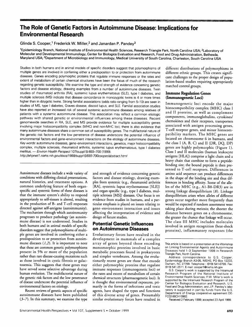
Inmune Regulation GenesOmnunogenetic Loci)Immunogenetic loci encode the majorhistocompatibility complex (MHC) class Iand II proteins, as well as complementcomponents, immunoglobulins, cytokines/chemokines and their receptors, transportersassociated with antigen processing genes,T-cell receptor genes, and minor histocom-patibility markers. The MHC genes arelocated on chromosome 6 in humans, andthe class I (A, B, C) and II (DR, DQ, DP)genes are highly polymorphic (Figure 1).Class I and II molecules (human leukocyteantigens [HLA]) comprise a light chain and aheavy chain that combine to form a peptide-binding site; the bound peptide is then pre-sented to T-cell receptors. Differences inamino acid sequence can produce differencesin the shape of the binding site and thus dif-ferences in binding affinity. Some of the alle-les of the MHC (e.g., Al-B8-DR3) are instrong linkage disequilibrium (8). Linkagedisequilibrium arises when alleles of differentgenes occur together more frequently thanwould be expected if random assortment weretaking place during meiosis; the shorter thedistance between genes on a chromosome,the greater the chance that linkage will occur.The class III MHC includes moleculesinvolved in antigen recognition (heat-shockproteins), inflammatory responses (the
This article is based on a presentation at the Workshopon Linking Environmental Agents and AutoimmuneDiseases held 1-3 September 1998 in ResearchTriangle Park, North Carolina.
Address correspondence to G.S. Cooper,Epidemiology Branch A3-05, NIEHS, PO Box 12233,Durham, NC 27709. Telephone: (919) 541-0799. Fax:(919) 541-251 1. E-mail: [email protected]
G.S. Cooper's work is supported by the IntramuralResearch Program of the National Institute ofEnvironmental Health Sciences; F.W. Miller's work issupported by the Intramural Research Program of theCenter for Biologics Evaluation and Research, U.S.Food and Drug Administration; and J.P. Pandey's labo-ratory is supported in part by funds from the U.S.Department of Energy cooperative agreement DE-FCO2-98CH10902.
Received 2 February 1999; accepted 23 April 1999.
Environmental Health Perspectives * Vol 107, Supplement 5 * October 1999 693
COOPER ET AL.
Class 11 (HLA) Class Ill (non-HLA) Class I (HLA)
Centromere
o--111-O--Q-0-O-D-sE- --O -1-O---4-t-----O ---
C DP TAP DA DR C C C C Bf C HSP TNF0 Y 4 Y 4 2L P B P A
2 21 1A B
B C A PRL
Figure 1. Genes of the human major histocompatibility complex, located on the short arm of chromosome 6.Abbreviations: HSP, heat shock protein; PRL, prolactin, TAP, transporters associated with antigen processing; TNF,tumor necrosis factor. Class (A, B, C) and class 11 genes (DR, DO, DP), shaded in grey, comprise the human leukocyteantigens. These molecules can be expressed on most cells, bind to peptides, and present the bound peptide to T-cellreceptors. Other immune-regulating genes are shaded white: TAP, complement components (C4B, C4A, Bf, C2), HSP,TNF. Genes with nonimmunologic functions are shaded in black: collagen (COLilA), 21a- and 21 i-hydroxylase(CYP21 and CYP21 P, respectively), and PRL.
complement proteins), and macrophageactivation (tumor necrosis factor [TNF]) (9).Other cytokines that are not encoded by theMHC play important roles in stimulating Tcells and B cells (e.g., interleukin [IL]-2, IL-6,IL-12, interferons) and therefore could beinvolved in autoimmune responses. Geneticvariability in the structure of immunoglobu-lins (immunoglobulin allotypes) and T-cellreceptors can also influence immune respon-
siveness to self-antigens and foreign antigens.Prolactin may also have important
immune-modulating influences affecting therisk of autoimmune disease (10). The pro-lactin gene is located close to the MHCregion of chromosome 6, and Brennan et al.(11) recently reported associations betweengenetic markers close to the prolactin genein SLE patients who also had DRB1*0301and in RA patients who had DRB1*0401.Thus, linkage disequilibrium may occur
between the class I, class II, and class IIIgenes of the MHC and also between theMHC genes and other nearby genes that are
not directly involved in immune regulation.Linkage disequilibrium between TNF-a andDRB may explain the conflicting resultsfrom studies of TNF-a polymorphisms,DRB alleles, and either the incidence or
clinical presentation ofRA (12-15).
Metabolism GenesThe metabolism of drugs, chemicals, anddietary constituents can require several differ-ent steps involving oxidation (sometimesreferred to as phase I) and conjugation ofoxygenated (electrophilic) intermediaries intohydrophilic compounds that are more easilyexcreted (phase 2). Oxidation enzymes
include cytochrome P450 enzymes (e.g.,CYPlAI, encoding arylhydrocarbon hydrox-ylase, and CYP2D6, encoding debrisoquinehydroxylase), myeloperoxidase, alcohol dehy-drogenase, and aldehyde dehydrogenase.Glutathione S-transferase, epoxide hydrolase,
sulfotransferase, and N-acetyltransferase(NAT) can act as phase II enzymes (16-18).The liver is the primary site of metabolism ofdrugs and other compounds, but additionalsteps can occur in the bladder, lung, colon,and other tissues. One of the isoforms ofNAT, NAT-1, is present in leukocytes (19),and myeloperoxidase is present in neu-
trophils (20). The toxicologic or carcino-genic activity of the metabolites along a
pathway varies; conjugated compounds are
generally but not always less reactive.Polymorphisms in many of these enzyme-
encoding genes have been reported. Thesepolymorphisms result in relatively slow andfast metabolism phenotypes, resulting in dif-ferences in exposures to the parent com-
pound and to specific metabolites.Polymorphisms in receptor genes such as thearomatic hydrocarbon receptor gene may
also influence metabolic activity (17).
Much of the work with respect tometabolism has focused on drug-inducedlupus. Drug-induced lupus shares some of theclinical and autoantibody features of SLE butdiffers in other respects. These syndromes areunintended outcomes of many commonlyused drugs such as procainamide, hydralazine,isoniazid, and penicillamine (21,22). Oneimportant aspect of drug-induced lupus isthat the condition most often resolvesafter the medication is discontinued (23).N-Acetyltransferase activity has been associ-ated with the development of drug-inducedlupus, with slow acetylation conferring higherrisk for developing specific autoantibodies orother features of this condition (24,25).
Studies of NAT activity in idiopathic(non-drug-induced) SLE have found littleevidence of an association (26-29). Thesestudies used a phenotypic assessment ofNATactivity based on a dapsone challenge ratherthan polymerase chain reaction-based tech-niques that can identify the genotype for eachof two NAT isoforms (NAT-1 and NAT-2).Only one of these studies also assessed expo-sure to aromatic amines (from dark hair dyesand from smoking) (29).
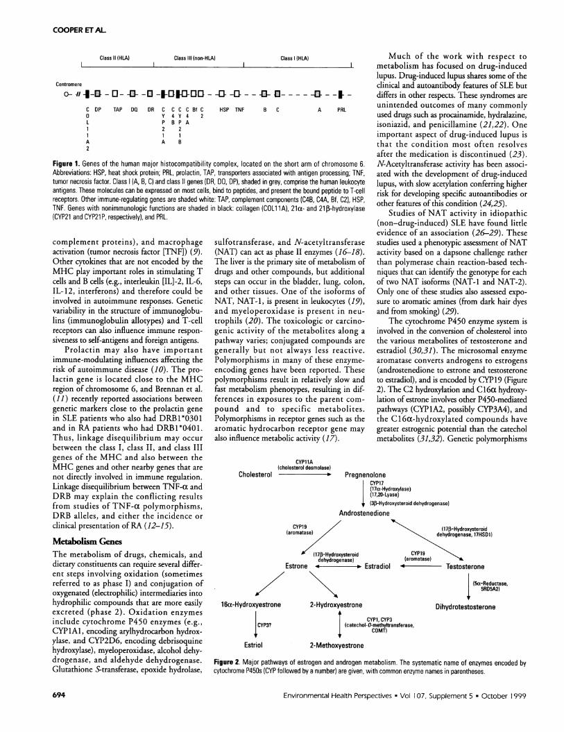
The cytochrome P450 enzyme system isinvolved in the conversion of cholesterol intothe various metabolites of testosterone andestradiol (30,31). The microsomal enzymearomatase converts androgens to estrogens(androstenedione to estrone and testosteroneto estradiol), and is encoded by CYP19 (Figure2). The C2 hydroxylation and Cl6a hydroxy-lation of estrone involves other P450-mediatedpathways (CYP1A2, possibly CYP3A4), andthe C16a-hydroxylated compounds havegreater estrogenic potential than the catecholmetabolites (31,32). Genetic polymorphisms
CYP11A(cholesterol desmolase)
Cholesterol PregnenoloneCYP17(17a-Hydroxylase)(17,20-Lyase)
I (3p-Hydroxysteroid dehydrogenase)Androstenedione
CYP19(aromatase)
(17p-Hydroxysteroiddehydrogenase)
Estrone . Estradiol
(17p-Hydroxysteroiddehydrogenase, 17HSD1)
CYP19(aromatase)4 Testosterone
(5a-Reductase,5RD5A2)
1 6c-Hydroxyestrone 2-Hydroxyestrone Dihydrotestosterone
CYP1, CYP3CYP3? (catechol-O-methyltransferase,
1 COMT)
Estriol 2-Methoxyestrone
Figure 2. Major pathways of estrogen and androgen metabolism. The systematic name of enzymes encoded bycytochrome P450s (CYP followed by a number) are given, with common enzyme names in parentheses.
Environmental Health Perspectives * Vol 107, Supplement 5 * October 1999
A2
694
THE ROLE OF GENETIC FACTORS IN AUTOIMMUNE DISEASES
and inducibility by specific medications,dietary components, and environmentalcontaminants may influence the activity ofvarious P450 enzymes (31,33). Although thereare no studies of predisease hormone levels andSLE risk, reduced androgen levels have beenreported in male and female SLE patients(34), and Lahita et al. (35) reported increased16a hydroxylation of estradiol in SLE patientsand their relatives compared to controls.
It is important to note that there aremultiple steps in most metabolic processesthat involve different enzymes and differentgenes. Determining the overall significanceof variation of one enzyme in a systemrequires consideration of all the steps, partic-ularly with respect to possible rate-limitingsteps, in the pathway. There may be factorsthat affect variation within pathways(inducibility of enzymes), and there also maybe competing pathways involving differentenzymes. Thus, metabolism of exogenousand endogenous compounds is an importantpotential source of variability in risk forautoimmune diseases, but its full importanceis not yet understood.
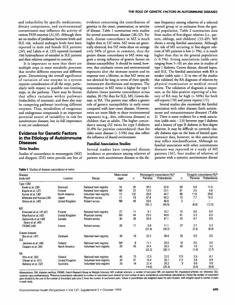
Evidence for Genetic Factorsin the Etiology of AutoimmuneDiseasesTwin StudiesStudies of concordance in monozygotic (MZ)and dizygotic (DZ) twins provide one line of
evidence concerning the contribution ofgenetics to the onset, presentation, or severityof disease. Table 1 summarizes twin studiesfor several autoimmune diseases (36-52). Foreach, disease concordance in MZ is muchhigher than in DZ twins. MZ twins are genet-ically identical, but DZ twins share on averageonly 50% of genes in common, thus thegreater disease concordance in MZ twins sug-gests a strong influence of genetic factors ondisease susceptibility. It should be noted, how-ever, that stochastic events and environmentalexposures alter the immune system and itsresponse over a lifetime, so that MZ twins arenot identical for long in terms of their specificimmunocyte distributions and receptors. Theconcordance in MZ twins is higher for type Idiabetes (mean pairwise concordance acrossstudies, 30.1%) than for SLE, MS, Graves dis-ease, or RA. This pattern may reflect a greaterrole of genetic susceptibility in early-onsetcompared with later-onset diseases. However,twins are more likely to share environmentalexposures (e.g., diet, infectious diseases) aschildren than as adults. The higher concor-dance among DZ twins for type I diabetes(6.8% for pairwise concordance) than forolder-onset diseases (< 3.5%) may also reflectthe influence of shared environment.
Familial Association StudiesSeveral studies have compared diseaseincidence or prevalence among relatives ofpatients with autoimmune diseases to the dis-
ease frequency among relatives of a selectedcontrol group or to estimates from the gen-eral population. Table 2 summarizes datafrom studies of first-degree relatives (i.e., par-ents, siblings, and children) (53-69). MSshows a strong familial association. Althoughthe risk of MS occurring in first-degree rela-tives of MS patients is low (< 5%), it is muchhigher than that in the general population(< 0.5%). Strong associations (odds ratioranging from 5-10) are also seen in studies oftype I diabetes, Graves disease, discoid lupus,and SLE. The familial association with RA isweaker (odds ratio < 2) in two of the studiesthat validated the RA diagnosis of relatives byphysical examination or medical recordreview. The validation of diagnosis is impor-tant, as the false-positive reporting of a his-tory of RA may be high (> 50%) for bothself-reports (70) and proxy reports (71).
Several studies also examined the familialassociation with other diseases (both autoim-mune and nonautoimmune diseases) (Table2). There is some evidence for a weak associa-tion (odds ratio - 2.0) between type I diabetesand a history of type II diabetes in first-degreerelatives. It may be difficult to correctly clas-sify diabetes type on the basis of limited ques-tionnaire data, however, so this associationmay reflect misclassification. Although nofamilial association with other autoimmunediseases was reported in a study of MSpatients (56), four studies of relatives ofpatients with a systemic autoimmune disease
Table 1. Studies of disease concordance in twins.
Disease Mean onset Monozygotic concordance (%)a Dizygotic concordance (%)astudy (ref) Location Design (age) n Pairwise Probandwise n Pairwise Probandwise
Type DMKyvik et al. (36) Denmark National twin registry 16 26 38.5 53.0 69 5.8 11.0Kaprio et al. (37) Finland National twin registry NR 23 13.0 23.1 81 2.5 4.8Kumar et al. (38) North America Volunteer twin registry 13 132 28.8 44.7 86 11.6 20.8Matsuda and Kuzuya (39) Japan Physician survey 1 1 19 47.4 64.3 13 7.7 14.3Olmos et al. (40) United Kingdom Patient survey NR 49 30.6 46.9 0 - -
(30.1) (45.5) (6.8) (12.5)MS
Kinnunen et al. (41,42) Finland National twin registry 27 11 9.1 16.7 10 0.0 0.0Mumford et al. (43) United Kingdom Physician survey NR 44 25.0 40.0 61 3.3 6.3Sadovnick et al. (44) Canada Patient survey 30 26 30.8 47.1 43 4.7 8.9Ebers et al. (45)FRGMS (46) France Patient survey 29 17 5.9 11.1 37 2.7 5.3
(21.4) (34.2) (3.3) (6.4)Graves disease
Brix et al. (47) Denmark National twin registry 39 18 22.2 36.4 33 0.0 0.0SLE
Jarvinen et al. (48) Finland National twin registry NR 9 11.1 20.0 10 0.0 0.0Deapen et al. (49) North America Volunteer twin registry 28 45 24.4 39.3 62 1.6 3.2
(22.2) (36.1) (1.4) (2.7)RA
Aho et al. (50) Finland National twin registry 45 73 12.3 22.0 173 3.5 6.7Silman et al. (51) United Kingdom Volunteer twin registry 38 91 15.4 26.7 112 3.6 6.9Bellamy et al. (52) Australia Volunteer twin registry 39 14 21.4 35.3 9 0.0 0.0
(14.6) (25.4) (3.4) (6.6)
Abbreviations: DM, diabetes mellitus; FRGMS, French Research Group on Mutiple Sclerosis; MS, multiple sclerosis, n, number of twin pairs; NR, not reported; RA, rheumatoid arthritis; ref, reference; SLE,systemic lupus erythematosus. Pairwise concordance calculated as number of concordant pairs divided by total number of pairs; probandwise concordance calculated as 2 times the number of concordantpairs divided by the sum of the number of discordant pairs plus 2 times the number of concordant pairs. Values in parentheses are weighted mean for each disease, with weights equal to number of pairsin each study.
Environmental Health Perspectives * Vol 107, Supplement 5 * October 1999 695
COOPER ETAL.
Table 2. Studies of familial associations with autoimmune diseases in first-degree relatives.
Disease Design, data source,a Familial association Familial associationstudy (ref) Location number of patients with same diseaseb with other diseasesb
Type DMAltobelli et al. (53) Italy CC, Q (parents), 136 4.0(1.610.2) Type II DM: 1.6(0.92-2.8)Dahiquist et al. (54) Sweden CC, Q (parents), 339 7.8 (3.6-16.8) Type It OM: 2.1 (0.35-14.3)Cederholm and Wibell (55) Sweden CC, Q (patients), 161 7.0 (4.2-11.9) Type II DM: 2.5 (1.4-4.4)
MSMidgard et al. (56) Norway CC, 0 (patients), 155 12.6 (1.7-552) Autoimmune diseases:c 1.2 (0.81-1.7)Robertson et al. (57) United Kingdom Cohort, exams, 674 9.2 NRSadovnick et al. (58) Canada Cohort, exams, 815 30-50 NR
Graves disease Serbia CC, Q (patients), 100 7.2(0.85-60) NRJankovi et al. (59)
Discoid lupus United Kingdom CC, exams, 37 7.2 (2.9-17.6) SLE: 8.9 (1.3 99)Lawrence et al. (60)
SLEStrom et al. (61) United States CC, G (patients), 195 2.0 (0.6-7.0) Autoimmune diseases:d2.3 (1.2-4.6)Nagata et al. (62) Japan CC, Q (patients), 282 NR Autoimmune diseases:e 5.2 (1.1-25)Lawrence et al. (60) United Kingdom CC, exams, 36 3.5 (2.2-142) NR
RAKoumantaki et al. (63) Greece CC, Q (patients), 126 4.4 (1.7-11.1) NRJones et al. (64) United Kingdom CC, exams, 207 1.6 (0.3-8.7) NRdel Junco et al. (65) United States Cohort, records, exams, 78 1.7 (1.0-2.9) NRLin et al. (66) United States CC, records, 29 15.5 (2.0-122) Autoimmune diseases:' 3.6 (1.2-14.5)
RA and others:f1 1.4 (2.5-47)Myositis United States CC, 0 (relatives), 21 NR Autoimmune diseases:9 7.9 (2.-21.9)
Ginn et al. (67)Systemic sclerosis Greece CC, 0 (patients), 166 NR Cancer: 3.8 (2.2-6.7)
Sakkas et al. (68)Sjogren syndrome United Kingdom CC, Q (patients), 42 1.9 (p< 0.01) Clinical thyroid disease: 6.6 (3.5-12.3)
Foster et al. (69) Autoimmune diseases:h 2.5
Abbreviations: CC, case-control; DM, diabetes mellitus; MS, multiple sclerosis; NR, not reported; 0, questionnaire or interview; RA, rheumatoid arthritis; ref, reference; SLE, systemic lupus erythematosus.'Questionnaire or interview asked either of patients or of their relatives; exams = physical examination of relatives reported to have the disease of interest; records = medical record review of relativesreported to have the disease of interest. bOdds ratio or risk ratio and 95% confidence interval or p-value. eRA, psoriasis, goitre, DM. dRA, inflammatory bowel disease, SLE, and other autoimmune dis-eases. 'Collagen diseases, including SLE. fAutoimmune thyroid disease, Type DM, rheumatic fever, ankylosing spondylitis, myasthenia gravis. #Includes autoimmune thyroid disease, RA, Type DM, pso-riasis, Sjogren syndrome, pernicious anemia, Takayasu arteritis, ulcerative colitis, hemolytic anemia, dermatomyositis, idiopathic thrombocyptopenic purpura, and other autoimmune diseases. kType DM,RA, pernicious anemia, SLE. Statistical significance not reported; odds ratio based on 7 cases in 140 relatives of probands compared to estimated population prevalence of 2%.
(SLE, RA, myositis, or Sjogren syndrome)(61,66,67,69) reported an increased risk ofother autoimmune diseases (the definition ofwhich varied between studies but thatincluded SLE, RA, and thyroid disease). Thisassociation may reflect a common etiologicpathway with shared genetic or environmentalinfluences between these diseases.
Two studies have shown familial linksbetween autoimmune diseases and cancer.One examined family history of cancer amongpatients with systemic sclerosis and found anodds ratio of 3.8 (95% confidence interval[CI], 2.2-6.7) (Table 2) (68). The otherexamined family history of autoimmune dis-eases among patients with multiple myeloma;the odds ratio for any reported autoimmunedisease (e.g., RA, SLE, and pernicious anemia)was 3.0 (95% CI, 1.3-7.1) (72). These obser-vations raise the possibility ofcommon patho-genic mechanisms involving cancer andautoimmune diseases, such as dysregulation ofapoptosis and detoxification pathways.
Gene Association StudiesGene association studies compare the frequencyof a specified genetic marker (measuredthrough either phenotypic assays or genotyp-ing) in patients and in a control group. Muchof the work with respect to autoimmune
diseases has focused on the MHC genes. Onecomplication in the design and interpretationof these studies, however, is the degree ofethnic variability in the prevalence of specificMHC alleles. Ethnicity in this context refersnot to broad racial groups but rather to muchsmaller groups defined by specific historical,migration, and sociocultural patterns. This isparticularly problematic in geographic areasthat have been the destination of significantimmigration. Another complication is thedegree of linkage disequilibrium between thegenes of the MHC, which may obscure theidentification of the effects of specific genes,particularly in early studies that relied on sero-logic measures of antigens (e.g., DR2 or DR3).Examples of gene association studies in SLEthat address ethnicity are shown in Table 3(73-78). The studies by Schur et al. (75) andGoldstein and Sengar (76) analyzed ethnicgroups within broad racial categories (e.g.,French Canadian and non-French Canadian).Both reported evidence of different associa-tions among different ethnic groups, althoughthe small sample size in the Goldstein andSengar study resulted in variable estimates thatmake it difficult to definitely interpret theobserved differences.
The selection of controls in population-based gene association studies is very
important, as it may be difficult to adequatelyaccount for genetic admixture of the popula-tion. Alternative designs such as geneassociation analyses using case-parent triadsavoid this problem and do not requireassumptions about type of inheritance or dis-ease penetrance (8,79) that are needed inother analytic approaches.
Pedigree Studies: Segregation Analysis,Link Analysis, and Genome ScaithesSegregation analysis is the first step inidentifying the relation between an individual'sgenotype and the resulting phenotype (80).Using appropriate statistical methods, onecompares the inheritance of the diseasewithin families with that expected under spe-cific models. The models may evaluatea) whether there is a single major generesponsible for the autoimmune disease,b) whether the susceptibility to the disease iscontrolled by many genes (polygenic inheri-tance), and c) the environmental transmis-sion model. The model that is mostcompatible with the observed family data isadopted. Identification of a major gene doesnot mean that it is the only gene responsiblefor the disease; rather, its effect is large enoughto be discernible from those of the other genesimplicated in the etiology of the disease.
Environmental Health Perspectives * Vol 107, Supplement 5 * October 1999696

THE ROLE OF GENETIC FACTORS IN AUTOIMMUNE DISEASES
Table 3. Case-control studies of major histocompatibility complex associations with SLE in two or more ethnic groups.
Controls PatientsGene Percent Percent Odds ratio (95%study (ref) Population Source n positive n positive confidence interval)C4A null
Howard et al. (73)a United States, blacks Blood donors 35 7 35 20 3.3 (1.0-12.2)United States, whites Blood donors 63 10 63 25 3.2(1.5-7.1)
Dunckley et al. (74)a Australia, whites Blood donors 197 17 63 32 2.3 (1.4-3.7)Chinese Unspecified 76 19 75 30 1.9 (1.1- 3.3)Japanese Unspecified 50 12 51 35 3.8 (1.8-8.5)
Schur et al. (75)b United States, English/Irish Non-SLE relatives 144 22 27 41 2.5 (0.97-6.5)United States, other whites Non-SLE relatives 310 12 62 11 0.94 (0.34-2.3)
Goldstein and Sengar (76)a Quebec, French University employees 44 6 43 12 2.2 (0.64-8.5)Quebec, non-French University employees 36 10 43 31 4.3 (1.6-11.7)
Reveille et al. (77)C United States, blacks Blood donors 73 20 88 20 0.99 (0.43-2.3)United States, Hispanics Blood donors, university employees 119 13 68 19 1.6 (0.67-3.9)United States, whites Unspecified 186 20 69 30 1.8 (0.90-3.5)
DR3Howard et al. (73)d United States, blacks Blood donors 35 26 35 31 1.3 (0.41-4.3)
United States, whites Blood donors 63 25 63 38 1.8(0.79-4.2)Schur etal. (75)b United States, English/Irish Non-SLE relatives 144 19 27 41 2.9(1.1-7.4)
United States, other whites Non-SLE relatives 310 10 62 16 1.7 (0.74-4.0)Goldstein and Sengar (76)e Quebec, French University employees 43 12 43 23 2.4(0.65-9.6)
Quebec, non-French University employees 35 20 43 58 5.6 (1.8-17.8)Reveille et al. (77)f United States, blacks Blood donors 88 14 88 14 1.0 (0.39-2.6)
United States, Hispanics Blood donors, university employees 105 9 70 20 2.7 (1.0-7.2)United States, whites Unspecified 200 25 67 51 3.1 (1.7-5.7)
TNF-a-238ARudwaleit et al. (78)g United Kingdom, whites Blood donors 96 9 49 2 0.20 (0.00-1.5)
South Africa, blacks Unspecified 81 19 49 24 1.4 (0.56-3.7)TNFa-r3mA
Rudwaleit et al. (78)h United Kingdom, whites Blood donors 96 28 49 47 2.3 (1.0-4.9)South Africa, blacks Unspecified 81 35 49 27 0.68 (0.29-1.6)
Abbreviations: n, number; ref, reference; SLE, systemic lupus erythematosus; TNF, tumor necrosis factor. JGene frequency, based on phenotypic measurement; total number used in calculations is equal totwice the number of patients. hAllele frequency, based on phenotypic measurement; total number used in calculations is total number of haplotypes. cAllotype frequency; based on phenotypic measure-ment; total number used in calculations is number of patients. dSerologic measurement of DR3; total number used in calculations is number of patients. 0DR3(17) specificity based on analysis of restrictionfragment length polymorphisms; total number used in calculations is number of patients. fDR3*0301 allele frequency; total number used in calculations is number of patients. 'Frequency of TNF-cr238, theG to A substitution at the -238 postition of the promotor region of TNF-a, which results in the TNF-A variant. *Frequency of TNF-ae3-8, the G to A substitution at postition -308 of the promotor region ofTNF-a, which results in the TNF-2 variant.
Recently, more powerful methods of segrega-tion analyses, called complex segregationanalyses, have been developed (81). These canbe applied to both quantitative and qualitativetraits and can elucidate complex patterns ofgenetic/environmental transmission.
An early segregation analysis involving 18selected kindreds suggested that autoimmu-nity is controlled by a single autosomal domi-nant gene (82). The postulated majorautoimmune gene has not been mapped, butin two studies of familial patterns of auto-immune diseases, linkage to HLA or geneticmarkers of human immunoglobulin gammaor kappa chain (GM and KM) allotypes wasexcluded (82-84). Recent investigations aremore consistent with the belief that auto-immunity is polygenetic (1). Development ofspecific autoantibodies or an autoimmunedisease may depend on the epistatic interac-tions of autoimmunity-predisposing genesand environmental factors.
Linkage implies cosegregation of alleles attwo different loci. Linkage of a marker locusand a disease provides much stronger evi-dence for a substantial genetic component inthe etiology of the disease than that providedby segregation analysis. It is important to
remember that the association analysis dis-cussed in the previous section specifies arelationship between an allele and a disease,whereas linkage denotes a close physicallocalization of a marker locus and the puta-tive locus for the disease. Loci are linked butnot their alleles (unless there is linkagedisequilibrium/allelic association). In otherwords, the marker allele segregating with thedisease may be different in different families.
Different analytical strategies have beendeveloped that take advantage of informationprovided by genetic markers in families ofindividuals affected by autoimmune diseases.These linkage approaches include the log-odds score method, the affected-sibpairmethod, the affected-pedigree-membermethod, the variance component method,and linkage disequilibrium-based approaches(80,85-87). Each of these has intrinsicadvantages and disadvantages, and the choicebest suited for a given study depends uponmany factors, including the epidemiology ofthe disorder investigated, the state of knowl-edge about the nature and frequencies ofgenetic risk factors and linked loci, and theresources available. All of these approachesare efficient in defining genetic linkages for
well-defined monogenic traits when largenumbers of individuals are available for analy-ses. They have limitations, however, whenapplied to rare, complex disorders in whichmultiple genes, or gene-environment interac-tions are likely to play pathogenic roles. It hasbeen suggested that family-based associationstudies (e.g., case-parent triad designs)employing a large number of candidate genesare more powerful than other approaches indissecting the genetic contribution to suchdisorders (88).
Until recently most linkage andassociation analyses in autoimmune diseasesstudied candidate genes coding for HLA,the T-cell receptor, GM and KM allotypes,the complement components, and other rel-evant proteins. Now, with the availability ofvery polymorphic microsatellite markersscattered throughout the genome, one cansearch the entire genome for autoimmunitygenes without knowledge of their mode ofinheritance or function. Genomewidesearches in RA, SLE, and MS provide evi-dence for multiple susceptibility genesinvolving MHC and non-MHC loci(89-93). A recent comparison of the linkageresults from 23 published genomewide scans
Environmental Health Perspectives * Vol 107, Supplement 5 * October 1999 697
COOPER ET AL.
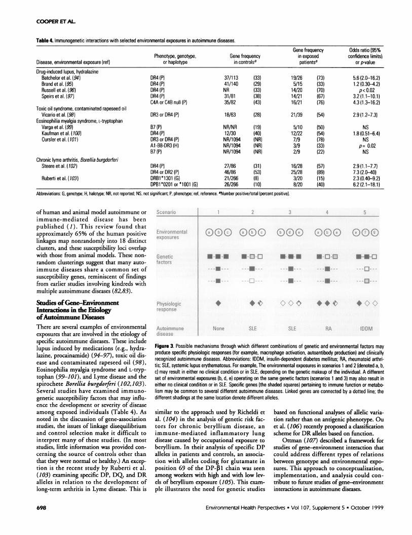
Table 4. Immunogenetic interactions with selected environmental exposures in autoimmune diseases.
Gene frequency Odds ratio (95%Phenotype, genotype, Gene frequency in exposed confidence limits)
Disease, environmental exposure (ref) or haplotype in controlsa patientsa or p-valueDrug-induced lupus, hydralazine
Batchelor et al. (94) DR4 (P) 37/113 (33) 19/26 (73) 5.6(2.0-16.2)Brand et al. (95) DR4 (P) 41/140 (29) 5/15 (33) 1.2(0.30-4.2)Russell et al. (96) DR4 (P) NR (33) 14/20 (70) p< 0.02Speirs et al. (97) DR4 (P) 31/81 (38) 14/21 (67) 3.2 (11-10.1)
C4A or C4B null (P) 35/82 (43) 16/21 (76) 4.3 (1.3-16.2)Toxic oil syndrome, contaminated rapeseed oil
Vicario et al. (98) DR3 or DR4 (P) 18/63 (28) 21/39 (54) 2.9 (1.2-7.3)Eosinophilia myalgia syndrome, [-tryptophan
Varga et al. (99) B7 (P) NR/NR (19) 5/10 (50) NSKaufman et al. (100) DR4 (P) 12/30 (40) 12/22 (54) 1.8 (0.51-6.4)Oursler et al. (101) DR3 or DR4 (P) NR/1 094 (NR) 7/9 (78) NS
A1-88-DR3 (H) NR/1 094 (NR) 3/9 (33) p= 0.02B7 (P) NR/1094 (NR) 2/9 (22) NS
Chronic lyme arthritis, Borellia burgdorferiSteere et al. (102) DR4 (P) 27/86 (31) 16/28 (57) 2.9 (1.1-7.7)
DR4 or DR2 (P) 46/86 153) 25/28 (89) 7.3(2.0-40)Ruberti et al. (103) DRB1*1301 (G) 21/266 (8) 3/20 (15) 2.3(0.40-9.2)
DPB1*0201 or *1001 (G) 26/266 (10) 8/20 (40) 6.2 (2.1-18.1)Abbreviations: G, genotype; H, halotype; NR, not reported; NS, not significant; P, phenotype; ref, reference. &Number positive/total (percent positive).
of human and animal model autoimmune orimmune-mediated disease has beenpublished (1). This review found thatapproximately 65% of the human positivelinkages map nonrandomly into 18 distinctclusters, and these susceptibility loci overlapwith those from animal models. These non-random clusterings suggest that many auto-immune diseases share a common set ofsusceptibility genes, reminiscent of findingsfrom earlier studies involving kindreds withmultiple autoimmune diseases (82,83).
Studies ofGene-EnvironmentInterctions in the EtiologyofAuto une DiseasesThere are several examples of environmentalexposures that are involved in the etiology ofspecific autoimmune diseases. These includelupus induced by medications (e.g., hydra-lazine, procainamide) (94-97), toxic oil dis-ease and contaminated rapeseed oil (98),Eosinophilia myalgia syndrome and L-tryp-tophan (99-101), and Lyme disease and thespirochete Borellia burgdorferi (102,103).Several studies have examined immuno-genetic susceptibility factors that may influ-ence the development or severity of diseaseamong exposed individuals (Table 4). Asnoted in the discussion of gene-associationstudies, the issues of linkage disequilibriumand control selection make it difficult tointerpret many of these studies. (In moststudies, little information was provided con-cerning the source of controls other thanthat they were normal or healthy.) An excep-tion is the recent study by Ruberti et al.(103) examining specific DP, DQ, and DRalleles in relation to the development oflong-term arthritis in Lyme disease. This is
Scenario
t_|......sum
*: .......
ii3iiliz ; ;e.e
..,.... .. 1, ;:, ,.....,;,
.^
*. -
fj,...,0;.......- -
......., . .- . ... ... ..;1.'. ...1 1.
* :. .&:;2 3- 4:-
. . U~- - --S----$4--... i ; ; ; t X
:,;t'*'S:'wo~~~~~~~~~~~~~~~~~~~~~~~~~~~~~~~~~~~~~~~~~~~~~~~~~~~~~~.;1,n
.~~~~~~~~~~~~~~~~~~~~~~.....
. . > w * ar.J..tEams~~~~~~~~~~~~~~~~~~~~~. :I.A<~ ,*.:i.-Z'*
"D* *;!;lrl S^fltt l~~~~~~~~~~~~~~~~~~~~~~~~~~~~~~~~~~~1
. .+ ..+. 'Vt t5t + ''@,*S~~~~~~~~~~~~~~~~~~t.x
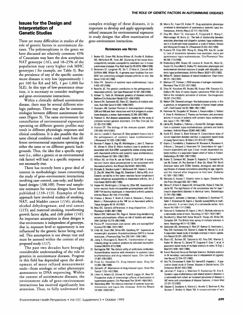
Figure 3. Possible mechanisms through which different combinations of genetic and environmental factors mayproduce specific physiologic responses (for example, macrophage activation, autoantibody production) and clinicallyrecognized autoimmune diseases. Abbreviations: IDDM, insulin-dependent diabetes mellitus; RA, rheumatoid arthri-tis; SLE, systemic lupus erythematosus. For example, The environmental exposures in scenarios 1 and 2 (denoted a, b,c) may result in either no clinical condition or in SLE, depending on the genetic makeup of the individual. A differentset of environmental exposures (b, d, e) operating on the same genetic factors (scenarios 1 and 3) may also result ineither no clinical condition or in SLE. Specific genes (the shaded squares) pertaining to immune function or metabo-lism may be common to several different autoimmune diseases. Linked genes are connected by a dotted line; thedifferent shadings at the same location denote different alleles.
similar to the approach used by Richeldi etal. (104) in the analysis of genetic risk fac-tors for chronic beryllium disease, animmune-mediated inflammatory lungdisease caused by occupational exposure toberyllium. In their analysis of specific DPalleles in patients and controls, an associa-tion with alleles coding for glutamate inposition 69 of the DP-31 chain was seenamong workers with high and with low lev-els of beryllium exposure (105). This exam-ple illustrates the need for genetic studies
based on functional analyses of allelic varia-tion rather than on antigenic phenotype. Ouet al. (106) recently proposed a classificationscheme for DR alleles based on function.
Ottman (107) described a framework forstudies of gene-environment interaction thatcould address different types of relationsbetween genotype and environmental expo-sures. This approach to conceptualization,implementation, and analysis could con-tribute to future studies of gene-environmentinteractions in autoimmune diseases.
Environmental Health Perspectives * Vol 107, Supplement 5 * October 1999
s.
-- ah. I:Li.1,
698
THE ROLE OF GENE7IC FACTORS IN AUTOIMMUNE DISEASES
Issues for the Design andInterpretation ofGenetic StudiesThere are many difficulties in studies of therole of genetic factors in autoimmune dis-eases. The polymorphisms in the genes wehave discussed are relatively common: 45%of Caucasians may have the slow acetylationNAT genotype (16), and 10-25% of thepopulation may carry higher risk MHCgenotypes ( for example, see Table 3). Butthe prevalence of any of the specific autoim-mune diseases is very low (approximately 1per 100 for RA and MS, 1 per 1,000 forSLE). In this type of low-penetrance situa-tion, it is necessary to consider multigeneand gene-environment interactions.
Within a clinically defined autoimmunedisease, there may be several different etio-logic pathways. There may also be commonetiologies between different autoimmune dis-eases (Figure 3). The same environment (orconstellation of environmental exposures)operating on different genetic profiles mayresult in different physiologic responses andclinical conditions. It is also possible that thesame clinical condition could result from dif-ferent environmental exposures operating oneither the same or on different genetic back-grounds. Thus, the idea that a specific expo-sure (i.e., either a genetic or an environmentalrisk factor) will lead to a specific response isnot necessarily true.
There has recently been a great deal ofinterest in methodologic issues concerningthe study of gene-environment interactionsinvolving case-control, case-only, and family-based designs (108,109). Power and sample-size estimates for various designs have beenpublished (110-113). Examples of thisapproach have involved studies of smoking,NAT, and bladder cancer (114); alcohol,alcohol dehydrogenase, and oral cancer(115); and maternal smoking, transforminggrowth factor alpha, and cleft palate (116).An important assumption in these designs isthat environment is independent of genotype,that is, exposure level or opportunity is notinfluenced by the genetic factor being stud-ied. This assumption is not always true andmust be assessed within the context of anyproposed study (117).
The past two decades have broughtconsiderable understanding of the role ofgenetics in autoimmune diseases. Progressin this field has depended upon the devel-opment of more refined measurementtools-from serologic or other phenotypicassessments to DNA sequencing. Withinthe context of autoimmune diseases, theenvironment side of gene-environmentinteractions has received significantly lessattention. Thus, to fully understand the
complex etiology of these diseases, it isimportant to develop and apply appropriatelyrefined measures for environmental exposuresin study designs that allow examination ofgene-environment interactions.
REFERENCES AND NOTES
1. Becker KG, Simon RM, Bailey-Wilson JE, Freidlin B, BiddisonWE, McFarland HF, Trent JM. Clustering of non-major histo-compatibility complex susceptibility candidate loci in humanautoimmune diseases. Proc NatI Acad Sci 95:9979-9984 (1998).
2. Remmers EF, Longman RE, Du Y, O'Hare A, Cannon GW,Griffiths MM, Wilder RL. A genome scan localizes five non-MHC loci controlling collagen-induced arthritis in rats. NatGenet 14:82-85 (1996).
3. Schur PH Genetics of systemic lupus erythematosus. Lupus4:425-437 (1995).
4. Reveille JD. The genetic contribution to the pathogenesis ofrheumatoid arthritis. Curr Opin Rheumatol 10:187-200 (1998).
5. Aitman TJ, Todd JA. Molecular genetics of diabetes mellitus.Bailliere Clin Endocrinol Metab 9:631-656 (1995).
6. Dyment DA, Sadnovich AD, Ebers GC. Genetics of multiple scle-rosis. Hum Mol Genet 6:1693-1698 (1997).
7. Dogoujon JM, Cambon-Thomsen A. Immunoglobulin allotypes(GM and KM) and their interactions with HLA antigens in autoim-mune diseases: a review. Autoimmunity 22:245-260 (1995).
8. Thomson G. HLA disease associations: models for the study ofcomplex human genetic disorders. Crit Rev Clin lab Sci32:183-219 (1995).
9. Huston DP. The biology of the immune system. JAMA278:1804-1814 (1997).
10. Jara LJ, Lavalle C, Espinoza LR. Does prolactin have a role inthe pathogenesis of systemic lupus erythematosus?J Rheumatol 19:1333-1336 (1992).
11. Brennan P, Hajeer A, Ong KR, Worthington J, John S, ThomsonW, Silman A, Oilier B. Allelic markers close to prolactin areassociated with HLA-DRB1 susceptibility alleles among womenwith rheumatoid arthritis and systemic lupus erythematosus.Arthritis Rheum 40:1383-1386(1997).
12. Wilson AG, de Vries N, van de Putte LB, Duff GW. A tumournecrosis factor alpha polymorphism is not associated withrheumatoid arthritis. Ann Rheum Dis 54:601-603 (1995).
13. Mulcahy B, Waldron-Lynch F, McDermott MF, Adams C, AmosCl, Zhu DK, Ward RH, Clegg DO, Shanahan F, Molloy MG, et al.Genetic variability in the tumor necrosis factor-lymphotoxinregion influences susceptibility to rheumatoid arthritis. Am JHum Genet 59:676-683 (1996).
14. Hajeer AH, Worthington J, Silman AJ, Oilier WE. Association oftumor necrosis factor microsatellite polymorphisms with HLA-DRB1*04-bearing haplotypes in rheumatoid arthritis patients.Arthritis Rheum 39:1109-1114 (1996).
15. Vinasco J, Beraun Y, Nieto A, Fraile A, Mataran L, Pareja E,Martin J. Polymorphism at the TNF loci in rheumatoid arthritis.Tissue Antigens 49:74-78 (1997).
16. May DG. Genetic differences in drug disposition. J ClinPharmacol 34:881-897 (1994).
17. Nebert DW, McKinnon RA, Puga A. Human drug-metabolizingenzyme polymorphisms: effects on risk of toxicity and cancer.DNA Cell Biol 15:273-280 (1996).
18. Perera FP. Environment and cancer: who are susceptible?Science 278:1068-1073 (1997).
19. Cribb AE, Grant DM, Miller MA, Spielberg SP Expression ofmonomorphic arylamine N-acetyltransferase (NAT1) in humanleukocytes. J Pharmacol Exp Ther 259:1241-1246 (1991).
20. Jiang X, Khursigara G, Rubin RL. Transformation of lupus-inducing drugs to cytotoxic products by activated neutrophils.Science 266:810-813 (1994).
21. Burlingame RW. The clinical utility of antihistone antibodies.Autoantibodies reactive with chromatin in systemic lupuserythematosus and drug-induced lupus. Clin Lab Med17:367-378 (1997).
22. Price EJ, Venables PJ. Drug-induced lupus. Drug Saf12:283-290(1995).
23. Uetrecht JP. Mechanism of drug-induced lupus. Chem ResToxicol 1:133-143 (1988).
24. Litwin A, Adams LE, Zimmer H, Foad B, Loggie JH, Hess EV.Prospective study of immunologic effects of hydralazine inhypertensive patients. Clin Pharmacol Ther 29:447-456 (1981).
25. Reidenberg MM. The chemical induction of systemic lupus ery-thematosus and lupus-like illnesses. Arthritis Rheum24:1004-1009 (1981).
26. Morris RJ, Freed CR, Kohier PF. Drug acetylation phenotypeunrelated to development of spontaneous systemic lupus ery-thematosus. Arthritis Rheum 22:777-780 (1979).
27. Ong ML, Mant TG, Veerapen K, Fitzgerald D, Wang F,Manivasagar M, Bosco JJ The lack of relationship betweenacetylator phenotype and idiopathic systemic lupus erythemato-sus in a South-east Asian population: a study of Indians, Malaysand Malaysian Chinese. Br J Rheumatol 29:462-464 (1990).
28. Kumana CR, Chan MM, Wong KL, Wong RW, Kou M, lauderIJ. Lack of association between slow acetylator status andspontaneous lupus erythematosus. Clin Pharmacol Ther48:208-213 (1990).
29. Reidenberg MM, Drayer DE, Lorenzo B, Strom BL, West SL,Snyder ES, Freundlich B, Stolley PD. Acetylation phenotypes andenvironmental chemical exposure of people with idiopathic sys-temic lupus erythematosus. Arthritis Rheum 36:971-973 (1993).
30. White PC. Genetic diseases of steroid metabolism. Vitam Horm49:131-195 (1994).
31. Martucci CP, Fishman J. P450 enzymes of estrogen metabolism.Pharmacol Ther 57:237-257 (1993).
32. Shou M, Korzekwa KR, Brooks EN, Krausz KW, Gonzalez FJ,Gelboin HV. Role of human hepatic cytochrome P450 1A2 and3A4 in the metabolic activation of estrone. Carcinogenesis18:207-214 (1997).
33. Nebert DW. Elevated estrogen 16ca-hydroxylase activity: is thisa genotoxic or nongenotoxic biomarker in human breast cancerrisk? J NatI Cancer Inst 85:1888-1891 (1993).
34. Folomeev M, Dougados M, Beaune J, Kouyoumdjian J, NahoulK, Amor B, Alekberova Z. Plasma sex hormones and aromataseactivity in tissues of patients with systemic lupus erythemato-sus. Lupus 1:191-195 (1992).
35. Lahita RG, Bradlow L, Fishman J, Kunkel HG. Estrogen metabo-lism in systemic lupus erythematosus: patients and familymembers. Arthritis Rheum 25:843-846 (1982).
36. Kyvik KO, Green A, Beck Nielsen H. Concordance rates ofinsulin dependent diabetes mellitus: a population based studyof young Danish twins. Br Med J 311:913-917 (1995).
37. Kaprio J, Tuomilehto J, Koskenvuo M, Romanov K, Reunanen A,Eriksson J, Stengard J, Kesaniemi YA. Concordance for type 1(insulin dependent) and type 2 (non- insulin dependent) dia-betes mellitus in a population based cohort of twins in Finland.Diabetologia 35:1060-1067 (1992).
38. Kumar D, Gemayel NS, Deapen D, Kapadia D, Yamashita PH,Lee M, Dwyer JH, Roy Burman P, Bray GA, Mack TM. NorthAmerican twins with IDDM. Genetic, etiological, and clinicalsignificance of disease concordance according to age, zygosity,and the interval after diagnosis in first twin. Diabetes42:1351-1363 (1993).
39. Matsuda A, Kuzuya T. Diabetic twins in Japan. Diabetes ResClin Pract 24(suppl):S63-S67 (1994).
40. Olmos P, A'Hern R, Heaton DA, Millward BA, Risley D, Pyke DA,Leslie RD. The significance of the concordance rate for type 1(insulin dependent) diabetes in identical twins. Diabetologia31:747-750 (1988).
41. Kinnunen E, Juntunen J, Ketonen L, Koskimies S, Konttinen YT,Salmi T, Koskenvuo M, Kaprio J. Genetic susceptibility to multi-ple sclerosis. A co-twin study of a nationwide series. ArchNeurol 45:1108-1111 (1988).
42. Kinnunen E, Koskenvuo M, Kaprio J, Aho K. Multiple sclerosis ina nationwide series of twins. Neurology 37:1627-1629 (1987).
43. Mumford CJ, Wood NW, Kellar Wood H, Thorpe JW, Miller DH,Compston DA. The British Isles survey of multiple sclerosis intwins. Neurology 44:11-15 (1994).
44. Sadovnick AD, Armstrong H, Rice GP, Bulman D, Hashimoto L,Paty DW, Hashimoto SA, Warren S, Hader W, Murray TJ, et al.A population based study of multiple sclerosis in twins: update.Ann Neurol 33:281-285 (1993).
45. Ebers GC, Bulman DE, Sadovnick AD, Paty DW, Warren S,Hader W, Murray TJ, Seland TP, Duquette P, Grey T, et al. Apopulation based study of multiple sclerosis in twins. N EngI JMed 315:1638-1642 (1986).
46. French Research Group on Multiple Sclerosis. Multiple sclerosisin 54 twinships: concordance rate is independent of zygosity.Ann Neurol 32:724-727 (1992).
47. Brix TH, Christensen K, Holm NV, Harvald B, Hegedus L A pop-ulation based study of Graves' disease in Danish twins. ClinEndocrinol 48:397-400 (1998).
48. Jarvinen P, Kaprio J, Makitalo R, Koskenvuo M, Aho K.Systemic lupus erythematosus and related systemic diseases ina nationwide twin cohort: an increased prevalence of disease inMZ twins and concordance of disease features. J Intern Med2311:67-72 (1992).
49. Deapen D, Escalante A, Weinrib L, Horwitz D, Bachman B, RoyBurman P, Walker A, Mack TM. A revised estimate of twin
Environmental Health Perspectives * Vol 107, Supplement 5 * October 1999 699

COOPER ET AL.
concordance in systemic lupus erythematosus. Arthritis Rheum35:311- 318(1992).
50. Aho K, Koskenvuo M, Tuominen J, Kaprio J. Occurrence ofrheumatoid arthritis in a nationwide series of twins. JRheumatol 13:899-902 (1986).
51. Silman AJ, MacGregor Aj, Thomason W, Holligan S, Carthy D,Farhan A, Oilier WE. Twin concordance rates for rheumatoidarthritis: results from a nationwide study. Br J Rheumatol32:903-907 (1993).
52. Bellamy N, Duffy D, Martin N, Mathews J. Rheumatoid arthritisin twins: a study of aetiopathogenesis based on the AustralianTwin Registry. Ann Rheum Dis 51:588-593 (1992).
53. Altobelli E, Chiarelli F, Valenti M, Verrotti A, Blasetti A,Di-Orio F. Family history and risk of insulin-dependent diabetesmellitus: a population-based case-control study. Acta Diabetol35:57-60 (1998).
54. Dahlquist G, Blom L, Tuvemo T, Nystrom L, Sandstrom A, WallS. The Swedish childhood diabetes study-results from a nineyear case register and a one year case-referent study indicatingthat type 1 (insulin-dependent) diabetes mellitus is associatedwith both type 2 (non-insulin-dependent) diabetes mellitus andautoimmune disorders. Diabetologia 32:2-6 (1989).
55. Cederholm J, Wibell L. Familial influence on type 1 (insulinde-pendent) diabetes mellitus by relatives with either insulin-treated or type 2 (non-insulin-dependent) diabetes mellitus.Diabetes Res 18:109-113 (1991).
56. Midgard R, Gronning M, Riise T, Kvale G, Nyland H. Multiplesclerosis and chronic inflammatory diseases. A case-controlstudy. Acta Neurol Scand 93:322-328 (1996).
57. Robertson NP, Fraser M, Deans J, Clayton D, Walker N,Compston DA. Age-adjusted recurrence risks for relatives ofpatients with multiple sclerosis. Brain 1 19(Pt 2):449-455 (1996).
58. Sadovnick AD, Baird PA, Ward RH. Multiple sclerosis: updatedrisks for relatives. Am J Med Genet 29:533-541 (1988).
59. Jankovic SM, Radosavljevic R, Marinkovic JM. Risk factors forGraves' disease. EurJ Epidemiol 13:1518 (1997).
60. Lawrence JS, Martins CL, Drake GL. A family survey of lupuserythematosus. 1: Heritability. J Rheumatol 14:913-921 (1987).
61. Strom BL, Reidenberg MM, West S, Snyder ES, Freundlich B,Stolley PD. Shingles, allergies, family medical history, oral con-traceptives, and other potential risk factors for systemic lupuserythematosus. Am J Epidemiol 140:632-642 (1994).
62. Nagata C, Fujita S, Iwata H, Kurosawa Y, Kobayashi K,Kobayashi M, Motegi K, Omura T, Yamamoto M, Nose T, et al.Systemic lupus erythematosus: a case-control epidemiologicstudy in Japan. Int J Dermatol 34:333-337 (1995).
63. Koumantaki Y, Giziaki E, Linos A, Kontomerkos A, Kaklamanis P,Vaiopoulos G, Mandas J, Kaklamani E. Family history as a riskfactor for rheumatoid arthritis: a case-control study.J Rheumatol 24:1522-1526 (1997).
64. Jones MA, Silman AJ, Whiting S, Barrett EM, Symmons DP.Occurrence of rheumatoid arthritis is not increased in the firstdegree relatives of a population based inception cohort ofinflammatory polyarthritis. Ann Rheum Dis 55:89-93 (1996).
65. del Junco D, Luthra HS, Annegers F, Worthington JW, KuriandLT. The familial aggregation of rheumatoid arthritis and its rela-tionship to the HLA-DR4 association. Am J Epidemiol119:813-829 (1984).
66. Lin JP, Cash JM, Doyle SZ, Peden S, Kanik K, Amos Cl, Balc SJ,Wilder RL. Familial clustering of rheumatoid arthritis with otherautoimmune diseases. Hum Genet 103:475-482 (1998).
67. Ginn LR, Lin JP, Plotz PH, Bale SJ, Wilder RL, Mbauya A, MillerFW. Familial autoimmunity in pedigrees of idiopathic inflamma-tory myopathy patients suggests common genetic risk factors formany autoimmune diseases. Arthritis Rheum 41:400-405 (1998).
68. Sakkas LI, Moore DF, Akritidis NC. Cancer in families with sys-temic sclerosis. Am J Med Sci 310:223-225 (1995).
69. Foster H, Fay A, Kelly C, Charles P, Walker D, Griffiths I. Thyroiddisease and other autoimmune phenomena in a family study ofprimary Sjogren's syndrome. Br J Rheumatol 32:36-40 (1993).
70. Linet MS, Harlow SD, McLaughlin JK, McCaffrey LD. A compar-ison of interview data and medical records for previous medicalconditions and surgery. J Clin Epidemiol 42:1207-1213 (1989).
71. Kwoh CK, Venglish C, Lynn AH, Whitley DM, Young E,Chakravarti A. Age, sex, and the familial risk of rheumatoidarthritis. Am J Epidemiol 144:15-24 (1996).
72. Linet MS, Mclaughlin JK, Harlow SD, Fraumeni JF. Family his-
tory of autoimmune disorders and cancer in multiple myeloma.lntJ Epidemiol 17:512-513(1988).
73. Howard PF, Hochberg MC, Bias WB, Arnett FC Jr, McLean RH.Relationship between C4 null genes, HLA D region antigens,and genetic susceptibility to systemic lupus erythematosus inCaucasian and black Americans. Am J Med 81:187-193 (1986).
74. Dunckley H, Gatenby PA, Hawkins B, Naito S, Serjeantson SW.Deficiency of C4A is a genetic determinant of systemic lupuserythematosus in three ethnic groups. J Immunogenet14:209-218 (1987).
75. Schur PH, Marcus Bagley D, Awdeh Z, Yunis EJ, Alper CA. Theeffect of ethnicity on major histocompatibility complex comple-ment allotypes and extended haplotypes in patients with sys-temic lupus erythematosus. Arthritis Rheum 33:985-992 (1990).
76. Goldstein R, Sengar DP. Comparative studies of the major histo-compatibility complex in French Canadian and non-FrenchCanadian Caucasians with systemic lupus erythematosus.Arthritis Rheum 36:1121-1127 (1993).
77. Reveille JD, Moulds JM, Ahn C, Friedman AW, Baethge B,Roseman J, Straaton KV, Alarcon GS. Systemic lupus erythe-matosus in three ethnic groups. I: The effects of HLA class 11, C4,and CR1 alleles, socioeconomic factors, and ethnicity at diseaseonset. LUMINA Study Group. Lupus in minority populations,nature versus nurture. Arthritis Rheum 41:1161-1172 (1998).
78. Rudwaleit M, Tikly M, Khamashta M, Gibson K, Klinke J, HughesG, Wordsworth P. lnterethnic differences in the association oftumor necrosis factor promoter polymorphisms with systemiclupus erythematosus. J Rheumatol 23:1725-1728 (1996).
79. Spielman RS, Ewens WR. The TDT and other family-based testsfor linkage disequilibrium and association. Am J Hum Genet59:983-989 (1996).
80. Khoury MJ, Beatty TH, Cohen BH. Fundamentals of GeneticEpidemiology. New York:Oxford University Press, 1993.
81. Jarvik GP. Complex segregation analyses: uses and limitations.Am J Hum Genet 63:942-946 (1998).
82. Bias WB, Reveille JD, Beatty TH, Meyers DA, Arnett FC.Evidence that autoimmunity in man is a Mendelian dominanttrait. Am J Hum Genet 39:584-602 (1986).
83. Reveille JD, Wilson RW, Provost TT, Bias WB, Arnett FC.Primary Sjogren's syndrome and other autoimmune diseases infamilies. Prevalence and immunogenetic studies in six kindreds.Ann Intern Med 101:748-756 (1984).
84. Pandey JP, Fudenberg HH. Immunogenetic markers in auto-immune disease. Ann Intem Med 101:868-869 (1984).
85. Elston RC. Methods of linkage analysis and the assumptionsunderlying them. Am J Hum Genet 63:931-934 (1998).
86. Schaid DJ. Transmission disequilibrium, family controls, andgreat expectations. Am J Hum Genet 63:935-941 (1998).
87. Weeks DE, Lathrop GM. Polygenic disease: methods for map-ping complex disease traits. Trends Genet 11:513-519 (1995).
88. Risch N, Merikangas K. The future of genetic studies of com-plex diseases. Science 273:1516-1517 (1996).
89. Cornelis F, Faurd S, Martinez M, Prud'homme JF, Fritz P, Dib C,Alves H, Barrera P, de Vries N, Balsa A, et al. New susceptibil-ity locus for rheumatoid arthritis suggested by a genome-widelinkage study. Proc Nat Acad Sci USA 95:10746-10750 (1998).
90. Moser KL, Neas BR, Salmon JE, Yu H, Gray-McGuire C, AsundiN, Bruner GR, Fox J, Kelly J, Henshall S, et al. Genome scan ofhuman systemic lupus erythematosus: evidence for linkage onchromosome lq in African-American pedigrees. Proc NatI AcadSci 95:14869-14874 (1998).
91. Gaffney PM, Kearns G, Shark KB, Ortmann WA, Selby SA,Malmgren ML, Rohif KE, Ockenden TC, Messner RP, King RA, etal. A genome-wide search for susceptibility genes in humansystemic lupus erythematosus sib-pair families. Proc NatI AcadSci USA 95:14875-14879 (1998).
92. Ebers GC, Kukay K, Bulman DE, Sadovnick AD, Rice G, AndersonC, Armstrong H, Cousin K, Bell RB, Hader W, et al. A full genomesearch in multiple sclerosis. Nat Genet 13:472-476 (1996).
93. Sawcer S, Jones HB, Feakes R, Gray J, Smaldon N, ChatawayJ, Robertson N, Clayton D, Goodfellow PN, Compston A.A genome screen in multiple sclerosis reveals susceptibility locion chromosome 6p21 and 17q22. Nat Genet 13:464-468 (1996).
94. Batchelor JR, Welsh KI, Tinoco RM, Dollery CT, Hughes GR,Bernstein R, Ryan P, Naish PF, Aber GM, Bing RF, et al.Hydralazine-induced systemic lupus erythematosus: influence ofHLA-DR and sex on susceptibility. Lancet 1:1 107-110911980).
95. Brand C, Davidson A, Littlejohn G, Ryan P. Hydralazine-inducedlupus: no association with HLA-DR4 [Letter]. Lancet 1:462 (1984).
96. Russell Gl, Bing RF, Jones JA, Thurston H, Swales JD.Hydralazine sensitivity: clinical features, autoantibody changesand HLA-DR phenotype. Q J Med 65:845-852 (1987).
97. Speirs C, Fielder AH, Chapel H, Davey NJ, Batchelor JR.Complement system protein C4 and susceptibility tohydralazine-induced systemic lupus erythematosus. Lancet1:922-924 (1989).
98. Vicario JL, Serrano-Rios M, SanAndres F, ArnaizVillena A.HLADR3, DR4 increase in chronic stage of Spanish oil disease[Letter]. Lancet 1:276 (1982).
99. Varga J, HeimanPatterson TD, Emery DL, Griffin R, Lally EV,Uitto JJ, Jimenez SA. Clinical spectrum of the systemic mani-festations of the eosinophilia-myalgia syndrome. SeminArthritis Rheum 19:313-328 (1990).
100. Kaufman LO, Gruber BL, Gregersen PK. Clinical follow-up andimmunogenetic studies of 32 patients with eosinophilia-myal-gia syndrome. Lancet 337:1071-1074 (1991).
101. Oursler JR, Farmer ER, Roubenoff R, Mogavero HS, WatsonRM. Cutaneous manifestations of the eosinophilia-myalgia syn-drome. Br J Dermatol 127:138-146 (1992).
102. Steere AC, Dwyer E, Winchester R. Association of chronic Lymearthritis with HLADR4 and HLADR2 alleles. N EngI J Med323:219-223 (1990).
103. Ruberti G, Begovich AB, Steere AC, Klitz W, Erlich HA, FathmanCG. Molecular analysis of the role of the HLA class 11 genesDRB1, DQA1, DQB1, and DPB1 in susceptibility to Lyme arthri-tis. Hum Immunol 31:20-27 (1991).
104. Richeldi L, Sorrentino R, Saltini C. HLA-DPB1 glutamate 69: agenetic marker of beryllium disease. Science 262:242-244(1993).
105. Richeldi L, Kreiss K, Mroz MM, Zhen B, Tartoni P, Saltini C.Interaction of genetic and exposure factors in the prevalence ofberylliosis. Am J lnd Med 32:337-340 (1997).
106. Ou D, Mitchell LA, Tingle AJ. A new categorization of HLA DRalleles on a functional basis. Hum Immunol 59:665-676 (1998).
107. Ottman R. Gene-environment interaction: definitions and studydesigns. Prev Med 25:764-770(1996).
108. Khoury MJ, James LM. Population and familial relative risks ofdisease associated with environmental factors in the presenceof gene-environment interaction. Am J Epidemiol137:1241-1250 (1993).
109. Khoury MJ, Flanders WD. Nontraditional epidemiologicapproaches in the analysis of gene environment interaction:case control studies with no controls! Am J Epidemiol144:207-213 (1996).
110. Yang Q, Khoury MJ. Evolving methods in genetic epidemiology.Ill: Gene environment interaction in epidemiologic research.Epidemiol Rev 19:33-43(1997).
111. Hwang SJ, Beaty TH, Liang KY, Coresh J, Khoury MJ. Minimumsample size estimation to detect gene-environment interactionin case-control designs. Am J Epidemiol 140:1029-1037 (1994).
112. Foppa I, Spiegelman D. Power and sample size calculations forcase control studies of gene-environment interactions with a poly-tomous exposure variable. Am J Epidemiol 146:596-604. (1997).
113. Goldstein AM, Falk RT, Korczak JF, Lubin JH. Detecting geneenvironment interactions using a case-control design. GenetEpidemiol 14:1085-1089(1997).
114. Taylor JA, Umbach DM, Stephens E, Castranio T, Paulson D,Robertson C, Mohler JL, Bell DA. The role of N-acetylation poly-morphisms in smoking associated bladder cancer: evidence of agene-gene exposure three-way interaction. Cancer Res58:3603-3610 (1998).
115. Harty LC, Caporaso NE, Hayes RB, Winn DM, Bravo-Otero E,Blot WJ, Kleinman DV, Brown LM, Armenian HK, Fraumeni JFJr, et al. Alcohol dehydrogenase 3 genotype and risk of oralcavity and pharyngeal cancers. J Natl Cancer Inst89:1698-1705 (1997).
116. Hwang SJ, Beaty TH, Panny SR, Street NA, Joseph JM, GordonS, Mcintosh I, Francomano CA. Association study of transform-ing growth factor alpha (TGF oa) TaqI polymorphism and oralclefts: indication of gene-environment interaction in a popula-tion-based sample of infants with birth defects. Am J Epidemiol141:629-636 (1995).
117. Garcia Closas M, Thompson WD, Robins JM. Differential misclas-sification and the assessment of gene-environment interactions incase-control studies. Am J Epidemiol 147:426-433 (1998).
700 Environmental Health Perspectives * Vol 107, Supplement 5 * October 1999