the role of endoscopy in the evaluation and management of patients … · guideline the role of...
TRANSCRIPT

C00h
w
GUIDELINE
opyright ª 2016 by the16-5107/$36.00ttp://dx.doi.org/10.1016
ww.giejournal.org
The role of endoscopy in the evaluation and managementof patients with solid pancreatic neoplasia
Prepared by: ASGE STANDARDS OF PRACTICE COMMITTEE
Mohamad A. Eloubeidi, MD, MHS, FASGE, G. Anton Decker, MBBCh, MRCP, MHA,Vinay Chandrasekhara, MD, Krishnavel V. Chathadi, MD, Dayna S. Early, MD, FASGE, John A. Evans, MD,Robert D. Fanelli, MD, MHA, FACS, FASGE, Deborah A. Fisher, MD, MHS, FASGE,Kimberly Foley, RN, BSN, CGRN, Joo Ha Hwang, MD, PhD, FASGE, Terry L. Jue, MD,Jenifer R. Lightdale, MD, MPH, FASGE, Shabana F. Pasha, MD, John R. Saltzman, MD, FASGE,Ravi Sharaf, MD, Amandeep K. Shergill, MD, Brooks D. Cash, MD, FASGE, Previous Committee Chair,John M. DeWitt, MD, FASGE, Chair
This document was reviewed and approved by the Governing Board of the American Society for GastrointestinalEndoscopy.
This is one of a series of statements discussing the use ofGI endoscopy in common clinical situations. The Stan-dards of Practice Committee of the American Society forGastrointestinal Endoscopy (ASGE) prepared this docu-ment that updates a previously issued consensus state-ment and a technology status evaluation report on thistopic.1 In preparing this guideline, a search of themedical literature was performed by using PubMedbetween January 1975 and May 2015, with the use ofthe search terms “pancreatic AND malignancy,”“endoscopy,” “EUS,” and “ERCP.” Additional referenceswere obtained from the bibliographies of the identifiedarticles and from recommendations of expertconsultants. When limited or no data existed from well-designed prospective trials, emphasis is given to resultsfrom large series and reports from recognized experts.Recommendations for appropriate use of endoscopy arebased on a critical review of the available data andexpert consensus at the time the documents are drafted.Further controlled clinical studies may be needed toclarify aspects of recommendations contained in thisdocument. This document may be revised as necessaryto account for changes in technology, new data, orother aspects of clinical practice. The recommendationswere based on reviewed studies and were graded onthe strength of the supporting evidence (Table 1).2 Thestrength of individual recommendations is based bothon the aggregate evidence quality and an assessment ofthe anticipated benefits and harms. Weakerrecommendations are indicated by phrases such as “wesuggest,” whereas stronger recommendations aretypically stated as “we recommend.”
American Society for Gastrointestinal Endoscopy
/j.gie.2015.09.009
This guideline is intended to be an educational deviceto provide information that may assist endoscopists inproviding care to patients. It is not a rule and shouldnot be construed as establishing a legal standard ofcare or as encouraging, advocating, requiring, ordiscouraging any particular treatment. Clinicaldecisions in any particular case involve a complex anal-ysis of the patient’s condition and available courses ofaction. Therefore, clinical considerations may lead anendoscopist to take a course of action that varies fromthese recommendations and suggestions.
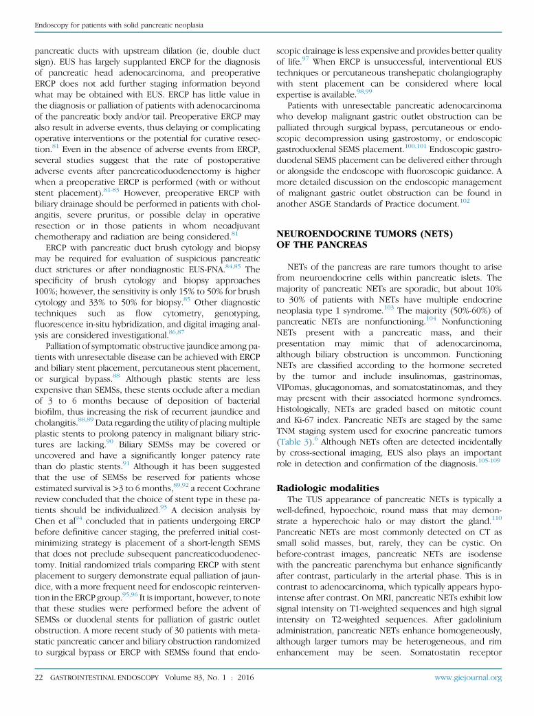
This document reviews the approach to the evaluationand treatment of the patient with suspected solid pancreaticneoplasia. Table 2 outlines the types of neoplasia discussedin this guideline. A discussion of the role of endoscopy forcystic lesions of the pancreas can be found in anotherASGE document.3 Solid lesions of the pancreas can beclassified as primary or metastatic, benign or malignant,and arising from the exocrine or endocrine pancreas. Themost common and potentially serious solid lesion of thepancreas, pancreatic adenocarcinoma, arises from theexocrine pancreas. An algorithm of the recommendedapproach to pancreatic adenocarcinoma diagnosis andstaging is presented in Figure 1.
PRESENTATION AND CLINICAL EVALUATION
Patients with suspected solid pancreatic neoplasia maypresent with obstructive jaundice, abdominal pain,anorexia, weight loss, acute pancreatitis, new onset orpoorly controlled diabetes, or steatorrhea. The physical ex-amination can include findings such as jaundice, musclewasting, pertinent skin lesions, palpable adenopathy, hepa-tomegaly, or masses. Occasionally these lesions will be
Volume 83, No. 1 : 2016 GASTROINTESTINAL ENDOSCOPY 17
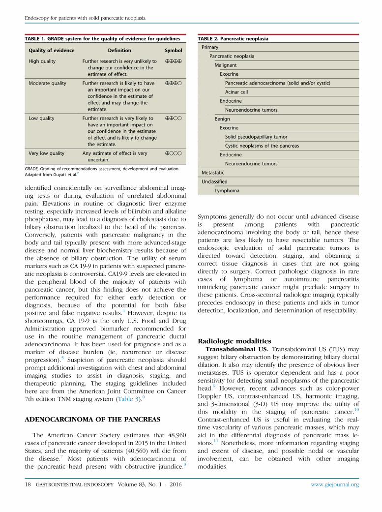
TABLE 1. GRADE system for the quality of evidence for guidelines
Quality of evidence Definition Symbol
High quality Further research is very unlikely tochange our confidence in theestimate of effect.
4444
Moderate quality Further research is likely to havean important impact on ourconfidence in the estimate ofeffect and may change theestimate.
444B
Low quality Further research is very likely tohave an important impact onour confidence in the estimateof effect and is likely to changethe estimate.
44BB
Very low quality Any estimate of effect is veryuncertain.
4BBB
GRADE, Grading of recommendations assessment, development and evaluation.Adapted from Guyatt et al.2
TABLE 2. Pancreatic neoplasia
Primary
Pancreatic neoplasia
Malignant
Exocrine
Pancreatic adenocarcinoma (solid and/or cystic)
Acinar cell
Endocrine
Neuroendocrine tumors
Benign
Exocrine
Solid pseudopapillary tumor
Cystic neoplasms of the pancreas
Endocrine
Neuroendocrine tumors
Metastatic
Unclassified
Lymphoma
Endoscopy for patients with solid pancreatic neoplasia
identified coincidentally on surveillance abdominal imag-ing tests or during evaluation of unrelated abdominalpain. Elevations in routine or diagnostic liver enzymetesting, especially increased levels of bilirubin and alkalinephosphatase, may lead to a diagnosis of cholestasis due tobiliary obstruction localized to the head of the pancreas.Conversely, patients with pancreatic malignancy in thebody and tail typically present with more advanced-stagedisease and normal liver biochemistry results because ofthe absence of biliary obstruction. The utility of serummarkers such as CA 19-9 in patients with suspected pancre-atic neoplasia is controversial. CA19-9 levels are elevated inthe peripheral blood of the majority of patients withpancreatic cancer, but this finding does not achieve theperformance required for either early detection ordiagnosis, because of the potential for both falsepositive and false negative results.4 However, despite itsshortcomings, CA 19-9 is the only U.S. Food and DrugAdministration approved biomarker recommended foruse in the routine management of pancreatic ductaladenocarcinoma. It has been used for prognosis and as amarker of disease burden (ie, recurrence or diseaseprogression).5 Suspicion of pancreatic neoplasia shouldprompt additional investigation with chest and abdominalimaging studies to assist in diagnosis, staging, andtherapeutic planning. The staging guidelines includedhere are from the American Joint Committee on Cancer7th edition TNM staging system (Table 3).6
ADENOCARCINOMA OF THE PANCREAS
The American Cancer Society estimates that 48,960cases of pancreatic cancer developed in 2015 in the UnitedStates, and the majority of patients (40,560) will die fromthe disease.7 Most patients with adenocarcinoma ofthe pancreatic head present with obstructive jaundice.8
18 GASTROINTESTINAL ENDOSCOPY Volume 83, No. 1 : 2016
Symptoms generally do not occur until advanced diseaseis present among patients with pancreaticadenocarcinoma involving the body or tail, hence thesepatients are less likely to have resectable tumors. Theendoscopic evaluation of solid pancreatic tumors isdirected toward detection, staging, and obtaining acorrect tissue diagnosis in cases that are not goingdirectly to surgery. Correct pathologic diagnosis in rarecases of lymphoma or autoimmune pancreatitismimicking pancreatic cancer might preclude surgery inthese patients. Cross-sectional radiologic imaging typicallyprecedes endoscopy in these patients and aids in tumordetection, localization, and determination of resectability.
Radiologic modalitiesTransabdominal US. Transabdominal US (TUS) may
suggest biliary obstruction by demonstrating biliary ductaldilation. It also may identify the presence of obvious livermetastases. TUS is operator dependent and has a poorsensitivity for detecting small neoplasms of the pancreatichead.9 However, recent advances such as color-powerDoppler US, contrast-enhanced US, harmonic imaging,and 3-dimensional (3-D) US may improve the utility ofthis modality in the staging of pancreatic cancer.10
Contrast-enhanced US is useful in evaluating the real-time vascularity of various pancreatic masses, which mayaid in the differential diagnosis of pancreatic mass le-sions.11 Nonetheless, more information regarding stagingand extent of disease, and possible nodal or vascularinvolvement, can be obtained with other imagingmodalities.
www.giejournal.org

Clinical suspicion ofpancreatic adenocarcinoma
or transabdominal USsuggestive
CT scan or MRI
Suspect resectablepancreatic
adenocarcinoma
Suspect unresectablepancreatic
adenocarcinoma
EUS ± FNA
Confirmresectablepancreatic
adenocarcinoma
Confirmunresectable
pancreaticadenocarcinoma
Non-diagnosticspecimen
Alternativediagnoses
PalliationConsider repeatEUS, CT FNA,
ERCP withbrushings andBx, or surgery
Surgery ±neoadjuvant
therapy
Obtain tissuediagnosis by mostappropriate means
Figure 1. Algorithm for evaluation and management of patients with suspected pancreatic adenocarcinoma.
Endoscopy for patients with solid pancreatic neoplasia
CT and magnetic resonance imaging. CT is themostwidely available modality for the noninvasive assessment oftumor resectability and detection of liver metastases, infor-mation that allows further planning of tissue confirmationand palliative care. CT imaging has significantly improvedwith the introduction of multiple-detector CT, which allowshigh-resolution 3-D imaging and multiplanar image recon-struction. Faster injection of iodinated contrast mediumand precisely timed post-injection image acquisition aretechniques that have improved the sensitivity of CT for de-tecting pancreatic adenocarcinoma.12
CT is insensitive for the detection of pancreatic le-sions <2 cm in size.12-14 It is very sensitive for identificationof larger tumors and can accurately stage and assess resect-ability by detection of tumor extension, liver metastases,and invasion of vascular structures.15-18 If CT findings
www.giejournal.org
highly suggest a resectable pancreatic carcinoma, and thepatient is deemed to be an operative candidate, it maybe reasonable to refer the patient directly for surgicalresection (eg, pancreaticoduodenectomy).19,20 CT-guidedbiopsy of pancreatic masses has a reported sensitivity upto 95%.21,22 However, needle-track seeding has been re-ported with this technique.23,24 In 1 study, peritoneal carci-nomatosis was observed to be significantly more commonamong patients with pancreatic masses who underwentpercutaneous sampling rather than EUS-guided biopsy(16.3% vs 2.2%, respectively, P < .025).25
The role of magnetic resonance imaging (MRI) for eval-uation of pancreatic malignancy continues to evolve.26
Although CT historically has been more sensitive thanMRI at the detection of pancreatic carcinoma,17,27 a recentstudy concluded that MRI was superior to CT for tumor
Volume 83, No. 1 : 2016 GASTROINTESTINAL ENDOSCOPY 19

TABLE 3. TNM staging of pancreatic adenocarcinoma
Primary tumor (T)
Tx Primary tumor cannot be assessed
T0 No evidence of primary tumor
Tis Carcinoma in situ
T1 Tumor limited to the pancreas, 2 cm or less in greatest dimension
T2 Tumor limited to the pancreas, more than 2 cm in greatest dimension
T3 Tumor extends beyond the pancreas but without involvement of the celiac axis or superior mesenteric artery
T4 Tumor involves the celiac axis or the superior mesenteric artery (unresectable primary tumor)
Regional lymph nodes (N)
NX Regional lymph nodes cannot be assessed
N0 No regional lymph node metastasis
N1 Regional lymph node metastasis
Distant metastasis (M)
M0 No distant metastasis
M1 Distant metastasis
Anatomical stage/prognosis groups
Stage 0 Tis N0 M0
Stage IA T1 N0 M0
Stage IB T2 N0 M0
Stage IIA T3 N0 M0
Stage IIB T1 N1 M0
T2 N1 M0
T3 N1 M0
Stage III T4 Any N M0
Stage IV Any T Any N M1
Reproduced with permission from the AJCC. AJCC cancer staging manual, 7th ed.6
Endoscopy for patients with solid pancreatic neoplasia
detection and performed similarly for the evaluation ofresectability.28 MRI may reliably detect smaller, non-contour-deforming tumors compared with CT.13,29 MRIalso more accurately detects and characterizes smaller he-patic metastases.30,31
In 1 study that compared the diagnostic performance(detection, local staging) of multiphasic 64-detector CTwith gadobenate dimeglumine-enhanced 3.0-T MRI in pa-tients suspected of having pancreatic cancer, both CTand MRI were found to be equally suited for detectingand staging pancreatic cancer.32 Therefore, the choice ofimaging modality for detection and staging of pancreaticcancer depends on test availability and local expertise.
Positron emission tomography and integratedPET/CT. Positron emission tomography (PET) is a tech-nique based on differential metabolic activity of neoplasticand nonneoplastic tissue. It most often uses 18
fluorodexy-glucose (18FDG), a tracer of glucose metabolism, as anadjunct to conventional imaging. PET may be used forthe diagnosis and staging of pancreatic cancer but alsofor postoperative surveillance to detect local and distantrecurrence or metastases.33 The development andstandardization of PET integrated with CT technology
20 GASTROINTESTINAL ENDOSCOPY Volume 83, No. 1 : 2016
(PET/CT) has dramatically enhanced the diagnosticcapabilities of these 2 modalities for pancreatic cancer,particularly for masses <2 cm in size or CT findings thatare considered equivocal.14,34 A recent meta-analysis of51 studies involving 3857 patients compared the diagnosticperformance of 18FDG PET alone, 18FDG PET/CT, and EUSfor diagnosing pancreatic cancer.35 The study concludedthat the pooled sensitivity estimate for 18FDG PET/CT of90.1% (95% confidence interval [CI], 85.5%-93.6%) wassignificantly greater than that of 18FDG PET alone(88.4%; 95% CI, 86.3%-90.3%) or EUS (81.2%; 95%CI, 78.7%-83.5%; P < .001 for all comparisons). However,EUS had the highest specificity for diagnosing pancreaticcancer (93.2%; 95% CI, 91.7%-94.5%) and wassignificantly better than 18FDG PET (83.1%; 95%CI, 79.6%-86.3%) and 18FDG PET/CT (80.1%; 95%CI, 73.1%-86.0%; P < .001 for all comparisons).35
Endoscopic modalitiesEUS. Although EUS is more operator dependent
compared with CT and MRI, it is the most sensitive testin expert hands to detect pancreatic mass lesionsor pancreatic adenocarcinoma, particularly when lesions
www.giejournal.org

Endoscopy for patients with solid pancreatic neoplasia
are equivocal by CT or <2 centimeters in size.12,14,36-38 In asystematic review of 9 studies and 678 patients, DeWittet al39 concluded that EUS was more sensitive than CTfor the detection of pancreatic adenocarcinoma (91%-100% vs 53%-91%), but the 2 studies were equivalent forloco-regional tumor staging.
EUS also allows tissue acquisition for pathology diag-nosis, but sampling may not be necessary before surgeryin resectable tumors.19,20 However, in some situations, anonoperative pathology diagnosis in patients with other-wise resectable lesions may be desired. For example, endo-scopic tissue diagnosis is helpful for the diagnosis ofmedically treated conditions that may mimic neoplasmsor tumors such as autoimmune pancreatitis40-42 or lym-phoma, for permitting patient enrollment into a neoadju-vant chemotherapy protocol,43 or for preoperativepatient counseling.44 EUS-guided tissue sampling can beperformed by FNA (EUS-guided FNA [EUS-FNA]) or byEUS-guided fine-needle core biopsy (EUS-FNB). EUS-FNAhas a sensitivity and specificity of up to 95% and 100%,respectively45-48 and is the preferred method for makinga definitive cytology diagnosis of a pancreatic mass, evenwhen results of other biopsy methods are negative orequivocal for malignancy.49,50 This approach has beenshown to be cost-effective as well.51 Immediateevaluation and feedback from an on-site cytopathologistduring sampling increases diagnostic yield by 10% to15%.52,53 Although EUS-FNA with cytopathology usually isadequate for a diagnosis of adenocarcinoma and neuroen-docrine tumors (NETs), it may not provide sufficient mate-rial for complete histologic examination for diseases suchas lymphoma, well-differentiated carcinoma, or autoim-mune pancreatitis.54,55 EUS-FNB has not been shown tobe superior to EUS-FNA for determining the etiology ofpancreatic masses but should be considered if EUS-FNAis nondiagnostic and a histologic diagnosis isrequired.44,56-60 FNB is technically difficult for samplingof pancreatic head masses because of the stiffness of theneedle and the acute angulation of the endoscoperequired for biopsy from this location. More-flexible nee-dles have been developed recently that may circumventthis problem and allow better transduodenal sampling ofpancreatic head masses that require core tissue to betterdetermine the nature of the lesion.61
Potential adverse events from EUS-guided sampling ofpancreatic masses include a 0.5% to 2% risk of pancreatitisor bleeding.45,47,48,62,63 Tumor seeding with EUS-FNA hasbeen reported, but the risk appears to be exceedinglysmall, and reports are currently limited to isolatedcases.64-66 It remains unclear whether the risk of tumorseeding with EUS-FNA is related to the number of passesrequired to obtain adequate diagnostic samples. Forpancreatic head masses, the small risk of tumor seedingwith this technique is further mitigated in that any poten-tial site of seeding would likely be included in the resectionspecimen. A recent study by Beane et al67 showed
www.giejournal.org
that preoperative EUS-FNA is not associated with adverseperioperative or long-term outcomes in patients undergo-ing distal pancreatectomy for solid neoplasms of thepancreas. Another study that evaluated survival afterpancreatic cancer surgery in patients with and withoutprior EUS-FNA showed that survival was slightly better inthe EUS-FNA group, although results were not statisticallysignificant.68
EUS traditionally has been performed before ERCP withstent placement because of the potential negative impactof the biliary stent on the accuracy of EUS staging.69,70
However, recent studies suggest that staging accuracymay not be compromised by an indwelling stent.71,72
EUS before ERCP also may identify unresectable pancreaticadenocarcinoma and help triage patients to biliary self-expandable metal stent (SEMS) placement at subsequentERCP.
In patients with pancreatic cancer–related pain, EUS-guided celiac plexus neurolysis (EUS-CPN) may be consid-ered. Performed via a transgastric approach, EUS-CPN at-tempts to ablate the neurons of the celiac ganglia throughthe injection of cytolytic agents such as alcohol or phenol.CPN is the preferred therapy in patients with cancer-related pain.73 A meta-analysis by Kaufman et al74 of 5studies including 199 patients found that EUS-CPN waseffective in alleviating abdominal pain in 72% of patients.A double-blind, controlled trial found that early EUS-CPN re-duces pain and may moderate morphine consumption inpatients with newly diagnosed, painful, inoperablepancreatic cancer.75 CPN can cause transient diarrhea,hypotension, and abdominal pain. Although CPN is a veryeffective and safe procedure, major adverse eventsincluding reversible and permanent paralysis, organpuncture, and gastric necrosis have been described.76,77
A more detailed discussion of the technical and/or proce-dural aspects of CPN can be found elsewhere.78
EUS-guided fiducial placement has been used to aid inimage-guided radiation therapy. Fiducials can be placedwith either 19-gauge or 22-gauge needles. Recently, pre-loaded fiducials on a 22-gauge needle have becomecommercially available. The procedure is very similar inconcept to EUS-guided FNA and can be performed withor without fluoroscopy. The rate of adverse events fromfiducial placement is comparable to that of EUS-FNA ofthe pancreas. Adverse events include mild pancreatitis, mi-nor bleeding, and fiducial migration, requiring a repeatprocedure.79
EUS-guided fine-needle tattooing (EUS-FNT) has beenreported to aid in the localization of pancreatic tumors inpatients undergoing laparoscopic distal pancreatectomy.This is particularly helpful for cases in which abdominal im-aging does not detect a lesion. In one study, the carbonparticle tattoo injected by EUS-FNT was durable and visiblein all 13 cases that underwent preoperative EUS-FNT.80
ERCP. ERCP findings suggestive of a pancreatichead malignancy include strictures of both the bile and
Volume 83, No. 1 : 2016 GASTROINTESTINAL ENDOSCOPY 21
Endoscopy for patients with solid pancreatic neoplasia
pancreatic ducts with upstream dilation (ie, double ductsign). EUS has largely supplanted ERCP for the diagnosisof pancreatic head adenocarcinoma, and preoperativeERCP does not add further staging information beyondwhat may be obtained with EUS. ERCP has little value inthe diagnosis or palliation of patients with adenocarcinomaof the pancreatic body and/or tail. Preoperative ERCP mayalso result in adverse events, thus delaying or complicatingoperative interventions or the potential for curative resec-tion.81 Even in the absence of adverse events from ERCP,several studies suggest that the rate of postoperativeadverse events after pancreaticoduodenectomy is higherwhen a preoperative ERCP is performed (with or withoutstent placement).81-83 However, preoperative ERCP withbiliary drainage should be performed in patients with chol-angitis, severe pruritus, or possible delay in operativeresection or in those patients in whom neoadjuvantchemotherapy and radiation are being considered.81
ERCP with pancreatic duct brush cytology and biopsymay be required for evaluation of suspicious pancreaticduct strictures or after nondiagnostic EUS-FNA.84,85 Thespecificity of brush cytology and biopsy approaches100%; however, the sensitivity is only 15% to 50% for brushcytology and 33% to 50% for biopsy.85 Other diagnostictechniques such as flow cytometry, genotyping,fluorescence in-situ hybridization, and digital imaging anal-ysis are considered investigational.86,87
Palliation of symptomatic obstructive jaundice among pa-tients with unresectable disease can be achieved with ERCPand biliary stent placement, percutaneous stent placement,or surgical bypass.88 Although plastic stents are lessexpensive than SEMSs, these stents occlude after a medianof 3 to 6 months because of deposition of bacterialbiofilm, thus increasing the risk of recurrent jaundice andcholangitis.88,89 Data regarding the utility of placingmultipleplastic stents to prolong patency in malignant biliary stric-tures are lacking.90 Biliary SEMSs may be covered oruncovered and have a significantly longer patency ratethan do plastic stents.91 Although it has been suggestedthat the use of SEMSs be reserved for patients whoseestimated survival is >3 to 6 months,89,92 a recent Cochranereview concluded that the choice of stent type in these pa-tients should be individualized.93 A decision analysis byChen et al94 concluded that in patients undergoing ERCPbefore definitive cancer staging, the preferred initial cost-minimizing strategy is placement of a short-length SEMSthat does not preclude subsequent pancreaticoduodenec-tomy. Initial randomized trials comparing ERCP with stentplacement to surgery demonstrate equal palliation of jaun-dice, with a more frequent need for endoscopic reinterven-tion in the ERCP group.95,96 It is important, however, to notethat these studies were performed before the advent ofSEMSs or duodenal stents for palliation of gastric outletobstruction. A more recent study of 30 patients with meta-static pancreatic cancer and biliary obstruction randomizedto surgical bypass or ERCP with SEMSs found that endo-
22 GASTROINTESTINAL ENDOSCOPY Volume 83, No. 1 : 2016
scopic drainage is less expensive and provides better qualityof life.97 When ERCP is unsuccessful, interventional EUStechniques or percutaneous transhepatic cholangiographywith stent placement can be considered where localexpertise is available.98,99
Patients with unresectable pancreatic adenocarcinomawho develop malignant gastric outlet obstruction can bepalliated through surgical bypass, percutaneous or endo-scopic decompression using gastrostomy, or endoscopicgastroduodenal SEMS placement.100,101 Endoscopic gastro-duodenal SEMS placement can be delivered either throughor alongside the endoscope with fluoroscopic guidance. Amore detailed discussion on the endoscopic managementof malignant gastric outlet obstruction can be found inanother ASGE Standards of Practice document.102
NEUROENDOCRINE TUMORS (NETS)OF THE PANCREAS
NETs of the pancreas are rare tumors thought to arisefrom neuroendocrine cells within pancreatic islets. Themajority of pancreatic NETs are sporadic, but about 10%to 30% of patients with NETs have multiple endocrineneoplasia type 1 syndrome.103 The majority (50%-60%) ofpancreatic NETs are nonfunctioning.104 NonfunctioningNETs present with a pancreatic mass, and theirpresentation may mimic that of adenocarcinoma,although biliary obstruction is uncommon. FunctioningNETs are classified according to the hormone secretedby the tumor and include insulinomas, gastrinomas,VIPomas, glucagonomas, and somatostatinomas, and theymay present with their associated hormone syndromes.Histologically, NETs are graded based on mitotic countand Ki-67 index. Pancreatic NETs are staged by the sameTNM staging system used for exocrine pancreatic tumors(Table 3).6 Although NETs often are detected incidentallyby cross-sectional imaging, EUS also plays an importantrole in detection and confirmation of the diagnosis.105-109
Radiologic modalitiesThe TUS appearance of pancreatic NETs is typically a
well-defined, hypoechoic, round mass that may demon-strate a hyperechoic halo or may distort the gland.110
Pancreatic NETs are most commonly detected on CT assmall solid masses, but, rarely, they can be cystic. Onbefore-contrast images, pancreatic NETs are isodensewith the pancreatic parenchyma but enhance significantlyafter contrast, particularly in the arterial phase. This is incontrast to adenocarcinoma, which typically appears hypo-intense after contrast. On MRI, pancreatic NETs exhibit lowsignal intensity on T1-weighted sequences and high signalintensity on T2-weighted sequences. After gadoliniumadministration, pancreatic NETs enhance homogeneously,although larger tumors may be heterogeneous, and rimenhancement may be seen. Somatostatin receptor
www.giejournal.org
Endoscopy for patients with solid pancreatic neoplasia
scintigraphy can be a useful tool to localize NETs and todetect metastases.111
Endoscopic modalitiesEUS. Pancreatic NETs typically appear solid, hypoe-
choic, and homogenous, with distinct margins on EUS.Rarely they may be cystic and confused with other cystic le-sions of the pancreas.3 EUS is superior to TUS, CT, MRI, andsomatostatin receptor scintigraphy for the localization ofNETs, with a sensitivity of 82% to 93%.105-109,111 Despiteimproved cross-sectional imaging, EUS remains superior toCT for detection of pancreatic NETs, particularly for insuli-nomas.107 EUS also permits tissue acquisition, which isparticularly useful in small or nonfunctioningtumors.107,109,112
ERCP. ERCP does not have a primary role in the diag-nosis of pancreatic NETs. For rare lesions that compressthe pancreatic duct or cause biliary obstruction, ERCPmay have an important therapeutic role.113
SOLID PSEUDOPAPILLARY TUMORS (SPTs)
SPTs of the pancreas are rare tumors that predominantlyaffect young women in the third decade of life. Unlikepancreatic adenocarcinoma, these tumors have a low malig-nant potential, and usually surgery is curative.114 Rarely,SPTs may develop in extrapancreatic locations. The mostcommon presentation is pain or a palpable abdominalmass, but other nonspecific symptoms such as nausea andvomiting may occur. SPTs are often large, with a mediansize of 6 cm to 7 cm, and frequently they are discoveredincidentally.114,115 Although radiologic and endoscopic imag-ing are important in the evaluation of these tumors, the diag-nosis of SPTs can be difficult. In the largest series of SPTsreported to date, only 52 of 718 cases (7%) had a preopera-tive diagnosis confirmed by biopsy.114
Radiologic modalitiesOn TUS, SPTs appear as well-defined, heterogeneous,
solid masses that may contain areas of cystic degenerationor hemorrhage.116 Data are lacking regarding thesensitivity and specificity of TUS for diagnosing SPTs. Thetypical CT appearance of an SPT consists of a large, well-circumscribed, heterogeneous mass with solid and cysticcomponents. Areas of internal hemorrhage and a capsulealso may be visualized.117 These tumors are typicallyavascular or hypovascular. The overall appearance of anSPT on MRI is similar to that of CT, but MRI allowsbetter identification of cystic portions or hemorrhagewithin the tumor.117,118
Endoscopic modalitiesEUS. In a retrospective, multicenter study of 28 cases,
EUS-FNA confirmed the diagnosis of SPT in 75%(21/28).119 Sonographically, SPTs were echo poor and
www.giejournal.org
solid in 50% (14/28), mixed solid and cystic in 39%(11/28), and cystic alone in 11% (3/28). Irregularcalcification was seen in 21% (6/28). Other reports havesupported the role of EUS-FNA in the diagnosis ofSPTs.120,121 Preoperative sampling of SPTs may not benecessary because both a positive diagnosis and nondiag-nostic specimen do not change planned surgical manage-ment, which provides the best chance for long-term cure.122
METASTATIC DISEASE
Metastases to the pancreas are rare and do not showpredilection for any region of the pancreas.123 The mostcommon metastasis to the pancreas is renal cellcarcinoma, but a variety of other cancers includingmelanoma, breast, lung, and colorectal cancers havebeen reported. There is often a long delay between theoriginal diagnosis and the appearance of pancreaticmetastasis, and multiple metastases may be present atthe time of diagnosis.124 Metastases to the pancreas canresult in biliary or pancreatic duct obstruction, pain, orpancreatitis and may be resectable.125,126 CT and MRI find-ings may mimic primary adenocarcinoma of the pancreasbut are more likely to show peripheral or homogenouscontrast enhancement rather than the hypoenhancementof primary pancreatic adenocarcinoma.127,128 The roles ofEUS and ERCP in metastatic disease of the pancreas aresimilar to those described for pancreatic adenocarci-noma.129 Diagnostic EUS findings in metastatic diseasemay be different than in pancreatic adenocarcinoma.DeWitt et al130 reviewed the EUS-FNA features in 24 pa-tients with metastases to the pancreas and found that met-astatic lesions were more likely to have well-definedmargins than primary pancreatic adenocarcinoma. A recentreport found that EUS-FNA confirmed the origin of metas-tasis in the majority of cases.131 Clinical history of a priormalignancy should prompt consideration of a potentialmetastatic lesion to the pancreas, therefore extrabiopsies for immunostains or core biopsy should beconsidered.132 EUS-FNA by using a 22-gauge needle withimmunostains has excellent diagnostic yield in patientswith unusual, neuroendocrine, and metastatic lesionsof the pancreas.132 The ability to procure a coretissue biopsy may even enhance the EUS-FNA potentialto diagnose these lesions in the future.
LYMPHOMA
Primary lymphoma of the pancreas is extremely rare andcan present as a focal or diffuse mass, frequently mimickingmore common neoplasms such as adenocarcinoma or in-flammatory processes such as pancreatitis.133-136 In 1 study,Khashab et al137 showed that EUS-FNA with flow cytometrywas superior to EUS-FNA without flow cytometry in the
Volume 83, No. 1 : 2016 GASTROINTESTINAL ENDOSCOPY 23

Endoscopy for patients with solid pancreatic neoplasia
evaluation of 16 patients suspected to have pancreatic pri-mary pancreatic lymphoma.
The endoscopic evaluation is identical to that of themore common pancreatic neoplasms. If lymphoma is sus-pected (eg, coexistent abdominal lymphadenopathy orother findings), a cytologic sample for flow cytometry orcore biopsy should be obtained.
SCREENING FOR PANCREATIC CANCER
In view of the dismal prognosis of pancreatic cancer atthe time of diagnosis, screening programs have been pro-posed in the last decade in an attempt to detect pancreaticcancer at an early stage and potentially improve survival.Population-based endoscopic or image-based screeningprograms are not feasible or cost-effective, given the rela-tively low incidence of the disease. However, screeningmay be desirable in high-risk individuals. High-risk individ-uals include patients with hereditary pancreatitis, Peutz-Jeghers syndrome, Lynch syndrome, familial breast-ovarian cancer syndrome, familial atypical multiple molemelanoma, and familial pancreatic cancer syndrome. Accu-mulating data indicate that clinically available abdominalimaging tests such as EUS and MRI and/or MRCP can detectasymptomatic precursor lesions, such as intraductal papil-lary mucinous neoplasm, PanIN, and invasive malignantpancreatic neoplasms, such as ductal adenocarcinoma, inindividuals with an inherited predisposition. Two largestudies in screening in high-risk individuals reported theirresults.138,139 One study from the United States reportedthat screening of asymptomatic high-risk individualsfrequently (42% of 216 patients) detects small pancreaticcysts, including curable, noninvasive, high-grade neo-plasms. EUS and MRI detect pancreatic lesions betterthan CT.139 A Dutch study recently reported that EUSand/or MRI detected clinically relevant pancreatic lesionsin 6% of high-risk individuals. Both imaging techniqueswere complementary. MRI was found to be very sensitivefor the detection of cystic lesions of any size. MRI, how-ever, might have some important limitations with regardto the timely detection of solid lesions.138 Screening isbest performed within research protocols or registriesinvolving multidisciplinary teams with expertise ingenetics, gastroenterology, radiology, surgery, andpathology.140
RECOMMENDATIONS
� We recommend that imaging evaluation of patients withsuspected solid pancreatic neoplasia include EUS andmultidetector pancreas protocol CT scans with selectiveuse of MRI and PET-CT when appropriate.4444
� We recommend that EUS be performed for evaluationof pancreatic masses and suspected malignancy, partic-
24 GASTROINTESTINAL ENDOSCOPY Volume 83, No. 1 : 2016
ularly when CT detection or evaluation of resectability isequivocal.4444
� We recommend that biopsy of a suspected primary ormetastatic pancreatic tumor should be individualizedbased on need for preoperative chemotherapy, resect-ability, and feasibility of surgery.4444
� We suggest that EUS-guided celiac plexus neurolysis beconsidered in patients with pancreatic cancer–relatedpain.444B
� We do not recommend preoperative ERCP in patientswith obstructive jaundice because of resectable adeno-carcinoma of the pancreas in the absence of cholangitisunless a substantial delay in operative resection of asymptomatic patient is anticipated.444B
� We recommend that patients with symptomatic, unre-sectable adenocarcinoma of the pancreas with biliaryand/or gastroduodenal obstruction undergo attemptedpalliation with endoscopic stent placement as thepreferred therapeutic modality.444B
� We recommend EUS � FNA for localization and charac-terization of suspected pancreatic neuroendocrine tu-mors and metastatic solid pancreatic neoplasia.444B
� We suggest EUS-guided fiducial placement into apancreatic malignancy if image-guided radiation therapyis considered.444B
� We suggest that screening with EUS and MRCP shouldbe offered to high-risk individuals for pancreatic cancer.44BB
DISCLOSURE
K. Chathadi is a member of the speakers bureau forBoston Scientific. D. Fisher is a consultant for Epigenom-ics Inc. J. DeWitt is a consultant for Olympus Americaand Boston Scientific. All other authors disclosed nofinancial relationships relevant to this publication.
Abbreviations: 3-D, 3-dimensional; 18FDG, 18fluorodexyglucose; ASGE,American Society for Gastrointestinal Endoscopy; CPN, celiac plexusneurolysis; EUS-FNA, EUS-guided FNA; EUS-FNB, EUS-guided fine-needlecore biopsy; EUS-FNT, EUS-guided fine-needle tattooing; MRI, magneticresonance imaging; NET, neuroendocrine tumor; PET, positronemission tomography; SEMS, self-expandable metal stent; SPT, solidpseudopapillary tumor; TUS, transabdominal US.
REFERENCES
1. ASGE Standards of Practice committee; Adler DG, Baron TH, Davila RE,et al. ASGE guideline: the role of ERCP in diseases of the biliary tractand the pancreas. Gastrointest Endosc 2005;62:1-8.
2. Guyatt G, Oxman AD, Akl EA, et al. GRADE guidelines: 1. Introduction-GRADE evidence profiles and summary of findings tables. J Clin Epi-demiol 2011;64:383-94.
3. ASGE Standards of Practice committee; Jacobson BC, Baron TH, AdlerDG, et al. ASGE guideline: The role of endoscopy in the diagnosis andthe management of cystic lesions and inflammatory fluid collectionsof the pancreas. Gastrointest Endosc 2005;61:363-70.
www.giejournal.org

Endoscopy for patients with solid pancreatic neoplasia
4. Partyka K, Maupin KA, Brand RE, et al. Diverse monoclonal antibodiesagainst the CA 19-9 antigen show variation in binding specificity withconsequences for clinical interpretation. Proteomics 2012;12:2212-20.
5. Winter JM, Yeo CJ, Brody JR. Diagnostic, prognostic, and predictivebiomarkers in pancreatic cancer. J Surg Oncol 2013;107:15-22.
6. Edge S, Byrd DR, Compton CC, et al, eds. AJCC Cancer StagingManual, 7th ed. New York: Springer; 2010.
7. American Cancer Society. Cancer Facts & Figures 2015. Atlanta: Amer-ican Cancer Society; 2015.
8. Hidalgo M. Pancreatic cancer. N Engl J Med 2010;362:1605-17.9. Freeny PC. Pancreatic carcinoma: What is the best imaging test? Pan-
creatology 2001;1:604-9.10. Hirooka Y, Goto H, Ito A, et al. Recent advances in US diagnosis of
pancreatic cancer. Hepatogastroenterology 2001;48:916-22.11. Takeda K, Goto H, Hirooka Y, et al. Contrast-enhanced transabdominal
ultrasonography in the diagnosis of pancreatic mass lesions. Acta Ra-diol 2003;44:103-6.
12. Bronstein YL, Loyer EM, Kaur H, et al. Detection of small pancreatictumors with multiphasic helical CT. AJR Am J Roentgenol 2004;182:619-23.
13. Saisho H, Yamaguchi T. Diagnostic imaging for pancreatic cancer:computed tomography, magnetic resonance imaging, and positronemission tomography. Pancreas 2004;28:273-8.
14. Sahani DV, Bonaffini PA, Catalano OA, et al. State-of-the-art PET/CT ofthe pancreas: current role and emerging indications. Radiographics2012;32:1133-58; discussion 58-60.
15. Valls C, Andia E, Sanchez A, et al. Dual-phase helical CT of pancreaticadenocarcinoma: assessment of resectability before surgery. AJR AmJ Roentgenol 2002;178:821-6.
16. Soriano A, Castells A, Ayuso C, et al. Preoperative staging and tumorresectability assessment of pancreatic cancer: prospective studycomparing endoscopic ultrasonography, helical computed tomogra-phy, magnetic resonance imaging, and angiography. Am J Gastroen-terol 2004;99:492-501.
17. Mortele KJ, Ji H, Ros PR. CT and magnetic resonance imaging inpancreatic and biliary tract malignancies. Gastrointest Endosc2002;56:S206-12.
18. Raman SP, Horton KM, Fishman EK. Multimodality imaging ofpancreatic cancer-computed tomography, magnetic resonance im-aging, and positron emission tomography. Cancer J 2012;18:511-22.
19. Hartwig W, Schneider L, Diener MK, et al. Preoperative tissue diag-nosis for tumours of the pancreas. Brit J Surg 2009;96:5-20.
20. Wolfgang CL, Herman JM, Laheru DA, et al. Recent progress inpancreatic cancer. CA-Cancer J Clin 2013;63:318-48.
21. Erturk SM, Mortele KJ, Tuncali K, et al. Fine-needle aspiration biopsy ofsolid pancreatic masses: comparison of CT and endoscopic sonogra-phy guidance. AJR Am J Roentgenol 2006;187:1531-5.
22. Paulsen SD, Nghiem HV, Negussie E, et al. Evaluation of imaging-guided core biopsy of pancreatic masses. AJR Am J Roentgenol2006;187:769-72.
23. Bergenfeldt M, Genell S, Lindholm K, et al. Needle-tract seeding afterpercutaneous fine-needle biopsy of pancreatic carcinoma. Casereport. Acta Chir Scand 1988;154:77-9.
24. Warshaw AL. Implications of peritoneal cytology for staging of earlypancreatic cancer. Am J Surg 1991;161:26-9; discussion 9-30.
25. Micames C, Jowell PS, White R, et al. Lower frequency of peritonealcarcinomatosis in patients with pancreatic cancer diagnosed byEUS-guided FNA vs. percutaneous FNA. Gastrointest Endosc2003;58:690-5.
26. Vachiranubhap B, Kim YH, Balci NC, et al. Magnetic resonance imag-ing of adenocarcinoma of the pancreas. Top Magn Reson Imaging2009;20:3-9.
27. Schima W, Fugger R, Schober E, et al. Diagnosis and staging of pancre-atic cancer: comparison of mangafodipir trisodium-enhanced MR im-aging and contrast-enhanced helical hydro-CT. AJR Am J Roentgenol2002;179:717-24.
www.giejournal.org
28. Park HS, Lee JM, Choi HK, et al. Preoperative evaluation of pancreaticcancer: comparison of gadolinium-enhanced dynamic MRI with MRcholangiopancreatography versus MDCT. J Magn Reson Imaging2009;30:586-95.
29. Vellet AD, Romano W, Bach DB, et al. Adenocarcinoma of the pancre-atic ducts: comparative evaluation with CT and MR imaging at 1.5 T.Radiology 1992;183:87-95.
30. Holalkere NS, Sahani DV, Blake MA, et al. Characterization of smallliver lesions: added role of MR after MDCT. J Comput Assist Tomogr2006;30:591-6.
31. Sahani DV, Shah ZK, Catalano OA, et al. Radiology of pancreaticadenocarcinoma: current status of imaging. J Gastroenterol Hepatol2008;23:23-33.
32. Koelblinger C, Ba-Ssalamah A, Goetzinger P, et al. Gadobenatedimeglumine-enhanced 3.0-T MR imaging versus multiphasic 64-detector row CT: prospective evaluation in patients suspected of hav-ing pancreatic cancer. Radiology 2011;259:757-66.
33. Murakami K. FDG-PET for hepatobiliary and pancreatic cancer: ad-vances and current limitations. World J Clin Oncol 2011;2:229-36.
34. Delbeke D, Martin WH. Update of PET and PET/CT for hepatobiliaryand pancreatic malignancies. HPB (Oxford) 2005;7:166-79.
35. Tang S, Huang G, Liu J, et al. Usefulness of 18F-FDG PET, combinedFDG-PET/CT and EUS in diagnosing primary pancreatic carcinoma: ameta-analysis. Eur J Radiol 2011;78:142-50.
36. Muller MF, Meyenberger C, Bertschinger P, et al. Pancreatic tumors:evaluation with endoscopic US, CT, and MR imaging. Radiology1994;190:745-51.
37. DeWitt J, Devereaux B, Chriswell M, et al. Comparison of endoscopicultrasonography and multidetector computed tomography for de-tecting and staging pancreatic cancer. Ann Intern Med 2004;141:753-63.
38. Gress FG, Hawes RH, Savides TJ, et al. Role of EUS in the preoperativestaging of pancreatic cancer: a large single-center experience. Gastro-intest Endosc 1999;50:786-91.
39. Dewitt J, Devereaux BM, Lehman GA, et al. Comparison of endoscopicultrasound and computed tomography for the preoperative evalua-tion of pancreatic cancer: a systematic review. Clin GastroenterolHepatol 2006;4:717-25; quiz 664.
40. Gardner TB, Levy MJ, Takahashi N, et al. Misdiagnosis of autoimmunepancreatitis: a caution to clinicians. Am J Gastroenterol 2009;104:1620-3.
41. Takahashi N, Fletcher JG, Hough DM, et al. Autoimmune pancreatitis:differentiation from pancreatic carcinoma and normal pancreas onthe basis of enhancement characteristics at dual-phase CT. AJR AmJ Roentgenol 2009;193:479-84.
42. Ghazale A, Chari ST, Smyrk TC, et al. Value of serum IgG4 in the diag-nosis of autoimmune pancreatitis and in distinguishing it frompancreatic cancer. Am J Gastroenterol 2007;102:1646-53.
43. Crane CH, Varadhachary G, Wolff RA, et al. The argument for pre-operative chemoradiation for localized, radiographically resectablepancreatic cancer. Best Pract Res Clin Gastroenterol 2006;20:365-82.
44. Levy MJ, Reddy RP, Wiersema MJ, et al. EUS-guided trucut biopsy inestablishing autoimmune pancreatitis as the cause of obstructivejaundice. Gastrointest Endosc 2005;61:467-72.
45. Lai R, Stanley MW, Bardales R, et al. Endoscopic ultrasound-guidedpancreatic duct aspiration: diagnostic yield and safety. Endoscopy2002;34:715-20.
46. Varadarajulu S, Tamhane A, Eloubeidi MA. Yield of EUS-guided FNA ofpancreatic masses in the presence or the absence of chronic pancre-atitis. Gastrointest Endosc 2005;62:728-36, quiz 51, 53.
47. Eloubeidi MA, Jhala D, Chhieng DC, et al. Yield of endoscopicultrasound-guided fine-needle aspiration biopsy in patients with sus-pected pancreatic carcinoma. Cancer 2003;99:285-92.
48. Eloubeidi MA, Chen VK, Eltoum IA, et al. Endoscopic ultrasound-guided fine needle aspiration biopsy of patients with suspectedpancreatic cancer: diagnostic accuracy and acute and 30-day compli-cations. Am J Gastroenterol 2003;98:2663-8.
Volume 83, No. 1 : 2016 GASTROINTESTINAL ENDOSCOPY 25

Endoscopy for patients with solid pancreatic neoplasia
49. Gress F, Gottlieb K, Sherman S, et al. Endoscopic ultrasonography-guided fine-needle aspiration biopsy of suspected pancreatic cancer.Ann Intern Med 2001;134:459-64.
50. Harewood GC, Wiersema MJ. Endosonography-guided fine needleaspiration biopsy in the evaluation of pancreatic masses. Am J Gastro-enterol 2002;97:1386-91.
51. Chen VK, Arguedas MR, Kilgore ML, et al. A cost-minimization analysisof alternative strategies in diagnosing pancreatic cancer. Am J Gastro-enterol 2004;99:2223-34.
52. Klapman JB, Logrono R, Dye CE, et al. Clinical impact of on-site cyto-pathology interpretation on endoscopic ultrasound-guided fine nee-dle aspiration. Am J Gastroenterol 2003;98:1289-94.
53. Layfield LJ, Bentz JS, Gopez EV. Immediate on-site interpretation offine-needle aspiration smears: a cost and compensation analysis. Can-cer 2001;93:319-22.
54. Thomas T, Kaye PV, Ragunath K, et al. Efficacy, safety, and predictivefactors for a positive yield of EUS-guided Trucut biopsy: a large ter-tiary referral center experience. Am J Gastroenterol 2009;104:584-91.
55. Levy MJ. Endoscopic ultrasound-guided trucut biopsy of thepancreas: prospects and problems. Pancreatology 2007;7:163-6.
56. Shah SM, Ribeiro A, Levi J, et al. EUS-guided fine needle aspirationwith and without trucut biopsy of pancreatic masses. JOP 2008;9:422-30.
57. Varadarajulu S, Fraig M, Schmulewitz N, et al. Comparison of EUS-guided 19-gauge Trucut needle biopsy with EUS-guided fine-needleaspiration. Endoscopy 2004;36:397-401.
58. Aithal GP, Anagnostopoulos GK, Tam W, et al. EUS-guided tissue sam-pling: comparison of “dual sampling” (Trucut biopsy plus FNA) with“sequential sampling” (Trucut biopsy and then FNA as required).Endoscopy 2007;39:725-30.
59. Larghi A, Verna EC, Stavropoulos SN, et al. EUS-guided trucut needlebiopsies in patients with solid pancreatic masses: a prospective study.Gastrointest Endosc 2004;59:185-90.
60. Mizuno N, Bhatia V, Hosoda W, et al. Histological diagnosis of autoim-mune pancreatitis using EUS-guided trucut biopsy: a comparisonstudy with EUS-FNA. J Gastroenterol 2009;44:742-50.
61. Varadarajulu S, Bang JY, Hebert-Magee S. Assessment of the technicalperformance of the flexible 19-gauge EUS-FNA needle. GastrointestEndosc 2012;76:336-43.
62. Eloubeidi MA, Tamhane A, Varadarajulu S, et al. Frequency of majorcomplications after EUS-guided FNA of solid pancreatic masses: aprospective evaluation. Gastrointest Endosc 2006;63:622-9.
63. Eloubeidi MA, Gress FG, Savides TJ, et al. Acute pancreatitis afterEUS-guided FNA of solid pancreatic masses: a pooled analysisfrom EUS centers in the United States. Gastrointest Endosc2004;60:385-9.
64. Paquin SC, Gariepy G, Lepanto L, et al. A first report of tumor seedingbecause of EUS-guided FNA of a pancreatic adenocarcinoma. Gastro-intest Endosc 2005;61:610-1.
65. Chong A, Venugopal K, Segarajasingam D, et al. Tumor seeding afterEUS-guided FNA of pancreatic tail neoplasia. Gastrointest Endosc2011;74:933-5.
66. Ahmed K, Sussman JJ, Wang J, et al. A case of EUS-guided FNA-related pancreatic cancer metastasis to the stomach. Gastrointest En-dosc 2011;74:231-3.
67. Beane JD, House MG, Cote GA, et al. Outcomes after preoperativeendoscopic ultrasonography and biopsy in patients undergoing distalpancreatectomy. Surgery 2011;150:844-53.
68. Ngamruengphong S, Swanson KM, Shah ND, et al. Preoperative endo-scopic ultrasound-guided fine needle aspiration does not impair sur-vival of patients with resected pancreatic cancer. Gut 2015;64:1105-10.
69. Chen CH, Tseng LJ, Yang CC, et al. Preoperative evaluation of peri-ampullary tumors by endoscopic sonography, transabdominal so-nography, and computed tomography. J Clin Ultrasound 2001;29:313-21.
26 GASTROINTESTINAL ENDOSCOPY Volume 83, No. 1 : 2016
70. Cannon ME, Carpenter SL, Elta GH, et al. EUS compared with CT, mag-netic resonance imaging, and angiography and the influence ofbiliary stenting on staging accuracy of ampullary neoplasms. Gastro-intest Endosc 1999;50:27-33.
71. Fisher JM, Gordon SR, Gardner TB. The impact of prior biliary stentingon the accuracy and complication rate of endoscopic ultrasound fine-needle aspiration for diagnosing pancreatic adenocarcinoma.Pancreas 2011;40:21-4.
72. Shami VM, Mahajan A, Sundaram V, et al. Endoscopic ultrasound stag-ing is adversely affected by placement of a self-expandable metalstent: fact or fiction? Pancreas 2008;37:396-8.
73. Chak A. What is the evidence for EUS-guided celiac plexus block/-neurolysis? Gastrointest Endosc 2009;69:S172-3.
74. Kaufman M, Singh G, Das S, et al. Efficacy of endoscopic ultrasound-guided celiac plexus block and celiac plexus neurolysis for managingabdominal pain associated with chronic pancreatitis and pancreaticcancer. J Clin Gastroenterol 2010;44:127-34.
75. Wyse JM, Carone M, Paquin SC, et al. Randomized, double-blind,controlled trial of early endoscopic ultrasound-guided celiac plexusneurolysis to prevent pain progression in patients with newly diag-nosed, painful, inoperable pancreatic cancer. J Clin Oncol 2011;29:3541-6.
76. Fujii L, Clain JE, Morris JM, et al. Anterior spinal cord infarction withpermanent paralysis following endoscopic ultrasound celiac plexusneurolysis. Endoscopy 2012;44(suppl 2)UCTN: E265-6.
77. Loeve US, Mortensen MB. Lethal necrosis and perforation of thestomach and the aorta after multiple EUS-guided celiac plexus neu-rolysis procedures in a patient with chronic pancreatitis. GastrointestEndosc 2013;77:151-2.
78. Levy MJ, Wiersema MJ. Endoscopic ultrasound-guided pain controlfor intra-abdominal cancer. Gastroenterol Clin N 2006;35:153-65.
79. Luz LP, Al-Haddad MA, Sey MS, et al. Applications of endoscopic ul-trasound in pancreatic cancer. World J Gastroenterol 2014;20:7808-18.
80. Lennon AM, Newman N, Makary MA, et al. EUS-guided tattooingbefore laparoscopic distal pancreatic resection (with video). Gastro-intest Endosc 2010;72:1089-94.
81. Isenberg G, Gouma DJ, Pisters PW. The on-going debate about peri-operative biliary drainage in jaundiced patients undergoing pancrea-ticoduodenectomy. Gastrointest Endosc 2002;56:310-5.
82. van der Gaag NA, Rauws EA, van Eijck CH, et al. Preoperative biliarydrainage for cancer of the head of the pancreas. N Engl J Med2010;362:129-37.
83. BaronTH, KozarekRA. Preoperativebiliary stents in pancreatic cancerdproceed with caution. N Engl J Med 2010;362:170-2.
84. Hawes RH. Diagnostic and therapeutic uses of ERCP in pancreatic andbiliary tract malignancies. Gastrointest Endosc 2002;56:S201-5.
85. De Bellis M, Sherman S, Fogel EL, et al. Tissue sampling at ERCP in sus-pected malignant biliary strictures (Part 1). Gastrointest Endosc2002;56:552-61.
86. Pausawasdi N, Scheiman J. Endoscopic evaluation and palliation ofpancreatic adenocarcinoma: current and future options. Curr OpinGastroenterol 2007;23:515-21.
87. Layfield LJ, Ehya H, Filie AC, et al. Utilization of ancillary studies in thecytologic diagnosis of biliary and pancreatic lesions: the PapanicolaouSociety of Cytopathology guidelines for pancreatobiliary cytology. Di-agn Cytopathol 2014;42:351-62.
88. Baron TH. Palliation of malignant obstructive jaundice. GastroenterolClin North Am 2006;35:101-12.
89. Kaassis M, Boyer J, Dumas R, et al. Plastic or metal stentsfor malignant stricture of the common bile duct? Results of arandomized prospective study. Gastrointest Endosc 2003;57:178-82.
90. Lawrence C, Romagnuolo J, Payne KM, et al. Low symptomatic pre-mature stent occlusion of multiple plastic stents for benign biliarystrictures: comparing standard and prolonged stent change intervals.Gastrointest Endosc 2010;72:558-63.
www.giejournal.org

Endoscopy for patients with solid pancreatic neoplasia
91. Davids PH, Groen AK, Rauws EA, et al. Randomised trial of self-expanding metal stents versus polyethylene stents for distal malig-nant biliary obstruction. Lancet 1992;340:1488-92.
92. Yeoh KG, Zimmerman MJ, Cunningham JT, et al. Comparative costsof metal versus plastic biliary stent strategies for malignant obstruc-tive jaundice by decision analysis. Gastrointest Endosc 1999;49:466-71.
93. Moss AC, Morris E, Mac Mathuna P. Palliative biliary stents for ob-structing pancreatic carcinoma. Cochrane Database Syst Rev 2006:CD004200.
94. Chen VK, Arguedas MR, Baron TH. Expandable metal biliarystents before pancreaticoduodenectomy for pancreatic cancer:a Monte-Carlo decision analysis. Clin Gastroenterol Hepatol2005;3:1229-37.
95. Shepherd HA, Royle G, Ross AP, et al. Endoscopic biliary endoprosthe-sis in the palliation of malignant obstruction of the distal commonbile duct: a randomized trial. Brit J Surg 1988;75:1166-8.
96. Smith AC, Dowsett JF, Russell RC, et al. Randomised trial of endo-scopic stenting versus surgical bypass in malignant low bile ductobstruction. Lancet 1994;344:1655-60.
97. Artifon EL, Sakai P, Cunha JE, et al. Surgery or endoscopy for palliationof biliary obstruction due to metastatic pancreatic cancer. Am J Gas-troenterol 2006;101:2031-7.
98. ASGE Technology Committee; Kaul V, Adler DG, Conway JD, et al. In-terventional EUS. Gastrointest Endosc 2010;72:1-4.
99. Harewood GC, Baron TH, LeRoy AJ, et al. Cost-effectiveness anal-ysis of alternative strategies for palliation of distal biliary obstruc-tion after a failed cannulation attempt. Am J Gastroenterol2002;97:1701-7.
100. Adler DG, Baron TH. Endoscopic palliation of malignant gastric outletobstruction using self-expanding metal stents: experience in 36 pa-tients. Am J Gastroenterol 2002;97:72-8.
101. Yim HB, Jacobson BC, Saltzman JR, et al. Clinical outcome of the useof enteral stents for palliation of patients with malignant upper GIobstruction. Gastrointest Endosc 2001;53:329-32.
102. ASGE Standards of Practice Committee; Fukami N, Anderson MA,Khan K, et al. The role of endoscopy in gastroduodenal obstructionand gastroparesis. Gastrointest Endosc 2011;74:13-21.
103. Alexakis N, Neoptolemos JP. Pancreatic neuroendocrine tumours.Best Pract Res Clin Gastroenterol 2008;22:183-205.
104. Vagefi PA, Razo O, Deshpande V, et al. Evolving patterns in the detec-tion and outcomes of pancreatic neuroendocrine neoplasms: theMassachusetts General Hospital experience from 1977 to 2005.Arch Surg 2007;142:347-54.
105. Ardengh JC, Rosenbaum P, Ganc AJ, et al. Role of EUS in the preop-erative localization of insulinomas compared with spiral CT. Gastroint-est Endosc 2000;51:552-5.
106. Anderson MA, Carpenter S, Thompson NW, et al. Endoscopic ultra-sound is highly accurate and directs management in patients withneuroendocrine tumors of the pancreas. Am J Gastroenterol2000;95:2271-7.
107. Khashab MA, Yong E, Lennon AM, et al. EUS is still superior tomultidetector computerized tomography for detection ofpancreatic neuroendocrine tumors. Gastrointest Endosc2011;73:691-6.
108. Sugiyama M, Abe N, Izumisato Y, et al. Differential diagnosis ofbenign versus malignant nonfunctioning islet cell tumors of thepancreas: the roles of EUS and ERCP. Gastrointest Endosc 2002;55:115-9.
109. ASGE Standards of Practice Committee; Gan SI, Rajan E, Adler DG,et al. Role of EUS. Gastrointest Endosc 2007;66:425-34.
110. Rockall AG, Reznek RH. Imaging of neuroendocrine tumours(CT/MR/US). Best Pract Res Clin Endocrinol Metab 2007;21:43-68.
111. Zimmer T, Ziegler K, Bader M, et al. Localisation of neuroendo-crine tumours of the upper gastrointestinal tract. Gut 1994;35:471-5.
www.giejournal.org
112. Pais SA, Al-Haddad M, Mohamadnejad M, et al. EUS for pancreaticneuroendocrine tumors: a single-center, 11-year experience. Gastro-intest Endosc 2010;71:1185-93.
113. ASGE Standards of Practice Committee; Chathadi KV, Khashab MA,Acosta RD, et al. The role of endoscopy in ampullary and duodenaladenomas. Gastrointest Endosc 2015;82:773-81.
114. Papavramidis T, Papavramidis S. Solid pseudopapillary tumors of thepancreas: review of 718 patients reported in English literature. J AmColl Surg 2005;200:965-72.
115. de Castro SM, Singhal D, Aronson DC, et al. Management of solid-pseudopapillary neoplasms of the pancreas: a comparison with stan-dard pancreatic neoplasms. World J Surg 2007;31:1130-5.
116. Buetow PC, Buck JL, Pantongrag-Brown L, et al. Solid and papillaryepithelial neoplasm of the pancreas: imaging-pathologic correlationon 56 cases. Radiology 1996;199:707-11.
117. Yu MH, Lee JY, Kim MA, et al. MR imaging features of small solid pseu-dopapillary tumors: retrospective differentiation from other smallsolid pancreatic tumors. AJR Am J Roentgenol 2010;195:1324-32.
118. Yao X, Ji Y, Zeng M, et al. Solid pseudopapillary tumor of thepancreas: cross-sectional imaging and pathologic correlation.Pancreas 2010;39:486-91.
119. Jani N, Dewitt J, Eloubeidi M, et al. Endoscopic ultrasound-guidedfine-needle aspiration for diagnosis of solid pseudopapillary tumorsof the pancreas: a multicenter experience. Endoscopy 2008;40:200-3.
120. Bardales RH, Centeno B, Mallery JS, et al. Endoscopic ultrasound-guided fine-needle aspiration cytology diagnosis of solid-pseudopapillary tumor of the pancreas: a rare neoplasm of elusiveorigin but characteristic cytomorphologic features. Am J Clin Pathol2004;121:654-62.
121. Master SS, Savides TJ. Diagnosis of solid-pseudopapillary neoplasmof the pancreas by EUS-guided FNA. Gastrointest Endosc 2003;57:965-8.
122. Levy P, Auber A, Ruszniewski P. Do not biopsy solid pseudopapillarytumors of the pancreas! Endoscopy 2008;40:959; author reply 60.
123. Minni F, Casadei R, Perenze B, et al. Pancreatic metastases: observa-tions of three cases and review of the literature. Pancreatology2004;4:509-20.
124. Baron TH. Endoscopic US for metastases to the pancreas: chasing thesatellites. Gastrointest Endosc 2005;61:697-9.
125. Jarufe N, McMaster P, Mayer AD, et al. Surgical treatment of metasta-ses to the pancreas. Surgeon 2005;3:79-83.
126. Sperti C, Pasquali C, Liessi G, et al. Pancreatic resection for metastatictumors to the pancreas. J Surg Oncol 2003;83:161-6; discussion 6.
127. Tsitouridis I, Diamantopoulou A, Michaelides M, et al. Pancreatic me-tastases: CT and MRI findings. Diagn Interv Radiol 2010;16:45-51.
128. Moon SG, Han JK, Kim TK, et al. Biliary obstruction in metastaticdisease: thin-section helical CT findings. Abdom Imaging 2003;28:45-52.
129. Valiozis I, Zekry A, Williams SJ, et al. Palliation of hilar biliary obstruc-tion from colorectal metastases by endoscopic stent insertion. Gas-trointest Endosc 2000;51:412-7.
130. DeWitt J, Jowell P, Leblanc J, et al. EUS-guided FNA of pancreatic me-tastases: a multicenter experience. Gastrointest Endosc 2005;61:689-96.
131. El Hajj II, LeBlanc JK, Sherman S, et al. Endoscopic ultrasound-guidedbiopsy of pancreatic metastases: a large single-center experience.Pancreas 2013;42:524-30.
132. Eloubeidi MA, Tamhane AR, Buxbaum JL. Unusual, metastatic, orneuroendocrine tumor of the pancreas: a diagnosis with endoscopicultrasound-guided fine-needle aspiration and immunohistochem-istry. Saudi J Gastroenterol 2012;18:99-105.
133. Tanaka T, Matsugu Y, Koide K, et al. Malignant lymphoma of thepancreas. Digest Dis Sci 1996;41:402-4.
134. Kondo T, Hayakawa T, Shibata T, et al. Pancreatic involvement by lym-phoma simulates pancreatic carcinoma. J Clin Gastroenterol 1989;11:594-6.
Volume 83, No. 1 : 2016 GASTROINTESTINAL ENDOSCOPY 27

Endoscopy for patients with solid pancreatic neoplasia
135. Lin H, Li SD, Hu XG, et al. Primary pancreatic lymphoma: report of sixcases. World J Gastroenterol 2006;12:5064-7.
136. Sheth S, Fishman EK. Imaging of uncommon tumors of the pancreas.Radiol Clin North Am 2002;40:1273-87; vi.
137. Khashab M, Mokadem M, DeWitt J, et al. Endoscopic ultrasound-guided fine-needle aspiration with or without flow cytometry forthe diagnosis of primary pancreatic lymphomada case series. Endos-copy 2010;42:228-31.
28 GASTROINTESTINAL ENDOSCOPY Volume 83, No. 1 : 2016
138. Harinck F, Konings IC, Kluijt I, et al. A multicentre comparative pro-spective blinded analysis of EUS and MRI for screening of pancreaticcancer in high-risk individuals. Gut. Epub 2015 May 18.
139. Canto MI, Hruban RH, Fishman EK, et al. Frequent detection ofpancreatic lesions in asymptomatic high-risk individuals. Gastroenter-ology 2012;142:796-804; quiz e14-5.
140. Shin EJ, Canto MI. Pancreatic cancer screening. Gastroenterol ClinNorth Am 2012;41:143-57.
www.giejournal.org