the role of botulinum toxin type a in the clinical ... · toxins 2015, 7, 3388-3404;...
TRANSCRIPT

Toxins 2015, 7, 3388-3404; doi:10.3390/toxins7093388
toxins ISSN 2072-6651
www.mdpi.com/journal/toxins
Review
The Role of Botulinum Toxin Type A in the Clinical Management of Refractory Anterior Knee Pain
Barbara J. Singer 1,*, Benjamin I. Silbert 2, Peter L. Silbert 1 and Kevin P. Singer 1
1 Centre for Musculoskeletal Studies, School of Surgery M424, the University of Western,
35 Stirling Highway, Nedlands, WA 6009, Australia; E-Mails: [email protected] (P.L.S.);
[email protected] (K.P.S.) 2
Faculty of Medicine, Dentistry and Health Science, the University of Western Australia,
35 Stirling Highway, Crawley, WA 6009, Australia; E-Mail: [email protected]
* Author to whom correspondence should be addressed; E-Mail: [email protected];
Tel.: +61-8-6488-7079.
Academic Editor: Bahman Jabbari
Received: 2 June 2015 / Accepted: 17 August 2015 / Published: 25 August 2015
Abstract: Anterior knee pain is a highly prevalent condition affecting largely young to
middle aged adults. Symptoms can recur in more than two thirds of cases, often resulting in
activity limitation and reduced participation in employment and recreational pursuits.
Persistent anterior knee pain is difficult to treat and many individuals eventually consider a
surgical intervention. Evidence for long term benefit of most conservative treatments or
surgical approaches is currently lacking. Injection of Botulinum toxin type A to the distal
region of vastus lateralis muscle causes a short term functional “denervation” which
moderates the influence of vastus lateralis muscle on the knee extensor mechanism and
increases the relative contribution of the vastus medialis muscle. Initial data suggest that,
compared with other interventions for anterior knee pain, Botulinum toxin type A injection,
in combination with an active exercise programme, can lead to sustained relief of symptoms,
reduced health care utilisation and increased activity participation. The procedure is less
invasive than surgical intervention, relatively easy to perform, and is time- and cost-effective.
Further studies, including larger randomized placebo-controlled trials, are required to
confirm the effectiveness of Botulinum toxin type A injection for anterior knee pain and to
elaborate the possible mechanisms underpinning pain and symptom relief.
OPEN ACCESS

Toxins 2015, 7 3389
Keywords: anterior knee pain; muscle imbalance; botulinum toxin type A
1. Anterior Knee Pain
1.1. Pathophysiology of Anterior Knee Pain
Anterior knee pain (AKP), also known as patellofemoral joint pain, is a highly prevalent condition
associated with considerable pain and related symptoms, resulting in activity limitation [1]. This
condition, often considered to be a sub-set of the broader category of patellofemoral pain, is typically
associated with a well-defined group of presenting signs and symptoms, including aching or sharp pain
behind the patella, minimal or no effusion, and worsening pain on activities that increase joint loading
such as kneeling, squatting, prolonged sitting, and ascending or descending stairs [2]. Anterior knee pain
is the most frequently diagnosed orthopaedic condition in young to middle aged adults, representing up
to one in six knee complaints presenting to primary health care settings [3]. It is more common in women
than men [4]. Some authors have suggested that it may predispose affected individuals to the
development of articular degeneration in later life [5,6].
Although the causes of AKP are poorly understood, many potential contributors have been identified [7].
These include: anatomical abnormalities of the patella and/or trochlear groove, mal-alignment of the lower
extremity (due to femoral ante-version, external torsion of the tibia, and/or hyper-pronation of the foot),
muscle imbalance and soft tissue tightness. The classification system proposed by Witrouw et al. [8],
which distinguishes mal-alignment from neuromuscular dysfunction, is widely utilized [9]. With respect
to the latter category of patellofemoral joint dysfunction, profound disruption of knee extensor force
production and motor control can occur due to inhibition of the medial part of the quadriceps muscle
(vastus medialis (VM)) related to injury, effusion, or persistent pain [10]. This inhibition results in
relative over-activity of the lateral component of the quadriceps, the vastus lateralis (VL) muscle,
which may lead to abnormal patellar tracking. Coordinated recruitment of the entire quadriceps muscle is
required for normal knee joint stability and movement. The view that VM muscle weakness may be an
important contributor to AKP receives some support from a long term observational study by Natri et al. [11]
which found that knee extensor torque was the best predictor of recovery, such that those with the least
strength difference between limbs reported the lowest ratings for pain and knee related disability at seven
year follow-up [11]. In addition to quadriceps muscle “strength”, abnormalities in the relative timing of
muscle activation of the medial and lateral components of this muscle have been the focus of
considerable study. A review by Chester et al. [12] concluded that data related to the timing of VM
versus VL muscle activation during knee extension are heterogeneous and that timing anomalies are also
seen in pain free individuals. However, in a prospective cohort study of 79 pain-free recruits who
undertook a six week strenuous military training program, Van Tiggelen et al. [13] reported that delayed
onset of electromyographic (EMG) activity of the VM relative to VL muscle was a major risk factor for
the 32% of individuals who developed AKP during the training period. More recently, Chen et al. [14]
reported longer electro-mechanical delays (EMD) in the VM muscle of 26 individuals with AKP
compared with age- and gender-matched controls during an isometric maximal quadriceps contraction.
Moreover, EMD in the VL muscle of individuals with AKP was shorter than in controls, suggesting that

Toxins 2015, 7 3390
the timing of onset of the components of quadriceps muscle activation may contribute to patellar
mal-tracking. Whether this is true motor delay, given their common innervation, or a result of pain
inhibition of VM muscle remains equivocal. Excessive lateral patellar tracking associated with muscle
imbalance has been suggested to be a major contributor to the development of AKP and joint dysfunction
for several decades [15]. Abnormal patella tilt and mal-tracking have been shown to correlate with longer
EMD’s of VM muscle and alterations in the ratio of VL:VM activation in people with AKP [16,17].
Lower limb alignment is evaluated by the so-called “Q angle” which has been purported to represent
the line of potential force exerted by the quadriceps muscle. The “dynamic Q angle” has been quantified
during tasks such as single limb squats and step downs [18,19]. An abnormal motor pattern secondary
to contralateral pelvic drop, ipsilateral hip adduction and internal rotation, knee abduction and tibial
external rotation with hyper-pronation of the weight-bearing foot, has been postulated to contribute to
AKP in some individuals. As a consequence of this finding, a number of investigators have suggested
that weakness of the abductor and external rotator muscles of the hip may be implicated in the
development of AKP [20], particularly in women [21]. However, the nature of this association has been
questioned, with a recent systematic review claiming that disordered hip muscle activation may arise
secondary to chronic knee pain and dysfunction, rather than having a causative relationship [22].
Finally, excessive foot pronation inducing medial tibial torsion has also been associated with the
development of AKP [7].
1.2. Management of Anterior Knee Pain
Conservative treatment for AKP includes a range of options (reviewed by [7,9,23]). Patellofemoral
orthoses or taping may be used in an attempt to restore more normal (passive) alignment of the joint.
However, Swart et al. [24] concluded from their meta-analysis of eighteen studies, only three of which
had a low risk of bias, that there is moderate evidence for no additive effect on pain of knee braces
compared with exercise therapy alone and conflicting evidence on function. Likewise, only low level
evidence exists to support the suggestion that taping the patellofemoral joint may produce greater
short-term improvement in pain and function than placebo, and the mechanism for this effect is unclear [25].
The efficacy of some conservative strategies has been shown to be related to the aetiology of AKP, with
bracing only relieving symptoms in those individuals accurately classified as “mal-trackers” [26].
Although isolated contraction of VM independent of VL muscle is physiologically not possible, specific
“VM muscle strengthening” exercises are a component of most physiotherapy programs, to correct the
presumed imbalance in activation of these muscles [9]. Electrical muscle stimulation can achieve
VM-specific muscle training and one report of a ten week program of twice daily stimulation in
established cases of AKP demonstrated VM hypertrophy and sustained improvement in symptoms,
both of which were still present at follow up three and a half years post intervention [27]. Weakness of
the hip abductors and external rotators may be corrected with specific muscle re-training [28] and,
in selected cases, with external bracing [29–31], and there is evidence that knee pain may be reduced by
foot orthoses in some individuals [32].
While short term benefits from some non-surgical interventions for AKP such as multi-modal
physiotherapy [32], exercise [33] and taping [25] have been reported, the long term effectiveness of
these treatments, apart from electrical muscle stimulation [27] has never been convincingly

Toxins 2015, 7 3391
demonstrated. Indicators to predict outcomes of various conservative treatment approaches are also
lacking, with a recent systematic review highlighting the paucity of the data on which to base the
selection of these interventions [34].
1.3. Refractory Anterior Knee Pain
More than two thirds of AKP cases do not respond to conservative therapy with symptoms recurring
over time [35,36], and many affected individuals may manage their symptoms by progressively
withdrawing from sports participation and restricting pain-provoking activities such as cycling,
squatting, kneeling or prolonged sitting. In severe and recalcitrant cases of AKP, surgical intervention
has been recommended [23]. However, surgical approaches are generally based on the putative
“diagnosis” of tracking abnormalities derived from non-weightbearing CT imaging, despite the fact that
this investigation may not accurately predict mal-tracking during physiological loading [37]. Surgical
interventions for those with demonstrated patella tracking abnormalities or instability range from passive
re-alignment of the patellar tendon with or without lateral capsular release, to the more radical tibial
tubercle translocation [23]. The success rate of both conservative and surgical approaches is limited and
many individuals experience chronic pain and dysfunction [35,36]. In addition, direct and indirect costs
(e.g., loss of productivity), associated with surgical intervention are considerable, as are the risks of
increased morbidity. One review comparing outcomes of 344 knees treated with surgery versus 403
conservatively treated joints reported that the risk of developing patellofemoral osteoarthritis was
doubled in the surgically managed cohort [37]. Consequently, efficacious non-surgical interventions for
this prevalent and disabling condition are urgently needed.
2. Botulinum Toxin Type A (BoNT-A)
2.1. Clinical Uses of BoNT-A
Botulinum toxin type A is one of seven serotypes of the Clostridium botulinum neurotoxin. Following
intramuscular injection, BoNT-A produces a functional “denervation” via inhibition of acetylcholine
release at the neuromuscular junction. In animal models, restoration of muscle function generally occurs
within three months, due initially to formation of new terminal nerve sprouts and eventually to recovery
of the parent terminal [38]. The use of intramuscular BoNT-A injection to address focal muscle over-activity
is well established in the management of focal or axial dystonia in adults [39] and children [40] with
acquired brain injury. Treatment of musculoskeletal conditions utilizing intramuscular BoNT-A
injection is less commonly reported [41]; however, in the majority of such conditions, BoNT-A has been
utilised to relieve muscle spasm and associated pain [42].
2.2. The Role of BoNT-A in Managing AKP
To date, a small number of studies have investigated a highly novel treatment of chronic AKP using
BoNT-A injection [43–47]. This intervention has involved inducing transient weakening of the distal
part of the VL muscle via intramuscular injection of BoNT-A, usually in addition to a standard
physiotherapy exercise program to strengthen and improve control of hip and knee musculature.
The approach to AKP using BoNT-A injection to the distal third of the VL muscle has much in common
Toxins 2015, 7 3392
with the original application of this neurotoxin to alleviate muscle imbalance causing strabismus, as
pioneered by ophthalmologist Dr. Alan Scott [48]. In AKP, the VL muscle is not “overactive” in the same
way that a dystonic muscle (e.g., in cervical dystonia) is abnormally active; however due to a loss of balance
in the amplitude, and possibly timing, of activation of the VL versus the VM muscle, the extensor mechanism
may be disrupted and consequently patella tracking may be altered. This represents only one of many
potential contributors to AKP, but it is the sub-set of individuals with demonstrable or suspected quadriceps
muscle imbalance who have largely been targeted for intervention using BoNT-A to date [43,45,47].
2.3. Clinical Trials of Safety and Efficacy of BoNT in AKP
In an initial open label pilot study [43] eight female subjects with chronic AKP (mean symptom
duration 5 years, range 1–19 years), who had failed conservative management, were injected with
BoNT-A (500 U Dysport®; Ipsen, Paris, France) to VL muscle and underwent a twelve week
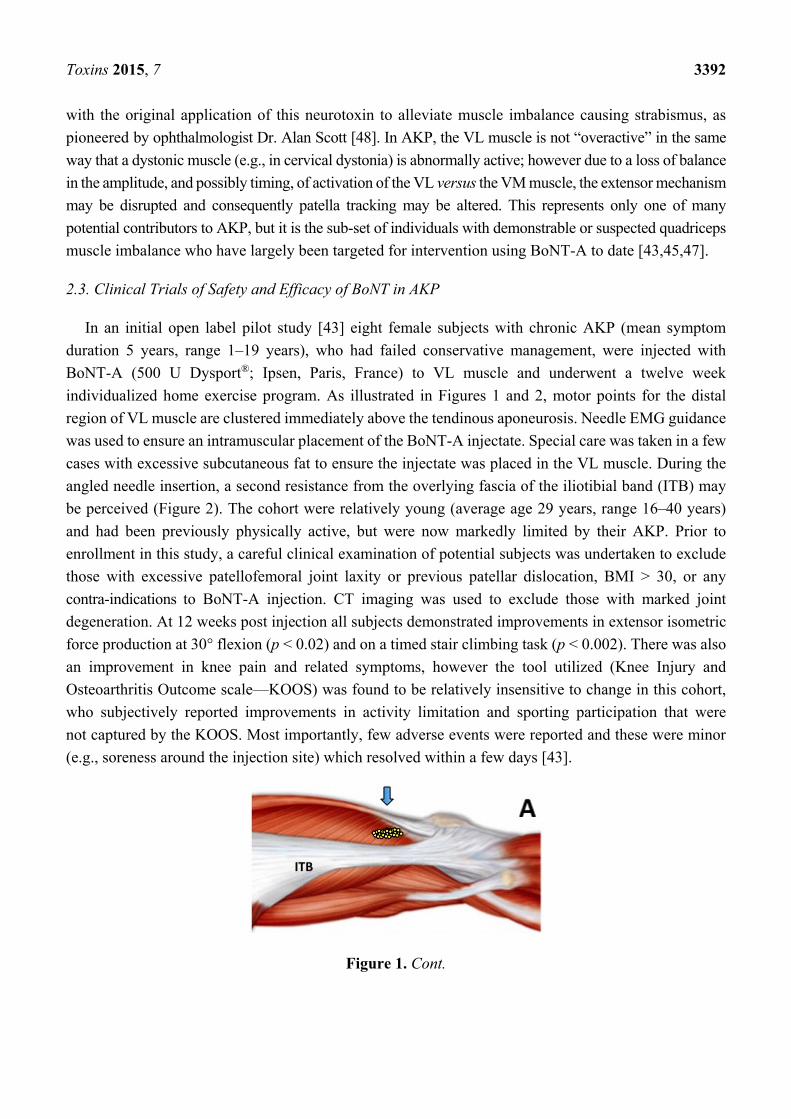
individualized home exercise program. As illustrated in Figures 1 and 2, motor points for the distal
region of VL muscle are clustered immediately above the tendinous aponeurosis. Needle EMG guidance
was used to ensure an intramuscular placement of the BoNT-A injectate. Special care was taken in a few
cases with excessive subcutaneous fat to ensure the injectate was placed in the VL muscle. During the
angled needle insertion, a second resistance from the overlying fascia of the iliotibial band (ITB) may
be perceived (Figure 2). The cohort were relatively young (average age 29 years, range 16–40 years)
and had been previously physically active, but were now markedly limited by their AKP. Prior to
enrollment in this study, a careful clinical examination of potential subjects was undertaken to exclude
those with excessive patellofemoral joint laxity or previous patellar dislocation, BMI > 30, or any
contra-indications to BoNT-A injection. CT imaging was used to exclude those with marked joint
degeneration. At 12 weeks post injection all subjects demonstrated improvements in extensor isometric
force production at 30° flexion (p < 0.02) and on a timed stair climbing task (p < 0.002). There was also
an improvement in knee pain and related symptoms, however the tool utilized (Knee Injury and
Osteoarthritis Outcome scale—KOOS) was found to be relatively insensitive to change in this cohort,
who subjectively reported improvements in activity limitation and sporting participation that were
not captured by the KOOS. Most importantly, few adverse events were reported and these were minor
(e.g., soreness around the injection site) which resolved within a few days [43].
Figure 1. Cont.

Toxins 2015, 7 3393
Figure 1. Motor points (arrow) of the distal branch of the femoral nerve (adapted from
Botter et al. [49]) (A). Dissection showing the distal branch of the femoral nerve to Vastus
Lateralis (small arrows), with the Iliotibial band (ITB) reflected posteriorly (B). As illustrated in
(C), multiple injection sites, using EMG guidance, were employed to ensure spread of injectate
within the distal VL muscle. VLA p = vastus lateralis aponeurosis of the knee joint capsule;
RF = rectus femoris muscle; VM = vastus medialis; p = patella. Reprinted with permission
from [45]. Copyright 2011 BMJ Publishing Group Ltd.
Figure 2. Coronal plane MRI (A) to highlight the relationship of the Iliotibial band to the
Vastus Lateralis muscle (red arrows). Axial plane MRI to highlight the anatomy at the level
of the superior pole of the patella (B). Note the proximity of the lateral joint capsule
(supra-patella pouch) to the distal end of the VL and the overlying ITB to the VL muscle
(red arrows); Legend: VM = vastus medialis, VL = Vastus lateralis, ITB = Iliotibial band,
BF = Biceps femoris, SM = Semimebranosus, ST = Semitendinosus, ADD = Adductors,
QT = Quadriceps tendon.

Toxins 2015, 7 3394
In a subsequent double blinded, placebo injection controlled trial by the same group [45],
24 individuals with refractory AKP (mean duration 6 years) were randomly allocated to receive 500 U
Dysport® (n = 14) or the same volume placebo (n = 10) injection to the distal area of the VL muscle,
again using EMG guidance to confirm placement. Prior to randomization, in addition to clinical
evaluation, VL:VM muscle imbalance was derived from surface EMG recordings during isometric knee
extension in 30° flexion. Abnormal VL:VM ratios from surface EMG during a stair climbing task,
in addition to self-reported pain (at least 2/10) on provocative tasks such as squatting or stairs, have been
shown to be a highly sensitive and specific way of identifying people with AKP [50]. Individuals who
did not meet a priori criteria for quadriceps imbalance were not included in this trial. All subjects also
met the exclusion criteria outlined for the open label pilot study above [43]. Enrolled subjects were again
a relatively young group, with a mean age of 29.5 years (range 15–48 years). Mean baseline Anterior
Knee Pain Scale (AKPS) scores (BoNT-A group 65/100; placebo 69/100) indicated moderate pain and
disability [51]. Following intramuscular BoNT-A or same volume placebo injection and a twelve week
individualized home exercise program, BoNT-A injected subjects demonstrated significantly greater
improvement in knee pain and disability (measured using the AKPS) compared with those receiving
placebo injection (p < 0.03), as well as reporting increased participation in sporting and daily living
activities. Mean change in the experimental group exceeded the minimal clinically important difference
(14 points) [52] for the AKPS (Figure 3). Statistically significant differences from baseline in self-reported
pain (assessed using a visual analogue scale) during pain provoking activities were demonstrated only
in the group who received BoNT-A injection (Figure 3). The ratio of VL:VM activation during an
isometric quadriceps contraction was reversed at 2 weeks post injection (in the BoNT-A group) and
remained significantly different from baseline at all time-points post injection (p < 0.001) (Figure 4).
Subtle changes in ratio for the placebo group may have reflected the influence of the exercise
intervention. Static quadriceps muscle force production at 30° knee extension was maintained or
improved in BoNT-A injected subjects, despite focal atrophy of the distal component of VL muscle in
the treated limb (Figure 5A), supporting the hypothesis that temporarily reducing VL activity would
“dis-inhibit” the VM muscle. No measureable change in patellofemoral joint alignment was found from
repeated CT assessment at 12 weeks despite the focal distal atrophy of VL muscle (Figure 5B).
Subjective and objective improvements in BoNT-A injected subjects were maintained at the 24 week
follow-up [45]. In phase two of this study, prior to unmasking of the data, five subjects elected to receive
open label injection of BoNT-A, on the basis that they believed their symptoms were unchanged. Upon
unmasking of the data, all of these subjects were subsequently found to have received placebo injection
in phase one, and most subsequently reported clinically significant improvement following open label
BoNT-A injection.

Toxins 2015, 7 3395
Figure 3. The mean (±SE) change between baseline and 12 weeks post-injection for
self-reported knee-related disability and activity induced pain is depicted for BoNT-A
injected (blue) versus placebo groups (white). Statistically significant differences (*) were seen
between groups for pain on kneeling, walking, squatting and Anterior Knee Pain Scale (AKPS)
scores. Reprinted with permission from [45]. Copyright 2011 BMJ Publishing Group Ltd.
Figure 4. A significant change (*) in VM muscle activation compared to baseline (W0), during
static maximal knee extensor contraction at 30° knee flexion, was seen in the BoNT-A
injected group only by week 2 (W2) and was sustained at week 12 (W12) (#). The dotted line
represents a normal ratio of VL:VM activation as assessed from surface EMG. Surface EMG
from one subject highlights the relative reversal of VL and VM activation profiles seen in
BoNT-A injected subjects only at week 2 (W2) post-injection. Reprinted with permission
from [45]. Copyright 2011 BMJ Publishing Group Ltd.

Toxins 2015, 7 3396
Figure 5. Axial CT images to highlight the anticipated focal atrophy seen in VL muscle at
the 12 week follow-up review in the treated limb (*) following BoNT-A injection (A).
Despite the induced atrophy, no change was recorded in force production nor congruency of
the patellofemoral joint during quadriceps contraction at 30° knee flexion (B).
More recently, an open label investigation of twelve men with bilateral symptoms of AKP was
undertaken by Chen and colleagues [47]. Subjects with excessive patellar lateral subluxation or tilting
evident on Merchant’s view, previous injury or surgery, or severe degeneration of the medial tibia were
excluded. The most affected VL muscle was injected with 100U BOTOX® (Allergan plc, Dublin, Ireland)
using electrical stimulation to guide injection. The Western Ontario and McMaster Universities
Osteoarthritis (WOMAC) Index, as well as eccentric and concentric quadriceps peak torque production
at 60°/s using isokinetic dynamometry were measured at four, eight and twelve weeks post injection.
Subjects reported improvement in the WOMAC subscales for pain (p < 0.014) and function (p < 0.029),
but not stiffness (p < 0.147), for the injected limb. Interestingly, improvements on the WOMAC were
also seen in the non-injected limb over the twelve week period, but these were not significant.
Improvements in flexion peak torque (eccentric contraction) were seen in both limbs, which may reflect
a training effect of repeated testing. The authors reported that quadriceps force production was not
impaired by BoNT-A injection of VL, nor were there any adverse events reported post injection [47].
2.4. Long Term Effects of BoNT-A Injection in AKP
In addition to these “proof of principle” studies, self-reported outcomes of BoNT-A injection for AKP in
the longer term have been investigated [46]. In this study, two groups were surveyed: (a) an unselected cohort
of individuals (apart from those under workers compensation or other compensation/insurance schemes)
who were referred by orthopaedic surgeons for treatment of refractory AKP using BoNT-A
(Dysport® or BOTOX®) prior to consideration for surgery. Cases were reviewed from a single private
neurology clinic (n = 53), none of whom had undergone EMG evaluation to establish quadriceps muscle
imbalance prior to injection. In a second series (b), volunteers who had participated in the two previous
research studies [43,45] and for whom contact details were available (n = 23) were surveyed. Responses

Toxins 2015, 7 3397
were received from 46 of the 53 privately treated patients. Thirty-eight of these reported initial benefit
from injection, which was ongoing in 29. Amelioration of symptoms had lasted for a mean ± SD of 25 ± 21
months. Nine individuals reported symptom recurrence after an average of 14 ± 21 months. Ten had
undergone knee surgery post-injection, six of whom had not benefitted initially from BoNT-A injection;
with only two reporting improvement in their symptoms following surgery. In the second cohort, 19 of
23 previous research participants responded. All had benefited from BoNT-A injection during the initial
six month follow up period associated with these studies [43,45]. Symptomatic benefit, with a mean ± SD
duration of 44 ± 20 months, had persisted in 15 of the 19 respondents. Four subjects had experienced
some return of AKP symptoms (after 21 ± 12 months), although in two cases they were less severe than
previously. Two had proceeded to surgical intervention, with one reporting an improvement in
symptoms. This audit provides support for the long-term efficacy of intramuscular injection of BoNT-A
to remediate chronic AKP which has been unresponsive to conservative management [45]. From these
data, of the 29 cases who were considering surgical management for their AKP, the single BoNT-A
injection obviated the need for surgery in 25 individuals (86%).
2.5. Possible Mechanisms of Effect of BoNT-A Injection in AKP
Intramuscular injection of BoNT-A for chronic AKP is clearly associated with a reduction in
self-reported pain and knee related disability, however further research is needed to elucidate the
mechanism(s) via which BoNT-A injection may induce these improvements. On the basis of the studies
reported above, we postulate that improvements in quadriceps motor control result from the opportunity
to “unmask” the VM muscle, which is afforded by temporary weakening of VL muscle.
The improvement in control of quadriceps activation has a sustained positive effect on recruitment of
the knee extensor mechanism and consequently on patellar tracking. However BoNT injection may also
contribute an anti-nociceptive effect via other mechanisms, in addition to this mechanistic change.
For instance, some authors have suggested that elevated stress at the cartilage-bone interface may be
responsible, in part, for AKP [51]. Wojtys et al. [53] have reported that the retinaculum, fat pad,
periosteum, and subchondral plate of the patella are all extensively innervated with nociceptors.
Peripheral sensitization can occur when there is injury causing joint swelling or tissue disruption.
The depolarisation threshold of the articular nerves (which contain A-delta, A-beta and C fibres) may be
reduced such that previously non-nociceptive inputs (pressure, motion etc.) start to be perceived as
painful. In chronic oedema in rodent models, secondary central sensitization has been demonstrated to
occur due to progressive hyper-excitability of the spinal nociceptive system [54]. In AKP it is proposed
that a combination of mechanical and neural adaptations to abnormal joint stresses may combine to
produce chronic joint pain, which is usually activity related, particularly in tasks involving significant
patellofemoral loading.
Studies in rat models have shown that injection of BoNT-A can inhibit formalin-induced oedema and
glutamate release, subsequently dampening down or preventing spinal sensitization [55]. In addition, in
capsaicin induced rat models of interstitial cystitis, BoNT-A induced blockade of substance P (SP) [56]
and calcitonin gene related protein (CGRP) [57,58] release has been demonstrated. Neuropeptides such
as SP and CGRP are thought to be the main mediators of neurogenic inflammation [58]. Intra-muscular
injection of BoNT-A has also been shown to inhibit release of a number of other cytokines and

Toxins 2015, 7 3398
neuropeptides which could reduce central and peripheral sensitization [59–61]. In vivo models of BoNT-A
induced anti-nociception in humans have mostly used sub- or intra-dermal injection. In control subjects,
intra-dermal BoNT-A injection has not been shown to have an effect on thresholds for pain induced by
electrical stimulation, hot/cold, mechanical pressure or capsaicin [62–65]. However, intradermal
injection of BoNT-A in individuals with neuropathic pain secondary to diabetic neuropathy has been
shown to produce significantly greater self-reported pain relief than placebo injection [66]. Hence the
response to BoNT-A may depend on the state of the “host nervous system”. Another possibility is that,
following injections into the distal third of the VL muscle, including the distal musculo-tendinous
portion of the muscle (Figures 1 and 2), some diffusion of BoNT-A may have occurred into adjacent
structures, and even into the joint. Spread of injected toxin across muscle fascia (into adjacent muscles)
has been demonstrated in animal models [67,68]. If this were the case, pain relief due to blockade of a
range of nociceptors (SP, CGRP) and excitatory neurotransmitters (most notably glutamate) may have
contributed to the reported symptom relief in this population with chronic AKP. In a series of randomized
controlled investigations examining the effect of intra-articular injection of BoNT-A for refractory
osteoarthritis or pain associated with joint arthroplasty, researchers have reported short term benefit with
regard to pain reduction and self-reported improvement in function and quality of life [69]. It would not
be unreasonable therefore to speculate that the possible leakage, in some cases, of BoNT-A into the
superior aspect of the knee joint capsule (refer Figure 2), together with the focal effect of the drug on
muscle mechanics and pain mechanisms, may contribute to the clinical effects seen following BoNT-A
injection in cases with AKP.
2.6. Clinical Algorithm for the Use of BoNT-A in AKP
Anterior knee pain is a common condition which primarily affects active people from adolescence to
middle age, resulting in significant economic and social costs from pain and activity limitation. In this
context, we do not regard injection of botulinum toxin as a “first line” of treatment for people presenting
with AKP. However, for the majority of individuals who go on to have recurrent symptoms following
initial conservative management [35,36], alternative effective non-surgical treatments are urgently
needed to avoid chronicity of pain and activity limitation. Surgery is recognised as a last resort for highly
selected cases with severe pain and disability associated with patella mal-tracking and instability [23].
Intramuscular injection of BoNT-A into the distal VL muscle produces reversible dose-related weakness,
and can confer a “window of opportunity” to effect a lasting change in the balance of activation between
VL and VM muscles, restoring more normal control of knee extension, and thereby contributing to long
term symptom relief [43–47]. The audit data reported here [46] support the long term efficacy of
intramuscular injection of BoNT-A (providing a mean of two years of symptom relief) to remediate
chronic AKP which had been unresponsive to conservative management, even in relatively unselected
cases who possibly had a range of contributing factors. In this case series, BoNT-A injection was
associated with reduced reliance on pain relieving drugs and physiotherapy attendance (Figure 6), and
in many cases, avoided the need for surgery [46]. To date, the only other conservative intervention for
AKP to demonstrate long term efficacy is electrical muscle stimulation to the VM muscle [27].
Although BoNT-A injection is relatively costly, it can be argued that the point when the administration
of other less evidence based interventions equals the cost of a single BoNT-A treatment is quickly
Toxins 2015, 7 3399
reached, particularly as VL:VM imbalance is commonly demonstrated in this condition [9].
Therefore early consideration of BoNT-A injection in the clinical management of this condition could
be considered to be a cost effective strategy to moderate the risk of progressive activity limitation and
the development of secondary morbidities [5,6].
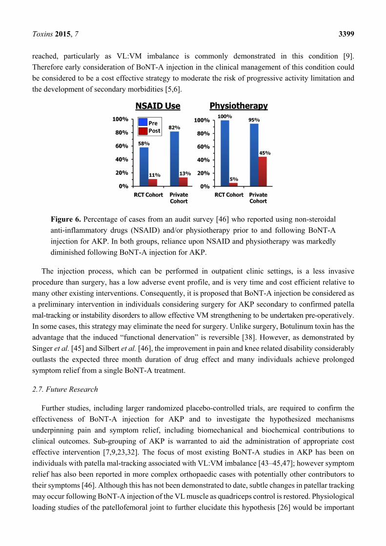
Figure 6. Percentage of cases from an audit survey [46] who reported using non-steroidal
anti-inflammatory drugs (NSAID) and/or physiotherapy prior to and following BoNT-A
injection for AKP. In both groups, reliance upon NSAID and physiotherapy was markedly
diminished following BoNT-A injection for AKP.
The injection process, which can be performed in outpatient clinic settings, is a less invasive
procedure than surgery, has a low adverse event profile, and is very time and cost efficient relative to
many other existing interventions. Consequently, it is proposed that BoNT-A injection be considered as
a preliminary intervention in individuals considering surgery for AKP secondary to confirmed patella
mal-tracking or instability disorders to allow effective VM strengthening to be undertaken pre-operatively.
In some cases, this strategy may eliminate the need for surgery. Unlike surgery, Botulinum toxin has the
advantage that the induced “functional denervation” is reversible [38]. However, as demonstrated by
Singer et al. [45] and Silbert et al. [46], the improvement in pain and knee related disability considerably
outlasts the expected three month duration of drug effect and many individuals achieve prolonged
symptom relief from a single BoNT-A treatment.
2.7. Future Research
Further studies, including larger randomized placebo-controlled trials, are required to confirm the
effectiveness of BoNT-A injection for AKP and to investigate the hypothesized mechanisms
underpinning pain and symptom relief, including biomechanical and biochemical contributions to
clinical outcomes. Sub-grouping of AKP is warranted to aid the administration of appropriate cost
effective intervention [7,9,23,32]. The focus of most existing BoNT-A studies in AKP has been on
individuals with patella mal-tracking associated with VL:VM imbalance [43–45,47]; however symptom
relief has also been reported in more complex orthopaedic cases with potentially other contributors to
their symptoms [46]. Although this has not been demonstrated to date, subtle changes in patellar tracking
may occur following BoNT-A injection of the VL muscle as quadriceps control is restored. Physiological
loading studies of the patellofemoral joint to further elucidate this hypothesis [26] would be important

Toxins 2015, 7 3400
to refine eligibility criteria and the indications for BoNT-A injection, and to optimize treatment protocols
by exploring the duration of effect and determinants of the BoNT-A dose-response relationship. Given
the paucity of evidence supporting the long-term effectiveness of most non-surgical and surgical
treatment options for AKP, a single intramuscular BoNT-A injection, with appropriate concurrent
clinical management, would appear to offer a cost-effective alternative to remediate this common
musculoskeletal condition.
3. Conclusions
Intramuscular BoNT-A injection into the distal region of VL muscle is safe, and confers a cost and
time effective alternative to both ongoing conservative management and/or surgery for individuals with
refractory AKP. A key finding of this original use of BoNT-A is the sustained improvement in knee pain
and symptoms, and reduced reliance on therapeutic and pharmaceutical management. From clinical audit
data of long term outcomes it is notable that many cases did not progress to a surgical intervention after
injection of BoNT-A for their chronic AKP symptoms. Clinical improvement reported in the literature
extends well past the expected three month duration of drug effect, and is associated with increased
activity and sports participation.
Acknowledgments
The authors wish to thank our colleagues Professor John Dunne and Dr. Swithin Song, of Royal Perth
Hospital, Perth WA, for their support of research [43,45] reported in this manuscript. Funding was
received from the Raine Medical Research Foundation at The University of Western Australia, Perth,
Australia to support data collection [45].
Author Contributions
All authors contributed to the design and data collection for various research studies reported in the
manuscript [43,45,46]. Barbara J. Singer and Kevin P. Singer drafted this review and Benjamin I. Silbert
and Peter L. Silbert contributed equally to reviewing and critiquing manuscript drafts.
Conflicts of Interest
Ipsen Pharma supplied BoNT-A (Dysport) to the open label pilot study and randomized controlled
trial reported herein [43,45] at no cost and provided financial support for computed tomography imaging
in the latter study [45]. Barbara J. Singer, Kevin P. Singer and Peter L. Silbert have received conference
attendance fees from companies producing BoNT-A but have no financial interest in any of the products
mentioned in this article and this manuscript has been written without interference.
References
1. Taunton, J.E.; Ryan, M.B.; Clement, D.B.; McKenzie, D.C.; Lloyd-Smith, D.R.; Zumbo, B.D.
A retrospective case-control analysis of 2002 running injuries. Br. J. Sports Med. 2002, 36, 95–101.
2. Kujala, U.M.; Jaakkola, L.H.; Koskinen, S.K.; Taimela, S.; Hurme, M.; Nelimarkka, O. Scoring of
patellofemoral disorders. Arthroscopy 1993, 9, 159–163.

Toxins 2015, 7 3401
3. Teitge, R.A. Patellofemoral syndrome a paradigm for current surgical strategies. Orthop. Clin. N. Am.
2008, 39, 287–311.
4. Roush, J.R.; Curtis Bay, R. Prevalence of anterior knee pain in 18–35 year-old females. Int. J. Sports
Phys. Ther. 2012, 7, 396–401.
5. Lonner, J.H. Patellofemoral arthroplasty. Orthopedics 2010, 33, 653.
6. Thomas, M.J.; Wood, L.; Selfe, J.; Peat, G. Anterior knee pain in younger adults as a precursor to
subsequent patellofemoral osteoarthritis: A systematic review. BMC Musculoskelet. Disord. 2010,
11, doi:10.1186/1471-2474-11-201.
7. Hiemstra, L.A.; Kerslake, S.; Irving, C. Anterior knee pain in the athlete. Clin. Sports Med. 2014,
33, 437–459.
8. Witvrouw, E.; Werner, S.; Mikkelsen, C.; Van Tiggelen, D.; Vanden Berghe, L.; Cerulli, G.
Clinical classification of patellofemoral pain syndrome: Guidelines for non-operative treatment.
Knee Surg. Sports Traumatol. Arthrosc. 2005, 13, 122–130.
9. Werner, S. Anterior knee pain: An update of physical therapy. Knee Surg. Sports Traumatol. Arthrosc.
2014, 22, 2286–2294.
10. Spencer, J.; Hayes, K.C.; Alexander, I.J. Knee joint effusion and quadriceps reflex inhibition in man.
Arch. Phys. Med. Rehabil. 1984, 65, 171–177.
11. Natri, A.; Kannus, P.; Järvinen, M. Which factors predict the long term outcome in chronic
patellofemoral pain syndrome? A 7-yr prospective follow up study. Med. Sci. Sports Exerc. 1998,
30, 1572–1577.
12. Chester, R.; Smith, T.O.; Sweeting, D.; Dixon, J.; Wood, S.; Song, F. The relative timing of VMO
and VL in the aetiology of anterior knee pain: A systematic review and meta-analysis.
BMC Musculoskelet. Disord. 2008, doi:10.1186/1471-2474-9-64.
13. Van Tiggelen, D.; Cowan, S.; Coorevits, P.; Duvigneaud, N.; Witvrouw, E. Delayed vastus medialis
obliquus to vastus lateralis onset timing contributes to the development of patellofemoral pain in
previously healthy men: A prospective study. Am. J. Sports Med. 2009, 37, 1099–1105.
14. Chen, H.Y.; Chien, C.C.; Wu, S.K.; Liau, J.J.; Jan, M.H. Electromechanical delay of the vastus
medialis obliquus and vastus lateralis in individuals with patellofemoral pain syndrome.
J. Orthop. Sports Phys. Ther. 2012, 42, 791–796.
15. Fulkerson, J.P.; Shea, K.P. Mechanical basis for patellofemoral pain and cartilage breakdown.
In Articular Cartilage and Knee Joint Function: Basic Science and Arthroscopy; Ewing, J.W., Ed.;
Raven Press: New York, NY, USA, 1990; pp. 93–101.
16. Pal, S.; Draper, C.E.; Fredericson, M.; Gold, G.E.; Delp, S.L.; Beaupre, G.S.; Besier, T.F.
Patellar maltracking correlates with vastus medialis activation delay in patellofemoral pain patients.
Am. J. Sports Med. 2011, 39, 590–598.
17. Pal, S.; Besier, T.F.; Draper, C.E.; Fredericson, M.; Gold, G.E.; Beaupre, G.S.; Delp, S.L.
Patellar tilt correlates with vastus lateralis: Vastus medialis activation ratio in maltracking
patellofemoral pain patients. J. Orthop. Res. 2012, 30, 927–933.
18. Ireland, M.L.; Willson, J.D.; Ballantyne, B.T.; Davis, I.M. Hip strength in females with and without
patellofemoral pain. J. Orthop. Sports Phys. Ther. 2003, 33, 671–676.
19. Powers, C.M. The influence of altered lower-extremity kinematics on patellofemoral joint dysfunction:
A theoretical perspective. J. Orthop. Sports Phys. Ther. 2003, 33, 639–646.

Toxins 2015, 7 3402
20. Barton, C.J.; Lack, S.; Malliaras, P.; Morrissey, D. Gluteal muscle activity and patellofemoral pain
syndrome: A systematic review. Br. J. Sports Med. 2013, 47, 207–214.
21. Prins, M.R.; van der Wurff, P. Females with patellofemoral pain syndrome have weak hip muscles:
A systematic review. Aust. J. Physiother. 2009, 55, 9–15.
22. Rathleff, M.S.; Rathleff, C.R.; Crossley, K.M.; Barton, C.J. Is hip strength a risk factor for
patellofemoral pain? A systematic review and meta-analysis. Br. J. Sports Med. 2014, 48, 1088.
23. McCarthy, M.M.; Strickland, S.M. Patellofemoral pain: An update on diagnostic and treatment
options. Curr. Rev. Musculoskelet. Med. 2013, 6, 188–194.
24. Swart, N.M.; van Linschoten, R.; Bierma-Zeinstra, S.M.; van Middelkoop, M. The additional effect
of orthotic devices on exercise therapy for patients with patellofemoral pain syndrome: A systematic
review. Br. J. Sports Med. 2012, 46, 570–577.
25. Callaghan, M.J.; Selfe, J. Patellar taping for patellofemoral pain syndrome in adults.
Cochrane Database Syst. Rev. 2012, 4, doi:10.1002/14651858.CD006717.pub2.
26. Draper, C.E.; Besier, T.F.; Santos, J.M.; Jennings, F.; Fredericson, M.; Gold, G.E.; Beaupre, G.S.;
Delp, S.L. Using real-time MRI to quantify altered joint kinematics in subjects with patellofemoral
pain and to evaluate the effects of a patellar brace or sleeve on joint motion. J. Orthop. Res. 2009,
27, 571–577.
27. Werner, S.; Arvidsson, H.; Arvidsson, I.; Eriksson, E. Electrical stimulation of vastus medialis and
stretching of lateral thigh muscles in patients with patello-femoral symptoms. Knee Surg. Sports
Traumatol. Arthrosc. 1993, 1, 85–92.
28. Souza, R.B.; Powers, C.M. Predictors of hip internal rotation during running: An evaluation of hip
strength and femoral structure in women with and without patellofemoral pain. Am. J. Sports Med.
2009, 37, 579–587.
29. Powers, C.M.; Ward, S.R.; Chan, L.D.; Chen, Y.J.; Terk, M.R. The effect of bracing on patella
alignment and patellofemoral joint contact area. Med. Sci. Sports Exerc. 2004, 36, 1226–1232.
30. Powers, C.M.; Doubleday, K.L.; Escudero, C. Influence of patellofemoral bracing on pain, knee
extensor torque, and gait function in females with patellofemoral pain. Physiother. Theory Pract.
2008, 24, 143–150.
31. Wilson, N.A.; Mazahery, B.T.; Koh, J.L.; Zhang, L.Q. Effect of bracing on dynamic patellofemoral
contact mechanics. J. Rehabil. Res. Dev. 2010, 47, 531–541.
32. Collins, N.J.; Bisset, L.M.; Crossley, K.M.; Vicenzino, B. Efficacy of nonsurgical interventions for
anterior knee pain: Systematic review and meta-analysis of randomized trials. Sports Med. 2012,
42, 31–49.
33. Van der Heijden, R.A.; Lankhorst, N.E.; van Linschoten, R.; Bierma-Zeinstra, S.M.;
van Middelkoop, M. Exercise for treating patellofemoral pain syndrome. Cochrane Database Syst. Rev.
2015, 1, doi:10.1002/14651858.CD010387.
34. Lack, S.; Barton, C.; Vicenzino, B.; Morrissey, D. Outcome predictors for conservative patellofemoral
pain management: A systematic review and meta-analysis. Sports Med. 2014, 44, 1703–1716.
35. Stathopulu, E.; Baildam, E. Anterior knee pain: A long term follow up. Rheumatology (Oxford)
2003, 42, 380–382.
36. Rathleff, M.S.; Rasmussen, S.; Olesen, J.L. Unsatisfactory long-term prognosis of conservative
treatment of patellofemoral pain syndrome. Ugeskr. Laeger 2012, 174, 1008–1013.

Toxins 2015, 7 3403
37. Smith, T.O.; Song, F.; Donell, S.T.; Hing, C.B. Operative versus non-operative management of
patellar dislocation. A meta-analysis. Knee Surg. Sports Traumatol. Arthrosc. 2011, 19, 988–998.
38. De Paiva, A.; Meunier, F.A.; Molgo, J.; Aoki, K.R.; Dolly, J.O. Functional repair of motor endplates
after botulinum toxin type A poisoning. Proc. Natl. Acad. Sci. USA 1999, 96, 3200–3205.
39. Intiso, D. Therapeutic use of botulinum toxin in neurorehabilitation. J. Toxicol. 2012, 2012,
doi:10.1155/2012/802893.
40. Guettard, E.; Roze, E.; Abada, G.; Lemesle, C.; Vidailhet, M.; Laurent-Vannier, A.; Chevignard, M.P.
Management of spasticity and dystonia in children with acquired brain injury with rehabilitation
and botulinum toxin A. Dev. Neurorehabil. 2009, 12, 128–138.
41. Singer, K.P.; Singer, B.J. The role of botulinum toxin injection in achieving balanced muscle
function. Disabil. Rehabil. 2007, 29, 1759–1760.
42. Jabbari, B.; Machado, D. Treatment of refractory pain with botulinum toxins—An evidence-based
review. Pain Med. 2011, 12, 1594–1606.
43. Singer, B.J.; Silbert, P.L.; Dunne, J.W.; Song, S.; Singer, K.P. An open label pilot investigation of
the efficacy of Botulinum toxin type A [Dysport] injection in the rehabilitation of chronic anterior
knee pain. Disabil. Rehabil. 2006, 28, 707–713.
44. Drake, D.F.; Pidcoe, P.E.; Ericksen, J. Botulinum toxin type A for nonsurgical lateral release in
patellofemoral pain syndrome: A case study. Mil. Med. 2011, 176, 696–698.
45. Singer, B.J.; Silbert, P.L.; Dunne, J.W.; Song, S.; Singer, K.P. Treatment of refractory anterior knee
pain using botulinum toxin type A (Dysport) injection to the distal vastus lateralis muscle:
A randomised placebo controlled crossover trial. Br. J. Sports Med. 2011, 45, 640–645.
46. Silbert, B.I.; Singer, B.J.; Silbert, P.L.; Gibbons, J.T.; Singer, K.P. Enduring efficacy of Botulinum
toxin type A injection for refractory anterior knee pain. Disabil. Rehabil. 2012, 34, 62–68.
47. Chen, J.T.; Tang, A.C.; Lin, S.C.; Tang, S.F. Anterior knee pain caused by patellofemoral pain
syndrome can be relieved by Botulinum toxin type A injection. Clin. Neurol. Neurosurg. 2015,
129 (Suppl. S1), S27–S29.
48. Scott, A.B. Botulinum toxin injection into extraocular muscles as an alternative to strabismus surgery.
J. Pediatr. Ophthalmol. 1980, 17, 21–25.
49. Botter, A.; Oprandi, G.; Lanfranco, F.; Allasia, S.; Maffiuletti, N.A.; Minetto, M.A. Atlas of the
muscle motor points for the lower limb: Implications for electrical stimulation procedures and
electrode positioning. Eur. J. Appl. Physiol. 2011, 111, 2461–2471.
50. Ferrari, D.; Kuriki, H.U.; Silva, C.R.; Alves, N.; Mícolis de Azevedo, F. Diagnostic accuracy of the
electromyography parameters associated with anterior knee pain in the diagnosis of patellofemoral
pain syndrome. Arch. Phys. Med. Rehabil. 2014, 95, 1521–1526.
51. Crossley, K.M.; Bennell, K.L.; Cowan, S.M.; Green, S. Analysis of outcome measures for persons with
patellofemoral pain: Which are reliable and valid? Arch. Phys. Med. Rehabil. 2004, 85, 815–822.
52. Watson, C.J.; Propps, M.; Ratner, J.; Zeigler, D.L.; Horton, P.; Smith, S.S. Reliability and
responsiveness of the lower extremity functional scale and the anterior knee pain scale in patients
with anterior knee pain. J. Orthop. Sports Phys. Ther. 2005, 35, 136–146.
53. Wojtys, E.M.; Beaman, D.N.; Glover, R.A.; Janda, D. Innervation of the human knee joint by
substance-P fibers. Arthroscopy 1990, 6, 254–263.

Toxins 2015, 7 3404
54. Telleria-Diaz, A.; Ebersberger, A.; Vasquez, E.; Schache, F.; Kahlenbach, J.; Schaible, H.G.
Different effects of spinally applied prostaglandin D2 on responses of dorsal horn neurons with knee
input in normal rats and in rats with acute knee inflammation. Neuroscience 2008, 156, 184–192.
55. Cui, M.; Khanijou, S.; Rubino, J.; Aoki, K.R. Subcutaneous administration of botulinum toxin A
reduces formalin-induced pain. Pain 2004, 107, 125–133.
56. Lucioni, A.; Bales, G.T.; Lotan, T.L.; McGehee, D.S.; Cook, S.P.; Rapp, D.E. Botulinum toxin
type A inhibits sensory neuropeptide release in rat bladder models of acute injury and chronic
inflammation. BJU Int. 2008, 101, 366–370.
57. Rapp, D.E.; Turk, K.W.; Bales, G.T.; Cook, S.P. Botulinum toxin type A inhibits calcitonin
gene-related peptide release from isolated rat bladder. J. Urol. 2006, 175, 1138–1142.
58. Birklein, F.; Schmelz, M. Neuropeptides, neurogenic inflammation and complex regional pain
syndrome (CRPS). Neurosci. Lett. 2008, 437, 199–202.
59. Arezzo, J.C. Possible mechanisms for the effects of botulinum toxin on pain. Clin. J. Pain 2002,
18, S125–S132.
60. Sheean, G. Botulinum toxin for the treatment of musculoskeletal pain and spasm. Curr. Pain
Headache Rep. 2002, 6, 460–469.
61. Aoki, K.R. Review of a proposed mechanism for the antinociceptive action of botulinum toxin
type A. Neurotoxicology 2005, 26, 785–793.
62. Blersch, W.; Schulte-Mattler, W.J.; Przywara, S.; May, A.; Bigalke, H.; Wohlfarth, K. Botulinum
toxin A and the cutaneous nociception in humans: A prospective, double-blind, placebo-controlled,
randomized study. J. Neurol. Sci. 2002, 205, 59–63.
63. Voller, B.; Sycha, T.; Gustorff, B.; Schmetterer, L.; Lehr, S.; Eichler, H.G.; Auff, E.; Schnider, P.
A randomized, double-blind, placebo controlled study on analgesic effects of botulinum toxin A.
Neurology 2003, 61, 940–944.
64. Sycha, T.; Samal, D.; Chizh, B.; Lehr, S.; Gustorff, B.; Schnider, P.; Auff, E. A lack of
antinociceptive or antiinflammatory effect of botulinum toxin A in an inflammatory human pain
model. Anesth. Analg. 2006, 102, 509–516.
65. Schulte-Mattler, W.J.; Opatz, O.; Blersch, W.; May, A.; Bigalke, H.; Wohlfahrt, K. Botulinum toxin A
does not alter capsaicin-induced pain perception in human skin. J. Neurol. Sci. 2007, 260, 38–42.
66. Yuan, R.Y.; Sheu, J.J.; Yu, J.M.; Chen, W.T.; Tseng, I.J.; Chang, H.H.; Hu, C.J. Botulinum toxin
for diabetic neuropathic pain: A randomized double-blind crossover trial. Neurology 2009, 72,
1473–1478.
67. Shaari, C.M.; George, E.; Wu, B.L.; Biller, H.F.; Sanders, I. Quantifying the spread of botulinum
toxin through muscle fascia. Laryngoscope 1991, 101, 960–964.
68. George, E.F.; Zimbler, M.; Wu, B.L.; Biller, H.F.; Sanders, I. Quantitative mapping of the effect of
botulinum toxin injections in the thyroarytenoid muscle. Ann. Otol. Rhinol. Laryngol. 1992, 101,
888–892.
69. Singh, J.A. Use of botulinum toxin in musculoskeletal pain. F1000Research 2013, 2, 52.
© 2015 by the authors; licensee MDPI, Basel, Switzerland. This article is an open access article
distributed under the terms and conditions of the Creative Commons Attribution license
(http://creativecommons.org/licenses/by/4.0/).