the respiratory system - scientistcindy.com · divisions of the respiratory tract • the...
TRANSCRIPT

TheRespiratory SystemBY SCIENTIST CINDY

Divisions of the Respiratory Tract
• The respiratory tract is the path of air from the nose to the lungs. It is divided into two sections: Upper Respiratory Tract and the Lower Respiratory Tract.
• The upper respiratory tract consists of the Nostrils, Nasal Cavities, Pharynx, Epiglottis, and the Larynx.
• The lower respiratory tract consists of the Trachea, Bronchi, Bronchioles, and the Lungs.
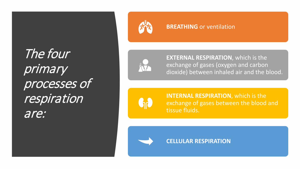
The four primary processes of respiration are:
BREATHING or ventilation
EXTERNAL RESPIRATION, which is the exchange of gases (oxygen and carbon dioxide) between inhaled air and the blood.
INTERNAL RESPIRATION, which is the exchange of gases between the blood and tissue fluids.
CELLULAR RESPIRATION

The secondary processes of respiration include:
REGULATION OF BLOOD pH, which occurs in coordination with the kidneys
DEFENSE AGAINST MICROBES
CONTROL OF BODY TEMPERATURE due to loss of evaporate during expiration

The Pathway of Air1. nasal cavities 2. pharynx (nasopharynx,
oropharynx, and laryngopharynx)
3. larynx4. trachea5. bronchi (right bronchus
and left bronchus)

Breathing and Lung Mechanics
Ventilation • There are two
phases of ventilation;
• Inspiration • Expiration

Breathing and Lung
Mechanics –Ventilation
Ventilation is the exchange of air between the external
environment and the alveoli.

Breathing and Lung
Mechanics –Ventilation
Air moves by bulk flow from an area of high pressure to low
pressure.

Breathing and Lung Mechanics – Ventilation• Air will move in or out
of the lungs depending on the pressure in the alveoli.
• The body changes the pressure in the alveoli by changing the volume of the lungs.
• As volume increases pressure decreases and as volume decreases pressure increases.

Changes in Intrapulmonary Pressure
Changes in pressure are driven by changing the volume of the lungs.• Gas pressure DECREASES
when lung volume INCREASES
• Gas pressure INCREASES when lung volume DECREASES
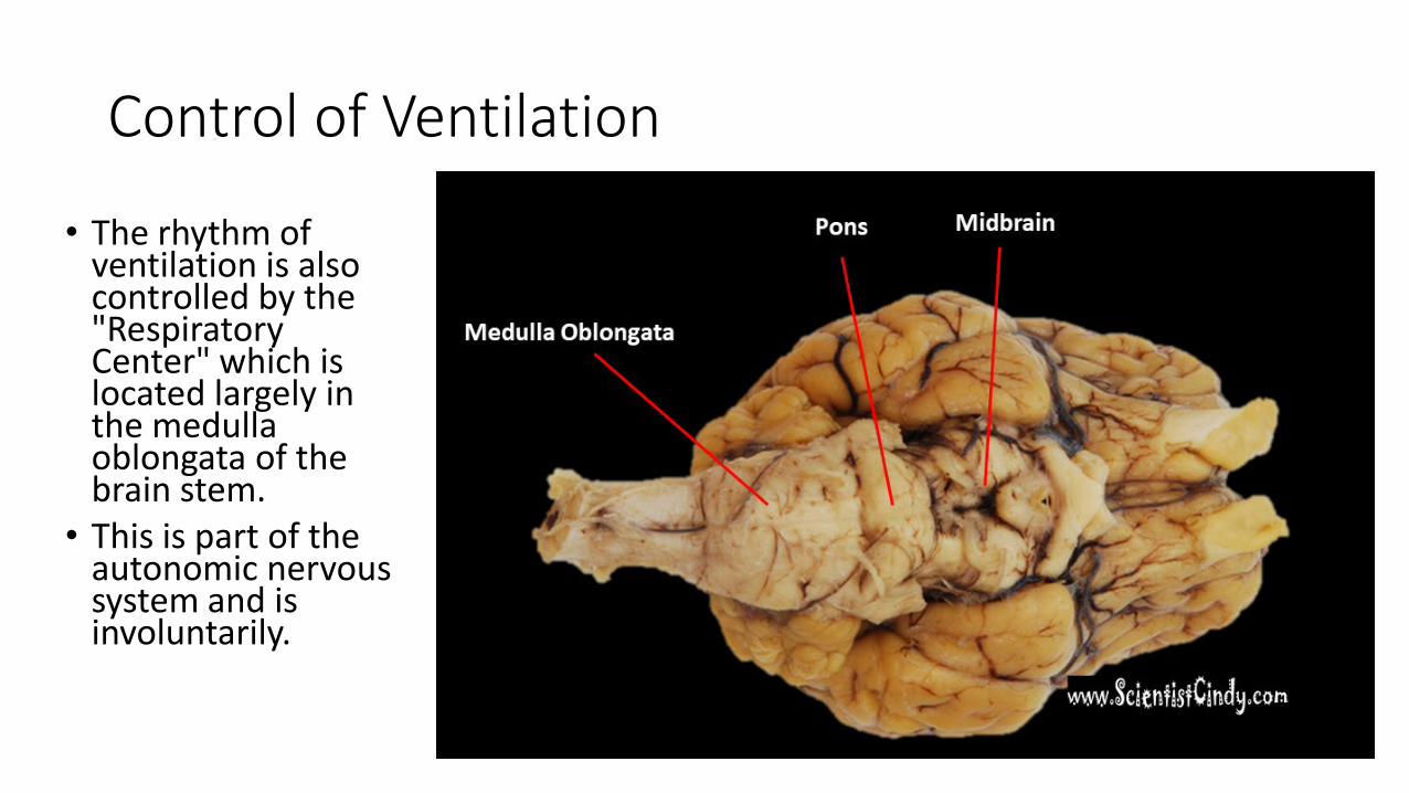
Control of Ventilation
• The rhythm of ventilation is also controlled by the "Respiratory Center" which is located largely in the medulla oblongata of the brain stem.
• This is part of the autonomic nervous system and is involuntarily.

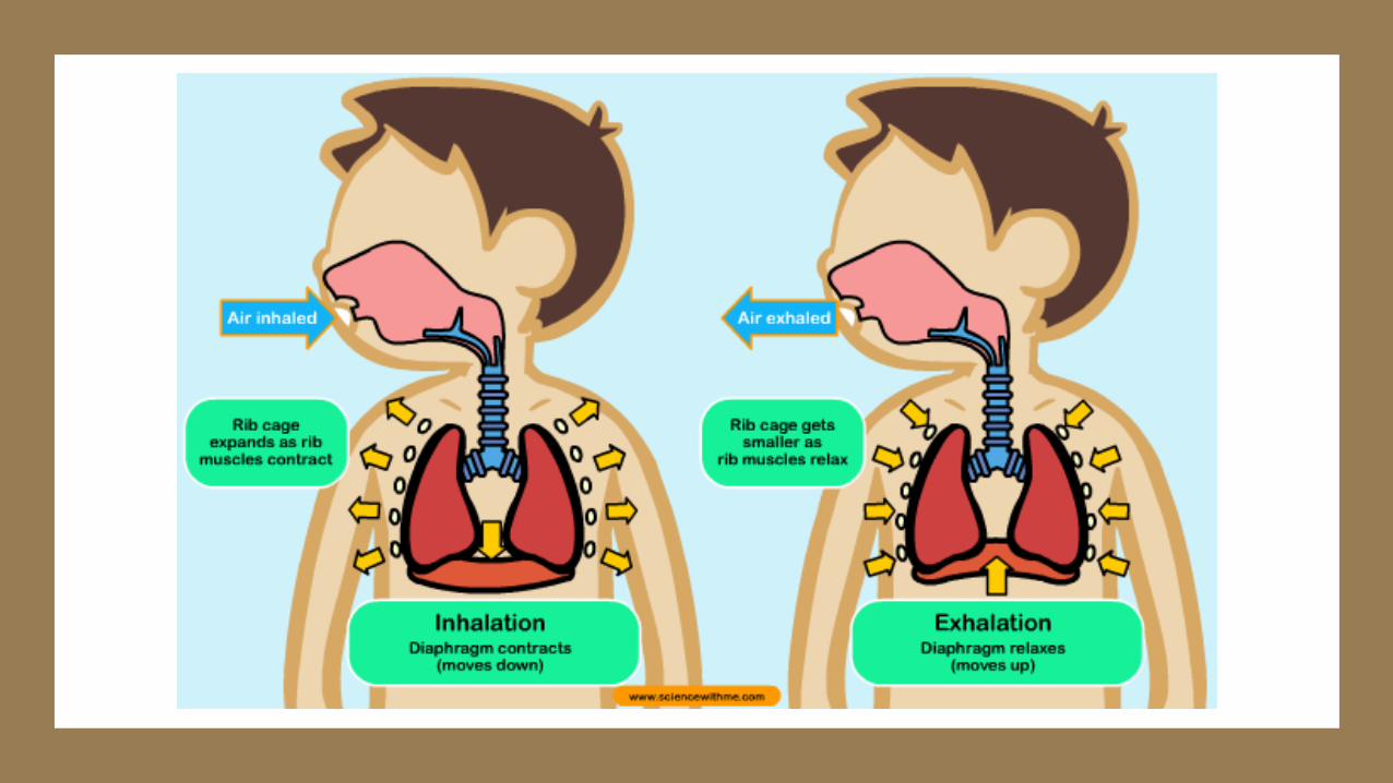
Inspiration (Inhalation)During normal quiet breathing,
• Inspiration is initiated by contraction of the diaphragm -
• When the diaphragm contracts it moves downward toward the abdomen.
• This downward movement of the diaphragm
• Enlarges lung volume• Decreases lung pressure• Air moves from low pressure
to high pressure, so air flows INTO the lungs.

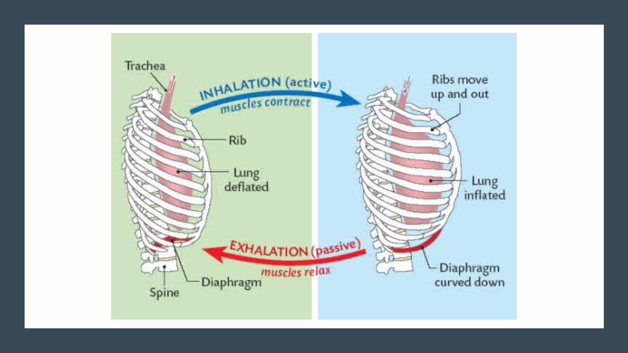
Inspiration (Inhalation)During forceful breathing,
• Inspiration is assisted by the intercostal muscles
• initiated by contraction of the intercostal muscles -
• When the intercostal muscles contract, the rib cage moves…
• Upward and Outward• This upward and outward
movement of the rib cage• Enlarges lung volume• Decreases lung pressure• Air moves from low pressure
to high pressure, so air flows INTO the lu

Expiration (Exhalation)During normal quiet breathing,
• Expiration is initiated by RELAXATION of the diaphragm -
• When the diaphragm RELAXES it moves UPWARD toward the thorax (chest).
• This upward movement of the diaphragm
• Decreases lung volume• Increases lung pressure• Air moves from low pressure
to high pressure, so air flows OUT OF the lungs.
During quiet breathing, expiration is normally a passive process and does not require muscles to work (rather it is the result of the muscles relaxing).

Expiration (Exhalation)During forceful breathing,
• Expiration is initiated by the RELAXATION of the intercostal muscles
• RELAXATION of the intercostal muscles causes the rib cage to move downward and Inward
• This Downward and Inward movement of the rib cage
• Decreases lung volume• Increases lung pressure• Air moves from low pressure
to high pressure, so air flows OUT OF the lungs.

Homeostasis is maintained by the respiratory system in two ways:
gas exchange
regulation of blood pH

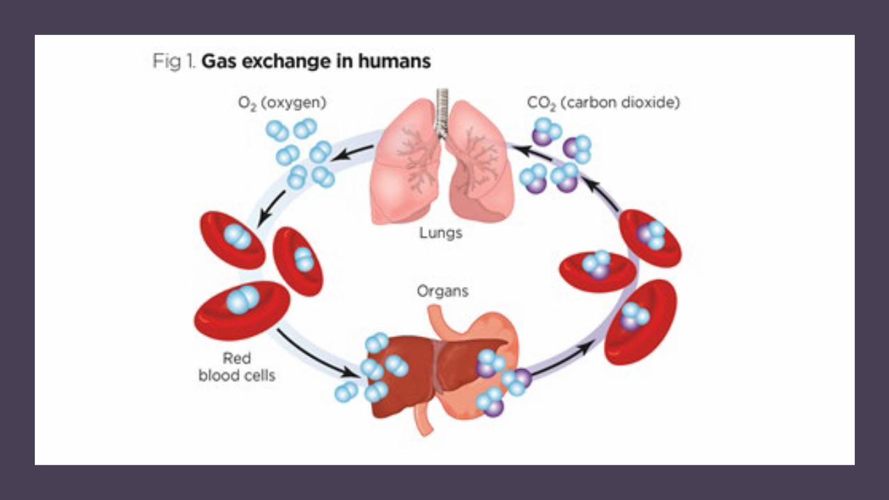
Gas exchange
Gas exchange is performed by the lungs
• supplying oxygen
• eliminating carbon dioxide
Gas exchange
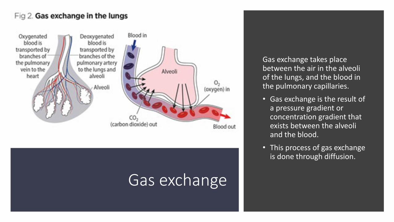
Gas exchange takes place between the air in the alveoli of the lungs, and the blood in the pulmonary capillaries. • Gas exchange is the result of
a pressure gradient or concentration gradient that exists between the alveoli and the blood.
• This process of gas exchange is done through diffusion.
Gas exchange
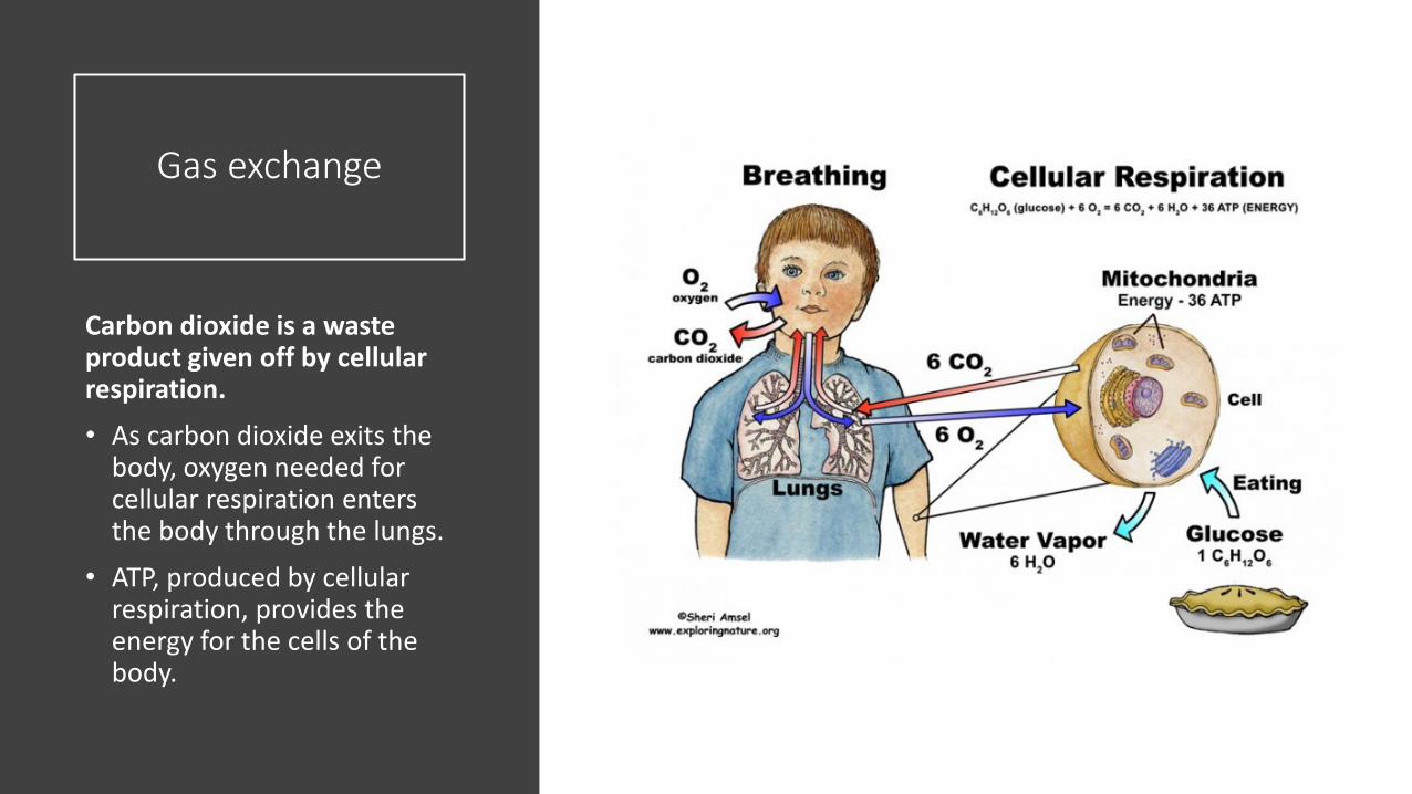
Carbon dioxide is a waste product given off by cellular respiration. • As carbon dioxide exits the
body, oxygen needed for cellular respiration enters the body through the lungs.
• ATP, produced by cellular respiration, provides the energy for the cells of the body.

External Respiration
External respiration is the exchange of gas between the air in the alveoli and the blood within the pulmonary capillaries. • In external respiration, gases diffuse in either direction across the
walls of the alveoli. • Oxygen diffuses from the air in the lungs into the blood when we inhale.• Carbon dioxide diffuses out of the blood into the lungs to be expelled from
the body during exhalation.

carbon dioxide (CO2)
• Most of the carbon dioxide is carried to the lungs in plasma as bicarbonate ions (HCO3-).
• When blood enters the pulmonary capillaries, the bicarbonate ions and hydrogen ions are converted first into carbonic acid (H2CO3) and then converted into carbon dioxide (CO2) and water.
• This chemical reaction removes hydrogen ions.• This neutralizes acidity (low pH).
De-oxygenated blood
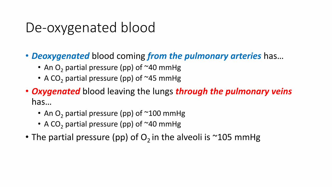
• Deoxygenated blood coming from the pulmonary arteries has…• An O2 partial pressure (pp) of ~40 mmHg• A CO2 partial pressure (pp) of ~45 mmHg
• Oxygenated blood leaving the lungs through the pulmonary veins has…
• An O2 partial pressure (pp) of ~100 mmHg• A CO2 partial pressure (pp) of ~40 mmHg
• The partial pressure (pp) of O2 in the alveoli is ~105 mmHg

Internal Respiration
Internal respiration is the exchanging of gases at the cellular level.
The Passage Way From the Trachea to the Bronchioles
• The inferior portion of the trachea to form the right primary bronchus and left primary bronchus.
• The point at which the trachea branches to form these bronchi is called the Carina.
• The Bronchial Tree is a series of respiratory tubes that branch off into smaller and smaller tubes as they run throughout the lungs.

Bronchial Tree
• The lungs are attached to the heart and trachea through structures that are called the roots of the lungs.
• The roots of the lungs are the bronchi, pulmonary vessels, bronchial vessels, lymphatic vessels, and nerves.
• These structures enter and leave at the hilus of the lung
• There are a number of terminal bronchioles connected to respiratory bronchioles which then advance into the alveolar ducts that then become alveolar sacs.
• Each bronchiole terminates in an elongated space enclosed by many air sacs called alveoli which are surrounded by blood capillaries.
Alveoli
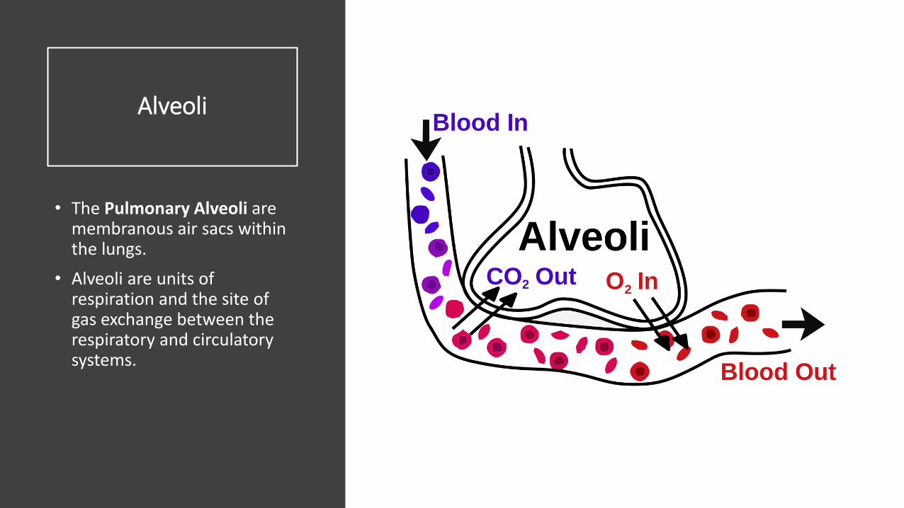
• The Pulmonary Alveoli are membranous air sacs within the lungs.
• Alveoli are units of respiration and the site of gas exchange between the respiratory and circulatory systems.

Oxygen Diffusion
• Oxygen diffuses alveolus into the capillaries
• Capillaries are permeable to oxygen.
• Once in the capillary• ~ 5% of the oxygen
will be dissolved in the blood plasma.
• ~ 95% of the oxygen will bind to hemoglobin in red blood cells.

Cellular Respiration
It is in the mitochondria of the cells where oxygen is actually consumed and carbon dioxide produced.
Oxygen is produced as it combines with hydrogen ions to form water at the end of the electron transport chain.
As cells take apart the carbon molecules from glucose, these get released as carbon dioxide.

Each body cell releases carbon dioxide into nearby capillaries by diffusion.
• The concentration of carbon dioxide is higher in the body cells than in the blood.
• In the capillaries…• some of the carbon dioxide is
dissolved in plasma• some of the carbon dioxide is
taken up by red blood cells and bound to hemoglobin
• most carbon dioxide enters the red blood cells where it binds with water to form carbonic acid.
• carbon dioxide then travels to the capillaries surrounding the lung
• water molecule leaves, causing it to turn back into carbon dioxide.
• It then enters the lungs where it is exhaled into the atmosphere.

Carbon Dioxide
1. Carbon Dioxide enters the red blood cells at capillary beds.
2. Carbon Dioxide binds with water to form carbonic acid.
3. Carbonic Acid then travels to the capillaries surrounding the lung.
4. Carbonic Acid is transformed into water and carbon dioxide.
5. Carbon Dioxide diffuses from the red blood cells in the capillaries to the alveoli of the lungs.
6. The Carbon Dioxide is exhaled out of the body through the lungs.

Stimulation of Breathing
• There are two pathways of motor neuron stimulation of the respiratory muscles.
1. The first is the control of voluntary breathing by the cerebral cortex.
2. The second is involuntary breathing controlled by the medulla oblongata.

Stimulation of Breathing
chemoreceptors that since pH levels are located in the…
• The aorta• The carotid arteries• The medulla oblongata

Stimulation of Breathing
As carbon dioxide levels increase, there is a buildup of carbonic acid, which releases hydrogen ions and lowers pH, making the blood more ACIDIC.
Chemoreceptors do not respond to changes in oxygen levels
Chemoreceptors do respond to changes pH
pH levels are dependent upon plasma carbon dioxide levels.
In other words, the level of CO2 is the driving force for breathing.
Regulation of Breathing
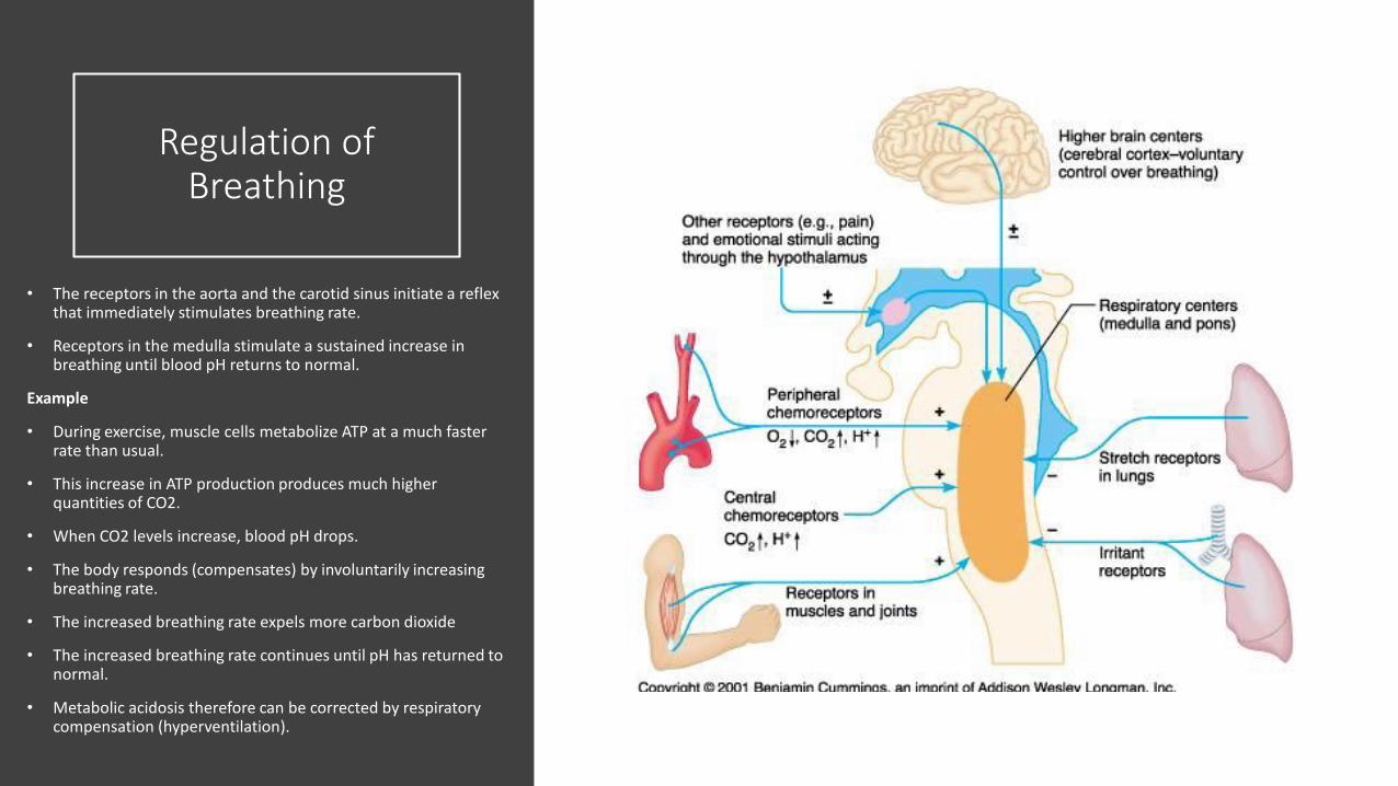
• The receptors in the aorta and the carotid sinus initiate a reflex that immediately stimulates breathing rate.
• Receptors in the medulla stimulate a sustained increase in breathing until blood pH returns to normal.
Example
• During exercise, muscle cells metabolize ATP at a much faster rate than usual.
• This increase in ATP production produces much higher quantities of CO2.
• When CO2 levels increase, blood pH drops.
• The body responds (compensates) by involuntarily increasing breathing rate.
• The increased breathing rate expels more carbon dioxide
• The increased breathing rate continues until pH has returned to normal.
• Metabolic acidosis therefore can be corrected by respiratory compensation (hyperventilation).

pH is the concentration of hydrogen ions (H+).
Buffers are molecules which take in or release ions in order to maintain the H+ ion concentration at a certain level.
Buffers
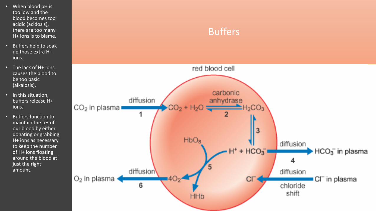
• When blood pH is too low and the blood becomes too acidic (acidosis), there are too many H+ ions is to blame.
• Buffers help to soak up those extra H+ ions.
• The lack of H+ ions causes the blood to be too basic (alkalosis).
• In this situation, buffers release H+ ions.
• Buffers function to maintain the pH of our blood by either donating or grabbing H+ ions as necessary to keep the number of H+ ions floating around the blood at just the right amount.

Bicarbonate Buffer System
The most important buffer we have in our bodies is a mixture of carbon dioxide (CO2) and bicarbonate ion (HCO3).
CO2 forms carbonic acid (H2CO3) when it dissolves in water and acts as an acid giving up hydrogen ions (H+) when needed.
HCO3 is a base and soaks up hydrogen ions (H+) when there are too many of them. In a nutshell, blood pH is determined by a balance between bicarbonate and carbon dioxide.
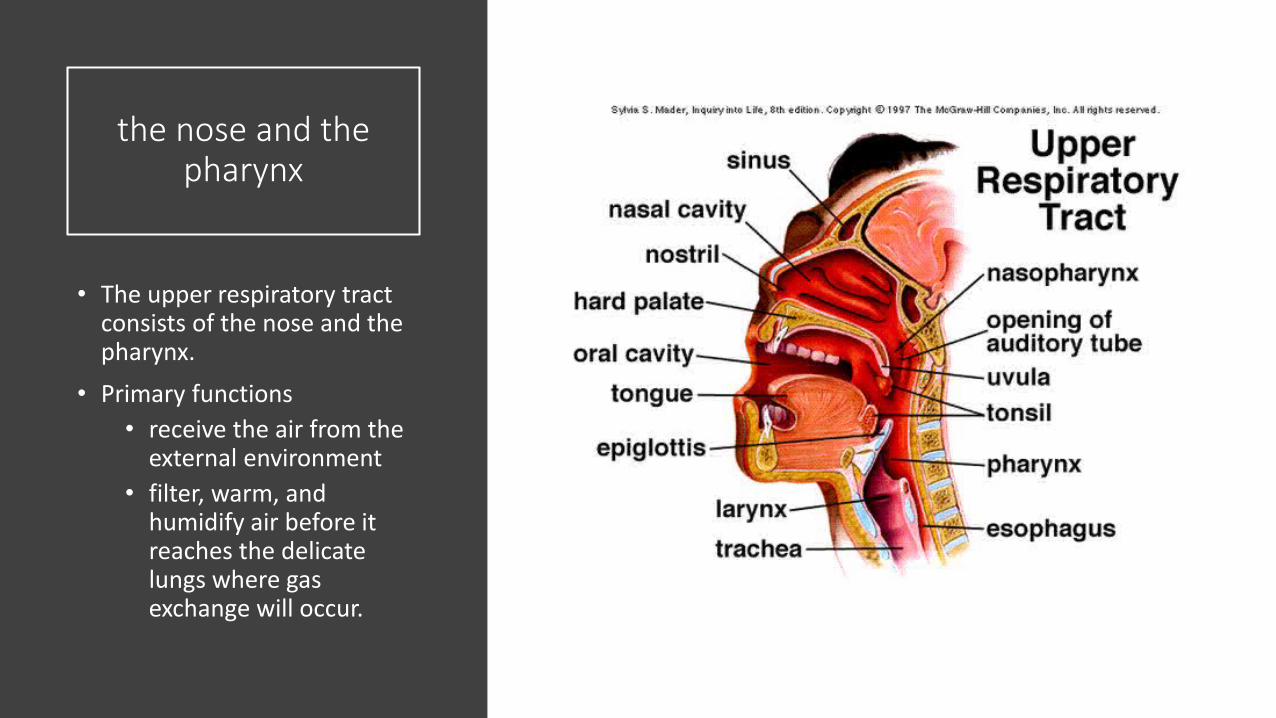
the nose and the pharynx
• The upper respiratory tract consists of the nose and the pharynx.
• Primary functions• receive the air from the
external environment• filter, warm, and
humidify air before it reaches the delicate lungs where gas exchange will occur.

the larynx, the trachea, and the
bronchi
• The lower respiratory tract includes
• the larynx• the trachea• the bronchi

Larynx
• The larynx contains two important structures:
• the epiglottis• the vocal cords
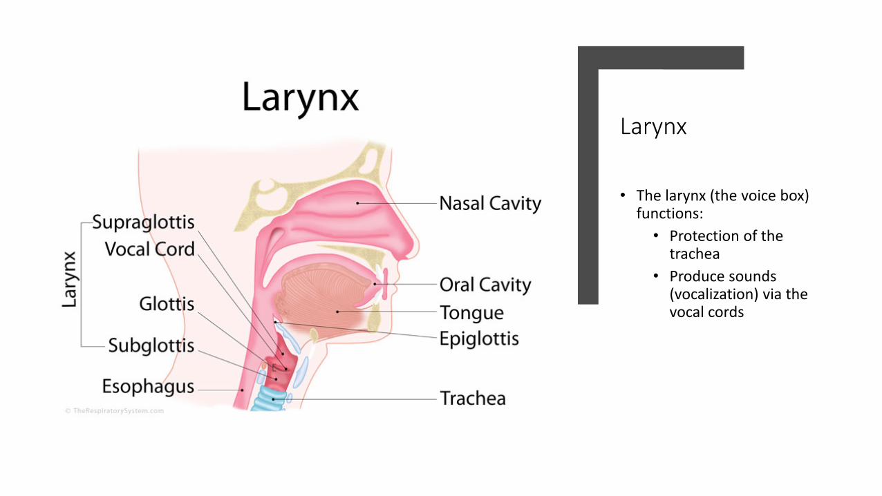
Larynx
• The larynx (the voice box) functions:
• Protection of the trachea
• Produce sounds (vocalization) via the vocal cords
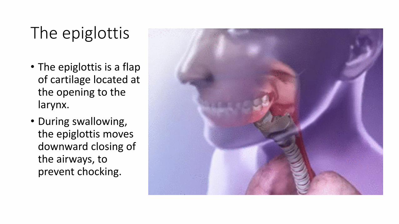
The epiglottis
• The epiglottis is a flap of cartilage located at the opening to the larynx.
• During swallowing, the epiglottis moves downward closing of the airways, to prevent chocking.

The vocal cords
•The vocal cords consist of two folds of connective tissue that stretch and vibrate when air passes through them,
causing vocalization.
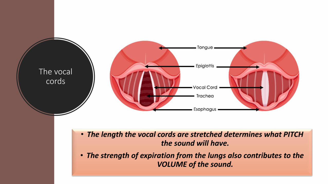
The vocal cords
• The length the vocal cords are stretched determines what PITCH the sound will have.
• The strength of expiration from the lungs also contributes to the VOLUME of the sound.

Acid-base Balance
• Blood is normally slightly basic, with a normal pH range of 7.35 to 7.45.
• Usually the body maintains the pH of blood close to 7.40.

Acid-base Balance
Breathing plays a major role in acid-base balance in the body.
Changes in pulmonary ventilation (breathing rate, depth of respiration) can help adjust pH when disturbances in pH occur.

Acid-base Balance
The two main organs that help balance the pH of blood are the:
Lungs - removes carbon dioxide (an acid) through breathing (respiration).
Kidneys - removes acids through urine (excretion).

CO2 is transport
CO2 is transported in the blood stream by three different means: • as dissolved CO2 gas in the plasma• in the form of bicarbonate in the blood plasma• by binding to hemoglobin in the erythrocytes
• Both methods of transporting CO2 in the plasma can influence the pH of the plasma.

What happens to CO2
• ~ 90% of the CO2 is absorbed by the erythrocytes. • ~ 20% binds to hemoglobin
• ~ 70% reacts with water to form carbonic acid • The carbonic acid formed subsequently dissociates into H+
and bicarbonate. • H+ binds to specific amino acid side chains on the
hemoglobin• The bicarbonate is transported out to the blood plasma. • The bicarbonate formed acts as a buffer against pH changes.
• In other words, if the concentration of hydrogen ions increases, bicarbonate will bind to the free hydrogen ions to form carbonic acid.

CO2 is transport
• Some of the CO2 dissolved in the plasma can react spontaneously with water to form carbonic acid.
• Thus increased CO2 in the plasma tend to decrease plasma pH through increased carbonic acid formation.

Carbon Dioxide Exchange and pH Balance.
• Carbon Dioxide (CO2) reversibly reacts with water to form Carbonic Acid (H2CO3).
• Carbonic acid dissociates into bicarbonate and hydrogen ions.
Carbonic Acid Carbon Dioxide Water
↔ H+ + HCO3-CO2+ H2O ↔ H2CO3
Carbonate Ion Hydrogen Ion
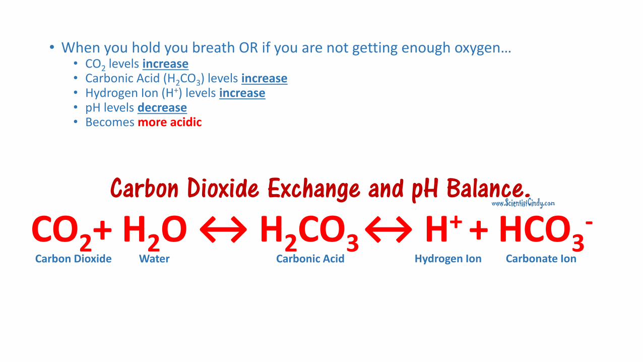
Carbon Dioxide Exchange and pH Balance.
• When you hold you breath OR if you are not getting enough oxygen…• CO2 levels increase• Carbonic Acid (H2CO3) levels increase• Hydrogen Ion (H+) levels increase• pH levels decrease • Becomes more acidic
Carbonic Acid Carbon Dioxide Water
↔ H+ + HCO3-CO2+ H2O ↔ H2CO3
Carbonate Ion Hydrogen Ion

RESULT OF HOLDING YOUR BREATH or NOT
GETTING ENOUGH OXYGEN
• carbon dioxide accumulates in the blood.
• the pH of the blood decreases, becoming more acidic!
• Blood pH balance• Acidosis is when your blood
pH drops below 7.35 and becomes too acidic.
• Alkalosis is when your blood pH is higher than 7.45 and becomes too alkaline.
• Low pH is called acidosis.• Low blood pH is a more common medical problem than high blood pH.• Acidosis can be a warning sign that a health condition isn’t properly
controlled.• Some health conditions cause natural acids to build up in your blood.
• Acids that can lower blood pH include:• lactic acid• keto acids• sulphuric acid• phosphoric acid• hydrochloric acid• carbonic acid• Diet - In a healthy person, diet does not affect blood pH.

RESULT OF RAPID BREATHING or
HYPERVENTALATION
• Alkalosis is caused by an overabundance of bicarbonate or a low level of carbon dioxide.
• People may have irritability, muscle twitching, muscle cramps, or even muscle spasms.

Alkalosis happens when your blood pH is higher than the normal range. • There are several causes of high blood pH.
• Dehydration (Fluid loss)• Losing too much water from your body can increase blood pH.• This happens because you also lose some blood electrolytes (salts
and minerals) with water loss. • These include sodium and potassium.
• Causes of fluid loss are excess:• sweating• vomiting• diarrhea• Diuretic drugs may cause you to urinate too much • Kidney problems
• - kidneys may not remove enough alkaline substances (such as bicarbonate) through the urine