the reproductive biology and laboratory maintenance of
TRANSCRIPT

The Reproductive Biology and Laboratory Maintenance of Vivipara angularis Muller (Prosobranchia: Viviparidae)
Imelda F. Pagulayan Institute of Biology Universit y of the Phi lippines Diliman, Quezon City
and Joel C . Cepillo Natural Science Research Instit ute University of the Philippines Diliman, Quezon City
A BSTRACT
Monthly sampling o f Vivipara angularis Muller in the UP lagoon was done to obtain a year-long trend. Physical conditions of the collection site during sampling were noted. Sex was determined from th e appearance of the curve of "hooked" right tentacle (the copulatory organ of the male), as opposed to the fine, pointed one of the female. Biometric data (shell length and width) of the monthly samples were obtained by the use of a Vernier caliper and were correlated to life cycle and sexual dimorphism. The rep roductive systems of the male and female Vivipara were studied by gross dissection and histological sections. This sensitive species of mollusks was tested for its food preference to develop feeding strategies for its maintenance in the laborator y. Protein content of laboratory -reared snails was assayed. This could serve as a means to come up with an economical protein alternative for human and animal consumption.
535

536 Transactions of the National A cademy of Science and Technology
INTRODUCTION
In the field of malacology {the study of mollusks}, a great number of researchers have indulged in the various aspects of reproduction among oviparous gastropods. The viviparous species, however, is often " ignored." This is probably because it is difficult to rear under laboratory conditions. As a consequence of its mode of reproduction, which, is viviparity, its fecundity is low.
Variations in the shell features of viviparids impose a problem in nomenclature. However, in the Philippines, only Vivipara angu/aris {Fig. 2) and Be/lamya philippinensis have been reported to be snails of medical importance belonging to Family Viviparidae {4, 15). The earliest report found on the species was done by Alonte in 1930. He studied the biology of V. angularis Muller, a common duck-feed snail in Laguna de Bay {1) which is known as one of the edible freshwater mollusks in Manila {10). Different local names given to this snail in the various regions of the Phiippines {susong, pangpang, egue in Tag.; ege in Bis. ; leddeg in lloc.; and betocol in Zam.), reflect its wide distribution in the country. It is usually found inhabiting shallow stagnant or still waters such as lakes, ponds, streams, lagoons, ricefields, canals and ditches {8). However, the problem of low fecundity aggravated by collection for both human and animal consumption contributes to its extinction. Environmental disturbances comprise a secondary threat to its existence.
Apart from the studies mentioned above, no other local reports have been encountered, most especially on the reproductive biology, ecological distribution and life cycle of Vivipara angularis.
This investigation aimed to: (a) obtain biometric data (shell length and width, body weight) which may be related to its life cycle and sexual dimorphism; (b) study some aspects of its reproductive biology through sampling from selected natural populations; {c) study its reproductive anatomy through gross dissection and histological sections; (d) study its feeding habits in the f ield and develop feeding strategies for Vivipara maintained in the laboratory; and {e} identify food types preferred by the snail and food types that improve its protein content. It is hoped that the data generated by this study will add to the scant existing knowledge on this subject .

Transactions of the National Academy of Science and Technology 537
METHODOLOGY
I. Field Collection
Monthly sampling of Vivipara angularis Muller (Fig. 2) in the UP Lagoon (Fig. 1 ) b~gan in March 1990 and continued until a year-long sample had been obtained. Physical conditions like air, water, and substrate temperature of the collection site during sampling were noted.
II. Culture Method
All collected samples were sieved and washed systematically to remove mud. Free-living young were separated from adults. All snai ls were reared in glass aquaria (Fig. 3) provided with continuous aeration. Adult snails were fed with lettuce leaves and dacaying leaves. Cultures were cleaned and replaced with decholorinated water twice a week.
Ill. Laboratory Rearing Experiments
Young vivipara (Fig. 4) expelled by laboratory-reared gravid females were collected daily and placed in separate basins. The basins were washed twice a week to remove excreta. For three weeks, the snails were fed with lettuce leaves. They served as the stock cu ltures for growth, reproduction and protein content determination experiments. Snails that survived after three weeks were grouped as follows: one pair, two pairs, three pairs and four pairs. They were placed separately in basins with designated food types: mud, lettuce, dacaying leaves and algae, respectively. Biometric data {shell length and width, weight and number of whorls) at the start of pairing, mating stage, first and last gestation and death were noted.
Immature and mature vivipara are being tested f or their food preference since these snails are known to shift their nutrition at certain stages of their life cycle. The same procedure as above has been followed. However, this stage of the project is ongoing and the initial results are still subject to retesting.
IV. Sex Determination and Biometric Data
Sex determination was based on the appearance of the truncated right tentacle in males (Fig. 5) as opposed to the fine, pointed left tentacle of the male, and left and right tentacles of

538 Transactlons of the National A• 'demy of Science and Technology
the females (Fig. 6). Shell length and width were measured with a Vernier caliper. Shell height was the maximum distance from the apex to the outer edge of the aperture while shell width was the measurement of the biggest whorl (3). Snails were weighed on a mettler balance. Female snails were counted and classified as immature, mature (with ova), gravid (with uterine young} or barren (with empty uteri). Dissection was done to obtain a direct count of uterine young. This permitted increased accuracy in the assessment of female reproductive output.
V . Gross Dissection and Histological Sections
Specimens were relaxed in water with menthol crystals. This relaxation technique took time since the species are operculated. The specimen is pinned to a paraffin block and dissection is carried out with a Will stereomicroscope and stainless steel fine-point forceps and scalpels. Gross dissection revealed most features . The study of the reproductive structures was carried out on both fresh and preserved specimens. Specimens were preserved with 70% ethyl alcohol and a few drops of glycerol to prevent "dryness or stiffness" of tissues. Photographs were taken using a stereomicroscope with camera attachment.
Preparation of histological sections to observe cellular composition and organization of reproductive organs involved fixation in Zenker's fluid, dehydration in ethyl alcohol, clearing in xylene/ paraffin embedding, sectioning at 7u thickness and staining with the standard hematocylineosin method.
VI. Determination of Protein Content
Snails from the July 1990 collection were fed with mud and lettuce leaves for nine months w hile snails collected in May 1990 were fed with lettuce leaves for 11 months. Samples taken from these laboratory-reared snails were subjected to protein content analysis by the Bradford method. Shell length and width as well as weight, were noted before and after the snails were deshelled. A physiological buffer (Carriker's solution) was added to the tissue and homogenized in a glass homogenizer. Protein content was determined using the Bradford method since it is more accurate than the Biuret method.

Transactions of the National Academy of Science and Technology 539
RESULTS
I. Biometric Data
Biometric data obtained from the nine sampling periods showed that generally, females were longer (Fig. 7), wider and heavier than males. Sub-adult and adult snails mostly had four whorls. In very few instances, snails with shell length and width as low as 14.8 mm and 14.3 mm, respectively, when dissected for presence of uterine young or ova were classified as mature while those below these measurements were classified as immature. Most of the snails with a shell length of 18.0 mm and shell width of 16.0 mm were gravid. Dissection of gravid females revealed that more developed young (with shell) were located anterior to midportion of the pallial oviduct while embryos/ova enclosed in white transluscent capsules occupied the midportion to posterior region of the pallial oviduct. Uterine young were found to be wider than they were long. They were usually threewhorled.
In the course of the present investigation, it was discovered that there is marked sexual dimorphism of size in Vivipara angularis as observed by Van Cleave { 1932) in Viviparus contectoides (14). A single female snail with a shell length of 31 mm was found in the collection used for this study. Male specimens of the species encountered in the collection never exceeded 26 mm. Van Cleave ( 1932) also noted that the close correlation between height and diameter for the entire population curve shows no basis for sex recognition (14). Thus, in all the later studies, the single measurement of height was taken into consideration.
A. Female Biometric Data
In December 1990, and January and February 1991 the bar graphs {Fig. 9), as well as the line graphs (Figs. 15- 17), showed an average shell length of around 18 - 19 mm. During these months, the snails had the highest % of uterine young and a very low % of uterine ova (Fig. 9}. This indicates that these months cover the period of gestation, when most snails give birth to their young. In March, April and June 1990 (Figs. 8 and 9}, the % of uterine young gradually decreased until it reached its lowest in June. However, a markedly high percentage of uterine young

540 Transactions of the National Academy of Science and Technology
was obtained in May 1990. This was not expected considering the general trend of the sample. Such a deviation may be accounted for by the physical conditions of the collection site. Four days before the May sampling period a sudden downpour filled the drying lagoon. This might have affected the activities of the snails.
Biometric data obtained in July 1 990 {Fig. 1 0) indicate that many Vivipara have shell length of 14-16 mm. These were the growing snails delivered during the gestation period. As stated earlier, at this size, most of the snails were immature. This is consistent with the low % of uterine young versus a high % of uterine ova (Fig. 8) during this month:
In August 1990 {Fig. 11), there was a reversal in ratio of uterine young and ova, the % of uterine young being higher than the % of uterine ova. This was a result of the maturation of snails from the previous month (Figs. 1 0 and 11), as shown by the graph peaking at points when shell length was longer. This was also true for the September and October biometric data {Figs. 12 and 13).
In Fig. 7 the bar graph shows that females are biggest in November 1 990, when they attain their oldest stage and then die the next month (December 1990). Post gravid snails of the November collection mostly had a shell length of 22 mm and shell width of 18.5 mm. They contained less number of uterine young as shown by a slightly lower % of uterine young in this month compared to December (Fig. 8) . This may be accounted for by the fact that post gravid snails had expelled their young in the earlier months.
B. Male Biometric Data
In April 1990, most mature male snails had died but left some immature ones {Fig. 9). A sudden increase in the number of snails with shell length characteristic of the immature and maturing snails was observed in the July biometric data {Fig. 1 0) . This may be similar to earlier observations of Van Cleave (1932) in V. contectoides that there is marked growth increment in the first few months after birth and a slow growth increase thereafter. Since male vivipara are generally smaller in size than females, it is therefore, safe to say, that males reach sexual maturation when they are smaller in size than mature females.

Transactions of the National Academy of Science and Technology 541
In August to December 1990 (Figs. 11 -15), most snails had reached sexual maturity w ith a shell length of 15-18 mm. Some of the snails attained sizes characteristic of old ones and later died. This is shown by the steadily decreasing number of snails with shell length of 20 mm.
II. Laboratory Rearing Experiments
Initial resu lts of young vivipara paired and fed separately with lettuce leaves and mud showed a high survival rate in contrast to those fed with algae and decaying leaves. Moreover, the optimum number of snails with normal growth was four per basin.
Ill. Gross Dissection
A. Female Reproductive Organ
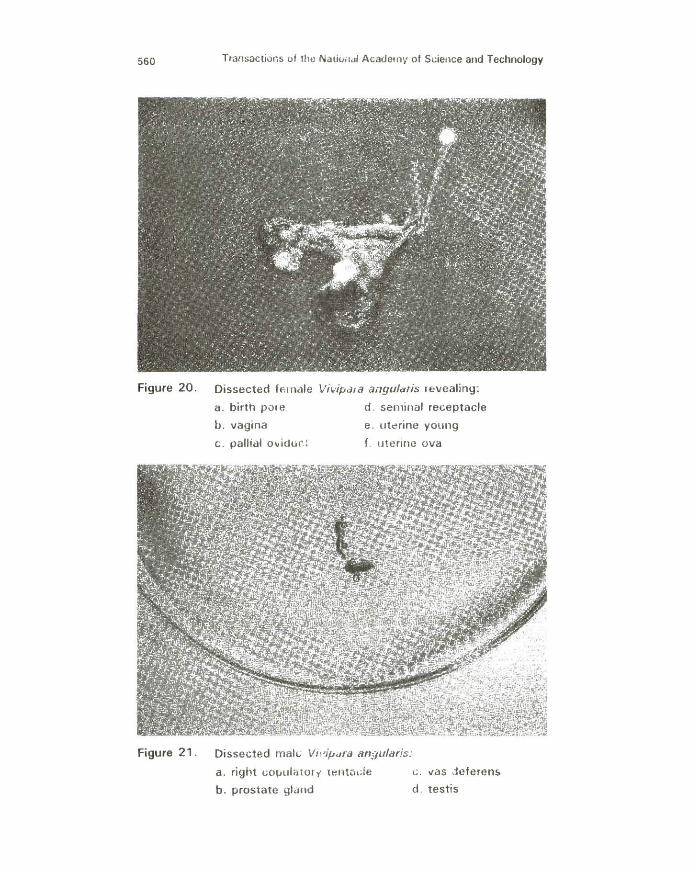
The birth pore of the female reproductive system f ound on the anterior surface of the vagina exhibits bright orange flecks above a speckled gray background of melanin (Figs. 18 and 20). The elliptical vagina is situated in the distal end of the pallial oviduct and terminates at the edge of the mant le. It exhibits a grayish appearance. Originating at the posterior end of the mantle cavity and proceeding anteriorly to the narrower vagina, the pallia l oviduct lies parallel to the rectum at the extreme right edge of the mantle cavity and exhibits moderate to heavy concentrations of melanin in the epidermal covering .
In non-gravid females it is a large, comparatively thickwalled, white duct while in gravid females the organ is expanded and enlarged in diameter to form the brood pouch. It is relatively thin-walled and easily identified by the presence of developing young.
The white pink seminal receptacle is located anterior to the albumen gland. It is externally distinguishable from the pallial oviduct in gravid females by its narrower diameter. It is externally indistinguishable in non-gravid females. The V-shaped oviduct is located anteroventrally to the albumen gland and receives the ovary at its proximal end. Its distal end enters the posterior part of the seminal receptacle. The oviduct appears white for most of its length, and frequently pink to rose in the distal region. The faintly translucent, white ovary (often difficult to discern in living animals) is located in the apical whorl along the digestive gland. The ovary is a long, narrow duct which sometimes possesses a variable number of short lateral branches or small protrusions (2).

542 Transactions of the National Academy of Science and Technology
B. Male Reproductive Organ
Gross dissection revealed features of the male reproductive structure (Figs. 19 and 21). The curved, modified right tentacle of the male vivipara serves as its copulatory organ. It is mainly black with transverse bands of orange in an irregular pattern. The bean-shaped testis is rust orange, long and fusiform organ located along the right edge of the mantle (2). The vas deferens, a short, nearly transparent structure, leaves the testis at about the middle to posterior portion. It curves slightly at the mantle wall, passes through the posterior end of the food groove and then enters the prostate gland. The large creamy yellow prostate gland receives the vas deferens and passes forward beneath the groove towards the right tentacle. It has a smooth, thickly muscular appearance in contrast to the grainy appearance of the testis. It spans nearly t he entire length of the mantle cavity then narrows into a terminal vas deferens that passes through the right tentacle and opens at its tip.
IV. Histological Sections
Histological sections for both male and female reproductive organs of Vivipara angularis were done using the Paraffin method.
A . Female Reproductive Organ
The ovary (Fig. 18) is very small, translucent to orangebrown delicate structure lying on the digestive gland. It is often very hard to discern in live specimens and researchers have not yet been successful with its histology.
The seminal receptacle of spermathecae (Figs. 22 and 23) where sperm received during copulation is strored, is lined by tall columnar epithelium and surrounded by circular muscles.
The pallial oviduct (Figs. 22 and 24) is lined by mucosal cells thrown into folds, perhaps f or greater distension capacity when eggs, embryo, and young snails are contained in this structure. It is lined by simple columnar ciliated cells with light staining cytoplasm and deeply staining basal nuclei .
The vagina {Fig. 25), located at the distal end of the pallial oviduct, exhibits irregular folds in its mucosa. The surface epithelium is transitional in type. This ephithelium is stratified and composed of several layers of generally similar cells which are usually cuboidal with round nuclei.

Transactions of the National Academy of Science and Technology 543
B. Male Reproductive Organ
The right copulatory tentacle's surface contains flecks of bright fuchsia to red over a solid sub-epidermal concentrat ion of melanin (Figs. 26 and 27). It is lined by columnar cells with basally located nuclei. The seminal duct included within the tentacle is composed mostly of circular muscles with some interspersed connective tissues. The muscular tentacle can be accounted for by the need for a mechanism to effectively ejaculate the sperm during copulation.
The light st aining prostate gland (Fig. 28) is lined by simple cuboidal cells. A thin muscular coating of an external circular muscle layer and an internal longitudinal muscle layer around the entire tissue can be observed.
The vas deferens (Fig. 29) exhibits a narrow, irregular lumen and a thick muscular layer. The epithelium is pseudostratified columnar.
The t estis (Figs. 30 and 31) is composed of seminiferous tubules, each surrounded by an outer layer of connective tissue and an inner basement membrane. The basement membrane encloses the specialized germinal epithel ium of t he seminiferous tubules which consists of two cell types: the <:>upporting cell of Sertoli cells and the spermatogenic cells.
The Sertoli cells are triangular and tapering w ith very irregular outlines extending from the basement membrane to t he free luminal surface. The very distinct ovoid or angular nucleus is outlined with fine, sparse chromatin.
The spermatogenic cells are arranged in rows, mostly superimposed on Sertoli cells, obscuring their cytoplasm. The most primitive or immature spermatogenic cells, the spermatogonia, are sit uated adjacent to the basement membrane of the seminiferous tubules. Spermatogonia divide mitotically to produce several generations of cell s.
The primary spermatocytes, the largest germ cells in the seminiferous tubules, occupy the middle of the germinal epithelium. The primary spennatocytes give rise to secondary spermatocyte by meiotic division.
The secondary spermatocytes are distinctly smaller t han the primary spermatocytes. The nuclear chromatin is less dense.
The spermatids are smaller cells with small nuclei containing both fine and larger chromatin granules. The cells become closely

544 Transactions of the National Academy of Science and Technology
associated with the surface of Sertoli cells, and in this environment differentiate into spermatozoa by the spermiogenesis process.
The small, deep-staining heads of spermatozoa are embedded in the cytoplasm of Sertoli cells w hile their t ails extend into the lumen of the seminiferous tubule. Most of the findings of this study are in accordance with the descriptions of DiFiore {1989)5 .
V. Protein Content
Preliminary results of protein concentration analysis of snails fed with different food types (sample 1 - lettuce only; samle 2 -lettuce and mud) indicat e that feeding with lettuce and mud could improve the protein content of V. angularis twofold (Fig. 32).
DISCUSSION
Vivipara angulans inhabiting the slow moving waters of the UP lagoon were subjected t o field and laboratory analyses of population samples taken at monthly intervals over a period of one year. Gross dissection and histological sect ions elucidated the existence of sexual dimorphism, disputing the claim of Alonte { 1930) that this snail is hermaphroditic (1 ). Although shell features offered no means of identifying sexes, sex determination was based on the appearance of curved or "hooked" right tentacle in males as opposed to fine left tentacle of males and right and left tentacles of females. THe right tentacle serves as its intromittent copulatory organ. Sexual dimorphism was also observable in size, f emales being generally larger than males. These findings further establish that Vivipara angularis is indeed sexually dimorphic.
Biometric data obtained through a scheme of monthly sampling covering a period of one year, described the life cycle of V. angularis. Gestation period was observed to cover a span of seven months, starting in August, and peaking in February of the next year {Fig. 8). Most of the female snails in these months are gravid, having a shell length of around 18-19 mm and containing uterine young in a graded series of development. The percentage of uterine young gradually decreased and reached its lowest in June, the time when gravid snails have expelled all their uterine young. In March t o June, snails delivered during the

Transactions of the National Academy of Science and Technology 545
gestation period developed into immature snails and became sexually mature in June. Since the growth period was from March to June, and the start of the gestation period was in August, a short mating period was in early June.
A very high percentage of uterine ova in June indicated that the snails had just undergone copulation. Growth of uterine ova into uterine young started in July as evidenced by the slight increase in the percentage of uterine young from June to July. More developed young were observed in August, when the gestation period commences.
The initial results in the laboratory rearing experiments show that there was an observably high survival rate in snails fed with mud and lettuce. It is interesting to note that they were also found to have higher protein content than snails fed with lettuce only. However, the results are not conclusive as the data obtained were from preliminary expreiments only.
Figure 1 . The collection site: U .P. lagoon

546
Figure 2 .
Figure 3 .
Transactions of the National Academy of Science and Technology
The specimen: Vivipara angu/aris
Snails were reared in glass aquaria provided w ith continuous aeration.

Transactions of the National Academy of Science and Technology 547
Figure 4 .
Figure 5.
Gravid female snails with newly-expelled young. They were fed with lettuce leaves.
. ... •'
. ,~j :, . ~~t/.{ ;;,;~.} : ~. ··-~·h~::·
~· .J : ·· (
Male Vivipars sngulsris showing their "hooked" right copulatory ten tacles

! 0 c • ...l
;; £ Ill
• 0
2
t
548 Transactions of the National Academy of Science and Technology
Figure 6. Female 'VIvi!Mra sngulsrl• showing their fine, pointed left and right tentacles.
VJ.fpon:> ..,~.dam for tt.e whole year 2•-r----------------------------------------------------------,
liar Apr llay J.., Jul Aug Sept Oat Nov Dea Jan F4b
Figure 7 . Average Shell Length of Male and Female

Transactions of the National Academy of Science and Technology 549
from Dl~ed Femat. V. aniJUiarh' 100-r----------------------------------~-------------------------,
eo
eo
70 eo
50
30
20
10
Jul AUIJ Sept Oot Feb
Dl_..,tlan P.rfod ~ Uterine 0..0 !ZZl uterine y....,IJ
Figure 8 . Percentage of Uterine Ova and Young

20 H;t 11!1
17 ,. ,5
14 .. ,s 'ij ,2 ~ 11 '0 10
.! .. E e ::> z 7
• e • s 2 , 0
0 1 2 s 4 e e 7 e
Coll-t.d In April 1••o
81011121S14181e1718182021222S2428282721!128SOS132SS34S5
Sh-ell L.notft (mm) c l"'•mGI• ... WGI•
Figure 9 . Biometric Data of V. angularis
Ul Ul 0
~ ii! :J
l!l (')
ct. 0 a 2. g. CD
z I» ct. 0 :J !!!. > (') I» a. (D
3 < 2. (/) (')
~· (') CD I» :J a. ~ g. :J 0 0
(C
<

20 19 us 17 ,Cl ,~
14
~ 13 Q ,2 ~ 11 0 10 • fl "E e ::> :z. 7
Cl e .. 3 2 , 0
0 1 2 3 .. !I Cl 7 e
Coll-t~ tn July 1flfl0
• 10111213141e1e171e1•::zo:t12223242!12cs272e:t•:so:s1:s:t:s:s:s4:se
SMII L--.ottoo (mm) a l"•mal• 'r Wal•
Figure 10. Biometric Data of V. angularis
f n g. :I
"' 0 -g. CD
z C> g. :I !!!.. >
l g. C/)
!2. ! ~ ~ Q.
-! CD n ;r :I 0 0
<C <
01 01 -

552
0 • II ..
Transac1ions of the National Academy of Science and Technology
IC
~ ., ., ., fC ., ., 0 ., • fC t) fC
" fC G tl IC fl .. tl ., tl fl tl
tl 0 fl • ... t) ... " G ... ., ... .. ... ., ... t4
o~at-~~-.~~ ... o•«~t-~IC~W>flrO Nrrrrr.-rrr,..
.! tl l
.... ~~o £ £ ...,.
£ 0
i ..J
=· 1-c ID£
:. D
-0
Ill ... co c u
·;: -a> E 0
ii5
..... ..... a> ... ::s Ol u:

Transactions of the National Academy of Science and Technology 553
_, ~ , ., , ~ , , 0 , II ~ ., t4 ,.. ~ ~ ~ ., fl .. tl ., .! ti ID tl l ·~
D tl ..!!! ... • tl ::i II ~ .. 0 ....... N Ill
" • £ • ... ::::.: .0 ~ E E
... .. ..... 0
l ,.. f Ill
A ... .. Cl D Ill • 0
Ill .. ~ C) .g
£ .. .. .. =· Q) .., .. 1ii E
t .,
0
1 .. 41lf iS N •
& ... .. N
0 ..... 0 ~
II :I C)
I) il: ,.. Cl
C) .. ., fl
o~.,,..~~•~~~cect-e~~~.,N·O Nrr"r"rr~r-.r,..r

554
0 • •
Transactions of the National Academy of Science and Technology
O~a~·~·.,~rO~CI~·~·.,ffrO N.-r~rr.- ... r r r
.. .. .. 0
• Cl
~
• ., • ., "
.! a l
D
-0 IU ~ 0 u ·c:: ... G)
E 0 ill

%0 18 11!
17 ,4111 ,~
14
.!! 13 15 12 ~ 11 -o 1D
.! " ~ e :z: 7
fJ s 4
3 % , D
0 1 % 3 4 & e 7 8 • 10111%1314181817181.%0%1%%%3%428%827282.303132333435
Bt-..11 L..._O+h (mm) a l'•mal• .. Mal•
Figure 14. Biometric Data of V. angularis
Ul Ul Ul

556
0 ~ • ...
-~
Transactions of the National Academy of Science and Technology
If)
~ ., ., ~ ., .... ~ ., tl oN Cl
~ oN Cl oN ., ~ oN ! .,
tl
t1 l t4
o~m~~lfl~~~ ... o•c~CIG~.,NrO ~r.-rrr .. rrT"'r
-0
IU rti 0 u
·.::: ... Cl)
E 0 lil
Lt) ...

%0 19 H!
17 18
1!1 .. 14 =ij 13
15 12
'0 1 1 .. 10 .! 9 E ::> ~ ::
7
oe ~ 4
3 2 , 0
0 1 ~ 3 • e oe 7 e
0611~•d In J oi'\UCII"Y 11tl~1
e 10111%131 4 1&1o&171e1e:toZ122%3242&Ze:t7 ze:te30313233343&
SMII L'""CIIth (t"r"' t"r"' ) a l"• mal• Y M o l•
Figure 16 . Biometric Data of V. angu/aris
-1 OJ :::l
!3 (") d . 0 :::l
"' s. g. <11
z Ql ::: . 0 ::l !!!.. )> (") Ql c. <11
~ s. (/) 0 iii' ::l
£ Ql ::l c. -1 <D 0 =r :J 0 0 (Q -<
c.n c.n -..j

20 1» H!l
17 ,. ,~
14 .. 13 'ij 12 ~ 1 1 0 •o .. .3 e E ;>
$
% 7 6 5 4
s 2
0
0 , 2 3 • 5 e 7 e •
Collected in February 1991
10111~1314151e171811t2021222324252e27282~t303132333435
sh•ll L...,oth (m M'l) = ,..,.,.HJI• 'rf Mal•
Figure 17 . Biometric Data of V. angularis

Transactions of the National Academy of Science and Technology 559
Figure 18. Vivip11ra angularis, female, with mantle cut open along right edge and deflected to animal's left~ Oviducal pouch cut open revealing juvenile and embryo
Figure 19. Vivipara angularis, male, with mantle cut open along right edge and deflected to animal 's left {so deflecting rectum from right to left of animal)
560 Transactions of tht.l Natio;t,JI Acadetny of Science and Technology
Figure 20. Dissected female Vi~·ipa1a ai1gula!is revealing:
a. birth p01e d . seminal receptacle
b. vagina e. ut~rine young
c . pallial ovidur.: f. uterine ova
Figure 21. Dissected mal t. Vhdpara angularfs:
a. right t;OJJulatorr t~ntude c . vas -ieferen~
b. prostate glund d . testis

Transactions of d1e Nationill Ac;JdP-r!lV of SciP.nco at>c1 Technology 561
Figure 22. The smallor seminal receptacle !SRI is medial to the bigger pallial oviduct (POl. Both structures are surrounded by circular muscles !CM). Note tllat the p<lllial oviduct has a biggP-r lumen Ill .
Figure 23. Higher magnification of s!.'minf.'ll mceptade showing tall t:olumnar epithelium IEl swroundP.d by circular rnuscles ICMI

562 Transactions of the National Academy of Science and Technology
Figure 24. Cross section of the mucosal folds of the pallial oviduct { 1 OOX} lined by simple columnar ciliated epithelium (E) with darker staining basally located nucleus INJ
Figure 25. Transverse section of vagina (10Xl reveals its irregular lumen IL}. transitional type of epithelium (TE} and connective tissue {CT}.

Transactions of the National Academy of Science and Technology 563
Figure 26. Cross section of the right copulatory tentacle (10X). The seminal duct (SOl is surrounded by a thick layer of circular muscles (CM) and connective tissue (CT). Black pigments called melanin (Ml are found in the sub-epidermal layer.
Figure 27. Higher magnification of the surface epithelium (SE) of the right tentacle showing brightly pigmented columnar cells with basal nucleus IN) and melanin (M).

564 Transactions of the National Academy of Science and Technology
Figure 28. Transverse section of prostate gland with thin muscle layer (M) and circular lumen (LI. It is lined by simple cuboidal cells.
Figure 29. Vas deferens (40Xl exhibiting a pseudostratified columnar epithelium (E). irregular lumen (L). and thick muscular layer (M)

Transactions of the National Ac3d&my of Sci~nc.e and Technology 565
Figure 30. Seminiferous tubules of testis illustrating the Sertoli cells (SJ and spc1matozoa ISPi with deep staining nuclei (N) and faintly staining tails IT). The smaller spermatids (SDI are also shown in this picture.
Figure 31 . Another eros~; section of testis showing seminiferous tubules enveloped hy bar.entent membrane (B). spermatozoa (SP) with :Jc:!p s:taining heads or nuclei (N) and the triangular Sertoli cells (S) closely associated with the spermatozoa.

566
?
i.B 1.8
\ , ;t'
1.~
, , J
1.~ , -~ ,~ ,, , a.& n.e Q,,
o.o -0 -~
n.4 O. J •'
0.2 .' s
0 .1
D
Transactions of the National Academy of Science and Tedwlology
Brad ford .ll.nol)"sis
_,.
/
~
:10 DO
.-------· ___ .u-~--
110
_ ... -------- ! .~--··-"
100
Figure 32. Protein determination of soluble extracts of V. 11ngularis fed with two different food types
STANDARD
x :ug BSA
5 10 20 40 80
100
SAMPLES
y: Ave. 00 at 595 nm
0.2093 0.3777 0.6363 1.2867 1.6730 1.9366
Regression Output : Constant 0.265661 Std Err of Y Est 0.182565 R Squared 0.947813 No. of Observations 6 Degrees of Freedom 4
X Coefficient (s) 0,017747 Std Err of Coef. 0.002082
DI LUTION A.BS 595 AVERAGE ug ug x OI L. ug/ul SUP ABS 595 Extract Factor
(ug/4 ul sup.)
Sample 1 [fed with lettuce; wt . = .218 g. deshelled)
1:25
Sample 2
0.408 0.403
0.4 1 7.89
(fed with lettuce and mud; wt. = .391 g, deshelled)
197.25 49.31
1:25 0 .554 0.587 0.545
0.56 16.34 408.50 102.13

Transactions of the National Academy of Science and Technology 567
REFERENCES
1. Alontet Fidel H. 1930. Biology of Vivipara angularis Muller, a common freshwater snail in Laguna de Bay. The Philippine Agriculturist 19(5). 307-325.
2 Berry, A.J. 1974. The anatomy of West Malaysian snails of parasitological significance. Malayan Nature Journal 27. 131-165.
3. Browne, A.A. 1978. Growth, mortality, fecundity, biomass and productivity of four lake populations of the prosobranch snail, Viviparus georgianus. Ecology 59(4). 742-750.
4. Chin, T.S. 1983. Helminths of medical importance found in edible freshwater snails commonly sold in public markets of Metro Manila. Masteral Thesis. University of the Philippines, Institute of Public Health Library, p. 62.
5 . Di Fiore, M .S. 1989. Atlas of Normal Histology. 6th ed. Philadelphia, U.S.A.: Lea and Febiger.
6. Monzon, A.B. 1985. Snails of medical importance in Family Viviparidae of Thailand. Paper read at the Training Course in Applied Malacology held in Bangkok, Thailand, NovemberDecember, 1985.
7. Pagulayan, I. F. 1986. Comparative characterization of the footmuscle, hemolymph, hepatopancreas proteins of Radix quadrasi and Mixas cumingiana (Pulmonata: Lymnaeidae). Unpublished material.
8. . 1987. The biology of Vivipara sp. Unpublished material.
9. SEAMEO-TROPMED Technical Report on snails of medical importance in Southeast Asia. Bangkok, 1 5-18 June 1985. pp. 12-13.
10. Talavera, F. and L.A. Faustino. 1933. Edible mollusks of Manila. Philippline Journal of Science 50(1 ): 1-48, Pits. 1-18.
11 . Vail, V.A. 1977. Comparative reproductive anatomy of 3 viviparid gastropods. Malacologia 16. 519-540.
12. . 1978. Seasonal reproductive patterns in three viviparid gastropods. Malacologia 17(1). 73-97.
13. Van Cleave. H.J. and D.A. Altringer. Studies on the life cycle of Campeloma rufum, a freshwater snail. The American Naturalist 16. 913-920.

568 Transactions of the National Academy of Science and Technology
14. Van Cleave, H.J. and L.G. Lederer. 1932. Studies on the life cycle of the snail, Viviparus contectoides. Journal of Morphology 53. 499-522.
15. Velasquez, C.C. 1964. Observations on the life cycle of Eupharyphium paramurinum sp. {Trematoda: Echinostomatidae). Journal of Parasitology 50(1). 106-111.