the renin-angiotensin-aldosterone system in congestive failure in
TRANSCRIPT
The Renin-Angiotensin-Aldosterone System in Congestive
Failure in Conscious Dogs
IZVx WATKINS, JR., JAMES A. BURTON, EDGARHABE, JAMLS R. CANT,FRANKaIn W. SMrm, and A. CLIFFORD BARGER with the technicalassistance of SARA E. MCNEIL and STEPHEN M. SmERRuL
From the Department of Physiology and Department of Medicine,Massachusetts General Hospital, Harvard Medical School,Boston, Massachusetts 02115
A B S T R A C T The role of the renin-angiotensin-aldos-terone system in the development of congestive failurehas been assessed in the conscious dog by use of thenonapeptide converting enzyme inhibitor. Constrictionof the pulmonary artery or thoracic inferior vena cavawas maintained for 2 wk while daily measurements weremade of plasma renin activity, plasma aldosterone,plasma volume, hematocrit, serum sodium and potassiumconcentrations, sodium and water balance, body weight,and arterial, caval, and atrial pressures. The initial re-sponse to constriction was a reduction in blood pressure,a rise in plasma renin activity, plasma aldosterone, andwater intake, and nearly complete sodium retention. Inthe days after moderate constriction plasma volume andbody weight increased (with development of ascites andedema); blood pressure, sodium excretion, plasma reninactivity, and plasma aldosterone returned to normal. Inanimals in which blood pressure was not restored, plasmarenin activity and plasma aldosterone remained elevatedthroughout the period of constriction. Single injectionsof converting enzyme inhibitor reduced blood pressurewhen plasma renin activity was elevated. Chronic infu-sion of the inhibitor in dogs with thoracic inferior venacaval constriction prevented the restoration of bloodpressure and suppressed the rise in plasma aldosterone;sodium retention and volume expansion were less thanin control experiments. Thus the renin-angiotensin-al-dosterone system plays an essential role in the mainte-nance of blood pressure during the genesis of congestive
This work was presented in preliminary form at the 59thAnnual Meeting of the Federation of American Societiesfor Experimental Biology, Atlantic City, N. J., 1975.
Received for publication 20 November 1975 and in revisedform 6 February 1976.
failure. Initially, the restoration of blood pressure is de-pendent upon circulating angiotensin II; in the laterstages, blood pressure is dependent upon the increase inplasma volume.
INTRODUCTION
The increased renal venous renin activity first observedin patients with congestive heart failure by Merrill et al.(1) 30 years ago, suggested that renin may play a rolein the maintenance of blood pressure in cardiac failure.Several years before aldosterone was identified, Demingand Luetscher (2) also reported that patients with con-gestive failure excreted increased amounts of a salt-retaining steroid, now known to be aldosterone. How-ever, the plasma levels of renin and aldosterone in pa-tients and experimental animals with congestive heartfailure have subsequently been reported as high, low, ornormal (3-9). As a result of this variability, the role ofthe renin-angiotensin-aldosterone (RAA)1 system in thesyndrome has not been clearly defined.The recent availability of inhibitors of the renin-
angiotensin system, as well as an appropriate animalmodel, has provided means for a more precise analysis ofthe problem. Congestive failure has been induced in theconscious trained dog by inflation of a previously im-planted constricting cuff around the thoracic inferiorvena cava (TIVC) or the pulmonary artery. The re-sponse of the RAA system was monitored from the timeof constriction to the development of frank congestivefailure, thus enabling us to study the sequence of events
'Abbreviations used in this paper: BP, blood pressure;CEI, converting enzyme inhibitor; PA, plasma aldosterone;PRA, plasma renin activity; PV, plasma volume; RAA,renin-angiotensin-aldosterone; TIVC, thoracic inferior venacava.
The Journal of Clinical Investigation Volume 57 June 1976 -1606-16171606

in the progressive course of the disease uncomplicatedby anesthesia and surgery. Pulmonary artery stenosis inthe dog produces a right-sided congestive heart failuresimilar to that seen in man, with cardiac enlargement,elevated right ventricular end diastolic pressure, lowcardiac output, inability to exercise, and retention ofsodium and water with resultant ascites and edema.TIVC constriction, although not a model of cardiac fail-ure, simulates the hemodynamic consequences of im-paired cardiac performance (10, 11).By the use of the nonapeptide converting enzyme in-
hibitor (CEI) (12-14) which blocks the conversion ofangiotensin I to the physiologically active pressor agentangiotensin II, we have been able to assess quantitativelythe role of the RAA system in the regulation of systemicpressure during the development of congestive failure.We have also examined the role of the renin-angiotensinsystem in the regulation of plasma aldosterone levels,sodium excretion, and water balance. Finally, the non-apeptide has enabled us to show that the ability of theanimals to compensate for the circulatory impairmentimposed by constriction is markedly impaired if thegeneration of angiotensin II and the rise in plasma al-dosterone is suppressed.
METHODSMongrel dogs (20-30 kg) were kept in metabolic cages ina room of constant temperature and fed a fixed diet con-taining approximately 75 meq of sodium and of potassium.Free access to water was allowed and 24-h intake andurinary output measured. The animals were trained to liequietly on a padded table in an air-conditioned room. Aftertraining the animals were prepared surgically for the ex-periments.
Surgical proceduresFor surgical procedures the animals were anesthetized
with intravenous pentobarbital sodium (30 mg/kg). Understerile conditions, with the animal on artificial ventilation,thoracotomy was performed through the left 4th intercostalspace. The pericardial sac was opened widely exposing thepulmonary artery and the TIVC. A 2.0 X 1.5-cm inflatablesilastic rubber cuff was placed around the main pulmonaryartery and a 2 X 2-cm cuff was placed around the TIVC.A polyvinyl chloride catheter (0.04 inch ID, 0.07 inch OD)was placed in the right atrium through the atrial appendage;in several dogs a catheter was also implanted in the leftatrium. The abdominal aorta and vena cava were approachedretroperitoneally through a left flank incision and werecatheterized with polyvinyl chloride catheters (0.025 inchID, 0.056 inch OD) using the techniques of Herd andBarger (15). The intravascular catheters and the cathetersattached to the inflatable cuffs were exteriorized throughsubcutaneous tunnels over the left flank and protected bya cotton jacket. The animals were allowed to recover for2 wk before experiments were started.
Experimental protocolThoracic inferior vena caval constriction. For 3 days
before caval constriction the following control measure-
ments were made between 8 and 10 a.m.: inferior vena caval,atrial, and aortic pressures; plasma renin activity (PRA)(16) and plasma aldosterone (PA) (17) ; plasma volume(PV); and hematocrit; 24-h urinary sodium and potassiumexcretion, serum sodium and potassium concentrations, waterbalance (intake minus urinary output), and body weightwere also measured. The caval constriction was done in twostages on the first 2 days to produce a 10-mm Hg rise inabdominal caval pressure. Preliminary experiments indi-cated that some animals did not tolerate a more rapid ormore severe constriction. The initial constriction, raising theinferior vena caval pressure by 5 mm Hg, was done over a
period of 5-10 min by inflating the caval cuff with saline;after the desired constriction was obtained the catheter was
clamped. Blood samples for PRA and PA were taken at15 min before and at 15, 30, and 45 min after constrictionwhile pressures were continuously recorded. The animal wasthen returned to the cage. 5 h later all pressures were againmeasured and blood samples taken for PRA and PA. Ifelevated caval pressure had not been maintained, minoradjustments in the inflation of the cuff were made.
24 h later the cuff was further inflated to raise the cavalpressure an additional 5 mm Hg. Samples for PRA andPA were taken before and 15 min after inflation. 5 h laterthe measurements were repeated. After 48 h, all measure-ments listed previously were done daily. At the end of 2 wk,or after a steady state of salt balance had been achieved, theconstriction was released. The measurements were continuedfor 3 days after release.Acute converting enzyme blockade during thoracic inferior
vena caval constriction. Thoracic caval constriction wasperformed in two stages as described above. On days 3, 5, 7,and 10, while pressures were measured in two dogs, 5 mgof the CEI (< Glu-Trp-Pro-Arg-Pro-Gln-Ile-Pro-Pro) wasgiven intravenously as a single bolus. In previous experi-ments in this laboratory, no rise in plasma bradykinin wasdetected with such a dose, or during constant infusion asbelow (18). Blood samples for PRA were taken before and15 min after injections.Chronic converting enzyme blockade during thoracic in-
ferior vena caval constriction. Before inflation of the cavalcuff, a 5-10-mg bolus of the inhibitor was given intravenouslyfollowed by a constant infusion for 3 days. The infusionpump, secured to the shoulder harness of the dog, infusedthe inhibitor through the caval catheter. The completenessof blockade was tested 2-4 times daily by the injection of 1,g of angiotensin I. In the normal animal this injectioncaused a 30-35-mm Hg rise in arterial pressure; duringadequate blockade no change in pressure was produced. Inthe first experiment with chronic infusion of the CEI, 5mg/h were administered. On the 2nd day, a small rise inarterial pressure was observed after the injection of 1 ,ug ofangiotensin I. Therefore, the dose of the inhibitor was raisedto 10 mg/h for the remainder of the infusion. In the follow-ing two experiments with chronic converting enzyme block-ade, a dose of 10 mg/h was used throughout the 3-day in-fusion period. This dose produced effective blockade inexperiments 1 and 2 as evidenced by the lack of a pressorresponse to injected angiotensin I. In the 3rd experiment,10 mg/h did not completely block the pressor response toangiotensin I in the latter part of the experiment; on severaloccasions a 5-10-mm Hg rise in blood pressure (BP) was
produced. However, because of the limited supply of theCEI the dose was not raised.Pulmonary artery constriction. The experimental proto-
col for the pulmonary artery constriction was similar to
Renin and Aldosterone in Congestive Failure 1607
MEANTIVCPRESSUREmm Hg
MEANAORTICPRESSURE
mm Hg
MEAN 1
RIGHTATRIALPRESSURE _ ,
mm Hg t t
PLASMARENIN
ACTIVITY
ng/ ml /h
10
0 ASCITES i
150HEART
100 RATE
50
600-
PLASMA 400ALDOSTERONE
pg/mi
200 Ld * * * i
2 3 4 5 6 7 8 9 10 11 12 13 14 15 16 17 18 19
t t DAY OF EXPERIMENTTIVC INCREASE RELEASE
CONSTRICTION CONSTRICTION CONSTRICTION
URINARYSODIUM +-POTASSIUMEXCRETIONmeq/24 h
No -
K-
PLASMAVOLUME
ml
27
BODYWEIGHT 26
Kg 25
HEMATOCRIT
1608 L. Watkins, Jr., J. Burton, E. Haber, J. Cant, F. Smith, and A. Barger
Z41 2 3 4 5 6 7 B 9 10 1I 12 13 14 15 16 17 IS 19
t t DAY OF EXPERIMENT IT IVC INCREASE RELEASE
CONSTRICTION CONSTRICTION CONSTRICTION
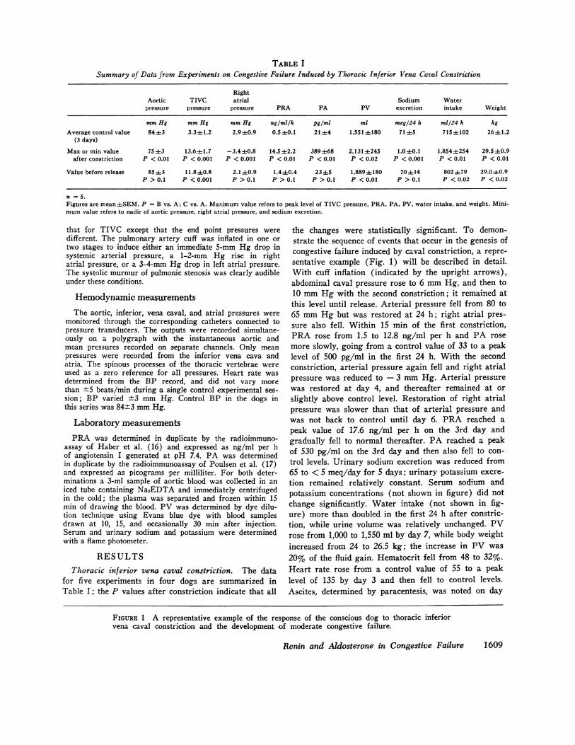
TABLE ISummary of Data from Experiments on Congestive Failure Induced by Thoracic Inferior Vena Caval Constriction
RightAortic TIVC atrial Sodium Waterpressure pressure pressure PRA PA PV excretion intake Weight
mm Hg mm Hg mm Hg ng/mi/h pg/mi ml meq/24 h mi/24 h kgAverage control value 84±==3 3.34±1.2 2.9±40.9 0.5±40.1 21±44 1,551±4-180 71±45 715±-4102 26±41.2
(3 days)
Max or min value 75±--3 13.6±4-1.7 -3.4±-0.8 14.5±42.2 389±-68 2,131±-i245 1.0±40.1 1,854±A254 29.5±=10.9after constriction P < 0.01 P < 0.001 P < 0.001 P < 0.01 P < 0.01 P < 0.02 P < 0.001 P < 0.01 P < 0.01
Value before release 85±43 11.8±40.8 2.1±-0.9 1.4±40.4 23±-45 1.889±-180 70±--14 802±479 29.0±-40.9P > 0.1 P < 0.001 P > 0.1 P > 0.1 P > 0.1 P < 0.01 P > 0.1 P < 0.02 P < 0.02
a = 5.Figures are mean ±-SEM. P = B vs. A; C vs. A. Maximum value refers to peak level of TIVC pressure, PRA, PA, PV. water intake, and weight. Mini-mum value refers to nadir of aortic pressure, right atrial pressure, and sodium excretion.
that for TIVC except that the end point pressures weredifferent. The pulmonary artery cuff was inflated in one ortwo stages to induce either an immediate 5-mm Hg drop insystemic arterial pressure, a 1-2-mm Hg rise in rightatrial pressure, or a 3-4-mm Hg drop in left atrial pressure.The systolic murmur of pulmonic stenosis was clearly audibleunder these conditions.
Hemodynamic measurements
The aortic, inferior, vena caval, and atrial pressures weremonitored through the corresponding catheters connected topressure transducers. The outputs were recorded simultane-ously on a polygraph with the instantaneous aortic andmean pressures recorded on separate channels. Only meanpressures were recorded from the inferior vena cava andatria. The spinous processes of the thoracic vertebrae wereused as a zero reference for all pressures. Heart rate wasdetermined from the BP record, and did not vary morethan ±+5 beats/min during a single control experimental ses-sion; BP varied -±3 mm Hg. Control BP in the dogs inthis series was 84±3 mm Hg.
Laboratory measurements
PRA was determined in duplicate by the radioimmuno-assay of Haber et al. (16) and expressed as ng/ml per hof angiotensin I generated at pH 7.4. PA was determinedin duplicate by the radioimmunoassay of Poulsen et al. (17)and expressed as picograms per milliliter. For both deter-minations a 3-ml sample of aortic blood was collected in aniced tube containing Na2EDTA and immediately centrifugedin the cold; the plasma was separated and frozen within 15min of drawing the blood. PV was determined by dye dilu-tion technique using Evans blue dye with blood samplesdrawn at 10, 15, and occasionally 30 min after injection.Serum and urinary sodium and potassium were determinedwith a flame photometer.
RESULTSThoracic inferior vena caval constriction. The data
for five experiments in four dogs are summarized inTable I; the P values after constriction indicate that all
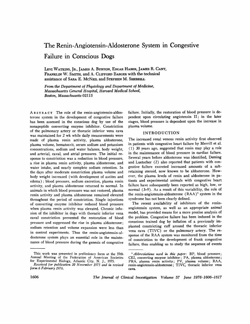
the changes were statistically significant. To demon-strate the sequence of events that occur in the genesis ofcongestive failure induced by caval constriction, a repre-sentative example (Fig. 1) will be described in detail.With cuff inflation (indicated by the upright arrows),abdominal caval pressure rose to 6 mm Hg, and then to10 mm Hg with the second constriction; it remained atthis level until release. Arterial pressure fell from 80 to65 mm Hg but was restored at 24 h; right atrial pres-sure also fell. Within 15 mmn of the first constriction,PRA rose from 1.5 to 12.8 ng/ml per h and PA rosemore slowly, going from a control value of 33 to a peaklevel of 500 pg/ml in the first 24 h. With the secondconstriction, arterial pressure again fell and right atrialpressure was reduced to - 3 mm Hg. Arterial pressurewas restored at day 4, and thereafter remained at or
slightly above control level. Restoration of right atrialpressure was slower than that of arterial pressure andwas not back to control until day 6. PRA reached a
peak value of 17.6 ng/ml per h on the 3rd day andgradually fell to normal thereafter. PA reached a peakof 530 pg/ml on the 3rd day and then also fell to con-trol levels. Urinary sodium excretion was reduced from65 to < 5 meq/day for 5 days; urinary potassium excre-tion remained relatively constant. Serum sodium andpotassium concentrations (not shown in figure) did notchange significantly. Water intake (not shown in fig-ure) more than doubled in the first 24 h after constric-tion, while urine volume was relatively unchanged. PVrose from 1,000 to 1,550 ml by day 7, while body weightincreased from 24 to 26.5 kg; the increase in PV was
20% of the fluid gain. Hematocrit fell from 48 to 32%.Heart rate rose from a control value of 55 to a peaklevel of 135 by day 3 and then fell to control levels.Ascites, determined by paracentesis, was noted on day
Renin and Aldosterone in Congestive Failure
FIGURE 1 A representative example of the response of the conscious dog to thoracic inferiorvena caval constriction and the development of moderate congestive failure.
1609
20MEAN TIVCPRESSURE 10
m- Hg
0 t t110 | ASCITES
600
MEAN
RIGHT ATRIAL
PRESSUREOt
mm Hg
RENIN i _ng/ml/h 10; . .
7
600
PLASMCA 400 f\ /ALDOSTERONEI\/ \/ \
pg/mI 300t lVV \
100.
10-
0 2 3 4 5 6 7 8 9 10 1I1 12 13 14 IS 16 17 l8 I9t t DAY OF EXPERIMENT
TIVC INCREASE RELEASECONSTRICTION CONSTRICTION CONSTRICTION
500
450
400
URNR 500
SODIUMb4. 300
POTASSIUMb250_
EXCRETION
mq/24h 200
I5O
100
SO L.____.__ _ __ '---
PLASMA 1400 r' 4-- - - . /
4oUE100\\ ,,^
300
2iW.25 -
mliIO
Kg/2h 0
1 2 3 4 S 6 7 8 9 10 1 1 12 13 14 15 16 17 IS 19
t t DAY OF EXPERIMENT lTIVC INCREASE RELEASE
COSTRICTIONI CONSTRICTION CONSTRICTION
1610 L. Watkins, Jr., 1. Burton, E. Haber, J. Cant, F. Smith, and A. Barger
1
5mg CEI 5mg CEI 5mg CEI 5mg CEI100
MEANAORTIC 90 1
PRESSUREmm g 80
0- control0
*-15min after70CEI
60
50
40 [IPLASMARENIN 30 -
ACTIVITY
ng/ml/h 20 -
I-control 10
O-15min after ~I iCEI
3000rPLASMA 2500VOLUME
ml 2000
10-Ic* *--a
lo4 5 6 7 8
DAY OF EXPERIMENT9 10 11
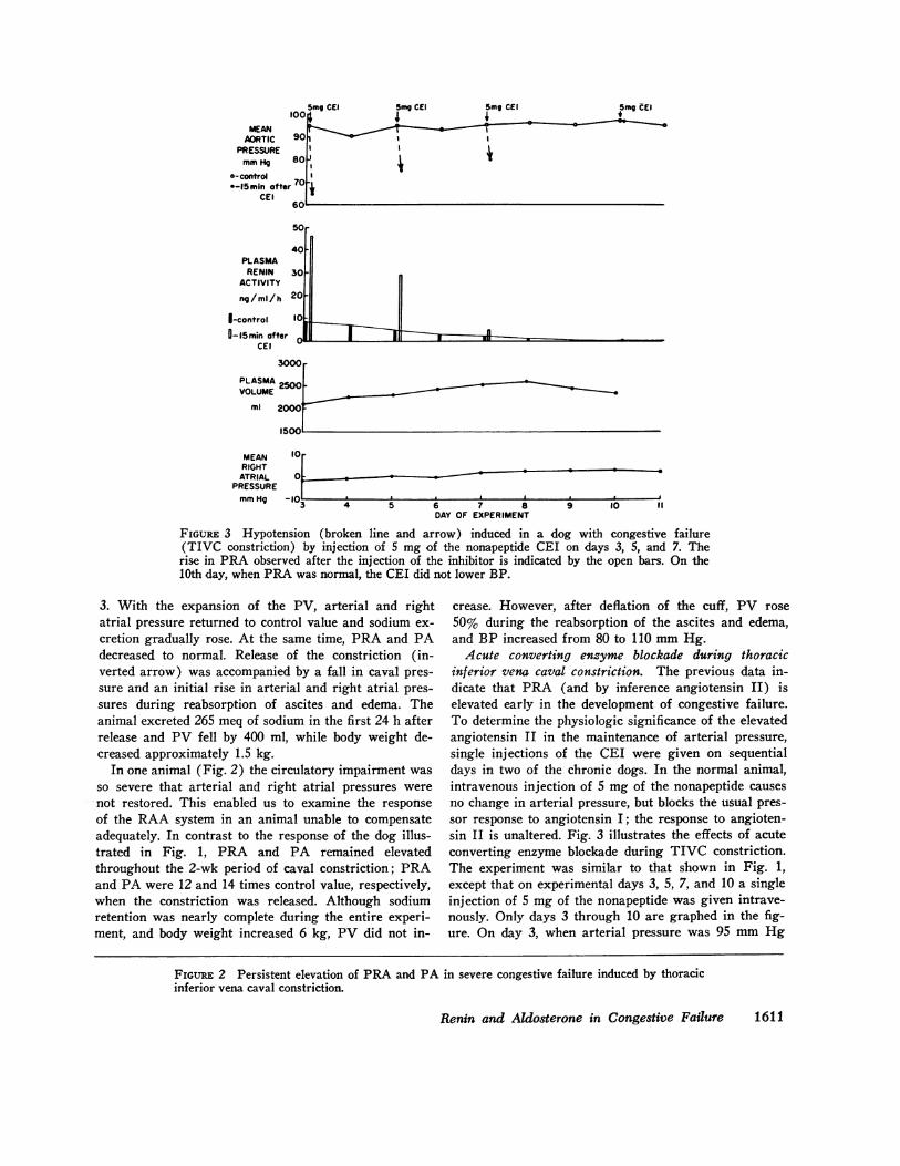
FIGURE 3 Hypotension (broken line and arrow) induced in a dog with congestive failure(TIVC constriction) by injection of 5 mg of the nonapeptide CEI on days 3, 5, and 7. Therise in PRA observed after the injection of the inhibitor is indicated by the open bars. On the10th day, when PRA was normal, the CEI did not lower BP.
3. With the expansion of the PV, arterial and rightatrial pressure returned to control value and sodium ex-cretion gradually rose. At the same time, PRA and PAdecreased to normal. Release of the constriction (in-verted arrow) was accompanied by a fall in caval pres-sure and an initial rise in arterial and right atrial pres-sures during reabsorption of ascites and edema. Theanimal excreted 265 meq of sodium in the first 24 h afterrelease and PV fell by 400 ml, while body weight de-creased approximately 1.5 kg.
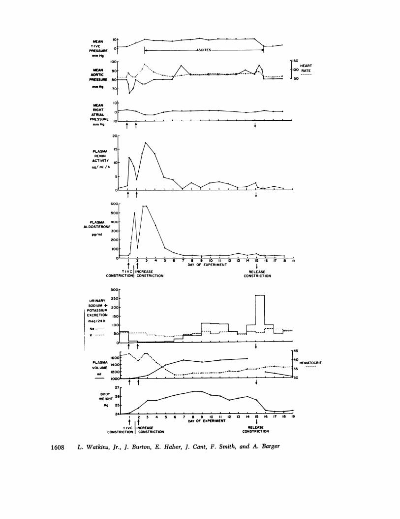
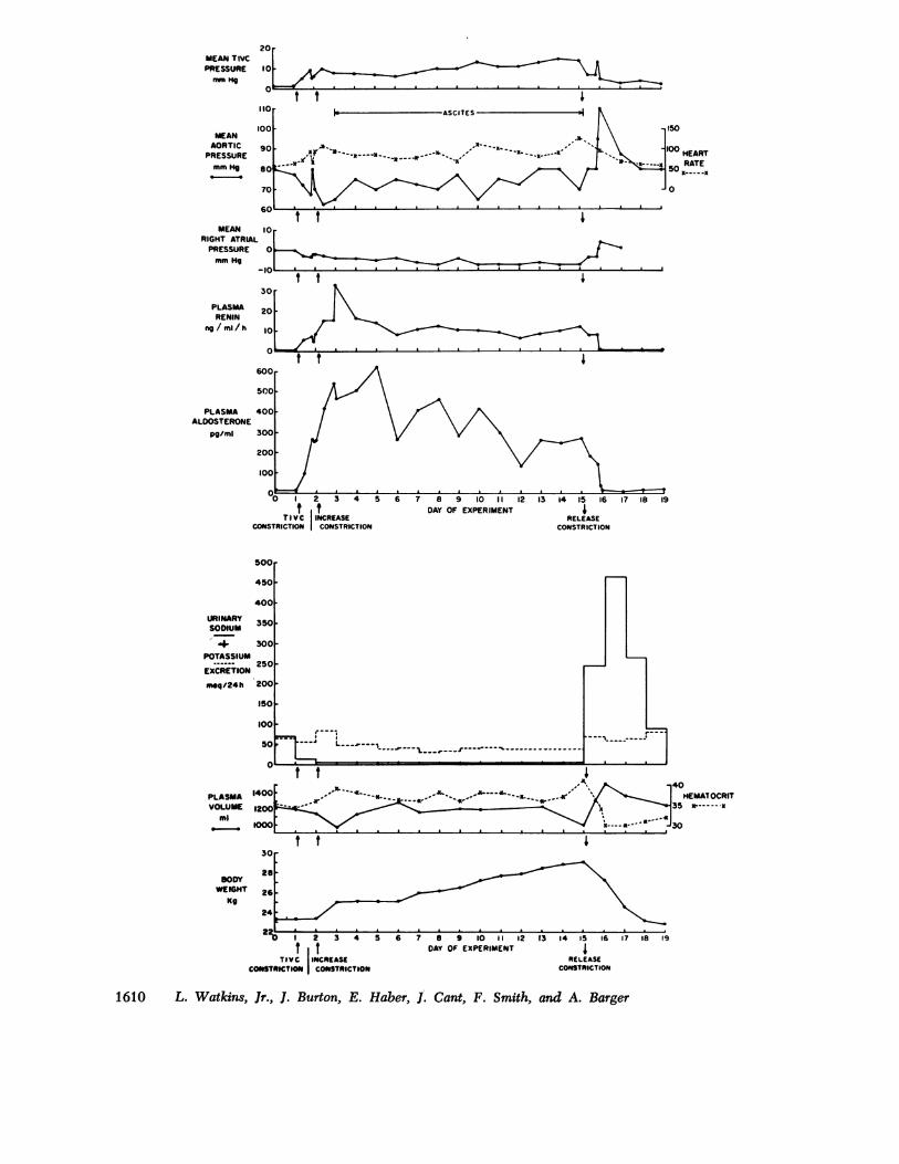
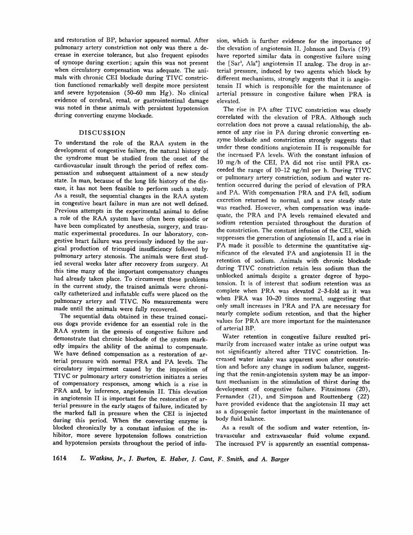
In one animal (Fig. 2) the circulatory impairment wasso severe that arterial and right atrial pressures were-not restored. This enabled us to examine the responseof the RAA system in an animal unable to compensateadequately. In contrast to the response of the dog illus-trated in Fig. 1, PRA and PA remained elevatedthroughout the 2-wk period of caval constriction; PRAand PA were 12 and 14 times control value, respectively,when the constriction was released. Although sodiumretention was nearly complete during the entire experi-ment, and body weight increased 6 kg, PV did not in-
crease. However, after deflation of the cuff, PV rose50% during the reabsorption of the ascites and edema,and BP increased from 80 to 110 mm Hg.Acute converting enzyme blockade during thoracic
inferior vena caval constriction. The previous data in-dicate that PRA (and by inference angiotensin II) iselevated early in the development of congestive failure.To determine the physiologic significance of the elevatedangiotensin II in the maintenance of arterial pressure,single injections of the CEI were given on sequentialdays in two of the chronic dogs. In the normal animal,intravenous injection of 5 mg of the nonapeptide causesno change in arterial pressure, but blocks the usual pres-sor response to angiotensin I; the response to angioten-sin II is unaltered. Fig. 3 illustrates the effects of acuteconverting enzyme blockade during TIVC constriction.The experiment was similar to that shown in Fig. 1,except that on experimental days 3, 5, 7, and 10 a singleinjection of 5 mg of the nonapeptide was given intrave-nously. Only days 3 through 10 are graphed in the fig-ure. On day 3, when arterial pressure was 95 mm Hg
Renin and Aldosterone in Congestive Failure 1611
MEANRIGHTATRIAL
PRESSUREmm Hg
FIGURE 2 Persistent elevation of PRA and PA in severe congestive failure induced by thoracicinferior vena caval constriction.
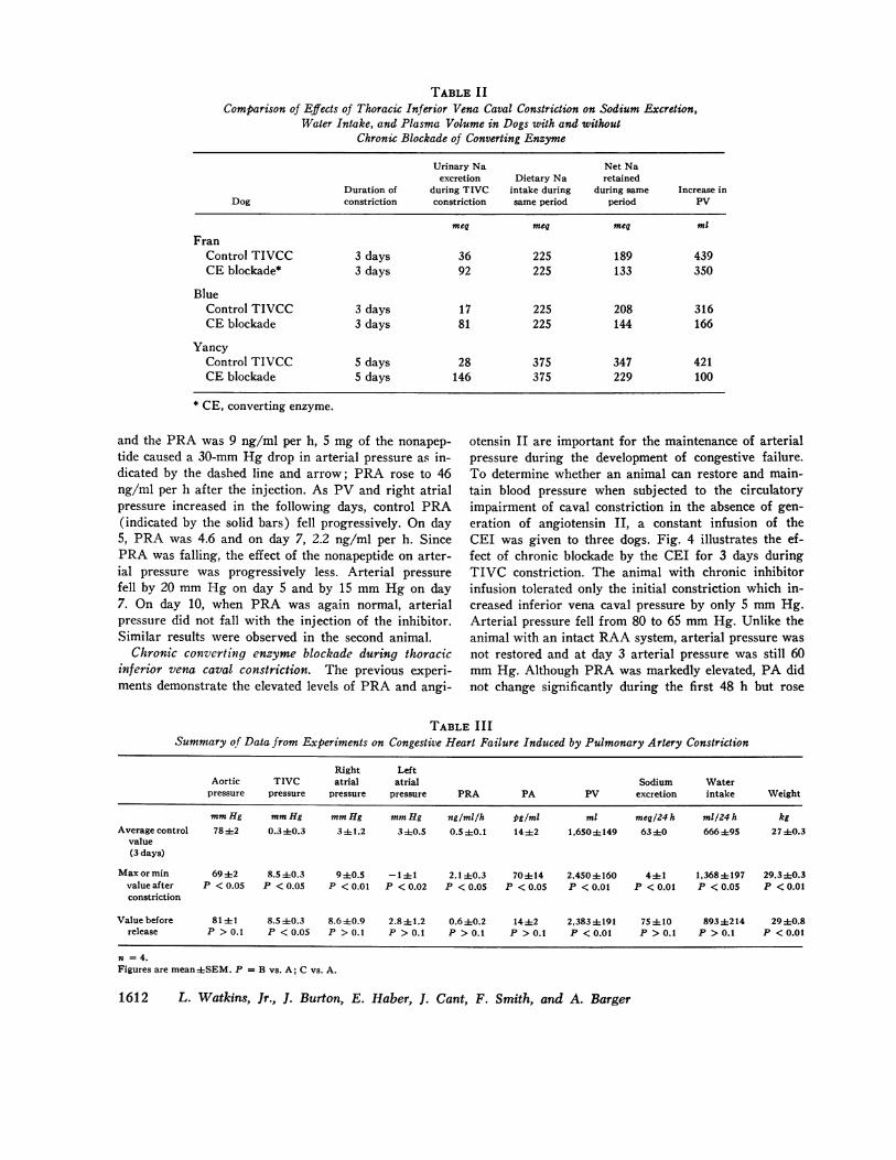
TABLE IIComparison of Effects of Thoracic Inferior Vena Caval Constriction on Sodium Excretion,
Water Intake, and Plasma Volume in Dogs with and withoutChronic Blockade of Converting Enzyme
Urinary Na Net Naexcretion Dietary Na retained
Duration of during TIVC intake during during same Increase inDog constriction constriction same period period PV
meq meq meq ml
FranControl TIVCC 3 days 36 225 189 439CE blockade* 3 days 92 225 133 350
BlueControl TIVCC 3 days 17 225 208 316CE blockade 3 days 81 225 144 166
YancyControl TIVCC 5 days 28 375 347 421CE blockade 5 days 146 375 229 100
* CE, converting enzyme.
and the PRA was 9 ng/ml per h, 5 mg of the nonapep-tide caused a 30-mm Hg drop in arterial pressure as in-dicated by the dashed line and arrow; PRA rose to 46ng/ml per h after the injection. As PV and right atrialpressure increased in the following days, control PRA(indicated by the solid bars) fell progressively. On day5, PRA was 4.6 and on day 7, 2.2 ng/ml per h. SincePRA was falling, the effect of the nonapeptide on arter-ial pressure was progressively less. Arterial pressurefell by 20 mm Hg on day 5 and by 15 mm Hg on day7. On day 10, when PRA was again normal, arterialpressure did not fall with the injection of the inhibitor.Similar results were observed in the second animal.
Chronic converting enzyme blockade during thoracicinferior vena caval constriction. The previous experi-ments demonstrate the elevated levels of PRA and angi-
otensin II are important for the maintenance of arterialpressure during the development of congestive failure.To determine whether an animal can restore and main-tain blood pressure when subjected to the circulatoryimpairment of caval constriction in the absence of gen-eration of angiotensin II, a constant infusion of theCEI was given to three dogs. Fig. 4 illustrates the ef-fect of chronic blockade by the CEI for 3 days duringTIVC constriction. The animal with chronic inhibitorinfusion tolerated only the initial constriction which in-creased inferior vena caval pressure by only 5 mm Hg.Arterial pressure fell from 80 to 65 mm Hg. Unlike theanimal with an intact RAA system, arterial pressure wasnot restored and at day 3 arterial pressure was still 60mm Hg. Although PRA was markedly elevated, PA didnot change significantly during the first 48 h but rose
TABLE IIISummary of Data from Experiments on Congestive Heart Failure Induced by Pulmonary Artery Constriction
Right LeftAortic TIVC atrial atrial Sodium Waterpressure pressure pressure pressure PRA PA PV excretion intake Weight
mm Hg mm Hg mm Hg mm Hg ng/ml/h pg/ml ml meq/24 h ml/24 h kgAverage control 7842 0.3±0.3 341.2 3±0.5 0.5 ±0.1 14±2 1.650±149 63±0 666495 2740.3
value(3 days)
Maxormin 6942 8.5±0.3 9±0.5 -141 2.140.3 70414 2.450±160 441 1,368±197 29.340.3valueafter P <0.05 P <0.05 P <0.01 P <0.02 P <0.05 P <0.05 P <0.01 P <0.01 P <0.05 P <0.01constriction
Valuebefore 81±1 8.540.3 8.640.9 2.8±1.2 0.6±0.2 14±2 2,383±191 75410 8934214 29±0.8release P > 0.1 P < 0.05 P > 0.1 P > 0.1 P > 0.1 P > 0.1 P < 0.01 P > 0.1 P > 0.1 P < 0.01
n = 4.Figures are mean±SEM. P = B vs. A; C vs. A.
1612 L. Watkins, Jr., J. Burton, E. Haber, J. Cant, F. Smith, and A. Barger
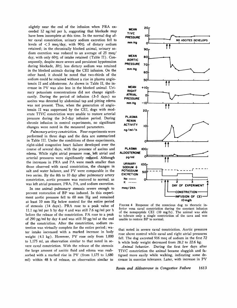
slightly near the end of the infusion when PRA ex-ceeded 12 ng/ml per h, suggesting that blockade mayhave been incomplete at this time. In the normal dog af-ter caval constriction, urinary sodium excretion fell tolevels of < 5 meq/day, with 90% of dietary sodiumretained; in the chronically blocked animal, urinary so-dium excretion was reduced to an average of 25 meq/day, with only 60% of intake retained (Table II). Con-sequently, despite more severe and persistent hypotensionduring blockade, 30% less dietary sodium was retainedin the blocked animals during the CEI infusion. On theother hand, it should be noted that two-thirds of thesodium could be retained without a rise in plasma angio-tensin II and aldosterone. As shown in Table II, the in-crease in PV was also less in the blocked animal. Uri-nary potassium concentrations did not change signifi-cantly. During the period of infusion (3-5 days) noascites was detected by abdominal tap and pitting edemawas not present. Thus, when the generation of angio-tensin II was suppressed by the CEI, dogs with mod-erate TIVC constriction were unable to restore arterialpressure during the 3-5-day infusion period. Duringchronic infusion in control experiments, no significantchanges were noted in the measured parameters.Pulmonary artery constriction. Four experiments were
performed in three dogs and the data are summarizedin Table III. Under the conditions of these experiments,right-sided congestive heart failure developed over thecourse of several days, with the presence of ascites andedema. While right atrial pressure rose, left atrial andarterial pressures were significantly reduced. Althoughthe increases in PRA and PA were much smaller thanthose observed with caval constriction, the changes insalt and water balance, and PV were comparable in thetwo series. By the 8th to 10 day after pulmonary arteryconstriction, aortic pressure was restored to normal, aswas left atrial pressure, PRA, PA, and sodium excretion.
In one animal pulmonary stenosis severe enough toprevent restoration of BP was induced. In this experi-ment aortic pressure fell to 60 mm Hg and remainedat least 10 mm Hg below control for the entire periodof stenosis (14 days). PRA rose to a peak value of11.1 ng/ml per h by day 4 and was still 7.6 ng/ml per hbefore the release of the constriction. PA rose to a peakof 290 pg/ml by day 4 and was still 50 pg/ml at the endof the constriction. After the constriction, sodium re-tention was virtually complete for the entire period; wa-ter intake increased with a marked increase in bodyweight (4.1 kg). However, PV rose only from 1,000to 1,175 ml, an observation similar to that noted in se-vere caval constriction. With the release of the stenosisthe large amount of ascitic fluid and edema was reab-sorbed with a marked rise in PV (from 1,175 to 1,680ml) within 48 h of release, an observation similar to
MEANTIVCPRESSURE
mm Hg
20
10 , oNO ASCITES DEVELOPS
I. I.
90i
MEANAORTIC
PRESSURE
mm Hg
MEANRIGHTATRIAL
PRESSUREmm Hg
PLASMARENIN
ACTIVITY
ng/ml/h
PLASMAALDOSTERONE
pg/ml
URINARYSODIUM +POTASSIUMEXCRETIONNa
80
ft
701
60
+10
0
100
t
ft
100
50r ----, _ _ _n~ ~IIa --
a
K----. I 2 3 4DAY OF EXPERIMENT
meq/ 24hCONSTRICTIONINFUSION OF CEI
10 mg/hFIGURE 4 Response of the conscious dog to thoracic in-ferior vena caval constriction during the constant infusionof the nonapeptide CEI (10 mg/h). The animal was ableto tolerate only a single constriction of the cava and wasunable to restore BP to normal.
that noted in severe caval constriction. Aortic pressurerose above control while caval and right atrial pressuresfell. The dog excreted 916 meq of sodium in the first 72h while body weight decreased from 28.2 to 22.6 kg.Animal behavior. During the first few days after
TIVC constriction the animal became sluggish and fa-tigued more easily while walking, indicating some de-crease in exercise tolerance. Later, with increase in PV
Renin and Aldosterone in Congestive Failure
_-e.
I
1613
and restoration of BP, behavior appeared normal. Afterpulmonary artery constriction not only was there a de-crease in exercise tolerance, but also frequent episodesof syncope during exertion; again this was not presentwhen circulatory compensation was adequate. The ani-mals with chronic CEI blockade during TIVC constric-tion functioned remarkably well despite more persistentand severe hypotension (50-60 mm Hg). No clinicalevidence of cerebral, renal, or gastrointestinal damagewas noted in these animals with persistent hypotensionduring converting enzyme blockade.
DISCUSSIONTo understand the role of the RAA system in thedevelopment of congestive failure, the natural history ofthe syndrome must be studied from the onset of thecardiovascular insult through the period of reflex com-pensation and subsequent attainment of a new steadystate. In man, because of the long life history of the dis-ease, it has not been feasible to perform such a study.As a result, the sequential changes in the RAA systemin congestive heart failure in man are not well defined.Previous attempts in the experimental animal to definea role of the RAA system have often been episodic orhave been complicated by anesthesia, surgery, and trau-matic experimental procedures. In our laboratory, con-gestive heart failure was previously induced by the sur-gical production of tricuspid insufficiency followed bypulmonary artery stenosis. The animals were first stud-ied several weeks later after recovery from surgery. Atthis time many of the important compensatory changeshad already taken place. To circumvent these problemsin the current study, the trained animals were chroni-cally catheterized and inflatable cuffs were placed on thepulmonary artery and TIVC. No measurements weremade until the animals were fully recovered.The sequential data obtained in these trained consci-
ous dogs provide evidence for an essential role in theRAA system in the genesis of congestive failure anddemonstrate that chronic blockade of the system mark-edly impairs the ability of the animal to compensate.We have defined compensation as a restoration of ar-terial pressure with normal PRA and PA levels. Thecirculatory impairment caused by the imposition ofTIVC or pulmonary artery constriction initiates a seriesof compensatory responses, among which is a rise inPRA and, by inference, angiotensin II. This elevationin angiotensin II is important for the restoration of ar-terial pressure in the early stages of failure, indicated bythe marked fall in pressure when the CEI is injectedduring this period. When the converting enzyme isblocked chronically by a constant infusion of the in-hibitor, more severe hypotension follows constrictionand hypotension persists throughout the period of infu-
sion, which is further evidence for the importance ofthe elevation of angiotensin II. Johnson and Davis (19)have reported similar data in congestive failure usingthe [Sar1, Ala8] angiotensin II analog. The drop in ar-terial pressure, induced by two agents which block bydifferent mechanisms, strongly suggests that it is angio-tensin II which is responsible for the maintenance ofarterial pressure in congestive failure when PRA iselevated.The rise in PA after TIVC constriction was closely
correlated with the elevation of PRA. Although suchcorrelation does not prove a causal relationship, the ab-sence of any rise in PA during chronic converting en-zyme blockade and constriction strongly suggests thatunder these conditions angiotensin II is responsible forthe increased PA levels. With the constant infusion of10 mg/h of the CEI, PA did not rise until PRA ex-ceeded the range of 10-12 ng/ml per h. During TIVCor pulmonary artery constriction, sodium and water re-tention occurred during the period of elevation of PRAand PA. With compensation PRA and PA fell, sodiumexcretion returned to normal, and a new steady statewas reached. However, when compensation was inade-quate, the PRA and PA levels remained elevated andsodium retention persisted throughout the duration ofthe constriction. The constant infusion of the CEL, whichsuppresses the generation of angiotensin II, and a rise inPA made it possible to determine the quantitative sig-nificance of the elevated PA and angiotensin II in theretention of sodium. Animals with chronic blockadeduring TIVC constriction retain less sodium than theunblocked animals despite a greater degree of hypo-tension. It is of interest that sodium retention was ascomplete when PRA was elevated 2-3-fold as it waswhen PRA was 10-20 times normal, suggesting thatonly small increases in PRA and PA are necessary fornearly complete sodium retention, and that the highervalues for PRA are more important for the maintenanceof arterial BP.Water retention in congestive failure resulted pri-
marily from increased water intake as urine output wasnot significantly altered after TIVC constriction. In-creased water intake was apparent soon after constric-tion and before any change in sodium balance, suggest-ing that the renin-angiotensin system may be an impor-tant mechanism in the stimulation of thirst during thedevelopment of congestive failure. Fitzsimons (20),Fernandez (21), and Simpson and Routtenberg (22)have provided evidence that the angiotensin II may actas a dipsogenic factor important in the maintenance ofbody fluid balance.As a result of the sodium and water retention, in-
travascular and extravascular fluid volume expand.The increased PV is apparently an essential compensa-
1614 L. Watkins, Jr., J. Burton, E. Haber, J. Cant, F. Smith, and A. Barger
tory response in congestive failure. As a consequence o:the increased PV, ventricular end-diastolic volume isrestored or elevated, augmenting ventricular perform-ance. When expansion of the PV was sufficient to re-store arterial pressure, and PRA was again normal, in-jection of the CEI had no effect on arterial pressure,indicating that angiotensin II no longer played a sig-nificant role in the maintenance of arterial pressure.With further expansion of the extravascular fluid vol-ume, the ascites and edema characteristic of right-sidedcongestive failure developed.
In the animals that were unable to compensate, PVwas not significantly increased, although ascites andedema were more severe than in the compensated ani-mal. Similar data were reported by Ross et al. (23) whofound that dogs with severe congestive failure (tricuspidinsufficiency and pulmonic stenosis) had normal or re-duced total blood volume. These observations on PVin the compensated vs. the uncompensated animal mayhelp to explain the variable results reported in patientswith congestive heart failure.
Release of the TIVC constriction was invariably as-sociated with a marked but transient rise in arterialpressure; right atrial pressure increased while inferiorvena caval pressure decreased. Arterial and atrial pres-sures stabilized at control values within 48-72 h. Theeffects on PRA and PA depended upon the levels beforerelease. If PRA and PA levels were elevated, they fellpromptly upon release; if they were at control levels nosignificant change was noted. In the animals that wereable to compensate, PV fell to control levels within 72 h;in animals with severe failure PV initially rose duringthe mobilization of ascites and edema and then graduallyfell to control. Diuresis and natriuresis invariably fol-lowed the release of the constriction, the magnitude de-pending upon the total amount of sodium and waterretained during the period of constriction. The samegeneral pattern followed pulmonary artery release ex-cept that right atrial pressure fell while left atrialpressure increased with release.The mechanisms controlling renin release in conges-
tive failure have not been well defined. By comparingthe changes in PRA after TIVC constriction with thatobserved after pulmonary artery constriction, some im-portant clues have emerged, particularly as regards therelative role of high and low pressure receptors. WithTIVC constriction BP fell and PRA increased markedly,suggesting the rise in PRA may be related to a fall inarterial pressure through activation of the renal baro-ceptor mechanism as proposed by Witty et al. (24).When a comparable drop in arterial pressure was in-duced by pulmonary artery constriction the rise in PRAwas much less. Since the fall in arterial pressure wassimilar in both models (a 10-15-mm Hg decrease) some
f other stimulus in addition to renal artery hypotensions may have been responsible for the greater release of
renin after TIVC constriction, or for the lesser re-lease after pulmonary artery constriction. From ex-periments such as that illustrated in Fig. 3, it is ap-parent that high levels of PRA may be sustained in theabsence of arterial hypotension.Although the role of vagal cardiac afferents in the
regulation of renin secretion is not yet clear, a growingbody of evidence suggests that cardiopulmonary pres-sures (atrial, pulmonary venous, or ventricular end-diastolic) may modify the release of renin. Reports in-dicate that high cardiopulmonary pressures may sup-press renin release, while low pressures or blockade ofvagal afferents may stimulate the release of renin (25-28). In the present experiments with TIVC constriction,right and left atrial pressures were reduced, whereaswith pulmonary artery constriction right atrial pressureincreased while left atrial pressure was decreased. In theTIVC experiments PRA returned to control level si-multaneously with the restoration of atrial pressures;arterial pressures were usually back to normal severaldays earlier, probably as a result of the high levels ofcirculating angiotensin II. After pulmonary artery con-striction, PRA returned to normal when left atrial pres-sure was back to control level. Thus, the high PRAnoted with TIVC constriction may result from the com-bined effects of renal artery hypotension and reflexneurogenic stimuli resulting from decreased cardiopul-monary pressures. In contrast, with pulmonary arteryconstriction, the fall in the left atrial pressure may becounterbalanced by the rise in right atrial pressure. Con-sequently, the major stimulus to renin release with pul-monary artery constriction may be the renal artery hy-potension. In support of such a view is the quantitativelysimilar rise in PRA observed when renal perfusion pres-sure was reduced to comparable levels by either pulmo-nary artery or renal artery constriction (29).
In summary, the compensatory responses to mildTIVC or pulmonary artery constriction may be dividedinto an early and late stage. In the early stage of TIVCC,arterial and atrial pressures fell; after pulmonary arteryconstriction, right atrial pressure rose while left atrialpressure fell. PRA, angiotensin II generation, and PAincreased. The elevation of systemic angiotensin II wasessential for the maintenance of arterial pressure; in-trarenally, the increase in angiotensin II and in aldos-terone may be important in the retention of sodium andwater. As a consequence of the salt and water retention,intravascular and extravascular fluid volumes expanded.In the later stage, if compensation was adequate, arterialand atrial pressures returned toward normal and PRAand PA fell to control levels. Sodium excretion was re-stored and the animal with venous congestion, ascites,
Renin and Aldosterone in Congestive Failure 1615

and edema reached a new steady state. As a result of theexpanded PV, ventricular performance was restored oraugmented and the elevated levels of angiotensin II wereno longer essential for the maintenance of arterial pres-sure. When circulatory impairment was severe or com-pensation was inadequate, PRA and PA remained ele-vated during the entire period of constriction. A singleinjection of the CEI caused a fall in arterial pressurewhen PRA was elevated, but produced no change whenPRA was normal. Chronic infusion of the CEL, whichsuppressed the generation of angiotensin II and in turna rise in aldosterone, markedly impaired the ability ofthe animal to compensate. After constriction with CEI,severe hypotension persisted and the ability to conservesodium and water was diminished. By examination ofthe sequential changes in the RAA system during theinitiation and maintenance of congestive failure, thevariability in PRA and PA levels reported in the liter-ature may now be understood. Early in the developmentof the failure they may be elevated; during the later orcompensated stage they may be normal. The particularPRA or PA level observed in congestive failure de-pends, then, upon the state of compensation.We conclude that the RAA system plays an essential
homeostatic role in the development of congestive fail-ure and is important for both restoration of arterial pres-sure and the expansion of intravascular volume. Sec-ondly, changes in cardiopulmonary pressures may beimportant in the regulation of renin secretion. Finally,the nonapeptide converting enzyme inhibitor has provento be a useful pharmacologic tool to define the role ofthe RAA system in the development of congestive failure.
ACKNOWLEDGMENTSWe thank Dr. J. S. Parsons of the National Institute forMedical Research, London, for the use of the Mill-Hill mini-ature infusion pump. We also thank Merck Sharp & Dohmefor the thrombolysin and Mrs. Birthe Creutz for secretarialassistance.This research was supported by U. S. Public Health Ser-
vice grants 5 RO1 HL02493 and 5 TO1 GM01541.
REFERENCES1. Merrill, A. J., J. L. Morrison, and E. S. Brannon. 1946.
Concentration of renin in renal venous blood in patientswith chronic heart failure. Anm. J. Med. 1: 468-472.
2. Deming, Q. B., and J. A. Luetscher, Jr. 1950. Bioassayof desoxycorticosterone-like material in urine. Proc. Soc.Exp. Biol. Med. 73: 171-175.
3. Brown, J. J., D. L. Davies, V. W. Johnson, A. F. Lever,and J. I. S. Robertson. 1970. Renin relationships in con-gestive cardiac failure, treated and untreated. Am. HeartJ. 80: 329-342.
4. Ayers, C. R., R. E. Bowden, and J. P. Schrank. 1972.Mechanisms of sodium retention in congestive heartfailure. Adv. Exp. Med. Biol. 17: 227-243.
5. Belleau, L., H. Mion, S. Simard, P. Granger, E. Ber-tranou, W. Nowaczynski, R. Boucher, and J. Genest.1970. Studies on the mechanism of experimental con-
gestive heart failure in dogs. Can. J. Physiol. Pharma-col. 48: 450-456.
6. Sanders, L. L., and J. C. Melby. 1964. Aldosterone andthe edema of congestive heart failure. Arch. Intern. Med.113: 331-341.
7. Davis, J. O., M. M. Pechet, W. C. Ball, Jr., and M. J.Goodkind. 1957. Increased aldosterone secretion in dogswith right-sided congestive heart failure and in dogswith thoracic inferior vena cava constriction. J. Clin.Invest. 36: 689-694.
8. Genest, J., P. Granger, J. de Champlain, and R. Boucher.1968. Endocrine factors in congestive heart failure. Am.J. Cardiol. 22: 35-42.
9. Davis, J. 0. 1962. Adrenocortical and renal hormonalfunction in experimental cardiac failure. Circulation. 25:1002-1014.
10. Davis, J. 0. 1965. The physiology of congestive heartfailure. Handb. Physiol. Section 2. Circulation. 3: 2071-2122.
11. Barger, A. C. 1956. The pathogenesis of sodium reten-tion in congestive heart failure. Metab. Clin. Exp. 5:480-489.
12. Ferreira, S. H. 1965. A bradykinin-potentiating factor(BPF) present in the venom of Bothrops jararaca. Brit.J. Pharmacol. Chemother. 24: 163-169.
13. Bakhle, Y. S. 1971. Inhibition of angiotensin I con-verting enzyme by venom peptides. Brit. J. Pharniacol.43: 252-254.
14. Ondetti, M. A., N. J. Williams, E. F. Sabo, J. Plu;6ec,E. R. Weaver, and 0. Kocy. 1971. Angiotensin-convert-ing enzyme inhibitors from the venom of Bothropsjararaca. Isolation, elucidation of structure, and synthe-sis. Biochemistry. 10: 4033-4039.
15. Herd, J. A., and A. C. Barger. 1964. Simplied techniquefor chronic catheterization of blood vessels. J. Appl.Physiol. 19: 791-792.
16. Haber, E., T. Koerner, L. B. Page, B. Kliman, and A.Purnode. 1969. Application of a radioimmunoassay forangiotensin I to the physiologic measurements of plasmarenin activity in normal human subjects. J. Clin. Endo-crinol. Metab. 29: 1349-1355.
17. Poulsen, K., J. Sancho, and E. Haber. 1974. A simplifiedradioimmunoassay for plasma aldosterone employing anantibody of unique specification. Clin. Immunol. Im-munopathol. 2: 373-380.
18. Miller, E. D., Jr., A. I. Samuels, E. Haber, and A. C.Barger. 1975. Inhibition of angiotensin conversion andprevention of renal hypertension. Am. J. Physiol. 228:448-453.
19. Johnson, J. A., and J. 0. Davis. 1973. Angiotensin II:Important role in the maintenance of arterial bloodpressure. Science (Wash. D. C.). 179: 906-907.
20. Fitzsimons, J. T. 1969. The role of a renal thirst factorgestive heart failure in dogs. Trans. Assoc. Am. Physi-(Lond.). 201: 349-368.
21. Fernandez, L. A. 1972. Effects of renin and angiotensinon water intake. Medicine (B. Aires). 32: (Suppl. I)63-67.
22. Simpson, J. B., and A. Routtenberg. 1973. Subfornicalorgan: site of drinking elicitation by angiotensin II.Science (Wash. D. C.). 181: 1172-1175.
23. Ross, J. F., A. C. Barger, S. C. Finch, R. S. Ross, H.L. Price, and E. J. Freireich. 1953. The blood volumebefore and following experimentally produced con-gestive heart failure in dogs Trans. Assoc. Am. Physi-cians Phila. 66: 278-293.
1616 L. Watkins, Jr., J. Burton, E. Haber, J. Cant, F. Smith, and A. Barger
24. Witty, R. T., J. 0. Davis, R. E. Shade, J. A. Johnsonand R. L. Prewitt. 1972. Mechanisms regulating reninrelease in dogs with thoracic caval constriction. Circ.Res. 31: 339-347.
25. Brennan, L. A., Jr., R. L. Malvin, K. E. Jochim, andD. E. Roberts. 1971. Influence of right and left atrialreceptors on plasma concentrations of ADH and renin.Am. J. Physiol. 221: 273-278.
26. Mancia, G., J. C. Romero, and J. T. Shepherd. 1975.Continuous inhibition of renin release in dogs by vagallyinnervated receptors in the cardiopulmonary region.Circ. Res. 36: 529-535.
27. Kurz, K. D., J. A. Hasbargen, and J. E. Lehr. 1975.Effects of increased left atrial transmural pressure onrenin secretion. Fed. Proc. 34: 367. (Abstr.)
28. Epstein, M., D. S. Pins, J. Sancho, and E. Haber. 1975.Suppression of plasma renin and plasma aldosteroneduring water immersion in normal man. J. Clin. Endo-crinol. Metab. 41: 618-625.
29. Tagawa, H., F. D. Gutmann, E. Haber, E. D. Miller,Jr., A. I. Samuels, and A. C. Barger. 1974. Reversiblerenovascular hypertension and renal arterial pressure.Proc. Soc. Exp. Biol. Med. 146: 975-982.
Renin and Aldosterone in Congestive Failure 1617