the relationship of the lingual nerve to the 3rd molar region a 3-d analysis
TRANSCRIPT
7/23/2019 The Relationship of the Lingual Nerve to the 3rd Molar Region a 3-D Analysis
http://slidepdf.com/reader/full/the-relationship-of-the-lingual-nerve-to-the-3rd-molar-region-a-3-d-analysis 1/95
The Relationship of the Lingual Nerve to the 3rd
MolarRegion: A Three Dimensional Analysis
by
Justin W.J. Garbedian
A thesis submitted in conformity with the requirementsfor the degree of Master of Science
Graduate Department of DentistryUniversity of Toronto
© Copyright by Justin W.J. Garbedian (2009)

7/23/2019 The Relationship of the Lingual Nerve to the 3rd Molar Region a 3-D Analysis
http://slidepdf.com/reader/full/the-relationship-of-the-lingual-nerve-to-the-3rd-molar-region-a-3-d-analysis 2/95ii
The Relationship of the Lingual Nerve to the 3rd Molar
Region: A Three Dimensional Analysis
Justin W.J. Garbedian
Master of Science
Graduate Department of Dentistry
University of Toronto
2009
Abstract
The objective of this study was to: (1) model the course of the lingual nerve (LN) in the
third molar region using digitized data and (2) investigate landmarks to aid in predicting
the position of LN. A MicroScribe 3-DX digitizer and Autodesk® Maya® 8.5 were used
to create 3-D in-situ models of LN for seven human cadaveric specimens. Regression
analysis demonstrated that an anteriorly positioned lingula is directly proportional to the
vertical distance of the LN relative to the alveolar crest (p < 0.05). A superiorly
positioned mylohyoid ridge was also directly proportional to the vertical distance of the
LN relative to the alveolar crest (p < 0.05). The LN is positioned closer to the alveolar
crest in specimens where the mylohyoid ridge is positioned superiorly (p = 0.001). This
study demonstrated a novel way of quantifying the relative position of the LN using 3-D
computer modeling.
7/23/2019 The Relationship of the Lingual Nerve to the 3rd Molar Region a 3-D Analysis
http://slidepdf.com/reader/full/the-relationship-of-the-lingual-nerve-to-the-3rd-molar-region-a-3-d-analysis 3/95iii
Acknowledgments
I wish to express my deepest gratitude and thanks to Dr. Anne Agur (Supervisor) for her
guidance, constructive criticism and support throughout these past few years. I truly
value the countless hours you sacrificed during the revision process. Your dedication toyour graduate students is unparalleled.
To my advisory committee members Dr. George Sándor and Dr. Cameron Clokie, thank
you for your careful revisions and unwavering support.
A special thanks to Mayooren and Kejeandra Ravichandiran for your expertise and
tremendous contribution to this thesis. You are both brilliant individuals and I wish you
great success in medical school!
I would like to thank Drs. George Sándor, Cameron Clokie and Howard Holmes for your
dedication to our profession and mentorship throughout my residency. I feel truly
blessed to have been taught by such outstanding individuals and surgeons.
In addition, I would like to express my gratitude to all my former and present surgical co-
residents. It was a pleasure to work with all of you.
I wish to thank my family who have supported and encouraged me throughout my many
years of studies. A special thank you to my father and mother for your endless love and
support. My accomplishments in life are the result of your hard work ethic and countless
sacrifices. Thanks to my sister and brother for their endless support and encouragement.
Lastly, I would like to express my profound thanks to my beautiful wife Melanie.
Without your love and encouragement, I would not have been able to overcome the many
challenges over the past four years. I am very fortunate to have you in my life and thank
you so much for your enduring support.

7/23/2019 The Relationship of the Lingual Nerve to the 3rd Molar Region a 3-D Analysis
http://slidepdf.com/reader/full/the-relationship-of-the-lingual-nerve-to-the-3rd-molar-region-a-3-d-analysis 4/95
iv
Table of Contents
Abstract ii
Acknowledgements iii
Table of Contents iv
List of Tables vii
List of Figures ix
List of Abbreviations xi
Chapter 1: Introduction 1
1.1 Contents of the Thesis 2
Chapter 2: Review of the Literature 3
2.1. Anatomy of the Lingual Nerve 32.1.1 Gross anatomy and Innervation 3
2.1.2 Anatomical Variations 52.1.2.1 Infratemporal and pterygomandibular regions 5
2.1.2.2 Retromolar region 72.1.2.3 Position of LN in third molar region. 8
2.1.3 Cross-section Morphology 112.1.4 Microanatomy 12
2.2 Imaging Studies of the LN 12
2.2.1 Introduction 122.2.2 Magnetic Resonance Imaging (MRI) 13
2.2.3 Ultrasonography 14
2.3 Lingual Nerve Injury 142.3.1 Introduction 14
2.3.2 Incidence of LN Injury 142.3.3 Factors Influencing LN Injuries 15
2.3.4 Classification of LN Injuries 182.3.4.1 Seddon’s Classification 18
2.3.4.2 Sunderland’s Classification 18
2.4 Management of LN Injuries 20
2.5 Conclusion of Literature Review 26

7/23/2019 The Relationship of the Lingual Nerve to the 3rd Molar Region a 3-D Analysis
http://slidepdf.com/reader/full/the-relationship-of-the-lingual-nerve-to-the-3rd-molar-region-a-3-d-analysis 5/95
v
Chapter 3: Objectives and Hypotheses 27
3.1 Objectives 27
3.2 Hypotheses 27
3.3 Significance 28
Chapter 4: Materials and Methods 29
4.1 Specimens 29
4.2 Dissection Protocol 29
4.3 Method of Fixation 30
4.4 Digitization and Serial Dissection 32
4.4.1 Selection of Reference Points 324.4.2 Digitization of the Oral Mucosa 33
4.4.3 Digitization of the Medial Pterygoid Muscle 334.4.4 Lingual Nerve 34
4.4.4.1 Reference Points Used for Studying the Positionof the LN 34
4.4.4.2 Technique to Capture Data Co-ordinates for 3-DReconstruction of LN 36
4.4.5 Digitization of the Mylohyoid muscle, Mandible andMandibular Second Molar 38
4.5 Creation of a 3-D Model 39
4.6 Vertical and Horizontal Measurements of LN from Mandibular
Alveolus 40
4.7 Data Analysis 41
Chapter 5: Results 43
5.1 Specimen Demographics 43
5.2 Three Dimensional Modeling 43
5.3 Lingual Nerve 485.3.1 Gross Morphology 48
5.3.2 Distance from Mucosa to LN / Validation of Measurementfrom 3-D Model 49
5.3.3 Morphology of LN 505.3.4 Vertical Position of LN 52
5.3.5 Horizontal Position of LN 54

7/23/2019 The Relationship of the Lingual Nerve to the 3rd Molar Region a 3-D Analysis
http://slidepdf.com/reader/full/the-relationship-of-the-lingual-nerve-to-the-3rd-molar-region-a-3-d-analysis 6/95
vi
5.4 Relative Position of Mylohyoid Ridge in Relation to the Position
of the LN 55
5.5 Relative Position of Lingula in Relation to LN 59
5.6 Results Summary 61
Chapter 6: Discussion 63
6.1 Introduction 63
6.2 Origin of the LN 63
6.3 Cross-sectional shape 64
6.4 LN Accessory Branches 64
6.5 Digitization and Method of LN Reconstitution 65
6.6 Anatomic Position of the LN in the Mandibular Third Molar
Region 666.6.1 Distance of LN to Oral Mucosa 66
6.6.2 Vertical Distance of LN to Alveolar Crest 676.6.3 Horizontal Distance of LN to Lingual Plate 68
6.7 Association Between the Position of the LN and Location
of Mylohyoid Ridge 69
6.8 Association Between the Position of the LN and Locationof Lingula 71
Chapter 7: Conclusions 73
Chapter 8: Future Directions 75
Chapter 9: References 77
7/23/2019 The Relationship of the Lingual Nerve to the 3rd Molar Region a 3-D Analysis
http://slidepdf.com/reader/full/the-relationship-of-the-lingual-nerve-to-the-3rd-molar-region-a-3-d-analysis 7/95
vii
List of Tables
Table 2.1 Literature Summary of LN position in 3rd
molar region 10
Table 2.2 Literature Summary of Cross - Sectional Morphology of LN 11
Table 2.3 Summary of LN Position using MRI 13
Table 5.1 Observations of the Course of LN 49
Table 5.2 Distances from Retromolar Pad to Reference Points on LN 50
Table 5.3 Distribution of LN Cross Sectional Morphology 52
Table 5.4 Vertical Distances from LN to Alveolar Crest 53
Table 5.5
Horizontal Distance from LN to Lingual Plate54
Table 5.6 Regression Analysis Source Data: Mylohyoid Ridge Ratio
and Minimum Vertical Distance between LN and Alveolar
Crest in Regions A, B and C. 56
Table 5.7 Data from Regression Analysis of Mylohyoid Ridge and
Vertical Distance to LN 56
Table 5.8 Regression Analysis Source Data: Mylohyoid Ridge Ratio and
Minimum Horizontal Distance between LN and Lingual Plate
in Regions A, B and C. 57
Table 5.9 Data from Regression Analysis of Mylohyoid Ridge & Horizontal
Distance to LN 58
Table 5.10 Regression Analysis Source Data: Lingula Ratio and Minimum
Vertical Distance between LN and Alveolar Crest in Regions
A, B and C. 59
Table 5.11 Data from Regression Analysis of Lingula Position and Vertical
Distance to LN 60

7/23/2019 The Relationship of the Lingual Nerve to the 3rd Molar Region a 3-D Analysis
http://slidepdf.com/reader/full/the-relationship-of-the-lingual-nerve-to-the-3rd-molar-region-a-3-d-analysis 8/95
viii
Table 5.12 Regression Analysis Source Data: Lingula Ratio and Minimum
Horizontal Distance between LN and Alveolar Crest in Regions
A, B and C. 61
Table 5.13
Data for Regression Analysis of Lingula Position and Horizontal
Distance to LN 61

7/23/2019 The Relationship of the Lingual Nerve to the 3rd Molar Region a 3-D Analysis
http://slidepdf.com/reader/full/the-relationship-of-the-lingual-nerve-to-the-3rd-molar-region-a-3-d-analysis 9/95
ix
List of Figures
Figure 2.1 Protocol for LN Injuries (Adapted from Robinson et al. (2004)) 22
Figure 4.1 A. Approach utilized for hemimaxillectomy. B. Medial view
of cadaveric head illustrating reconstruction plates.
C. MicroScribe-3DX digitizer D. Image demonstrating
“penning technique” with digitizer. 31
Figure 4.2 A. Reference points used to study the relationship of the LN in
the third molar region. B. Diagram depicting the regions (A, B & C)
used to measure the vertical and horizontal distances from the LN
to the mandibular alveolus. 35
Figure 4.3 A. Medial view of specimen displaying the superior and medial
penning of the LN. B. Medial view of specimen demonstrating
the cross-section of the LN 37
Figure 4.4 Schematic diagram of serial sectioning of LN and digitization
of cross-sectional perimeter 37
Figure 4.5 Medial view of mandible displaying “penning technique” 39
Figure 4.6 Coronal section of the mandible in the region of the second molar
illustrating the vertical and horizontal measurements to LN 40
Figure 4.7 Medial view of mandible illustrating the ratios used to
describe the position of the mylohyoid ridge and lingula 42
Figure 5.1 3-D Model of reconstructed specimen from digitized data
(multiple views) 44
Figure 5.2 Reconstitution of LN using Autodesk® Maya® Software 47

7/23/2019 The Relationship of the Lingual Nerve to the 3rd Molar Region a 3-D Analysis
http://slidepdf.com/reader/full/the-relationship-of-the-lingual-nerve-to-the-3rd-molar-region-a-3-d-analysis 10/95
x
Figure 5.3 Cross-sectional morphology of LN: A. Site of cross-sections.
B. Pie graphs of cross-section shapes in regions A, B and C.
C. Examples of oval, round, and ribbon cross-sectional shapes 51
Figure 5.4 Average Vertical Distance to LN in Regions A, B and C
for each Specimen 52
Figure 5.5 Average Vertical Distance to LN by Region 53
Figure 5.6 Average Horizontal Distance to LN in Regions A, B and
C for each Specimen 54
Figure 5.7 Average Horizontal Distance to LN by Region 55
Figure 5.8 Regression Analysis of Mylohyoid Ridge Ratio to Vertical
Position of LN 57
Figure 5.9 Regression Analysis of Mylohyoid Ridge Ratio To Horizontal
Position of LN 58
Figure 5.11 Regression Analysis of Lingula Ratio to Vertical Position of LN 60
7/23/2019 The Relationship of the Lingual Nerve to the 3rd Molar Region a 3-D Analysis
http://slidepdf.com/reader/full/the-relationship-of-the-lingual-nerve-to-the-3rd-molar-region-a-3-d-analysis 11/95
xi
List of Abbreviations
CN Cranial Nerve
CN V 3 Mandibular division of the Trigeminal Nerve
HR High Resolution
IAN Inferior Alveolar Nerve
LN Lingual Nerve
LP Lateral Pterygoid Muscle
M1 Mandibular First Molar
M2 Mandibular Second Molar
M3 Mandibular Third Molar
Md Mandible
MH Mylohyoid muscle
MRI Magnetic Resonance Imaging
Mu Mucosa
PETRA Phase Encode Time Reduction Acquisition
Ref Reference point
Sd Submandibular Duct
SMd Submandibular Gland
S/N Signal to noise ratio
TG Tongue
3-D Three Dimensional

7/23/2019 The Relationship of the Lingual Nerve to the 3rd Molar Region a 3-D Analysis
http://slidepdf.com/reader/full/the-relationship-of-the-lingual-nerve-to-the-3rd-molar-region-a-3-d-analysis 12/95
1
Chapter 1Introduction
Knowledge of the position of the lingual nerve (LN) is invaluable to the surgeon
operating in the mandibular third molar region. Iatrogenic injury to the nerve can result
in significant morbidity to the patient and potential medical-legal repercussions for the
surgeon. Previous studies using cadaveric dissections, clinical observation during third
molar surgery and high-resolution magnetic resonance imaging have yielded inconsistent
data in documenting the position of the LN (Kiesselbach and Chamberlain, 1984; Pogrel
et al., 1995; Miloro et al.,1997; Behnia et al., 2000; Hölzle and Wolff 2001). These
studies show large variations in the vertical and horizontal location of the LN with
respect to the mandibular alveolar crest. Attempts to explain this variability has largely
been attributed to several confounding variables in study design. For example Miloro et
al. (1997) ascribe such inconsistency to lack of standardized measurements and fixation
of specimens. However, in their recent study Hölzle and Wolff (2001) did find a positive
correlate between vertical LN positioning and mandibular crest atrophy.
The lack of consensus amongst previous studies warrants further investigation.
An in-situ 3-D computer model of the LN and related structures may aid in clarifying the
position of the LN in the third molar region. The relationship of the LN with other
anatomical structures could also be explored with this data set. Such correlations could
be utilized in surgical treatment planning to predict the location of the LN and minimize
iatrogenic injury.

7/23/2019 The Relationship of the Lingual Nerve to the 3rd Molar Region a 3-D Analysis
http://slidepdf.com/reader/full/the-relationship-of-the-lingual-nerve-to-the-3rd-molar-region-a-3-d-analysis 13/95
2
1.1 Contents of the Thesis
The present thesis consists of eight chapters presented in the following sequence:
Chapter 1 provides a brief discussion on the ramifications of lingual nerve (LN) damage
and the rationale for further investigation into its precise position.
Chapter 2 presents the literature describing the anatomical variations of the LN as it
courses from its origin from the mandibular branch of the trigeminal nerve (CN V 3) to
the tongue. The relationship of the LN to bony features in the mandibular third molar
region will be reviewed in detail followed by an introduction to the factors involved in
LN damage and the classifications for peripheral, along with management strategies for
LN injuries.
Chapter 3 outlines the hypotheses, objectives and significance of the study.
Chapter 4 describes the dissection approach, digitization and computer modeling of the
oral mucosa, medial pterygoid muscle, LN, mylohyoid muscle, medial surface of the
mandible and the mandibular second molar.
Chapter 5 is a summary of the results. Qualitative findings such as the shape of the LN
and presence of accessory branches are first described. Quantitative data describing the
position of the LN with respect to the oral mucosa and mandible was then presented. The
final section report describes the quantative data with a regression analyses between the
LN and the mylohyoid ridge and the LN and the lingula.
Chapter 6 is a discussion of the results of this thesis and how it compares with previous
studies. The clinical relevance of these findings is also presented.
Chapter 7 and 8 list the conclusions of this thesis and suggest possible future directions.
7/23/2019 The Relationship of the Lingual Nerve to the 3rd Molar Region a 3-D Analysis
http://slidepdf.com/reader/full/the-relationship-of-the-lingual-nerve-to-the-3rd-molar-region-a-3-d-analysis 14/95
3
Chapter 2Review of the Literature
2.1 Anatomy of the Lingual Nerve
2.1.1 Gross Anatomy and Innervation
The tongue is a complex organ that is involved in speech, mastication, swallowing,
taste, respiration and sensation (Zur et al., 2004; McGuire, 2005). The following cranial
nerves innervate the tongue:
• The hypoglossal nerve (CN XII) provides the motor supply to the muscles of the
tongue except for the palatoglossus muscle, which is innervated by the accessory
nerve (CN XI) via the vagus nerve (CN X) (Liegbott, 1986).
• The glossopharyngeal nerve (CN IX) supplies general and special sensory sensation
to the posterior one-third of the mucosa of the tongue.
• The LN, a branch of the mandibular division of the trigeminal nerve (CN V3)
provides sensory innervation to the ipsilateral two-thirds of the mucous membranes
of the tongue, floor of mouth and the mandibular lingual gingiva (Liegbott, 1986; Zur
et al., 2004).
• The chorda tympani, a branch of the facial nerve (CN VII) “hitchhikes” along the
course of the LN to transmit special visceral afferent fibres (preganglionic
parasympathetic) to the submandibular and sublingual salivary glands and special
sensory afferents (taste) to the anterior two-thirds of the tongue (Liebgott, 1986; Zur
et al., 2004; Fonseca, 2005).
In a recent anatomical study, Saigusa et al. (2006), suggested that a portion of the
superior and inferior longitudinal muscles of the human tongue may be innervated by
motor fibres originating from the CN V motor root.

7/23/2019 The Relationship of the Lingual Nerve to the 3rd Molar Region a 3-D Analysis
http://slidepdf.com/reader/full/the-relationship-of-the-lingual-nerve-to-the-3rd-molar-region-a-3-d-analysis 15/95
4
The origin of the LN begins as a branch from the posterior division of CN V3 in
the infratemporal fossa. While in the infratemporal fossa, the chorda tympani joins the
LN at an acute angle as it exits through the pterygotympanic fissure of the glenoid fossa
(Girod et al., 1989). The LN, along with the chorda tympani, then descends medially to
the lateral pterygoid muscle and inferior alveolar nerve.
The nerve continues to descend, in an anterior direction, to enter the
pterygomandibular space. While in this space, the LN travels between the medial aspect
of the mandibular ramus and the lateral aspect of the medial pterygoid muscle. The LN
then enters the oral cavity after passing the anterior edge of the medial pterygoid muscle.
The LN courses from a more lateral to medial position as it approaches the
mandibular third molar due to the oblique flare in the mandible in this region. As the LN
approaches the third molar, its position with respect to the alveolar bone, is variable.
Several studies have tried to clarify this complex 3-D relationship with little consistency
(Kiesselbach and Chamberlain, 1984; Pogrel et al., 1995; Miloro et al., 1997; Behnia et
al., 2000; Hölzle and Wolff, 2001; Karakas et al., 2007). The results of these studies are
compared in table 2.1.
As the LN courses along the floor of the mouth, the mylohyoid muscle bounds it
inferiorly. In the region of the mandibular second molar, the submandibular ganglion can
be found suspended inferiorly from the LN by autonomic secretory fibres. The LN then
begins to course anteromedially, looping itself under the submandibular (Wharton’s)
duct. The nerve then passes upward onto the genioglossus muscle to enter the ventral
mucosa of the tongue anterior to the circumvallate papillae (Zur et al., 2004)
As the LN enters the tongue, it gives off two main branches: a medial and a
lateral. The medial branch sends 2-4 small branches to the medial part of the

7/23/2019 The Relationship of the Lingual Nerve to the 3rd Molar Region a 3-D Analysis
http://slidepdf.com/reader/full/the-relationship-of-the-lingual-nerve-to-the-3rd-molar-region-a-3-d-analysis 16/95
5
ventrolateral tongue, while the lateral branch travels along the lateral border of the tongue
to send 2-5 large branches to the anterior tip of tongue (Zur et al., 2004). Both medial
and lateral branches are generally restricted to the ventrolateral portions of the tongue
leaving few, surgically significant, LN fibres in the mid-body of the tongue.
Furthermore, the LN has consistent anastomotic connections with the hypoglossal nerve
in the body of the tongue (Fitzgerald and Law, 1958; Zur et al., 2004). This neural
organization is consistent with the lingua-hypoglossal reflex and is important in the reflex
control of tongue movement (Zur et al., 2004).
2.1.2 Anatomical Variations
2.1.2.1 Infratemporal and Pterygomandibular Regions
The branching of the LN from the posterior division of CN V3 has classically
been described at the level of the otic ganglion (Leibgott, 1986). A cadaveric study by
Kim et al. (2004) clarified this division by illustrating multiple branching patterns of the
LN from the inferior alveolar nerve (IAN). In 65.6% specimens (21 out of 32) the
bifurcation of the LN and IAN was located between the sigmoid notch and the otic
ganglion. Eight cases (25.0%; 8 out of 32), demonstrated branching patterns in the upper
half of the ramus, while only one case (3.1%; 1 out of 32) bifurcated just superior to the
lingula. The remaining specimens (6.2 %; 2 out of 32) had a plexiform branching pattern
from CN V3. Racz and Maros (1981) reported similar plexform bifurcations in 8.3%
(4 out of 48) of the specimens. A recent cadaveric study by Erdogmus et al. (2008)
supports the multiple branching patterns of the LN and IAN. The majority of specimens
(66.7%; 14 out of 21) bifurcated above the level of the sigmoid notch. The remaining
cases bifurcated between the sigmoid notch and the mandibular lingula (33.3%; 7 out
7/23/2019 The Relationship of the Lingual Nerve to the 3rd Molar Region a 3-D Analysis
http://slidepdf.com/reader/full/the-relationship-of-the-lingual-nerve-to-the-3rd-molar-region-a-3-d-analysis 17/95
6
of 21). Some interconnections between the LN and IAN were observed, 1-3 branches in
47.61% (10 out of 21) of the specimens; however, no plexiform branching pattern was
noted.
Kim et al. (2004) also measured the distance of the bifurcation to stable anatomic
landmarks. The bifurcation was noted to occur approximately 14.3 mm
(range: 7.8 - 24.1 mm) inferior to the foramen ovale and 16.5 mm (range: 4.9 - 24.3 mm)
superior to the tip of the hamulus. This reported variation in LN and IAN bifurcation
patterns has contributed to the failure rate (13-29%) of mandibular nerve block anesthesia
in dentistry (Blanton and Jeske, 2003).
The chorda tympani, a branch of the facial nerve (CN VII) unites with the LN as
it descends in the infratemporal fossa. Nerve fibers from the chorda tympani hitchhike
along the LN as special sensory fibers to provide taste sensation from the anterior
two-thirds of the tongue and presynaptic parasympathetic fibers to the submandibular
ganglion. In a cadaveric study by Erdogmus et al. (2008), the junction of the LN and
chorda tympani was frequently (80.95%; 34 out of 42) found to be located at the depth of
the lateral pterygoid and superior to the upper edge of the medial pterygoid muscles. The
remaining cases (19.04%; 8 out of 42) demonstrated a junction in the upper half of the
medial pterygoid muscle. The average distance of the junction of the LN and the chorda
tympani from the foramen ovale was 15.1 ± 5.8 mm (Erdogmus et al., 2008).
As the LN travels anteroinferiorly in the pterygomandibular region, it generally
courses between the lateral surface of the pterygoid muscle and the mandible.
Occasionally, the LN has been reported to give off branches to the posterolateral part of
the medial pterygoid muscle from the lateral surface (Sakamoto and Akita, 2004).
Sakamoto and Akita (2004) reported an atypical spatial relationship of the LN through
7/23/2019 The Relationship of the Lingual Nerve to the 3rd Molar Region a 3-D Analysis
http://slidepdf.com/reader/full/the-relationship-of-the-lingual-nerve-to-the-3rd-molar-region-a-3-d-analysis 18/95
7
the medial pterygoid muscle. This alternative course had the LN penetrating the medial
pterygoid muscle and separating the anterolateral muscle bundle into an accessory
bundle. This accessory bundle inserted into an area superior to the mylohyoid line in the
retromolar region and was innervated by a small branch from the lingual nerve on its
medial surface.
2.1.2.2 Retromolar Region
Several studies have reported collateral nerve twigs from the LN innervating the
retromolar region (Kiesselbach and Chamberlain, 1984; Girod et al, 1989; Kim et al,
2004; Erdogmus et al., 2008). Erdogmus et al. (2008) noted that the LN provided several
small nerve branches to the paralingual area but failed to quantify these “variational
nerve innervations”. Kim et al (2004) claimed that 81.2% (26 out of 32) of their
specimens had collateral nerve twigs to the retromolar area. These collateral branches
were distributed on the lingual gingival in proximity to the mandibular third molar and
retromolar mucosa. An average of one nerve twig was reported with a range of zero to
three branches. Kim et al. (2004) suggested that this branching pattern should not be
considered a variation but a normal innervation pattern of the LN. Furthermore, this
anatomic collateral innervation may help to explain the incomplete anaesthesia that is
occasionally noted following the administration of local anaesthetic during a mandibular
block and long buccal blocks.
As the LN approaches the floor of mouth, communicating branches between the
LN and the mylohyoid nerve have been reported (Racz and Maros, 1981; Kim et al.,
2004; Sakamoto and Akita, 2004; Erdogmus et al., 2008). Erdogmus et al. (2008)
reported an incidence of 11.9% (5 out of 42). Kim et al. (2004) reported a similar

7/23/2019 The Relationship of the Lingual Nerve to the 3rd Molar Region a 3-D Analysis
http://slidepdf.com/reader/full/the-relationship-of-the-lingual-nerve-to-the-3rd-molar-region-a-3-d-analysis 19/95
8
incidence of 12.5% (4 out of 32) in Korean specimens. The LN innervated the
superomedial surface of the mylohyoid muscle and communicated with the mylohyoid
nerve within the muscle (Sakamoto and Akita, 2004). This communication has been
called the “mylohoid or sublingual curl” and provides an alternative route for collateral
sensory transmission.
2.1.2.3 Position of LN in Third Molar Region
As the LN courses anteriorly to the retromolar region, it follows the contours of
the medial aspect of the mandible. For many years, the scientific literature was lacking
qualitative and quantitative data for the position of the LN in the region of the third
molar. It was this lack of knowledge that prompted Kiesselbach and Chamberlain (1984)
to evaluate the position and shape of the LN in the third molar region. This study
evaluated 34 adult cadaver heads and 256 in-vivo observations of the LN during
mandibular third molar extractions. The horizontal distance varied amongst specimens
with an average distance of 0.58 ± 0.9 mm from the lingual plate. In 62% (21 out of 34)
of the specimens, the nerve was in contact with the lingual plate. Vertical distance of the
LN below the alveolar crest was 2.28 ± 1.96 mm. A surprising finding of this study was
that in 17.6% (6 out of 34) of the cadaveric specimens and 4.5% (12 out of 256) of the
in-vivo cases the LN was positioned at the level of the alveolar crest or higher.
Pogrel et al. (1995) attempted to verify the findings by Kiesselbach and
Chamberlain by dissecting 20 cadavers (40 sides) and measuring the location of LN with
respect to osseous points. This study used reproducible osseous landmarks for measuring
the distance to the LN and accounted for the presence or absence of posterior mandibular
teeth. The findings confirmed the variability of the nerve in the sagittal and coronal
7/23/2019 The Relationship of the Lingual Nerve to the 3rd Molar Region a 3-D Analysis
http://slidepdf.com/reader/full/the-relationship-of-the-lingual-nerve-to-the-3rd-molar-region-a-3-d-analysis 20/95
9
planes. The closest distance of the nerve to the mandible was a mean of 3.45 ± 1.48 mm
(range: 1 - 7mm), which differs from the findings by Kiesselbach and Chamberlain. The
LN showed a close proximity to the lingual cortex of the mandible for a mean distance of
27.7 mm. The mean vertical distance of the superior aspect of the LN to the alveolar
crest was 8.32 ± 5.69 mm. In 3 cases (out of 40), the LN was at the level (one case) or
superior (two cases) to the alveolar crest, which is in agreement with the findings
presented by Kiesselbach and Chamberlain (1984). No statistical relationship was found
with regards to the presence or absence of the mandibular third molar and LN position.
Furthermore, the position of the LN nerve on one side was not correlated to its position
on the contralateral side.
A potential limitation in cadaveric studies is the iatrogenic displacement of the
LN during the dissection process. To circumvent this, Behnia et al. (2000) used two clips
on the lingual aspect of the posterior gingiva to stabilize the location of the LN.
Although this technique may limit LN displacement during dissection, it introduces
compression distortion. However, the study was novel, in that the dissections were
performed on fresh cadavers within 24 hours of death. Furthermore, this study exceeded
previous sample sizes with the evaluation of 669 LNs. The mean horizontal and vertical
distances of the LN in the third molar region are summarized in Table 2.1. An
unexpected finding in this study was that in one case the LN was located in the
retromolar pad (Behnia et al, 2000).
In 2001, Hölzle et al., examined the position of the LN in the third molar region
with respect to atrophy of the alveolar crest. The investigators also found that the
distance from the LN decreases as the degree of mandibular atrophy increases. Vertical

7/23/2019 The Relationship of the Lingual Nerve to the 3rd Molar Region a 3-D Analysis
http://slidepdf.com/reader/full/the-relationship-of-the-lingual-nerve-to-the-3rd-molar-region-a-3-d-analysis 21/95
10
and horizontal distances of the LN to the alveolar crest were variable to previous
anatomical studies (Table 2.1).
All cadaveric studies discussed above utilized calipers for measuring the vertical
and horizontal distances of the LN. Karakas et al. (2007) in contrast, used radiographs to
quantify in the horizontal and vertical measurements of LN with respect to the mandible.
A 3 mm wire was placed along the undisturbed position of LN in 21 sagittally sectioned
cadaveric heads. Radiographs were then taken in the superior and lateral planes and
vertical and horizontal distances of the LN to mandible were then measured from the
radiographs using digital calipers. The mean vertical and horizontal distances were
9.5 ± 5.2 mm and 4.1 ± 1.9 mm respectively (Table 2.1). The horizontal measurements
are markedly greater to those presented by Kiesselbach and Chamberlain (1984) and
Hölzle and Wolff (2001). Karakas et al. (2007) postulated that this difference might be
due to a superiorly positioned mylohyoid muscle and/or greater mylohyoid muscle
volume.
Table 2.1: Literature Summary of LN position in 3rd
molar region
Measurement (mm) Percentage (%) of
Specimens
Study Type of
Study
N
Vertical Horizontal Above
Alveolar
Crest
Contact with
lingual plate
Kiesselbach
&
Chamberlain
Cad
IV
34
256
2.28 +/- 1.96 0.58 +/- 0.9 17.6 62
Pogrel et al. Cad 40 8.32 +/- 4.05 3.45 +/- 1.48 15 0
Behnia et al. Cad 669 3.01 +/- 0.42 2.06 +/- 1.10 14.05
(0.15 in
RMP)
23.27
Hölzle and
Wolff
Cad 68 7.83 +/- 1.65 0.86 +/- 1.00 8.82 57.4
Karakas et al. Cad 21 9.56 +/- 5.28 4.19 +/- 1.99 4.7 0
N = Sample Size; Cad = cadaveric study; IV = in-vivo study; RMP = retromolar pad

7/23/2019 The Relationship of the Lingual Nerve to the 3rd Molar Region a 3-D Analysis
http://slidepdf.com/reader/full/the-relationship-of-the-lingual-nerve-to-the-3rd-molar-region-a-3-d-analysis 22/95
11
2.1.3 Cross-section Morphology
The cross-sectional morphology of the LN is variable along its course in the oral
cavity, e.g. circular or oval to flat or ribbon-like (Kiesselbach and Chamberlain, 1984;
Miloro et al., 1997; Kim et al., 2004). The reported cross sectional morphologies are
summarized in Table 2.2. Kiesselbach and Chamberlin (1985) found the LN to be round
(61.7%; 21 out of 34), oval (17.6%; 6 out of 34) or flat/ribbon-like (20.5%; 7 out of 34)
in the third molar region with no correlation to the distance from the medial surface of
the mandible. Miloro at al. (1997) noted that of the 20 LNs examined, 9 (45%) were
round, 6 (30%) were elliptical, and 5 (25%) were kidney-bean shaped. Kim et al. (2004)
studied the shape of the LN in the retromolar, third molar and second molar region. A
circular (40.6%; 13 out of 32) or oval (40.6%; 13 out of 32) morphology of the LN
predominated in the retromolar region whereas an oval (59.4%; 19 out of 32) or ribbon-
shape (62.5 %; 20 out of 32) was most common in the third and second molar region
respectively. The ribbon-like shape was not observed in the retromolar or third molar
region by Kim et al. (2004).
Table 2.2: Literature Summary of Cross - Sectional Morphology of LN
Cross Sectional MorphologyStudy N Location
Circular Oval Flat Ribbon-like
Kiesselbach &
Chamberlain
34 M3 31.7% 17.6% 20.5%
RM 40.6% 40.6% 18.8% 0
M3 25% 59.4% 15.6% 0
Kim et al. 32
M2 3.1% 18.8% 15.6% 62.5%
Miloro et al. 20 M3 45% 30% (elliptical) 25% (kidney bean)
N = Sample Size; M3 = Third molar; M2 = Second Molar; RM = Retromolar

7/23/2019 The Relationship of the Lingual Nerve to the 3rd Molar Region a 3-D Analysis
http://slidepdf.com/reader/full/the-relationship-of-the-lingual-nerve-to-the-3rd-molar-region-a-3-d-analysis 23/95
12
2.1.4 Microanatomy
The microanatomy of the LN is similar to other peripheral nerves. The LN has a
polyfascicular pattern, which consists of many fascicles of varying sizes but without
fascicular grouping (Svane et al., 1986; Abby et al., 1987). The mean number of
fascicles for the LN at the third molar region is 8.5 to 10 +/- 4.0 (Girod et al., 1989).
Abby et al. (1987) reported 15-18 fascicles in this region, which decreased to 9 fascicles
as the nerve entered the tongue. The mean total cross-sectional area of the LN fascicle is
1.87 mm +/- 0.38 mm2 (Girod et al., 1989). The number and size of fascicles is critical to
the microsurgeon as repair of a nerve requires correlation between the donor and host
nerve.
2.2 Imaging Studies of the LN
2.2.1 Introduction
There are limited imaging studies in the literature evaluating the position of the
LN (Miloro et al., 1997; Karakas et al., 2007 and Olson et al., 2007). As previously
discussed, plain film radiography was utilized by Karakas et al. (2007) to aid in
measuring the position of the LN in cadaveric specimens. This technique provides no
diagnostic merit, as it requires the placement of a metal wire along the LN. Only one
study in the literature has evaluated the position of the human LN using magnetic
resonance imaging (MRI) (Miloro et al., 1997). This study represents the first and only
documentation of in-vivo measurements of the position of LN in the third molar region.
The use of ultrasonography has recently shown promise in locating the LN in cadaveric
pigs (Olsen and Troulis, 2007).

7/23/2019 The Relationship of the Lingual Nerve to the 3rd Molar Region a 3-D Analysis
http://slidepdf.com/reader/full/the-relationship-of-the-lingual-nerve-to-the-3rd-molar-region-a-3-d-analysis 24/95
13
2.2.2 Magnetic Resonance Imaging (MRI)
MRI was previously unable to identify the course of nerves due to distortion
(signal to noise ratio (S/N)) (Miloro et al., 1997). Advancements made through high
resolution (HR-MRI) and specific imaging protocols such as phase encode time reduction
acquisition (PETRA) has been applied to the LN by Miloro et al (1997). Ten healthy
volunteers with present mandibular third molars underwent axial and coronal HR-MRI
PETRA of the mandible, bilaterally. The mandible was examined from the lingula to the
mental foramen as reconstructed 1.5 mm slices. The image closest to the center of the
tooth was selected to measure the distance from the lateral edge of the LN to the lingual
plate (horizontal dimension) and from the superior edge of the LN to the lingual crest
(vertical dimension). The LN was only noted to be superior to the lingual alveolar crest in
two cases and there was no bilateral correlation. Results are summarized in Table 2.3.
Miloro also studied the position of the tongue with respect to LN position and found no
correlation.
Table 2.3: Summary of LN Position using MRI
Sample
Size
Mean Nerve
Diameter (mm)
Measurement (mm) Percentage (%) of Specimens
Vertical Horizontal Above
alveolar crest
Contact with
lingual plate
20 2.54 2.75 ± 0.97 2.53 ± 0.67 10 25
Although Miloro et al. demonstrated that HR-MRI could be utilized to image the
LN, its clinical application is limited primarily due to expense and availability.
Furthermore, documenting in-situ LN injuries would be limited to complete transections,
as subtle injuries would be indiscernible (Miloro et al.,1997).

7/23/2019 The Relationship of the Lingual Nerve to the 3rd Molar Region a 3-D Analysis
http://slidepdf.com/reader/full/the-relationship-of-the-lingual-nerve-to-the-3rd-molar-region-a-3-d-analysis 25/95
14
2.2.3 Ultrasonography
A recent study by Olson et al. (2007) demonstrated the use of ultrasonography in
visualizing the LN in cadaver pigs. After sufficient training, all three evaluators could
correctly identify the LN. The average distance of the LN to the lingual cortex was
1 mm. This application of ultrasonography proves promising in humans, as the pig’s LN
is approximately of the size. Furthermore, the evaluators could accurately identify an
intact, partially transected or fully transected nerve injury 63% of the time.
2.3 Lingual Nerve Injury
2.3.1 Introduction
The LN allows for complex sensory-discriminative-secretomotor function.
Damage to it can result in temporary or permanent general sensory changes to the
anterior two-thirds of the tongue and floor of mouth. This loss of sensory function can
result in changeable degrees of anesthesia, paraethesia, dyaesthesia or hypoasesthsia,
which can be permanent or transitional. This damage can cause drooling, tongue biting, a
burning sensation, pain and speech changes (Fielding et al., 1997; Pichler and Beirne,
2001; Renton and McGurk, 2001; Graff-Radford and Evans, 2003). Damage to the LN
can also result in loss of taste to the anterior two-thirds of the tongue and altered salivary
secretion on the involved side.
2.3.2 Incidence of LN Injury
While some iatrogenic damage to the LN is unavoidable, such as with the
removal of malignant masses, unfortunately the majority of injuries are the result of
elective and iatrogenic procedures (Holmes and Lam, 2002; Robinson et al., 2004). The
cause of LN injuries include, for example, the surgical insertion of dental implants

7/23/2019 The Relationship of the Lingual Nerve to the 3rd Molar Region a 3-D Analysis
http://slidepdf.com/reader/full/the-relationship-of-the-lingual-nerve-to-the-3rd-molar-region-a-3-d-analysis 26/95
15
(Berberi et al.,1993), inadvertent instrumentation during mandibular osteotomies (Zuniga
and Phillips, 1997; Jacks et al., 1998; Becelli et al., 2004), administration of local
anesthetic (Hillerup and Jensen, 2005) and removal of mandibular third molars.
Holmes and Lam (2002) suggest that approximately 75% of LN injuries are a
result of third molar removal. This is not surprising since removal of impacted third
molars is one of the most commonly performed procedures within an oral and
maxillofacial surgeon’s office. In 2001, Britain reported impacted third molars to be the
eighth most commonly performed surgery with approximately 150,000 procedures per
year (Renton and McGurk, 2001). The incidence of LN damage during mandibular third
molar surgery is inconsistent within the literature. The majority of studies report an
incidence of 0.6% to 2.0% (Pogrel and Renault, 1995; Brann et al., 1999; Behnia et al.,
2000, Bataineh, 2001; Hölzle and Wolff, 2001; Renton and McGurk, 2001; Chossegros et
al., 2002; Karakas et al., 2007); however, the prevalence has been reported as high as
23% (Middlehurst et al., 1988). Fortunately, the majority of LN injuries result in
temporary LN disturbances, with only approximately 0.5 – 1.0 % reporting permanent
LN sensory dysfunction (Blackburn and Bramley, 1989; Jerjes et al., 2006).
2.3.3 Factors Influencing LN Injuries
Several factors contribute to the variability of LN paresthesia following third
molar surgery. Variations in surgical technique, in particular the use of a lingual
retractor, are well documented in the literature (Rood, 1983; Wofford and Miller, 1987;
Mason, 1988; Obiechina, 1990; Rood, 1992; Schultz-Mosgau and Reich, 1993; Robinson
and Smith, 1996; Brann et al., 1999). Although a topic of debate, many studies agree that
avoidance of lingual retraction reduces the incidence of temporary lingual nerve
7/23/2019 The Relationship of the Lingual Nerve to the 3rd Molar Region a 3-D Analysis
http://slidepdf.com/reader/full/the-relationship-of-the-lingual-nerve-to-the-3rd-molar-region-a-3-d-analysis 27/95
16
disturbance and does not increase the incidence of permanent damage (Robinson and
Smith, 1996; Batanineh, 2001; Chossegros et al., 2002). A meta-analysis in 2001
corroborates this finding by showing no difference in permanent LN injury rates whether
a lingual retractor was used or not (Pichler and Beirne, 2001). Greenwood et al. (1994)
argued in support of a broad retractor to protect the LN rather than a Howarth periosteal
elevator since it produces less paresthesia at one month. On the other hand, Appiah-
Anane and Appiah-Anane (1997) demonstrated a 0.2% incidence of LN damage by
avoiding the use of a lingual flap and by preserving the lingual plate of the mandible.
Ultimately, the decision to use a lingual flap or retractor is at the discretion of the
surgeon.
The classification of impacted mandibular third molars can also affect the
incidence of LN injuries. Mesioangular impacted third molars account for 43% of LN
paresthesia cases, while distoangular impactions account for only 6% (Peterson, 1993).
In contrast, Bataineh (2001) failed to find an association between angulation of the tooth
and LN paresthesia.
The relationship between age and LN paresthesia following third molar surgery is
inconclusive. Bruce et al. (1980) and Chiapasco et al. (1995) found a positive correlation
between older patients and risk of LN damage. In the study by Bruce et al. (1980), the
incidence of LN damage was 1.8% for patients with a mean age of 46.5 years, compared
to 0.6% for those with a mean age of 20 years. In contrast, many studies have failed to
show a correlation of LN damage to the age of the patient (Brann et al., 1999; Bataineh,
2001; Jerjes et al., 2006;).
Studies have shown a greater incidence of LN paresthesia following surgery
performed under general anesthetic versus local anesthetic (Bruce et al., 1980; Brann et

7/23/2019 The Relationship of the Lingual Nerve to the 3rd Molar Region a 3-D Analysis
http://slidepdf.com/reader/full/the-relationship-of-the-lingual-nerve-to-the-3rd-molar-region-a-3-d-analysis 28/95
17
al, 1999). Brann et al (1999) reported a 5-fold increase in the incidence of LN
paresthesia under general anesthesia compared to local anesthetic alone. This finding
could not be explained by age, preoperative pathology or surgical difficulty. Brann et al.
(1999) rationalized that this higher incidence rate may be complicated by the positioning
of the patient in the supine position, extent of flap exposure / bone removal or greater
amount of surgical force generated while a patient is under a general anesthetic. The
surgeon’s experience plays a role in the incidence of LN damage (Sisk et al., 1986;
Gülicher and Gerlach, 2001; Bataineh, 2001; Jerjes et al., 2006). A prospective study
evaluating several predictors of tongue paresthesia, concluded that the seniority of the
surgeon to be the only significant predictor (P = 0.022) in determining permanent LN
paresthesia (Jerjes et al., 2006). The prevalence of permanent LN paresthesia was 4
times more likely to occur in a group of trainees. This discrepancy is likely to due
inexperience, improper use of force and mishandling of surgical instrumentation.
7/23/2019 The Relationship of the Lingual Nerve to the 3rd Molar Region a 3-D Analysis
http://slidepdf.com/reader/full/the-relationship-of-the-lingual-nerve-to-the-3rd-molar-region-a-3-d-analysis 29/95
18
2.3.4 Classification of LN Injuries
Several classification schemes exist for describing sensory disturbances
secondary to peripheral nerve damage: physiologic, symptomatic, anatomic,
histopathologic, and pathophysiologic (McGuire, 2005). While no single classification
scheme is ideal, the Seddon (1943) and/or Sunderland (1951) classification is widely
used as they correlate severity with prognosis.
2.3.4.1 Seddon’s Classification
In 1943, Seddon developed a peripheral nerve classification based on the severity
of injury affecting the endoneurium, perineurium, epineurium and supporting tissues.
Mild insult to the nerve was termed neuropraxia. With this type of injury, a conduction
block occurs without axonal degeneration. Most injuries to the LN due to the use of a
lingual flap retractor are classified as neuropraxia. Resolution of these sensory deficits is
within hours to days and is complete. Axonotmesis denotes a more serious injury with
preservation of the epineurium but varying degrees of afferent fiber degeneration.
Incomplete sensory recovery occurs with the potential of neuroma formation.
Neurotmesis describes the most severe injury with poor axonal regeneration due to severe
disruption or complete discontinuity of all connective layers of the peripheral nerve.
Clinical examination reveals anesthesia in the nerve distribution and no sensory recovery.
2.3.4.2 Sunderland’s Classification
In 1951, Sunderland expanded Seddon’s classification to further emphasize the
mechanism of injury and offer insight into managing such injuries. Under this
classification scheme, Sunderland describes five levels of injuries with the first-degree

7/23/2019 The Relationship of the Lingual Nerve to the 3rd Molar Region a 3-D Analysis
http://slidepdf.com/reader/full/the-relationship-of-the-lingual-nerve-to-the-3rd-molar-region-a-3-d-analysis 30/95
19
injury being sub-classified into three types. All Sunderland first-degree injuries are
equivalent to Seddon’s neuropraxia. Within this degree of injury, axonal conduction is
temporarily blocked but the continuity of the axon is preserved. First-degree type I
injuries are due to mild traction or compression which results in transient ischemia.
Recovery of the sensory deficit usually occurs within twenty-four hours. First-degree
type II injuries are the result of more severe traction or compression causing
intrafascicular edema, decreased blood flow and a conduction blockade (Graff-Radford
and Evans, 2003). Normal sensation returns as the edema subsides which takes days to
weeks. First-degree type III injuries are the result of further traction and compression
causing localized mechanical disruption of myelin sheaths and demyelinization. This
injury usually requires one to two months for normal sensation to return. Sunderland’s
second, third and fourth degree injuries correspond with Seddon’s classification of
axonotmesis. A second-degree injury results in Wallerian degeneration of the axon
without disruption to the endoneurium, perineurium or epineurium. Sensory recovery is
usually complete in 2-4 months but may take up to one year for complete recovery.
Third-degree injuries occur due to moderate or severe crush or traction insults. Wallerian
degeneration occurs with disruption of the endoneurium. This breach of the
endoneurium will not allow for axonal regeneration resulting in some degree of sensory
loss. Fourth-degree Sunderland injuries occur when the endoneurium and perineurium
have been violated due to severe trauma. Neuronal loss occurs with the possibility of
neuroma formation or intraneural fibrosis. Fifth-degree injuries correlate with Seddon’s
Neurotmesis classification. This type of injury is the result of transection, avulsion or
laceration of the nerve, which causes a disruption of all three connective tissue sheaths.
Unfortunately, this injury results in complete anesthesia with the potential for

7/23/2019 The Relationship of the Lingual Nerve to the 3rd Molar Region a 3-D Analysis
http://slidepdf.com/reader/full/the-relationship-of-the-lingual-nerve-to-the-3rd-molar-region-a-3-d-analysis 31/95
20
neuropathic responses such as allodynia, hyperalgesia or hyperpathia (Fonseca, 2005).
MacKinnon and Dellon, in 1988, added a sixth degree injury to Sunderland’s
classification to describe variable degrees of injury that can co-exist within a single nerve
(Mackinnon and Dellon, 1988).
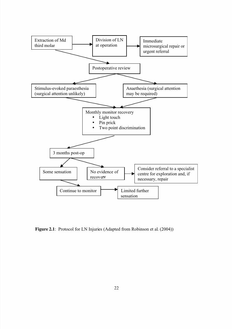
2.4 Management of LN Injuries
Robinson et al. (2004), suggest, “patients who are ultimately left with a minor
degree of hypoanaesthesia cope well with the sensory deficit are unlikely to benefit from
intervention, and are probably best left untreated. In contrast, patients who have either
substantial sensory deficit or the painful sensory disorder of dyaesthesia may benefit
from intervention, and so must be identified and managed in a manner that will optimize
the outcome”. In his 2004 article, Robinson provides an algorithm (Figure 2.1) for the
management of LN injuries as a result of third molar removal.
In the few patients in which LN laceration is noted at the time of third molar
removal the algorithm suggests immediate microsurgical repair. Robinson recommends
“the use of an operating microscope and insertion of 6 to 8, 8-0 monofilament polyamide
epineural sutures”. Variations of this technique are found in the literature with only
minor changes in suture size and number (Dobson, 1997; Fonseca, 2009 ). This
technique is only possible if a “tension free repair” is possible (Dobson, 1997).
Unfortunately, most LN injuries go unnoticed at the time of injury and it is not until post-
operative follow-up or complaint by the patient that they are recognized.
The timing of surgical intervention in LN injuries is paramount. The
observational period of three months recommended by Robinson et al. (2004) is to help
distinguish between varying degrees of injury. Stimulus evoked paresthesia suggest
7/23/2019 The Relationship of the Lingual Nerve to the 3rd Molar Region a 3-D Analysis
http://slidepdf.com/reader/full/the-relationship-of-the-lingual-nerve-to-the-3rd-molar-region-a-3-d-analysis 32/95
21
neuropraxia-like injuries and thus require no intervention. Complete anesthesia of the
LN distribution implies an axonotmesis or neurotmesis and may warrant surgical
intervention. The delay of surgical intervention has been attributed to poorer outcomes
(Mozsary and Middleton, 1984; Meyer, 1992; Pogrel and Kaban, 1993; Donoff, 1995).
Positive surgical outcomes decrease with the passing of time due a higher incidence of
Wallerian degeneration, atrophy and fibrosis of the distal portion of the nerve (Wolford
and Stevao, 2003). Hillerup and Jensen (2007) reported the fastest recovery occurred
during the initial 6 months after injury. Riediger and Cornelius (1989) suggested that
repairs conducted after twelve months of the injury are questionable.
7/23/2019 The Relationship of the Lingual Nerve to the 3rd Molar Region a 3-D Analysis
http://slidepdf.com/reader/full/the-relationship-of-the-lingual-nerve-to-the-3rd-molar-region-a-3-d-analysis 33/95
22
Figure 2.1: Protocol for LN Injuries (Adapted from Robinson et al. (2004))
3 months post-op
Some sensation No evidence ofrecover
Continue to monitor Limited further
sensation
Consider referral to a specialist
centre for exploration and, ifnecessary, repair
Stimulus-evoked paraesthesia
(surgical attention unlikely)
Anaethesia (surgical attention
may be required)
Monthly monitor recovery•
Light touch
• Pin prick• Two point discrimination
Postoperative review
Extraction of Md
third molar
Division of LN
at operationImmediatemicrosurgical repair or
urgent referral

7/23/2019 The Relationship of the Lingual Nerve to the 3rd Molar Region a 3-D Analysis
http://slidepdf.com/reader/full/the-relationship-of-the-lingual-nerve-to-the-3rd-molar-region-a-3-d-analysis 34/95
23
Microsurgical techniques for nerve repair have been used for many years.
According to Hillerup and Jensen (2007), microsurgical repair should only be considered
in nerves with persistent, total or near total loss of function beyond 3-6 months and the
potential benefit of repair will justify microsurgery. Comparing the literature on
microsurgical repair of LN is problematic due to lack of standardized outcome
assessments and small sample sizes (Graff-Radford and Evans, 2003; Robinson et al.,
2004). Wiethölter et al. (1990) and Wolford and Stevao (2003) report better results for
IAN repair with end-to-end anastomosis versus nerve grafting. Robinson and Smith
(1996) found similar success with direct repair of the LN, stating that it was the most
effective repair procedure. A retrospective study conducted by Rutner et al. in 2005
investigated an external neurolysis procedure in combination with an internal neurolysis,
neuroma excision or primary neurorrhaphy for LN injuries. The outcomes of this study
demonstrated a 90% improvement in neurosensory function. Robinson and Smith (2000)
also demonstrated positive outcomes with direct neurorrhaphy of the LN. Successful
outcomes of LN repair may appear to be case specific. Collin and Donoff (1990)
reported a higher success rate for LN repair in patients with anesthesia (77%) versus
pain-paresthesia (42%). Gregg (1990) and Pogrel and Kaban (1993) on the other hand
reported good pain reduction following repair. Zuniga et al. (1997) evaluated
chemosensory (taste) regeneration following nerve repair with a 50% improvement in 12
patients. Notably they reported that taste is usually compensated overtime. Recently, a
bioabsorbable collagen nerve cuff (NeuraGen) has shown promising results when used in
conjunction with direct repair of IAN and LN injuries (Farole, 2008). The NeuraGen
cuff is believed to minimize scar ingrowth and concentrate growth factors at the site of
injury.
7/23/2019 The Relationship of the Lingual Nerve to the 3rd Molar Region a 3-D Analysis
http://slidepdf.com/reader/full/the-relationship-of-the-lingual-nerve-to-the-3rd-molar-region-a-3-d-analysis 35/95
24
While direct apposition is desirable, this treatment modality is not always
possible. The nature of LN damage often precludes the ability to obtain tension free co-
adaptation and thus a bridge is required between the proximal and distal ends of the
nerve. The sural, greater auricular and medial antebrachial cutaneous nerves have been
used as donor nerves for the repair of LN injuries (Fonseca 2009). The major
disadvantage of this technique is the creation of paresthesia at the donor site. Wolford
and Miller (2003) reports less favourable results with LN grafting. Lower success rates
are likely associated with higher technical difficulty, limited access and potential
incompatibility in shape, size and fascicle numbers between the grafted nerve and LN.
Cable grafting of the LN defect has been advocated to improve the match between donor
and host (Wolford, 1992). Dobson and Kaban (1997) criticize this technique by stating;
“given the variable results after autogenous nerve grafts to reconstruct IAN or LN, any
donor site morbidity [which can be quite significant] calls into question the use of
autogenous nerve grafts’. An alternative to nerve grafts has lead to the use of autogenous
veins and arteries. The use of vascular entubulation for LN repair has shown mixed
results (Miloro, 2001; Pogrel and Maghen, 2002). Poor outcomes are likely due to the
vein collapse and impediment of axonal growth (Wolford and Stevao, 2003; Fonseca
2009).
Grafting with alloplastic grafts is an attractive option for LN repair due to graft
availability and avoidance of donor site morbidity. Several studies involving animal
models have demonstrated the ability of nerves to regenerate across defects via tubes and
conduits (Dellon and Mackinnon, 1986). Initial trials with permanent conduits were
largely unsuccessful with LN repairs. 1n 1998, Pogrel and Kaban reported a 28%
increase in sensory improvement with LN and IAN defects reconstructed with Gortex.

7/23/2019 The Relationship of the Lingual Nerve to the 3rd Molar Region a 3-D Analysis
http://slidepdf.com/reader/full/the-relationship-of-the-lingual-nerve-to-the-3rd-molar-region-a-3-d-analysis 36/95
25
Unfavourable outcomes were likely the result of localized compression and alterations in
the blood-nerve barrier by the permanent conduit. Bioabsorbable polyglycolic acid
(PGA) conduits have been developed to circumvent the problems associated with
permanent conduits. Mackinnon and Dellon (1990) first reported the use of PGA
conduits for reconstructing digital nerve repairs. The results of this study demonstrated
nerve regeneration across gaps up to 3 cm in length. The use of PGA conduits for CN V3
repair is lacking in the literature. In 1992, Crawley and Dellon (1992) reported an
isolated case in which the IAN was reconstructed with a PGA conduit. A successful
outcome was obtained, with clinical improvements beginning 6 months after surgery.
Ways to improve artificial nerve conduits are currently been developed. The
incorporation of Schwann cells into nerve conduits is currently being investigated in
animal models (Kim et al., 2007). Schwann cells promote axonal extension and neuronal
survival and their use may greatly facilitate nerve conduit performance (Ichihara et al.,
2008).
Non-surgical therapy is also an option for management of LN injury. The use of
oral and topical therapies, neural blockade and behavioral strategies have provided mixed
outcomes (Graff-Radford and Evans, 2003). Pharmaceuticals such as tricyclic
antidepressants have also been used with varying degrees of success (Gregg, 1978).
While many attempts have been made to correct injuries to the LN, treatment is
not a simple foregone conclusion. The affects on the patient can be debilitating and
permanent. While surgeons try to minimize iatrogenic events, without a clear
visualization of the LN their best efforts will remain insufficient.

7/23/2019 The Relationship of the Lingual Nerve to the 3rd Molar Region a 3-D Analysis
http://slidepdf.com/reader/full/the-relationship-of-the-lingual-nerve-to-the-3rd-molar-region-a-3-d-analysis 37/95
26
2.5 Conclusion of Literature Review
Anatomical studies have failed to be succinct in predicting the path of the LN.
The lack of standardized measurements for the LN and possible displacement of the
nerve due to the use of positional clips has contributed to the variability of the data.
Imaging studies of the LN are limited and await improved advances in technology.
A 3-D computer model of the course of the LN in the third molar region has not
been described in the literature. Such a model would allow for documentation of the
position of the LN in multiple planes. Furthermore, potential factors predicting the
course of the LN could be evaluated using this model. Such a diagnostic tool would aid
in risk stratification for LN damage during mandibular third molar surgery.

7/23/2019 The Relationship of the Lingual Nerve to the 3rd Molar Region a 3-D Analysis
http://slidepdf.com/reader/full/the-relationship-of-the-lingual-nerve-to-the-3rd-molar-region-a-3-d-analysis 38/95
27
Chapter 3Objectives and Hypotheses
3.1 Objectives
1. To digitize the LN from the pterygomandibular region to the floor of the mouth,
medial surface of mandible, medial pterygoid muscle, mylohyoid muscle, second
mandibular molar and if present, the third mandibular molar.
2. To reconstruct a 3-D geometric computer model in Autodesk® Maya® of the
digitized data to outline the course of the LN in relation to surrounding tissue.
3. To determine whether a relationship exists between the position of the LN and lingula
and/or mylohyoid ridge.
4. To quantify the horizontal and vertical distance of the LN to the mandibular alveolus
3.2 Hypotheses
Hypothesis #1
H0: The height of the mylohyoid ridge is not associated with the position of the lingual
nerve.
H1: A superiorly positioned mylohyoid ridge is directly proportional to the vertical
position of the lingual nerve in the mandibular third molar region.
Hypothesis #2
H0: The anteroposterior position of the lingula is not associated with the position of the
lingual nerve.
H1: An anteriorly positioned lingula is directly proportional to the vertical position of the
lingual nerve in the mandibular third molar region.

7/23/2019 The Relationship of the Lingual Nerve to the 3rd Molar Region a 3-D Analysis
http://slidepdf.com/reader/full/the-relationship-of-the-lingual-nerve-to-the-3rd-molar-region-a-3-d-analysis 39/95
28
3.3 Significance
A macroanatomical study with 3-D modeling of cadaveric specimens of the
course of lingual nerve will provide insight into the location of the nerve from multiple
perspectives. Furthermore, the database will allow investigation of various hard and soft
tissue landmarks in determining the predictability of the course of the lingual nerve.
Clarification of the location of this nerve will provide clinical guidance to ensure LN
protection during third molar surgery.

7/23/2019 The Relationship of the Lingual Nerve to the 3rd Molar Region a 3-D Analysis
http://slidepdf.com/reader/full/the-relationship-of-the-lingual-nerve-to-the-3rd-molar-region-a-3-d-analysis 40/95
29
Chapter 4Materials and Methods
4.1 Specimens
Seven formalin embalmed cadaveric sagitally sectioned heads (2 males / 2
females) were utilized in this study. The inclusion criteria consisted of no visible
craniofacial deformity and the presence of a mandibular 2nd
molar.
4.2 Dissection Protocol
The cadaver head was first placed on the dissection table with the midsagitally
sectioned surface facing down. The inferior border of the mandible was then palpated to
localize the angle of the mandible. A dissecting probe was inserted through the skin until
the bony mandible was reached. Using a # 15 scalpel blade, a full thickness flap was
raised in a subperiosteal fashion to expose the angle of the mandible and a portion of the
lateral ramus. To obtain adequate exposure, the inferior lobe of the masseter muscle as
well as the inferior lobe of the parotid gland was removed.
Next, a hemi-maxillectomy was performed using a modified Weber Fergusson
approach (Figure 4.1A). This approach allowed for an unobstructed view of the medial
aspect of the mandible with minimal disturbance to underlying anatomy. The first
incision was made along the radix of the nose and carried laterally across the infra-orbital
rim. The incision was then extendend inferiorly along the nasal crease to the philtrum.
Next, an intra-oral incision was made approximately 5 mm superior to the mucogingival
junction at the midline of the maxilla, which was then carried laterally to the region of
the first molar. The skin flap was dissected off the maxilla in a subperiosteal fashion. As
the dissection proceeded laterally, the infra-orbital nerve was encountered and transected

7/23/2019 The Relationship of the Lingual Nerve to the 3rd Molar Region a 3-D Analysis
http://slidepdf.com/reader/full/the-relationship-of-the-lingual-nerve-to-the-3rd-molar-region-a-3-d-analysis 41/95
30
to allow greater flap mobility and exposure. The subperiosteal envelope was continued
until the pterygomaxillary fissure and the junction of the hard and soft palate was
identified. Next a mucosal incision was made parallel to the posterior border of the hard
palate. This incision was then carried just posterior to the maxillary tuberosity to connect
to the previously described subperiosteal dissection in the region of the pterygomaxillary
fissure.
Using a series of osteotomes and a fissure bur on a Dremel rotary tool (Dremel,
Racine, Wisconson), maxillary osteotomies were made to free the hemimaxilla. The
osteotomy started at the nasofrontal suture and continued along the inferior orbital rim.
The lateral osteotomy was made along the zygomaticomaxillary buttress. Next, a curved
osteotome was placed along the pterygomaxillary fissure and directed medially. While
palpating the hamulus intraorally, the osteotome was advanced until it disjoined the
posterior maxilla from the pterygoid plate. This approach allowed minimal disruption to
the medial pterygoid muscle and underlying lingual nerve. The hemimaxilla was then
infractured medially and remaining bony interferences were relieved with rongeurs.
4.3 Method of Fixation
In order to ensure accurate reconstruction of the digitized LN, the mandible and
associated masticatory muscles needed to be stabilized with a 2.4 mm mandibular
reconstruction titanium plate. The in-line bend of these plates also minimized
interferences while digitizing. A minimum of two plates were used in all specimens to
secure the mandible in the closed position (Figure 4.1B). The first plate was positioned
on the medial aspect of the sagittally cut mandible and spanned posteriorly to the mastoid
process. A second plate was then placed from the medial aspect of the mandible (just
superior to the first reconstruction plate) to the calvarial bone just superior to the frontal

7/23/2019 The Relationship of the Lingual Nerve to the 3rd Molar Region a 3-D Analysis
http://slidepdf.com/reader/full/the-relationship-of-the-lingual-nerve-to-the-3rd-molar-region-a-3-d-analysis 42/95
31
sinus. The Dremel rotary tool and the plating system’s drill bits were then used to pre-
drill all fixation screws. Pre-drilling was necessary to facilitate screw placement and
limit propagation of fractures within the bone. Washers were often required between the
plate and bone to ensure the plate was passive and not torquing the mandible medially
when fixated.
Figure 4.1: A. Approach utilized for hemimaxillectomy. Red dashed line represents theosteotomy. B. Medial view of cadaveric head illustrating reconstruction plates.C. MicroScribe-3DX digitizer D. Image demonstrating “penning technique” with
digitizer.
A B
C D

7/23/2019 The Relationship of the Lingual Nerve to the 3rd Molar Region a 3-D Analysis
http://slidepdf.com/reader/full/the-relationship-of-the-lingual-nerve-to-the-3rd-molar-region-a-3-d-analysis 43/95
32
4.4 Digitization and Serial Dissection
4.4.1 Selection of Reference Points
The digitization process required three fixed points on the mandible to enable a
3-D computer reconstruction of the specimen. The retromolar pad, angle of the mandible
and the occlusal surface of the second mandibular molar were selected, as they were
easily identified landmarks and accessible from all angles during the digitization process.
Accurate triangulation of these data coordinates was accomplished by placing fixation
screws in all reference points except the occlusal tooth surface. The screw at the
mandibular angle was inserted until its head was flush with the surrounding bone. A
similar approach was taken at the retromolar pad, except the screw head was only
advanced to the level of the oral mucosa. It was important to avoid over advancement of
this screw, as this would cause surrounding tissue distortion. The tooth did not receive a
fixation screw due to potential fracture. To circumvent this, a divot was made in the
center of the tooth using a fissure bur on the Dremel rotary tool.
To ensure that the specimen did not move during the digitization process, the
cadaveric head was placed in a vise that had been fixated to the workstation. A calvarial
wedge of bone was removed (lateral to the midsagittal plane) to allow for fixation of the
vise to the skull.
The MicroScribe-3DX digitizer was the only device utilized throughout this study
(Figure 4.1C & D). The fine digitizing tip was used as it allowed for more accurate
plotting of delicate structures with minimal tissue disruption and fit into the center of the
reference point Phillip’s screw head. Digitization began with the capturing of the
reference points. These points were recorded in the same sequence for all specimens.
7/23/2019 The Relationship of the Lingual Nerve to the 3rd Molar Region a 3-D Analysis
http://slidepdf.com/reader/full/the-relationship-of-the-lingual-nerve-to-the-3rd-molar-region-a-3-d-analysis 44/95
33
4.4.2 Digitization of the Oral Mucosa
The retromolar pad was first demarcated and marked for digitization using a fine
diameter paint pen. Points were placed approximately 2-3 mm apart and the
MicroScribe-3DX digitizer was utilized to digitize the circumference of the retromolar.
The data was captured with a computer. The area of the oral mucosa to be digitized was
then defined and marked with the paint pen:
• The superoinferior boundaries extended from the lingual gingival sulcus to the
depth of the lingual vestibule (floor of mouth).
• The posterior and anterior boundaries included the entire retromolar area to the
level of the palatoglossus arch and the mid-sagittal osteotomy of the mandible
respectively.
The MicroScribe-3DX digitizer was then used to record data points of the oral mucosa.
4.4.3 Digitization of the Medial Pterygoid Muscle
The posterior oral mucosa overlying the medial pterygoid was carefully removed
using a #15 blade. Dissection of the oral mucosa continued anteriorly until the LN was
identified deep to the anterior margin of the medial pterygoid. Once the entire deep
surface of the medial pterygoid muscle was exposed, the paint pen was used to demarcate
muscle fiber bundles. Points were placed approximately 2-3 mm apart. Next, the fiber
bundles were digitized from origin to insertion and then removed using sharp dissection
with a #15 blade and tissue forceps. Underlying muscle fibers within a layer were
sequentially digitized and removed until the entire muscle was digitized.

7/23/2019 The Relationship of the Lingual Nerve to the 3rd Molar Region a 3-D Analysis
http://slidepdf.com/reader/full/the-relationship-of-the-lingual-nerve-to-the-3rd-molar-region-a-3-d-analysis 45/95
34
4.4.4 Lingual Nerve
4.4.4.1 Reference Points Used for Studying the Position of the LN
The course of the lingual nerve to be measured extended from the posterior
margin of the medial pterygoid to the region the nerve first turned medially towards the
tongue. These boundaries were marked with a paint pen. Three reference marks were
then placed along the superior border of the LN prior to digitization (Figure 4.2A). The
first reference point corresponded to the location at which the LN began to diverge
medially into the tongue (defined as #1). The distance from point #1 to the reference
screw was labeled I. The second point (identified as # 2), was the position of the nerve
perpendicular to the reference screw head. The distance between point #2 and the
reference screw was labeled II. The last reference point (labeled #3), marked the closest
position of LN to the reference screw in the retromolar pad. The distance between point
#3 and the reference screw was labeled III and represented the shortest distance from the
superior aspect of LN to the oral mucosa. Distances I, II and III were measured using a
digital boley gauge and recorded to the hundredth of a millimeter. The same distances
were then measured using the Autodesk® Maya ® measuring software. A paired t-test
was then conducted to validate the accuracy of these measuring techniques. The
statistical null hypothesis that there is no difference between the two measurements was
accepted with a p < 0.05.
The course of LN was divided into three sections to evaluate its position relative
to the mandibular alveolus (Figure 4.2B). The first section (labeled A) was the area
between points 1 and 2, and represented the region of the second molar. The region
between points 2 and 3 corresponded to the third molar region and was labeled B.

7/23/2019 The Relationship of the Lingual Nerve to the 3rd Molar Region a 3-D Analysis
http://slidepdf.com/reader/full/the-relationship-of-the-lingual-nerve-to-the-3rd-molar-region-a-3-d-analysis 46/95
35
The area posterior to point 3 was labeled section C and represented the retromolar region.
The superior extent of the area in section C was defined by the height of the mandibular
alveolus.
Figure 4.2: A. Reference points used to study the relationship of the LN in the thirdmolar region. Point 1 represents the point at which the nerve courses medially towards
the tongue. Point 2 is the superior aspect of the LN below the reference screw in theretromolar pad. Point 3 is the closest position of the LN to the reference screw. The
distances from points 1, 2 and 3 to the reference screw are labeled as I, II and IIIrespectively. B. Diagram depicting the regions (A, B & C) used to measure the vertical
and horizontal distances from the LN to the mandibular alveolus.

7/23/2019 The Relationship of the Lingual Nerve to the 3rd Molar Region a 3-D Analysis
http://slidepdf.com/reader/full/the-relationship-of-the-lingual-nerve-to-the-3rd-molar-region-a-3-d-analysis 47/95
36
4.4.4.2 Technique to Capture Data Co-ordinates for 3-D Reconstruction of LN
The most superior aspect of the nerve was first marked with points (1-3mm) apart
using a fine diameter paint pen. These points were then digitized and transferred to a
computer. This line represented the most superior in-situ course of the LN. Penning was
then performed on the medial surface of the nerve, with points being separated by
approximately 1-3 mm (Figure 4.3A). Digitization was conducted in a superior to
inferior direction in a serial manner and represented the in-situ medial perimeter of the
LN. To digitize the remaining lateral aspect of the nerve, a coronal approach was used.
Using a #15 blade, the nerve was first transected at point 1, in a medial to lateral
direction (Figure 4.3B). Approximately 5 minutes was allowed to pass, prior to
digitizing the circumference of the sectioned LN. This time lapse allowed for tissue
rebound following nerve transection. The circumference of the cross-sectioned LN was
then digitized in-situ with the aid of a dissection microscope. This process was repeated
in an anterior to posterior direction along the course of the LN. Serial sectioning of the
nerve was made to coincide with the penning points along the medial surface of the LN
(Figure 4.4). The dissection blade was changed frequently to minimize tissue distortion.
At each cross-section of the nerve, the morphology of LN was evaluated. Three clinical
descriptors were used to classify the shape of the LN: round, oval and ribbon-shaped.
The transection and digitization of LN cross-sections ended at the posterior limit of the
medial pterygoid muscle.

7/23/2019 The Relationship of the Lingual Nerve to the 3rd Molar Region a 3-D Analysis
http://slidepdf.com/reader/full/the-relationship-of-the-lingual-nerve-to-the-3rd-molar-region-a-3-d-analysis 48/95
37
Figure 4.3: A. Medial view of specimen displaying the superior and medial penning ofthe LN (Blue and Green dots respectively). B. Medial view of specimen demonstrating
the cross-section of the LN. Second molar (M2); Mandible (Md); Mylohyoid muscle(Mh); Submandibular duct (Sd); Lingual nerve (LN).
Figure 4.4: Schematic diagram of serial sectioning of LN and digitization of cross-
sectional perimeter.
7/23/2019 The Relationship of the Lingual Nerve to the 3rd Molar Region a 3-D Analysis
http://slidepdf.com/reader/full/the-relationship-of-the-lingual-nerve-to-the-3rd-molar-region-a-3-d-analysis 49/95
38
4.4.5 Digitization of the Mylohyoid Muscle, Mandible and Mandibular Second
Molar
Digitization of the mylohyoid muscle proceeded after removal of the LN. Muscle
fiber bundles within the superficial layer of the muscle were marked and digitized in a
similar manner as the medial pterygoid. Digitization began at the fiber bundle
attachment at the mylohyoid ridge to their termination at the midsagittal plane.
Using a periosteal elevator, the remaining soft tissue and muscle attachments
were removed from the medial surface of the mandible. Subperiosteal dissection was
limited along the condylar neck to avoid displacement of the mandible from its fixated
position. Penning of the medial surface of the mandible with a fine tip paint pen then
ensued in a superior to inferior direction (Figure 4.5). The marked points were
approximately 2-3 mm apart, with closer markings in areas of greater curvature. The
mandible was digitized along these points to replicate the osseous topography. The
perimeter of the mylohoid ridge and lingula were then outlined with points. These points
were digitized and combined to the mandible data set.

7/23/2019 The Relationship of the Lingual Nerve to the 3rd Molar Region a 3-D Analysis
http://slidepdf.com/reader/full/the-relationship-of-the-lingual-nerve-to-the-3rd-molar-region-a-3-d-analysis 50/95
39
Figure 4.5: Medial view of mandible displaying “penning technique”. The mylohyoid
ridge (MR) and lingula (Li) are outlined in red marker.
The second mandibular molar was the last structure digitized. The perimeter of
the tooth was marked with a fine paint pen to capture the cusp and fossa anatomy. These
data points were then digitized and lofted using Autodesk® Maya® Software to create a
3-D creation of the tooth.
4.5 Creation of a 3-D Model
The animation software, Autodesk® Maya®, along with custom plug-ins
developed in our laboratory were used to transform the digitized points into 3-D models
of the specimens. The creation of an in-situ 3-D model was obtained by merging the
co-ordinate data of the oral mucosa, medial ptyergoid muscle, LN and branches,
mylohyoid muscle, second molar tooth and mandible. A program script developed in our
laboratory was used to recreate the in-situ 3-D course of the LN. The program script
combined these digitized points to correspond with the undisturbed superior and medial
M2Li
MR

7/23/2019 The Relationship of the Lingual Nerve to the 3rd Molar Region a 3-D Analysis
http://slidepdf.com/reader/full/the-relationship-of-the-lingual-nerve-to-the-3rd-molar-region-a-3-d-analysis 51/95
40
data co-ordinates of the LN. The circumferential data co-ordinates were then lofted with
Autodesk® Maya® to create an accurate in-situ reconstruction of the 3-D course of the
LN.
4.6 Vertical and Horizontal Measurements of LN from Mandibular
Alveolus
The 3-D model of the specimen was used to measure the location of the LN. The
distance between the lateral edge of the LN and the mandibular alveolus (horizontal
distance) was measured using the Autodesk® Maya® distance tool (Figure 4.6).
Horizontal measurements were taken in 2-3 mm increments along regions A, B and C as
illustrated in Figure 4.4. A similar process was used to measure the vertical distance of
the LN. This dimension was between the superior aspect of the LN and the mandibular
alveolar crest (Figure 4.6).
Figure 4.6: Coronal section of themandible in the region of the
second molar illustrating thevertical (X) and horizontal (Y)
measurements to LN (red).
7/23/2019 The Relationship of the Lingual Nerve to the 3rd Molar Region a 3-D Analysis
http://slidepdf.com/reader/full/the-relationship-of-the-lingual-nerve-to-the-3rd-molar-region-a-3-d-analysis 52/95
41
4.7 Data Analysis
Mean vertical and horizontal measurements from the LN to the mandibular
alveolus were calculated for each specimen using SPSS 16.0 (SPPSS Inc, Chicago, IL).
The mean vertical distance for each region (A, B and C) was then tabulated. A one-way
ANOVA with Tukey’s post-hoc test was used to compare the means of vertical distances
across regions A, B and C. A similar analysis was carried out for the mean horizontal
distance for each region.
A regression analysis using SPSS 16.0 (SPPSS Inc, Chicago, IL) was conducted
to test possible associations between the LN position (vertical and horizontal) and the
location of the lingula and mylohyoid ridge. The minimum vertical and horizontal
distances of the LN to mandible in each region (A, B and C) was used in the regression
analysis. This minimum distance was selected as it represents the location of greatest
vulnerability of the LN during third molar surgery and thus is clinically significant.
Ratios were calculated to determine the position of the lingula (A/B) and mylohyoid
ridge (X/Y) relative to the ramus and body of the mandible respectively, as shown in
Figure 4.7. The lingual ratio was determined by measuring the distance from the anterior
border of the ramus to the anterior lingula relative to the entire width of the ramus
(Figure 4.7). The mylohyoid ridge ratio was the distance from the alveolar crest to the
superior border of the mylohyoid ridge relative to the distance from the mandibular crest
to the inferior border of the mandible (Figure 4.7). The measurements used to determine
the position of the mylohyoid ridge were conducted in the retromolar region to account
for errors mandibular crest atrophy.

7/23/2019 The Relationship of the Lingual Nerve to the 3rd Molar Region a 3-D Analysis
http://slidepdf.com/reader/full/the-relationship-of-the-lingual-nerve-to-the-3rd-molar-region-a-3-d-analysis 53/95
42
Figure 4.7: Medial view of mandible illustrating the ratios used to describe the position
of the mylohyoid ridge (yellow outline) and lingula (green outline). The height from thesuperior border of the posterior mylohyoid ridge to the alveolar crest (X) over the height
of the mandible (Y) represented the mylohyoid ratio. The distance from the anteriorextent of the lingula to the anterior border of the ascending ramus (A) divided by the
width of the ascending ramus (B) represented the lingula ratio.

7/23/2019 The Relationship of the Lingual Nerve to the 3rd Molar Region a 3-D Analysis
http://slidepdf.com/reader/full/the-relationship-of-the-lingual-nerve-to-the-3rd-molar-region-a-3-d-analysis 54/95
43
Chapter 5Results
5.1 Specimen Demographics
Seven sagittally sectioned cadaveric heads with a mean age of 60.5 ± 19.9 years
(Range: 40 – 79 years) were dissected, digitized and modeled.
5.2 Three Dimensional Modeling
A 3-D in-situ model of the LN in relationship to the surrounding architecture was
created for all seven specimens from the digitized data (Figure 5.1 A & B). Using this
model, the LN could be viewed / studied from any angle (Figure 5.1 C, D, E, F & G).
Superimposing the medial pterygoid and mylohyoid muscle onto the mandible provided
insight into the relationship between the LN and surrounding muscles. The surface of the
mandible and oral mucosa was lofted to recreate their topography. The medial pterygoid
and mylohyoid muscles remained as lines to show the path of the fiber bundles. These
structures could be created with some translucency such that the relationship to the LN
remained visible (text continues on page 47).

7/23/2019 The Relationship of the Lingual Nerve to the 3rd Molar Region a 3-D Analysis
http://slidepdf.com/reader/full/the-relationship-of-the-lingual-nerve-to-the-3rd-molar-region-a-3-d-analysis 55/95
44
Figure 5.1: 3-D Model of Specimen. A. Medial view of raw data set. B. Medial viewof transformed data set into 3-D model. Mandible (Md), grey; medial pterygoid muscle
(Mp), red; mucosa (Mu), pink; mylohyoid muscle (Mh), yellow; lingual nerve (LN-arrow), green; mandibular second molar (M2); mandibular third molar (M3). Compass
legend: Superior (S), Inferior (I), Medial (M), Lateral (L), Anterior (A), and Posterior (P)
Mp
Mu
Mh
MdM1M2
LN
S
A P
I
M
L
A
B
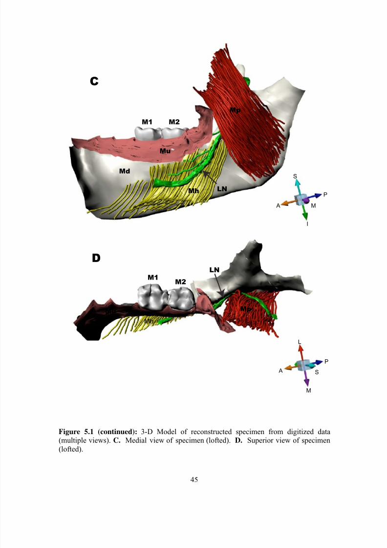
7/23/2019 The Relationship of the Lingual Nerve to the 3rd Molar Region a 3-D Analysis
http://slidepdf.com/reader/full/the-relationship-of-the-lingual-nerve-to-the-3rd-molar-region-a-3-d-analysis 56/95
45
Figure 5.1 (continued): 3-D Model of reconstructed specimen from digitized data(multiple views). C. Medial view of specimen (lofted). D. Superior view of specimen
(lofted).
C
D
S
P
A M
I
S A
P
L
M
Md
Mp
Mu
M2M1
Mh
M2M1
MpMu
Mh
LN
LN

7/23/2019 The Relationship of the Lingual Nerve to the 3rd Molar Region a 3-D Analysis
http://slidepdf.com/reader/full/the-relationship-of-the-lingual-nerve-to-the-3rd-molar-region-a-3-d-analysis 57/95
46
Figure 5.1 (continued): 3-D Model of specimen reconstructed from digitized and lofteddata (multiple views). E. Inferior view (lofted). F. Posterior view (lofted). G. Anteriorview (lofted).
E
F G
S
M
L
I
S
L
I
MP
A
A
M
P
L
I
Mu
Mu
Mp
Mp
Mp
Md
Md
M2M1
Mh
Mh
Md
Mh
LN
LN
LN
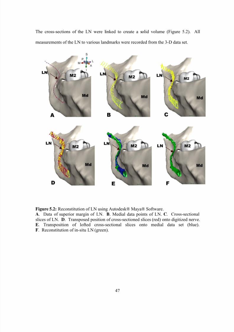
7/23/2019 The Relationship of the Lingual Nerve to the 3rd Molar Region a 3-D Analysis
http://slidepdf.com/reader/full/the-relationship-of-the-lingual-nerve-to-the-3rd-molar-region-a-3-d-analysis 58/95
47
The cross-sections of the LN were linked to create a solid volume (Figure 5.2). All
measurements of the LN to various landmarks were recorded from the 3-D data set.
Figure 5.2: Reconstitution of LN using Autodesk® Maya® Software.A. Data of superior margin of LN. B. Medial data points of LN. C. Cross-sectional
slices of LN. D. Transposed position of cross-sectioned slices (red) onto digitized nerve.E. Transposition of lofted cross-sectional slices onto medial data set (blue).
F. Reconstitution of in-situ LN (green).

7/23/2019 The Relationship of the Lingual Nerve to the 3rd Molar Region a 3-D Analysis
http://slidepdf.com/reader/full/the-relationship-of-the-lingual-nerve-to-the-3rd-molar-region-a-3-d-analysis 59/95
48
5.3 Lingual Nerve
5.3.1 Gross Morphology
The LN branched from CN V3 in the infratemporal region for the majority of
specimens (85.6%; 6 out of 7). In one specimen the LN originated from the CN V3 in the
pterygomandibular region, between the sigmoid notch and the lingula.
Inferior to the origin of LN, the nerve passed lateral to the medial pterygoid
muscle without penetrating the muscle belly. In this region, the LN was surrounded by
an abundant amount of perineural fat.
After passing anterior to the medial pterygoid muscle, 1-3 small accessory
branches from the LN could be seen in the retromolar region (Table 5.1). These
accessory branches resided on the surface of the mylohyoid muscle and generally
traveled in a superior direction. In this region, the LN was in direct contact with the
lingual plate of the mandible in three specimens, whereas the remaining four specimens
had variable amount of connective tissue (e.g. perineural fat) between the LN and the
mandible. Quantification of the distance between the LN and the mandible are presented
in section 5.3.5 of this thesis.
In this study, no communicating LN branches to the mylohyoid nerve were
observed.
7/23/2019 The Relationship of the Lingual Nerve to the 3rd Molar Region a 3-D Analysis
http://slidepdf.com/reader/full/the-relationship-of-the-lingual-nerve-to-the-3rd-molar-region-a-3-d-analysis 60/95
49
Table 5.1: Observations of the Course of LN
SPECIMEN PENETRATION
OF MP
ORIGIN OF LN ACCESSORY
NERVE
BRANCHES
CONTACT
WITH
BONE
1 NO PTERYGOMANDIBULAR 2 NO
2 NO INFRATEMPORAL 3 NO3 NO INFRATEMPORAL 3 YES
4 NO INFRATEMPORAL 3 NO
5 NO INFRATEMPORAL 1 YES
6 NO INFRATEMPORAL 2 YES
7 NO INFRATEMPORAL 1 NOMP = Medial Pterygoid Muscle; LN = Lingual Nerve
5.3.2 Distance from Mucosa to LN / Validation of Measurement from 3-D Model
The mean shortest distance from the oral mucosa in the retromolar pad to the
most superior aspect of the LN (distance III) was 9.0 ± 1.4 mm and 9.2 ± 1.7 mm with
digital calipers and Autodesk® Maya® 8.5 respectively (Table 5.2). The vertical
distance to point 2 on the LN (distance II) was approximately two times longer than the
distance to point 3 (distance III). The measurements for distance II were 17.3 ± 3.5 mm
and 18.0 ± 2.9 mm using digital calipers and Autodesk® Maya® 8.5 respectively. The
distance at which the LN turned medially towards the tongue was 26.3 ± 4.2 mm and
26.4 ± 4.3 mm using digital calipers and Autodesk® Maya® 8.5 respectively. Distance I
measured at the point where the LN turned medially towards the tongue was the longest
of the three distances measured.
The measurements (distances I, II and III) made directly from the specimens
using digital calipers and from the 3-D model using Autodesk® Maya® 8.5 were
compared using paired t-tests (p < 0.05). No statistical significant differences were found
between the two measuring techniques (Table 5.2).

7/23/2019 The Relationship of the Lingual Nerve to the 3rd Molar Region a 3-D Analysis
http://slidepdf.com/reader/full/the-relationship-of-the-lingual-nerve-to-the-3rd-molar-region-a-3-d-analysis 61/95
50
Table 5.2: Distances from Retromolar Pad to Reference Points on LN
Distance I (mm) Distance II (mm) Distance III (mm)
Specimen Digital
caliper
Autodesk®
Maya®
Digital
caliper
Autodesk®
Maya®
Digital
caliper
Autodesk®
Maya®
1 29.4 28.2 22.4 21.1 7.3 7.8
2 20.3 20.4 14.7 15.6 11.1 11.9
3 26.5 25.9 15.3 15.9 9.6 9.5
4 29.6 29.2 14.8 14.1 8.4 8.0
5 29.2 30.4 13.7 19.6 7.9 7.3
6 20.3 20.5 19.9 18.8 8.2 8.9
7 28.5 29.9 20.6 21.2 10.5 11.0
Mean 26.3 26.4 17.3 18.0 9.0 9.2
S.D. 4.2 4.3 3.5 2.9 1.4 1.7p value 0.96 0.69 0.78
5.3.3 Morphology of LN
Each region of LN (A, B and C as outlined in Figure 5.3) was sectioned in order
to evaluate cross sectional morphology. A range of 9-17 cross-sections were taken for
region A; 7-12 for region B; and 14-22 for region C. The cross-sectional morphology of
the LN differed between regions (Figure 5.3). The largest proportion of oval shaped
cross-sections was found in region A (60%) followed by region B (50%). Regions B
contained the most round shaped cross-sections (21%). The LN appeared to be
compressed by the medial pterygoid muscle in region C. The large percentages of oval
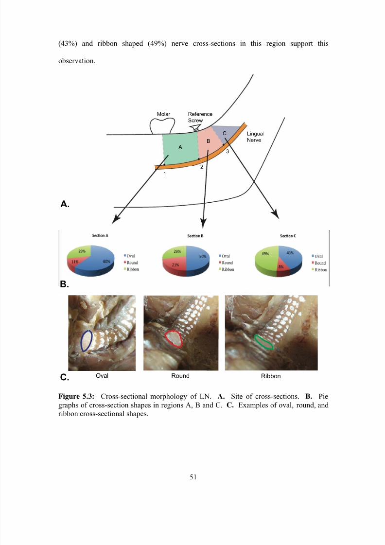
7/23/2019 The Relationship of the Lingual Nerve to the 3rd Molar Region a 3-D Analysis
http://slidepdf.com/reader/full/the-relationship-of-the-lingual-nerve-to-the-3rd-molar-region-a-3-d-analysis 62/95
51
(43%) and ribbon shaped (49%) nerve cross-sections in this region support this
observation.
Figure 5.3: Cross-sectional morphology of LN. A. Site of cross-sections. B. Pie
graphs of cross-section shapes in regions A, B and C. C. Examples of oval, round, andribbon cross-sectional shapes.

7/23/2019 The Relationship of the Lingual Nerve to the 3rd Molar Region a 3-D Analysis
http://slidepdf.com/reader/full/the-relationship-of-the-lingual-nerve-to-the-3rd-molar-region-a-3-d-analysis 63/95
52
Table 5.3: Distribution of LN Cross Sectional Morphology
Region A* Region B* Region C*Specimen
Oval Round Ribbon Oval Round Ribbon Oval Round Ribbon
1 11/17 2/17 4/17 1/10 9/10 0/10 12/22 1/22 9/22
2 10/14 4/14 0/14 5/10 5/10 0/10 20/20 0/20 0/20
3 13/15 0/15 2/15 6/7 0/7 1/7 2/20 1/20 17/204 4/9 0/9 5/9 8/8 0/8 0/8 3/14 1/14 10/145 11/17 2/17 4/17 1/10 9/10 0/10 12/21 1/21 8/21
6 6/17 1/17 10/17 8/12 1/12 3/12 4/19 0/19 15/197 10/14 4/14 0/14 5/10 5/10 0/10 20/20 0/20 0/20
* Data is presented as a fraction: number of sections with oval, round or ribbon cross-sectional
shape/total number of sections in region.
5.3.4 Vertical Position of LN
The vertical distances of the LN to the alveolar crest for each specimen are
summarized in Table 5.4 and Figure 5.4. A value of zero was given when the LN was at
the same vertical height as the alveolar crest. This was noted in Specimen 5. A negative
value of -3.9 mm was observed in region C of specimen 4. This denotes a measurement
in which the LN was above the alveolar crest.
Figure 5.4: Average Vertical Distance to LN in Regions A, B and C for each Specimen

7/23/2019 The Relationship of the Lingual Nerve to the 3rd Molar Region a 3-D Analysis
http://slidepdf.com/reader/full/the-relationship-of-the-lingual-nerve-to-the-3rd-molar-region-a-3-d-analysis 64/95
53
Table 5.4: Vertical Distances from LN to Alveolar Crest
Vertical Distance to LN (mm)
Region A Region B Region C
Specimen Mean Range Mean Range Mean Range
1 16.5±0.8 15.5-17.8 11.3±3.9 5.3-17.4 8.5±2.2 5.1-12.1
2 13.3±1.4 11.9-15.8 12.0±4.6 2.4-17.4 4.9±0.3 4.6-5.6
3 16.0±3.9 8.9-20.2 3.7±1.4 1.9-5.1 0.1±1.7 -0.9-2.0
4 12.6±2.7 5.8-14.9 3.9±1.1 2.8-4.9 -3.9±0.0 -3.9-3.9
5 14.8±1.5 12.3-17.4 7.9±3.7 3.0-13.6 3.2±2.4 0.0-7.8
6 12.1±0.4 11.6-12.9 9.5±2.8 5.4-12.6 3.8±0.7 3.0-5.0
7 13.9±1.4 11.4-15.5 8.4±2.3 4.0-10.7 3.3±0.4 2.8-4.0
Mean 14.2±1.7a 8.1±3.3
b 2.8±3.9
b,c
If the superscript letters differ, then the result is statistically significant. If the letter is the same,
then the result is not statistically different.
As depicted in Figure 5.5, the mean vertical distance to the LN from the alveolar
crest is highly variable amongst specimens. Region C was the most variable with a mean
vertical distance of 2.8 ± 3.9 mm.
Figure 5.5: Average Vertical Distance to LN by Region

7/23/2019 The Relationship of the Lingual Nerve to the 3rd Molar Region a 3-D Analysis
http://slidepdf.com/reader/full/the-relationship-of-the-lingual-nerve-to-the-3rd-molar-region-a-3-d-analysis 65/95
54
5.3.5 Horizontal Position of LN
The mean horizontal distance between alveolar bone and the LN are summarized
by specimen in Table 5.5. If the LN had direct contact with the lingual plate, the
horizontal distance was zero. This was noted in three specimens (2, 3 and 7). The mean
distances for each specimen are graphically displayed in Figure 5.6 and illustrate a
non-uniform distribution.
Table 5.5: Horizontal Distance from LN to Lingual Plate
Horizontal Distance to LN (mm)
Region A Region B Region C
Specimen Mean Range Mean Range Mean Range
1 9.2±0.8 7.2-10.3 4.2±2.4 1.3-7.8 6.2±5.7 1.1-19.3
2 4.5±1.0 3.5-5.9 3.8±3.2 0.0-8.4 1.8±2.7 0.0-9.1
3 3.1±1.0 1.6-5.2 0.2±0.3 0.0-0.7 7.5±6.1 0.0-20.1
4 4.6±2.0 010-7.1 0.6±0.4 0.1-1.0 7.4±5.9 1.0-16.4
5 7.3±1.4 5.0-9.7 1.7±1.2 0.2-4.7 4.1±4.3 0.5-14.8
6 3.2±0.6 2.3-3.9 2.3±1.1 0.8-3.7 4.2±3.9 0.3-11.4
7 3.6±1.2 2.0-5.4 1.8±0.8 0.8-3.0 3.5±3.5 0.0-10.6
Mean 4.1±1.7a,b
2.4±2.0a 7.9±4.1
b,c
If the superscript letters differ, then the result is statistically significant. If the letter is the same,
then the result is not statistically different.
Figure 5.6: Average Horizontal Distance to LN in Regions A, B and C for each
Specimen

7/23/2019 The Relationship of the Lingual Nerve to the 3rd Molar Region a 3-D Analysis
http://slidepdf.com/reader/full/the-relationship-of-the-lingual-nerve-to-the-3rd-molar-region-a-3-d-analysis 66/95
55
The average horizontal distance to the LN by region was tabulated and plotted in
Figure 5.7. The horizontal distance to LN was 2.4 ± 2.0 mm in region B. The largest
variability was found in region C with a mean horizontal distance of 7.9 ± 4.1 mm.
Figure 5.7: Average Horizontal Distance to LN by Region
5.4 Relative Position of Mylohyoid Ridge in Relation to the Position of
the LN
The height of the mylohyoid ridge was calculated as a ratio to the height of the
mandible and correlated with the vertical and horizontal distance from the alveolar bone
to LN in each region. The data is presented in Table 5.6. The assumption that the
position of the mylohyoid ridge affects the location of the LN generated the following
hypotheses:
H1: A superiorly positioned mylohyoid ridge is directly proportional to the
vertical position of the lingual nerve in the third molar region.

7/23/2019 The Relationship of the Lingual Nerve to the 3rd Molar Region a 3-D Analysis
http://slidepdf.com/reader/full/the-relationship-of-the-lingual-nerve-to-the-3rd-molar-region-a-3-d-analysis 67/95
56
H1: A superiorly positioned mylohyoid ridge is directly proportional to the
horizontal position of the lingual nerve in the third molar region.
A scatter plot of the data was plotted with a line of best-fit using Excel version
12.1.7 (Microsoft Inc, Redmond, WA) and a regression analysis preformed with SPSS
(SPSS Inc, Chicago, IL). No outliers were identified. When a regression analysis was
performed (Table 5.7, Figure 5.8), a statistically significant relationship was found
between the vertical distance of the LN in the third molar region (Region B) and the
height of the mylohyoid ridge (p = 0.002). There was no statistically significant
relationship within regions A (p = 0.324) and C (p = 0.136).
Table 5.6: Regression Analysis Source Data: Mylohyoid Ridge Ratio and Minimum
Vertical Distance between LN and Alveolar Crest in Regions A, B and C.
Minimum Vertical Distance between LN and
Alveolar Crest (mm)
Specimen Mylohyoid Ridge
Ratio
Region A Region B Region C
1 0.805 5.8 2.8 3.9
2 0.851 8.9 1.9 -0.9
3 0.843 12.3 3.0 0.04 0.724 11.6 5.4 3.0
5 0.752 11.3 4.0 2.8
6 0.733 15.5 5.3 5.1
7 0.818 11.9 2.4 4.6
Table 5.7: Data from Regression Analysis of Mylohyoid Ridge and Vertical Distance toLN
Regression Analysis
Region A Region B Region C
R 2 0.193 0.884 0.387
F-value 1.198 38.288 3.159
Slope (m) -25.360 -24.895 -26.946y-intercept (b) 31.063 23.180 23.905
Significance 0.324 0.002 0.136

7/23/2019 The Relationship of the Lingual Nerve to the 3rd Molar Region a 3-D Analysis
http://slidepdf.com/reader/full/the-relationship-of-the-lingual-nerve-to-the-3rd-molar-region-a-3-d-analysis 68/95
57
Figure 5.8: Regression Analysis of Mylohyoid Ridge Ratio to Vertical Position of
LN.
A statistically significant relationship (p = 0.009) was also present between the horizontal
distance in Section B and the height of the mylohyoid ridge (Tables 5.8 and Figure 5.9).
Table 5.8: Regression Analysis Source Data: Mylohyoid Ridge Ratio and MinimumHorizontal Distance between LN and Lingual Plate in Regions A, B and C.
Minimum Horizontal Distance between LN and Lingual
Plate (mm)
Specimen Mylohyoid
Ridge Ratio
Region A Region B Region C
1 0.805 1.0 0.1 1.02 0.851 1.6 0.0 0.03 0.843 5.0 0.2 0.5
4 0.724 2.3 0.8 0.35 0.752 2.0 0.8 0.0
6 0.733 7.2 1.3 1.17 0.818 3.5 0.0 0.0

7/23/2019 The Relationship of the Lingual Nerve to the 3rd Molar Region a 3-D Analysis
http://slidepdf.com/reader/full/the-relationship-of-the-lingual-nerve-to-the-3rd-molar-region-a-3-d-analysis 69/95
58
Table 5.9: Data from Regression Analysis of Mylohyoid Ridge & Horizontal
Distance to LN
Regression Analysis
Region A Region B Region C
R 2 0.047 0.776 0.079F-value 0.246 17.365 0.430Slope (m) -9.063 -8.300 -2.530y-intercept (b) 10.376 7.005 2.417
Significance 0.641 0.009 0.541
Figure 5.9: Regression Analysis of Mylohyoid Ridge Ratio To Horizontal Position of
LN.
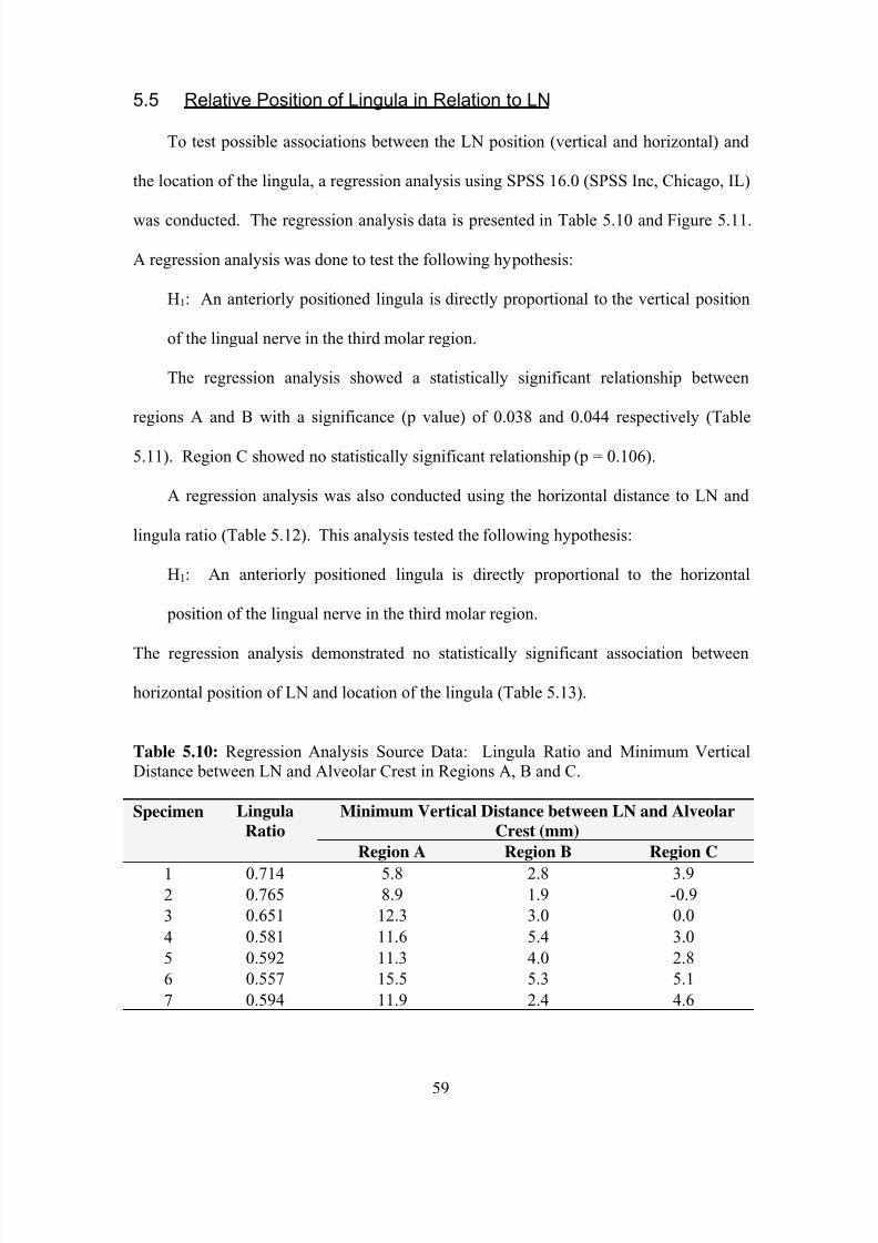
7/23/2019 The Relationship of the Lingual Nerve to the 3rd Molar Region a 3-D Analysis
http://slidepdf.com/reader/full/the-relationship-of-the-lingual-nerve-to-the-3rd-molar-region-a-3-d-analysis 70/95
59
5.5 Relative Position of Lingula in Relation to LN
To test possible associations between the LN position (vertical and horizontal) and
the location of the lingula, a regression analysis using SPSS 16.0 (SPSS Inc, Chicago, IL)
was conducted. The regression analysis data is presented in Table 5.10 and Figure 5.11.
A regression analysis was done to test the following hypothesis:
H1: An anteriorly positioned lingula is directly proportional to the vertical position
of the lingual nerve in the third molar region.
The regression analysis showed a statistically significant relationship between
regions A and B with a significance (p value) of 0.038 and 0.044 respectively (Table
5.11). Region C showed no statistically significant relationship (p = 0.106).
A regression analysis was also conducted using the horizontal distance to LN and
lingula ratio (Table 5.12). This analysis tested the following hypothesis:
H1: An anteriorly positioned lingula is directly proportional to the horizontal
position of the lingual nerve in the third molar region.
The regression analysis demonstrated no statistically significant association between
horizontal position of LN and location of the lingula (Table 5.13).
Table 5.10: Regression Analysis Source Data: Lingula Ratio and Minimum VerticalDistance between LN and Alveolar Crest in Regions A, B and C.
Minimum Vertical Distance between LN and Alveolar
Crest (mm)
Specimen Lingula
Ratio
Region A Region B Region C
1 0.714 5.8 2.8 3.92 0.765 8.9 1.9 -0.9
3 0.651 12.3 3.0 0.0
4 0.581 11.6 5.4 3.0
5 0.592 11.3 4.0 2.8
6 0.557 15.5 5.3 5.1
7 0.594 11.9 2.4 4.6

7/23/2019 The Relationship of the Lingual Nerve to the 3rd Molar Region a 3-D Analysis
http://slidepdf.com/reader/full/the-relationship-of-the-lingual-nerve-to-the-3rd-molar-region-a-3-d-analysis 71/95
60
Table 5.11: Data from Regression Analysis of Lingula Position and Vertical Distance to
LN
Regression Analysis
Region A Region B Region C
R 2 0.612 0.589 0.437F-value 7.887 7.177 3.874
Slope (m) - 30.667 -13.810 - 19.444
y-intercept (b) 30.556 12.314 15.005
Significance 0.038 0.044 0.106
Figure 5.11: Regression Analysis of Lingula Ratio to Vertical Position of LN

7/23/2019 The Relationship of the Lingual Nerve to the 3rd Molar Region a 3-D Analysis
http://slidepdf.com/reader/full/the-relationship-of-the-lingual-nerve-to-the-3rd-molar-region-a-3-d-analysis 72/95
61
Table 5.12: Regression Analysis Source Data: Lingula Ratio and Minimum Horizontal
Distance between LN and Alveolar Crest in Regions A, B and C.
Minimum Vertical Distance between LN and Lingual
Plate (mm)
Specimen Lingula
Ratio
Region A Region B Region C1 0.714 0.6 1.3 1.1
2 0.765 1.0 0.0 0.0
3 0.651 1.0 0.0 0.0
4 0.581 1.0 0.1 1.0
5 0.592 1.8 0.2 0.5
6 0.557 0.3 0.8 0.3
7 0.594 1.5 0.6 0.0
Table 5.13: Data for Regression Analysis of Lingula Position and Horizontal Distanceto LN
Regression Analysis
Region A Region B Region C
R 2 0.022 0.000 0.007
F-value 0.110 0.002 0.037
Slope (m) - 0.945 - 0.120 - 0.526y-intercept (b) 1.631 0.507 0.755
Significance 0.754 0.967 0.854
5.6 Results Summary
A medial dissection approach was employed to capture the course of the LN in 7
specimens. This approach minimized the amount of tissue distortion to allow for
accurate digitization and 3-D computational reconstitution of the specimen. The modeled
specimen could then be rotated in 3-D space to appreciate and quantify the relationship of
the LN to various soft and hard tissue structures: oral mucosa, medial pterygoid muscle,
mylohyoid muscle, and the medial surface of the mandible.
The LN is a posterior branch of CN V3. In 6 out of 7 specimens, the LN was found
to originate in the infratemporal fossa, however in one specimen, the LN branched from
CN V3 in the pterygomandibular region. The LN did not penetrate the medial pterygoid

7/23/2019 The Relationship of the Lingual Nerve to the 3rd Molar Region a 3-D Analysis
http://slidepdf.com/reader/full/the-relationship-of-the-lingual-nerve-to-the-3rd-molar-region-a-3-d-analysis 73/95
62
muscle and maintained a lateral position to the muscle. Anterior to the medial pterygoid
muscle the LN, in all specimens, had 1-3 accessory branches. The shape of the LN
changed along its course to the tongue. An oval shape was predominant in regions A and
B, however in region C the nerve was mainly ribbon shaped.
The mean shortest distance from the oral mucosa of the retromolar pad to the LN as
measured manually (using digital calipers) was 9.0 ± 1.4 mm and computationally a
mean distance of 9.2 ± 1.7 mm was obtained using the Autodesk® Maya® distance tool.
There was no significant difference between the two methods of measurements.
The average vertical and horizontal distance to LN from the mandibular alveolus
was variable amongst the three regions (A, B and C). The mean vertical distance from
the alveolar crest to LN was 14.2 ± 1.67 mm, 9.1 ± 3.3 mm and 2.8 ± 3.6 mm for regions
A, B and C respectively. The mean horizontal distances for sections A, B, and C were
4.1 ± 1.6 mm, 2.4 ± 2.0 mm and 7.85 ± 4.1 mm respectively.
Several positive correlations were observed between the distance to LN and relative
position of the mylohyoid ridge or lingula. The vertical distance of the LN in region B
was positively correlated with the height of the mylohyoid ridge (p=0.002). No
correlations were found in sections A (p=0.324) and C (p=0.136). An anteriorly
positioned lingula was positively correlated with the vertical positioning of the LN in
regions A (p = 0.038) and B (p = 0.044). Evaluation of the position of the mylohyoid
ridge with respect to the horizontal position of the LN yielded a positive correlation
(p=0.009) only in region B. Regression analysis was also conducted to determine if a
relationship between the position of the lingula and horizontal position of the LN was
present, but no significant association was noted.

7/23/2019 The Relationship of the Lingual Nerve to the 3rd Molar Region a 3-D Analysis
http://slidepdf.com/reader/full/the-relationship-of-the-lingual-nerve-to-the-3rd-molar-region-a-3-d-analysis 74/95
63
Chapter 6Discussion
6.1 Introduction
Three-dimensional reconstruction of the in-situ course of the LN along the third
molar region has not been described in the literature. The method developed in this study
is superior to traditional cadaveric dissections in that a 3-D model of the specimen is
created which can be viewed and evaluated from any perspective. Furthermore,
Autodesk® Maya® allows for magnification and measurement of delicate anatomical
structures in the model without fear of tissue distortion or iatrogenic displacement.
6.2 Origin of the LN
In the current study, the majority (6 out of 7) of specimens demonstrated
branching from the posterior division of CN V3 in the infra-temporal region. Kim et al.
(2004) and Erdogmus et al. (2008) also reported a high percentage of LN branching in
the infra-temporal region (21 out of 32 and 14 out of 21 respectively). An explanation
for higher incidence reported in the current study may be attributed to the small sample
size. No plexiform pattern between the LN and IAN was observed in the current study,
which is in accordance to the findings presented by Erdogmus et al. (2008).
Only one specimen demonstrated a branching pattern inferior to the sigmoid
notch. This is clinically significant as it can contribute to the failure rate of LN
anesthesia during IAN blocks. The Gow-Gates and Akinosi techniques have been
advocated when traditional IAN and LN anesthetic techniques fail (Meechan, 1999). The
success of these techniques relies on inserting the anesthetic needle more superiorly so
that local anesthetic is deposited near the branching of the LN and IAN. This clinical

7/23/2019 The Relationship of the Lingual Nerve to the 3rd Molar Region a 3-D Analysis
http://slidepdf.com/reader/full/the-relationship-of-the-lingual-nerve-to-the-3rd-molar-region-a-3-d-analysis 75/95
64
finding supports the anatomical observations that the majority of LNs originate in the
infra-temporal region.
6.3 Cross-sectional Shape
The cross-sectional morphology of the LN changes along its course towards the
tongue. Observations from this study suggest the shape of the nerve is dependent on
overlying structures. For example, the highest percentile of ribbon shaped cross-sections
was observed in region C where the LN is often found tightly compressed between the
mandible and medial pterygoid muscle. This is in contrast to the findings presented by
Kim et al. (2004) in which the highest percentage (62.5%; 20 out of 32) of ribbon-like
cross-sections was found in the second molar region. This anatomical difference may be
related to racial differences as Kim et al. (2004) dissected Korean cadavers. In the
current study, a higher percentage of oval and round shapes (50% and 21% respectively)
were found in the third molar region. This proportion is similar to Kim’s (2004) finding
of predominately oval or circular morphology (59.4%; 19 out of 32, and 25%; 8 out of
32, respectively) in this region. Knowledge of cross-sectional shape is clinically relevant
since autogenous nerve grafts should mimic the morphology of the nerve defect (Wolford
and Stevao 2003).
6.4 LN Accessory Branches
Collateral branches (Range: 1-3) from the LN in the retromolar and third molar
region were found in all specimens in the current study. This finding supports Kim et
al.’s (2004) claim that this is not an anatomical variation, rather the predominant pattern.
The presence of the collateral LN branches may explain incomplete anesthesia in this

7/23/2019 The Relationship of the Lingual Nerve to the 3rd Molar Region a 3-D Analysis
http://slidepdf.com/reader/full/the-relationship-of-the-lingual-nerve-to-the-3rd-molar-region-a-3-d-analysis 76/95
65
region and the finding of localized paraesthesia of the lingual mucosa in the third molar
region following third molar surgery. Evaluation of the 3-D models suggests these
accessory branches are particularly vulnerable to lingual plate perforation during third
molar surgery misadventures.
6.5 Digitization and Method of LN Reconstitution
Previous cadaveric studies used to evaluate the position of the LN have been
criticized due to potential tissue distortion and manipulation of the LN position (Miloro
et al,. 1997; Behnia et al. 2000). Specimen distortion due to the fixation process has been
shown to be negligible (Cutts, 1988). In the current study, several methods were utilized
to minimize iatrogenic displacement of LN during dissection. A hemi-maxillectomy was
utilized to provide an unobstructed view and allow for greater flexibility of the digitizing
arm within this confined space. During the osteotomies, the pterygoid plates were
preserved which minimized disruption to the medial pterygoid muscle and underlying
LN. The mesoneurium surrounding the LN was left intact to minimize displacement.
Magnification (2.5 X Surgical Loupes, Design for Vision, Inc. Ronkonkoma, NY) was
used to carefully expose the superior and medial surface of the LN. Digitization of this
undisturbed surface allowed for accurate 3-D reconstitution of digitized co-ordinates in
Autodesk® Maya® 8.5. In addition, the dissection and digitization process was
conducted by one operator to minimize inter-operator variability.
Creating accurate 3-D models through serial dissection and digitization has been
validated by several studies done in our laboratory (Kertesz, 2005; Sansalone, 2006).
Mahoney et al. (1999) examined the consistency of this technique with an intra-observer
reliability between 0.12 and 0.7 mm, and an inter-observer reliability between 0.15 and

7/23/2019 The Relationship of the Lingual Nerve to the 3rd Molar Region a 3-D Analysis
http://slidepdf.com/reader/full/the-relationship-of-the-lingual-nerve-to-the-3rd-molar-region-a-3-d-analysis 77/95
66
0.32 mm. The current study also validated the accuracy of Autodesk® Maya® 8.5
distance tool software to measure distances to LN from the reference screw in the center
of the retromolar pad. These distances were also measured manually with digital
calipers. A paired t-test between these two measuring techniques demonstrated no
statistically significant difference (p < 0.05).
6.6 Anatomic Position of the LN in the Mandibular Third Molar Region
6.6.1 Distance of LN to Oral Mucosa
The center of the retromolar pad was selected as the reference point for oral
mucosa measurements to the LN since the underlying basal bone endures minimal
resorption (Zarb, 2003). Furthermore, it lies in close proximity to the distal extent of
incisions required for the surgical extraction of impacted mandibular third molars. The
retromolar pad was free of pathology in all specimens. This was important since a
hyperplastic retromolar pad would exaggerate the distance from the oral mucosa to the
LN. The mean shortest distance from the oral mucosa in the retromolar pad to the LN
was 9.22 ± 1.72 mm. According to data presented by Hölzle and Wolff (2001), the LN
lies considerably closer to the oral mucosa with a mean distance of 4.41 ± 1.44 mm.
Comparison of these two distances is not possible since the measuring points utilized on
the oral mucosa are dissimilar between the two studies. Hölzle and Wolff (2001)
simulated an incision up the ascending ramus for third molar surgery and measured the
distance to the LN from the most distal part of the incision. Use of this type of incision is
highly unlikely as a more lateral incision is usually used in the clinical setting and thus
represents a poor choice for standardized measurements to the LN.
7/23/2019 The Relationship of the Lingual Nerve to the 3rd Molar Region a 3-D Analysis
http://slidepdf.com/reader/full/the-relationship-of-the-lingual-nerve-to-the-3rd-molar-region-a-3-d-analysis 78/95
67
The retromolar pad serves as a surgical landmark for outlining safe incisions
during third molar surgery. In the current study, the LN was observed to be medial to the
retromolar pad in all specimens. Behnia et al. (2000) reported a rare observation (1 out
of 669) of the LN traveling in the retromolar region. In view of these findings, a mucosal
incision lateral to the retromolar pad is recommended to minimize iatrogenic damage to
the LN during third molar surgery.
6.6.2 Vertical Distance of LN to Alveolar Crest
The results of the current study were compared to the findings of Pogrel (1995).
Pogrel’s study was unique in that it used definitive reference points for measuring the
vertical distance to LN. In the current study, the vertical LN distances in region B
correspond to the vertical distance measured by Pogrel. The mean vertical distance from
the alveolar crest to the LN in region B was 8.1 ± 2.9 mm in this study, while Pogrel
(1995) reported a distance of 8.3 ± 4.1 mm. The similarity between these distances
stresses the importance of comparing standardized measurements. Previous studies
lacked standardized methods to measure the position of the LN and should be interpreted
with caution.
Variability of results reported in the literature may also be due to anatomical
variation and age related changes. Anatomical variations such as the position of the
mylohyoid ridge (see section 6.6) and lingula (see section 6.7) may also contribute to
variable vertical positions of the LN. The loss of muscle tone and connective tissue
tension with advanced age may alter the vertical position of the LN. For example, Hölzle
and Wolff (2001) concluded a significant relationship between the degree of mandibular
crest atrophy and the distance of the LN to the crest. In addition, Miloro et al. (1997)

7/23/2019 The Relationship of the Lingual Nerve to the 3rd Molar Region a 3-D Analysis
http://slidepdf.com/reader/full/the-relationship-of-the-lingual-nerve-to-the-3rd-molar-region-a-3-d-analysis 79/95
68
reported a vertical distance of 2.53 ± 0.67mm in ten young (mean age 24.7 years)
volunteers using MRI. These measurements were made from a single coronal MRI slice
in the third molar region.
6.6.3 Horizontal Distance of LN to Lingual Plate
Previous literature has used manual or digital calipers to measure the horizontal
distance of the LN to the lingual plate. This method introduces potential errors as the
calipers can inadvertently move the position of the LN. A novel method of using in-situ
3-D models to measure the horizontal distance to LN was introduced in the current study.
The ability to manipulate and magnify the 3-D model allowed for multiple horizontal
measurements to be conducted along the course of the LN with no iatrogenic
displacement.
In region B (third molar region), the LN was in contact with the alveolar bone in
28.57% (2 out of 7) of the specimens, similar to the findings of Behnia et al. (2000) who
reported this relationship in 22.27% (149 out of 669) specimens. In the current study the
mean distance to the alveolar bone was 2.4 ± 2.0 mm. This distance is similar to the
measurement of 2.1 ± 1.1 mm reported by Behnia et al. (2000). Miloro (2001) reported a
comparable distance of 2.5 ± 0.7 mm using MRI imaging.
In region C, the LN had direct contact with the lingual plate in three specimens
(42.85%; 3 out of 7). The greatest distance between the LN and the lingual plate was
7.9 ± 4.1 mm in region C. Differences of lateral flaring of the mandible in this region
may help account for the large variability.
No specimens demonstrated LN contact with alveolar bone in region A. This is
not surprising as the LN begins to course medially towards the tongue in this region. The
7/23/2019 The Relationship of the Lingual Nerve to the 3rd Molar Region a 3-D Analysis
http://slidepdf.com/reader/full/the-relationship-of-the-lingual-nerve-to-the-3rd-molar-region-a-3-d-analysis 80/95
69
average mean distance to the bone in this region was 4.1 ± 1.7 mm. This increased
distance to the LN in this region would inherently lower the risk of damage to LN due to
lingual plate perforations.
6.7 Association Between the Position of the LN and Location of Mylohyoid
Ridge
In 2007, Karakas et al. suggested the position of the mylohyoid muscle and/or the
volume of the muscle may account for differences in the position of the LN but did not
support this statement with findings. Evidence for this relationship was not found in the
literature.
The current study examined the potential relationship between the location of the
mylohyoid ridge and the position of the LN. A regression analysis demonstrated a
positive association between the location of the mylohyoid ridge and the vertical and
horizontal distances between the LN and the alveolus in region B. No association was
found in regions A and C. The mylohyoid ridge is the origin for the mylohyoid muscle,
which forms the floor of the mouth and thus limits the inferior displacement of the LN.
The positive association found in region B, means that a more superiorly positioned
mylohyoid ridge results in a LN positioned closer to the alveolar crest. However, as the
LN courses anteriorly, this relationship is lost, possibly due to muscle laxity or decrease
in muscle volume. In the current study, the presence of more perineural tissue around the
LN in sections A and C may account for greater variability and the loss of an association
with the mylohyoid ridge. No relationship was found with the position of the LN in
section C, as there was minimal mylohyoid muscle in this region.

7/23/2019 The Relationship of the Lingual Nerve to the 3rd Molar Region a 3-D Analysis
http://slidepdf.com/reader/full/the-relationship-of-the-lingual-nerve-to-the-3rd-molar-region-a-3-d-analysis 81/95
70
Regression analysis also found a relationship between the position of the
mylohyoid ridge and the horizontal position of the LN suggesting a more superiorly
positioned mylohyoid ridge is associated with the LN lying closer to the lingual plate of
the mandible in region B. The gingiva may account for this association. The lingual
gingiva transitions from unattached mucosa inferiorly, to firm attached gingiva
superiorly. Thus, a superiorly positioned mylohyoid ridge causes the LN to course under
attached gingiva and as a result be tightly bound to the medial wall of the mandibular
alveolus.
Atrophy of the alveolar crest is likely not a factor in the association between the
position of the LN and location of the mylohyoid ridge since all specimens had
mandibular second molars present with no furcation involvement. Furthermore, the
distances used to measure the mylohyoid ridge ratio were at the posterior extent of the
ridge anatomy and beyond the tooth-bearing region.
The association between the mylohyoid ridge and the position of the LN has
clinical implications. The mylohyoid ridge runs diagonally anteroinferiorly from the
region of the third molar to the premolar region and is visible on panorex radiographs
(White, 2000). Since the mylohyoid ridge is visible on panorex radiographs, this imaging
modality could be used to classify the risk of the LN during oral and maxillofacial
procedures. The ratio of the distance from the superior border of the mylohyoid ridge to
the height of the mandible developed in the current study could potentially be used to
predict the location of the LN from panorex radiographs. Variable magnification
amongst panorex machines would not be a factor with the use of a ratio. This ratio may
allow for universal method for approximating the relative position of the LN in the third
molar region.

7/23/2019 The Relationship of the Lingual Nerve to the 3rd Molar Region a 3-D Analysis
http://slidepdf.com/reader/full/the-relationship-of-the-lingual-nerve-to-the-3rd-molar-region-a-3-d-analysis 82/95
71
6.8 Association Between the Position of the LN and Location of Lingula
In the current study, as well as in previous studies, the LN was found to course
anterior to the lingula and IAN (Behnia et al., 2000; Hölzle and Wolff, 2001; Erdogmus
et al., 2008). The relationship between the lingula and LN has not been discussed in the
literature. In the current study, regression analysis demonstrated a positive correlation
between the vertical position of the LN and lingula in regions A and B. Thus, when the
lingula is situated more anteriorly, the LN is also translated in this direction to maintain
its relationship to the IAN. Anterior translation of the LN would then result in the LN
lying closer to the alveolar crest in the region of the third molar.
In 1992, Bell reported that the IAN foramina are located in a more superior and
posterior position in children compared to adults. Corroborating this finding, with the
relationship observed in this study would suggest that children are at a lower risk of LN
damage compared to adults. In a prospective study of 2134 consecutive mandibular third
molar surgeries, Renton and McGurk (2001) reported a mean age of 36 years for patients
with permanent LN injury versus a mean age of 29 years for all subjects. Chiapasco et
al. (1995) reported an increased incidence of LN injury with subjects over 24 years of
age. In 1991, Herpy and Goupil reported a 25% incidence of temporary LN injury if the
patient was over 40 years of age. Although many studies support this claim between age
and risk of LN damage, others provide conflicting evidence (Middlehurst et al., 1988;
Lopes et al., 1995; Black, 1997; Fielding et al., 1997; Valmaesed-Catellon et al., 2000).
Unfortunately, this study could not evaluate anatomical differences between adults and
children, as all specimens were over 40 years of age (range 40 – 79). Regardless, the
current study provides a possible novel explanation to the potential risk factor of age and
incidence of LN damage. In addition, the findings of the current study, along with Bell’s

7/23/2019 The Relationship of the Lingual Nerve to the 3rd Molar Region a 3-D Analysis
http://slidepdf.com/reader/full/the-relationship-of-the-lingual-nerve-to-the-3rd-molar-region-a-3-d-analysis 83/95
72
observation, suggest that early removal of third molars may limit adverse outcomes
associated with LN damage. This paradigm requires further investigation.
Furthermore, the positive relationship between the lingula and LN position may
aid in risk stratification of impacted mandibular third molars. Distoangular impactions
are considered the most problematic since their removal often requires surgical
intervention along the mandibular ramus (Peterson, 2003). In 1997, Fielding reported the
highest incidence of LN paresthesia with distoangular impacted teeth. In these situations,
an anteriorly positioned lingula would place the LN closer to the anterior border of the
ramus and thus at greater risk to LN damage.

7/23/2019 The Relationship of the Lingual Nerve to the 3rd Molar Region a 3-D Analysis
http://slidepdf.com/reader/full/the-relationship-of-the-lingual-nerve-to-the-3rd-molar-region-a-3-d-analysis 84/95
73
Chapter 7Conclusions
The conclusions of the thesis are as follows:
• A hemi-maxillectomy and a medial dissection allowed for the digitization of the
oral mucosa, medial pterygoid muscle, LN, mylohyoid muscle, medial aspect of
the mandible and the second mandibular molar. Three-dimensional models and a
database of the position of the LN within the mandibular 3rd molar region was
constructed from digitized data using Autodesk® Maya®.
• The origin of the LN occurred in the infratemporal region for the majority
(85.57%) of specimens.
• Small accessory branches (range: 1-3) of the LN were observed in the retromolar
region for all specimens.
• The LN displayed a variety of cross-sectional shapes within the oral cavity. In
the current study, an oval-round morphology accounted for 71% of cross-
sectional shapes in the third molar region.
• The mean shortest distance from the oral mucosa to the most superior aspect of
the LN was 9.0 ± 1.4 mm and 9.2 ± 1.7 mm using digital calipers and Autodesk®
Maya® software respectively. Thus computation of length measurements yielded
no statistical difference (p < 0.05) between these two methods.
• The horizontal and vertical positioning of the LN is highly variable. The vertical
measurement of the LN to the alveolar crest in the third molar region (section B)
was 8.1 ± 2.9 mm. The mean horizontal distance of the LN to the medial surface
of the mandibular alveolus in this region was 2.1 ± 1.7 mm.

7/23/2019 The Relationship of the Lingual Nerve to the 3rd Molar Region a 3-D Analysis
http://slidepdf.com/reader/full/the-relationship-of-the-lingual-nerve-to-the-3rd-molar-region-a-3-d-analysis 85/95
74
• The vertical distance of the LN to the alveolar crest in the third molar region
(region B) was positively correlated with the height of the mylohyoid ridge
(p = 0.001). However, no correlation was found in sections A (p = 0.327) and C
(p = 0.198). Thus, a superiorly positioned mylohyoid ridge is directly
proportional to the vertical position of the LN from the alveolar crest in the third
molar region. A superiorly positioned mylohyoid ridge in this region was also
correlated (p = 0.009) with the LN lying closer to the alveolar bone.
• An anteriorly positioned lingula was directly proportional to the vertical distance
of the LN from the alveolar crest in regions A and B (p < 0.05). There was no
association between the position of the lingula and the horizontal distance of the
LN and the lingual plate of the mandible.
7/23/2019 The Relationship of the Lingual Nerve to the 3rd Molar Region a 3-D Analysis
http://slidepdf.com/reader/full/the-relationship-of-the-lingual-nerve-to-the-3rd-molar-region-a-3-d-analysis 86/95
75
Chapter 8Future Directions
The current study has demonstrated a novel way of studying the position of the
LN using 3-D modeling. Although a potential relationship of the position of the LN with
the mylohyoid ridge and lingula was established, further examination with a larger
sample size would be required to produce more significant correlations.
Additional studies could evaluate the clinical relevance of imaging the
relationship between the LN and mylohyoid ridge and/or lingula. A suitable and
convenient imaging modality would be the use of a panorex as it is routinely used prior to
third molar surgery. Furthermore, the lingula and mylohyoid ridge are easily identified
on a panorex. A potential study could involve obtaining panorex images of cadaveric
mandibles prior to dissection and stratifying the potential risk to the LN based on the
location of the mylohyoid ridge and lingula from the panorex image. The cadaver could
then be dissected and a 3-D model created. Data from the 3-D model could then be used
to measure distances to the LN and test the validity of using panorex images as predictors
to the position of the LN. Other imaging modalities such as MRI could also be used in a
similar manner. Advancements in MRI technology would allow for 3-D volumetric
image reconstruction, which could then be compared to the digitized 3-D model.
Histology may provide additional insight into the cross-sectional microanatomy
of the LN. Cross-sectional histological examination of the LN would show the number
of nerve fascicles in various positions along its course. This information would be
useful in ensuring the number of fascicles of the donor nerve graft corresponds with the
number of fascicles in the damaged LN segment.

7/23/2019 The Relationship of the Lingual Nerve to the 3rd Molar Region a 3-D Analysis
http://slidepdf.com/reader/full/the-relationship-of-the-lingual-nerve-to-the-3rd-molar-region-a-3-d-analysis 87/95
76
The future use of ultrasonography in depicting the position of the LN remains
exciting. Although human trials are pending, this imaging modality may provide an
inexpensive, low risk alternative to the prevention and surgical intervention process of
LN injuries (Olsen and Troulis, 2007).

7/23/2019 The Relationship of the Lingual Nerve to the 3rd Molar Region a 3-D Analysis
http://slidepdf.com/reader/full/the-relationship-of-the-lingual-nerve-to-the-3rd-molar-region-a-3-d-analysis 88/95
77
Chapter 9References
Abby PA, LaBanc JP, Lupkiewicz S, Stewart CM. (1987). Fascicular characterization ofthe human lingual nerve: implications for injury and repair. Journal of Oral Maxillofacial
Surgery. 45:M43.
Appiah-Anane S, Appiah-Anane MG. (1997). Protection of the lingual nerve duringoperations on the mandibular third molar: a simple method. British Journal of Oral and
Maxillofacial Surgery 35(3): 170-2
Bataineh AB. (2001). Sensory nerve impairment following mandibular third molarsurgery. Journal of Oral Maxillofacial Surgery. 59 (9):1012-7
Becelli R, Fini G, Renzi G, Giovannetti F, Roefaro E. (2004). Complications of
bicortical screw fixation observed in 482 mandibular sagittal osteotomies." Journal of
Craniofacial Surgery 15(1): 64-8.
Behnia H, Kheradvar A, Shahrokhi M (2000). An anatomical study of the lingual nervein the third molar region. Journal of Oral Maxillofacial Surgery. 58: 649-51
Bell WH. (1992). Mandibular Advancement in Children Special Considerations. In Bell
WH, Modern Practice in Orthognathic and Reconstruction Surgery. 3: 2516
Berberi A, Le Breton G, Mani J, Woimant H, Nasseh I. (1993). Lingual paresthesiafollowing surgical placement of implants: report of a case. International Journal of Oral
Maxillofacial Implants. 8(5): 580-2
Bird, E. V., F. M. Boissonade, et al. (2003). Neuropeptide expression following ligationof the ferret lingual nerve. Arch Oral Biology 48(7): 541-546.
Black CG. (1997). Sensory impairment following lower third molar surgery: a
prospective study in New Zealand. New Zealand Dental Journal. 93: 68-71.
Blackburn CW, Bramley PA. (1989). Lingual nerve damage associated with the removal
of lower third molars. British Journal of Dentistry. 167(3) 103-7
Brann CR, Brickley MR, Shepherd JP. (1999). Factors influencing nerve damage duringlower third molar surgery. British Journal of Dentistry. 186(10) : 514-6
Bruce RA, Fredrickson GC, Small GS (1980). Age of patients and morbidity associated
with mandibular molar surgery. Journal of American Dental Association. 101: 240-245
7/23/2019 The Relationship of the Lingual Nerve to the 3rd Molar Region a 3-D Analysis
http://slidepdf.com/reader/full/the-relationship-of-the-lingual-nerve-to-the-3rd-molar-region-a-3-d-analysis 89/95
78
Chiapasco M, Crescentini M, Romanoni G. (1995). Germectomy or delayed removal ofmandibular impacted third molars: the relationship between age and incidence of
complications. Journal of Oral Maxillofacial Surgery. 53(4): 418-22Chossegros C, Guyot L, Cheynet F, Belloni D, Blanc JL. (2002). Is lingual nerve
protection necessary for lower third molar germectomy? A prospective study of 300 procedures. International Journal of Oral Maxillofacial Surgery. 31(16) :620-4
Colin WB, Donoff RB. (1990) Electrodiagnostic evaluation of the uninjured rabbit
inferior alveolar nerve. Journal of Oral Maxillofacial Surgery 48(2):170-3.
Crawley WA, Dellon. A. (1992). Inferior alveolar nerve reconstruction with a
polyglycolic acid bioresorbable nerve conduit. Plastic reconstructional surgery 90: 300.
Cutts A. (1988). Shrinkage of muscle fibres during the fixation of cadaveric tissue.Journal of Anatomy. 160: 75-78
Dellon AL, MacKinnon SE. (1986). Gore-tex tubing as a conduit for repair of lingual and
inferior alveolar nerve continuity defects: A preliminary report Journal of Oral andMaxillofacial Surgery 56(3): 321-322
Dobson TB, Kaban. L. (1997). Recommendations for the management of trigeminal
nerve defects based on critical appraisal of the literature. Journal of Oral MaxillofacialSurgery 55: 1380-6.
Donoff RB (1995). Surgical management of the inferior alveolar nerve injuries, PartI:
The case for early repair. Journal of Oral Maxillofacial Surgery 53: 1327.
Erdogmus S, Govsa F, Celik S. (2008). Anatomic position of the lingual nerve in the
mandibular third molar region as potential risk factors for nerve palsy. Journal ofCraniofacial Surgery . 19(1): 264-70
Farole A, J. B. (2008). A Bioabsorbable collagen nerve cuff (NeuraGen) for repair oflingual and inferior alveolar nerve injuries: a case series. Journal of Oral Maxillofacial
Surgery 66(10): 2058-2062.
Fielding A, F., Racheile D, P, Frazier G. (1997). Lingual Nerve Parathesia followingthird molar surgery. Oral Surgery Oral Med Oral Pathol Oral Radiol Endod 84: 345-348.
Fitzgerald MJT, Law ME. (1958). The peripheral connexons between the lingual and
hypoglossal nerves. Journal of Anatomy. 92: 178-88.
Fonseca RJ (2005). Oral and Maxillofacial Trauma. St. Louis, Elsevier Saunders.
Fonseca RJ (2009). Oral and Maxillofacial Surgery, Second Edition. St. Louis, ElsevierSaunders.
7/23/2019 The Relationship of the Lingual Nerve to the 3rd Molar Region a 3-D Analysis
http://slidepdf.com/reader/full/the-relationship-of-the-lingual-nerve-to-the-3rd-molar-region-a-3-d-analysis 90/95
79
Girod, SC, Neukam FW, Girod B, Reumann K, Semrau H. (1989). The fascicularstructure of the lingual nerve and the chorda tympani: an anatomic study. Journal of Oral
Maxillofacial Surgery. 47(6): 607-9
Graff-Radford S B, Evans. R. W. (2003). Lingual Nerve Injury. Headache 43: 975-83.
Greenwood M, Langton SG, Rood JP (1994). A comparison of broad and narrowretractors for lingual nerve protection during lower third molar surgery. British Journal of
Oral and Maxillofacial Surgery 32(2): 114-7
Gregg, J. (1978). Post-traumatic trigeminal neuralgia :response to physiological, surgicaland pharmacological therapies. International Dental Journal 28: 43-51.
Gregg, J. (1990). Studies of traumatic neuralgia in maxillofacial region: symptom
complexes and response to microsurgery. Journal or oral maxillofacial surgery 48: 135-140.
Gülicher D, Gerlach KL. (2001). Sensory impairment of the lingual and inferior alveolarnerves following removal of impacted mandibular third molars. International Journal ofOral Maxillofacial Surgery. 30(4): 306-12
Haas DA, L. D. (1982). A 21-year retrospective study of reports or parasthesia following
local anesthetic administration. Journal of Canadian Dental Association 61: 961-967.
Herpy AK, Goupil MT. (1991). A monitoring and evaluation study of third molar surgerycomplications at a major medical center. Mil Med. 156(1): 10-2
Hillerup, S. and . Jensen, R (2006). "Nerve injury caused by mandibular block analgesia."
International Journal of Oral Maxillofacial Surgery: 35(5): 437-43
Holmes, H. I. and D. K. Lam (2002). Lingual nerve damage during removal ofmandibular third molars: a comparison of two surgical methods. Oral Health 92(5): 13-
23.
Hölzle FW, Wolff KD. (2001). Anatomic position of the lingual nerve in the mandibularthird molar region with special consideration of an atrophied mandibular crest: an
anatomical study. International Journal of Oral Maxillofacial Surgery. 30: 333-8
Ichihara S, Inada Y, Nakamura T. (2008). Artificial nerve tubes and their application for
repair of peripheral nerve injury: an update of current concepts. Injury 39(4): 29-39.
Jacks, S. C., J. R. Zuniga, et al. (1998). A retrospective analysis of lingual nerve sensory
changes after mandibular bilateral sagittal split ostoeotomy. Journal of Oral MaxillofacialSurgery 56(6): 700-4.
Jerjes W, Swinson B, Moles DR, El-Maaytah M, Banu B, Upile T, Kumar M, Al
Khawalde M, Vourvachis M, Hadi H, Kumar S, Hopper C. (2006). Permanent sensory

7/23/2019 The Relationship of the Lingual Nerve to the 3rd Molar Region a 3-D Analysis
http://slidepdf.com/reader/full/the-relationship-of-the-lingual-nerve-to-the-3rd-molar-region-a-3-d-analysis 91/95
80
nerve impairment following third molar surgery: a prospective study. Oral Surg OralMed Oral Pathol Oral Radiol Endod. 102(4):e1-7.
Joshi A, R. J. (2002). External neurolysis of the lingual nerve. International journal of
oral maxillofacial surgery 31: 40-43.
Kertesz, Y. (2005). Architecture of the human lateral pterygoid muscle: A 3-Dimentional analysis. Master’s Thesis, University of Toronto, Ontario, Canada
Karakas P, Uzel M, Koebke J. (2007). The relationship of the lingual nerve to the third
molar region using radiographic imaging. British Journal of Dentistry. 203 (1): 29-31
Kiesselbach JE, Chamberlain JG. (1984). Clinical and anatomical observations on therelationship of the lingual nerve to the mandibular third molar region. Journal of Oral
Maxillofacial Surgery 42: 565-7.
Kim SY, Hu KS, Chung IH, Lee EW, Kim HJ. (2004) Topographic anatomy of the
lingual nerve and variation in the communication pattern of the mandibular nerve branches. Surgery Radiologic Anatomy. 26: 128-35.
Kim SM, Lee SK, Lee JH. (2007). Peripheral nerve regeneration using a threedimensionally cultured Schwann cell conduit. Journal of Craniofacial Surgery.
18(3):475-88.
Kipp DP, G. B., Weiss WW (1980). Dyesthesia after mandibular third molar surgery: A
retrospective study and analysis of 1377 surgical procedures. Journal of AmericanDental Association 100: 185.
Lopes V, Mumenya R, Feinman C, Harris M. (1995). Third molar surgery: an audit of theindications for surgery, post- operative complaints and patient satisfaction. BritishJournal of Oral Maxillofacial Surgery 33: 33-5
Liebgott, B. (1986). The Anatomical Basis of Dentistry. Philadelphia, Mosby.
Mason, D. A. (1988). Lingual nerve damage following third molar surgery. International
Journal of Oral Maxillofacial Surgery 17: 290 - 4.
MacKinnon SE, Dellon AL. (1990). Clinical nerve reconstruction with a bioabsorbable polgyglycolic acid tube. Plastic Reconstructive Surgery 85: 416
Mahoney, M.H., Marley, K.I., Agur, A.M., and McKee, H.H. (1999). Reliabilty of using
a 3D-X Microscribe Digitizer to model human muscle architecture. Abstract – 1999Summer Undergraduate Research Program, Institute of Medical Science, University of
Toronto, Ontario, Canada.
McGuire, T. (2005), Effects of Lingual Nerve Damage on Orofacial Reflexes. Master’sThesis, University of Toronto, Ontario, Canada.
7/23/2019 The Relationship of the Lingual Nerve to the 3rd Molar Region a 3-D Analysis
http://slidepdf.com/reader/full/the-relationship-of-the-lingual-nerve-to-the-3rd-molar-region-a-3-d-analysis 92/95
81
Meechan JG. (1999). How to overcome failed local anesthesia. British Journal ofDentistry. 186(1): 15-20
Meyer RA (1992). Applications of microneurosurgery tooth repair of trigeminal nerve
injuries. Philidelphia, W.B Saunders.
Middlehurst RJ, Barker GR, Rood JP. (1988). Postoperative morbidity with mandibularthird molar surgery: a comparison of two techniques. Journal of Oral Maxillofacial
Surgery. 46(6): 474-6
Miloro M, H. L., Slone W, Chakeres D (1997). Assessment of the lingual nerve in thethird molar region using magnetic resonance imaging. Journal of Oral Maxillofacial
Surgery 55: 134-7.
Miloro M. (2001). The use of autogenous vein grafts for inferior alveolar and lingualnerve reconstruction. Journal of Oral Maxillofacial Surgery. 59: 988
Mozsary PG, Middleton. R A. (1984). Microsurgical reconstruction of the lingual nerve.Journal of Oral Maxillofacial Surgery 42: 415-420.
Obiechina AE. (1990). Paraesthesia after mandibular third molar extractions in
Nigerians. Odontostomatol Trop. 13(4):113-4.
Olsen J, P. M., Troulis M (2007). Using Ultrasound to visualize the lingual nerve. Journalof Oral Maxillofacial Surgery 65: 2295-2300.
Peterson, LJ. (2003). Oral and Maxillofacial Surgery, 4th
Edition. St. Louis, Mosby.
Pichler JW, Beirne O. (2001). Lingual flap retraction and prevention of lingual nervedamage associated with third molar surgery: a systematic review of the literature. OralSurgery Oral Med Oral Pathol Oral Radiol Endod 91: 395-401.
Pogrel MA, Kaban. L. (1993). Injuries to the inferior alveolar and lingual nerves. Journal
of Californian dental association 21: 50-45.
Pogrel MA, Renaut A, Schmidt B (1995). The relationship of the lingual nerve to themandibular third molar region: an anatomical study. Journal of Oral MaxillofacialSurgery 53: 1178.
Pogrel MA, Kaban LB (1998). "Gore-Tex tubing as a conduit for repair for inferioralveolar and lingual nerve repair: a preliminary report." Journal of Oral MaxillofacialSurgery 56: 319-321.
Pogrel MA, Maghen A. (2002). The use of autogenous vein grafts for the inferior
alveolar and lingual nerve reconstruction. Journal of Oral Maxillofacial Surgery. 59:985
7/23/2019 The Relationship of the Lingual Nerve to the 3rd Molar Region a 3-D Analysis
http://slidepdf.com/reader/full/the-relationship-of-the-lingual-nerve-to-the-3rd-molar-region-a-3-d-analysis 93/95
82
Racz VL, Maros T. (1981). The anatomic variants of the lingual nerve in human. AnatAnz. 149: 64 -71.
Reidiger D, E. M., Cornelius CP (1989). Micronerve surgery of the inferior and lingual
nerve with special consideration for nerve replacement. San Francisco, QuintessencePublishing.
Renton T, McGurk M. (2001). Evaluation of factures predictive of lingual nerve injury in
third molar surgery. British Journal of Oral and Maxillofacial Surgery 39: 423 - 8.
Robert RC, B. P., Pogrel MA (2005). Frequency of trigeminal nerve injuries followingthird molar removal. Journal of Oral Maxillofacial Surgery 63: 732-735.
Robinson, L., Yates, Smith (2004). Current Management of damage to the Inferior
alveolar and lingual nerves as a result of removal of third molars. British Journal of Oraland Maxillofacial Surgery 42: 285-92.
Robinson PP, Smith K. (1996). A study on the efficacy of late lingual nerve repair.British Journal of Oral and Maxillofacial Surgery 34: 96-103.
Robinson PP, Smith. K. (2000). A study on the efficacy of late lingual nerve repair.British Journal of Oral and Maxillofacial Surgery 38: 255-263.
Rood JP. (1983). Lingual split technique. Damage to inferior alveolar and lingual nerves
during removal of impacted mandibular third molars. British Journal of Dentistry. 154
(12): 402-3
Rood JP. (1983). Degrees of injury to the inferior alveolar nerve sustained during the
removal of impacted mandibular third molars by the lingual split technique. BritishJournal or Oral Surgery). 21(2): 103-16
Rood JP (1992). Permanent damage to inferior alveolar and lingual nerves during the
removal of impacted mandibular third molars. Comparison of two methods of boneremoval. British Journal of Dentistry. 172(3): 108-10
Rutner TW, Ziccardi VB, Janal MN. (2005). Long-term outcome assessment for lingual
nerve microsurgery. Journal of Oral Maxillofacial Surgery 63(8):1145-9.
Saigusa H, Tanuma K, Yamashita K, Saigusa M, Niimi S (2006) Nerve fiber analysis for
the lingual nerve of the human adult subjects. Surgical Radiologic Anatomy 28(1): 59-65
Sakamoto Y, Akita K. (2004). Spatial relationships between masticatory muscles and
their innervating nerves in man with special reference to the medial pterygoid muscle andits accessory muscle bundle. Surgical Radiologic Anatomy. 26(2) 122-7

7/23/2019 The Relationship of the Lingual Nerve to the 3rd Molar Region a 3-D Analysis
http://slidepdf.com/reader/full/the-relationship-of-the-lingual-nerve-to-the-3rd-molar-region-a-3-d-analysis 94/95
83
Sansalone, C (2006). The relationship of the lateral pterygoid muscle and articular discto the retrodiscal tissue: A three dimensional analysis. Master’s Thesis, University of
Toronto, Ontario, Canada.
Schultze-Mosgau S, Reich RH. (1993). Assessment of inferior alveolar and lingual nervedisturbances after dentoalveolar surgery, and of recovery of sensitivity. International
Journal of Oral Maxillofacial Surgery 22(4): 214 - 7.
Seddon HJ. Three types of nerve injury. Brain 1943;66:238.
Sisk AL, Hammer WB, Shelton DW, Joy ED Jr. (1986). Complications following
removal of impacted third molars: the role of the experience of the surgeon. Journal ofOral Maxillofacial Surgery. 44 (11): 855-9.
Sunderland S. A classification of peripheral nerve injuries producing loss of function.
Brain 1951;74:491.
Svane TJ, Wolford LM, Milam SB, Bass RK. (1986) Fascicular characteristics of thehuman inferior alveolar nerve. Journal of Oral Maxillofacial Surgery. 44: 431 -4.
Valmaseda-Castellón E, Berini-Aytés L, Gay-Escoda C. (2000). Lingual nerve damageafter third lower molar surgical extraction. Oral Surg Oral Med Oral Pathol Oral Radiol
Endod. 90(5):567-73
Van Gool, A. V., J. J. Ten Bosch, et al. (1977). Clinical consequences of complaints andcomplications after third molar removal of the mandibular third molar. International
Journal of Oral Surgery 6(1): 29-37.
White SC. (2000). Oral Radiology. Principles and Interpretation. Mosby. St. Louis
Wiethölter H, Riediger D, Ehrenfeld M, Cornelius CP. (1990). [Results ofmicrosurgery of sensory peripheral branches of the mandibular nerve] Fortschr Kiefer
Gesichtschir. 35:128-34.
Wofford DT, Miller RI (1987). Prospective study of dysesthesia following odontectomyof impacted mandibular third molars. Journal of Oral Maxillofacial Surgery. 45(1): 15-
9.
Wolford LM. (1992) Autogenous nerve graft repair of the trigeminal nerve. OralMaxillofacial Surgery Clinical North America. 4:447-457.
Wolford LM, Stevao EL (2003). Considerations in nerve repair. Proc (Bayl Univ Med
Cent). 16(2):152-6.
Zuniga JR, Chen. N., Phillips C. (1997). Chemosensory and somatosensory regenerationafter lingual nerve repair in humans. Journal of Oral Maxillofacial Surgery 55: 2-13.

7/23/2019 The Relationship of the Lingual Nerve to the 3rd Molar Region a 3-D Analysis
http://slidepdf.com/reader/full/the-relationship-of-the-lingual-nerve-to-the-3rd-molar-region-a-3-d-analysis 95/95
Zur KB, Liancai M, Sanders I, (2004). Distribution pattern of the human lingual nerve.Clinical Anatomy, 17: 88-92.
Zarb, GA (2003). Prosthodontic Treatment for Edentulous Patient: Complete Denture
and Implant – Supported Prosthesis. Portland, Mosby.