the relationship between dlpfc activity during
TRANSCRIPT

Balderston et al. Translational Psychiatry (2017) 7:1266 DOI 10.1038/s41398-017-0006-5 Translational Psychiatry
ART ICLE Open Ac ce s s
The relationship between dlPFC activityduring unpredictable threat and CO2-induced panic symptomsNicholas L. Balderston 1, Jeffrey Liu1, Roxann Roberson-Nay2, Monique Ernst1 and Christian Grillon1
AbstractPanic disorder is characterized by sudden, repeated, and unexpected attacks of intense fear and overwhelming anxietyabout when another attack may strike. Patients with panic disorder and healthy individuals with a history of panicattacks show a hypersensitivity to unpredictable threats, suggesting a possible link between panic and sustainedanxiety. The purpose of this study was to determine the degree to which induced symptoms of panic relate to fearand anxiety, as well as activity in the neural systems that mediate and regulate these affective states. Psychological andphysiological symptoms of panic were assessed during an 8-min 7.5% CO2 challenge task. Psychological, physiological,and neural symptoms of fear and anxiety were measured during two sessions (one psychophysiology and onefunctional magnetic resonance imaging where subjects experienced several blocks of no threat (N), predictable shock(P), and unpredictable shock (U; NPU threat task). We used a principle component analysis to characterize panicsusceptibility (PS), and found that PS significantly predicted dorsolateral prefrontal cortex (dlPFC) activity to theunpredictable cue during the NPU threat task. When examining the weighted beta coefficients from this analysis, weobserved that self-reported fear/anxiety during the CO2 challenge negatively loaded onto dlPFC activity during theNPU task. Consistent with this observation, dlPFC activity during the unpredictable cue was also negatively correlatedwith anxiety during the NPU sessions. Together, these results suggest that panic symptoms and anxiety are regulatedby the same prefrontal cognitive control system.
IntroductionPanic disorder is characterized by sudden, repeated, and
unexpected attacks of intense fear and anxiety1. Not onlydo individuals with panic disorder suffer from theseparalyzing attacks, but they also express intense worry andoverwhelming anxiety about when another attack maystrike1. Although this disorder may profoundly impact thequality of life of the affected individuals, we know littleabout the etiology of this disorder, or the neural andcognitive systems that maintain and regulate panicsymptoms2.
Many symptoms of panic attacks, such as breathingproblems, dizziness, and numbness, can be traced to therespiratory system2. Accordingly, elevated CO2 bloodlevels are thought to contribute to panic attacks2. Tostudy panic attacks, researchers have developed a CO2
challenge to experimentally induce panic symptoms.During this challenge, enriched air (5–7.5% CO2) isinhaled for up to 20min, resulting in elevated symptomsof anxiety and panic, especially in those susceptible topanic disorder3–7. Although effective for identifying panicsusceptibility (i.e., elevated psychophysiological arousaland intense feelings of fear/anxiety in response to the CO2
administration), little is known about how CO2-inducedpanic symptoms relate to fear and anxiety.Responses to threats are heterogeneous. One recog-
nized distinction is that between fear, an emergencyreaction to a proximal and/or predictable threat, and
© The Author(s) 2017OpenAccessThis article is licensedunder aCreativeCommonsAttribution 4.0 International License,whichpermits use, sharing, adaptation, distribution and reproductionin any medium or format, as long as you give appropriate credit to the original author(s) and the source, provide a link to the Creative Commons license, and indicate if
changesweremade. The images or other third partymaterial in this article are included in the article’s Creative Commons license, unless indicated otherwise in a credit line to thematerial. Ifmaterial is not included in the article’s Creative Commons license and your intended use is not permitted by statutory regulation or exceeds the permitted use, you will need to obtainpermission directly from the copyright holder. To view a copy of this license, visit http://creativecommons.org/licenses/by/4.0/.
Correspondence: Nicholas L Balderston ([email protected])1Section on Neurobiology of Fear and Anxiety, National Institute of MentalHealth, National Institutes of Health, Bethesda, MD, USA2Virginia Institute for Psychiatric and Behavioral Genetics, Department ofPsychiatry, Virginia Commonwealth University, Richmond, VA, USA
1234
5678
9012
3456
7890

anxiety, a more sustained state of apprehension inresponse to a distal and/or unpredictable threat8,9, whichare mediated by distinct core neural systems9. Acute fearis supported by the amygdala, while sustained anxiety issupported by the bed nucleus of the stria terminalis(BNST)9. There is evidence to suggest that panic symp-toms may be more related to sustained anxiety than toacute fear. For instance, panic disorder patients10,11 andindividuals with a history of panic attack12 show hyper-sensitivity to unpredictable but not predictable threats,and patients with Urbach–Wiethe disease can experiencepanic attacks, even without a healthy amygdala13,14. Theseresults suggest that CO2-induced panic symptoms maynot be mediated by the canonical acute fear circuit;however, less is known about CO2-induced panic andsustained anxiety. The so-called “NPU threat task” is thegold standard for experimentally studying fear and anxietyin humans15, and is part of the Research Domain Criteriamatrix put forth by the National Institute of MentalHealth (NIMH)16. The NPU threat task consists of peri-ods of No shock, Predictable shocks, and Unpredictableshocks. The predictable condition can evoke acute fear,while the unpredictable condition can evoke anxiety.In spite of these core neural differences, fear and anxiety
also engage common neural systems. For instance, bothacute fear and sustained anxiety recruit a large network ofstructures important for emotional expression (fear net-work; FN), including the dorsal anterior cingulate cortex/dorsomedial prefrontal cortex (dmPFC), the anteriorinsula, and the thalamus17–24. Similarly, both fear andanxiety disengage regions of the default mode network(DMN), which may play a key role in planning and self-referential processing25–28. Finally, fear and anxiety areboth regulated by similar cognitive systems29–42. Forinstance, tasks that engage regions of the cognitive controlnetwork, such as the dorsolateral prefrontal cortex(dlPFC), reduce fear and anxiety possibly through implicitemotion regulation43–47. Therefore, although the coresystems mediating fear and anxiety are different, theseemotional states rely on similar auxiliary systems.Because the amygdala may not be necessary for panic, it
is clear that panic, like anxiety, is mediated by a coresystem separate from the fear system13,14. Given thatindividuals with panic disorder or a history of panic attackshow heightened anxiety to unpredictable threat10,11, onemight hypothesize that anxiety and panic share commoncore systems. In addition, the contributions of these other,auxiliary, systems to the expression of panic symptomsare less clear. Therefore, one might hypothesize that theability to engage, disengage, and regulate activity in thesecanonical networks (e.g., FN, DMN, cognitive control,etc.) may represent a core component of these similaremotional states, and thus the disorders are characterizedby overexpression of these states48,49. Therefore, the
purposes of this study are to (1) determine the degree towhich panic-susceptible individuals show elevated mea-sures of fear and anxiety during the NPU threat task, and(2) determine the degree to which panic-susceptibleindividuals show abnormal reactivity in the neural sys-tems that mediate or regulate fear and anxiety. Accord-ingly, we recruited healthy subjects, and screened themfor panic susceptibility using a maintained 7.5% CO2
challenge, and then used their self-report and physiolo-gical responses to predict behavioral and neural responsesin the NPU threat task.
Materials and methodsParticipantsEighty-four healthy, right-handed volunteers from the
Washington DC area were recruited by advertisements,word of mouth, and medical referrals into the presentstudy. Sample size was maximized based on availablescanning resources, and it surpassed the minimum samplesize needed to obtain the effects described in Schmitz andGrillon15. Potential participants were given a compre-hensive evaluation by the clinical staff at the NationalInstitute of Health Clinical Center in Bethesda, MD.Participants were excluded if they had (1) current or pasthistory of any axis I psychiatric disorder as assessed bySCID-I/NP (2) first-degree family history of mania, schi-zophrenia, or other psychoses, (3) current or past historyof any psychotropic or illicit drug use confirmed by anegative urine screen, (4) brain abnormalities on MRI asassessed by a licensed radiologist, or (5) medical condi-tions or that interfered with the objectives of the study.Nine participants withdrew or could not be scheduled
for all three experimental sessions. Two participants wereexcluded on the basis of performance (e.g., falling asleepor not paying attention), six participants were excluded onthe basis of contaminated data sets (e.g., movement,excessive noise, etc.), and 4 participants were excluded onthe basis of missing self-reports, leaving 63 completers (28female; M (SD): 27 (5.7) yo). All participants gave writteninformed consent approved by the NIMH CombinedNeuroscience Institutional Review Board and receivedfinancial compensation.
ProcedureOverviewThe purpose of this study was to identify relationships
between panic susceptibility and psychological, psycho-physiological, and neural measures of fear and anxiety. Tocharacterize panic susceptibility, we administered a 7.5%CO2 challenge and collected several psychological andpsychophysiological measures of panic symptomology. Tocharacterize the psychological and psychophysiologicalaspects of fear and anxiety, we administered a laboratoryversion of the NPU threat task, during which shocks are
Balderston et al. Translational Psychiatry (2017) 7:1266 Page 2 of 11

delivered predictably or unpredictably. Fear was definedas the response to the cue during the predictable blocks,and anxiety was defined as the response during theunpredictable blocks compared to the neutral blocks.Finally, to characterize the neural aspects of fear andanxiety, we administered a functional magnetic resonanceimaging (fMRI) version of the NPU threat task. The studyconsisted of two visits on separate days. During thelaboratory visit, subjects completed the CO2 challengeand NPU threat task. During the MRI visit, subjectscompleted the NPU threat task without startle probes.Separate NPU visits for psychophysiology and fMRIrecordings were conducted because the hardware toadminister the white-noise probes or collect the appro-priate psychophysiological measures (i.e., the acousticstartle response) in the MRI scanner were not available.Visit and NPU block orders were counterbalanced acrossparticipants. Additional methodological details can befound in the Supplementary Methods.
CO2 challenge procedureSubjects were seated and affixed with a silicone face-
mask (Hans Rudolph Inc.) that covered their mouth andnose. The facemask was connected through gas-impermeable tubing to a non-diffusing gas bag (HansRudolph Inc.), via a three-way stop cock, which allowedthe researcher to manually switch from room air to the7.5% CO2 mixture. Once fitted with mask, participantsbreathed 5min of room air (Pre-CO2), followed by 8minof 7.5% CO2 (CO2-inhalation), followed by 5min of roomair (recovery). The mask was removed after the recoveryperiod. Subjects were blind to CO2 onset, but informedthat they could withdraw at any point.
NPU laboratory sessionElectrodes to measure the startle response and deliver
the shocks were attached to the subject, and the subjectwas given headphones for the startle response. Next, thesubjects underwent a standard startle habituation blockwhere they received nine unsignaled white-noise pre-sentations (used to probe the acoustic startle reflex).Afterward, a shock workup procedure was done to set thelevel of shock (used to induce anxiety).The NPU task consisted of three types of blocks: Neu-
tral (N), Predictable (P), and Unpredictable (U). Duringeach block, an 8-s cue was presented three times. Thecues were simple geometric shapes with three, four, or fivesides that were colored orange (RGB color: 255, 128, 0),teal (RGB color: 0, 128, 255), or purple (RGB color: 128, 0,255). Different cues were used for the N, P, and U blocks,and both the color and the shape of the cues weredetermined randomly for each subject at the start of theexperiment. During the N blocks, subjects were informedthat they would not receive a shock, regardless of the
presence or absence of the cue. During the P blocks,subjects were informed that they could receive a shock,but only during the cue. During the U blocks, subjectswere informed that they could receive a shock anytime.This information was provided both before the experi-ment and throughout each block via text prompts.Throughout, subjects were informed that they wouldreceive periodic white-noise presentations (for startlemeasurements). Subjects were instructed to continuouslyrate their anxiety using an online likert-type scale.There were two runs consisting of alternating N, P, and
U blocks with the following sequences: PNUNUNP orUNPNPNU. Six white-noise probes were administered(three during the cue and three during the IntertrialInterval (ITI)). Ten shocks were randomly distributedacross the predictable (during the cue) and unpredictableblocks, with five shocks occurring in each block type. Therun order was counterbalanced across subjects.
NPU fMRI sessionSubjects were prepped to go into the MRI scanner
(given earplugs, situated on the scanner table, etc).Afterward, a shock workup procedure was done to set thelevel of shock. Once situated, subjects received a struc-tural scan (T1), an 8-min pre-NPU resting EPI scan, 2 EPIscans during the NPU threat task, and an 8-min post-NPU resting EPI scan. The procedure for the NPU threattask was identical to that of the laboratory session, withthe exception that no startle probes were presented. Theresting scans were not analyzed for this study.
MaterialsFor the CO2 challenge, we collected several psycholo-
gical (Diagnostic Symptom Questionnaire (DSQ)1, Sub-jective Units of Distress Scale (SUDS)50) andpsychophysiological (tidal lung volume (LV), capnography(CO2%), heart rate (HR), heart rate variability (HRV), skinconductance, and respiratory rate (RR)) measures of panicsymptomology. During the NPU laboratory session wecollected startle and anxiety ratings. During the NPUfMRI session we collected BOLD and analyzed the cue-evoked activity for the N, P, and U conditions. For a fulldiscussion of the methods see the SupplementaryMethods.
AnalysisThe analysis strategy was 2-fold. First, we considered
the effects of each manipulation on the correspondingdependent measures. For the CO2 challenge we examinedthe change in each measure from Pre-CO2 period to CO2
period. For the NPU laboratory session, we identifiedbehavioral measures that reflected sustained anxietyduring the unpredictable blocks by calculating anxiety-potentiated startle (APS) and anxiety-potentiated ratings
Balderston et al. Translational Psychiatry (2017) 7:1266 Page 3 of 11
(APR). We also calculated behavioral measures thatreflected acute fear by calculating fear-potentiated startle(FPS) and fear-potentiated ratings (FPRs). For the NPUfMRI session, we conducted a one-way ANOVA on thecue-evoked betas, corrected for multiple comparisonsusing cluster thresholding, and examined the post hocpairwise comparisons at the cluster level.Next, we examined the relationship between the mea-
sures of each experiment. The goal was to explain asmuch variability in the data using the fewest possiblecomparisons. Accordingly, we first reduced the data fromeach experiment using a principal component analysis(PCA), and used the component scores (regressors ofinterest) in a general linear model (GLM) to predictanxiety and/or panic symptoms for the two remaining
experiments. Finally, to characterize each GLM we com-bined the PCA item loadings for each component withthat component’s coefficient, yielding weighted coeffi-cients for each item entered into the GLM. For a completediscussion of data processing and analysis, see the Sup-plementary Materials.
ResultsCO2
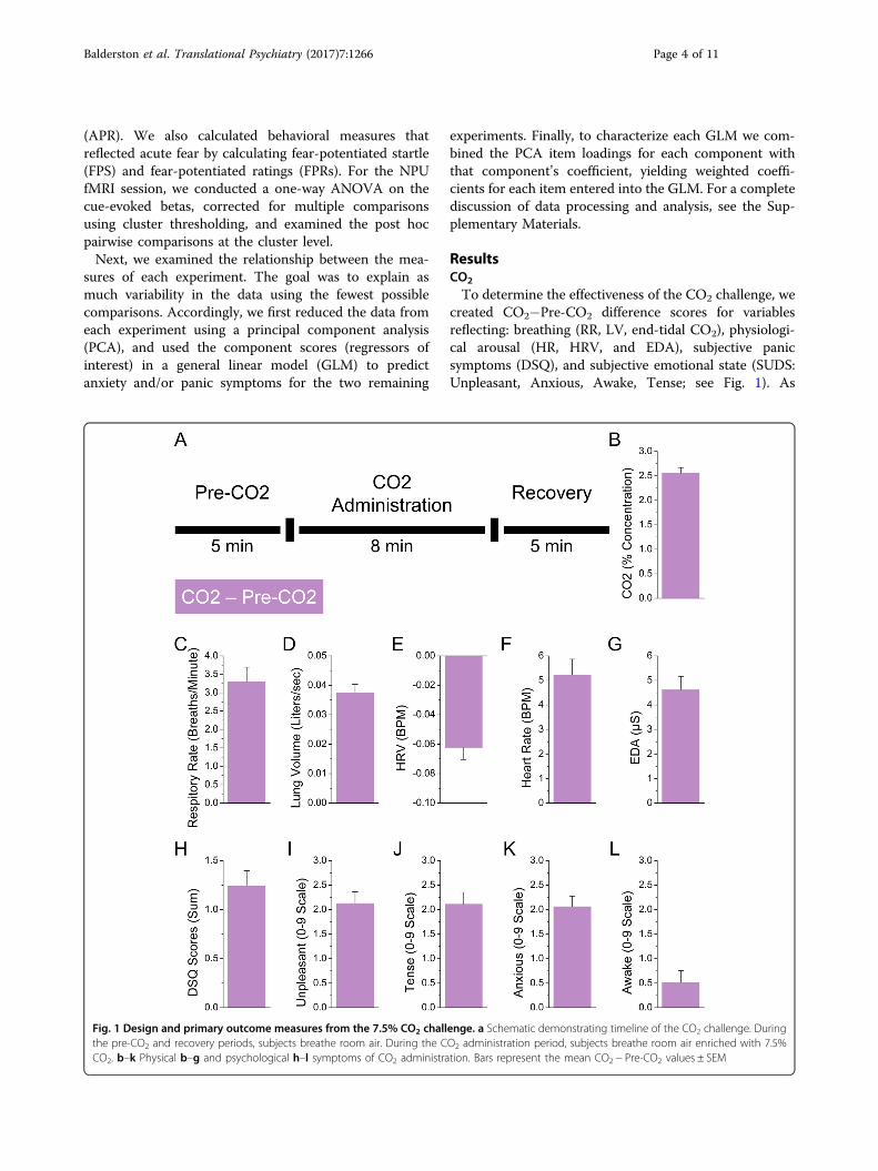
To determine the effectiveness of the CO2 challenge, wecreated CO2−Pre-CO2 difference scores for variablesreflecting: breathing (RR, LV, end-tidal CO2), physiologi-cal arousal (HR, HRV, and EDA), subjective panicsymptoms (DSQ), and subjective emotional state (SUDS:Unpleasant, Anxious, Awake, Tense; see Fig. 1). As
Fig. 1 Design and primary outcome measures from the 7.5% CO2 challenge. a Schematic demonstrating timeline of the CO2 challenge. Duringthe pre-CO2 and recovery periods, subjects breathe room air. During the CO2 administration period, subjects breathe room air enriched with 7.5%CO2. b–k Physical b–g and psychological h–l symptoms of CO2 administration. Bars represent the mean CO2 − Pre-CO2 values ± SEM
Balderston et al. Translational Psychiatry (2017) 7:1266 Page 4 of 11

expected, subjects showed increased breathing (RR: t(62)= 8.24; p< 0.001; d= 1.05, LV: t(62)= 13.57; p< 0.001;d= 1.72, CO2: t(62)= 22.41; p< 0.001; d= 2.85),increased arousal (HR: t(62)= 8.04; p< 0.001; d= 1.02,HRV: t(62)=−7.75; p< 0.001; d=−0.98, EDA: t(62)=8.46; p< 0.001; d= 1.07), elevated symptoms of panic(DSQ: t(62)= 8.01; p< 0.001; d= 1.02), and an increasein negative emotional state (Unpleasant: t(62)= 9.04; p<0.001; d= 1.15, Anxious: t(62)= 9.6; p< 0.001; d= 1.22,Awake: t(62)= 2.08; p= 0.042; d= 0.26, Tense: t(62)=8.88; p< 0.001; d= 1.13).
NPU laboratoryTo determine the effectiveness of the laboratory session
of the NPU threat task, we analyzed the online ratings andstartle magnitudes for the N, P, and U blocks using 3(Block: N, P, U)× 2 (Interval: Cue vs. ITI) repeated-measures ANOVA (See Fig. 2). For both ratings andstartle, we found a significant main effect for both block(Ratings: F(2, 124)= 97.99; p< 0.001, Startle: F(2, 124)=73.46; p< 0.001) and interval (Ratings: F(1, 62)= 54.14;p< 0.001, Startle: F(1, 62)= 95.58; p< 0.001), as well as a
significant Block × Interval Interaction (Ratings:F(2, 124)= 48.25; p< 0.001, Startle: F(2, 124)= 41.81; p<0.001).To characterize the interaction, we quantified fear and
anxiety from the ratings and startle measures. For fear, wesubtracted the rating (i.e., FPR) and startle (i.e., FPS)magnitude during the predictable ITI period from thepredictable cue period. For anxiety, we subtracted therating (i.e., APR) and startle (i.e., APS) magnitude duringthe unpredictable blocks from the neutral blocks duringboth the cue and ITI. As expected, both ratings and startleincreased during the predictable cue compared to thepredictable ITI, indicating an acute increase in fearbrought on by the predictable cue (FPR: t(62)= 7.36;p< 0.001; d= 0.93, FPS: t(62)= 10.3; p< 0.001; d= 1.3).Similarly, both ratings and startle increased during theunpredictable cue (APR-CUE: t(62)= 11; p< 0.001,d= 1.39; APS-CUE: t(62)= 7.78; p< 0.001; d= 0.98) andITI periods (APR-ITI: t(62)= 11.1; p< 0.001; d= 1.4,APS-ITI: t(62)= 8.37; p< 0.001; d= 1.05), indicating asustained increase in anxiety that was present for theentire unpredictable blocks.
Fig. 2 Design and behavioral outcome measures from the NPU sessions. a Schematic demonstrating design of the NPU paradigm. Colored linesindicate blocks of neutral, predictable, and unpredictable threat. Geometric shapes indicate visual cues presented during blocks. Lightning boltsrepresent timing of shock delivery. b Average anxiety ratings during the fMRI session for the neutral (N), predictable (P), and unpredictable (U) blockswhen the cue was present (Cue) or absent (ITI) from the screen. c Startle magnitude (t-scores) during the laboratory session for the neutral (N),predictable (P), and unpredictable (U) blocks when the cue was present (Cue) or absent (ITI) from the screen. d Average anxiety ratings during thelaboratory session for the neutral (N), predictable (P), and unpredictable (U) blocks when the cue was present (Cue) or absent (ITI) from the screen.Bars represent the mean ± SEM
Balderston et al. Translational Psychiatry (2017) 7:1266 Page 5 of 11

NPU fMRI sessionFor the fMRI data, we performed one-way (N, P, U)
repeated-measures voxelwise ANOVA on the cue-evokedactivity, and extracted the clusters that survived correc-tion for multiple comparisons (see SupplementaryTable 1). We then grouped these clusters into twoco-activation networks based on their pattern of activityacross conditions (see Fig. 3).Regions in the first co-activation network (FN),
including the dmPFC and bilateral insula, showed a pat-tern of activity consistent with fear only (i.e., P ≠ N andU). Specifically, these regions showed significantly moreactivity to the predictable than the unpredictable cue(P>U: t(62)= 7.99; p< 0.001; d= 1) or neutral cue(P>N: t(62)= 11.75; p< 0.001; d= 1.45).Regions in the second co-activation network (DMN),
including the ventromedial prefrontal cortex and
posterior cingulate cortex, showed a pattern of activityconsistent with both fear and anxiety (i.e., P and U ≠ N).However, unlike the FN, there was significantly lessactivity to the predictable cue (P>N: t(62)=−7.89;p< 0.001; d=−0.97) and the unpredictable cue (U>N: t(62)=−4.96; p< 0.001; d=−0.62) compared to theneutral cue. Although not reported, the pattern of resultsfor each cluster matches that of the corresponding co-activation network.The final cluster (Right dlPFC) showed a pattern of
activity consistent with both fear and anxiety (i.e., P and U≠ N), but unlike the regions in the DMN this clusterexhibited significantly more activity to the predictable cue(P>N: t(62)= 7.28; p< 0.001; d= 0.91) and unpredict-able cue (U >N: t(62)= 4.38; p< 0.001; d= 0.55) com-pared to the neutral cue.We did not observe any significant effects in the BNST.
Fig. 3 Main effects from the analysis of the BOLD data from the NPU fMRI session. a Statistical parametric map and corresponding graphicrepresentation for regions of the fear network (FN). b Statistical parametric map and corresponding graphic representation for regions of the DMN. cStatistical parametric map and corresponding graphic representation for regions of the right dorsolateral prefrontal cortex (dlPFC). Bars represent themean ± SEM
Balderston et al. Translational Psychiatry (2017) 7:1266 Page 6 of 11

PCATo identify regressors of interest, we conducted inde-
pendent PCAs including all variables from a givenexperiment. For the CO2 experiment, we entered 26variables into the PCA, and identified six componentswith an eigenvalue >1 (see Fig. 4a). These six componentsexplained 69.09% of the variability. For the NPU labora-tory session, we entered 12 variables into the PCA, andidentified three components with an eigenvalue >1 (seeSupplementary Fig. 2). These three components explained69.77% of the variability. For the NPU fMRI session, weentered 33 variables into the PCA, and identified 7components with an eigenvalue >1 (see SupplementaryFig. 2). These seven components explained 79.25% of thevariability.
GLMsTo determine whether the primary outcome measures
of each experiment were affected by individual differencesin responding in the other experiments, we conducted aseries of GLMs (One per measure, see Supplementary Fig.3) using the subject scores for the signal components fromone experiment to predict the outcome measures fromanother experiment. This was done in a systematic fash-ion, whereby the signal components from each experi-ment were used to predict each outcome measure for theremaining experiments. The subsequent r2 values areplotted in Supplementary Fig. 3. Although several of theseGLMs were significant at the 0.05 level (see hatched barsin Supplementary Fig. 3), only one GLM (CO2 challengecomponents → Anxiety-related dlPFC activity) was
Fig. 4 Statistical relationship between measures from the CO2 challenge and NPU sessions. a Screenplot demonstrating outcome of theprincipal components analyses for the CO2 challenge. Components with an eigenvalue >1 are considered signal components, while those with aneigenvalue <1 are considered noise components. b Variability (r2) in dependent measures from the NPU sessions accounted for by the signalcomponents in the CO2 challenge. Filled bars are significant after correcting for multiple comparisons. Hatched bars are trends, but not significantafter correcting for multiple comparisons. c Weighted beta coefficients showing contributions of specific items from the CO2 challenge to the PCA/regression model predicting anxiety-related dlPFC activity. d Correlation between anxiety-related dlPFC activity and anxiety (as derived via PCA fromNPU startle and ratings during fMRI and laboratory sessions)
Balderston et al. Translational Psychiatry (2017) 7:1266 Page 7 of 11

significant after correcting for multiple comparisons (seeshaded bar in Fig. 4b).
Weighted beta coefficientsTo characterize the relationship between predictor
variables from the CO2 challenge and anxiety-relateddlPFC activity, we computed weighted coefficients for thisGLM (see Fig. 4c). CO2 challenge items related to fear ornegative affect tended to load negatively on anxiety-related dlPFC activity (f(6, 56)= 4.02; p= 0.002; FDR=0.016; r2= 0.3), suggesting that individuals with highanxiety-related dlPFC activity report less fear during theCO2 challenge. A similar but less robust pattern wasobserved for fear-related dlPFC activity (f(6, 56)= 2.35; p= 0.043; FDR= 0.17; r2= 0.2; see Supplementary Fig. 4).
Anxiety/dlPFC correlationsBecause individuals who exhibited larger anxiety-related
dlPFC responses during the NPU fMRI session reportedless anxiety during the CO2 challenge, we hypothesizedthat these dlPFC responses may regulate anxiety moregenerally. Thus, we correlated anxiety ratings from theunpredictable condition during both sessions and startlefrom the NPU Laboratory session with dlPFC responsesfrom the NPU fMRI session. For all measures we calcu-lated Unpredictable >Neutral difference scores. Becausethe dlPFC responses were cue-evoked, we includedseparate scores for the cue and ITI periods for the anxietyratings and startle measures. Importantly, across mea-sures (Ratings vs. Startle), studies (fMRI session vs.Laboratory session), and intervals (Cue vs. ITI), the cor-relations with anxiety-related dlPFC responses werenegative (see Supplementary Table 2). Although many ofthese correlations were only trends, the pattern is con-sistent across measures, and these measures themselveswere likely correlated. Therefore, we combined the mea-sures using a PCA and extracted a single value componentto anxiety (eigenvalue= 3.6). As expected, the anxietycomponent was significantly negatively correlated withanxiety-related dlPFC activity (r(62)=−0.29; p= 0.023;see Fig. 4d). When applying a similar approach to fear andfear-related dlPFC activity, no significant correlationswere found (all p values> 0.05).
DiscussionThe purpose of this study was to determine the degree to
which panic susceptibility is related to (1) fear and anxietyduring the NPU threat task and (2) reactivity in the neuralsystems that mediate fear and anxiety. Accordingly, weexposed individuals to a 7.5% CO2 challenge and twoversions (laboratory and fMRI) of the NPU threat task.Contrary to our first hypothesis, we did not find evidencethat panic symptoms were mediated by the same coreneural system as anxiety. However, we did find evidence
that panic symptoms to CO2 may be regulated by the sameneural system that regulates anxiety to unpredictablethreat. When comparing across experimental sessions, wefound that CO2-related behavioral changes were asso-ciated only with dlPFC activity during the unpredictablecue (see Fig. 4b). The activity of the dlPFC is commonlyconsidered to mediate cognitive control51,52, and ourresults suggest that the dlPFC regulates anxiety in theseparadigms48. Weighted coefficients from our analysisshowed that fear-related DSQ symptom items negativelyloaded onto dlPFC activity (see Fig. 4c). Similarly,anxiety-related dlPFC was negatively correlated withanxiety during the NPU sessions (see Fig. 4d). Together,these results suggest that panic symptoms and anxiety areregulated by the same prefrontal cognitive controlsystem48.In addition to this novel finding, we replicate many
previous reports about the CO2 challenge and the NPUthreat task. During the CO2 challenge, subjects showedincreased respiration, physiological arousal, and self-reported anxiety (see Fig. 1). During the laboratory NPUsession, subjects showed the traditional pattern of ele-vated startle and self-reported anxiety during the pre-dictable cue (fear), and unpredictable cue and ITI periods(anxiety)8,15,53–55 (see Fig. 2). During the NPU fMRI ses-sion, subjects showed distinct patterns of BOLD activityrelated to fear and anxiety22,56–61 (see Fig. 3). During bothpredictable and unpredictable cues, subjects showeddecreased DMN activity (see Fig. 3b), and increased dlPFCactivity (see Fig. 3c). However, only the predictable cueincreased FN activity (see Fig. 3a). This FN result isconsistent with the fact that only the predictable cueinformed the probability of shock15.Previous research has shown that panic attacks are not
necessarily mediated by the same mechanisms as fear14,62.However, there is evidence that history of panic attacks isrelated to startle magnitude during unpredictablethreat10–12. There are two possible explanations for thisobservation. First, panic susceptibility may be driven byelevated responding in anxiety-related neural sys-tems22,56–61. Alternatively, panic susceptibility may bedriven by attenuated responding in anxiety-regulatingneural systems63–65. We found that panic symptoms canpredict dlPFC activity, which is negatively associated withanxiety, which supports the second hypothesis, and sug-gests that the dlPFC regulates panic symptoms andanxiety. Consistent with this, anxiety patients show dlPFCdeficits during complex working memory (WM) tasks27.In contrast, we found no evidence that panic symptomsare associated with anxiety-related regions, which failsto support the expression hypothesis (see Fig. 4b).Thus, more work is needed to understand the distinctneural systems mediating the expression of anxiety andpanic.
Balderston et al. Translational Psychiatry (2017) 7:1266 Page 8 of 11

These findings can explain the previously counter-intuitive finding that CO2 administration actually reducesstartle magnitude66–68. According to our results, onecould argue that the dlPFC is a common regulatory sys-tem engaged by 7.5% CO2 and by unpredictable threatbecause these challenges evoke a similar defensiveresponse. Thus, evoking either defensive response shouldlead to the regulation of both responses. According to thishypothesis, startle is reduced during CO2 administrationbecause 7.5% CO2 administration engages the dlPFC,which regulates the ongoing activity in the neural systemengaged by unpredictable threat. Therefore, it should alsobe possible to reduce CO2-related panic symptoms usingsustained unpredictable threat, as a proof of concept. Thishypothesis is testable within the context of the rapid 35%CO2 challenge. In this paradigm, subjects inhale a singlebreath of air with 35% CO2, which results in panic attacksin susceptible individuals69–71. Therefore, using unpre-dictable threat to engage the system that mediates anxietyshould reduce the likelihood of experiencing a panicattack in this paradigm.It may also be possible to test the regulation hypothesis
using other techniques to drive dlPFC activity72–82. Wehave shown previously that WM, known to activate thedlPFC, is sufficient to reduce APS during threat83,84. Weknow of no studies examining the effect of WM on panicsymptoms. Future studies might accomplish this byadministering single breaths of 35% CO2 during periods oflow vs. high WM load69–71. According to the regulationhypothesis, subjects should experience fewer panic attacksduring high WM blocks compared to low WM blocks.The N-back WM task would be ideal because it provideslong durations of steady-state WM engagement, whereWM load can be parametrically manipulated withinsubjects79–82. It may also be possible to test the regulationhypothesis using noninvasive neuromodulation. Forinstance, one could administer single breaths of 35% CO2
while the subject receives electrical (transcranial directcurrent stimulation; tDCS72–74) or magnetic (transcranialmagnetic stimulation; TMS75–78) to the right dlPFC.According to the regulatory hypothesis, subjects shouldexperience fewer panic attacks during excitatory (i.e.,anodal tDCS or high-frequency TMS) compared to inhi-bitory (i.e., cathodal tDCS or low-frequency TMS) orsham stimulation of the dlPFC.
Strengths and limitationsThere were a number of strengths and limitations with
current work that should be noted. Among the primarystrengths, we used well-validated, experimental, transla-tional techniques for inducing symptoms of fear (pre-dictable shock threat), anxiety (unpredictable shockthreat), and panic (7.5% CO2 inhalation)
4,15, and includedan adequate sample size (n= 63) to test the hypotheses.
Among the primary limitations, we did not directly assayneural activity during the CO2 challenge, and we did notreport the findings related to sustained anxiety from theunpredictable blocks in the fMRI study. For a full dis-cussion of these strengths and limitations, see the Sup-plementary Materials.
ConclusionsThe purpose of this study was to determine the degree to
which panic symptoms correlated with fear, anxiety, andthe neural systems that mediate/regulate fear and anxiety.We found evidence that panic symptoms and anxietymay be regulated by similar prefrontal cognitive controlmechanisms. Accordingly, these results raise severaltestable hypotheses about the effect of cognitive controlon panic symptoms, and warrant future studies on thistopic.
AcknowledgmentsFinancial support of this study was provided by the Intramural ResearchProgram of the National Institute of Mental Health, ZIAMH002798 (ClinicalTrial.gov Identifier: NCT00047853: Protocol ID 02-M-0321).
Competing interestsThe authors declare that they have no competing financial interests.
Publisher's note: Springer Nature remains neutral with regard to jurisdictionalclaims in published maps and institutional affiliations.
Supplementary informationThe online version of this article (https://doi.org/10.1038/s41398-017-0006-5)contains supplementary Material.
Received: 26 February 2017 Revised: 11 May 2017 Accepted: 15 May 2017
References1. American Psychiatric Association. Diagnostic and Statistical Manual of Mental
Disorders: Dsm-5 (Amer Psychiatric Pub Incorporated, Arlington, VA, 2013).2. Battaglia, M., Ogliari, A., D’Amato, F. & Kinkead, R. Early-life risk factors for panic
and separation anxiety disorder: insights and outstanding questions arisingfrom human and animal studies of CO2 sensitivity. Neurosci. Biobehav. Rev. 46,455–464 (2014).
3. Bailey, J. E., Kendrick, A., Diaper, A., Potokar, J. P. & Nutt, D. J. A validation of the7.5% CO2 model of GAD using paroxetine and lorazepam in healthy volun-teers. J. Psychopharmacol. 21, 42–49 (2007).
4. Bailey, J. E., Dawson, G. R., Dourish, C. T. & Nutt, D. J. Validating the inhalation of7.5% CO2 in healthy volunteers as a human experimental medicine: a modelof generalized anxiety disorder (GAD). J. Psychopharmacol. 25, 1192–1198(2011).
5. Seddon, K. et al. Effects of 7.5% CO2 challenge in generalized anxiety disorder.J. Psychopharmacol. 25, 43–51 (2011).
6. Bailey, J. E., Argyropoulos, S. V., Kendrick, A. H. & Nutt, D. J. Behavioral andcardiovascular effects of 7.5% CO2 in human volunteers. Depress. Anxiety 21,18–25 (2005).
7. Bystritsky, A., Craske, M., Maidenberg, E., Vapnik, T. & Shapiro, D. Autonomicreactivity of panic patients during a CO2 inhalation procedure. Depress. Anxiety11, 15–26 (2000).
8. Grillon, C. Models and mechanisms of anxiety: evidence from startle studies.Psychopharmacology 199, 421–437 (2008).
9. Davis, M., Walker, D. L., Miles, L. & Grillon, C. Phasic vs sustained fear in rats andhumans: role of the extended amygdala in fear vs anxiety. Neuropsycho-pharmacology 35, 105–135 (2010).
Balderston et al. Translational Psychiatry (2017) 7:1266 Page 9 of 11

10. Grillon, C. et al. Increased anxiety during anticipation of unpredictable but notpredictable aversive stimuli as a psychophysiologic marker of panic disorder.Am. J. Psychiatry 165, 898–904 (2008).
11. Grillon, C., Ameli, R., Goddard, A., Woods, S. W. & Davis, M. Baseline and fear-potentiated startle in panic disorder patients. Biol. Psychiatry 35, 431–439(1994).
12. Grillon C. et al. Distinct responses to predictable and unpredictable threat inanxiety pathologies: effect of panic attack. Biol. Psychiatry Cogn. Neurosci.Neuroimaging 1–7 (2016).Grillon C, O’Connell K, Lieberman L, Alvarez G, GeraciM, Pine DS et al. Distinct Responses to Predictable and Unpredictable Threat inAnxiety Pathologies: Effect of Panic Attack. Biol Psychiatry Cogn NeurosciNeuroimaging 2, 575-581 (2016).
13. Khalsa, S. S. et al. Panic anxiety in humans with bilateral amygdala lesions:pharmacological induction via cardiorespiratory interoceptive pathways. J.Neurosci. 36, 3559–3566 (2016).
14. Feinstein, J. S. et al. Fear and panic in humans with bilateral amygdaladamage. Nat. Neurosci. 16, 270–272 (2013).
15. Schmitz, A. & Grillon, C. Assessing fear and anxiety in humans using the threatof predictable and unpredictable aversive events (the NPU-threat test). Nat.Protoc. 7, 527–532 (2012).
16. Insel, T. et al. Research Domain Criteria (RDoC): toward a new classificationframework for research on mental disorders. Am. J. Psychiatry 167, 748–751(2010).
17. Fullana, M. A. et al. Neural signatures of human fear conditioning: an updatedand extended meta-analysis of fMRI studies. Mol. Psychiatry 21, 500–508(2015).
18. Vytal, K. E., Overstreet, C., Charney, D. R., Robinson, O. & Grillon, C. Sustainedanxiety increases amygdala-dorsomedial prefrontal coupling: a mechanism formaintaining an anxious state in healthy adults. J. Psychiatry Neurosci. 39, 1–9(2014).
19. Hasler, G. et al. Cerebral blood flow in immediate and sustained anxiety. J.Neurosci. 27, 6313–6319 (2007).
20. Etkin, A. & Wager, T. D. Functional neuroimaging of anxiety: a meta-analysis ofemotional processing in PTSD, social anxiety disorder, and specific phobia.Am. J. Psychiatry 164, 1476–1488 (2007).
21. Baur, V., Hänggi, J., Langer, N. & Jäncke, L. Resting-state functional and struc-tural connectivity within an insula-amygdala route specifically index state andtrait anxiety. Biol. Psychiatry 73, 85–92 (2013).
22. Andreatta, M. et al. Initial and sustained brain responses to contextual con-ditioned anxiety in humans. Cortex 63, 352–363 (2015).
23. Robinson, O., Charney, D. R., Overstreet, C., Vytal, K. E. & Grillon, C. The adaptivethreat bias in anxiety: amygdala-dorsomedial prefrontal cortex coupling andaversive amplification. Neuroimage 60, 523–529 (2012).
24. Wheelock, M. D. et al. Threat-related learning relies on distinct dorsal prefrontalcortex network connectivity. Neuroimage 102, 904–912 (2014).
25. Tao, Y. et al. The structural connectivity pattern of the default mode networkand its association with memory and anxiety. Front. Neuroanat. 9, 152 (2015).
26. Broyd, S. J. et al. Default-mode brain dysfunction in mental disorders: a sys-tematic review. Neurosci. Biobehav. Rev. 33, 279–296 (2009).
27. Balderston, N. L. et al. Anxiety patients show reduced working memory relatedDlpfc activation during safety and threat. Depress. Anxiety 12, 1–12 (2016).
28. Tomasi, D. & Volkow, N. D. Functional connectivity hubs in the human brain.Neuroimage 57, 908–917 (2011).
29. Reinholdt-Dunne, M. L., Mogg, K. & Bradley, B. P. Attention control: relation-ships between self-report and behavioural measures, and symptoms ofanxiety and depression. Cogn. Emot. 9931, 1–11 (2012).
30. Reinholdt-Dunne, M. L., Mogg, K. & Bradley, B. P. Effects of anxiety andattention control on processing pictorial and linguistic emotional information.Behav. Res. Ther. 47, 410–417 (2009).
31. Grillon, C., Robinson, O., Mathur, A. & Ernst, M. Effect of attention control onsustained attention during induced anxiety. Cogn. Emot. 9931, 1–13 (2015).
32. Braver, T. S., Cole, M. W. & Yarkoni, T. Vive les differences! Individual variation inneural mechanisms of executive control. Curr. Opin. Neurobiol. 20, 242–250(2010).
33. Derryberry, D. & Reed, Ma Anxiety-related attentional biases and their reg-ulation by attentional control. J. Abnorm. Psychol. 111, 225–236 (2002).
34. Eysenck, M. W., Derakshan, N., Santos, R. & Calvo, M. G. Anxiety and cognitiveperformance: attentional control theory. Emotion 7, 336–353 (2007).
35. Berggren, N. & Derakshan, N. Attentional control deficits in trait anxiety: whyyou see them and why you don’t. Biol. Psychol. 92, 440–446 (2013).
36. Berggren, N. & Derakshan, N. The role of consciousness in attentional controldifferences in trait anxiety. Cogn. Emot. 27, 923–931 (2013).
37. Coombes, S. A., Higgins, T., Gamble, K. M., Cauraugh, J. H. & Janelle, C. M.Attentional control theory: anxiety, emotion, and motor planning. J. AnxietyDisord. 23, 1072–1079 (2009).
38. Armstrong, T., Zald, D. H. & Olatunji, B. O. Attentional control in OCD and GAD:specificity and associations with core cognitive symptoms. Behav. Res. Ther. 49,756–762 (2011).
39. Price, R. B., Eldreth, Da & Mohlman, J. Deficient prefrontal attentional control inlate-life generalized anxiety disorder: an fMRI investigation. Transl. Psychiatry 1,e46 (2011).
40. Najmi, S., Amir, N., Frosio, K. E. & Ayers, C. The effects of cognitive load onattention control in subclinical anxiety and generalised anxiety disorder. Cogn.Emot. 9931, 1–14 (2014).
41. Najmi, S., Kuckertz, J. M. & Amir, N. Attentional impairment in anxiety: ineffi-ciency in expanding the scope of attention. Depress. Anxiety 29, 243–249(2012).
42. Morrison, A. S. & Heimberg, R. G. Attentional control mediates the effect ofsocial anxiety on positive affect. J. Anxiety Disord. 27, 56–67 (2013).
43. Cieslik, E. C. et al. Is there one DLPFC in cognitive action control? Evidence forheterogeneity from Co-activation-based parcellation. Cereb. Cortex 23,2677–2689 (2013).
44. Basten, U., Stelzel, C. & Fiebach, C. J. Trait anxiety and the neural efficiency ofmanipulation in working memory. Cogn. Affect. Behav. Neurosci. 12, 571–588(2012).
45. Harding, I. H., Yücel, M., Harrison, B. J., Pantelis, C. & Breakspear, M. Effectiveconnectivity within the frontoparietal control network differentiates cognitivecontrol and working memory. Neuroimage 106, 144–153 (2015).
46. Fales, C. L. et al. Anxiety and cognitive efficiency: differential modulation oftransient and sustained neural activity during a working memory task. Cogn.Affect. Behav. Neurosci. 8, 239–253 (2008).
47. Peers, P. V., Simons, J. S. & Lawrence, A. D. Prefrontal control of attention tothreat. Front. Hum. Neurosci. 7, 24 (2013).
48. Cole, M. W., Repovš, G. & Anticevic, A. The frontoparietal control system: acentral role in mental health. Neuroscientist 20, 652–664 (2014).
49. Sylvester, C. M. et al. Functional network dysfunction in anxiety and anxietydisorders. Trends Neurosci. 35, 527–535 (2012).
50. Wolpe J. The practice of behavior therapy, 2nd ed. Pergamon: New York, NY,1973.
51. Levy, B. J. & Anderson, M. C. Purging of memories from conscious awarenesstracked in the human brain. J. Neurosci. 32, 16785–16794 (2012).
52. Koechlin, E., Ody, C. & Kouneiher, F. The architecture of cognitive control in thehuman prefrontal cortex. Science 302, 1181–1185 (2003).
53. Schmitz, A. et al. Measuring anxious responses to predictable and unpre-dictable threat in children and adolescents. J. Exp. Child Psychol. 110, 159–170(2011).
54. Nelson, B. D., Hajcak, G. & Shankman, S. A. Event-related potentials to acousticstartle probes during the anticipation of predictable and unpredictable threat.Psychophysiology 52, 887–894 (2015).
55. Kaye, J. T., Bradford, D. E. & Curtin, J. J. Psychometric properties of startle andcorrugator response in NPU, affective picture viewing, and resting state tasks.Psychophysiology 53, 1241–1255 (2016).
56. Robinson, O., Krimsky, M. & Grillon, C. The impact of induced anxiety onresponse inhibition. Front. Hum. Neurosci. 7, 69 (2013).
57. Robinson, O. et al. Anxiety-potentiated amygdala–medial frontal coupling andattentional control. Transl. Psychiatry 6, e833 (2016).
58. Alvarez, R. P., Chen, G., Bodurka, J., Kaplan, R. & Grillon, C. Phasic and sustainedfear in humans elicits distinct patterns of brain activity. Neuroimage 55,389–400 (2011).
59. Cornwell, B. R., Echiverri, A. M., Covington, M. F. & Grillon, C. Modality-specificattention under imminent but not remote threat of shock. Psychol. Sci. 19,615–622 (2008).
60. Gold, A. L., Morey, Ra & McCarthy, G. Amygdala-prefrontal cortex functionalconnectivity during threat-induced anxiety and goal distraction. Biol. Psychiatry77, 1–9 (2014).
61. Torrisi, S. et al. The neural basis of improved cognitive performance by threatof shock. Soc. Cogn. Affect. Neurosci. 11, 1677–1686 (2016).
62. Wiest, G., Lehner-Baumgartner, E. & Baumgartner, C. Panic attacks in an indi-vidual with bilateral selective lesions of the amygdala. Arch. Neurol. 63,1798–1801 (2006).
Balderston et al. Translational Psychiatry (2017) 7:1266 Page 10 of 11

63. Forster, S., Nunez Elizalde, A. O., Castle, E., Bishop, S. J. & Elizalde, A. O. N.Unraveling the Anxious Mind: Anxiety, Worry, and Frontal Engagement inSustained Attention Versus Off-Task Processing. Cereb. Cortex 25, 609–618(2015).
64. Ironside, M., O’Shea, J., Cowen, P. J. & Harmer, C. J. Frontal cortex stimulationreduces vigilance to threat: implications for the treatment of depression andanxiety. Biol. Psychiatry 79, 823–830 (2016).
65. Shackman, A. J. et al. Right dorsolateral prefrontal cortical activity and beha-vioral inhibition. Psychol. Sci. 20, 1500–1506 (2014).
66. Pappens, M., De Peuter, S., Vansteenwegen, D., Van den Bergh, O. & Van Diest,I. Psychophysiological responses to CO2 inhalation. Int. J. Psychophysiol. 84,45–50 (2012).
67. Ceunen, E., Vlaeyen, J. W. S. & Van Diest, I. Atypical modulation of startle inwomen in face of aversive bodily sensations. Int. J. Psychophysiol. 88, 157–163(2013).
68. Pinkney, V., Wickens, R., Bamford, S., Baldwin, D. S. & Garner, M. Defensive eye-blink startle responses in a human experimental model of anxiety. J. Psycho-pharmacol. 28, 874–880 (2014).
69. Nardi, A. E. et al. Psychopathological profile of 35% CO2 challenge test-induced panic attacks: a comparison with spontaneous panic attacks. Compr.Psychiatry 47, 209–214 (2006).
70. Wetherell, M. A. et al. The four-dimensional stress test: psychological, sympa-thetic-adrenal-medullary, parasympathetic and hypothalamic-pituitary-adrenalresponses following inhalation of 35% CO2. Psychoneuroendocrinology 31,736–747 (2006).
71. Battaglia, M. & Perna, G. The 35% CO2 challenge in panic disorder: optimi-zation by receiver operating characteristic (ROC) analysis. J. Psychiatr. Res. 29,111–119 (1995).
72. Balconi, M. & Vitaloni, S. The tDCS effect on alpha brain oscillation for correctvs. incorrect object use. The contribution of the left DLPFC. Neurosci. Lett. 517,25–29 (2012).
73. Fregni, F. et al. Anodal transcranial direct current stimulation of prefrontalcortex enhances working memory. Exp. Brain Res. 166, 23–30 (2005).
74. Brunoni, A. R. & Vanderhasselt, M. A. Working memory improvement withnon-invasive brain stimulation of the dorsolateral prefrontal cortex: a sys-tematic review and meta-analysis. Brain Cogn. 86, 1–9 (2014).
75. Rogasch, N. C., Daskalakis, Z. J. & Fitzgerald, P. B. Cortical inhibition of distinctmechanisms in the dorsolateral prefrontal cortex is related to workingmemory performance: a TMS-EEG study. Cortex 64, 68–77 (2015).
76. Gratton, C., Lee, T. G., Nomura, E. M. & D’Esposito, M. The effect of theta-burstTMS on cognitive control networks measured with resting state fMRI. Front.Syst. Neurosci. 7, 124 (2013).
77. Schicktanz, N. et al. Continuous theta burst stimulation over the left dorso-lateral prefrontal cortex decreases medium load working memory perfor-mance in healthy humans. PLoS ONE 10, 1–10 (2015).
78. Shajahan, P. M. et al. Left dorso-lateral repetitive transcranial magnetic sti-mulation affects cortical excitability and functional connectivity, but does notimpair cognition in major depression. Prog. Neuro-Psychopharmacol. Biol.Psychiatry 26, 945–954 (2002).
79. Carlson, S. et al. Distribution of cortical activation during visuospatial n-backtasks as revealed by functional magnetic resonance imaging. Cereb. Cortex 8,743–752 (1998).
80. Owen, A. M., McMillan, K. M., Laird, A. R. & Bullmore, E. N-back workingmemory paradigm: a meta-analysis of normative functional neuroimagingstudies. Hum. Brain Mapp. 25, 46–59 (2005).
81. León-Domínguez, U., Martín-Rodríguez, J. F. & León-Carrión, J. Executive n-back tasks for the neuropsychological assessment of working memory. Behav.Brain Res. 292, 167–173 (2015).
82. Jaeggi, S. M., Buschkuehl, M., Perrig, W. J. & Meier, B. The concurrent validityof the N-back task as a working memory measure. Memory 18, 394–412(2010).
83. Vytal, K. E., Cornwell, B. R., Letkiewicz, A. M., Arkin, N. E. & Grillon, C. The complexinteraction between anxiety and cognition: insight from spatial and verbalworking memory. Front. Hum. Neurosci. 7, 93 (2013).
84. Balderston, N. L. et al. Working memory maintenance is sufficient to reducestate anxiety. Psychophysiology 53, 1660–1668 (2016).
Balderston et al. Translational Psychiatry (2017) 7:1266 Page 11 of 11