the relationship between dietary calcium intake, alone or
TRANSCRIPT

Food Standards Australia New Zealand
Diet-Disease Relationship Review
The Relationship Between Dietary Calcium Intake, Alone or in Association With Vitamin D Status, and Risk
of Developing Osteoporosis
Prof Ian R Reid MD, FRSNZ
Department of Medicine, University of Auckland, New Zealand
Correspondence to: Prof IR Reid
Department of Medicine University of Auckland
Private Bag 92019 Auckland
New Zealand
Tel: (+64 9) 3737 599 extn 86259 Fax: (+64 9) 3677 146
email: [email protected]

2
TABLE OF CONTENTS
INTRODUCTION 4
Calcium and Bone Biology 4
Vitamin D Metabolism 5
Exercise 6
Definitions of Osteoporosis 6
PART 1: CRITICAL APPRAISAL OF THE CANADIAN REVIEW OF THE CALCIUM-OSTEOPOROSIS RELATIONSHIP 8
General Comments 8
Responses to Questions in Template 11
Re-Analysis of Pivotal Studies Cited in the Review 12
Consideration of the Validity of the Canadian Review’s Conclusions 17
PART 2: REVIEW OF EVIDENCE RELEASED SINCE THE CANADIAN REVIEW 19
Randomised Controlled Trials 19 Children and Adolescents 19 Adults 20 Pending Studies 22
Observational Studies 22
Systematic Reviews 23
PART 3: RELEVANCE OF THE RELATIONSHIP TO AUSTRALIA AND NEW ZEALAND 26
Calcium Intakes and Vitamin D Status in Australia and New Zealand 26
Australians and New Zealanders of Asian Origin 27
Studies of Calcium Supplementation in Australia or New Zealand 27
PART 4: RELATIONSHIP OF DIETARY CALCIUM INTAKE TO BIOMARKERS OF OSTEOPOROSIS 29
Bone Mineral Density 29
Biochemical Markers of Bone Turnover 29
Other Biomarkers 30

3
Calcium Balance Studies 31
Biomarkers Conclusion 31
SUMMARY 32
Osteoporosis and Bone Biology 32
Assessment of the Canadian Review 33
Relevance to Australia and New Zealand 33
Evidence Published Since 2000 34
Conclusions 34
REFERENCES 36
APPENDICES 44
Appendix 1: Strategy Used in Medline Search 44
Appendix 2: Strategy Used in Weekly Current Contents Searches 60

4
INTRODUCTION In the last 20 years, there have been huge advances in our understanding of the biology of bone, the metabolism of vitamin D, and the pathophysiology of osteoporosis. A brief over-view of this material provides a biological context for the interpretation of the epidemiological and clinical studies that will follow.
Calcium and Bone Biology Bone is a connective tissue consisting of a protein matrix embedded in a mineral phase, which is made up of calcium and phosphate. During growth and renewal of bone, the bone-forming cells (osteoblasts) lay down the protein scaffold of bone, which is predominantly type 1 collagen, but also includes other proteins such as osteocalcin. Secondarily, calcium and phosphate precipitate between the fibres of this matrix, forming hydroxyapatite crystals. The amount of bone formed is determined by the amount of protein laid down by the osteoblasts. In the presence of normal circulating levels of calcium and phosphate, almost all the bone formed will become mineralised. The reshaping of bone during growth and the subsequent renewal of bone, involve the removal of packets of bone by resorbing cells, known as osteoclasts. These cells secrete acid to dissolve the mineral, and proteolytic enzymes to remove the matrix. When this occurs postpubertally as part of the bone remodelling sequence, it results in small areas of resorption dotted across the entire skeletal surface. Each of these packets of resorbed bone is then replaced with new bone by osteoblasts, resulting in complete skeletal renewal every 10 – 15 years. Calcium deficiency can impact on bone development by providing inadequate levels of calcium for mineralization to take place normally. This results in osteomalacia. In adults, osteomalacia results in bone pain. In children, osteomalacia is manifest by reduced skeletal growth and deformity of the bones (such as “knock knees” and “bow legs”). This symptom complex is referred to as rickets. In Western countries these days, rickets most commonly occurs as a result of congenital abnormalities of phosphate or vitamin D metabolism. It is also seen, occasionally, in children with very low calcium intakes or those that are deprived of vitamin D, often as a consequence of illness or lifestyle practices that result in marked sunlight deprivation. Osteomalacia can occur at any age in individuals who become severely vitamin D-deficient, again usually as a result of sunlight deprivation. While the development of rickets/osteomalacia is regarded as the classic skeletal manifestation of calcium/vitamin D deficiency, in recent years it has become apparent that more subtle deficiencies can exact a toll on the skeleton. The top priority in calcium homeostasis is the maintenance of the extracellular calcium concentration, since this is important to the normal functioning of excitable membranes such as those in the central nervous system, skeletal muscle and heart. Any decline in dietary calcium intake or vitamin D levels results in a reduced amount of absorbed calcium. This is compensated for by increased secretion of parathyroid hormone, which in turn increases activation of vitamin D, reduces urinary calcium loss, and increases bone resorption. This is probably the mechanism by which suboptimal calcium and vitamin D status accelerate bone loss. Restoration of optimal calcium/vitamin D status will reverse these processes, with declines in parathyroid hormone and bone resorption, increases in bone density, and hopefully, decreases in fracture risk.

5
In the past there has been a belief that the provision of increasing amounts of calcium will either promote bone growth or result in the accretion of calcium around bones, thereby augmenting their strength. There is no evidence whatsoever for calcium increasing osteoblast anabolic activity, and therefore no possibility that calcium can increase bone growth. Its therapeutic benefit is entirely as an anti-resorptive through decreasing parathyroid hormone secretion, and as a substrate for normal bone mineralisation. There are few data addressing the comparable efficacies of food calcium (e.g., dairy products) and calcium given as a mineral supplement. However, it is likely that the efficacies are comparable, based on similar therapeutic effects on bone density in trials that use mineral calcium(95) and those that use a dairy supplement containing a similar amount of calcium(62). Many opinions have been expressed over the years as to the ideal way to take calcium. No studies of hard endpoints, such as fracture or bone density, have compared divided dosing versus single dosing regimens, or regimens that differ by time of day. It has been suggested that divided dose regimens should lead to greater total calcium absorption, since the active transport of calcium takes place in the upper small bowel and is likely to be saturated by a large single dose. This is likely to be modified by the presence of food, which will slow the release of calcium from the stomach, and therefore greatly prolong the upper small bowel transit time. Another potential interaction of food with calcium, is that food acid may aid dissolution of relatively insoluble calcium salts (e.g. carbonate, phosphate). Thus, in subjects with achlorhydria, insoluble calcium salts are probably better absorbed when taken with food rather than fasting. Despite these theoretical considerations, formal studies suggest that these considerations make little difference. For instance, Scopacasa(104) compared a 1g calcium supplement given in the evening, with 500 mg calcium given twice a day. Measuring bone resorption markers over 24 hours, they found that the two regimens were comparable in the reduction in resorption produced. Karkkainen(54) compared the effects of calcium supplementation given at 9 am or 9 pm on serum calcium and parathyroid hormone concentrations, and found no difference. In the same study, the effect of a single 1 g dose of calcium was compared with that of four divided doses. Both produced comparable suppression of parathyroid hormone secretion, though no effects on turnover markers were apparent in this study.
Vitamin D Metabolism Vitamin D suffers from having been misnamed at the time of its discovery. It is in fact a pro-hormone, virtually all vitamin D being synthesised endogenously in the skin as a result of the interaction of 7-dehydrocholesterol with ultraviolet light. It is not an essential dietary constituent, and the unsupplemented diet contributes is minimally, in most parts of the world. The vitamin D synthesised in the skin is mostly converted to 25-hydroxyvitamin D in the liver, and this is the principal form in the circulation. Vitamin D can also be stored in adipose tissue. 25-hydroxyvitamin D has some biological activity, but this is increased a thousand-fold when it is further hydroxylated in the kidney to form 1,25-dihydroxyvitamin D, also known as calcitriol. This is the single most potent regulator of intestinal calcium absorption, though it can also stimulate bone resorption and may play roles in many other tissues and processes, such as hormone secretion and regulation of cell proliferation. Because the principal source of vitamin D is sunlight exposure, those most at risk of vitamin D deficiency are those who seldom venture outside or keep their skin completely covered, those with heavily pigmented skin (which acts as an impediment to the interaction of ultraviolet light with 7-dehydrocholesterol),

6
and the elderly. The amount of vitamin D produced by older individuals in response to a fixed sunlight exposure, can be reduced by 50-70% in comparison with young adults(46,69). Living at higher latitudes or in areas where there is air pollution will also reduce cutaneous vitamin D synthesis. In these groups, oral intake of vitamin D may become a more important means of maintaining adequate levels of this vitamin. It is worth emphasising that calcium and vitamin D are completely different compounds, with different roles in skeletal homeostasis. Often, trials are carried out of the two substances together. This may make sense in terms of optimising the beneficial therapeutic outcome, but it creates confusion as to whether any beneficial effects are attributable to one or other substance, or to their combination. While they both tend to increase the total amount of calcium absorbed from the gastrointestinal tract, their mechanisms of action are otherwise dissimilar. It is quite possible for individuals to be deficient in one but not the other, such as young adults who spend a lot of time in the sun but have very low intakes of dairy products. Therefore, it might not always necessary to administer both to an individual with suboptimal bone health. There is continuing debate as to what the optimal level of serum 25-hydroxyvitamin D is. This could be defined in terms of the level that minimises circulating parathyroid hormone concentrations. Some cross-sectional studies suggest that this threshold may be as high as 100 nmol/L(22). However, it has been shown that vitamin D supplementation only suppresses parathyroid hormone levels in subjects whose baseline serum 25-hydroxyvitamin D is <50 nmol/L(65,70). This suggests that 50 nmol/L is an appropriate threshold concentration for serum 25-hydroxyvitamin D, below which individuals are at risk. This level has been endorsed by the World Health Organisation Taskforce on Osteoporosis(36). Vitamin D (also known as calciferol) is measured both in mass units (usually µg) and in international units (40 IU = 1 µg).
Exercise The skeleton, like the muscles, responds to the loads placed upon it. Thus, there is a large body of evidence indicating that body weight is closely related to bone density, and there is both cross-sectional and prospective data indicating that exercise levels impact on skeletal strength. Most of the evidence suggests that increases in exercise have a small anabolic effect, mediated by osteoblasts. There is no evidence at the present time to suggest that the effects of exercise on density are mediated by the same mechanisms as those of calcium or vitamin D. Thus, these modalities operate independently, and may be additive. This suggests that they should be assessed independently of one another in determining what their value might be to skeletal health.
Definitions of Osteoporosis The definition of osteoporosis has evolved over the last 30 years, and this process is continuing. In general, “osteoporosis” refers to the skeletal frailty which is common amongst the elderly, particularly older women. In the 1960s and 1970s, the classic osteoporotic fracture was considered to be that of the vertebral body, contributing to the loss of height and stooping of older women. Subsequently, increased longevity led to a rapid increase in the total number of hip fractures, since these tend to occur 20–30 years later. Because of their attendant morbidity and mortality, these have tended to supplant vertebral fractures as the osteoporotic fracture of most concern. In 1994, the World Health Organisation chose to recast the definition of osteoporosis in terms of bone density alone. This has had many

7
advantages in terms of ease of use, but tends to obscure the fact that bone thinning per se is not of much concern to anyone, whereas bone fractures are. Therefore, there is currently a move away from simply considering bone density, and an attempt to achieve a more precise definition of fracture risk in terms of both bone density and other clinical risk factors such as age, fracture history, body weight, family history and other lifestyle variables. The definition of osteoporosis is obviously important in the present context in determining whether calcium/vitamin D has an impact on this condition. If the current WHO definition is accepted, then all that needs to be demonstrated is that calcium/vitamin D has an effect on bone density, and there is quite substantial evidence to support this contention. Trials to demonstrate an effect on fracture risk are much more demanding, and the quality and quantity of evidence in this regard, is very much less substantial. Studies which relate calcium intake during growth to fracture risk in senescence would need to cover almost the entire lifespan, and are virtually impossible to carry out prospectively. Therefore, high-level proof to address the effect of nutrition early in life on fracture risk is unlikely to ever be available.

8
PART 1: CRITICAL APPRAISAL OF THE CANADIAN REVIEW OF THE CALCIUM-OSTEOPOROSIS RELATIONSHIP
General Comments This section contains my comments on individual statements in the L’Abbé review, and is set out in the page order of that review.
• Page 4, paragraph 2. It is an exaggeration to say that many studies in the elderly have shown that calcium supplements prevent fracture. Very few have addressed this endpoint and none have unequivocally demonstrated a beneficial effect. The two most convincing studies used a combination of calcium and vitamin D, rather than calcium alone – this will be considered in more detail later in this section.
• Page 4, paragraph 3. The statement that calcium does not have a beneficial effect on bone density in the absence of an exercise intervention is without foundation. There are substantial numbers of papers showing that calcium alone has a positive effect on bone density, and there is also significant literature demonstrating that exercise has roughly comparable effects. The two may well be additive, since their mechanisms of action are probably entirely independent, but this is no reason to mandate that they must always be given together, and in most trials they have not been. The same logic could result in advocacy that hormone replacement therapy or a bisphosphonate should always accompany calcium supplementation.
• Page 5, paragraph 3. The final sentence of the proposed claim ‘An adequate intake of vitamin D is also necessary’ is not strictly correct. As discussed above, normal vitamin D status is an important component of optimal bone health. However, this is virtually always a result of adequate sunlight exposure rather than vitamin D intake. This statement perpetuates the erroneous belief that vitamin D is principally a nutrient.
• Pages 10 – 11. The concept of type 1 and type 2 osteoporosis was introduced by Riggs et al. almost two decades ago, and has been substantially repudiated by its own authors. The balance of evidence at the present time indicates that bone loss after the menopause can be stopped at any time with the use of oestrogen. While there may be other factors (such as suboptimal calcium and vitamin D status) that accelerate bone loss, there is no value in attempting to dichotomise two different diseases from what is a universal phenomenon.
• Page 15, paragraph 1. The importance of vitamin D fortification of food in maintaining an adequate dietary intake in Canada is discussed. In New Zealand, there is almost no vitamin D fortification of food, and the situation is similar in Australia, with the exception of margarine. The final sentence of this paragraph concludes that it is important to provide a reliable, safe and adequate source of vitamin D in the diet. However, dietary fortification with vitamin D was introduced to combat the problem of vitamin D deficiency in children. The major group suffering vitamin D deficiency at the present time in the West is the elderly. Their intake of all foods is often low and unpredictable, as a result of frailty and ill

9
health. Therefore, this is not a reliable way of ensuring normal vitamin D status across the elderly population, and most authorities regard the use of specific supplements as being more effective.
• Page 16, paragraph 2. L’Abbé discusses the potential dangers of vitamin D supplementation. Sustained grossly supraphysiological intakes of vitamin D can indeed cause hypercalcaemia, which in turn can cause transient or permanent kidney damage. However, at doses used in vitamin D supplementation (500–2000 IU/day) toxicity has never been described, and indeed the lowest intake of vitamin D ever associated with the toxicity is 10,000-40,000 IU/day(110). The consensus at the present time is that the vitamin D intakes necessary to optimise bone health provide a substantial margin for safety.
• Page 16-17. While some writers have considered protein intake to be potentially damaging to bone, the consensus is that this is not a major problem. Feeding with animal protein does increase urine calcium loss, but there is no convincing prospective or cross-sectional data linking high protein intakes with either low bone density or increased fracture rates. Quite the opposite, as pointed out by L’Abbé on page 17, protein malnutrition in the elderly may actually contribute to bone loss, and randomised controlled trials of protein supplements have been shown to have beneficial effects(102). The statement that North Americans have a higher incidence of osteoporosis in spite of having increased calcium consumption, is confounded by the effects of race and longevity on fracture rates in the United States and the Third World comparator nations.
• Page 17, paragraph 3. The situation with salt intake is somewhat similar to that with protein. Again, sodium loading does promote urinary calcium loss but many studies have failed to show any cross-sectional relationship between sodium intake (or urinary output) and bone density or fracture rate. Trials of reducing dietary salt intake have not produced significant beneficial effects on bone density.
• Page 19, paragraph 3. L’Abbé is dismissive of studies carried out in children which measured bone mineral density (BMD) rather than bone mineral content (BMC). BMC represents the total amount of bone present, so is influenced by bone size as well as by density. In general, bones of increased diameter have increased strength, but bones of increased length may break more easily. The latter is suggested both by the positive correlation between hip axis length and hip fracture risk(34), and by the positive relationship between height and hip fracture risk(97). Thus the issue is more complex than she suggests. For a given bone geometry, BMD would be expected to reduce fracture rates in children just as it does in adults, which is why many of the authors in the field have chosen to report BMD.
• Page 18-24. Consistent with her brief, L’Abbé has used cross-sectional as well as
intervention trials to look at the relationship between dietary calcium and bone density/fracture. There are major confounders in the analysis of such cross-sectional studies. First, larger individuals tend to consume more of any nutrient, including calcium. Because of limitations in the technology for BMD

10
measurement, larger individuals also tend to have larger areal BMDs. Therefore, a simple analysis of total calcium intake and BMD will tend to find a positive relationship. Younger individuals have higher bone densities, lower fracture risks and higher calcium intakes. Those who are free of intercurrent illness also tend to eat more and have healthier bones. All of these considerations will tend to provide a positive bias. There is considerable inaccuracy in the measurement of dietary calcium intake, both because of limitations in the instruments used, and because there is a substantial day-to-day variation in the calcium content of the diet. This consideration will tend to reduce the likelihood of a positive finding. Individuals who have osteoporosis or are aware that they are at risk (e.g., because of a past history of fracture or a family history of fracture) may address their concern by increasing their dietary calcium intake or use of calcium supplements. This consideration may tend to obscure any relationship that would otherwise be found. In a given study, it is almost impossible to determine what the balance of these confounding factors is. Therefore, most authorities have placed most emphasis in determining the role of calcium in bone health on prospective randomised controlled trials. This is consistent with the practice of regulatory agencies in considering claims for novel pharmaceuticals in this area.
• Page 22, paragraph 1. L’Abbé suggests that calcium has a lesser effect in early postmenopausal women. While some studies have shown this(100) others have demonstrated a beneficial effect of calcium in the early post-menopause(33). Certainly, baseline rates of bone loss are much higher in some early postmenopausal women, and this wider range of baseline values may result in loss of statistical power in randomised controlled trials in this age group. L’Abbé also suggests that those women with the lowest calcium intakes show the greatest benefit from calcium supplementation. While one study has shown this(21) others have not(95). It would seem logical that this should be the case, but it is far from an established fact.
• Page 23-24. The most important point in establishing the health benefits of calcium (and/or vitamin D) is the demonstration that they can reduce fracture numbers in the context of a randomised controlled trial. L’Abbé greatly exaggerates the extent and persuasiveness of the data in this respect. Really only two studies have convincingly suggested that calcium (in both cases combined with vitamin D) could reduce fracture – the studies of Chapuy(11) and Dawson-Hughes(23). There were trends to reduced fracture numbers in the Chevalley(15) and Recker(91) studies, but both studies were small and the findings are not statistically robust (see Re-Analysis of Pivotal Studies section below). Reid et al(96) also showed reduced fracture numbers as a result of an intervention with calcium alone, but again this study was underpowered for a fracture endpoint and the finding is not robust. L’Abbé is on very thin ice when she suggests that there is convincing evidence of anti-fracture efficacy in men. Of the studies she cites, Chevalley had only 19 men in a total population of 156, and Dawson-Hughes has 176 men out of a total population of 389. However, only five fractures occurred in men in the Dawson-Hughes study, so it is completely underpowered to assess anti-fracture efficacy in this group. The study of Orwoll(80) showed no therapeutic benefit of calcium and vitamin D in men at all. The study of Ringe(101) has been cited in error. This is a randomised controlled trial comparing fluoride therapy with calcium supplementation. There is no group not receiving either therapy, so it is totally uninformative in relation to the question

11
at hand. There is a major need for data on the effects of calcium and vitamin D supplementation in men.
Responses to Questions in Template The FSANZ brief for the present review set out a number of specific questions, in section 1(a). The responses to these are as follows:
• The Canadian review considered the major studies relevant to this question. As a result of the weight given to the observational studies, it is, if anything, over-inclusive.
• The Canadian review appears to interpret the findings of the original US review adequately and correctly.
• The Canadian review has drawn appropriately on the evidence, but has accorded it a greater weight than many authorities in the field would think appropriate. The current consensus is that there is convincing evidence that calcium and vitamin D administered to an elderly population which is deficient in both, will reduce fractures. There is also convincing evidence that calcium supplementation at any point in the lifespan will produce small beneficial effects on bone density. In the context of postmenopausal women, it is probable that calcium alone will reduce fractures. However, in children and young adults, the positive effects seen in bone density could possibly reduce fractures many years later, but probably only if the high calcium intakes are sustained. This is because most studies demonstrate loss of gains in bone density when the calcium intervention is discontinued.
• As discussed above, interpretation of some of the cited evidence was inappropriate.
• The review only considered the bioavailability of calcium very briefly (pages 28-29) and did not reach firm conclusions. It seems probable that more soluble calcium sources are more easily absorbed(21,45,55,93,109), but the available data are inconsistent(43). The outcome may vary depending on the duration of the study, and based on subject differences such as age, and the use of medications and presence of diseases which interfere with normal gastrointestinal function (e.g. inhibitors of gastric acid secretion or achlorhydria, respectively). The bioavailability of vitamin D was not considered in the L’Abbé review.
• I believe that L’Abbé did not adequately consider the limitations of some of the studies she discussed. This has been alluded to above and will be discussed subsequently in this appraisal.
• The L’Abbé review did not set out to determine required intakes for either calcium or vitamin D, but rather to address the more general question as to whether higher intakes of these compounds would have positive effects on bone health. Only limited conclusions can be drawn regarding the optimal dietary calcium intake. Randomised controlled trials demonstrate that individuals with baseline intakes of 500–900 mg/day show beneficial changes in bone density when those intakes are increased by a further 500–1000 mg/day. This suggests that a total calcium

12
intake of the order of 1.5 g/day is preferable to one of only 0.5 g/day. However, the current data do not address the question as to whether 2.0 grams or 2.5 grams are better than 1.5 g/day and, because of the scantiness of the safety data, it is not possible to determine what trade-offs start to be made with higher calcium intakes.
Re-Analysis of Pivotal Studies Cited in the Review Chapuy, M. C., M. E. Arlot, F. Duboeuf, J. Brun, B. Crouzet, S. Arnaud, P. D. Delmas and P. J. Meunier (1992)(11) "Vitamin-D3 and calcium to prevent hip fractures in elderly women." New England Journal of Medicine 327(23): 1637-1642 The Chapuy study was the first to really suggest that supplementation with calcium and vitamin D alone might prevent fractures. The study was carried out in 3270 women aged 69-106 years of age who were living in nursing homes or apartment houses for the elderly in France. They were randomly assigned to receive 1.2 g elemental calcium in the form of tricalcium phosphate powder in an aqueous suspension plus 800 IU vitamin D3, or matching placebo. Baseline calcium intake was 510 mg/day and mean baseline serum 25-hydroxyvitamin D levels were 33 ± 23 and 40 ± 26 nmol/L, in the placebo and supplemented groups respectively. Since current recommendations are that serum 25-hydroxyvitamin D levels should be greater than 50 nmol/L, a majority of subjects in the study would be considered significantly vitamin D deficient. At baseline, femoral bone density was inversely related to parathyroid hormone concentrations (r= -0.34, and after adjustment for age r= -0.25). The initial report from the study was at 18 months, by which time serum 25-hydroxyvitamin D had risen to 103 ± 23 nmol/L, and mean parathyroid hormone concentrations had reduced by almost one half (from 54 to 30 pg/mL). BMD of the total proximal femur increased by 2.7% in the supplemented group, in contrast to a decline of 4.6% in the placebo group, in a subset of 56 subjects in whom these measurements were made (P< 0.001). An intention-to-treat analysis showed the number of non-vertebral fractures to be 215 in the placebo group compared with 160 in the supplemented group (P< 0.001). The respective numbers for hip fractures were 110 and 80 (P= 0.004). There was a trend for non-hip fractures to occur at a lower frequency in the supplemented group from about 2 months into the study, whereas the hip fracture rates began to separate between the treatment groups at about 9 months. Subsequently a report of the progress of this study to 3 years was published(12). This included 2303 women in the intention-to-treat analysis, which demonstrated a reduction in the number of subjects with hip fractures from 178 to 137 (P< 0.02) and a reduction in the number of women with any fracture from 308 to 255 (P< 0.02). More recently the same authors have repeated the study with a new cohort(13). This consisted of 583 women, with a mean age of 85 years, living in institutions. Two different calcium-vitamin D preparations were used in this study, one third of the cohort being randomised to each. The remaining third of subjects were given placebo. The types and doses of calcium and vitamin D were as in the original study. The study duration was 2 years. Baseline calcium intakes were again low (mean 560 mg/day) and mean serum 25-hydroxyvitamin D was even lower than before (22 nmol/L). In the active treatment group, 6.9% suffered a hip

13
fracture compared with 11.1% of the placebo group. The percentages of subjects with non-vertebral fractures were 17.8% and 17.9% in the active and placebo groups, respectively. Neither of these differences was statistically significant, though that for hip fractures approached conventional levels of significance (P= 0.07). The original Chapuy study caused significant surprise in the bone community at the time, since it demonstrated an anti-fracture efficacy that most workers in the field would have only thought was possible with the use of a pharmaceutical intervention. The study was far larger than any comparable study reported previously, and the trend towards fewer fractures was evident over most of the 3 years of the study. As a result of the size and duration of the original study, the results were statistically robust – for instance, they were not different between the per-protocol and intention-to-treat analyses. The finding of similar effects on hip fractures in the smaller and shorter duplicate study suggest that this is a replicable finding. While these findings have not been seriously questioned, there are some points that should be made. This is a frail, elderly population with low vitamin D status and low dietary calcium intake. Therefore, the findings of fracture prevention cannot necessarily be extrapolated to younger women, men, or those with less marked reductions in dietary calcium and circulating vitamin D levels. The baseline and longitudinal measurements of serum 25-hydroxyvitamin D, parathyroid hormone, and bone density are all consistent with the mechanism of action outlined in the Introduction above. That is, that supplementation of calcium and vitamin D results in reduced circulating levels of parathyroid hormone, reduced bone resorption, increased bone density, leading in turn to fewer fractures. While the data are consistent with this scenario, other contributions to the prevention of fractures (e.g. by reductions in the number of falls as a result of the beneficial effects of vitamin D on muscle function) cannot be ruled out. A further important limitation of this study is that calcium and vitamin D were used in a fixed combination. Dose-response effects were not studied for either agent, and the study design gives no insight into the relative contributions of the two interventions to the outcome. Dawson-Hughes, B., S. S. Harris, E. A. Krall and G. E. Dallal (1997)(23) "Effect of calcium and vitamin D supplementation on bone density in men and women 65 years of age or older." New England Journal of Medicine 337(10): 670-676 This study was published several years after the Chapuy study already discussed, and reinforced the message that supplementation with these two substances could produce real differences in fracture rates. The study recruited healthy, ambulatory men and women aged 65 years or older, with bone densities in the age-appropriate normal range. Four hundred and forty-five subjects were enrolled in the study, of whom 389 completed all visits and were included in the intention-to-treat analysis. Three hundred and eighteen subjects continued to take the supplements throughout and were included in the per-protocol analysis. The intention-to-treat cohort consisted of 213 women and 176 men, with a mean age of 71 years. Mean baseline dietary calcium intake was approximately 750 mg/day. In the men, the mean baseline serum 25-hydroxyvitamin D was 83 nmol/L and in the women, 68 nmol/L. Subjects were randomised to take 500 mg/day of calcium, as the citrate-malate salt, and 700 IU cholecalciferol, or matching placebos for both. The interventions were associated with an increase in serum 25-hydroxyvitamin D of 40 nmol/L in the women, and 30 nmol/L in the men, with declines in serum parathyroid hormone concentrations of about 15 – 20 percent. There were significant beneficial effects on bone density of the femoral neck, lumbar spine and total

14
body. The between-groups differences over three years were 1.2 percent for the femoral neck, 0.9 percent for the spine and 1.0 percent for the total body, in the intention-to-treat cohort. They tended to be slightly larger in the per-protocol analysis and were statistically significant with both analyses. In the intention-to-treat cohort, five men and 32 women had at least one non-vertebral fracture during the study. The cumulative incidence of first fracture at three years was 5.9% in the intervention group and 12.9% in the placebo group (relative risk 0.5, P=0.02). Amongst the 318 subjects in the per-protocol cohort, the relative risk of a first non-vertebral fracture was 0.4 (P=0.03). There was no significant difference between the treatment groups in the percentage of subjects who fell. This study confirms the pattern of biochemical changes observed in the Chapuy study, suggesting that supplementation with calcium and vitamin D reduces parathyroid hormone concentrations, and thus bone turnover. Again, increases in bone density and substantial decreases in fracture rates were observed. Because it used a combined intervention, the same uncertainty regarding the relative contributions of calcium and vitamin D remains, though it is worth noting that the baseline serum levels of 25-hydroxyvitamin D were substantially higher in this group than in the Chapuy study. The present study extends the findings of Chapuy through having a substantial number of men in the cohort (though the number of fractures they contributed was very small) and by studying subjects living in their own homes rather than in institutions. It was also carried out in the United States providing generalisation of the Chapuy data to a different social environment. Together these two studies provide powerful evidence for fracture prevention and preservation of bone density in elderly subjects through the use of a calcium/vitamin D combination. Chevalley, T., R. Rizzoli, V. Nydegger, D. Slosman, C. H. Rapin, J. P. Michel, H. Vasey and J. P. Bonjour (1994)(15) "Effects of calcium supplements on femoral bone mineral density and vertebral fracture rate in vitamin-D-replete elderly patients." Osteoporosis International 4: 245-252 This much smaller study recruited 93 ambulatory healthy subjects (82 women, 11 men) with a mean age of 72 years. Thirteen of the 93 subjects did not complete the 18-month study. All were given a single dose of 300,000 IU vitamin D and then randomised to receive either placebo, or one of two different forms of calcium supplementation (calcium carbonate or what is described as osseino-mineral complex, presumably prepared from bone meal). The daily dose of elemental calcium was 800 mg. Baseline dietary calcium intake was 620 mg/day. The study also included a group of patients with a history of hip fracture, but the reason for their inclusion is obscure, since there was no appropriate comparator group – all hip fracture patients were given calcium supplements. Bone density changes in the femur showed no differences between the two forms of calcium supplements, so they were treated as a single group. The between-group differences in changes in femoral bone density were about 2%. This difference was significant in the femoral shaft but not in the femoral neck. Vertebral fracture rates were assessed from lateral

15
spine x-rays carried out at the beginning and end of the study. Using a definition of new fractures as a 20% decrease in any vertebral height (the conventional definition), fracture rates were 74 and 107 fractures-per-1000-patient-years in the calcium-supplemented and placebo groups, respectively (P>0.05). However, if fractures were defined as 20% decrease in the ratio of either the anterior or middle vertebral heights to the posterior height of the same vertebra, then there were 11 of 54 calcium-supplemented individuals who had a new fracture and 11 of 25 in the placebo group (P<0.05). During the study, there were four new non-vertebral fractures (two in the placebo group and two in the calcium-supplemented group). This is a very small study which would not meet any conventional assessments of statistical power to assess anti-fracture efficacy, even if the agent being assessed was a potent pharmaceutical. It seems reasonable to assume, therefore, that its original intent was to assess bone density, and the fracture endpoints were secondary or exploratory. It has been elevated to a level of prominence by L’Abbé and other reviewers because of its positive anti-fracture effect in what the authors quite openly admit is a post hoc analysis. This study contains a small number of male subjects, but no data are provided relating to the number of fractures that they suffered. Based on the other studies, it is likely that very few of the small number of fractures did occur in men, in which case this study tells us nothing about the anti-fracture efficacy of calcium in men. If the study is regarded as being one of a very large number of bone density studies, but one which happened to find a downward trend in fracture numbers, then the prominence it has achieved can be seen as being a result of a selective reporting of positive data. It seems reasonable to assume that most bone density studies will have recorded non-vertebral fracture rates, and presumably would have included these data in the study reports had the trends been favourable. This is not a criticism of the Chevalley study, but merely a caution against relying too heavily on studies that report fractures, but were clearly not designed with this as a primary endpoint. The same comments apply to the studies of Recker and Reid, reviewed below. Recker, R. R., S. Hinders, K. M. Davies, R. P. Heaney, M. R. Stegman, J. M. Lappe and D. B. Kimmel (1996)(91) "Correcting calcium nutritional deficiency prevents spine fractures in elderly women." Journal of Bone and Mineral Research 11(12): 1961-1966. This is a prospective, randomised, double-blind, placebo-controlled trial of mostly rural women aged over 60, living independently, who were consuming <1 g/day of calcium. They were randomly allocated to receive a calcium supplement of 1.2 g/day, in the form of the carbonate. Two hundred and fifty-one women entered the study, of whom 54 were excluded from the analysis because they underwent less than one year of observation. Bone density of the forearm was measured at six-month intervals and lateral spine x-rays were carried out annually. The mean duration of observation was 4.3 ± 1.1 years. For purposes of analysis, the study population was divided into those who had a prevalent vertebral fracture and those who did not. The mean dietary calcium intakes in the two groups were 400–450 mg/day. Serum 25-hydroxyvitamin D measured in a subset of 38 individuals was 63 ± 15 and 65 ± 23 nmol/L in the calcium and placebo subjects, respectively. Among the women with prevalent vertebral fractures, 15 out of 53 in the calcium group and 21 out of 41 in the placebo group experienced new vertebral fractures (P= 0.02). Amongst those who did not have a fracture at baseline, the fracture rates were comparable (12 out of 42

16
subjects in the calcium group, compared with 13 out of 61 subjects in the placebo group, P= 0.4). Bone density data were also presented according to baseline fracture status. In the prevalent fracture group, those on placebo lost 1.2 percent per year, whereas those allocated to calcium gained 0.3 percent per year (P<0.001). In those without prevalent fractures, there was a loss of 0.4 percent per annum in the placebo group and no change in those on calcium (P<0.2). Surprisingly, non-vertebral fractures are not reported. Like the Chevalley study, this report lends credibility to the idea that calcium supplementation might prevent fractures. However, the size of the study suggests that it is underpowered to address the effects of calcium supplementation on fracture risk, and the beneficial effects are only seen for a subset of fractures (radiographic vertebral deformities) in a subset of study subjects (those with prevalent vertebral deformities). Radiographic deformities are much more common than clinical fractures, and all fracture events are more common in those with a history of previous fracture. Therefore, it could be argued that the pattern of findings in this study is not surprising, since its power to assess clinical fractures in the broader study group is very low. However, the selectivity of the findings mean that this cannot be regarded as definitive evidence of anti-fracture efficacy from calcium supplements alone. Rather, like the Chevalley study, this represents a useful corroboration of the data presented by Chapuy and Dawson-Hughes. It must be remembered, however, that the Chevalley and Recker studies have used calcium alone and have not demonstrated the clear-cut benefits seen in the two trials which used combination therapies. These differences in outcomes may be related to the different interventions used, differences in the study subjects, or to the lower power of the Chevalley and Recker studies Reid, I. R., R. W. Ames, M. C. Evans, G. D. Gamble and S. J. Sharpe (1995)(96) "Long-term effects of calcium supplementation on bone loss and fractures in postmenopausal women- a randomized controlled trial." American Journal of Medicine 98: 331-335 This study was originally set up as a properly powered assessment of the effects of calcium supplementation on bone density over a period of two years(95). Having produced a positive effect on that endpoint over that interval, it was extended to four years to determine whether there was a maintenance or accumulation of the beneficial effect on bone density. Fractures were neither a primary nor a secondary endpoint of the study, they were merely collected as adverse event reports. They were included in the first report from the study at the suggestion of a referee, and showed a non-significant trend towards a reduction in those taking calcium. In the light of this finding, they were also analysed at the end of the four years of the study. One hundred and thirty five women were recruited into the initial study, of whom 122 completed to two years. Eighty-six agreed to continue for a further two years, and 78 completed to the year four endpoint. Their mean age at baseline was 58 years, and dietary calcium intakes were of the order of 700–750 mg/day. Baseline serum 25-hydroxyvitamin D levels were 95 ± 37 nmol/L. Both the two-year and four-year reports demonstrated significant effects of calcium supplementation in slowing bone loss throughout the skeleton. At most sites, this effect appeared to be greater in the first year than subsequently, though in the total body scans the effect appeared to be cumulative over time. Nine clinical fractures occurred in seven subjects in the placebo group (two proximal femur, three forearm, one vertebral and three phalanges/metatarsals) and two fractures in two subjects receiving calcium (one

17
metacarpal and one forearm). These differences in fracture rates were significant between the groups (P=0.04). No incident vertebral deformities were found in the course of the study. The interventions were discontinued at year four, but a further follow-up was undertaken on all available subjects at the end of ten years(113). One hundred and four women took part in this follow-up, 24% of whom had started hormone replacement therapy at the time they left the original study. The use of hormone replacement therapy was associated with positive changes in bone density and a reduced risk of vertebral fracture (relative risk 0.42, 95% CI 0.18–0.83). Other important determinants of change in bone density over the decade were weight and fat mass, time since menopause, and sex hormone concentrations. There was no evidence of a persisting beneficial effect of calcium use in years 1-4 on bone density or fracture risk six years after the termination of the study. This study is very similar in design and results to those of Chevalley and Recker. It is underpowered to definitively assess the effect of calcium on fracture. It did, however, suggest that non-vertebral fractures were less common, but had no capacity to assess the effect on vertebral fracture, since few occurred in this relatively young population of healthy women. Like the Recker and Chevalley studies, the emphasis on the fracture data is selective – if there had been no effect on fracture this would certainly not have been cited as a negative study, since it was not powered to address this endpoint. As with those other studies, it provides supportive but not conclusive evidence regarding the anti-fracture efficacy of calcium.
Consideration of the Validity of the Canadian Review’s Conclusions • In my opinion the Canadian review is a comprehensive summary of the evidence
available at the time it was conducted. There are some inaccuracies, which weaken its conclusions.
• The review gave adequate consideration to the circumstances under which the studies were carried out, but did not acknowledge the weakness of the evidence for anti-fracture efficacy in some of the smaller studies.
• The review did consider the suggested adverse effects of high calcium intake. The only adverse effects which are generally accepted are constipation, and possibly an increased risk of renal calculi in those who use calcium supplements. In contrast, those with a high dietary calcium intake may have a reduced risk of renal calculi, since calcium complexes oxalate in the diet, making the risk of calcium oxalate stone formation lower(19). In considering the non-bone effects of calcium and vitamin D, the trial did not mention a number of other positive effects that have been suggested. Thus, high calcium intakes may be associated with lower risk of colon neoplasia(114), hypertension and other cardiovascular diseases(72,99).
• As discussed above, the data reviewed by L’Abbé are convincing with respect to the anti-fracture efficacy of calcium/vitamin D combinations in the frail elderly, and with respect to positive effects on bone density across a broad age range. They suggest that fractures would probably be decreased by calcium alone in postmenopausal women, and that it is possible that calcium use earlier in life may
18
reduce fractures in old age.
• Because the L’Abbé paper is comprehensive in its literature review and because it builds on the earlier statements from the US Authorities, it does represent a suitable starting point for reconsideration of this issue in the Australian and New Zealand context.
• The consideration of phosphate content on bone health in the L’Abbé review is far from convincing. As she herself implies, this is a conclusion based on the cumulative nervousness of various academic nutritionists, rather than an evidence-based position. There is concern that high phosphate intakes will act as a stimulus to parathyroid hormone secretion, resulting in accelerated rates of bone loss. However, it has not been possible to demonstrate consistently in cross-sectional or prospective studies that phosphate intake does produce this effect. Certainly, no relationship between phosphate intake and fracture risk has been demonstrated, nor has it been shown that reductions in phosphate intake can produce beneficial effects on bone health. In the absence of such persuasive data, it is probably inappropriate for health authorities to make firm statements about the role of phosphate in bone health.

19
PART 2: REVIEW OF EVIDENCE RELEASED SINCE THE CANADIAN REVIEW
Randomised Controlled Trials The search strategy used to identify original studies published since January 2000 was that used by L’Abbé in the Canadian review. The search and its results from Medline are included as Appendix 1. This search was supplemented with references in the author’s own electronic database, which has been compiled from weekly searches of Current Contents over this period, using the search strategy set out in Appendix 2. As pointed out in the Introduction, the definition of osteoporosis is somewhat arbitrary, so the criteria for inclusion of studies that might be relevant to the risk of developing osteoporosis are similarly arbitrary. Trials which report BMD, BMC, or fractures have been included in this update, and are set out in Tables 1 and 2. Relevant studies which consider other endpoints (e.g. calcitropic hormones and biochemical markers of bone turnover) will be considered in part 4 of this report.
Children and Adolescents The study of Specker(108) is a substantial investigation of the effects of exercise and calcium supplementation in young children. Disappointingly, it did not show clear-cut beneficial effects of either intervention on BMD or BMC, though there was evidence that the circumference of the distal tibia increased more rapidly in the children randomised to increased physical activity. Calcium did not have an impact on this endpoint. Post-hoc analyses suggested a complex interaction between calcium intake and motor activity in the lower limb, but it is difficult to ascertain the significance of this finding considering the number of post-hoc statistical tests that have been carried out. Thus, this study tends to suggest that calcium and exercise have minimal impact in this age group, but it should be noted that the baseline calcium intakes of these children were already quite high. The study of Dibba(28,29) provides a substantial contrast. This is also a large study and was carried out in older children in the Gambia. Baseline calcium intake was very much lower, and there is a consistent benefit in BMD/BMC at the radial sites assessed. These investigators followed up the children over 2 years after the discontinuation of the intervention, and found persistence of a substantial benefit of the mid-radius (mainly cortical bone) but no statistically significant residuum in the trabecular bone of the distal radius. Because of the substantially different baseline calcium intakes, this study and that of Specker are not necessarily inconsistent with one another, and taken together suggest that calcium is a threshold nutrient in children. The papers of Bonjour(6) and Chevalley(16) provide further updates on a very interesting study published previously. This study was originally a one-year intervention with calcium-enriched foods in a group of 8 year-old Swiss girls. Beneficial effects on bone density were documented at the end of the intervention, and the Bonjour report indicates that these effects are still detectable 3½ years later. This is in contrast to most other studies in children or adults, which suggest that effects from a calcium intervention do not persist beyond the time of the intervention itself. The Chevalley paper revisits this cohort after a further 4 years and suggests that the calcium-supplemented groups have gone through puberty an average of 5 months earlier than the placebo subjects. At this time, a residual effect of calcium on bone

20
mass was only apparent at one of the 6 sites assessed (the radial metaphysis). However, if the cohort was divided in two according to median age at menarche, then a consistent benefit of calcium supplementation on bone density was seen in those with early menarche, and no effect in those with menarche age above the median value. Once again, it is difficult to know how much significance should be attached to these post hoc analyses, but it is an intriguing possibility that calcium may be one of the nutritional factors that impacts on age of menarche. The study of Iuliano-Burns(51) is particularly relevant to this review, since it was carried out in Australia. They assessed a reasonable-sized cohort of 9 year-old girls, looking at the interaction of calcium and exercise using a factorial design. The investigators failed to show any impact of exercise or calcium supplementation on total body or lumbar spine bone density. What appear to be post hoc analyses of sub-regions of the upper and lower limbs showed a calcium effect at some sites and an exercise effect at others, but the data do not provide a compelling case for either having a major biological impact. Despite the size of the total cohort, its division into four groups means that the study is under-powered to find biologically relevant effects, particularly when its short duration is taken into account. These results are consistent with a second Australian study(9). These authors assessed the impact of calcium supplementation alone in a much larger cohort, over a much longer period. They found effects on bone density of the order of about 1% between 6 and 18 months, but most of these effects had waned by the 2-year endpoint of the study. Thus, there are small effects of calcium on bone in Australian girls of this age group, but their biological significance is dubious. Two similar studies have been reported from Hong Kong(63) and China(31). Substantial cohorts of 9-10 year old girls were studied over 18-24 months, and positive effects on bone density of 1-3% were observed. These children had much lower baseline calcium intakes than the Australian studies referred to above. The Du study suggested that calcium and vitamin D provided independent and additive beneficial effects. In addition, the Du study suggested that the milk supplementation increased height and weight, though it is not possible to determine whether this was an effect of its calcium content or its other constituents. The Lau study suggested a dose-response, the beneficial effects on bone density being greater with a 1300 mg/day supplement than with half that amount. Several studies have reassessed the effects of calcium supplementation in adolescents. These studies have generally been carried out in Western populations, and have shown beneficial effects on bone density of less than 1%. The Merrilees(75) study is significant in that it studied the effect of a dairy product intervention in New Zealand over 2 years, and then followed up the subjects for a further 12 months. While a benefit was apparent at 2 years, there was no residual effect one year later.
Adults Table 2 sets out the results from studies carried out on adult populations. The Winters-Stone(112) study in women athletes suggests a benefit in the femur, but is under-powered. Shapses(105) also detected a benefit in the special context of women actively attempting to lose weight. Meier(74) et al report a small study of a disparate group of subjects, in whom calcium and vitamin D were only given during the winter months. This did eliminate the seasonal fluctuation in BMD and bone turnover markers which has been reported at high latitudes, but this is probably less of an issue in the milder climates of Australia and New Zealand.

21
Di Daniele(27) have demonstrated a beneficial effect of calcium and vitamin D on perimenopausal bone loss in Italian women. Cleghorn(17) carried out a crossover study using a calcium fortified milk in Australian women. When bone loss in the year that the women took milk was compared with that in the control year, a 1.8% difference in spine BMD was seen. There was no significant effect of the milk supplement on forearm bone loss. Lau(61,62) has carried out a randomised controlled trial in late postmenopausal women in Hong Kong, showing sustained beneficial effects of the order of 1% over 3 years of intervention. Interestingly, there was a suggestion that height loss was reduced during the first 2 years of the study. Chee(14) followed a similar protocol in Chinese women in Malaysia, with similar results. Peacock(83) reported a substantial study in both men and women in which the effects of a calcium supplement were compared with those of the vitamin D metabolite 25-hydroxyvitamin D. Over four years, bone densities were 2-3% higher in the calcium group compared with placebo. The benefit from the vitamin D supplement was somewhat less, but baseline serum 25-hydroxyvitamin D levels were of the order of 60 nmol/L which, does not suggest the presence of significant vitamin D deficiency in this cohort. Grados(37) and Harwood(41) assessed calcium and vitamin D supplementation in elderly women in France and Britain, respectively. Both showed substantial beneficial effects on BMD, and the Harwood study also suggested that falls were reduced by 50% in those receiving vitamin D. The Harwood study was carried out in patients who had recently had a hip fracture, and their mean 25-hydroxyvitamin D levels were markedly reduced at 28-30 nmol/L. It is likely that many of these subjects were suffering from myopathy as a result of vitamin D deficiency and this may have contributed to the dramatic effect on falls. The Larsen study(58) is of particular significance in that it assesses the effectiveness of a community-wide programme on fractures. A single municipality was divided into 4 comparable blocks, which were randomised to receive three different fracture prevention programmes, or no intervention. The programmes were: a home safety inspection by a community nurse associated with review of medications and advice regarding falls; an intervention with calcium and vitamin D; and a combination of the two. Fractures were ascertained from community hospital records. Approximately half of the eligible population took up the various interventions, and there was a 16% reduction in fracture incidence amongst the men and women offered the calcium and vitamin D programme. However, when the men and women are assessed separately, there is no trend towards benefit in the men, whereas there are statisitically significant effects in the women. While the methodology is not that of a simple randomised controlled trial, this study provides a valuable corroboration of the smaller trials of Chapuy and Dawson-Hughes, reviewed in Part 1 of this document. It suggests that a combined intervention with calcium and vitamin D can be instituted as a cost-effective community programme. Albertazzi(1) carried out a 6-month study comparing the effects of 2 different calcium supplements. While there were some effects on bone turnover markers, no significant effect on bone density was seen, consistent with the short duration of this study. The Dawson-Hughes(24) study is a follow-up of their earlier paper reviewed above. Two years after the discontinuation of the calcium and vitamin D supplements, no residual benefits in bone density of the spine and femoral neck were detectable, and in the total body scans a small residual benefit was only significant in the men. This is consistent with most other data.

22
Doetsch(30) assessed bone density at the site of recent humeral fractures. Subjects taking calcium and vitamin D appeared to have greater density at these sites, but the clinical significance of this during a phase of laying down and remodelling of fracture callus is uncertain. Two large studies have just been published from Britain. A 2-year, open, randomized study compared a combined intervention of 1g of calcium plus 800 IU vitamin D3 daily with dietary and falls-prevention advice in 3314 women aged 70 and over with one or more risk factors for hip fracture(86). The study found no significant effect of the calcium/vitamin D intervention on fracture risk (odds ratio for any fracture 1.01, 95%CI 0.71 to 1.43; odds ratio for hip fracture 0.75 95%CI 0.31 to 1.78). However, the findings are limited by several methodological flaws, including lack of a placebo control, insufficient statistical power to detect a reduction in fracture risk of <30%, and poor adherence with study medication (only about 60% of subjects continued to take the study medication). . The RECORD study recruited 5292 people aged 70 years or older (85% women) with a history of a low-trauma fracture(38). They were randomly assigned to take 800 IU/day vitamin D3, 1000 mg/day calcium, vitamin D3 plus calcium, or placebo, and followed for a median period of 45 months. The incidence of new, low-trauma fractures did not differ significantly between participants allocated to calcium and those who were not (hazard ratio 0·94, 95%CI 0·81–1·09). Combined therapy was also ineffective (HR 1·01, 95%CI 0·75–1·36). This study had a low compliance (~50%), there was significant contamination with other therapies for osteoporosis, and it was carried out in a population with prevalent fractures. However, these considerations do not completely explain why its results are different from most of its predecessors
Pending Studies Richard Prince recently presented data describing the effect of a 1g calcium supplementation in 1500 older women(90). The per protocol analysis suggested that calcium supplementation reduced clinical fracture risk by 34%. Further evaluation of these preliminary results will need to await publication of the full trial report. A similar study from Auckland is expected to report in the next few months, as will the very large calcium-vitamin D study that is a part of the Women’s health Initiative.
Observational Studies L’Abbé pointed out that there are a large number of observational studies relating calcium intake to bone density. Despite their number, they have contributed little to our understanding of the relationship between calcium intake and bone density because the relationships they find are weak and they are often confounded. Therefore I have followed the example of L’Abbé and not systematically searched the entire literature for such studies. However, there have been some longitudinal observational studies published since the year 2000 which are potentially of importance. Bailey(3)measured total body BMC annually on 6 occasions in 60 Canadian boys and 53 girls going through the pubertal transition. 24-hour dietary recall was recorded 2-3 times each year, and the mean of these measurements up until the time of peak growth was used as an estimate of calcium intake. This estimate showed a correlation with peak calcium accretion rate of 0.05 for boys and 0.07 for girls, explaining less than 1% of the variance in maximal bone growth.

23
Barr(4) carried out a similar study of 45 Canadian girls going through the pubertal transition. Nutrient intakes were assessed using 3-day diet records and a calcium food frequency questionnaire. Bone density was measured at baseline and 2 years later. Calcium intake was not related to baseline bone density at either the lumbar spine or total body, but it was related to the total body measurement 2 years later (P= 0.002) and to the change in total body BMD during the study (P= 0.04). Calcium intake explained 1.6-5.3% of the variance in total BMC. Molgaard(76) studied 192 Danish girls and 140 boys aged 5-19 years at baseline, and repeated the studies one year later. Calcium intake was assessed on three occasions using a food frequency questionnaire. Size-adjusted average BMC was positively associated with average calcium intake (P= 0.03 in girls, P= 0.07 in boys). However the rate of increase in BMC was not related to average calcium intake. Fisher(35) studied 192 girls from Pennsylvania at the ages of 5,7 & 9 years, collecting 24 hour dietary recalls at each visit, and measuring whole body BMC & BMD at the age of 9. The mean calcium intake from ages 5-9 was positively related to BMD at age 9 (P< 0.001) and was weakly related to BMC (P< 0.05). Higher calcium intakes were associated with higher energy intakes but not greater weight, possibly suggesting that these girls were more physically active. The effect of physical activity does not appear to have been adjusted for in these analyses, whereas it was in the studies of Barr and Molgaard, discussed above. Since bone density was only measured at the end of the assessment period, this is not a longitudinal study of bone growth, so not really comparable with the others reviewed in this section. Finally, Lloyd(66) reported a 10-year follow up of women studied between the ages of 12-22 years. Over this time, 45 days of food records were collected, and exercise histories were assessed by questionnaire. Daily calcium intakes ranged from 500-1900 mg/day, but were not associated with either BMC, BMD, or estimates of bone strength based on proximal femoral geometry and density. In contrast, exercise was related to both BMD and bone strength. Overall, the four longitudinal studies do not suggest a key role for calcium intake in determining skeletal growth during childhood and adolescence. They suggest that calcium intake only accounts for a few percent of the variance in rates of bone gain in individuals taking Western diets.
Systematic Reviews There have been several reviews of this area since the year 2000. Two of these can be characterised as consensus statements from learned societies. The first came from the North American Menopause Society(42). Their panel of experts concluded that:
“Adequate calcium intake (in the presence of adequate vitamin D intake) has been shown to prevent bone loss and reduce fracture risk in peri- and postmenopausal women. Although calcium is not as effective as antiresorptive agents … it is an essential component of antiresorptive agent therapy for osteoporosis. …Estimates of adequate intakes of calcium for peri- and postmenopausal women are based on evidence relating to osteoporosis prevention. At least 1,200 mg/day of calcium is required for most women; levels

24
greater than 2,500 mg/day are not recommended. To ensure adequate calcium absorption, a daily intake of 400-600 IU of vitamin D is recommended, either through sun exposure or through diet or supplementation…”
More recently, a European panel produced their own consensus statement(7), which included the following:
“- when given in appropriate doses, calcium and vitamin D have been shown to be pharmacologically active (particularly in patients with dietary deficiencies), safe, and effective for the prevention and treatment of osteoporotic fractures - calcium and vitamin D are an essential, but not sufficient, component of an integrated management strategy for the prevention and treatment of osteoporosis in patients with dietary insufficiencies, although maximal benefit in terms of fracture prevention requires the addition of antiresorptive therapy - it is apparent that awareness of the efficacy of calcium and vitamin D in osteoporosis is still low and further work needs to be done to increase awareness among physicians, patients, and women at risk”
The Cochrane Group has carried out a quantitative meta-analysis of the data in postmenopausal women. This was formally published in 2002(106) and has been updated online since then, most recently in 2005. This is generally regarded as a comprehensive review, though it does contain some significant inaccuracies. For instance, for our own study(96) it records all fractures as being vertebral, whereas the great majority were non-vertebral. Despite this and some other less important omissions, the meta-analysis is generally regarded as valid. From pooled data it found a relative risk of vertebral fracture of 0.77 (95% CI, 0.54-1.09) and a relative risk of non-vertebral fracture of 0.86 (95% CI, 0.43-1.72). The weighted mean differences for bone density change after treatment with calcium were as follows: total body 2.05% (95% CI, 0.24-3.86); lumbar spine 1.7% (95% CI, 0.9-2.4); hip 1.6% (95% CI, 0.7-2.6); distal radius 1.9% (95% CI, 0.3-3.5). This reinforces the data cited above indicating that calcium supplementation consistently produces beneficial effects on bone density in postmenopausal women, but at the present time there is not conclusive evidence that calcium alone can reduce fracture rates. The studies in which calcium and vitamin D were co-administered were excluded from this meta-analysis. The currently available version online does not include any new data and its conclusions are essentially the same as those of the 2002 document, the final conclusions of which were:
“Calcium supplementation alone has a small positive effect on bone density. The data show a trend toward reduction in vertebral fractures, but do not meaningfully address the possible effect of calcium on reducing the incidence of non-vertebral fractures.”
Very recently, a similar meta-analysis has been carried out for studies in those under the age of 25(57). They identified four cross-sectional studies that assessed only dairy or milk intake, 3 of which found no relationship of these variables to BMD. There were 13 studies which looked at total calcium intake in relation to BMD, 9 finding no relationship. The authors conclude that body weight, physical activity, pubertal status, height and age were the most consistent predictors of BMD in children and adolescents in these studies. They identified 13 retrospective studies, of which 7 were judged to be methodologically adequate. Three of these assessed dairy intake and produced conflicting results. The other four studies assessed total calcium intake, and did not find that it was consistently related to bone density. Ten prospective studies were identified, which again did not show consistent relationships

25
between calcium intake and bone density. There were 13 randomised controlled trials, 12 of which had durations of at least one year. Three of these assessed milk or dairy product supplements. Two of these showed more positive changes in bone density in the supplemented groups. In the 10 trials that assessed the effects of calcium supplementation, 9 showed a 1-6% increase in BMD or BMC at at least one bone measurement site, whereas one showed no effect. Only one of these 10 trials reported an increase in BMD that persisted 12 months post-treatment. The final conclusion of the authors is:
“Scant evidence supports nutrition guidelines focused specifically on increasing milk or other dairy product intake for promoting child and adolescent bone mineralization.”
An editorial(39) which accompanied the Lanou paper concluded:
“…I agree with Lanou et al that there is no direct evidence that calcium supplements at any level in childhood or adolescent have any impact on long-term bone health in adults, including osteoporosis.”
It can be concluded that the recent reviews of the paediatric area are less enthusiastic about the potential long-term benefits of calcium supplementation in children than was the L’Abbé review. While the ‘expert committees’ generally agree with L’Abbé’s conclusions regarding the value of calcium supplementation in postmenopausal women, the formal meta-analysis of Shea is less positive that there is fracture protection. This difference is substantially because they have not included studies in which vitamin D was co-administered with calcium. The meta-analysis of BMD data from postmenopausal women agrees with the L’Abbé conclusion that bone loss is slowed by calcium supplementation.

26
PART 3: RELEVANCE OF THE RELATIONSHIP TO AUSTRALIA AND NEW ZEALAND
Calcium Intakes and Vitamin D Status in Australia and New Zealand There has been a considerable amount of research regarding the role of calcium in osteoporosis carried out in Australasia. This focus is substantially attributable to the leadership of Professor BEC Nordin who has been an international leader in this area since the 1950s, initially in the United Kingdom and more recently in Adelaide. Therefore, there is fairly extensive documentation of calcium intakes and serum 25-hydroxyvitamin D levels in these counttries. Table 3 sets out the dietary calcium intakes and circulating levels of 25-hydroxyvitamin D, reported in some of the larger studies published from Australasia over the last decade or so. The Adelaide group was one of the first to provide substantial documentation of vitamin D levels in this part of the world. The study of Need(77) indicates that the average levels in the postmenopausal population are satisfactory, but that a significant proportion of older women had values less than 50 nmol/L, which is generally regarded as sub-optimal. Perth(26), Victoria(82), Queensland(73) and Auckland(68,98) all have generally comparable values, which is interesting in light of the variation in their sunshine hours and intensity. It has been suggested that people in hotter climates stay out of the sun, and are probably more likely to use sunscreens(65). These factors may tend to even out regional differences in serum 25-hydroxyvitamin D levels. In Hobart, a study of more elderly men and women showed somewhat lower values, with more than half being at insufficient levels(49). As has been reported elsewhere, individuals who are unwell(49,50) or who have recently had a hip fracture, have substantially lower levels of vitamin D. In the case of geriatric medical patients, this is probably because their other illnesses and general frailty cause them to spend less time outdoors. The same explanation probably applies to the hip fracture patients, though there is the further possibility that their vitamin D deficiency might directly contribute to their risk of fracture, through causing myopathy and accelerating bone loss. The study of Lips(65) directly compared the serum 25-hydroxyvitamin D levels of Australasian women with those of several thousand others from around the world. The New Zealand mean of 65 nmol/L was very similar to that in the United States (68 nmol/L). Canada had a mean value of 76 nmol/L, Australia 83 nmol/L, and Northern Europe 85 nmol/L. The higher values in Northern Europe may reflect a greater use of vitamin D supplements by the Europeans. Overall, these data suggest that the distribution of vitamin D concentrations are similar in Australasia to what has been reported from the northern hemisphere. Pasco(82) assessed vitamin D intakes over a wide age range in 861 women in Victoria. The median intake from the diet was 1.2 µg (50 IU) daily. Eight percent of women used supplements, which accounted for 43% of the total intake over the whole cohort. Other contributors were margarine (28%), fish (18%), and dairy products (3%). In New Zealand, where supplementation of margarines is less common, intakes would be less. In both countries, the diet is a relatively minor contributor to vitamin D status, since the measured levels of serum 25-hydroxyvitamin D imply a daily intake plus endogenous production of about 25 µg (1000 IU)(110). Table 3 also sets out calcium intakes from a number of different studies. Intakes in Perth and Auckland are of the order of 900 mg/day, which is comparable to some countries in Western

27
Europe, but generally higher than the intakes reported from the United States. In New South Wales and Victoria however, and in fracture patients from Western Australia, the intakes are 200-300 mg/day lower, which is comparable to the values reported from a number of United States studies. It seems unlikely that these differences between states are real. They are more likely to arise from the fact that the Auckland and Perth studies were of volunteers entering a clinical trial, who may be more conscious of issues related to calcium nutrition, whereas the Dubbo and Geelong studies were population-based samples. This explanation is supported by the values from Horwath’s population studies from New Zealand(47,48), which are similar to the Dubbo and Geelong data. Differences in the instruments used for assessing calcium intake may also contribute to these discrepancies in measured intakes. Overall calcium intakes are comparable to those found in other Western populations, suggesting that the conclusions drawn from trials in those populations are applicable in Australasia. The studies of Pasco and Horwath suggest there is not a major change in calcium intakes throughout the adult years. Comparison of these data with those from the Australasian studies in Table 1 and that of Parnell(81) in Table 3, indicates that girls and adolescents in Australasia have similar intakes. On the basis of these data, there do not appear to be substantial differences between the European populations in these countries and those in North America or Western Europe. Therefore, there is no reason to believe that the general conclusions with respect to the effects of calcium and vitamin D supplementation are different between these regions. Vitamin D levels are slightly lower in Polynesian populations compared with Europeans living in New Zealand(94), and are likely to be even lower in individuals with darker skin pigmentation, so these conclusions may not be generalisable to all population groups in Australasia. Also, bone density is higher and fracture incidence is much lower in Maori than in pakeha New Zealanders(18,78), so bone health is a much less critical issue for Maori. Data on bone density and facture incidence do not appear to be available for Australian Aborigines.
Australians and New Zealanders of Asian Origin There are few data describing the effects of calcium intake on bone health in specific ethnic groups in Australia and New Zealand. However, there is now a substantial literature addressing this question in several Asian countries. Studies in both children and adults in Asia (Tables 1 & 2) have documented increases in bone density following calcium supplementation, which are broadly comparable to those seen in European populations, despite the lower dietary calcium intakes in most of these cohorts. Chinese women show responses to low dose vitamin D supplementation comparable to those seen in Europeans(14), though dark-skinned individuals, particularly those of Indian origin, tend to have lower circulating 25-hydroxyvitamin D concentrations, possibly because of increased catabolism of vitamin D(2). Thus, these groups might require greater amounts of vitamin D to maintain optimal status, but otherwise, the conclusions of this review are applicable to the Asian residents of Australia and New Zealand. This suggestion is supported by the fact that, with increasing westernisation, the epidemiology of osteoporotic fractures in Asian populations has become very similar to what is seen in the West(60).
Studies of Calcium Supplementation in Australia or New Zealand Over the last 20 years, a substantial amount of work in this area has been carried out at research centres throughout both countries. Some of the most recent of these studies are set out in Tables 1&2 (e.g. Iuliano Burns, Cameron, Merrilees, Cleghorn) and have been discussed already. A number of earlier studies have also come from this part of the world.

28
The Perth group, led by Professor Richard Prince, have been major contributors in this area. In 1991 they published a study of 120 postmenopausal women with low bone density who were enrolled in a double-blind, placebo-controlled study comparing the effects of exercise, exercise plus calcium, and exercise plus hormone replacement therapy(89). Bone density was measured in the forearm, and bone loss was significantly reduced in the group receiving exercise and calcium when compared with those having exercise alone. As expected, the effects of hormone replacement therapy were greater than those of calcium. Subsequently, the same group reported a second randomised controlled trial of 168 late postmenopausal women who were randomised to receive placebo, milk powder, calcium tablets or calcium tablets plus an exercise regimen(88). This study demonstrated that either calcium supplements or milk powder produced beneficial effects on bone density in the proximal femur. At the femoral neck, exercise appeared to have a significant additional effect. A further follow-up of some of this cohort at 4 years demonstrated maintained benefit in those who continued to take their calcium supplementation(25). In Auckland, we have assessed the effects of calcium supplementation in clinical trials with durations ranging from 4 hours to 4 years. We were the first to show that calcium supplementation reduced bone resorption within 2-3 hours of administration(93), and we demonstrated that this effect was sustained over a period of 3 months(92). This has been confirmed several times by others(104). Subsequently we were the first to demonstrate that calcium supplementation has significant effects on spine and hip bone density in normal postmenopausal women on unselected calcium intakes(95), and an extension of this randomised controlled trial showed that the effects were sustained to 4 years, and were possibly associated with a decrease in clinical fractures(96). Professor John Wark in Melbourne has focussed on the effects of calcium supplementation in younger subjects. In 1997 they published a study of 42 pairs of female twins aged 10-17 years, in which one of each pair was randomly assigned to receive a calcium supplement and the other to receive placebo. At 18 months the spine bone density was 1.6% higher in the supplemented twin(79). Other work from this group has already been reviewed. These studies from both sides of the Tasman show similar responses to calcium supplementation to those found in Europe and North America. They span an age range from childhood to women in their 70’s. Based on these findings there is no reason to believe that the response of Australasians to calcium supplements is any different from that seen in other populations studied.
29
PART 4: RELATIONSHIP OF DIETARY CALCIUM INTAKE TO BIOMARKERS OF OSTEOPOROSIS
Bone Mineral Density As indicated in the Introduction, there is a diversity of definitions of osteoporosis, and therefore a lack of clarity regarding what is the disease process (fractures versus low bone density) and what is a biomarker of the disease. If fractures are taken as the definition of osteoporosis, then BMD meets all the criteria for a valuable biomarker. It is a powerful predictor of fracture risk and in most cases, agents which increase BMD decrease fracture risk. An exception to this latter statement occurs in the case of pharmaceutical doses of fluoride, which increase bone density without reducing fracture risk. This appears to be because they interfere with normal bone mineralisation, so while there is more bone it is of lower structural quality. In the context of calcium and/or vitamin D supplementation, there have never been any issues of compromised bone quality, so there is a broad consensus that positive effects of calcium/vitamin D supplementation on BMD indicate a high likelihood of reduced fracture risk. This conclusion is in broad agreement with that of Prentice et al from the PASSCLAIM process(87).
Biochemical Markers of Bone Turnover As already discussed, the World Health Organisation has defined osteoporosis purely in terms of bone density. If this definition is taken literally then low bone density is the endpoint and other biomarkers that might be related to it require consideration. The principal candidates in this respect are the biochemical markers of bone turnover. As discussed in the Introduction, bone tissue contains both bone forming cells (osteoblasts) and bone resorbing cells (osteoclasts). A number of biochemical tests which assess the activity of these respective cell types are now available. These include enzymes manufactured in the respective cells, which are released into the peripheral circulation (for example, alkaline phosphatase in the case of osteoblasts, and tartrate-resistant acid phosphatase [TRAP] in the case of osteoclasts). They also include proteins or fragments of proteins made in the osteoblast (for example osteocalcin, and fragments of type 1 collagen, such as P1NP) or components of bone matrix proteins released when bone is degraded by osteoclasts (for example, the pyridinolines and peptide fragments of type 1 collagen). Because the activities of osteoblasts and osteoclasts are tightly coupled (by mechanisms that are not fully understood), there is almost always a change in both formation and resorption markers in the same direction in response to a given stimulus. Attempts have been made to quantify the relative changes in formation and resorption markers to gain some measure of bone balance. However these are not widely accepted at present as robustly addressing this problem. For example, an anti-resorptive intervention such as calcium supplementation or a bisphosphonate, primarily acts to reduce osteoclast activity and thus biochemical markers of bone resorption. However, this is accompanied by a decrease in osteoblast markers as well. Because the primary site of action is the osteoclast and the reduction in activity of this cell is presumably greater than of the osteoblast, there is an increase in bone density. However in the case of therapy with glucocorticoid drugs such as prednisone, there is also a decrease in bone turnover but in this case the primary site of action is the osteoblast. There is a substantial reduction in bone formation with a lesser secondary reduction in bone resorption, and there is a profound loss of bone. A common cause of increased osteoblast markers is

30
states of hormonal or cytokine excess (e.g. primary hyperparathyroidism or bone disease of malignancy). Here, the primary driver to increase turnover is acting on the osteoclast, and the increase in osteoblast indices is entirely secondary, and bone loss usually occurs. Therefore, biochemical markers provide evidence that an intervention has an impact on bone cell metabolism, but they need to be interpreted in the context of other evidence regarding the mechanism of action of that intervention, and with bone density studies, for them to be informative. There is an ongoing reappraisal of the importance of bone turnover to fracture risk. There are now studies demonstrating that bone turnover is an independent predictor of fracture. However, there remains uncertainty as to whether all markers can predict fracture risk with equal effectiveness, and these tests are little used in clinical practice for fracture prediction at present. Since randomised controlled trials of anti-resorptive therapies in the management of osteoporosis became widespread 15 years ago, there has been surprise at the anti-fracture efficacy of agents such as bisphosphonates and oestrogen therapy in relation to the relatively small increases in bone density they produce. The same is certainly true for calcium and vitamin D, which produce changes in bone density of the order of 1%, but appear to reduce fractures by 20-50%. By way of comparison, in cross-sectional studies a 10% difference in bone density is generally associated with a 50% difference in fracture risk. This has led to the hypothesis that decreased bone turnover itself, independent of the changes in bone density this leads to, increases bone strength. While this remains controversial, a recent reanalysis of data from trials using the potent bisphosphonate risedronate, suggested that an individual’s fracture risk post-treatment was directly related to the level of bone turnover achieved(32). At the present time, there is not a consensus that reduced bone turnover itself prevents fractures, but there is a strengthening body of opinion in support of this contention. Therefore, there would currently be only minority support for accepting changes in bone turnover as evidence of a health benefit, and this is also the conclusion of the Prentice document(87). However, this is an area of very active research, and it is quite likely that the consensus may shift significantly in the next few years. While concluding that changes in markers alone are not adequate evidence for allowing a substance to make claims in relation to bone health, it should be noted that calcium/vitamin D have consistently been shown to have effects on a variety of bone turnover markers in a number of different study populations. Thus, there is evidence for reduced turnover markers following calcium use in adolescent girls(103), young men(40), during pregnancy(53), and in postmenopausal women(55). These effects are apparent within 2 hours of calcium administration(93). The early fall in turnover is probably mediated by secretion of calcitonin(116), though the longer term suppression of turnover is more probably related to reduced parathyroid hormone secretion(37,74,103).
Other Biomarkers While the great bulk of evidence of calcium’s effect on health has been measured in terms of changes in bone density and to a lesser extent, markers, there are other biological endpoints on which calcium has been shown to impact. For instance, calcium and vitamin D supplementation has been shown to reduce body sway, suggesting an effect on muscle function(85). It may also result in a reduction in fall frequency(5) but there is some inconsistency of the data surrounding this endpoint(59). Calcium has also been suggested to reduce tooth loss(56).

31
There has been a lot of recent interest in the effects of calcium supplementation on body weight. The data suggesting this has been mainly from small clinical or epidemiological studies(8,10,20,52,64,67,71,84,107,115). There is also evidence for beneficial effects of calcium supplementation on circulating cholesterol fractions(44,99).
Calcium Balance Studies Prior to the advent of bone densitometry and the availability of measurements of bone markers, calcium balance studies were the basis for determining optimal intakes of calcium. There is a fundamental problem with calcium balance studies, in that calcium and bone metabolism take many months to adjust to changes in intakes. It is seldom convenient to carry out studies over this prolonged period of time, so many take place over a week or two and, as a result, produce misleading results. For instance, if calcium intake is increased, marker studies indicate that bone resorption drops abruptly, but that it takes 3 or more months for the associated decline in osteoblast activity to occur. In the interim, there is a positive bone and calcium balance and this is reflected in the early increases in bone density. This positive calcium balance is not sustained, and therefore has no role in determining optimal calcium intake. These issues are exemplified by the study of Wastney(111) which assessed the effect of increased calcium intake on calcium kinetics in 12 year old girls. After one week on a high calcium diet, calcium retained in bone increased by 450 mg/day. If this improvement was maintained over a period of a year, then the girls would have a skeletal calcium content 165 g higher than that of their unsupplemented peers. This is equivalent to a between-groups difference of 20% of baseline bone mass. The data in Table 1 indicate that the real figure is about 1%. For this reason, calcium balance studies need to be interpreted with great care, and they only provide a very indirect indication of what is happening at the bone level.
Biomarkers Conclusion BMD is certainly acceptable as a biomarker in the terms set out in the report template. Biochemical marker data would be accepted as corroborative only. The other measures considered do not form a sound basis for a bone health claim.

32
SUMMARY
Osteoporosis and Bone Biology • Bone is a connective tissue consisting of a protein matrix, embedded in a mineral
phase made up of calcium and phosphate. During growth and renewal of bone, the bone-forming cells lay down the protein scaffold of bone, which is predominantly type 1 collagen. Secondarily, calcium and phosphate precipitate between the fibres of this matrix, forming hydroxyapatite crystals. The amount of bone at any time in life is determined by the balance between the amount of protein laid down by the bone-forming cells and that which has been removed by the bone resorbing cells.
• Severe deficiency of calcium and/or vitamin D, results in low circulating levels of calcium, and inadequate mineralisation of bone. This produces the clinical picture of osteomalacia in adults, or rickets in children.
• Less severe deficiency of calcium/vitamin D results in increased secretion of parathyroid hormone, which in turn increases bone resorption. This is the mechanism by which suboptimal calcium and vitamin D status accelerate bone loss, particularly in the elderly. Supplementation with calcium and vitamin D has been shown in randomised trials to reverse these processes, with declines in parathyroid hormone and bone resorption, increases in bone density, and decreases in fracture risk.
• There is no evidence that calcium directly stimulates bone growth.
• Vitamin D is not an important dietary constituent in Australia or New Zealand, virtually all being synthesised in the skin as a result of sunlight exposure.
• Various definitions of osteoporosis have been used, including the occurrence of fractures after minimal trauma, and bone density below a specific threshold. Obviously the choice of definition impacts on the degree of certainty that calcium/vitamin D impact on osteoporosis.
• Bone density is acceptable as a biomarker for osteoporosis.
• Changes in biochemical markers of bone turnover indicate an effect on bone metabolism, but generally require bone density or other data for meaningful interpretation. Less direct assessments, such as calcium balance, can be misleading.

33
Assessment of the Canadian Review The starting point of the present review was a similar process undertaken in Canada in 2000, by L’Abbé. The present author’s assessment of that analysis and the data it reviewed are as follows:
• The Canadian review was a comprehensive summary of the evidence available at the time it was conducted.
• The review gave adequate consideration to the circumstances under which the studies were carried out, but did not acknowledge the weakness of the evidence for anti-fracture efficacy in some of the smaller studies.
• The only adverse effects of high calcium intake which are generally accepted are constipation, and possibly an increased risk of renal calculi in those who use calcium supplements.
• The review did not set out to determine required intakes for either calcium or vitamin D, but rather to address the more general question of whether higher intakes of these compounds would have positive effects on bone health. Randomised controlled trials demonstrate that individuals with baseline intakes of 500–900 mg/day show beneficial changes in bone density when those intakes are increased by a further 500–1000 mg/day. This suggests that a total calcium intake of the order of 1.5 g/day is preferable to one of only 0.5 g/day
• The importance of the bioavailability of calcium remains uncertain. It seems probable that more soluble calcium sources are more easily absorbed, but the available data are inconsistent.
• The data are not convincing with respect to the effects of phosphate content on bone health. There is concern that high phosphate intakes will act as a stimulus to parathyroid hormone secretion, resulting in accelerated rates of bone loss. However, it has not been possible to demonstrate this consistently. In the absence of persuasive data, it is probably inappropriate for health authorities to make firm statements about the role of phosphate in bone health.
Relevance to Australia and New Zealand • Serum 25-hydroxyvitamin D concentrations and calcium intakes in the European
populations in Australasia are similar to those in North America or Western Europe.
• Trials of calcium supplementation in children and adults in Australia and New Zealand have produced similar results to those from North America or Western Europe. Trials in some Asian populations with very low baseline calcium intakes have found more striking treatment effects.
• There is no reason to believe that the general conclusions with respect to the effects of calcium and vitamin D supplementation are different between Australia/New Zealand and other Western countries.

34
Evidence Published Since 2000 • Four prospective longitudinal studies in children have been published in this period.
These studies suggested that calcium intake only accounts for a few percent of the variance in rates of bone gain in children and adolescents taking Western diets.
• Ten new randomised controlled trials in children or adolescents were published in this period. Most showed beneficial effects on bone density of about 1%, and these benefits were not sustained after discontinuation of the supplement.
• Thirteen new randomised controlled trials in adults were published in this period. Eleven showed beneficial effects on bone density of about 1-2%, and one found a decrease in fracture incidence in postmenopausal women randomised to calcium and vitamin D.
• Several societies working in the calcium/bone area have published statements
supporting higher calcium and vitamin D intakes.
• The Cochrane Group has carried out a quantitative meta-analysis of the data for calcium supplementation in postmenopausal women. The conclusions of this were: “Calcium supplementation alone has a small positive effect on bone density. The data show a trend toward reduction in vertebral fractures, but do not meaningfully address the possible effect of calcium on reducing the incidence of non-vertebral fractures.” They excluded studies of combined intervention with calcium and vitamin D from this meta-analysis.
• A systematic review of studies in children has recently been published, which reached a more negative conclusion: “Scant evidence supports nutrition guidelines focused specifically on increasing milk or other dairy product intake for promoting child and adolescent bone mineralization.” Despite its negative conclusion, it did report small positive effects of calcium on bone density in randomised controlled trials, though these did not usually persist after the cessation of the intervention.
Conclusions • The available data are convincing with respect to the anti-fracture efficacy of
calcium/vitamin D combinations in the frail elderly, particularly in women.

35
• The available data are convincing with respect to the positive effects of calcium supplementation on bone density across a broad age range, particularly in women.
• The available data suggest that fractures would probably be decreased by calcium alone in postmenopausal women.
• The available data suggest that it is possible that high calcium intakes earlier in life may reduce fractures in older people, if that high intake were sustained through into old age.
• There is little evidence that a period of several years of dietary calcium intake substantially above current mean levels in children, will produce lasting skeletal benefits.
• The beneficial effects of calcium supplementation have been demonstrated with a variety of forms of calcium.
• The beneficial effects of calcium supplementation have been demonstrated in healthy, unselected girls and women in Australia and New Zealand.
• Current evidence suggests that levels of 25-hydroxyvitamin D of at least 50 nmol/L are
necessary for optimal bone health.
36
REFERENCES 1. Albertazzi P, Steel SA, Howarth EM, Purdie DW 2004 Comparison of the effects of
two different types of calcium supplementation on markers of bone metabolism in a postmenopausal osteopenic population with low calcium intake : a double-blind placebo-controlled trial. Climacteric 7:33-40.
2. Awumey EMK, Mitra DA, Hollis BW, Kumar R, Bell NH 1998 Vitamin D metabolism is altered in Asian Indians in the southern United States - a clinical research center study. J Clin Endocrinol Metab 83:169-173.
3. Bailey DA, Heather ADM, McKay HA, Whiting S, Mirwald R 2000 Calcium accretion in girls and boys during puberty: A longitudinal analysis. J Bone Mineral Res 15:2245-2250.
4. Barr SI, Petit MA, Vigna YM, Prior JC 2001 Eating attitudes and habitual calcium intake in peripubertal girls are associated with initial bone mineral content and its change over 2 years. J Bone Mineral Res 16:940-947.
5. Bischoff HA, Stahelin HB, Dick W, Akos R, Knecht M, Salis C, Nebiker M, Theiler R, Pfeifer M, Begerow B, Lew RA, Conzelmann M 2003 Effects of vitamin D and calcium supplementation on falls: A randomized controlled trial. J Bone Mineral Res 18:343-351.
6. Bonjour JP, Chevalley T, Ammann P, Slosman D, Rizzoli R 2001 Gain in bone mineral mass in prepubertal girls 3.5 years after discontinuation of calcium supplementation: a follow-up study. Lancet 358:1208-1212.
7. Boonen S, Rizzoli R, Meunier PJ, Stone M, Nuki G, Syversen U, Lehtonen-Veromaa M, Lips P, Johnell O, Reginster JY 2004 The need for clinical guidance in the use of calcium and vitamin D in the management of osteoporosis: a consensus report. Osteoporos Int 15:511-519.
8. Buchowski MS, Semenya J, Johnson AO 2002 Dietary calcium intake in lactose maldigesting intolerant and tolerant African-American women. J Am Coll Nutr 21:47-54.
9. Cameron MA, Paton LM, Nowson CA, Margerison C, Frame M, Wark JD 2004 The effect of calcium supplementation on bone density in premenarcheal females: A co-twin approach. J Clin Endocrinol Metab 89:4916-4922.
10. Carruth BR, Skinner JD 2001 The role of dietary calcium and other nutrients in moderating body fat in preschool children. Int J Obes Relat Metab Disord 25:559-66.
11. Chapuy MC, Arlot ME, Duboeuf F, Brun J, Crouzet B, Arnaud S, Delmas PD, Meunier PJ 1992 Vitamin-D3 and calcium to prevent hip fractures in elderly women. N Engl J Med 327:1637-1642.
12. Chapuy MC, Arlot ME, Delmas PD, Meunier PJ 1994 Effect of calcium and cholecalciferol treatment for three years on hip fractures in elderly women. BMJ 308:1081-1082.
13. Chapuy MC, Pamphile R, Paris E, Kempf C, Schlichting M, Arnaud S, Garnero P, Meunier PJ 2002 Combined calcium and vitamin D-3 supplementation in elderly women: Confirmation of reversal of secondary hyperparathyroidism and hip fracture risk: The Decalyos II study. Osteoporos Int 13:257-264.
14. Chee WSS, Suriah AR, Chan SP, Zaitun Y, Chan YM 2003 The effect of milk supplementation on bone mineral density in postmenopausal Chinese women in Malaysia. Osteoporos Int 14:828-834.

37
15. Chevalley T, Rizzoli R, Nydegger V, Slosman D, Rapin CH, Michel JP, Vasey H, Bonjour JP 1994 Effects of calcium supplements on femoral bone mineral density and vertebral fracture rate in vitamin-D-replete elderly patients. Osteoporos Int 4:245-252.
16. Chevalley T, Rizzoli R, Hans D, Ferrari S, Bonjour JP 2005 Interaction between calcium intake and menarcheal age on bone mass gain: an eight-year follow-up study from prepuberty to postmenarche. Journal of Clinical Endocrinology & Metabolism 90:44-51.
17. Cleghorn DB, O'Loughlin PD, Schroeder BJ, Nordin BEC 2001 An open, crossover trial of calcium-fortified milk in prevention of early postmenopausal bone loss. Med J Aust 175:242-245.
18. Cundy TF, Cornish J, Evans MC, Gamble G, Stapleton J, Reid IR 1995 Sources of interracial variation in bone mineral density. J Bone Mineral Res 10:368-373.
19. Curhan GC, Willett WC, Knight EL, Stampfer MJ 2004 Dietary factors and the risk of incident kidney stones in younger women - Nurses' health study II. Arch Int Med 164:885-891.
20. Davies KM, Heaney RP, Recker RR, Lappe JM, Barger-Lux MJ, Rafferty K, Hinders S 2000 Calcium intake and body weight. J Clin Endocrinol Metab 85:4635-4638.
21. Dawson-Hughes B, Dallal GE, Krall EA, Sadowski L, Sahyoun N, Tannenbaum S 1990 A controlled trial of the effect of calcium supplementation on bone density in postmenopausal women. N Engl J Med 323:878-883.
22. Dawson-Hughes B, Harris SS, Dallal GE 1997 Plasma calcidiol, season, and serum parathyroid hormone concentrations in healthy elderly men and women. Am J Clin Nutr 65:67-71.
23. Dawson-Hughes B, Harris SS, Krall EA, Dallal GE 1997 Effect of calcium and vitamin D supplementation on bone, density in men and women 65 years of age or older. N Engl J Med 337:670-676.
24. Dawson-Hughes B, Harris SS, Krall EA, Dallal GE 2000 Effect of withdrawal of calcium and vitamin D supplements on bone mass in elderly men and women. Am J Clin Nutr 72:745-750.
25. Devine A, Dick IM, Heal SJ, Criddle RA, Prince RL 1997 A 4-year follow-up study of the effects of calcium supplementation on bone density in elderly postmenopausal women. Osteoporos Int 7:23-28.
26. Devine A, Wilson SG, Dick IM, Prince RL 2002 Effects of vitamin D metabolites on intestinal calcium absorption and bone turnover in elderly women. Am J Clin Nutr 75:283-288.
27. Di Daniele N, Carbonelli MG, Candeloro N, Iacopino L, De Lorenzo A, Andreoli A 2004 Effect of supplementation of calcium and Vitamin D on bone mineral density and bone mineral content in peri- and post-menopause women - A double-blind, randomized, controlled trial. Pharmacological Research 50:637-641.
28. Dibba B, Prentice A, Ceesay M, Stirling DM, Cole TJ, Poskitt EME 2000 Effect of calcium supplementation on bone mineral accretion in Gambian children accustomed to a low-calcium diet. Am J Clin Nutr 71:544-549.
29. Dibba B, Prentice A, Ceesay M, Mendy M, Darboe S, Stirling DM, Cole TJ, Poskitt EME 2002 Bone mineral contents and plasma osteocalcin concentrations of Gambian children 12 and 24 mo after the withdrawal of a calcium supplement. Am J Clin Nutr 76:681-686.
30. Doetsch AM, Faber J, Lynnerup N, Watjen I, Bliddal H, Danneskiold-Samsoe B 2004 The effect of calcium and vitamin D-3 supplementation on the healing of the proximal humerus fracture: A randomized placebo-controlled study. Calcif Tissue Int 75:183-188.

38
31. Du XQ, Zhu K, Trube A, Zhang Q, Ma GS, Hu XQ, Fraser DR, Greenfield H 2004 School-milk intervention trial enhances growth and bone mineral accretion in Chinese girls aged 10-12 years in Beijing. British Journal of Nutrition 92:159-168.
32. Eastell R, Barton I, Hannon RA, Chines A, Garnero P, Delmas PD 2003 Relationship of early changes in bone resorption to the reduction in fracture risk with risedronate. J Bone Mineral Res 18:1051-1056.
33. Elders PJ, Netelenbos JC, Lips P, van-Ginkel FC, Khoe E, Leeuwenkamp OR, Hackeng WH, van-der-Stelt PF 1991 Calcium supplementation reduces vertebral bone loss in perimenopausal women: a controlled trial in 248 women between 46 and 55 years of age. J Clin Endocrinol Metab 73:533-540.
34. Faulkner KG, Cummings SR, Black D, Palermo L, Gluer CC, Genant HK 1993 Simple measurement of femoral geometry predicts hip fracture - the Study of Osteoporotic Fractures. J Bone Mineral Res 8:1211-1217.
35. Fisher JO, Mitchell DC, Smiciklas-Wright H, Mannino ML, Birch LL 2004 Meeting calcium recommendations during middle childhood reflects mother-daughter beverage choices and predicts bone mineral status. Am J Clin Nutr 79:698-706.
36. Genant HK, Cooper C, Poor G, Reid I, Ehrlich G, Kanis J, Nordin BEC, Barrett-Connor E, Black D, Bonjour JP, Dawson-Hughes B, Delmas PD, Dequeker J, Eis SR, Gennari C, Johnell O, Johnston CC, Lau EMC, Liberman UA, Lindsay R, Martin TJ, Masri B, Mautalen CA, Meunier PJ, Miller PD, al. e 1999 Interim report and recommendations of the World Health Organization task-force for osteoporosis. Osteoporos Int 10:259-264.
37. Grados F, Brazier M, Kamel S, Duver S, Heurtebize N, Maamer M, Mathieu M, Garabedian M, Sebert JL, Fardellone P 2003 Effects on bone mineral density of calcium and vitamin D supplementation in elderly women with vitamin D deficiency. Joint Bone Spine 70:203-208.
38. Grant AM, Anderson FH, Avenell A, Campbell MK, Cooper C, Donaldson C, Francis RM, Gillespie WJ, Robinson CM, Torgerson DJ, Wallace WA, McPherson GC, MacLennan GS, McDonald AM, Grant M, Wallace WAGARTG 2005 Oral vitamin D3 and calcium for secondary prevention of low-trauma fractures in elderly people (Randomised Evaluation of Calcium Or vitamin D, RECORD): a randomised placebo-controlled trial. Lancet 365:1621-1628.
39. Greer FR 2005 Therapy is more than treatment. Pediatrics 115:792-794. 40. Guillemant J, Le HT, Maria A, Guillemant S 2000 Acute effects of oral calcium load
on parathyroid function and on bone resorption in young men. American Journal of Nephrology 20:48-52.
41. Harwood RH, Sahota O, Gaynor K, Masud T, Hosking DJ 2004 A randomised, controlled comparison of different calcium and vitamin D supplementation regimens in elderly women after hip fracture : The Nottingham Neck of Femur (NoNOF) Study. Age Ageing 33:45-51.
42. Heaney RP, Dawson-Hughes B, Gallagher JC, Marcus R, Nieves JW 2001 The role of calcium in peri- and postmenopausal women: consensus opinion of The North American Menopause Society [Review]. Menopause 8:84-95.
43. Heaney RP, Dowell MS, Bierman J, Hale CA, Bendich A 2001 Absorbability and cost effectiveness in calcium supplementation. J Amer Coll Nutr 20:239-246.
44. Heaney RP, Recker RR, Lappe JM 2003 Effects of calcium supplementation on serum lipid levels in postmenopausal women. Am J Med 114:620-621.
45. Heller HJ, Greer LG, Haynes SD, Poindexter JR, Pak CYC 2000 Pharmacokinetic and pharmacodynamic comparison of two calcium supplements in postmenopausal women. Journal of Clinical Pharmacology 40:1237-1244.

39
46. Holick MF, Matsuoka LY, Wortsman J 1989 Age, vitamin D, and solar ultraviolet. Lancet 2:1104-5.
47. Horwath C, Parnell WR, Birkbeck J, Wilson NC, Russell DG, Herbison GP 1991 Life in New Zealand Commision Report Volume 6: Nutrition. Universtiy of Otago, Dunedin.
48. Horwath C, Parnell WR, Wilson NC, Russell DG 2001 Attaining optimal bone status: lessons from the 1997 National Nutrition Survey. NZ Med J 114:138-141.
49. Inderjeeth CA, Nicklason F, Al-Lahham Y, Greenaway TM, Jones G, Parameswaran VV, David R 2000 Vitamin D deficiency and secondary hyperparathyroidism: clinical and biochemical associations in older non-institutionalised Southern Tasmanians. Aust N Z J Med 30:209-214.
50. Inderjeeth CA, Barrett T, Al-Lahham Y, Mulford J, Nicklason F, Reberger C 2002 Seasonal variation, hip fracture and vitamin D levels in Southern Tasmania. NZ Med J 115:183-185.
51. Iuliano-Burns S, Saxon L, Naughton G, Gibbons K, Bass SL 2003 Regional specificity of exercise and calcium during skeletal growth in girls: A randomized controlled trial. J Bone Mineral Res 18:156-162.
52. Jacqmain M, Doucet E, Despres JP, Bouchard C, Tremblay A 2003 Calcium intake , body composition, and lipoprotein-lipid concentrations in adults. Am J Clin Nutr 77:1448-1452.
53. Janakiraman V, Ettinger A, Mercado-Garcia A, Hu H, Hernandez-Avila M 2003 Calcium supplements and bone resorption in pregnancy - A randomized crossover trial. American Journal of Preventive Medicine 24:260-264.
54. Karkkainen MUM, Lamberg-Allardt CJE, Ahonen S, Valimaki M 2001 Does it make a difference how and when you take your calcium? The acute effects of calcium on calcium and bone metabolism. Am J Clin Nutr 74:335-342.
55. Kenny AM, Prestwood KM, Biskup B, Robbins B, Zayas E, Kleppinger A, Burleson JA, Raisz LG 2004 Comparison of the effects of calcium loading with calcium citrate or calcium carbonate on bone turnover in postmenopausal women. Osteoporos Int 15:290-294.
56. Krall EA, Wehler C, Garcia RI, Harris SS, Dawson-Hughes B 2001 Calcium and vitamin D supplements reduce tooth loss in the elderly. Am J Med 111:452-456.
57. Lanou AJ, Berkow SE, Barnard ND 2005 Calcium, dairy products, and bone health in children and young adults: a reevaluation of the evidence. Pediatrics 115:736-743.
58. Larsen ER, Mosekilde L, Foldspang A 2004 Vitamin D and calcium supplementation prevents osteoporotic fractures in elderly community dwelling residents: A pragmatic population-based 3-year intervention study. J Bone Mineral Res 19:370-378.
59. Latham NK, Anderson CS, Reid IR 2003 Effects of vitamin D supplementation on strength, physical performance, and falls in older persons: A systematic review. J Amer Geriatr Soc 51:1219-1226.
60. Lau EMC, Lee JK, Suriwongpaisal P, Saw SM, Das De S, Khir A, Sambrook P 2001 The incidence of hip fracture in four Asian countries: The Asian Osteoporosis Study (AOS). Osteoporos Int 12:239-243.
61. Lau EMC, Woo J, Lam V, Hong A 2001 Milk supplementation of the diet of postmenopausal Chinese women on a low calcium intake retards bone loss. J Bone Mineral Res 16:1704-1709.
62. Lau EMC, Lynn H, Chan YH, Woo J 2002 Milk supplementation prevents bone loss in postmenopausal Chinese women over 3 years. Bone 31:536-540.

40
63. Lau EMC, Lynn H, Chan YH, Lau W, Woo J 2004 Benefits of milk powder supplementation on bone accretion in Chinese children. Osteoporos Int 15:654-658.
64. Lin YC, Lyle RM, McCabe LD, McCabe GP, Weaver CM, Teegarden D 2000 Dairy calcium is related to changes in body composition during a two-year exercise intervention in young women. J Amer Coll Nutr 19:754-760.
65. Lips P, Duong T, Oleksik A, Black D, Cummings S, Cox D, Nickelsen T 2001 A global study of vitamin D status and parathyroid function in postmenopausal women with osteoporosis: Baseline data from the multiple outcomes of raloxifene evaluation clinical trial. J Clin Endocrinol Metab 86:1212-1221.
66. Lloyd T, Petit MA, Lin HM, Beck TJ 2004 Lifestyle factors and the development of bone mass and bone strength in young women. J Pediatr 144:776-782.
67. Lovejoy JC, Champagne CM, Smith SR, de Jonge L, Xie H 2001 Ethnic differences in dietary intakes, physical activity, and energy expenditure in middle-aged, premenopausal women: the Healthy Transitions Study. Am J Clin Nutr 74:90-5.
68. Lucas JA, Bolland MJ, Grey AB, Ames RW, Mason BH, Horne AM, Gamble GD, Reid IR 2005 Determinants of vitamin D status in older women living in a subtropical climate. Osteoporosis International (in press).
69. MacLaughlin J, Holick MF 1985 Aging decreases the capacity of human skin to produce vitamin D3. J Clin Invest 76:1536-8.
70. Malabanan A, Veronikis IE, Holick MF 1998 Redefining vitamin D insufficiency. Lancet 351:805-806.
71. McCarron DA 1983 Calcium and magnesium nutrition in human hypertension. Ann Intern Med 98:800-5.
72. McCarron DA, Heaney RP 2004 Estimated healthcare savings associated with adequate dairy food intake. Am J Hypertens 17:88-97.
73. McGrath JJ, Kimlin MG, Saha S, Eyles DW, Parisi AV 2001 Vitamin D insufficiency in south-east Queensland. Med J Aust 174:150-151.
74. Meier C, Woitge HW, Witte K, Lemmer B, Seibel MJ 2004 Supplementation with oral vitamin D-3 and calcium during winter prevents seasonal bone loss: A randomized controlled open-label prospective trial. J Bone Mineral Res 19:1221-1230.
75. Merrilees MJ, Smart EJ, Gilchrist NL, Frampton C, Turner JG, Hooke E, March RL, Maguire P 2000 Effects of dairy food supplements on bone mineral density in teenage girls. Eur J Nutr 39:256-262.
76. Molgaard C, Thomsen BL, Michaelsen KF 2001 The influence of calcium intake and physical activity on bone mineral content and bone size in healthy children and adolescents. Osteoporos Int 12:887-894.
77. Need AG, Morris HA, Horowitz M, Nordin C 1993 Effects of skin thickness, age, body fat, and sunlight on serum 25-hydroxyvitamin D. Am J Clin Nutr 58:882-5.
78. Norton R, Butler M, Currie R, Lee-Joe T, Campbell AJ, Reid IR, Gray H 1995 Hip fracture incidence among older people in Auckland: a population-based study. NZ Med J 108:426-428.
79. Nowson CA, Green RM, Hopper JL, Sherwin AJ, Young D, Kaymakci B, Guest CS, Smid M, Larkins RG, Wark JD 1997 A co-twin study of the effect of calcium supplementation on bone density during adolescence. Osteoporos Int 7:219-225.
80. Orwoll ES, Oviatt SK, McClung MR, Deftos LJ, Sexton G 1990 The rate of bone mineral loss in normal men and the effects of calcium and cholecalciferol supplementation. Ann Intern Med 112:29-34.

41
81. Parnell W, Scragg R, Wilson N, Schaaf D, Fitzgerald E 2003 NZ Food NZ Children: Key results of the 2002 National Children's Nutrition Survey. Ministry of Health, Wellington.
82. Pasco JA, Henry MJ, Nicholson GC, Sanders KM, Kotowicz MA 2001 Vitamin D status of women in the Geelong Osteoporosis Study: association with diet and casual exposure to sunlight. Med J Aust 175:401-405.
83. Peacock M, Liu GD, Carey M, McClintock R, Ambrosius W, Hui S, Johnston CC 2000 Effect of calcium or 25OH vitamin D-3 dietary supplementation on bone loss at the hip in men and women over the age of 60. J Clin Endocrinol Metab 85:3011-3019.
84. Pereira MA, Jacobs DR, Jr., Van Horn L, Slattery ML, Kartashov AI, Ludwig DS 2002 Dairy consumption, obesity, and the insulin resistance syndrome in young adults: the CARDIA Study. JAMA 287:2081-9.
85. Pfeifer M, Begerow B, Minne HW, Abrams C, Nachtigall D, Hansen C 2000 Effects of a short-term vitamin D and calcium supplementation on body sway and secondary hyperparathyroidism in elderly women. J Bone Mineral Res 15:1113-1118.
86. Porthouse J, Cockayne S, King C, Saxon L, Steele E, Aspray T, Baverstock M, Birks Y, Dumville J, Francis RM, Iglesias C, Puffer S, Sutcliffe A, Watt I, Torgerson DJ 2005 Randomised controlled trial of supplementation with calcium and cholecalciferol (vitamin D) for prevention of fractures in primary care. BMJ 330:1003-1006.
87. Prentice A, Bonjour JP, Branca F, Cooper C, Flynn A, Garabedian M, D MAl, Pannemans D, Weber P 2003 PASSCLAIM - Bone health and osteoporosis. Eur J Nutr 42:I28-I49.
88. Prince R, Devine A, Dick I, Criddle A, Kerr D, Kent N, Price R, Randell A 1995 The effects of calcium supplementation (milk powder or tablets) and exercise on bone density in postmenopausal women. J Bone Mineral Res 10:1068-1075.
89. Prince RL, Smith M, Dick IM, Price RI, Webb PG, Henderson NK, Harris MM 1991 Prevention of postmenopausal osteoporosis. A comparative study of exercise, calcium supplementation, and hormone-replacement therapy. N Engl J Med 325:1189-1195.
90. Prince RL, Devine A, Dhaliwal SS, Dick IM 2004 Results of a 5 year double blind, placebo controlled trial of calcium supplementation: clinical fracture outcomes. J Bone Mineral Res 19 (suppl 1):S3.
91. Recker RR, Hinders S, Davies KM, Heaney RP, Stegman MR, Lappe JM, Kimmel DB 1996 Correcting calcium nutritional deficiency prevents spine fractures in elderly women. J Bone Mineral Res 11:1961-1966.
92. Reid IR, Ibbertson HK 1986 Calcium supplements in the prevention of steroid-induced osteoporosis. Am J Clin Nutr 44:287-290.
93. Reid IR, Schooler BA, Hannon S, Ibbertson HK 1986 The acute biochemical effects of four proprietary calcium supplements. Aust N Z J Med 16:193-197.
94. Reid IR, Cullen S, Schooler BA, Livingston NE, Evans MC 1990 Calcitropic hormone levels in Polynesians, evidence against their role in inter-racial differences in bone mass. J Clin Endocrinol Metab 70:1452-1456.
95. Reid IR, Ames RW, Evans MC, Gamble GD, Sharpe SJ 1993 Effect of calcium supplementation on bone loss in postmenopausal women. N Engl J Med 328:460-464.
96. Reid IR, Ames RW, Evans MC, Gamble GD, Sharpe SJ 1995 Long-term effects of calcium supplementation on bone loss and fractures in postmenopausal women- a randomized controlled trial. Am J Med 98:331-335.

42
97. Reid IR 1996 Has improved nutrition contributed to the hip fracture epidemic. Molecular & Cellular Endocrinology 123:123-125.
98. Reid IR, Ames RW, Orr-Walker BJ, Clearwater JM, Horne AM, Evans MC, Murray MAF, McNeil AR, Gamble GD 2000 Hydrochlorothiazide reduces loss of cortical bone in normal postmenopausal women: A randomized controlled trial. Am J Med 109:362-370.
99. Reid IR, Mason B, Horne A, Ames R, Clearwater J, Bava U, Orr-Walker B, Wu F, Evans MC, Gamble GD 2002 Effects of calcium supplementation on serum lipid concentrations in normal older women: A randomized controlled trial. Am J Med 112:343-347.
100. Riis B, Thomsen K, Christiansen C 1987 Does calcium supplementation prevent postmenopausal bone loss? N Engl J Med 316:173-177.
101. Ringe JD, Dorst A, Kipshoven C, Rovati LC, Setnikar I 1998 Avoidance of vertebral fractures in men with idiopathic osteoporosis by a three year therapy with calcium and low-dose intermittent monofluorophosphate. Osteoporos Int 8:47-52.
102. Rizzoli R, Bonjour JP 2004 Dietary protein and bone health - Editorial. J Bone Mineral Res 19:527-531.
103. Rozen GS, Rennert G, Dodiuk-Gad RP, Rennert HS, Ish-Shalom N, Diab G, Raz B, Ish-Shalom S 2003 Calcium supplementation provides an extended window of opportunity for bone mass accretion after menarche. Am J Clin Nutr 78:993-998.
104. Scopacasa F, Need AG, Horowitz M, Wishart JM, Morris HA, Nordin BEC 2000 Inhibition of bone resorption by divided-dose calcium supplementation in early postmenopausal women. Calcif Tissue Int 67:440-442.
105. Shapses SA, Von Thun NL, Heymsfield SB, Ricci TA, Ospina M, Pierson RN, Stahl T 2001 Bone turnover and density in obese premenopausal women during moderate weight loss and calcium supplementation. J Bone Mineral Res 16:1329-1336.
106. Shea B, Wells G, Cranney A, Zytaruk N, Robinson V, Griffith L, Ortiz Z, Peterson J, Adachi J, Tugwell P, Guyatt G 2002 Meta-analysis of calcium supplementation for the prevention of postmenopausal osteoporosis. Endocrine Rev 23:552-559.
107. Skinner JD, Bounds W, Carruth BR, Ziegler P 2003 Longitudinal calcium intake is negatively related to children's body fat indexes. J Amer Diet Assoc 103:1626-1631.
108. Specker B, Binkley T 2003 Randomized trial of physical activity and calcium supplementation on bone mineral content in 3-to 5-year-old children. J Bone Mineral Res 18:885-892.
109. Talbot JR, Guardo P, Seccia S, Gear L, Lubary DR, Saad G, Roberts ML, Fradinger E, Marino A, Zanchetta JR 1999 Calcium bioavailability and parathyroid hormone acute changes after oral intake of dairy and nondairy products in healthy volunteers. Osteoporos Int 10:137-142.
110. Vieth R 1999 Vitamin D supplementation, 25-hydroxyvitamin D concentrations, and safety. Am J Clin Nutr 69:842-856.
111. Wastney ME, Martin BR, Peacock M, Smith D, Jiang XY, Jackman LA, Weaver CM 2000 Changes in calcium kinetics in adolescent girls induced by high calcium intake. J Clin Endocrinol Metab 85:4470-4475.
112. Winters-Stone KM, Snow CM 2004 One year of oral calcium supplementation maintains cortical bone density in young adult female distance runners. International Journal of Sport Nutrition and Exercise Metabolism 14:7-17.
113. Wu F, Ames R, Clearwater J, Evans MC, Gamble G, Reid IR 2002 Prospective 10-year study of the determinants of bone density and bone loss in normal

43
postmenopausal women, including the effect of hormone replacement therapy. Clin Endocrinol 56:703-711.
114. Wu K, Willett WC, Fuchs CS, Colditz GA, Giovannucci EL 2002 Calcium intake and risk of colon cancer in women and men. Journal of the National Cancer Institute 94:437-446.
115. Zemel MB, Shi H, Greer B, Dirienzo D, Zemel PC 2000 Regulation of adiposity by dietary calcium. FASEB J 14:1132-1138.
116. Zikan V, Stepan JJ 2004 Diminished acute response of osteoclasts to calcium load in thyroidectomized patients. Calcif Tissue Int 74:377-381.

44
APPENDICES
Appendix 1: Strategy Used in Medline Search Database: Ovid MEDLINE(R) <1966 to February Week 2 2005> Search Strategy: ---------------------------------------------------------------------------- ---- 1 calcium.m_titl. (77856) 2 calcium.mp. [mp=title, original title, abstract, name of substance word, subject heading word] (311517) 3 osteoporosis.mp. [mp=title, original title, abstract, name of substance word, subject heading word] (29851) 4 bone.mp. [mp=title, original title, abstract, name of substance word, subject heading word] (389120) 5 3 or 4 (399544) 6 1 and 5 (6112) 7 trial.mp. [mp=title, original title, abstract, name of substance word, subject heading word] (154545) 8 6 and 7 (166) 9 limit 8 to yr=2000 - 2005 (59) 10 from 9 keep 1-10 (10) 11 from 9 keep 1-59 (59) *************************** Citation <1> Unique Identifier 15507508 Authors Chevalley T. Rizzoli R. Hans D. Ferrari S. Bonjour JP. Institution Service of Bone Diseases [WHO Collaborating Center for Osteoporosis Prevention], Department of Rehabilitation and Geriatrics, University Hospitals, CH-1211 Geneva 14, Switzerland. [email protected] Title Interaction between calcium intake and menarcheal age on bone mass gain: an eight-year follow-up study from prepuberty to postmenarche. Source Journal of Clinical Endocrinology & Metabolism. 90(1):44-51, 2005 Jan. Citation <2> Unique Identifier 15100715 Authors Harrington M. Bennett T. Jakobsen J. Ovesen L. Brot C. Flynn A. Cashman KD. Institution Department of Food and Nutritional Sciences, University College, Cork, Ireland. Title Effect of a high-protein, high-salt diet on calcium and bone metabolism in postmenopausal women stratified by hormone replacement therapy use. Source European Journal of Clinical Nutrition. 58(10):1436-9, 2004 Oct.

45
Citation <3> Unique Identifier 15139387 Authors Cimma JP. Arnaud J. Labarere J. Guillard O. Nugues F. Marrauld A. Durand C. Farvacque JM. Bottari SP. Haque S. Institution Association Shahidul, Varces, France. Title Effect of consumption of food cooked in aluminium or stainless-steel pots on Bangladeshi children with calcium-deficient rickets: an eight month trial. Source Journal of Trace Elements in Medicine & Biology. 17(4):249-53, 2004. Citation <4> Unique Identifier 15472185 Authors Cameron MA. Paton LM. Nowson CA. Margerison C. Frame M. Wark JD. Institution Department of Medicine, University of Melbourne, Royal Melbourne Hospital, Parkville, Victoria 3050, Australia. Title The effect of calcium supplementation on bone density in premenarcheal females: a co-twin approach. Source Journal of Clinical Endocrinology & Metabolism. 89(10):4916-22, 2004 Oct. Citation <5> Unique Identifier 15311008 Authors Sakhaee K. Poindexter JR. Griffith CS. Pak CY. Institution Center for Mineral Metabolism and Clinical Research, University of Texas Southwestern Medical Center, 5323 Harry Hines Boulevard, Dallas, TX 75390-8885, USA. [email protected] Title Stone forming risk of calcium citrate supplementation in healthy postmenopausal women. Source Journal of Urology. 172(3):958-61, 2004 Sep. Citation <6> Unique Identifier 15259281 Authors Albertazzi P. Steel SA. Howarth EM. Purdie DW. Institution Centre for Metabolic Bone Disease, University of Hull, Hull Infirmary, UK. Title Comparison of the effects of two different types of calcium supplementation on markers of bone metabolism in a postmenopausal osteopenic population with low calcium intake: a double-blind placebo-controlled trial. Source Climacteric. 7(1):33-40, 2004 Mar.

46
Citation <7> Unique Identifier 15250845 Authors Wong SY. Lau EM. Lau WW. Lynn HS. Institution Department of Community and Family Medicine, The Chinese University of Hong Kong, Hong Kong. [email protected] Title Is dietary counselling effective in increasing dietary calcium, protein and energy intake in patients with osteoporotic fractures? A randomized controlled clinical trial. Source Journal of Human Nutrition & Dietetics. 17(4):359-64, 2004 Aug. Citation <8> Unique Identifier 15167303 Authors Chen YM. Ho SC. Lam SS. Ho SS. Woo JL. Institution Department of Community and Family Medicine, School of Pharmacy, the Chinese University of Hong Kong, China. Title Beneficial effect of soy isoflavones on bone mineral content was modified by years since menopause, body weight, and calcium intake: a double-blind, randomized, controlled trial.[see comment]. Comments Comment in: Menopause. 2004 May-Jun;11(3):239-41; PMID: 15167300 Source Menopause. 11(3):246-54, 2004 May-Jun. Citation <9> Unique Identifier 15129926 Authors Winters-Stone KM. Snow CM. Institution Bone Research Laboratory, Department of Exercise Science, Oregon State University, Corvallis, OR 97331-4501, USA. Title One year of oral calcium supplementation maintains cortical bone density in young adult female distance runners. Source International Journal of Sport Nutrition & Exercise Metabolism. 14(1):7-17, 2004 Feb. Citation <10> Unique Identifier 15105040 Authors Bell JA. Whiting SJ. Institution College of Pharmacy and Nutrition, University of Saskatchewan, Saskatoon, Saskatchewan, Canada. Title Effect of fruit on net acid and urinary calcium excretion in an acute feeding trial of women. Source Nutrition. 20(5):492-3, 2004 May.
47
Citation <11> Unique Identifier 15089940 Authors Wolfart S. Wegner SM. Kern M. Institution Department of Prosthodontics, Propaedeutics and Dental Materials, School of Dentistry, University Hospital Schleswig-Holstein, Campus Kiel, Germany. [email protected] Title Comparison of using calcium hydroxide or a dentine primer for reducing dentinal pain following crown preparation: a randomized clinical trial with an observation time up to 30 months. Source Journal of Oral Rehabilitation. 31(4):344-50, 2004 Apr. Abstract Citation <12> Unique Identifier 14974070 Authors Shea B. Wells G. Cranney A. Zytaruk N. Robinson V. Griffith L. Hamel C. Ortiz Z. Peterson J. Adachi J. Tugwell P. Guyatt G. Osteoporosis Methodology Group. Osteoporosis Research Advisory Group. Title Calcium supplementation on bone loss in postmenopausal women.[update of Cochrane Database Syst Rev. 2003;(4):CD004526; PMID: 14584022]. [Review] [41 refs] Source Cochrane Database of Systematic Reviews. (1):CD004526, 2004. Citation <13> Unique Identifier 14748937 Authors Harrington M. Bennett T. Jakobsen J. Ovesen L. Brot C. Flynn A. Cashman KD. Institution Department of Food and Nutritional Sciences, Cork, Republic of Ireland. Title The effect of a high-protein, high-sodium diet on calcium and bone metabolism in postmenopausal women and its interaction with vitamin D receptor genotype. Source British Journal of Nutrition. 91(1):41-51, 2004 Jan Citation <14> Unique Identifier 14717945 Authors Torres A. Garcia S. Gomez A. Gonzalez A. Barrios Y. Concepcion MT. Hernandez D. Garcia JJ. Checa MD. Lorenzo V. Salido E. Institution Nephrology Section, Hospital Universitario de Canarias, University of La Laguna, Tenerife, Canary Islands, Spain. [email protected] Title Treatment with intermittent calcitriol and calcium reduces bone loss after renal transplantation. Source Kidney International. 65(2):705-12, 2004 Feb.
48
Citation <15> Unique Identifier 14695863 Authors Harwood RH. Sahota O. Gaynor K. Masud T. Hosking DJ. The Nottingham Neck of Femur (NONOF) Study. Institution Health Care of the Elderly, Queen's Medical Centre, Nottingham NG7 2UH, UK. [email protected] Title A randomised, controlled comparison of different calcium and vitamin D supplementation regimens in elderly women after hip fracture: The Nottingham Neck of Femur (NONOF) Study. Source Age & Ageing. 33(1):45-51, 2004 Jan. Citation <16> Unique Identifier 14670550 Authors Moyad MA. Institution Department of Urology, University of Michigan Medical Center, 1500 East Medical Center Drive, Ann Arbor, MI 48109-0330, USA. [email protected] Title The potential benefits of dietary and/or supplemental calcium and vitamin D. [Review] [71 refs] Source Urologic Oncology. 21(5):384-91, 2003 Sep-Oct. Citation <17> Unique Identifier 14653832 Authors Bartram SA. Peaston RT. Rawlings DJ. Francis RM. Thompson NP. Institution Musculoskeletal Unit Department of Biochemistry, Freeman Hospital, Newcastle-upon-Tyne, UK. Title A randomized controlled trial of calcium with vitamin D, alone or in combination with intravenous pamidronate, for the treatment of low bone mineral density associated with Crohn's disease. Source Alimentary Pharmacology & Therapeutics. 18(11-12):1121-7, 2003 Dec. Citation <18> Unique Identifier 14652238 Authors Grau MV. Baron JA. Sandler RS. Haile RW. Beach ML. Church TR. Heber D. Institution Department of Community and Family Medicine, Dartmouth Medical School, Lebanon, NH, USA. [email protected] Title Vitamin D, calcium supplementation, and colorectal adenomas: results of a randomized trial.[see comment]. Comments Comment in: J Natl Cancer Inst. 2003 Dec 3;95(23):1736-7; PMID: 14652228, Comment in: J Natl Cancer Inst. 2004 May 19;96(10):805-6; author reply 806-7; PMID: 15150314 Source Journal of the National Cancer Institute. 95(23):1765-71, 2003 Dec 3.
49
Citation <19> Unique Identifier 12955469 Authors Grem JL. Quinn MG. Keith B. Monahan BP. Hamilton JM. Xu Y. Harold N. Nguyen D. Takimoto CH. Rowedder A. Pang J. Morrison G. Chen A. Institution National Cancer Institute-Navy Medical Oncology, National Naval Medical Center, Bethesda, MD 20889, USA. [email protected] Title A phase I and pharmacologic study of weekly gemcitabine in combination with infusional 5-fluorodeoxyuridine and oral calcium leucovorin. Source Cancer Chemotherapy & Pharmacology. 52(6):487-96, 2003 Dec. Citation <20> Unique Identifier 14607578 Authors Carr BR. Breslau NA. Peng N. Adams-Huet B. Bradshaw KD. Steinkampf MP. Institution Department of Obstetrics and Gynecology, The University of Texas Southwestern Medical Center, Dallas, Texas 75390-9032, USA. [email protected] Title Effect of gonadotropin-releasing hormone agonist and medroxyprogesterone acetate on calcium metabolism: a prospective, randomized, double-blind, placebo-controlled, crossover trial. Source Fertility & Sterility. 80(5):1216-23, 2003 Nov. Citation <21> Unique Identifier 14602746 Authors Grados F. Brazier M. Kamel S. Mathieu M. Hurtebize N. Maamer M. Garabedian M. Sebert JL. Fardellone P. Institution Department of Rheumatology, CHU, Amiens 80054, France. Title Prediction of bone mass density variation by bone remodeling markers in postmenopausal women with vitamin D insufficiency treated with calcium and vitamin D supplementation. Source Journal of Clinical Endocrinology & Metabolism. 88(11):5175-9, 2003 Nov. Citation <22> Unique Identifier 14584022 Authors Shea B. Wells G. Cranney A. Zytaruk N. Robinson V. Griffith L. Hamel C. Ortiz Z. Peterson J. Adachi J. Tugwell P. Guyatt G. Osteoporosis Methodology Group. Osteoporosis Research Advisory Group. Institution Institute of Population Health, University of Ottawa, 1 Stewart Street, Ottawa, K1N 6N5, Ontario, CANADA. [email protected] Title Calcium supplementation on bone loss in postmenopausal women.[update in Cochrane Database Syst Rev. 2004;(1):CD004526; PMID: 14974070]. [Review] [41 refs] Source Cochrane Database of Systematic Reviews. (4):CD004526, 2003.

50
Citation <23> Unique Identifier 14575942 Authors Jackson RD. LaCroix AZ. Cauley JA. McGowan J. Institution College of Medicine and Public Health, Ohio State University, Columbus, OH, USA. Title The Women's Health Initiative calcium-vitamin D trial: overview and baseline characteristics of participants. Source Annals of Epidemiology. 13(9 Suppl):S98-106, 2003 Oct. Citation <24> Unique Identifier 14533561 Authors Kubota M. Institution Department of Nutritional Science, Faculty of Health and Welfare Science, Okayama Prefectural University, 111 Kuboki, Soja-City, Okayama 719-1197, Japan. [email protected] Title [Optimization of calcium intake for the prevention of osteoporosis and osteoporotic fractures: a review of the evidence]. [Japanese] Source Nippon Eiseigaku Zasshi - Japanese Journal of Hygiene. 58(3):317-27, 2003 Sep. Citation <25> Unique Identifier 14507273 Authors Bolan CD. Cecco SA. Yau YY. Wesley RA. Oblitas JM. Rehak NN. Leitman SF. Institution Department of Transfusion Medicine, Warren Grant Magnuson Clinical Center, National Institutes of Health, Bethesda, Maryland 20892-1184, USA. [email protected] Title Randomized placebo-controlled study of oral calcium carbonate supplementation in plateletpheresis: II. Metabolic effects. Source Transfusion. 43(10):1414-22, 2003 Oct. Citation <26> Unique Identifier 13129457 Authors Larsen T. Thilsted SH. Biswas SK. Tetens I. Institution Department of Animal Health and Welfare, Danish Institute of Agricultural Sciences, PO Box 50, 8830 Tjele, Denmark. [email protected] Title The leafy vegetable amaranth (Amaranthus gangeticus) is a potent inhibitor of calcium availability and retention in rice-based diets. Source British Journal of Nutrition. 90(3):521-7, 2003 Sep.

51
Citation <27> Unique Identifier 12970289 Authors Winer KK. Ko CW. Reynolds JC. Dowdy K. Keil M. Peterson D. Gerber LH. McGarvey C. Cutler GB Jr. Institution Pediatric and Reproductive Endocrinology Branch, National Institute of Child Health and Human Development, National Institutes of Health, Bethesda, Maryland 20892, USA. [email protected] Title Long-term treatment of hypoparathyroidism: a randomized controlled study comparing parathyroid hormone-(1-34) versus calcitriol and calcium. Source Journal of Clinical Endocrinology & Metabolism. 88(9):4214-20, 2003 Sep. Citation <28> Unique Identifier 12970287 Authors Boivin G. Lips P. Ott SM. Harper KD. Sarkar S. Pinette KV. Meunier PJ. Institution Institut National de la Sante et de la Recherche Medicale Unite 403, Faculte de Medecine R. Laennec, 69372 Lyon Cedex 08, France. [email protected] Title Contribution of raloxifene and calcium and vitamin D3 supplementation to the increase of the degree of mineralization of bone in postmenopausal women. Source Journal of Clinical Endocrinology & Metabolism. 88(9):4199-205, 2003 Sep. Citation <29> Unique Identifier 12733733 Authors Sambrook PN. Kotowicz M. Nash P. Styles CB. Naganathan V. Henderson-Briffa KN. Eisman JA. Nicholson GC. Institution Institute of Bone & Joint Research, University of Sydney, Sydney, Australia. [email protected] Title Prevention and treatment of glucocorticoid-induced osteoporosis: a comparison of calcitriol, vitamin D plus calcium, and alendronate plus calcium. Source Journal of Bone & Mineral Research. 18(5):919-24, 2003 May. Citation <30> Unique Identifier 12733728 Authors Specker B. Binkley T. Institution E.A. Martin Program in Human Nutrition, South Dakota State University, Brookings, South Dakota 57007, USA. [email protected] Title Randomized trial of physical activity and calcium supplementation on bone mineral content in 3- to 5-year-old children. Source Journal of Bone & Mineral Research. 18(5):885-92, 2003 May.

52
Citation <31> Unique Identifier 12697371 Authors Lilliu H. Pamphile R. Chapuy MC. Schulten J. Arlot M. Meunier PJ. Institution CLP-Sante, 9-11 rue du Mont Aigoual, F-75015 Paris, France. [email protected] Title Calcium-vitamin D3 supplementation is cost-effective in hip fractures prevention. Source Maturitas. 44(4):299-305, 2003 Apr 25. Citation <32> Unique Identifier 12657345 Authors Janakiraman V. Ettinger A. Mercado-Garcia A. Hu H. Hernandez-Avila M. Institution Brigham and Women's Hospital, Department of Obstetrics and Gynecology, Boston, Massachusetts 02115, USA. [email protected] Title Calcium supplements and bone resorption in pregnancy: a randomized crossover trial. Source American Journal of Preventive Medicine. 24(3):260-4, 2003 Apr. Citation <33> Unique Identifier 12606887 Authors Hernandez-Avila M. Gonzalez-Cossio T. Hernandez-Avila JE. Romieu I. Peterson KE. Aro A. Palazuelos E. Hu H. Institution Centro de Investigacion en Salud Poblacional, Instituto Nacional de Salud Publica, Cuernavaca, Morelos, Mexico. [email protected] Title Dietary calcium supplements to lower blood lead levels in lactating women: a randomized placebo-controlled trial. Source Epidemiology. 14(2):206-12, 2003 Mar. Abstract Citation <34> Unique Identifier 12568412 Authors Bischoff HA. Stahelin HB. Dick W. Akos R. Knecht M. Salis C. Nebiker M. Theiler R. Pfeifer M. Begerow B. Lew RA. Conzelmann M. Institution Department of Orthopaedics, University of Basel, Basel, Switzerland. [email protected] Title Effects of vitamin D and calcium supplementation on falls: a randomized controlled trial.[see comment]. Comments Comment in: J Bone Miner Res. 2003 Jul;18(7):1342; author reply 1343; PMID: 12854846 Source Journal of Bone & Mineral Research. 18(2):343-51, 2003 Feb.

53
Citation <35> Unique Identifier 12510818 Authors Iuliano-Burns S. Saxon L. Naughton G. Gibbons K. Bass SL. Institution School of Human Movement, Victoria University, Melbourne, Australia. Title Regional specificity of exercise and calcium during skeletal growth in girls: a randomized controlled trial. Source Journal of Bone & Mineral Research. 18(1):156-62, 2003 Jan. Citation <36> Unique Identifier 12602080 Authors Willis MS. Institution Swedish Institute for Health Economics. Title The health economics of calcium and vitamin D3 for the prevention of osteoporotic hip fractures in Sweden. Source International Journal of Technology Assessment in Health Care. 18(4):791-807, 2002. Citation <37> Unique Identifier 12572358 Authors Li J. Li H. Wang S. Institution Department of Nutrition and Food Hygiene, Chongqing University of Medical Sciences, Chongqing 400016, China. Title [Effects of calcium supplementation on bone mineral accretion in adolescents]. [Chinese] Source Wei Sheng Yen Chiu/Journal of Hygiene Research. 31(5):363-6, 2002 Oct. Citation <38> Unique Identifier 12381942 Authors Phelps KR. Stern M. Slingerland A. Heravi M. Strogatz DS. Haqqie SS. Institution Samuel S. Stratton Department of Veterans Affairs Medical Center, Albany Medical College, and State University of New York School of Public Health, Albany, N.Y. 12208, USA. [email protected] Title Metabolic and skeletal effects of low and high doses of calcium acetate in patients with preterminal chronic renal failure. Source American Journal of Nephrology. 22(5-6):445-54, 2002 Sep-Dec.

54
Citation <39> Unique Identifier 12236088 Authors Gera I. Dori F. Keglevich T. Anton S. Szilagyi E. Windisch P. Institution Semmelweis Egyetem, Budapest. Title [Experience with the clinical use of beta-tri-calcium phosphate (Cerasorb) as a bone replacement graft material in human periodontal osseous defects]. [Hungarian] Source Fogorvosi Szemle. 95(4):143-7, 2002 Aug. Citation <40> Unique Identifier 12202470 Authors Shea B. Wells G. Cranney A. Zytaruk N. Robinson V. Griffith L. Ortiz Z. Peterson J. Adachi J. Tugwell P. Guyatt G. Osteoporosis Methodology Group and The Osteoporosis Research Advisory Group. Title Meta-analyses of therapies for postmenopausal osteoporosis. VII. Meta-analysis of calcium supplementation for the prevention of postmenopausal osteoporosis. [Review] [34 refs] Source Endocrine Reviews. 23(4):552-9, 2002 Aug. Citation <41> Unique Identifier 11916767 Authors Dawson-Hughes B. Harris SS. Institution Calcium and Bone Metabolism Laboratory, Jean Mayer US Department of Agriculture Human Nutrition Research Center on Aging, Tufts University, Boston, MA 02111, USA. [email protected] Title Calcium intake influences the association of protein intake with rates of bone loss in elderly men and women.[see comment]. Comments Comment in: Am J Clin Nutr. 2002 Apr;75(4):609-10; PMID: 11916747, Comment in: Am J Clin Nutr. 2003 May;77(5):1340-1; author reply 1341; PMID: 12716695 Source American Journal of Clinical Nutrition. 75(4):773-9, 2002 Apr. Citation <42> Unique Identifier 11904107 Authors Reid IR. Mason B. Horne A. Ames R. Clearwater J. Bava U. Orr-Walker B. Wu F. Evans MC. Gamble GD. Institution Department of Medicine, University of Auckland, Auckland, New Zealand. Title Effects of calcium supplementation on serum lipid concentrations in normal older women: a randomized controlled trial.[see comment]. Comments Comment in: Am J Med. 2003 May;114(7):620-1; author reply 621; PMID: 12753892 Source American Journal of Medicine. 112(5):343-7, 2002 Apr 1.

55
Citation <43> Unique Identifier 11840760 Authors An H. Yin S. Xu Q. Institution Department of Maternal and Child Nutrition, Institute of Nutrition and Food Hygiene, Chinese Academy of Preventive Medicine, Beijing 100050, China. Title [Effects of supplementing of calcium, iron and zinc on women's health during pregnancy]. [Chinese] Source Chung-Hua Yu Fang i Hsueh Tsa Chih [Chinese Journal of Preventive Medicine]. 35(6):365-9, 2001 Nov. Citation <44> Unique Identifier 11687186 Authors Kuschel CA. Harding JE. Institution Newborn Services, National Women's Hospital, Private Bag 92 189, Auckland, New Zealand. [email protected] Title Calcium and phosphorus supplementation of human milk for preterm infants. [Review] [18 refs] Source Cochrane Database of Systematic Reviews. (4):CD003310, 2001. Citation <45> Unique Identifier 11690570 Authors Krall EA. Wehler C. Garcia RI. Harris SS. Dawson-Hughes B. Institution Department of Health Policy and Health Services Research, Boston University Goldman School of Dental Medicine, 715 Albany Street, Boston, MA O2118, USA. Title Calcium and vitamin D supplements reduce tooth loss in the elderly. Source American Journal of Medicine. 111(6):452-6, 2001 Oct 15. Citation <46> Unique Identifier 11587253 Authors Cleghorn DB. O'Loughlin PD. Schroeder BJ. Nordin BE. Institution Department of Nutrition and Dietetics, Royal Adelaide Hospital, SA. Title An open, crossover trial of calcium-fortified milk in prevention of early postmenopausal bone loss. Source Medical Journal of Australia. 175(5):242-5, 2001 Sep 3. Abstract

56
Citation <47> Unique Identifier 11549632 Authors Ebeling PR. Wark JD. Yeung S. Poon C. Salehi N. Nicholson GC. Kotowicz MA. Institution Department of Diabetes, University of Melbourne Hospital, Melbourne, Parkville 3050, Australia. Title Effects of calcitriol or calcium on bone mineral density, bone turnover, and fractures in men with primary osteoporosis: a two-year randomized, double blind, double placebo study. Source Journal of Clinical Endocrinology & Metabolism. 86(9):4098-103, 2001 Sep. Citation <48> Unique Identifier 11531934 Authors Chen M. Chow SN. Institution Department of Obstetrics and Gynecology, College of Medicine, National Taiwan University and Hospital, Taipei, Taiwan. Title Additive effect of alfacalcidol on bone mineral density of the lumbar spine in Taiwanese postmenopausal women treated with hormone replacement therapy and calcium supplementation: a randomized 2-year study. Source Clinical Endocrinology. 55(2):253-8, 2001 Aug. Citation <49> Unique Identifier 11504259 Authors Alexander DI. Manson NA. Mitchell MJ. Institution Division of Orthopaedic Surgery, Dalhousie University, Halifax, NS. Title Efficacy of calcium sulfate plus decompression bone in lumbar and lumbosacral spinal fusion: preliminary results in 40 patients. Source Canadian Journal of Surgery. 44(4):262-6, 2001 Aug. Citation <50> Unique Identifier 11380487 Authors Oleksik A. Duong T. Popp-Snijders C. Pliester N. Asma G. Lips P. Institution Department of Endocrinology, Vrije Universiteit, Amsterdam, the Netherlands. Title Effects of the selective oestrogen receptor modulator-raloxifene-on calcium and PTH secretory dynamics in women with osteoporosis. Source Clinical Endocrinology. 54(5):575-82, 2001 May.

57
Citation <51> Unique Identifier 11154003 Authors Kelly CM. Wilkins RM. Gitelis S. Hartjen C. Watson JT. Kim PT. Institution Colorado Limb Consultants & Institute for Limb Preservation, Denver 80218, USA. Title The use of a surgical grade calcium sulfate as a bone graft substitute: results of a multicenter trial. Source Clinical Orthopaedics & Related Research. (382):42-50, 2001 Jan. Citation <52> Unique Identifier 11134120 Authors Davies KM. Heaney RP. Recker RR. Lappe JM. Barger-Lux MJ. Rafferty K. Hinders S. Institution Osteoporosis Research Center, Creighton University, Omaha, Nebraska 68131, USA. Title Calcium intake and body weight. Source Journal of Clinical Endocrinology & Metabolism. 85(12):4635-8, 2000 Dec. Citation <53> Unique Identifier 10999778 Authors Peacock M. Liu G. Carey M. McClintock R. Ambrosius W. Hui S. Johnston CC. Institution Department of Medicine, Indiana University School of Medicine, Indianapolis 46202, USA. Title Effect of calcium or 25OH vitamin D3 dietary supplementation on bone loss at the hip in men and women over the age of 60.[see comment]. Comments Comment in: J Clin Endocrinol Metab. 2000 Sep;85(9):3009-10; PMID: 10999777 Source Journal of Clinical Endocrinology & Metabolism. 85(9):3011-9, 2000 Sep. Citation <54> Unique Identifier 10966893 Authors Dawson-Hughes B. Harris SS. Krall EA. Dallal GE. Institution Calcium and Bone Metabolism Laboratory, Jean Mayer US Department of Agriculture Human Nutrition Research Center on Aging at Tufts University, Boston 02111, USA. [email protected] Title Effect of withdrawal of calcium and vitamin D supplements on bone mass in elderly men and women. Source American Journal of Clinical Nutrition. 72(3):745-50, 2000 Sep.

58
Citation <55> Unique Identifier 10934692 Authors Vallecillo G. Diez A. Carbonell J. Gonzalez Macias J. Institution Servicio de Medicina Interna y Enfermedades Infecciosas, Hospital del Mar, Barcelona. Title [Treatment of osteoporosis with calcium and vitamin D. Systematic review]. [Review] [49 refs] [Spanish] Source Medicina Clinica. 115(2):46-51, 2000 Jun 10. Citation <56> Unique Identifier 10929446 Authors Ringe JD. Coster A. Meng T. Schacht E. Umbach R. Institution Med. Klinik 4, Klinikum Leverkusen. Title [Therapy of glucocorticoid-induced osteoporosis with alfacalcidol/calcium and vitamin D/calcium]. [German] Source Zeitschrift fur Rheumatologie. 59(3):176-82, 2000 Jun Citation <57> Unique Identifier 10921533 Authors Chertow GM. Dillon MA. Amin N. Burke SK. Institution Department of Medicine, University of California, San Francisco 94143-0532, USA. Title Sevelamer with and without calcium and vitamin D: observations from a long-term open-label clinical trial. Source Journal of Renal Nutrition. 10(3):125-32, 2000 Jul. Citation <58> Unique Identifier 10914797 Authors De Leonardis D. Pecora GE. Institution Department of Surgery, University of Miami, School of Medicine, FL, USA. [email protected] Title Prospective study on the augmentation of the maxillary sinus with calcium sulfate: histological results. Source Journal of Periodontology. 71(6):940-7, 2000 Jun.

59
Citation <59> Unique Identifier 10663358 Authors Holzherr ML. Retallack RW. Gutteridge DH. Price RI. Faulkner DL. Wilson SG. Will RK. Stewart GO. Stuckey BG. Prince RL. Criddle RA. Kent GN. Bhagat CI. Dhaliwal SS. Jamrozik K. Institution Departments of Endocrinology and Diabetes, Sir Charles Gairdner Hospital, Nedlands, Australia. Title Calcium absorption in postmenopausal osteoporosis: benefit of HRT plus calcitriol, but not HRT alone, in both malabsorbers and normal absorbers. Source Osteoporosis International. 11(1):43-51, 2000.

60
Appendix 2: Strategy Used in Weekly Current Contents Searches
#22 Set Combination 1 or 2 or 3 or 4 or 5 or 6 or 7 or 8 or 9 or 10 or 11 or 12 or 13 or 14 or 15 or 16 or 17 or 18 or 19 or 21
#21 Journal Title clinical endocrinology #20 Topic/Subject milk or dairy #19 Topic/Subject osteomalac* or lactofer* #18 Topic/Subject selective same estrogen #17 Topic/Subject raloxifene or paget* #16 Topic/Subject calcitriol or alfacalcidol #15 Topic/Subject alendronate or risedronate or etidronate or pamidronate or ibandronate or zoledron* #14 Topic/Subject bisphosphonate* #13 Topic/Subject hormon* same therapy #12 Topic/Subject (estrogen or oestrogen) same therapy #11 Topic/Subject amylin #10 Topic/Subject calcium supplement* #9 Topic/Subject dietary calcium #8 Topic/Subject calcium intake #7 Topic/Subject calcium metabolism #6 Topic/Subject vitamin same d #5 Topic/Subject parathyroid #4 Topic/Subject hip fracture #3 Topic/Subject osteoporo* #2 Topic/Subject osteoclast* #1 Topic/Subject osteoblast*

Table 1: Randomised Controlled Trials of Calcium/Vitamin D on Bone Mineral Density in Those Aged <20 Years
Reference N Sex Age at Entry
(years)
Country Duration Intervention Dose of Elemental Calcium (mg/d)
Dietary Calcium Intake (mg/d)
Results Comments
Specker et al 2003
178 M/F 3-5 USA 1 year Calcium carbonate ± exercise
1000 (5 days a week)
923 No significant effects on BMC
Effects of calcium and exercise on bone geometry
Dibba et al, 2000, 2002
160 M/F 8-12 Gambia 12 months
Calcium carbonate
1000, 5 days a week
342 2-5% benefit in forearm BMD/BMC
Benefits at mid-radius persist 2 years after intervention, but not at distal radius
Bonjour et al, 2001
144 F 8 Switzerland 48 weeks Milk calcium phosphate enriched foods
850 ~900 Mean BMD increase, calcium vs placebo: 30.3% vs 25.6% at 4.5 years
Chevalley et al, 2005
144 F 8 Switzerland 48 weeks Milk calcium phosphate enriched foods
850 ~900 Mean BMD increase, calcium vs placebo: 50.7% vs 48.3% at 8.5 years
Menarche 5 months earlier in calcium group
Iuliano-Burns et al, 2003
66 F 9 Australia 8.5 months
Calcium-fortified foods
430 670 Benefits of calcium and exercise at some sites only

62
62
Lau et al, 2004
324 M/F 9-10 Hong Kong 18 months
Milk powder 1300 or 650 449 1.1-1.4% benefits at hip and spine with high dose
Du et al, 2004
698 F 10 China 2 years Milk fortified with calcium, or milk fortified with calcium + D
245 ± 3 µg calciferol
430 Additive increases (1-3%) in total body BMD independently from calcium and vit D
Cameron et al, 2004
129 F 8-13 Australia 2 years Calcium carbonate
1200 716 Benefit of ~1% at 6-18 months, NS by 24 months except at total body
Molgaard et al, 2004
113 F 12-14 Denmark 1 year Calcium carbonate
500 <713 or 1000-1304
Between-groups benefits of ~0.5% in BMD/BMC, mostly non-significant
No effect of baseline calcium intake on response
Volek et al, 2003
28 M
13-17 USA 12 weeks Milk 700 mL/d 740 980 1% greater ∆BMD with milk
Both groups in exercise programme
Rozen et al, 2003
100 F 14-15 Israel 1 year Calcium carbonate
1000 580 0.7% increase in ∆BMD at spine and total body, NS at hip
Effect mainly in those >2 years post-puberty

63
63
Merrilees et al, 2000
91 F 15-16 New Zealand
2 years intervention, 1 year follow-up
Dairy foods 410 755 Benefits at hip and spine at 2 years
No residual benefit 1 year later
* Differences between-groups are significant, unless indicated otherwise. NS = not significant

64
64
Table 2: Randomised controlled trials of calcium/vitamin D on Bone Mineral Density or Fracture in Adults
Reference N Sex Age at entry
(years)
Country Duration Intervention Dose of Elemental Calcium (mg/d)
Dietary Calcium Intake (mg/d)
Results* Comments
Winters-Stone et al,
2004
23 F 18-35 USA 1 year Calcium carbonate
1000 1150 No benefit at spine or hip, but 2%
benefit at femoral shaft
>50% dropout
Shapses et al, 2001
38 F 35-48 USA 6 months Calcium citrate
1000 990 Spine BMD increased 1.7% vs placebo, no effect
at total body
Women undergoing weight loss
Meier et al, 2004
43 M/F 33-78 Germany 1 year Calcium + calciferol (from Oct to March)
500 - Benefit to BMD of ~1% at femoral neck and spine
Winter supplementati
on only Di Daniele et al, 2004
120 F 45-55 Italy 2.5 years Calcium + vit D 200 IU/day
500 - 1.9% difference in total BMD (calcium
>placebo)
Cleghorn et al, 2001
115 F 51-53 Australia 1 year Calcium fortified milk
700 945 1.8% advantage from calcium for
spine BMD, NS at forearm
Crossover study
Lau et al, 2001 & 2002
185 F 55-59 Hong Kong
2 & 3 years
Milk powder 800 480 Benefits of ~1% at hip, spine and total
body at 2 years, with further small gains in year 3
Reduced height loss in
years 1-2

65
65
Chee et al, 2003
200 F 55-65 Malaysia 2 years Milk powder 1200 500 Benefits of 0.7-1.6% at hip, spine
and total body
Doetsch et al, 2004
30 F 58-88 Denmark 12 weeks Calcium + vit D 800 IU/day
1000 - 7.5% greater increase in BMD at
fracture site
BMD measured at
humeral fracture site
Peacock et al, 2000
438 F 316 M 122
60+ USA 4 years Calcium citrate malate
or 25(OH)D 15 µg/d
750 550 2-3% benefit with calcium vs placebo,
marginal benefit from 25(OH)D
Grados et al, 2003
192 F 65+ France 1 year Calcium carbonate + 400 IU/day
cholecalciferol
500 740 1-3% between groups differences
in ∆BMD (calcium>placebo)
Harwood et al, 2004
150 F 67-92 UK 1 year Calcium carbonate, ±
vit D
1000 - Total hip ∆BMD 4.6% greater with
Ca-D
Falls reduced 50% in vit D
groups Larsen et al, 2004
9605 M/F 66-103 Denmark 42 months
Calcium carbonate + 400 IU vit D
1000 16% reduction in fractures in Ca-D
group
No benefit seen in men
Albertazzi et al, 2004
153 F 60+ UK 6 months Tricalcium phosphate or
ossein hydroxyapatite
500 630 No significant effects on BMD
Dawson-Hughes et al, 2000
295 M/F 68+ USA 3 year trial, 2 years
follow-up
Calcium citrate malate + 700 IU vit D
500 ~750 Minimal residual benefit 2 years post
intervention
Porthouse et al, 2005
3314 F 70+ UK Median follow-up
25
Calcium carbonate +
800 IU
1000 1080 No effect on fracture rates
Unblinded. Low
compliance

66
66
months calciferol
RECORD, 2005
5292 M/F 70+ UK Median 45
months
Calcium carbonate ±
800 IU calciferol
1000 ? No effect on fracture rates
Low compliance
* Differences between-groups are significant, unless indicated otherwise
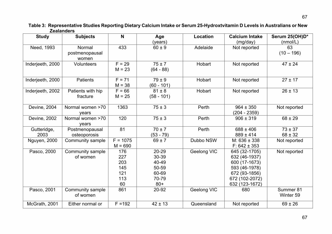
67
67
Table 3: Representative Studies Reporting Dietary Calcium Intake or Serum 25-Hydroxtvitamin D Levels in Australians or New Zealanders
Study Subjects N Age (years)
Location Calcium Intake (mg/day)
Serum 25(OH)D* (nmol/L)
Need, 1993 Normal postmenopausal
women
433 60 ± 9 Adelaide Not reported 63 (10 – 196)
Inderjeeth, 2000 Volunteers F = 29 M = 23
75 ± 7 (64 - 88)
Hobart Not reported 47 ± 24
Inderjeeth, 2000 Patients F = 71 M = 38
79 ± 9 (60 - 101)
Hobart Not reported 27 ± 17
Inderjeeth, 2002 Patients with hip fracture
F = 66 M = 25
81 ± 8 (58 - 101)
Hobart Not reported 26 ± 13
Devine, 2004 Normal women >70 years
1363 75 ± 3 Perth 964 ± 350 (204 - 2359)
Not reported
Devine, 2002 Normal women >70 years
120 75 ± 3 Perth 906 ± 319 68 ± 29
Gutteridge, 2003
Postmenopausal osteoporosis
81 70 ± 7 (53 - 79)
Perth 688 ± 406 889 ± 414
73 ± 37 68 ± 32
Nguyen, 2000 Community sample F = 1075 M = 690
69 ± 7 Dubbo NSW M: 636 ± 338 F: 642 ± 353
Not reported
Pasco, 2000 Community sample of women
176 227 203 145 121 113 60
20-29 30-39 40-49 50-59 60-69 70-79 80+
Geelong VIC 645 (32-1705) 632 (46-1937) 600 (17-1673) 593 (46-1978) 672 (93-1856)
672 (102-2072) 632 (123-1672)
Not reported
Pasco, 2001 Community sample of women
861 20-92 Geelong VIC 680 Summer 81 Winter 59
McGrath, 2001 Either normal or F =192 42 ± 13 Queensland Not reported 69 ± 26

68
68
with psychosis M= 201 (17-65) (12 – 175) Lips, 2001 Post menopausal
women with osteoporosis
Women N=7564
31-80 Multiple cities in Australia and Christchurch
Not reported Aust: 83 ± 32 NZ: 65 ± 29
Horwath, 1991 Community men and women
F = 906
M = 796
15 – 18 19 – 24 25 – 44 45 – 64
65+
15 – 18 19 – 24 25 – 44 45 – 64
65+
New Zealand 607 (172-1452)* 736 (260-1401) 642 (294-1329) 554 (230-1189) 474 (256-837)
1059 (359-2449) 727 (312-1558) 816 (351-1656) 631 (289-1141) 599 (217-1239)
Not reported
Horwath, 2001 Community men and women
F = 2709
M = 1927
15 – 18 19 – 24 25 – 44 45 – 64
65+
15 – 18 19 – 24 25 – 44 45 – 64
65+
New Zealand 740** 713 714 676 636
894 875 908 809 751
Not reported
Reid, 2000 Normal >5 years postmenopausal
185 63 ± 6
Auckland 1040 ± 490 57 ± 25
Parnell, 2003 School children M+F=3275 5-14 New Zealand 736 (481-1092)* Not reported Lucas, 2005 Normal, >5 years
postmenopausal 1606 74 ± 4
(54 - 85) Auckland 855± 384
(71 - 3200) 51 ± 19
(10 - 145)

69
69
Figures derived using the FSANZ dietary modelling
computer program, DIAMOND, and
data from the 1995 Australian and
1997 New Zealand National Nutrition
Surveys
Australians
13858 2+ Australia 840 (318-1470)*** Not reported
Data are mean ± SD, unless indicated otherwise. Where available, ranges are given in brackets 25(OH)D = 25-hydroxyvitamin D. Values can be converted to µg/L by dividing by 2.5 *Median (10th-90th centiles) ** Median *** Mean (10th-90th percentile)