the rabgap tbc1d1 plays a central role in exercise...
TRANSCRIPT

Jacqueline Stöckli,1,2,3,4 Christopher C. Meoli,1,2,3 Nolan J. Hoffman,1,2,3
Daniel J. Fazakerley,1,2,3 Himani Pant,3 Mark E. Cleasby,5 Xiuquan Ma,3
Maximilian Kleinert,3,6 Amanda E. Brandon,3 Jamie A. Lopez,3 Gregory J. Cooney,3,4
and David E. James1,2,3,7
The RabGAP TBC1D1 Plays a CentralRole in Exercise-Regulated GlucoseMetabolism in Skeletal MuscleDiabetes 2015;64:1914–1922 | DOI: 10.2337/db13-1489
Insulin and exercise stimulate glucose uptake into skel-etal muscle via different pathways. Both stimuli convergeon the translocation of the glucose transporter GLUT4from intracellular vesicles to the cell surface. Two Rabguanosine triphosphatases-activating proteins (GAPs)have been implicated in this process: AS160 for insulinstimulation and its homolog, TBC1D1, are suggested toregulate exercise-mediated glucose uptake into muscle.TBC1D1 has also been implicated in obesity in humansand mice. We investigated the role of TBC1D1 in glucosemetabolism by generating TBC1D12/2 mice and analyz-ing body weight, insulin action, and exercise. TBC1D12/2
mice showed normal glucose and insulin tolerance, withno difference in body weight compared with wild-typelittermates. GLUT4 protein levels were reduced by∼40% in white TBC1D12/2 muscle, and TBC1D12/2 miceshowed impaired exercise endurance together with im-paired exercise-mediated 2-deoxyglucose uptake intowhite but not red muscles. These findings indicate thatthe RabGAP TBC1D1 plays a key role in regulating GLUT4protein levels and in exercise-mediated glucose uptakein nonoxidative muscle fibers.
Insulin and exercise enhance muscle glucose uptake bytriggering GLUT4 vesicles to translocate from within thecell to the plasma membrane (PM) (1,2). These stimuliactivate different signaling pathways that converge onsimilar steps in the GLUT4 trafficking pathway. The
identification of the Rab guanosine triphosphatase(GTPase)-activating protein (GAP) AS160/TBC1D4 as anAkt substrate was exciting because this provided a linkbetween insulin signaling and GLUT4 trafficking (3).RabGAPs regulate the activity of Rab GTPases, whichplay an intimate role in eukaryotic vesicular trafficking(1,4). AS160 is localized to GLUT4 vesicles through itsinteraction with insulin-responsive aminopeptidase (IRAP),a constituent of GLUT4 vesicles (5–7). In the absence ofinsulin, AS160 is thought to be active, thus facilitatingintracellular sequestration of GLUT4 by rendering theRab associated with GLUT4 vesicles, possibly Rab10, in-active (8,9). Insulin stimulates Akt-dependent AS160phosphorylation and 14-3-3 binding, leading to inactiva-tion of AS160 GAP activity, increased GTP loading ofRab10, and increased GLUT4 translocation to the PM(1,10). Knockdown of AS160 in adipocytes increases PMGLUT4 levels, consistent with the role of AS160 as a neg-ative regulator (5,11,12).
TBC1D1 is a close homolog of AS160 that is highlyexpressed in skeletal muscle and so has been postulatedto play a role in exercise-mediated GLUT4 trafficking(13–17). AS160 and TBC1D1 have identical domain struc-tures. They share 47% amino acid identity and displaydistinct tissue expression: AS160 is highly expressed inheart, white adipose tissue (WAT), and oxidative muscles,such as soleus, whereas TBC1D1 is expressed in musclebut is absent from WAT (14). The suggested mechanism
1Charles Perkins Centre, University of Sydney, Sydney, New South Wales, Australia2School of Molecular Bioscience, University of Sydney, Sydney, New South Wales,Australia3Garvan Institute of Medical Research, Sydney, New South Wales, Australia4St Vincent’s Clinical School, Faculty of Medicine, University of New South Wales,Sydney, New South Wales, Australia5The Royal Veterinary College, University of London, London, U.K.6Molecular Physiology Group, Department of Nutrition, Exercise and Sports, AugustKrogh Centre, University of Copenhagen, Copenhagen, Denmark7School of Medicine, University of Sydney, Sydney, New South Wales, Australia
Corresponding author: David E. James, [email protected].
Received 28 September 2013 and accepted 24 December 2014.
J.A.L. is currently affiliated with the Peter MacCallum Cancer Centre, Departmentof Oncology, University of Melbourne, Parkville, Victoria, Australia.
© 2015 by the American Diabetes Association. Readers may use this article aslong as the work is properly cited, the use is educational and not for profit, andthe work is not altered.
1914 Diabetes Volume 64, June 2015
METABOLISM

for TBC1D1 regulation is primarily based on the well-known regulation of its close homolog AS160. TBC1D1interacts with IRAP (18), inactivates the same Rabs asAS160 (13), and binds 14-3-3 upon phosphorylation(16,19). Although Akt mediates AS160 phosphorylationon the crucial 14-3-3 binding sites, linking AS160 to in-sulin signaling, TBC1D1 binds 14-3-3 in response toAMPK activation, a kinase activated by exercise (2).
A mutation in TBC1D1 (R125W) is linked to humanobesity (20,21), although the precise role of this mutationin TBC1D1 function is not known. Several mouse modelswith reduced TBC1D1 expression have been described,including a congenic model that contains a locus fromthe Swiss Jim Lambert strain, and a gene trap knockout(22–25). These mice show no change in body weight orreduced body weight; reduced glucose uptake into isolatedwhite muscle in response to various agonists, includinginsulin, contraction, or AICAR, an AMPK agonist; and in-creased fatty acid oxidation at the whole-body level andin muscle. In general, no defect in whole-body insulin-mediated glucose metabolism was found (22–25).
We generated a TBC1D12/2 mouse model back-crossedonto the C57Bl6 background for .10 generations. Thesemice had no defect in whole-body insulin-mediated glucosemetabolism, unchanged body weight, and no difference inhigh-fat diet (HFD)–induced obesity or insulin resistancecompared with wild-type (WT) littermates. They did, how-ever, show impaired exercise endurance, which was likelycaused by impaired AMPK agonist–mediated glucose uptakeinto muscle in vitro and impaired exercise-mediated glucoseuptake in vivo, highlighting the important role of TBC1D1in exercise-regulated glucose metabolism in muscle.
RESEARCH DESIGN AND METHODS
MaterialsGeneral chemicals were from Sigma-Aldrich ChemicalCompany, unless otherwise stated. Antibodies were fromSigma-Aldrich (Flag), Cell Signaling Technology (TBC1D1,AMPK, pT172 AMPK), Santa Cruz Biotechnology (14-3-3),Mito Sciences (mito-profile), Symansis (p642-AS160), andMolecular Probes (cyclooxygenase, complex IV). Antibodiesagainst AS160 (5), GLUT4 (26), and GLUT1 (27) have beenpreviously described. BSA was obtained from Bovogen Bio-logicals, and protease inhibitors were from Roche.
Generation of TBC1D12/2 miceTBC1D12/2 mice were generated using a TBC1D1 gene-trap ES cell line from BayGenomics (#RRR502). The gene-trap vector insertion resulted in a truncated TBC1D1mRNA. Heterozygous mice were generated by the Austra-lian Phenomics Network ES to Mouse service at MonashUniversity. Mice were genotyped by PCR and real-time PCRusing the following primers within the gene-trap vector:59-gcggcaccgcgcctttcggcgg-39 and 59-ggaagggctggtcttcatccac-39.Mice were backcrossed onto C57Bl/6 background andheterozygous (+/2) TBC1D1 breeding pairs producedTBC1D12/2 and WT littermates for experiments. Mice
were group housed on a 12-h light/dark cycle with freeaccess to food and water. Mice were fed ad libitum a stan-dard laboratory chow (8% of calories from fat) or a HFD(Hugo’s Copha, 48% fat [7:1 lard-to-safflower oil ratio],32% carbohydrate, 20% protein). All experimental proce-dures were approved by the Garvan Institute/St Vincent’sHospital Animal Ethics Committee and followed the guide-lines issued by the National Health and Medical ResearchCouncil Australia.
Glucose and Insulin Tolerance Tests and InsulinMeasurementsMale littermates (12–16 weeks old) were fed standardchow or the HFD for 4 weeks. Mice were fasted for 6 hor overnight before glucose or insulin tolerance tests,respectively. Glucose (1 g/kg) or insulin (1 unit/kg) wasadministered by intraperitoneal injection, blood sampleswere obtained from the tail, and glucose was measuredusing an Accu-Chek Performa glucometer (Roche Diag-nostics). Insulin was measured from whole blood usingan insulin ELISA kit (Crystal Chem).
In Vivo ElectroporationIn vivo electroporation of DNA into mouse tibialisanterior (TA) muscle was done under anesthesia, aspreviously described (28). Briefly, DNA was injectedinto the TA muscle. Immediately after the injection, 8pulses of 200 V/cm and 20 ms at 1 Hz were administeredacross the distal limb via tweezer electrodes attached toan ECM-830 electroporator (BTX).
Exercise ExperimentsExercise experiments were performed on an Exer3/6mouse treadmill (Columbus Instruments) at a 5% incline.Mice (15–21 weeks) underwent a 2-day period of tread-mill running acclimatization that consisted of running for15 min at a speed of 10 m/min on day 1 and for 15 min at10 m/min, followed by 15 min at 13 m/min on day 2.Exercise was performed as indicated until exhaustion, de-fined as falling off the treadmill three times within 15 s.
In Vitro Glucose Uptake Into Isolated Muscle[3H]-2-deoxyglucose ([3H]-2DOG) uptake into isolated ex-tensor digitorum longus (EDL) and soleus muscles was per-formed as previously described (29). Muscles were incubatedin the absence or presence of 100 nmol/L insulin (Calbio-chem) or 2 mmol/L 5-aminoimidazole-4-carboxamideribonucleotide (AICAR; Toronto Research Chemicals) for20 min at 30°C.
Surgical Procedures and In Vivo Exercise-MediatedGlucose UptakeMice were anesthetized with isoflurane anesthesia forinsertion of a polyurethane catheter into the left carotidartery. The free catheter end was tunneled under the skin,externalized at the neck, and sealed. Mice were thensingly housed and monitored daily. Catheters wereflushed every 1–2 days with heparinized saline to main-tain patency. At 5–8 days after surgery, mice were fittedwith an extension catheter and run on a treadmill with
diabetes.diabetesjournals.org Stöckli and Associates 1915

speed gradually increasing to 16.5 m/min. [3H]-2DOG (0.2mCi/kg) was administered via the catheter, and bloodsamples were taken throughout the experiment. After20 min, mice were killed and tissues removed and snapfrozen. Exercise-mediated glucose uptake into red and whitequadriceps was measured using AG 1-X8 Resin (BioRad)to remove glucose 6-phosphate and measuring tracer inthe starting material (total [3H]-2DOG) and the flow-through (nonphosphorylated [3H]-2DOG) of the AG 1-X8column, as previously described (30).
Quantitative Real-Time RT-PCR AssaysRNA extraction was performed using TRIZOL reagent(Life Technologies, Carlsbad, CA), following the manu-facturer’s protocol. Omniscript RT Kit (Qiagen) wasused for cDNA synthesis. Real-time RT-PCR analysiswas performed on Light Cycler 480 (Roche Applied Sci-ence) with Universal Probe Master System. Primers andprobes for GLUT4 mouse gene were selected accordingto the Universal Probe Library System (Roche AppliedScience). The Cyclophilin gene was used as a control. Theprimers 59-gacggacactccatctgttg-39 and 59-gccacgatggagaca-tagc-39 were used for GLUT4, and 59-ttcttcataaccacagt-caagacc-39 and 59-accttccgtaccacatccat-39 were used forCyclophilin.
Glycogen MeasurementsGlycogen was measured as described (31). Briefly, tissueswere dissolved in 1 mol/L KOH, and glycogen was pre-cipitated twice with 95% ethanol. The glycogen pellet wasresuspended in amyloglucosidase solution (0.3 mg/mLamyloglucosidase in 0.25 mol/L acetate buffer [pH4.75]) and incubated overnight at 37°C. Glucose wasdetermined using a calorimetric glucose oxidase kit(ThermoFisher Scientific).
Triglyceride, Nonesterified Fatty Acids, and LactateMeasurementsCalorimetric assays were performed according to themanufacturer’s instructions to measure triglycerides(Roche) and nonesterified fatty acids (NEFAs; Wako) inplasma from mice. For lactate measurements, plasma wasdeproteinized, and lactate was measured as previouslydescribed (32,33).
Tissue Lysates, SDS-PAGE, and ImmunoblottingMice were killed by cervical dislocation for tissue iso-lation. Tissues were lysed (20 mmol/L HEPES [pH 7.4],250 mmol/L sucrose, 1 mmol/L EDTA, 2% SDS, proteaseinhibitors). Immunoblotting was carried out as previouslydescribed (5). Quantification of immunoblots was per-formed using Odyssey IR imaging system software.
Statistical AnalysisData are expressed as mean and SEM unless indi-cated otherwise. P values were calculated by t test,one-way ANOVA, or two-way ANOVA using GraphPadPrism software.
RESULTS
GLUT4 Protein Levels Are Reduced in Muscle FromTBC1D12/2 miceTBC1D12/2 mice were generated using a gene-trap ES cellline (Fig. 1). There was no detectable TBC1D1 protein inmuscles from TBC1D12/2 mice (Fig. 2A). The insertion ofthe gene trap resulted in a putative truncated TBC1D1construct, fused to b-galactosidase and neomycin trans-ferase. However, this putative TBC1D1 truncation wasnot detectable. AS160 protein levels were highest in heart,soleus, WAT, and red quadriceps and very low in whitequadriceps, TA, and EDL. AS160 levels were not changedin tissues from TBC1D12/2 mice compared with WT lit-termates (Fig. 2A and B). GLUT4 levels were significantlyreduced in muscle from TBC1D12/2 mice, with the mostpronounced decrease in muscles with low endogenousAS160 expression. GLUT4 protein levels were significantlyreduced in white quadriceps, TA, and EDL by 45%, 42%,and 37%, respectively (Fig. 2A and B). No significant changeoccurred in GLUT4 mRNA in TA muscle between the geno-types, indicating that reduced GLUT4 in TBC1D12/2 mus-cle occurs posttranscriptionally (Fig. 2C), consistent withrecent findings (22–24). There was no compensatory upreg-ulation in the level of GLUT1 (Fig. 2B).
TBC1D1 Overexpression Increases GLUT4 ProteinLevelsTo determine if TBC1D1 expression can rescue GLUT4levels, Flag-TBC1D1 cDNA was injected into the right legof mice, followed by in vivo electroporation. The left legwas injected with empty vector. One week later, TA
Figure 1—TBC1D12/2 mice genotyping. A: Diagram depicts endogenous protein and genomic DNA of WT and TBC1D12/2 (KO) location inRRR502 ES cell line with predicted truncated TBC1D1 protein. b-geo, fusion of b-galactosidase and neomycin transferase; CaM, calmod-ulin binding domain; pA, SV40 polyadenylation signal; PTB, phosphotyrosine binding domain; SA, splice acceptor site. B: TBC1D1 proteinand 14-3-3 loading control in TA muscle from WT and TBC1D1 KO mice.
1916 TBC1D1 and Exercise-Regulated Glucose Uptake Diabetes Volume 64, June 2015

muscle lysates from left and right legs were immunoblot-ted with antibodies against Flag, TBC1D1, and GLUT4.Flag-tagged TBC1D1 was only detected in the TA musclefrom the right leg (Fig. 3A). The TBC1D1 antibody selec-tively recognizes mouse but not human TBC1D1 andtherefore does not recognize the overexpressed TBC1D1.GLUT4 levels were increased in TA that expressedFlag-TBC1D1 compared with the control leg in WT andTBC1D12/2 mice by 67% and 79%, respectively (Fig. 3Aand B).
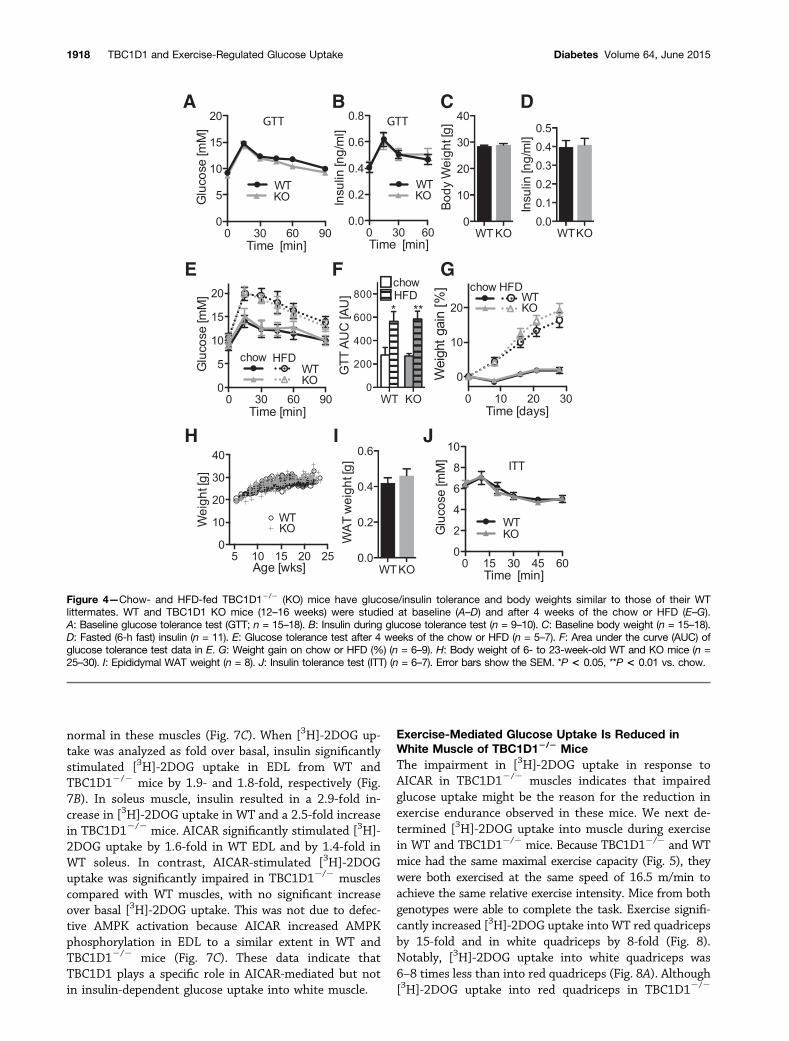
TBC1D12/2 Mice Have Normal Insulin-MediatedGlucose Metabolism and Body WeightNo difference was observed in glucose or insulin tolerancebetween TBC1D12/2 and WT littermates (Fig. 4A and J).In the insulin tolerance test for both genotypes, the glu-cose levels increased initially before dropping, as previ-ously reported (34), likely due to a stress response (Fig.4J). There was no difference in body weight in mice fedthe chow or HFD, epididymal fat pad weight, fasting in-sulin levels, insulin levels during glucose tolerance tests,or glucose tolerance in response to the HFD betweenTBC1D12/2 and WT mice (Fig. 4).
TBC1D12/2 Mice Show Impaired Exercise EnduranceWe next examined the exercise performance of TBC1D12/2
mice compared with WT littermates. Mice were subjected
to exercise running on a treadmill, and two different ex-ercise protocols were used to determine exercise endur-ance as determined by the total running time duringlow-intensity exercise, and maximal exercise capacity, de-termined by maximal running speed during high-intensityexercise. TBC1D12/2 mice showed a significant impair-ment in exercise endurance compared with WT littermates(Fig. 5A and B), but not in maximal exercise capacity(Fig. 5C).
There was no difference in muscle wet weight, mito-chondrial oxidative phosphorylation protein levels, muscleglycogen levels at rest or after exercise, plasma levels ofNEFA, and lactate between the genotypes (Fig. 6). There wasa significant difference in plasma TGs between TBC1D12/2
and WT mice after exercise (Fig. 6C).
AMPK Agonist-Mediated Glucose Uptake Into MuscleIs Impaired in TBC1D12/2 MiceWe next sought to examine whether the absence ofTBC1D1 affected insulin- or AMPK-induced glucoseuptake. EDL and soleus muscles were isolated fromTBC1D12/2 and WT littermates, mounted on muscle hold-ers, and incubated in vitro. Muscles were incubated in theabsence or presence of insulin or the AMPK agonist AICAR,and glucose uptake was measured using [3H]-2DOG traceraccumulation (Fig. 7). [3H]-2DOG uptake was signifi-cantly reduced in EDL from TBC1D12/2 mice, but thiswas not the case for soleus (Fig. 7A). This is likely dueto decreased GLUT4 levels in EDL from TBC1D12/2 micebecause insulin-stimulated AS160 phosphorylation was
Figure 2—GLUT4 protein levels are reduced in TBC1D12/2 muscle.Tissues from WT and TBC1D12/2 (KO) littermates were isolatedand immunoblotted with indicated antibodies. A: Representativeimmunoblots are shown. B: Immunoblots were quantified and nor-malized to WT protein levels. WT is shown in black bars and KO ingray bars (n = 3–4). C: GLUT4 mRNA levels in TA of WT and TBC1D1KO mice, normalized to WT (n = 3). RQ, red quadriceps; WQ, whitequadriceps. Error bars show the SEM. *P < 0.05, **P < 0.01.
Figure 3—TBC1D1 overexpression increases GLUT4 levels. A: Invivo electroporation of endotoxin-free Flag-TBC1D1 (human) DNAinto right leg (R) and endotoxin-free empty vector DNA into left leg(L) of WT and TBC1D12/2 (KO) mice. Mice were killed after 1 week,tissues were isolated, and TA muscles were immunoblotted withantibodies against Flag, TBC1D1, and GLUT4. Representativeimmunoblots are shown. B: Quantification of data in A (n = 3).The error bars show the SEM. *P < 0.05.
diabetes.diabetesjournals.org Stöckli and Associates 1917
normal in these muscles (Fig. 7C). When [3H]-2DOG up-take was analyzed as fold over basal, insulin significantlystimulated [3H]-2DOG uptake in EDL from WT andTBC1D12/2 mice by 1.9- and 1.8-fold, respectively (Fig.7B). In soleus muscle, insulin resulted in a 2.9-fold in-crease in [3H]-2DOG uptake in WT and a 2.5-fold increasein TBC1D12/2 mice. AICAR significantly stimulated [3H]-2DOG uptake by 1.6-fold in WT EDL and by 1.4-fold inWT soleus. In contrast, AICAR-stimulated [3H]-2DOGuptake was significantly impaired in TBC1D12/2 musclescompared with WT muscles, with no significant increaseover basal [3H]-2DOG uptake. This was not due to defec-tive AMPK activation because AICAR increased AMPKphosphorylation in EDL to a similar extent in WT andTBC1D12/2 mice (Fig. 7C). These data indicate thatTBC1D1 plays a specific role in AICAR-mediated but notin insulin-dependent glucose uptake into white muscle.
Exercise-Mediated Glucose Uptake Is Reduced inWhite Muscle of TBC1D12/2 MiceThe impairment in [3H]-2DOG uptake in response toAICAR in TBC1D12/2 muscles indicates that impairedglucose uptake might be the reason for the reduction inexercise endurance observed in these mice. We next de-termined [3H]-2DOG uptake into muscle during exercisein WT and TBC1D12/2 mice. Because TBC1D12/2 and WTmice had the same maximal exercise capacity (Fig. 5), theywere both exercised at the same speed of 16.5 m/min toachieve the same relative exercise intensity. Mice from bothgenotypes were able to complete the task. Exercise signifi-cantly increased [3H]-2DOG uptake into WT red quadricepsby 15-fold and in white quadriceps by 8-fold (Fig. 8).Notably, [3H]-2DOG uptake into white quadriceps was6–8 times less than into red quadriceps (Fig. 8A). Although[3H]-2DOG uptake into red quadriceps in TBC1D12/2
Figure 4—Chow- and HFD-fed TBC1D12/2 (KO) mice have glucose/insulin tolerance and body weights similar to those of their WTlittermates. WT and TBC1D1 KO mice (12–16 weeks) were studied at baseline (A–D) and after 4 weeks of the chow or HFD (E–G).A: Baseline glucose tolerance test (GTT; n = 15–18). B: Insulin during glucose tolerance test (n = 9–10). C: Baseline body weight (n = 15–18).D: Fasted (6-h fast) insulin (n = 11). E: Glucose tolerance test after 4 weeks of the chow or HFD (n = 5–7). F: Area under the curve (AUC) ofglucose tolerance test data in E. G: Weight gain on chow or HFD (%) (n = 6–9). H: Body weight of 6- to 23-week-old WT and KO mice (n =25–30). I: Epididymal WAT weight (n = 8). J: Insulin tolerance test (ITT) (n = 6–7). Error bars show the SEM. *P < 0.05, **P < 0.01 vs. chow.
1918 TBC1D1 and Exercise-Regulated Glucose Uptake Diabetes Volume 64, June 2015

mice was similar to that observed in WT mice, [3H]-2DOG
uptake into white quadriceps from TBC1D12/2 mice wasreduced by 54% (Fig. 8B).
DISCUSSION
Members of the RabGAP family have generated muchinterest in the context of glucose metabolism. AS160plays an important role in insulin-stimulated glucoseuptake in fat and muscle cells (1,35), and mutations inAS160 are associated with severe insulin resistance inhumans (36,37). The AS160 homolog TBC1D1 is involvedin contraction-mediated glucose uptake (24) and has beenimplicated in obesity (20,21). In the current study, weshow that TBC1D12/2 mice have no disruption inwhole-body insulin action but impaired exercise-regulatedmetabolism. This is based on the following: 1) AICAR-mediated [3H]-2DOG uptake into isolated muscle was im-paired in TBC1D12/2 mice (Fig. 7), 2) exercise-mediated[3H]-2DOG uptake into white quadriceps was significantlyreduced in TBC1D12/2 mice in vivo (Fig. 8), and 3)TBC1D12/2 mice exhibited reduced exercise endurance(Fig. 5).
The impairment in exercise endurance in TBC1D12/2
mice (Fig. 5) clearly implicates TBC1D1 as having an im-portant role in exercise-regulated glucose metabolism.This defect could not be attributed to changes in muscleweight, the levels of mitochondrial oxidative phosphory-lation proteins, or plasma lactate or NEFA levels, or mus-cle glycogen levels (Fig. 6), so it is likely due to theimpairment in the ability of the exercising white muscleto import extracellular glucose (Fig. 8). White and redmuscle fibers are both engaged during the treadmill exer-cise used for this test because we observed a decline inglycogen levels in both muscle types after a single bout ofexercise (Fig. 6), consistent with previous studies (38).Although glucose uptake into white muscle was consider-ably less than into red muscle (Fig. 8A), it is plausible thatthe loss of TBC1D1 results in an impairment of glucoseuptake into specific fibers that ultimately fatigue fasterand lead to an overall exercise impairment. These data
suggest that white muscle fibers likely play a crucial roleeven in endurance-style exercise and that a defect in thesefibers may be a limiting factor in long-term endurance.However, whether the defect in exercise-mediated glucoseuptake can be directly attributed to loss of TBC1D1 or theparallel decrease in GLUT4 protein levels is unclear. Thegreater defect we observed in AICAR compared withinsulin-dependent glucose uptake in EDL (Fig. 7) is con-sistent with the defect being primarily due to loss ofTBC1D1. Given that TBC1D1 is a major AMPK substratein muscle (16,19,39), this would support the view thatAMPK is a major determinant of exercise-regulated glu-cose metabolism in muscle. Although the role of AMPK inexercise-mediated glucose uptake has been controversial(40,41), a recent study demonstrated that muscle-specificAMPK b1/b22/2 mice also display impaired exercise en-durance (42).
Our data, combined with results of other studies, areconsistent with intramuscular energy stores, such asphosphocreatine and glycogen, in providing the energyneeded during the initial phase of exercise, followed bya gradually increasing reliance on extracellular glucosewhen exercise is sustained. An impairment in this processwould appear to have a major effect on endurance, givingrise to the concept that increased expression of TBC1D1and/or AMPK might lead to improved endurance isworthy of future study. One difference we observedbetween WT and TBC1D12/2 mice after exercise, in ad-dition to impaired exercise-mediated glucose uptake, wasa significant reduction in plasma TG levels (Fig. 6). Thatfatty acid oxidation is increased in the TBC1D12/2 miceduring exercise is possible, consistent with reports abouta switch in fuel usage in other TBC1D12/2 mouse models(22,23). However, this is unlikely to be the cause of im-paired endurance but rather a consequence of reducedglucose uptake.
The lack of any detectable body weight phenotype inTBC1D12/2 mice fed the chow or HFD (Fig. 4) was curi-ous in light of previous studies (22–25). Initial studiesused a congenic strain containing a Swiss Jim Lambert
Figure 5—TBC1D12/2 mice have impaired exercise endurance. A: Survival plot showing the percentage of WT and TBC1D12/2 (KO)littermates running at indicated times during an exercise endurance test (10 min at 10 m/min with an increase in running speed by 1 m/minevery 15 min). Total running time was determined until exhaustion. Individual data points are shown. B: Average and SEM of data shown inA (n = 15–17). C: Maximal running speed of WT and TBC1D1 KO littermates was determined during a maximal exercise capacity test (startat 8 m/min with an increase in running speed by 2 m/min every 1.5 min). Maximal running speed at exhaustion was determined (n = 15–17).The error bars show the SEM. ***P < 0.001.
diabetes.diabetesjournals.org Stöckli and Associates 1919

locus with a mutation in the TBC1D1 gene that resultedin reduced body weight on the HFD but not on chow(24,25). A more recent study with the same mouse modelreported reduced body weight on a chow diet (22). How-ever, a TBC1D12/2 gene-trap mouse model, similar to theone used in our study, reported reduced body weight onboth chow and the HFD (23). The discrepancy in the bodyweight phenotype is likely related to the mouse modelused, the breeding strategy used to generate mice for ex-perimental use (i.e., use of littermates vs. nonlittermates),the genetic background of the mice used, differences in theage of onset of the HFD, duration of the HFD, and specificcomposition (percentages and types of lipids) of the HFD.
Genetic background clearly plays a crucial role in metabo-lism in mice (34), and it is now well recognized that breed-ing strategies and the degree of backcrossing affect theeventual metabolic phenotype. For this reason, all of ourmetabolic studies were performed using mice that werebackcrossed onto a C57Bl/6 background for .10 genera-tions, and all animals (TBC1D12/2 and WT) were litter-mates obtained from TBC1D1+/2 breeding pairs.
We did, however, observe a significant reduction inGLUT4 levels in skeletal muscle from these mice (Fig. 2).This likely involves an important role for TBC1D1 inmaintaining the stability of the GLUT4 protein, becausewe did not observe any change in GLUT4 mRNA, consis-tent with previous reports (22,24). The reduction inGLUT4 levels could be rescued by overexpression ofTBC1D1 in TA muscle (Fig. 3). Notably, GLUT4 levelswere significantly reduced only in muscle types thatexpressed little AS160 (Fig. 2). This indicates that inmuscles expressing high levels of AS160, AS160
Figure 6—No difference in muscle tissue weights, mitochondrialoxidative phosphorylation protein levels, or glycogen. A: TA, EDL,and soleus were isolated and weighed from WT and TBC1D12/2
(KO) mice (n = 8). B: Muscle glycogen was measured in red quad-riceps (RQ) and white quadriceps (WQ) from WT and TBC1D1 KOlittermates after resting or exercise (Ex) (treadmill running for 55 min:10 min at 10 m/min with an increase in running speed by 1 m/minevery 15 min; n = 11–15). WT is shown in black bars and KO in graybars. C: TGs, lactate, and NEFAs were measured in plasma fromWT and TBC1D1 littermates after resting or exercise (see in B) (n =5–8). D: Indicated tissues were immunoblotted for mitochondrialoxidative phosphorylation proteins using a mito-profile cocktailthat includes antibodies against subunits of complex II (C-II), com-plex III (C-III), complex IV (C-IV), and complex V (C-V). Antibodyagainst 14-3-3 was used as a loading control (n = 3). Error barsshow the SEM. hr, hours. *P < 0.05, **P < 0.01.
Figure 7—AICAR-stimulated glucose uptake is impaired inTBC1D12/2 muscle. Isolated EDL or soleus muscle from WT orTBC1D12/2 (KO) littermates was incubated in the presence or ab-sence (B) of 100 nmol/L insulin (I) or 2 mmol/L AICAR (A), and[3H]-2DOG uptake was measured (n = 9–25). A: [3H]-2DOG uptakeis shown. B: [3H]-2DOG uptake data in A are presented as fold overbasal. C: EDL muscles were immunoblotted with indicated antibod-ies. Error bars show the SEM. ns, not significant. *P < 0.05 vs.basal, ****P < 0.0001 vs. basal. #P < 0.0001 vs. WT. §P < 0.01vs. WT. ‡P < 0.05 vs. WT.
1920 TBC1D1 and Exercise-Regulated Glucose Uptake Diabetes Volume 64, June 2015

compensates for the loss of TBC1D1 in stabilizing GLUT4levels in those muscles. Intriguingly, reduced GLUT4levels have been observed in red muscle and fat fromAS1602/2 mice (43). This is consistent with a majorrole for these RabGAPs in regulating intracellular reten-tion of GLUT4 in intracellular vesicles. In the absence ofstimulation, TBC1D1 and AS160 are localized to GLUT4vesicles via their interaction with IRAP. Under these cir-cumstances, TBC1D1 and AS160 are nonphosphorylated,and their GAP activity is likely on, thus maintaining a Rabinactive. This leads to efficient intracellular sequestrationof GLUT4. In the absence of TBC1D1 or AS160, GLUT4vesicles are not efficiently sequestered, resulting in entryof GLUT4 into the endocytic recycling system and ulti-mately leading to increased delivery to the lysosome andGLUT4 degradation. Consistent with this, the half-life ofthe GLUT4 protein is;50 h in the absence of insulin, andthis is reduced to ;15 h in insulin-stimulated adipocytes(44). Hence, we conclude that in the absence of TBC1D1,GLUT4 protein levels are reduced in certain muscles thatare normally enriched in TBC1D1 but not in AS160 ex-pression, possibly due to increased GLUT4 degradation.
Another question arising from the current studies iswhy, in view of a 40% reduction in total GLUT4 levelsin muscle, did we not observe any significant defect inwhole-body insulin action? Previous studies using muscle-specific GLUT4+/2 mice observed a concomitant impair-ment in glucose homeostasis and insulin action in muscle(45). However, GLUT4 levels in these studies were re-duced in all muscles, including oxidative type muscles,whereas that was not the case in TBC1D12/2 mice. Giventhat red muscles are much more insulin sensitive thanwhite muscles (46), this is consistent with a greater
contribution of red muscle to whole-body insulin action.Importantly, insulin-stimulated glucose uptake was nor-mal in isolated soleus from TBC1D12/2 mice (Fig. 7).Although there was an absolute reduction in insulin-stimulated [3H]-2DOG uptake into EDL from TBC1D12/2
mice compared with WT mice, likely due to the 40% re-duction in GLUT4 protein levels (Fig. 2), it is important tonote that the fold increase over basal with insulin wasalmost identical in TBC1D12/2 EDL and in WT EDL(Fig. 7). Thus, it seems likely that the insulin-dependentincrease in muscle glucose uptake, combined with thelesser contribution of white muscle to whole-body glucosemetabolism, contributed to normal whole-body glucoseand insulin tolerance in TBC1D12/2 mice (Fig. 4). This isconsistent with other studies using different TBC1D12/2
mouse models that also showed no defect in glucose andinsulin tolerance (22–24).
These studies provide further insights into the molec-ular regulation of glucose metabolism in muscle duringexercise, implicating a key role for the RabGAP TBC1D1 inthis process. We have not, however, been able to observeany significant role for this protein in obesity or whole-body insulin sensitivity.
Acknowledgments. The TBC1D12/2 mice were generated by the Aus-tralian Phenomics Network (APN) ES to Mouse service at Monash University. Theauthors thank Jørgen Jensen (Norwegian School of Sport Sciences, Oslo, Nor-way) for technical assistance with muscle isolation.Funding. This work was supported by National Health and Medical ResearchCouncil (NHMRC) project grants GNT1068469 to J.S., GNT1047067 to D.E.J., anda grant from the Diabetes Australia Research Trust to J.S. D.E.J. is an NHMRCSenior Principal Research Fellow.Duality of Interest. No potential conflicts of interest relevant to this articlewere reported.Author Contributions. J.S. performed most of the experiments,designed the study, and wrote the manuscript. C.C.M., N.J.H., D.J.F., H.P., M.K.,and G.J.C performed animal experiments. M.E.C. performed in vivo electro-poration. X.M. performed quantitative PCR. A.E.B. performed the mouse surgery.J.A.L. initiated the study and organized the generation of the animal model. D.E.J.designed the study and wrote the manuscript. D.E.J. is the guarantor of this workand, as such, had full access to all the data in the study and takes responsibilityfor the integrity of the data and the accuracy of the data analysis.
References1. Stöckli J, Fazakerley DJ, James DE. GLUT4 exocytosis. J Cell Sci 2011;124:4147–41592. Richter EA, Hargreaves M. Exercise, GLUT4, and skeletal muscle glucoseuptake. Physiol Rev 2013;93:993–10173. Sano H, Kane S, Sano E, et al. Insulin-stimulated phosphorylation of a RabGTPase-activating protein regulates GLUT4 translocation. J Biol Chem 2003;278:14599–146024. Hutagalung AH, Novick PJ. Role of Rab GTPases in membrane traffic andcell physiology. Physiol Rev 2011;91:119–1495. Larance M, Ramm G, Stöckli J, et al. Characterization of the role of the RabGTPase-activating protein AS160 in insulin-regulated GLUT4 trafficking. J BiolChem 2005;280:37803–378136. Martin S, Rice JE, Gould GW, Keller SR, Slot JW, James DE. The glucosetransporter GLUT4 and the aminopeptidase vp165 colocalise in tubulo-vesicularelements in adipocytes and cardiomyocytes. J Cell Sci 1997;110:2281–2291
Figure 8—Exercise-mediated glucose uptake is impaired in whitequadriceps from TBC1D12/2 mice. Resting or exercising mice(16.5 m/min treadmill running) were administered [3H]-2DOG viaintra-arterial injection. Mice were killed after 20 min, and red andwhite quadriceps (Quad) were collected. A: [3H]-2DOG uptake isshown. B: [3H]-2DOG uptake data in A are presented as fold overrest. WT is shown in black bars and KO in gray bars (n = 6–8). *P <0.05, **P < 0.01, ***P < 0.001.
diabetes.diabetesjournals.org Stöckli and Associates 1921

7. Peck GR, Ye S, Pham V, et al. Interaction of the Akt substrate, AS160, withthe glucose transporter 4 vesicle marker protein, insulin-regulated aminopepti-dase. Mol Endocrinol 2006;20:2576–25838. Mîinea CP, Sano H, Kane S, et al. AS160, the Akt substrate regulatingGLUT4 translocation, has a functional Rab GTPase-activating protein domain.Biochem J 2005;391:87–939. Sano H, Eguez L, Teruel MN, et al. Rab10, a target of the AS160 Rab GAP, isrequired for insulin-stimulated translocation of GLUT4 to the adipocyte plasmamembrane. Cell Metab 2007;5:293–30310. Ramm G, Larance M, Guilhaus M, James DE. A role for 14-3-3 in insulin-stimulated GLUT4 translocation through its interaction with the RabGAP AS160.J Biol Chem 2006;281:29174–2918011. Eguez L, Lee A, Chavez JA, et al. Full intracellular retention of GLUT4 re-quires AS160 Rab GTPase activating protein. Cell Metab 2005;2:263–27212. Brewer PD, Romenskaia I, Kanow MA, Mastick CC. Loss of AS160 Aktsubstrate causes Glut4 protein to accumulate in compartments that are primedfor fusion in basal adipocytes. J Biol Chem 2011;286:26287–2629713. Roach WG, Chavez JA, Mîinea CP, Lienhard GE. Substrate specificity andeffect on GLUT4 translocation of the Rab GTPase-activating protein Tbc1d1.Biochem J 2007;403:353–35814. Taylor EB, An D, Kramer HF, et al. Discovery of TBC1D1 as an insulin-,AICAR-, and contraction-stimulated signaling nexus in mouse skeletal muscle.J Biol Chem 2008;283:9787–979615. An D, Toyoda T, Taylor EB, et al. TBC1D1 regulates insulin- and contraction-induced glucose transport in mouse skeletal muscle. Diabetes 2010;59:1358–136516. Frøsig C, Pehmøller C, Birk JB, Richter EA, Wojtaszewski JF. Exercise-induced TBC1D1 Ser237 phosphorylation and 14-3-3 protein binding capacity inhuman skeletal muscle. J Physiol 2010;588:4539–454817. Jessen N, An D, Lihn AS, et al. Exercise increases TBC1D1 phosphorylationin human skeletal muscle. Am J Physiol Endocrinol Metab 2011;301:E164–E17118. Tan SX, Ng Y, Burchfield JG, et al. The Rab GTPase-activating proteinTBC1D4/AS160 contains an atypical phosphotyrosine-binding domain that in-teracts with plasma membrane phospholipids to facilitate GLUT4 trafficking inadipocytes. Mol Cell Biol 2012;32:4946–495919. Chen S, Murphy J, Toth R, Campbell DG, Morrice NA, Mackintosh C.Complementary regulation of TBC1D1 and AS160 by growth factors, insulin andAMPK activators. Biochem J 2008;409:449–45920. Stone S, Abkevich V, Russell DL, et al. TBC1D1 is a candidate for a severeobesity gene and evidence for a gene/gene interaction in obesity predisposition.Hum Mol Genet 2006;15:2709–272021. Meyre D, Farge M, Lecoeur C, et al. R125W coding variant in TBC1D1confers risk for familial obesity and contributes to linkage on chromosome 4p14in the French population. Hum Mol Genet 2008;17:1798–180222. Chadt A, Immisch A, de Wendt C, et al. Deletion of both Rab-GTPase-activating proteins TBC1D1 and TBC1D4 in mice eliminates insulin- and AICAR-stimulated glucose transport [published correction appears in Diabetes 2015;64:1492]. Diabetes 2015;64:746–75923. Dokas J, Chadt A, Nolden T, et al. Conventional knockout of Tbc1d1 in miceimpairs insulin- and AICAR-stimulated glucose uptake in skeletal muscle. En-docrinology 2013;154:3502–351424. Szekeres F, Chadt A, Tom RZ, et al. The Rab-GTPase-activating proteinTBC1D1 regulates skeletal muscle glucose metabolism. Am J Physiol EndocrinolMetab 2012;303:E524–E53325. Chadt A, Leicht K, Deshmukh A, et al. Tbc1d1 mutation in lean mouse strainconfers leanness and protects from diet-induced obesity. Nat Genet 2008;40:1354–135926. Hashiramoto M, James DE. Characterization of insulin-responsive GLUT4storage vesicles isolated from 3T3-L1 adipocytes. Mol Cell Biol 2000;20:416–427
27. James DE, Strube M, Mueckler M. Molecular cloning and characterization ofan insulin-regulatable glucose transporter. Nature 1989;338:83–8728. Cleasby ME, Davey JR, Reinten TA, et al. Acute bidirectional manipulation ofmuscle glucose uptake by in vivo electrotransfer of constructs targeting glucosetransporter genes. Diabetes 2005;54:2702–271129. Li J, Cantley J, Burchfield JG, et al. DOC2 isoforms play dual roles in insulinsecretion and insulin-stimulated glucose uptake. Diabetologia 2014;57:2173–218230. James DE, Kraegen EW, Chisholm DJ. Muscle glucose metabolism in ex-ercising rats: comparison with insulin stimulation. Am J Physiol 1985;248:E575–E58031. Hoehn KL, Turner N, Swarbrick MM, et al. Acute or chronic upregulation ofmitochondrial fatty acid oxidation has no net effect on whole-body energy ex-penditure or adiposity. Cell Metab 2010;11:70–7632. Prabhu AV, Krycer JR, Brown AJ. Overexpression of a key regulator of lipidhomeostasis, Scap, promotes respiration in prostate cancer cells. FEBS Lett2013;587:983–98833. Arola L, Herrera E, Alemany M. A new method for deproteinization of smallsamples of blood plasma for amino acid determination. Anal Biochem 1977;82:236–23934. Montgomery MK, Hallahan NL, Brown SH, et al. Mouse strain-dependentvariation in obesity and glucose homeostasis in response to high-fat feeding.Diabetologia 2013;56:1129–113935. Cartee GD, Funai K. Exercise and insulin: convergence or divergence atAS160 and TBC1D1? Exerc Sport Sci Rev 2009;37:188–19536. Dash S, Sano H, Rochford JJ, et al. A truncation mutation in TBC1D4 ina family with acanthosis nigricans and postprandial hyperinsulinemia. Proc NatlAcad Sci U S A 2009;106:9350–935537. Dash S, Langenberg C, Fawcett KA, et al. Analysis of TBC1D4 in patientswith severe insulin resistance. Diabetologia 2010;53:1239–124238. Furler SM, Goldstein M, Cooney GJ, Kraegen EW. In vivo quantification ofglucose uptake and conversion to glycogen in individual muscles of the rat fol-lowing exercise. Metabolism 1998;47:409–41439. Pehmøller C, Treebak JT, Birk JB, et al. Genetic disruption of AMPK sig-naling abolishes both contraction- and insulin-stimulated TBC1D1 phosphoryla-tion and 14-3-3 binding in mouse skeletal muscle. Am J Physiol EndocrinolMetab 2009;297:E665–E67540. Maarbjerg SJ, Jørgensen SB, Rose AJ, et al. Genetic impairment ofAMPKalpha2 signaling does not reduce muscle glucose uptake during treadmillexercise in mice. Am J Physiol Endocrinol Metab 2009;297:E924–E93441. Merry TL, Steinberg GR, Lynch GS, McConell GK. Skeletal muscle glucoseuptake during contraction is regulated by nitric oxide and ROS independently ofAMPK. Am J Physiol Endocrinol Metab 2010;298:E577–E58542. O’Neill HM, Maarbjerg SJ, Crane JD, et al. AMP-activated protein kinase(AMPK) beta1beta2 muscle null mice reveal an essential role for AMPK inmaintaining mitochondrial content and glucose uptake during exercise. Proc NatlAcad Sci U S A 2011;108:16092–1609743. Lansey MN, Walker NN, Hargett SR, Stevens JR, Keller SR. Deletion of RabGAP AS160 modifies glucose uptake and GLUT4 translocation in primary skeletalmuscles and adipocytes and impairs glucose homeostasis. Am J Physiol Endo-crinol Metab 2012;303:E1273–E128644. Sargeant RJ, Pâquet MR. Effect of insulin on the rates of synthesis anddegradation of GLUT1 and GLUT4 glucose transporters in 3T3-L1 adipocytes.Biochem J 1993;290:913–91945. Zisman A, Peroni OD, Abel ED, et al. Targeted disruption of the glucosetransporter 4 selectively in muscle causes insulin resistance and glucose in-tolerance. Nat Med 2000;6:924–92846. James DE, Jenkins AB, Kraegen EW. Heterogeneity of insulin action in in-dividual muscles in vivo: euglycemic clamp studies in rats. Am J Physiol 1985;248:E567–E574
1922 TBC1D1 and Exercise-Regulated Glucose Uptake Diabetes Volume 64, June 2015