the protective effects of alisol a 24-acetate from alisma
TRANSCRIPT

molecules
Article
The Protective Effects of Alisol A 24-Acetate fromAlisma canaliculatum on Ovariectomy Induced BoneLoss in VivoYun-Ho Hwang 1, Kyung-Yun Kang 1, Sung-Ju Lee 1, Sang-Jip Nam 2, Young-Jin Son 1 andSung-Tae Yee 1,*
Received: 2 December 2015; Accepted: 7 January 2016; Published: 9 January 2016Academic Editor: Christopher W.K. Lam
1 Department of Pharmacy, Sunchon National University, 255 Joongang-Ro, Seokhyeon-Dong,Suncheon 549-742, Korea; [email protected] (Y.-H.H.); [email protected] (K.-Y.K.);[email protected] (S.-J.L.); [email protected] (Y.-J.S.)
2 Department of Chemistry and Nano Science, Ewha Womans University, Seoul 120-750, Korea;[email protected]
* Correspondence: [email protected]; Tel.: +82-61-750-3752; Fax: +82-61-750-3708
Abstract: Alisma canaliculatum is a herb commonly used in traditional Korean medicine, and has beenshown in scientific studies to have antitumor, diuretic hepatoprotective, and antibacterial effects.Recently, the anti-osteoclastogenesis of alisol A 24-acetate from Alisma canaliculatum was investigatedin vitro. However, the influence of alisol A 24-acetate on osteoporosis in animals has not beeninvestigated. The present study was undertaken to investigate the anti-osteoporotic effect of alisol A24-acetate on bone mass in ovariectomized (OVX) mice and to identify the mechanism responsiblefor its effects. OVX mice were treated daily with 0.5 or 2 µg/g of alisol A 24-acetate for a period of sixweeks. It was found that these administrations significantly suppressed osteoporosis in OVX miceand improved bone morphometric parameters. The serum estradiol, bone alkaline phosphatase levels,regulatory T/Th17 cell numbers were significantly increased by alisol A 24-acetate as compared withuntreated OVX mice. In addition, TRAP activity was inhibited by alisol A 24-acetate in OVX mice.These results suggest alisol A 24-acetate effectively prevents bone loss in OVX mice, and that it canbe considered a potential therapeutic for the treatment of postmenopausal osteoporosis.
Keywords: osteoporosis; bone loss; alisol A 24-acetate; Alisma canaliculatum; regulator T cell;traditional Korean medicine
1. Introduction
Bone is a dynamic tissue that undergoes continual adaption during vertebrate life to preserveskeletal size and shape, and to regulate mineral homeostasis. Bone remodeling is the removaland formation of damaged bone to maintain skeletal integrity and mineral homeostasis, whereasbone homeostasis is maintained by a balance between bone-resorbing osteoclasts and bone-formingosteoblasts [1].
Osteoporosis is a common disease among the elderly, and a serious worldwide healthproblem [2,3]. It causes loss of bone mass and strength and deterioration of bone microarchitecture,which increase the risk of fragility fractures [4]. Osteoporosis especially affects post-menopausalwomen and is associated with estrogen deficiency, which enhances osteoclast production, and thus,disrupts osteoblast/osteoclast balance [5]. T cells are key inducers of bone loss in the presence ofestrogen deficiency [6], and CD4+ helper T cells are the central organizers of adaptive immunityand various immunological diseases. Naive CD4+ T cells undergo functional differentiation intocytokine-secreting effector cells. The effector differentiation of helper T cells into the Th1, Th2, Th17,
Molecules 2016, 21, 74; doi:10.3390/molecules21010074 www.mdpi.com/journal/molecules

Molecules 2016, 21, 74 2 of 11
and regulatory T cells (Treg) subsets is determined by the cytokine environment [7,8]. Th17 cellsamong the T cell subtype play an important role in the induction of inflammation by producingpro-inflammatory cytokines, such as IL-17A and IL-17F [9]. On the other hand, Treg cells have ananti-inflammatory role and maintain self-tolerance by secreting cytokines, such as, transforminggrowth factor (TGF)-β and IL-10 [10]. IL-17 secreted by Th17 cells induces differentiation of osteoclastprogenitors into mature osteoclasts in vitro. Moreover, treatment of human monocytes with onlyIL-17 induces osteoclastogenesis [11]. Whereas Th17 cells are key effector cells in diseases such asrheumatoid arthritis and osteoporosis, Treg cells are essential for dominant immunologic tolerance.Ovariectomy enhances Receptor activator of nuclear factor kappa-B ligand (RANKL), Tumor necrosisfactor-α (TNF-α), and IL-17, and inhibition of these cytokines is likely to afford effective skeletalprotection post-ovx [12].
The goal of osteoporosis therapy is to inhibit bone resorption by reducing osteoclastic productionor activity. Hormone replacement therapies (HRT) based on, for example, estrogens, selective estrogenreceptor modulators (SERMs), bisphosphonates, and calcitonin, inhibit bone loss. However, recentresults of the Women’s Health Initiative demonstrated that women taking estrogen/progestin HRT areat elevated risk of breast cancer, coronary heart disease, and pulmonary embolism [13]. Therefore, analternative treatment with fewer side effects is required for HRT.
The increasing interest shown in new and safer drugs from natural sources stems from safetyconcerns, and resulted in studies on the protective effects of alternative medicines on osteoporosis.Although many studies have been conducted on the treatment of osteoporosis, the problematic issueof side effects remains, and therefore, it would be useful to identify side-effect free natural compoundswith a positive effect on osteoporosis [14].
Alisma canaliculatum (common name water plantain or “taeksa” in Korea) is commonly usedin traditional Korean medicine [15]. This fruit has unique terpenoids, such as alisol A, B, and C;alisol A 24-acetate; alisol B 23-acetate; alisol C 23-acetate; alismalactone 23-acetate; alismols A, B,and C; sulfoorientalol A; oriediterpenol; and oriediterpenoside [16]. Furthermore, it has been shownto possess antibacterial [17], antitumor [18], and hepatoprotective [19] effects. In addition, alisolA-24 acetate from Alisma canaliculatum has been reported to inhibit osteoclast formation in vitro [20].However, the antiosteoporotic effects of alisol A-24 acetate on postmenopausal osteoporosis have notbeen examined in ovariectomized (OVX) mice.
In the present study, we hypothesized that alisol A-24 acetate might prevent bone loss induced byestrogen deficiency, and in the present study examined its effects on bone deterioration in OVX mice.
2. Materials and Methods
The experimental protocol was approved by the Institutional Animal Care and Use Committee orSunchon National University (permit number: SCNU IACUC-2015-05).
2.1. Isolation of Alisol A 24-Acetate from Alisma canaliculatum
Alisol A-24 acetate (AA; Figure 1A) prepared by Professor Nam Sang Jip at the Chemistry andNano Science Department of Ewha Womans University (Seoul, Korea), as described previously [20],dissolved in dimethyl sulfoxide (DMSO) and diluted with distilled water immediately prior to use.
2.2. Animals and Experimental Treatments
Eight-week-old female C3H/HeN mice (weighing 20–22 g) were purchased from Orientbio(Orientbio Inc, Iksan, Korea). Animals were housed in standard polycarbonate cages under controlledconditions (22 ˘ 2 ˝C, RH 50%–60%, and a 12-h light/dark cycle) and allowed free access to commercialrodent chow (DAE-HAN Biolink, Daejeon, Korea) and water. In OVX animals, both ovaries (theprimary source of endogenous estrogen) were removed under Zoletil-induced anesthesia. Animalswere allowed to recover from surgery for 5 days prior to experiments. Mice were divided into 5 groupsof 5 animals as follows: a sham-operated control group (a treatment naïve control group), which were

Molecules 2016, 21, 74 3 of 11
administered water containing DMSO (dimethyl sulphoxide), i.p; a vehicle treated OVX group, whichwere also administered water containing DMSO, i.p; an OVX water-soluble β-estradiol (OVX E2 group;0.03 µg/daily (s.c)) group as a positive control; and two OVX AA groups: a 0.5 µg OVX AA group anda 2 µg OVX AA group (animals were administered 0.5 or 2 µg/g BW (body weight) daily (i.p)). E2 andAA were administered for 6 weeks, and body weights were recorded weekly. At the end of the 6-weektreatment period (15 weeks), animals were sacrificed by cervical dislocation. Serum was collected andstored at ´80 ˝C until use, and the uteruses, spleens, thymuses, and tibias and femurs were removedand weighed. Femur and tibia lengths were measured using a Vernier caliper.
Molecules 2016, 21, 74 3 of 11
β-estradiol (OVX E2 group; 0.03 μg/daily (s.c)) group as a positive control; and two OVX AA groups: a 0.5 μg OVX AA group and a 2 μg OVX AA group (animals were administered 0.5 or 2 μg/g BW (body weight) daily (i.p)). E2 and AA were administered for 6 weeks, and body weights were recorded weekly. At the end of the 6-week treatment period (15 weeks), animals were sacrificed by cervical dislocation. Serum was collected and stored at −80 °C until use, and the uteruses, spleens, thymuses, and tibias and femurs were removed and weighed. Femur and tibia lengths were measured using a Vernier caliper.
Figure 1. (A) Molecular structure of alisol A 24-acetate. Effect on (B) body weight and (C) uterine weight after six weeks treatment. Each value represents the mean ± SD for n = 5. ### p < 0.001, significantly different from sham mice. * p < 0.05, ** p < 0.01 and *** p < 0.001, significantly different from OVX (ovariectomy) mice.
2.3. Measurements of Serum Ca, IP, and TCHO
Blood samples were maintained at room temperature for 1 h, and centrifuged at 5000 rpm for 5 min to obtain serum. Serum was separated immediately and stored at −80 °C. Serum calcium (Ca), inorganic phosphorus (IP), and total cholesterol (TCHO) levels were measured using a diagnostic slide kit and an automatic analyzer (Fuji Dri-Chem, Fuji, Japan).
2.4. Measurements of TRAP, E2 and BALP in Serum by ELISA
Tartrate-resistant acid phosphatase (TRAP) activity (a marker of bone resorption) and serum estradiol (E2) levels were measured using a TRAP enzyme-linked immunoassay (ELISA) kit (USCN Life Science, Wuhan, China) and an estradiol ELISA kit (Calbiotech, San Diego, CA, USA), respectively. Bone alkaline phosphatase (BALP) levels were measured using a BALP ELISA kit (Elabscience, Wuhan, China).
2.5. Flow Cytometry
To analyze intracellular cytokine levels, spleen cells were stimulated at 1 × 10⁶ cells/mL with 50 μg/mL of phorbolmyristate acetate (PMA) containing 1 uM innomycin for 5 h in the presence of 5 μg/mL of brefeline A for 3 h. These stimulated cells were stained with FITC-conjugated anti-CD4,
Figure 1. (A) Molecular structure of alisol A 24-acetate. Effect on (B) body weight and (C) uterineweight after six weeks treatment. Each value represents the mean ˘ SD for n = 5. ### p < 0.001,significantly different from sham mice. * p < 0.05, ** p < 0.01 and *** p < 0.001, significantly differentfrom OVX (ovariectomy) mice.
2.3. Measurements of Serum Ca, IP, and TCHO
Blood samples were maintained at room temperature for 1 h, and centrifuged at 5000 rpm for5 min to obtain serum. Serum was separated immediately and stored at ´80 ˝C. Serum calcium (Ca),inorganic phosphorus (IP), and total cholesterol (TCHO) levels were measured using a diagnostic slidekit and an automatic analyzer (Fuji Dri-Chem, Fuji, Japan).
2.4. Measurements of TRAP, E2 and BALP in Serum by ELISA
Tartrate-resistant acid phosphatase (TRAP) activity (a marker of bone resorption) and serumestradiol (E2) levels were measured using a TRAP enzyme-linked immunoassay (ELISA) kit (USCNLife Science, Wuhan, China) and an estradiol ELISA kit (Calbiotech, San Diego, CA, USA), respectively.Bone alkaline phosphatase (BALP) levels were measured using a BALP ELISA kit (Elabscience,Wuhan, China).
2.5. Flow Cytometry
To analyze intracellular cytokine levels, spleen cells were stimulated at 1 ˆ 106 cells/mL with50 µg/mL of phorbolmyristate acetate (PMA) containing 1 uM innomycin for 5 h in the presence of5 µg/mL of brefeline A for 3 h. These stimulated cells were stained with FITC-conjugated anti-CD4,

Molecules 2016, 21, 74 4 of 11
APC-conjugated anti-CD25 (BD Biosciences; San Diego, CA, USA), and then fixed and permeabilizedusing PE-conjugated anti-IL-17A or forkhead box P3 (Foxp3) (BD Biosciences; San Diego, CA, USA).All data were analyzed using FACScantoII (BD Bioscience).
2.6. Bone Structure Analysis
Bone morphometric parameters of femurs (cleaned of adherent soft tissues) were assessed usinga micro-computed tomography (micro-CT) system (Skyscan 1172, Kontich, Belgium). Scans weretaken at a source voltage of 49 kV and a source current of 200 µA. The resolution was set at 17.09 µmand the rotation step at 0.4˝. 2D and 3D images were obtained for visualization and display. Thestructural parameters for trabecular bone were analyzed using CTAn software (Skyscan). Bone volumedensities (BV/TV), bone surface/total volum (BS/TV), bone surface/bone volume (BS/BV), trabecularthickness/separation/number/pattern factor (Tb.Th, Tb.Sp, Tb.N, and Tb.Pf, respectively) values,structure model indices (SMIs), and bone mineral density (BMDs) of femurs were calculated. Thedistal femur metaphysis was used as a region of interest for the analysis.
2.7. Histological Analysis
Femurs were fixed in 4% paraformaldehyde, decalcified in 10% EDTA, dehydrated, embeddedin paraffin, sectioned at 5 µm, and stained with hematoxylin and eosin (H & E). The femoral regionsstudied were; the secondary spongiosa, the trabecular portion of the distal femur, 12 mm distal to theepiphyseal plate and extending to 6 mm. Sections (7 mm) were deparaffinized in 2-ethoxyethyl acetateand stained with Masson’s trichrome.
2.8. Statistical Analysis
Results are presented as the means ˘ SDs. The significances of differences were analyzed usingthe Student’s t-test. Probability values of less than 0.05 were considered significant.
3. Results and Discussion
3.1. Effects of AA on Body, Uterus, and Bone Weights in OVX Mice
As shown in Figure 1B, mice in all five experimental groups had similar initial body weights.At six weeks after surgery, the OVX group showed a significant increase in final body weight ascompared with the SHAM group (p < 0.001). Treatment with AA resulted in a significant reduction inOVX-induced weight gain in OVX mice at 0.5 and 2 µg/g daily (p < 0.001). Uterine weights of all OVXmice were significantly lower than in the SHAM group (p < 0.001), which confirmed the success of thesurgical procedure, and mice in the OVX groups exhibited atrophy of uterine tissue. Uterine weightsin the AA 2 µg/g group were significantly higher than in the OVX group (Figure 1C).
Table 1. Effect on AA (alisol A-24 acetate) on weight and length in bone of OVX mice.
Length (mm) Weight (mg)
Tibia Femur Tibia Femur
SHAM 19.528 ˘ 0.606 15.986 ˘ 0.074 45.9 ˘ 3.937 59.68 ˘ 2.791OVX 18.338 ˘ 0.638 # 15.538 ˘ 0.349 # 40.3 ˘ 4.702 51.48 ˘ 4.827 ##
E2 18.784 ˘ 0.071 16.344 ˘ 0.129 ** 45.26 ˘ 1.024 57.84 ˘ 1.44 *AA 0.5 18.556 ˘ 0.099 16.18 ˘ 0.154 ** 43.9 ˘ 2.747 56.32 ˘ 2.681AA 2 18.592 ˘ 0.112 15.826 ˘ 0.119 47.22 ˘ 2.734 * 58.84 ˘ 2.855 *
Each value represents the mean ˘ SD for n = 5. # p < 0.05 and ## p < 0.01 significantly different from sham mice.* p < 0.05 and ** p < 0.01 significantly different from OVX mice.
In a subsequent experiment, the effects of AA on bone weight and length were evaluated. Femurweights in the OVX control group were significantly lower than in the SHAM group. The femur

Molecules 2016, 21, 74 5 of 11
weights of OVX mice were suppressed in reduction by treatment with AA 2 µg/g (p < 0.05). Femurand tibia lengths were lower in the OVX group than in the SHAM group, and femur lengths weresignificantly greater in the AA 0.5 µg/g group than in the OVX group. Supplementation with AA at0.5 and 2 µg/g positively affected femur and tibia weights and lengths as compared with the OVXgroup (Table 1).
3.2. Effects of AA on the Balance between Th17 and Regulatory T Cell
We next investigated whether AA affects the population of new Th cells, that is, Th17 cells andregulatory T cells. Th17 cells and regulatory T cell numbers were determined using IL-17A or Foxp3as markers by flow cytometry. Mouse splenocytes obtained from animals in AA 2 µg/g groupsshowed slightly lower cytokine expression of Th17 cells related factors such as IL-17A. However,it is not a statistically significant difference. Nevertheless, AA significantly increased numbers ofCD4+CD25+Foxp3+ regulatory T cells in spleen tissues as compared with OVX group (p < 0.05)(Figure 2A). As shown in Figure 2B, the ratio of Th17 cell numbers to regulatory T cell numbers wassignificantly decreased by AA in OVX mice.
Molecules 2016, 21, 74 5 of 11
femur weights of OVX mice were suppressed in reduction by treatment with AA 2 μg/g (p < 0.05). Femur and tibia lengths were lower in the OVX group than in the SHAM group, and femur lengths were significantly greater in the AA 0.5 μg/g group than in the OVX group. Supplementation with AA at 0.5 and 2 μg/g positively affected femur and tibia weights and lengths as compared with the OVX group (Table 1).
3.2. Effects of AA on the Balance between Th17 and Regulatory T Cell
We next investigated whether AA affects the population of new Th cells, that is, Th17 cells and regulatory T cells. Th17 cells and regulatory T cell numbers were determined using IL-17A or Foxp3 as markers by flow cytometry. Mouse splenocytes obtained from animals in AA 2 μg/g groups showed slightly lower cytokine expression of Th17 cells related factors such as IL-17A. However, it is not a statistically significant difference. Nevertheless, AA significantly increased numbers of CD4+CD25+Foxp3+ regulatory T cells in spleen tissues as compared with OVX group (p < 0.05) (Figure 2A). As shown in Figure 2B, the ratio of Th17 cell numbers to regulatory T cell numbers was significantly decreased by AA in OVX mice.
Figure 2. AA treatment increase Treg cells and decrease Th17 cell. (A) After the isolation of splenocytes from AA treated mice or vehicle treated mice, the populations of IL-17 producing CD4+ T cells, and Foxp3 producing C25+ T cells were analyzed using antibodies specific for CD4, CD25, Foxp3 and IL-17 by intracellular flow cytometric analysis. (B) Treg/Th17 ratio. Each value represents the mean ± SD for n = 5. # p < 0.05 and ### p < 0.001, significantly different from sham mice. * p < 0.05, ** p < 0.01 and *** p < 0.001, significantly different from OVX mice.
3.3. Effects of AA on Serum Biochemical Markers (Ca and IP) and TCHO
The effects of six weeks of treatment on serum biochemical parameters (Ca and IP) and TCHO are shown in Table 2. The level of serum calcium was significantly lower in the AA-treated groups (0.5 μg/g and 2 μg/g) than in the OVX group (p < 005 and p < 0.01). The level of phosphorus was higher in the OVX group than in the SHAM group (p < 0.05), and was slightly lower in the AA treated groups than in OVX group. The total serum cholesterol (TCHO) was higher in the OVX group than in the SHAM group (p < 0.001), and was significantly lower in the AA 0.5 and 2 μg/g groups than in OVX group (p < 0.05 and p < 0.01, respectively).
Figure 2. AA treatment increase Treg cells and decrease Th17 cell. (A) After the isolation of splenocytesfrom AA treated mice or vehicle treated mice, the populations of IL-17 producing CD4+ T cells, andFoxp3 producing C25+ T cells were analyzed using antibodies specific for CD4, CD25, Foxp3 and IL-17by intracellular flow cytometric analysis. (B) Treg/Th17 ratio. Each value represents the mean ˘ SDfor n = 5. # p < 0.05 and ### p < 0.001, significantly different from sham mice. * p < 0.05, ** p < 0.01 and*** p < 0.001, significantly different from OVX mice.
3.3. Effects of AA on Serum Biochemical Markers (Ca and IP) and TCHO
The effects of six weeks of treatment on serum biochemical parameters (Ca and IP) and TCHOare shown in Table 2. The level of serum calcium was significantly lower in the AA-treated groups(0.5 µg/g and 2 µg/g) than in the OVX group (p < 005 and p < 0.01). The level of phosphorus washigher in the OVX group than in the SHAM group (p < 0.05), and was slightly lower in the AA treatedgroups than in OVX group. The total serum cholesterol (TCHO) was higher in the OVX group than inthe SHAM group (p < 0.001), and was significantly lower in the AA 0.5 and 2 µg/g groups than inOVX group (p < 0.05 and p < 0.01, respectively).
Molecules 2016, 21, 74 6 of 11
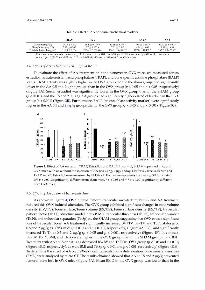
Table 2. Effect of AA on serum biochemical markers.
SHAM OVX E2 AA 0.5 AA 2
Calcium (mg/dl) 10.32 ˘ 0.303 10.8 ˘ 0.173 # 10.38 ˘ 0.277 * 10.4 ˘ 0.212 * 10.22 ˘ 0.205 **Phosphorus (mg/dl) 5.22 ˘ 0.507 7.7 ˘ 1.632 # 7.22 ˘ 0.963 6.86 ˘ 1.033 7.32 ˘ 1.094
Total cholesterol (mg/dl) 118.8 ˘ 9.834 197.4 ˘ 6.656 ### 156.6 ˘ 5.459 *** 177.8 ˘ 11.432 * 162.2 ˘ 14.533 **
Each value represents the mean ˘ SD for n = 5. # p < 0.05 and ### p < 0.001 significantly different from shammice. * p < 0.05, ** p < 0.01 and *** p < 0.001 significantly different from OVX mice.
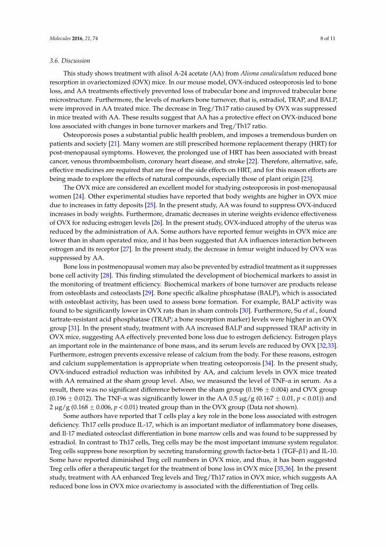
3.4. Effects of AA on Serum TRAP, E2, and BALP
To evaluate the effect of AA treatment on bone turnover in OVX mice, we measured serumestradiol, tartrate-resistant acid phosphatase (TRAP), and bone specific alkaline phosphatase (BALP)levels. TRAP activity was slightly higher in the OVX group than in the sham group, and significantlylower in the AA 0.5 and 2 µg/g groups than in the OVX group (p < 0.05 and p < 0.05, respectively)(Figure 3A). Serum estradiol was significantly lower in the OVX group than in the SHAM group(p < 0.001), and the 0.5 and 2.0 µg/g AA groups had significantly higher estradiol levels than the OVXgroup (p < 0.001) (Figure 3B). Furthermore, BALP (an osteoblast activity marker) were significantlyhigher in the AA 0.5 and 2 µg/g groups than in the OVX group (p < 0.05 and p < 0.001) (Figure 3C).
Molecules 2016, 21, 74 6 of 11
Table 2. Effect of AA on serum biochemical markers.
SHAM OVX E2 AA 0.5 AA 2 Calcium (mg/dl) 10.32 ± 0.303 10.8 ± 0.173 # 10.38 ± 0.277 * 10.4 ± 0.212 * 10.22 ± 0.205 **
Phosphorus (mg/dl) 5.22 ± 0.507 7.7 ± 1.632 # 7.22 ± 0.963 6.86 ± 1.033 7.32 ± 1.094 Total cholesterol (mg/dl) 118.8 ± 9.834 197.4 ± 6.656 ### 156.6 ± 5.459 *** 177.8 ± 11.432 * 162.2 ± 14.533 **
Each value represents the mean ± SD for n = 5. # p < 0.05 and ### p < 0.001 significantly different from sham mice. * p < 0.05, ** p < 0.01 and *** p < 0.001 significantly different from OVX mice.
3.4. Effects of AA on Serum TRAP, E2, and BALP
To evaluate the effect of AA treatment on bone turnover in OVX mice, we measured serum estradiol, tartrate-resistant acid phosphatase (TRAP), and bone specific alkaline phosphatase (BALP) levels. TRAP activity was slightly higher in the OVX group than in the sham group, and significantly lower in the AA 0.5 and 2 μg/g groups than in the OVX group (p < 0.05 and p < 0.05, respectively) (Figure 3A). Serum estradiol was significantly lower in the OVX group than in the SHAM group (p < 0.001), and the 0.5 and 2.0 μg/g AA groups had significantly higher estradiol levels than the OVX group (p < 0.001) (Figure 3B). Furthermore, BALP (an osteoblast activity marker) were significantly higher in the AA 0.5 and 2 μg/g groups than in the OVX group (p < 0.05 and p < 0.001) (Figure 3C).
Figure 3. Effect of AA on serum TRAP, Estradiol, and BALP. In control, SHAM -operated mice and OVX mice with or without the injection of AA (0.5 μg/g, 2 μg/g/day, I.P) for six weeks, Serum (A) TRAP and (B) Estradiol were measured by ELISA kit. Each value represents the mean ± SD for n = 4~5. ### p < 0.001, significantly different from sham mice. * p < 0.05 and *** p < 0.001 significantly different from OVX mice.
3.5. Effects of AA on Bone Microarchitecture
As shown in Figure 4, OVX altered femoral trabecular architecture, but E2 and AA treatment reduced this OVX-induced alteration. The OVX group exhibited significant changes in bone volume density (BV/TV), bone surface/bone volume (BS/BV), bone surface density (BS/TV), trabecular pattern factor (Tb.Pf), structure model index (SMI), trabecular thickness (Tb.Th), trabecular number (Tb.N), and trabecular separation (Tb.Sp) vs. the SHAM group, suggesting that OVX caused significant loss of trabecular bone. AA treatment significantly increased BV/TV, BS/TV, and Tb.N at doses of 0.5 and 2 μg/g vs. OVX mice (p < 0.01 and p < 0.001, respectively) (Figure 4A,C,G), and significantly increased Tb.Th at 0.5 and 2 μg/g (p < 0.05 and p < 0.001, respectively) (Figure 4F). In contrast, BS/BV, Tb.Pf, SMI, and Tb.Sp were higher in the OVX group than in the SHAM group (p < 0.001). Treatment with AA at 0.5 or 2.0 μg/g decreased BS/BV and Tb.Pf vs. OVX group (p < 0.05 and p < 0.01) (Figure 4B,D, respectively), as were SMI and Tb.Sp (p < 0.01 and p < 0.001, respectively) (Figure 4E,H). To determine the effect of AA on OVX-induced trabecular bone deterioration, bone mineral densities (BMD) were analyzed by micro-CT. The results obtained showed that AA at 0.5 and 2 μg/g prevented femoral bone loss in OVX mice (Figure 5A). Mean BMD in the OVX group was lower than in the SHAM group (p < 0.001), but was higher in the 2 μg/g AA group than in the OVX group (p < 0.05) (Figure 5B).
Figure 3. Effect of AA on serum TRAP, Estradiol, and BALP. In control, SHAM -operated mice andOVX mice with or without the injection of AA (0.5 µg/g, 2 µg/g/day, I.P) for six weeks, Serum (A)TRAP and (B) Estradiol were measured by ELISA kit. Each value represents the mean ˘ SD for n = 4~5.### p < 0.001, significantly different from sham mice. * p < 0.05 and *** p < 0.001 significantly differentfrom OVX mice.
3.5. Effects of AA on Bone Microarchitecture
As shown in Figure 4, OVX altered femoral trabecular architecture, but E2 and AA treatmentreduced this OVX-induced alteration. The OVX group exhibited significant changes in bone volumedensity (BV/TV), bone surface/bone volume (BS/BV), bone surface density (BS/TV), trabecularpattern factor (Tb.Pf), structure model index (SMI), trabecular thickness (Tb.Th), trabecular number(Tb.N), and trabecular separation (Tb.Sp) vs. the SHAM group, suggesting that OVX caused significantloss of trabecular bone. AA treatment significantly increased BV/TV, BS/TV, and Tb.N at doses of0.5 and 2 µg/g vs. OVX mice (p < 0.01 and p < 0.001, respectively) (Figure 4A,C,G), and significantlyincreased Tb.Th at 0.5 and 2 µg/g (p < 0.05 and p < 0.001, respectively) (Figure 4F). In contrast,BS/BV, Tb.Pf, SMI, and Tb.Sp were higher in the OVX group than in the SHAM group (p < 0.001).Treatment with AA at 0.5 or 2.0 µg/g decreased BS/BV and Tb.Pf vs. OVX group (p < 0.05 and p < 0.01)(Figure 4B,D, respectively), as were SMI and Tb.Sp (p < 0.01 and p < 0.001, respectively) (Figure 4E,H).To determine the effect of AA on OVX-induced trabecular bone deterioration, bone mineral densities(BMD) were analyzed by micro-CT. The results obtained showed that AA at 0.5 and 2 µg/g preventedfemoral bone loss in OVX mice (Figure 5A). Mean BMD in the OVX group was lower than in the

Molecules 2016, 21, 74 7 of 11
SHAM group (p < 0.001), but was higher in the 2 µg/g AA group than in the OVX group (p < 0.05)(Figure 5B).Molecules 2016, 21, 74 7 of 11
Figure 4. Effect of AA on trabecular morphometric parameters in distal femur of OVX mice. Mice were treated with vehicle, AA (0.5, 2 μg/g/day, I.P) for 6 weeks. (A) Bone volume/tissue volume (BV/TV); (B) bone surface/bone volume (BS/BV); (C) bone surface/tissue volume (BS/TV); (D) trabecular pattern factor (Tb.Pf); (E) structure model index (SMI); (F) trabecular thickness (Tb.Th); (G) trabecular number (Tb.N); and (H) trabecular separation (Tb.Sp) as analyzed with micro-CT Skyscan CTAn software. Each value represents the mean ± SD for n = 5. ### p < 0.001, significantly different from sham mice. * p < 0.05, ** p < 0.01 and *** p < 0.001 significantly different from OVX mice.
Figure 5. The effect of treatment with AA (alisol A-24 acetate) on the femur trabecular microarchitecture in OVX (ovaritectomy) mice: (A) two dimensional micro-computed tomography (micro-CT) images of the femoral trabecular bone; (B) bone mineral density (BMD) was assessed by micro-CT; and (C) histological analysis of femur with H & E and Masson’s trichrome staining. Magnification: 40-fold. Each value represents the mean ± SD for n = 5. ### p < 0.001, significantly different from sham mice. * p < 0.05 and *** p < 0.001 significantly different from OVX mice.
Figure 4. Effect of AA on trabecular morphometric parameters in distal femur of OVX mice. Mice weretreated with vehicle, AA (0.5, 2 µg/g/day, I.P) for 6 weeks. (A) Bone volume/tissue volume (BV/TV);(B) bone surface/bone volume (BS/BV); (C) bone surface/tissue volume (BS/TV); (D) trabecularpattern factor (Tb.Pf); (E) structure model index (SMI); (F) trabecular thickness (Tb.Th); (G) trabecularnumber (Tb.N); and (H) trabecular separation (Tb.Sp) as analyzed with micro-CT Skyscan CTAnsoftware. Each value represents the mean ˘ SD for n = 5. ### p < 0.001, significantly different fromsham mice. * p < 0.05, ** p < 0.01 and *** p < 0.001 significantly different from OVX mice.
Molecules 2016, 21, 74 7 of 11
Figure 4. Effect of AA on trabecular morphometric parameters in distal femur of OVX mice. Mice were treated with vehicle, AA (0.5, 2 μg/g/day, I.P) for 6 weeks. (A) Bone volume/tissue volume (BV/TV); (B) bone surface/bone volume (BS/BV); (C) bone surface/tissue volume (BS/TV); (D) trabecular pattern factor (Tb.Pf); (E) structure model index (SMI); (F) trabecular thickness (Tb.Th); (G) trabecular number (Tb.N); and (H) trabecular separation (Tb.Sp) as analyzed with micro-CT Skyscan CTAn software. Each value represents the mean ± SD for n = 5. ### p < 0.001, significantly different from sham mice. * p < 0.05, ** p < 0.01 and *** p < 0.001 significantly different from OVX mice.
Figure 5. The effect of treatment with AA (alisol A-24 acetate) on the femur trabecular microarchitecture in OVX (ovaritectomy) mice: (A) two dimensional micro-computed tomography (micro-CT) images of the femoral trabecular bone; (B) bone mineral density (BMD) was assessed by micro-CT; and (C) histological analysis of femur with H & E and Masson’s trichrome staining. Magnification: 40-fold. Each value represents the mean ± SD for n = 5. ### p < 0.001, significantly different from sham mice. * p < 0.05 and *** p < 0.001 significantly different from OVX mice.
Figure 5. The effect of treatment with AA (alisol A-24 acetate) on the femur trabecular microarchitecturein OVX (ovaritectomy) mice: (A) two dimensional micro-computed tomography (micro-CT) imagesof the femoral trabecular bone; (B) bone mineral density (BMD) was assessed by micro-CT; and(C) histological analysis of femur with H & E and Masson’s trichrome staining. Magnification: 40-fold.Each value represents the mean ˘ SD for n = 5. ### p < 0.001, significantly different from sham mice.* p < 0.05 and *** p < 0.001 significantly different from OVX mice.
Molecules 2016, 21, 74 8 of 11
3.6. Discussion
This study shows treatment with alisol A-24 acetate (AA) from Alisma canaliculatum reduced boneresorption in ovariectomized (OVX) mice. In our mouse model, OVX-induced osteoporosis led to boneloss, and AA treatments effectively prevented loss of trabecular bone and improved trabecular bonemicrostructure. Furthermore, the levels of markers bone turnover, that is, estradiol, TRAP, and BALP,were improved in AA treated mice. The decrease in Treg/Th17 ratio caused by OVX was suppressedin mice treated with AA. These results suggest that AA has a protective effect on OVX-induced boneloss associated with changes in bone turnover markers and Treg/Th17 ratio.
Osteoporosis poses a substantial public health problem, and imposes a tremendous burden onpatients and society [21]. Many women are still prescribed hormone replacement therapy (HRT) forpost-menopausal symptoms. However, the prolonged use of HRT has been associated with breastcancer, venous thromboembolism, coronary heart disease, and stroke [22]. Therefore, alternative, safe,effective medicines are required that are free of the side effects on HRT, and for this reason efforts arebeing made to explore the effects of natural compounds, especially those of plant origin [23].
The OVX mice are considered an excellent model for studying osteoporosis in post-menopausalwomen [24]. Other experimental studies have reported that body weights are higher in OVX micedue to increases in fatty deposits [25]. In the present study, AA was found to suppress OVX-inducedincreases in body weights. Furthermore, dramatic decreases in uterine weights evidence effectivenessof OVX for reducing estrogen levels [26]. In the present study, OVX-induced atrophy of the uterus wasreduced by the administration of AA. Some authors have reported femur weights in OVX mice arelower than in sham operated mice, and it has been suggested that AA influences interaction betweenestrogen and its receptor [27]. In the present study, the decrease in femur weight induced by OVX wassuppressed by AA.
Bone loss in postmenopausal women may also be prevented by estradiol treatment as it suppressesbone cell activity [28]. This finding stimulated the development of biochemical markers to assist inthe monitoring of treatment efficiency. Biochemical markers of bone turnover are products releasefrom osteoblasts and osteoclasts [29]. Bone specific alkaline phosphatase (BALP), which is associatedwith osteoblast activity, has been used to assess bone formation. For example, BALP activity wasfound to be significantly lower in OVX rats than in sham controls [30]. Furthermore, Su et al., foundtartrate-resistant acid phosphatase (TRAP; a bone resorption marker) levels were higher in an OVXgroup [31]. In the present study, treatment with AA increased BALP and suppressed TRAP activity inOVX mice, suggesting AA effectively prevented bone loss due to estrogen deficiency. Estrogen playsan important role in the maintenance of bone mass, and its serum levels are reduced by OVX [32,33].Furthermore, estrogen prevents excessive release of calcium from the body. For these reasons, estrogenand calcium supplementation is appropriate when treating osteoporosis [34]. In the present study,OVX-induced estradiol reduction was inhibited by AA, and calcium levels in OVX mice treatedwith AA remained at the sham group level. Also, we measured the level of TNF-α in serum. As aresult, there was no significant difference between the sham group (0.196 ˘ 0.004) and OVX group(0.196 ˘ 0.012). The TNF-α was significantly lower in the AA 0.5 µg/g (0.167 ˘ 0.01, p < 0.01)) and2 µg/g (0.168 ˘ 0.006, p < 0.01) treated group than in the OVX group (Data not shown).
Some authors have reported that T cells play a key role in the bone loss associated with estrogendeficiency. Th17 cells produce IL-17, which is an important mediator of inflammatory bone diseases,and Il-17 mediated osteoclast differentiation in bone marrow cells and was found to be suppressed byestradiol. In contrast to Th17 cells, Treg cells may be the most important immune system regulator.Treg cells suppress bone resorption by secreting transforming growth factor-beta 1 (TGF-β1) and IL-10.Some have reported diminished Treg cell numbers in OVX mice, and thus, it has been suggestedTreg cells offer a therapeutic target for the treatment of bone loss in OVX mice [35,36]. In the presentstudy, treatment with AA enhanced Treg levels and Treg/Th17 ratios in OVX mice, which suggests AAreduced bone loss in OVX mice ovariectomy is associated with the differentiation of Treg cells.

Molecules 2016, 21, 74 9 of 11
The clinically measurable properties of bone that have been shown to independently predicta future osteoporotic fracture are bone mineral density (BMD), and bone microarchitecture, thatis, trabecular bone volume, number, and thickness [37]. Ovariectomy is typically linked withdeteriorations in trabecular structure and BMD [38]. The present study shows that AA has a positiveeffect on trabecular morphometric parameters, that is, bone volume density (BV/TV), thickness,number, separation, and BMD in OVX mice. Furthermore, 2D images and histological analysis showedthat AA protects against bone loss in OVX mice. These results suggest that AA has inhibitory effect onOVZ-induced bone loss and deterioration.
4. Conclusions
In summary, we provide evidence that six weeks of 0.5 or 2 µg/g of AA administration toosteoporotic mice suppresses body weight increases and uterine weight reductions, and improvesbone biochemical markers, such as, Ca, TCHO, BALP, estradiol, and TRAP levels and Treg/Th17ratios. Furthermore, AA administration improved the femoral BMD and trabecular microstructure.We believe that alisol A-24 acetate from Alisma canaliculatum has potential for further development as anatural alternative for the management of postmenopausal osteoporosis.
Acknowledgments: This research was supported by the Suncheon Research Center for Natural Medicines andthe National Research Foundation of Korea (NRF) grant funded by the Ministry of Science, ICT & Future Planning(MSIP) (2015R1A4A1041219).
Author Contributions: Y.-H.H. performed experiments and analyzed the data. K.-Y.K., S.-J.L. and Y.-J.S.participate in the data analysis. S.-J.N. provided alisol A24-acetate. Y.-H.H. and S.-T.Y. conceived and designedthe study and wrote the paper.
Conflicts of Interest: The authors have no potential conflict of interest to declare.
References
1. Raggatt, L.J.; Partridge, N.C. Cellular and molecular mechanisms of bone remodeling. J. Biol. Chem. 2010,285, 25103–25108. [CrossRef] [PubMed]
2. Tseng, S.H.; Sung, C.H.; Chen, L.G.; Lai, Y.J.; Chang, W.S.; Sung, H.C.; Wang, C.C. Comparison of chemicalcompositions and osteoprotective effects of different sections of velvet antler. J. Ethnopharmacol. 2014, 151,352–360. [CrossRef] [PubMed]
3. Tantikanlayaporn, D.; Wichit, P.; Weerachayaphorn, J.; Chairoungdua, A.; Chuncharunee, A.; Suksamrarn, A.;Piyachaturawat, P. Bone sparing effect of a novel phytoestrogen diarylheptanoid from Curcuma comosaRoxb. in ovariectomized rats. PLoS ONE 2013. [CrossRef] [PubMed]
4. Raisz, L.G. Pathogenesis of osteoporosis: Concepts, conflicts, and prospects. J. Clin. Investig. 2005, 115,3318–3325. [CrossRef] [PubMed]
5. Lerner, U.H. Bone remodeling in post-menopausal osteoporosis. J. Dent. Res. 2006, 85, 584–595. [CrossRef][PubMed]
6. Tyagi, A.M.; Srivastava, K.; Mansoori, M.N.; Trivedi, R.; Chattopadhyay, N.; Singh, D. Estrogen deficiencyinduces the differentiation of IL-17 secreting Th17 cells: A new candidate in the pathogenesis of osteoporosis.PLoS ONE 2012. [CrossRef] [PubMed]
7. Zhu, J.; Paul, W.E. Heterogeneity and plasticity of T helper cells. Cell Res. 2010, 20, 4–12. [CrossRef] [PubMed]8. Yang, X.O.; Zhang, H.; Kim, B.S.; Niu, X.; Peng, J.; Chen, Y.; Kerketta, R.; Lee, Y.H.; Chang, S.H.; Corry, D.B.;
et al. The signaling suppressor CIS controls proallergic T cell development and allergic airway inflammation.Nat. Immunol. 2013, 14, 732–740. [CrossRef] [PubMed]
9. Bedoya, S.K.; Lam, B.; Lau, K.; Larkin, J. Th17 cells in immunity and autoimmunity. Clin. Dev. Immunol. 2013.[CrossRef] [PubMed]
10. Afzali, B.; Lombardi, G.; Lechler, R.I.; Lord, G.M. The role of T helper 17 (Th17) and regulatory T cells (Treg)in human organ transplantation and autoimmune disease. Clin. Exp. Immunol. 2007, 148, 32–46. [CrossRef][PubMed]

Molecules 2016, 21, 74 10 of 11
11. Alves, C.H.; Farrell, E.; Vis, M.; Colin, E.M.; Lubberts, E. Animal Models of Bone Loss in InflammatoryArthritis: From Cytokines in the Bench to Novel Treatments for Bone Loss in the Bedside-a ComprehensiveReview. Clin. Rev. Allergy Immunol. 2015. [CrossRef] [PubMed]
12. Tyagi, A.M.; Mansoori, M.N.; Srivastava, K.; Khan, M.P.; Kureel, J.; Dixit, M.; Shukla, P.; Trivedi, R.;Chattopadhyay, N.; Singh, D. Enhanced immunoprotective effects by anti-IL-17 antibody translates toimproved skeletal parameters under estrogen deficiency compared with anti-RANKL and anti-TNF-αantibodies. J. Bone Miner. Res. 2014, 29, 1981–1992. [CrossRef] [PubMed]
13. Downey, P.A.; Siegel, M.I. Bone biology and the clinical implications for osteoporosis. Phys. Ther. 2006, 86,77–91. [PubMed]
14. Lee, J.W.; Jhee, O.; Yuan, H.; Kim, T.; Kim, D.; Lee, M.; Om, A.; Lee, B.; Park, S.K.; Kang, J. Effect of Koreanoriental medicine extract on bone mass as compared with alendronate in ovariectomized rats. J. Med. Food.2005, 8, 369–376. [CrossRef] [PubMed]
15. Hossain, M.E.; Kim, G.M.; Lee, S.K.; Yang, C.J. Growth performance, meat yield, oxidative stability, andFatty Acid composition of meat from broilers fed diets supplemented with a medicinal plant and probiotics.Asian-Australas. J. Anim. Sci. 2012, 25, 1159–1168. [CrossRef] [PubMed]
16. Hossain, M.E.; Ko, S.Y.; Kim, G.M.; Firman, J.D.; Yang, C.J. Evaluation of probiotic strains for developmentof fermented Alisma canaliculatum and their effects on broiler chickens. Poult. Sci. 2012, 91, 3121–3131.[CrossRef] [PubMed]
17. Mikamo, H.; Kawazoe, K.; Izumi, K.; Sato, Y.; Tamaya, T. Effects of crude herbal ingredients on intrauterineinfection in a rat model. Curr. Ther. Res. 1998, 59, 122–127. [CrossRef]
18. Huang, Y.T.; Huang, D.M.; Chueh, S.C.; Teng, C.M.; Guh, J.H. Alisol B acetate, a triterpene from Alismatisrhizoma, induces Bax nuclear translocation and apoptosis in human hormone-resistant prostate cancer PC-3cells. Cancer Lett. 2006, 231, 270–278. [CrossRef] [PubMed]
19. Jang, M.K.; Han, Y.R.; Nam, J.S.; Han, C.W.; Kim, B.J.; Jeong, H.S.; Ha, K.T.; Jung, M.H. Protective Effects ofAlisma orientale Extract against Hepatic Steatosis via Inhibition of Endoplasmic Reticulum Stress. Int. J.Mol. Sci. 2015, 16, 26151–26165. [CrossRef] [PubMed]
20. Kim, K.J.; Leutou, A.S.; Yeon, J.T.; Choi, S.W.; Kim, S.H.; Yee, S.T.; Choi, K.H.; Nam, S.J.; Son, Y.J. TheInhibitory Effect of Alisol A 24-Acetate from Alisma canaliculatum on Osteoclastogenesis. Int. J. Endocrinol.2015. [CrossRef] [PubMed]
21. Ray, N.F.; Chan, J.K.; Thamer, M.; Melton, L.J. Medical expenditures for the treatment of osteoporoticfractures in the United States in 1995: Report from the National Osteoporosis Foundation. J. Bone Miner. Res.1997, 12, 24–35. [CrossRef] [PubMed]
22. Canonico, M.; Plu-Bureau, G.; Lowe, G.D.; Scarabin, P.Y. Hormone replacement therapy and risk of venousthromboembolism in postmenopausal women: Systematic review and meta-analysis. BMJ 2008, 336,1227–1231. [CrossRef] [PubMed]
23. Liu, Z.G.; Zhang, R.; Li, C.; Ma, X.; Liu, L.; Wang, J.P.; Mei, Q.B. The osteoprotective effect of Radix Dipsaciextract in ovariectomized rats. J. Ethnopharmacol. 2009, 123, 74–81. [CrossRef] [PubMed]
24. Wronski, T.J.; Dann, L.M.; Qi, H.; Yen, C.F. Skeletal effects of withdrawal of estrogen and diphosphonatetreatment in ovariectomized rats. Calcif. Tissue Int. 1993, 53, 210–216. [CrossRef] [PubMed]
25. Notomi, T.; Okimoto, N.; Okazaki, Y.; Nakamura, T.; Suzuki, M. Tower climbing exercise started 3 monthsafter ovariectomy recovers bone strength of the femur and lumbar vertebrae in aged osteopenic rats. J. BoneMiner. Res. 2003, 18, 140–149. [CrossRef] [PubMed]
26. Hidaka, S.; Okamoto, Y.; Nakajima, K.; Suekawa, M.; Liu, S.Y. Preventive effects of traditional Chinese(Kampo) medicines on experimental osteoporosis induced by ovariectomy in rats. Calcif. Tissue Int. 1997, 61,239–246. [CrossRef] [PubMed]
27. Park, J.A.; Ha, S.K.; Kang, T.H.; Oh, M.S.; Cho, M.H.; Lee, S.Y.; Park, J.H.; Kim, S.Y. Protective effect ofapigenin on ovariectomy-induced bone loss in rats. Life Sci. 2008, 82, 1217–1223. [CrossRef] [PubMed]
28. Sims, N.A.; Morris, H.A.; Moore, R.J.; Durbridge, T.C. Estradiol treatment transiently increases trabecularbone volume in ovariectomized rats. Bone 1996, 19, 455–461. [CrossRef]
29. Swaminathan, R. Biochemical markers of bone turnover. Clin. Chim. Acta. 2001, 313, 95–105. [CrossRef]30. Fahmy, S.R.; Soliman, A.M.; Sayed, A.A.; Marzouk, M. Possible antiosteoporotic mechanism of Cicer
arietinum extract in ovariectomized rats. Int. J. Clin. Exp. Pathol. 2015, 8, 3477–3490. [PubMed]
Molecules 2016, 21, 74 11 of 11
31. Su, S.J.; Yeh, Y.T.; Shyu, H.W. The preventive effect of biochanin A on bone loss in ovariectomized rats:Involvement in regulation of growth and activity of osteoblasts and osteoclasts. Evid. Based Complement.Altern. Med. 2013. [CrossRef]
32. Mohamed, M.K.; Abdel-Rahman, A.A. Effect of long-term ovariectomy and estrogen replacement on theexpression of estrogen receptor gene in female rats. Eur. J. Endocrinol. 2000, 142, 307–314. [CrossRef][PubMed]
33. Lim, D.W.; Lee, Y.; Kim, Y.T. Preventive effects of Citrus unshiu peel extracts on bone and lipid metabolismin OVX rats. Molecules 2014, 19, 783–794. [CrossRef] [PubMed]
34. Davis, J.W.; Ross, P.D.; Johnson, N.E.; Wasnich, R.D. Estrogen and calcium supplement use amongJapanese-American women: Effects upon bone loss when used singly and in combination. Bone 1995,17, 369–373. [CrossRef]
35. Lai, N.; Zhang, Z.; Wang, B.; Miao, X.; Guo, Y.; Yao, C.; Wang, Z.; Wang, L.; Ma, R.; Li, X.; et al. Regulatoryeffect of traditional Chinese medicinal formula Zuo-Gui-Wan on the Th17/Treg paradigm in mice with boneloss induced by estrogen deficiency. J. Ethnopharmacol. 2015, 166, 228–239. [CrossRef] [PubMed]
36. Liu, J.C.; Zhou, C.H.; Zhang, X.; Chen, Y.; Xu, B.L.; Cui, L.; Xu, D.H. Effect of 1,25-dihydroxyvitamin D3 onregulatory T cells in ovariectomized mice. Biomed. Environ. Sci. 2014, 27, 779–785. [PubMed]
37. Recker, R.; Masarachia, P.; Santora, A.; Howard, T.; Chavassieux, P.; Arlot, M.; Rodan, G.; Wehren, L.;Kimmel, D. Trabecular bone microarchitecture after alendronate treatment of osteoporotic women. Curr. Med.Res. Opin. 2005, 21, 185–194. [CrossRef] [PubMed]
38. Huang, G.; Wu, J.; Wang, S.; Wei, Y.; Chen, F.; Chen, J.; Shi, J.; Xia, J. Pycnogenol® treatment inhibits bonemineral density loss and trabecular deterioration in ovariectomized rats. Int. J. Clin. Exp. Med. 2015, 8,10893–10901. [PubMed]
Sample Availability: Samples of the compound Alisol A 24 acetate is available from the authors.
© 2016 by the authors; licensee MDPI, Basel, Switzerland. This article is an open accessarticle distributed under the terms and conditions of the Creative Commons by Attribution(CC-BY) license (http://creativecommons.org/licenses/by/4.0/).