the 'presyrinx' state: a reversible myelopathic … ajnr am j neuroradiol 20:7–20,...
TRANSCRIPT

7
AJNR Am J Neuroradiol 20:7–20, January 1999
The ‘‘Presyrinx’’ State: A Reversible MyelopathicCondition That May Precede Syringomyelia
Nancy J. Fischbein, William P. Dillon, Charles Cobbs, and Philip R. Weinstein
BACKGROUND AND PURPOSE: Alteration of CSF flow has been proposed to be an importantmechanism leading to the development of syringomyelia. We hypothesize that a ‘‘presyrinx’’ con-dition attributable to a potentially reversible alteration in normal CSF flow exists and that itsappearance may be caused by variations in the competence of the central canal of the spinal cord.
METHODS: Five patients with clinical evidence of myelopathy, no history of spinal cordtrauma, enlargement of the cervical spinal cord with T1 and T2 prolongation but no cavitation,evidence of altered or obstructed CSF flow, and no evidence of intramedullary tumor or aspinal vascular event underwent MR imaging before and after intervention that alleviatedobstruction to CSF flow.
RESULTS: Preoperatively, all patients had enlarged spinal cords and parenchymal T1 andT2 prolongation without cavitation. Results of MR examinations after intervention showedresolution of cord enlargement and normalization or improvement of cord signal abnormalities.In one patient with severe arachnoid adhesions who initially improved after decompression,late evolution into syringomyelia occurred in association with continued CSF obstruction.
CONCLUSION: Nontraumatic obstruction of the CSF pathways in the spine may result inspinal cord parenchymal T2 prolongation that is reversible after restoration of patency of CSFpathways. We refer to this MR appearance as the ‘‘presyrinx’’ state and stress the importanceof timely intervention to limit progression to syringomyelia.
Syringomyelia occurs in a number of clinical set-tings, most commonly after trauma (1–3) or in as-sociation with a Chiari malformation (4–7). Othercauses include infectious or inflammatory arach-noiditis (7–11), tumors of the spine and spinal cord(7), posterior fossa tumors (12), and cervical spon-dylosis (13). Syrinxes typically appear as well-de-fined discrete cavities containing fluid that is isoin-tense with CSF on all sequences (14, 15). Thepathophysiology of syringomyelia is controversial,but experimental models (16–18) and clinical stud-ies (19) have implicated alterations in CSF flow asa significant factor in the development and pro-gression of certain types of syringomyelia. The hy-pothesis that CSF normally flows along perivas-cular spaces within the parenchyma of the spinalcord to the central canal is supported by experi-mental studies (20–25). Variations in patency of thecentral canal of the spinal cord have been associ-
Received March 10, 1998; accepted after revision August 26.From the Departments of Radiology (N.J.F., W.P.D.) and
Neurosurgery (C.C., P.R.W.), University of California, SanFrancisco.
Address reprint requests to Nancy J. Fischbein, MD, De-partment of Radiology, Box 0628, University of California,San Francisco, 505 Parnassus Ave, San Francisco, CA 94143.
q American Society of Neuroradiology
ated with the development of different types of sy-ringomyelia (18, 26) and most likely play a role indetermining the location of a syrinx remote from afocus of CSF obstruction, such as is commonlyseen in the Chiari I malformation. The reversibilityof syringomyelia after restoration of CSF pathwayshas been well documented in patients undergoingposterior fossa decompression for Chiari malfor-mations, removal of extradural masses, and lysis ofadhesions (9, 12, 27).
We have identified five myelopathic patientswith no history of spine trauma who had underly-ing conditions associated with alterations of CSFflow. These patients had enlargement and T2 pro-longation of the spinal cord, without frank cavita-tion, on MR imaging studies that improved or re-versed after restoration of CSF flow pathways. Wehypothesized on the basis of historical, imaging,and surgical findings that obstruction to normalCSF flow pathways resulted in the cord enlarge-ment and MR signal abnormalities that reversed af-ter restoration of normal CSF pathways. We alsohypothesized that variation in patency of the centralcanal of the spinal cord plays a role in the genesisof this ‘‘presyrinx’’ state.
MethodsOver a 2-year period, five patients who met the following
criteria were identified prospectively: clinical evidence of my-

AJNR: 20, January 19998 FISCHBEIN
Summary of Patient Data
Patient Figure No. Age (y)/Sex History Clinical Condition Pre-op Previous Surgery
1 1 2/M Chiari I malformation Severe headaches, progressiveclumsiness in limb move-ments over the course of 1 y
Limited decompression (suboc-cipital craniectomy) forChiari I malformation
2 2 69/F Polymyalgia rheumatica, previ-ous epidural abscess associ-ated with osteomyelitis ofthe cervical spine
Progressive quadriparesis, pro-gressive loss of sensationand motor control in hands
Decompression of epidural ab-scess via C6 laminectomy18 mo earlier
3 4 41/M Remote h/o head trauma; basi-lar arachnoid adhesions; h/ohydrocephalus
Progressive neck pain andspastic quadriparesis
Posterior fossa decompressionVP shunt for hydrocephalus
4 3 40/F Grade III SAH caused by an-eurysm, s/p GDC coil em-bolization; course c/b S. epi-dermidis meningitis
Neck pain and quadriparesis Coil embolization of aneu-rysm, external ventriculardrainage of CSF
5 NS 77/F Severe cervical spondylosten-osis and mild basilar im-pression
Progressive weakness in botharms, R . L, and handnumbness; spasticity and in-coordination of legs; noneck pain
None
Note.—FM indicates foramen magnum; SAH, subarachnoid hemorrhage; NS, not shown; VP, ventriculoperitoneal; S. epidermidis, Staphylococcusepidermidis; h/o, history of; pre-op, preoperatively; post-op, postoperatively.
elopathy, no history of spinal cord trauma, enlargement of thecervical cord with parenchymal T1 and T2 prolongation butno frank cavitation, and no evidence of intramedullary tumoror a spinal vascular event as the cause of cord signal changes.No patient had evidence of active inflammatory or demyelin-ating disease or received steroid therapy. In addition, all pa-tients had obstruction to CSF flow at the level of the foramenmagnum or the spinal epidural or subarachnoid space as evi-denced by historical and/or imaging findings. Our patients in-cluded two males and three females and ranged in age from 2to 77 years. Clinical records and imaging studies for thesepatients were reviewed.
All patients underwent preoperative imaging on a 1.5-T MRsystem. Imaging sequences included conventional spin-echoT1-weighted images (500/14 [TR/TE], 4 mm thick, 256 3 256matrix) and fast spin-echo T2-weighted images (3000/105eff,echo train length of 8, 3 mm thick, 256 3 256 matrix) obtainedin the sagittal plane in addition to axial T1-weighted images.A proton density–weighted sequence was not obtained, sinceit is not part of our routine spine imaging protocol. Contrast-enhanced images were obtained in three patients and includedsagittal and axial T1-weighted sequences. MR examinationswere conducted on all patients after surgical intervention andincluded, at a minimum, sagittal T1-weighted and fast spin-echo T2-weighted sequences. In addition, one patient under-went CT myelography preoperatively, and one patient under-went CT myelography postoperatively. Only one patient hadan MR flow study performed preoperatively, using a cinephase-contrast technique (24/minimum [TR/TE], flip angle of308, 256 3 128 matrix, flow compensation and peripheral gat-ing applied, velocity-encoding gradient 5 5 cm/s) to evaluateCSF flow at the foramen magnum.
ResultsA summary of our patients’ histories, as well as
clinical and imaging findings, is presented in the
Table. All patients underwent MR imaging of thecervical spinal cord to evaluate myelopathic symp-toms, although in one case (patient 1) headacheswere the dominant clinical feature. The clinical pre-sentations were similar to that of patients with cen-tral cystic myelopathy (7, 28), including increasingloss of motor function or weakness in all patients,sensory changes in two patients, increased spastic-ity in two patients, and radicular pain in one pa-tient. The conditions that predisposed our patientsto alterations in CSF flow dynamics and myelop-athy included the following: Chiari I malformationwith severe tonsillar herniation; previous osteo-myelitis complicated by epidural abscess, menin-gitis, and arachnoiditis; basilar arachnoid adhesionsrelated to previous head trauma, traumatic sub-arachnoid hemorrhage, and posterior fossa surgery;subarachnoid hemorrhage complicated by menin-gitis, leading to severe hydrocephalus and tonsillarherniation; and rheumatoid arthritis with severecervical spondylosis and spinal stenosis accompa-nied by basilar impression. In all five cases, thelesion occurred in the cervical spinal cord.
Results of preoperative MR imaging revealed avariable degree of enlargement of the cervical cordin all patients. All patients had abnormal T1 andT2 prolongation of the cervical spinal cord signalextending over a variable distance. The T1 signalwas not as low as CSF in any case, and the marginsof the T1 signal abnormality were not sharply de-fined. No frank cavitation was observed in any pa-

AJNR: 20, January 1999 ‘‘PRESYRINX’’ STATE 9
TABLE 1: Continued.
MR Findings Pre-Op Surgical Intervention Clinical Findings Post-op MR Findings Post-op
Progressive T1 and T2 prolon-gation within upper cervicalcord; no cavity; tonsillarherniation with reduced CSFflow at FM
Aggressive suboccipital de-compression and partial re-section of cerebellar tonsils
Improved headaches, decreasedclumsiness
Near-resolution of T1 and T2prolongation in cord sub-stance, improved CSF flowat FM
Marked enlargement of mid-cervical cord; T1 and T2,prolongation from C3–C4 toT1–T2; large ventrolateralosteophyte at C3–C4 level
C3 through C7 laminectomy,lysis of subdural adhesions,and sectioning of dentateligaments at C3 and C4
Substantial improvement instrength, sensation in armsand legs within hours aftersurgery
Decrease in cord caliber, per-sistent T1 and T2 prolonga-tion below C3–C4 level,where the cord remains fo-cally adhesed
Cord enlargement from C1 toT3, with T1 and T2 prolon-gation
C6 and C7 laminectomy withexploration and myelotomybut no shunt as a syrinxcavity was not found
Slight improvement in righthand function, decreasedneck and arm pain
Progressive decrease in cordenlargement and T1 and T2prolongation, then late de-cline and progression tofrank syrinx formation
Cervical cord enlargement withT1 and T2 prolongation, en-larged ventricles with tonsil-lar herniation
VP shunting to relieve hydro-cephalus
Improved mental status, de-creased neck stiffness, reso-lution of quadriparesis
Complete resolution of cordenlargement and signal ab-normalities
Severe cervical spondylosten-osis, basilar impression, andatlantoaxial subluxation;cord enlargement and edemafrom cervicomedullary junc-tion to C6–C7
C1 to C6 laminectomies withmedial facetectomies, thenposterior fusion from C4 toC6
Stabilization of neurologicfindings, mild improvementin lower extremity spasticity
Decreased cord enlargementand significant decrease incord signal abnormality
tient. Mild parenchymal enhancement of the uppercervical cord was observed in one case; this patienthad no evidence of an acute clinical decline or sys-temic infectious or inflammatory condition, and herCSF profile was benign.
Evidence of a discrete level of obstruction tonormal CSF flow was detected preoperatively in allbut one patient. Patient 1 (Fig 1) had a Chiari Imalformation with markedly narrowed CSF spacesat the level of the foramen magnum; a cine phase-contrast CSF flow study showed markedly restrict-ed flow of CSF through the foramen magnum. Pa-tient 2 (Fig 2) underwent preoperative CTmyelography that showed a decreased flow of con-trast material around a swollen cervical spinal cordand a myelographic block at the C3–C4 levelcaused by a large ventrolateral osteophyte and pos-sibly adhesions as well; subtle increased density ofthe cord on delayed images was also found. Patient4 (Fig 3) had a markedly enlarged fourth ventricleand tonsillar herniation that resulted in obstructionto flow at the level of the foramen magnum. Patient5 had multilevel spondylostenosis and markedcompression at the level of the foramen magnumcaused by basilar impression and atlantoaxial sub-luxation. In all these cases, the level of obstructionwas rostral to the level of cord signal changes. Thelevel of obstruction in patient 3 was not clear pre-operatively; this patient had a history of previousposterior fossa surgery and basilar adhesions thatwere most likely causing obstruction to flow at thelevel of the foramen magnum, but he also had amild congenital spinal stenosis with superimposed
acquired stenosis at the C5–C6 and C6–C7 levels.In this case, it is unclear whether the spinal cordsignal changes were only caudal or both rostral andcaudal to the level of obstruction to CSF flow.
Four of five patients underwent surgical interven-tion directed at restoring patency of CSF pathways.Patient 3 (Fig 4) underwent laminectomy and intra-dural exploration for placement of a syringopleuralshunt, but this procedure was aborted because no syr-inx was found (see below). Procedures performed aresummarized in the Table. Only patient 3 underwentintraoperative sonographic evaluation of the spinalcord. Postoperatively, all patients experienced clinicalimprovement to varying degrees.
Postoperative MR imaging was performed in allpatients from 1 week to 1 year after surgery. Inall cases, the postoperative images showed a re-duction in cord caliber, as well as improvement orresolution of T1 and T2 signal abnormalities. Onepatient (patient 3, Fig 4) experienced a subsequentdeterioration in his clinical status after having hadsome improvement in clinical and imaging find-ings in the immediate postoperative period afterlower cervical laminectomy and myelotomy. Asecond postoperative image 6 weeks after surgeryshowed an increase in central parenchymal T2prolongation. No definite evidence was found forrecurrent CSF obstruction at the surgical level, butthe cord remained deformed at the level of theforamen magnum, and it was considered likelythat obstruction to normal CSF flow remained atthe foramen magnum level that had not been ad-dressed surgically; a cine phase-contrast CSF flow

AJNR: 20, January 199910 FISCHBEIN
study was not performed at this time, because thissequence was unavailable at the rehabilitation hos-pital where the patient was imaged. No further in-tervention was undertaken after that study for psy-chosocial reasons, and the patient was reexaminedby his neurosurgeon 10 months later, because ofsymptom progression. A third postoperative studyat this time revealed progression to frank syrinxformation. At this time, a syringopleural shunt waseasily placed, but the patient experienced onlyminimal symptomatic improvement.
DiscussionThe understanding of the pathogenesis of syrin-
gomyelia has been advanced by the important patho-logic studies of Milhorat et al (7, 27, 28). On the
basis of detailed histopathologic findings, they distin-guished among three types of spinal cord cavities: 1)dilatations of the central canal that communicate di-rectly with the fourth ventricle (communicating syr-inxes); 2) noncommunicating dilatations of the cen-tral canal that arise below a syrinx-free segment ofspinal cord; and 3) extracanalicular syrinxes that orig-inate in the spinal cord parenchyma and do not com-municate with the central canal. By correlating withclinical parameters, they were able to associate thesedistinct cavitary patterns with different mechanismsof pathogenesis. Communicating syrinxes were foundin association with hydrocephalus and were causedby obstruction of CSF circulation distal to the outletsof the fourth ventricle. Noncommunicating syrinxeswere associated with disorders of CSF dynamics inthe spinal subarachnoid space, such as the Chiari I

AJNR: 20, January 1999 ‘‘PRESYRINX’’ STATE 11
FIG 1. Patient 1.A, Sagittal T1-weighted image (600/8/2) shows a Chiari I malformation, with tonsillar herniation to the mid-C2 level and a pointed configuration
to the cerebellar tonsils (arrow).B, Sagittal T2-weighted image (3000/105eff /3) shows T2 prolongation within the spinal cord parenchyma at the C2–C3 level.C, Sagittal images from a cine phase-contrast flow study in systole (left panel) and diastole (right panel), sensitized to flow in the superior-
to-inferior direction (see text for parameters). Note the absence of flow-related phase change at the level of the foramen magnum, as well asprominent tonsillar motion in both systole and diastole, with the curved arrows indicating the position of the tonsillar tips in systole and diastole.Subtle linear low signal is present anterior to the tonsil in diastole (right panel, straight arrow), indicating minimal flow between the fourthventricle and the spinal subarachnoid space below the level of the foramen magnum.
D, Sagittal T1-weighted image (500/14/3) obtained 6 weeks later after limited extradural decompression of the foramen magnum. Cordexpansion and parenchymal hypointensity (curved arrows) are present in the upper cervical cord.
E, Sagittal T2-weighted image (3000/105eff /3) corresponding to D shows marked upper cervical cord T2 prolongation. This was presumedrelated to ongoing or increased obstruction to CSF flow.
F, Axial T1-weighted image (500/13/2) shows that the central parenchymal signal abnormality is somewhat ill-defined and not as low insignal intensity as CSF.
G, Cine phase-contrast flow study sensitized to motion in the superior-to-inferior direction (see text for parameters) shows prominent down-ward motion of the brain stem and cerebellar tonsils (which appear white), but no definite flow of CSF at the foramen magnum. The tip of thetonsil is indicated (curved arrow).
H, Sagittal T1-weighted image (600/8/2) after aggressive decompression of the foramen magnum, including duraplasty, lysis of arachnoidadhesions, and partial tonsillar resection shows that the upper cervical cord appears to be of normal caliber. Minimal parenchymal hypointensitypersists in the upper cervical spinal cord (curved arrow).
I, Sagittal T2-weighted image (4000/105eff /2) corresponding to H shows near-complete resolution of previously seen abnormal T2prolongation.
J, Cine phase-contrast flow study sensitized to motion in the superior-to-inferior direction (see text for parameters) no longer shows abnormaldownward motion of the brain stem or residual cerebellar tonsils. CSF flow is evident at the foramen magnum (curved arrows).
malformation, cervical spinal stenosis, basilar im-pression, and arachnoiditis. Extracanalicular or paren-chymal syrinxes were typically found in the water-shed area of the spinal cord, associated withconditions that cause direct injury to spinal cord tis-sue, such as trauma, infarction, and hemorrhage. Ad-ditionally, they found that concentrically enlargedcentral cavities (as are seen with communicating ornoncommunicating syrinxes) either were asympto-matic or were associated with bilateral, nonspecific
neurologic findings, such as spasticity, weakness, andsegmental pain.
The theory that noncommunicating syringomy-elia is related to alterations in CSF flow has re-ceived experimental support. The findings of sev-eral groups of investigators (20–22, 24) haveshown in animal models that, under normal circum-stances, CSF flows from the spinal subarachnoidspace into perivascular spaces of the spinal cordand, from there, along the interstitial spaces toward
AJNR: 20, January 199912 FISCHBEIN
the central canal. This net unidirectional flow ishypothesized to be driven by both pulsatile andbulk mechanisms (23), although it is unclearwhether the impetus to flow is actual arterial pul-sations within the spinal cord or the transmissionof intracranial arterial pulsations to the CSF in thespinal subarachnoid space. Both accentuation of ar-terial pulsations during systole as well as redirec-tion and accentuation of CSF pulsations transmittedthrough the subarachnoid space are theorized to ac-count at least in part for the formation and expan-sion of cysts in noncommunicating types of syrin-gomyelia, although additional experimental work isnecessary to investigate these hypotheses. The roleof adhesive arachnoiditis in syrinx formation hasalso been investigated experimentally (17). Sub-arachnoid block caused by adhesive arachnoiditis
may initiate the formation or enlargement of a syr-ingomyelic cavity, perhaps by redirecting and/oraccentuating transmission of the force of systolicarterial pulsations.
The development of noncommunicating syringo-myelia, or focal central canal dilatation remote fromthe site of CSF obstruction, may relate to variationsin the patency of the central canal among individuals.The findings of Milhorat et al (7) showed that non-communicating syrinxes were defined rostrally aswell as caudally by stenosis of the central canal. Anautopsy study of 232 patients without spinal cord ab-normalities by this same group (29) indicated thatstenosis of the central canal correlates with the ageof the patient. Varying degrees of stenosis were pres-ent at one or more levels in 3% of infants under 1year of age, 88% of adolescents and young adults

AJNR: 20, January 1999 ‘‘PRESYRINX’’ STATE 13
FIG 2. Patient 2.A, Midline sagittal T2-weighted image (4000/102eff /2) shows central T2 prolongation within the lower cervical spinal cord parenchyma
(arrows).B, Parasagittal T2-weighted image (4000/102eff /2) shows a large paracentral disk/osteophyte complex at the C3–C4 level (arrow).C, Contrast-enhanced sagittal T1-weighted image (650/15/2) with fat saturation shows intense meningeal enhancement along the
surface of the spinal cord. The patient was treated aggressively with broad-spectrum antibiotics and a decompressive laminectomy atC6 for a parasagittal epidural abscess (not shown).
D, Sagittal T1-weighted image (600/11/3) obtained 16 months later shows marked enlargement of the cervical spinal cord below theC3–C4 level. The parenchyma is hyperintense compared with CSF, and no evidence is detected of frank cavitation.
E, Sagittal T2-weighted image (4000/102/2) confirms the marked enlargement of the cervical cord below the C3–C4 level as well asextensive and confluent T2 prolongation within the central cord parenchyma. The cord surface is slightly irregular at C3–C4, suggestingpossible adhesions.
F, Contrast-enhanced sagittal T1-weighted image (650/11/2) shows no abnormal enhancement of the cord parenchyma. Faint linearincreased signal dorsal to the cord (arrows) likely represents residual thickening and fibrosis of the dura/epidural space related to theprevious intense inflammatory episode.
G, Cervical CT myelogram was obtained several days later, via a lumbar approach. Axial image at the C7–T1 level shows normal-appearing spinal cord surrounded by dense intrathecal contrast.
H, Axial image from the CT myelogram at the C5–C6 level shows marked cord enlargement and minimal intrathecal contrast alongthe right lateral cord.
I, Axial image from the CT myelogram at the C3–C4 level shows a narrow spinal canal, a left lateral calcified disk/osteophyte complex,and a lack of contrast around the spinal cord.
J, Delayed CT scan obtained 6 hours after the initial study shows subtle increased density of the peripheral parenchyma at the C3–C4 level consistent with penetration of contrast medium. The central cord (arrow) stands out in subtle contrast to the more denseperipheral white matter.
K, Postoperative sagittal T2-weighted image (4000/102eff /2) obtained after C3 to C7 laminectomy, lysis of subdural adhesions, andsectioning of the dentate ligaments at C3 and C4 shows a marked decrease in cord caliber. Parenchymal T2 prolongation persists, asdoes irregularity consistent with persistent adhesions/obstruction at the C3–C4 level. Because the patient was symptomatically improved,it was elected to monitor her with serial imaging studies rather than to reoperate.
AJNR: 20, January 199914 FISCHBEIN
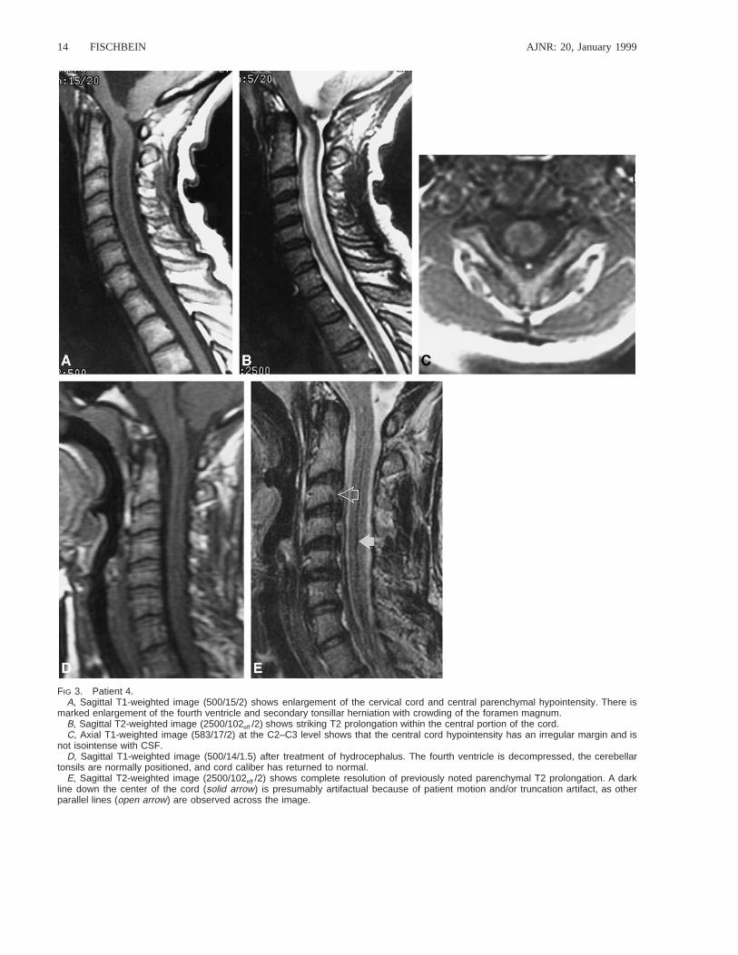
FIG 3. Patient 4.A, Sagittal T1-weighted image (500/15/2) shows enlargement of the cervical cord and central parenchymal hypointensity. There is
marked enlargement of the fourth ventricle and secondary tonsillar herniation with crowding of the foramen magnum.B, Sagittal T2-weighted image (2500/102eff /2) shows striking T2 prolongation within the central portion of the cord.C, Axial T1-weighted image (583/17/2) at the C2–C3 level shows that the central cord hypointensity has an irregular margin and is
not isointense with CSF.D, Sagittal T1-weighted image (500/14/1.5) after treatment of hydrocephalus. The fourth ventricle is decompressed, the cerebellar
tonsils are normally positioned, and cord caliber has returned to normal.E, Sagittal T2-weighted image (2500/102eff /2) shows complete resolution of previously noted parenchymal T2 prolongation. A dark
line down the center of the cord (solid arrow) is presumably artifactual because of patient motion and/or truncation artifact, as otherparallel lines (open arrow) are observed across the image.
AJNR: 20, January 1999 ‘‘PRESYRINX’’ STATE 15
(ages 13 to 29 years), and 100% of those over age65. They concluded that the frequency and extent ofcentral canal stenosis in humans almost certainly af-fects the clinical features of syringomyelia. Theoret-ically, a disturbance of CSF circulation in the spinalsubarachnoid space redirects fluid through the inter-stitial spaces of the spinal cord and eventually into apatent segment of the central canal. Focal obliterationof the central canal above this level prevents com-munication between the syrinx and the fourth ventri-cle, initiating the conditions required for establish-ment of noncommunicating syringomyelia.
Clinical studies also support the importance ofCSF flow patterns and subarachnoid space pressurewaves in the initiation and propagation of syrin-gomyelia. Oldfield et al (19) studied seven patientswith Chiari I malformations and syringomyelia us-ing MR imaging and intraoperative sonography. Onthe basis of their observations, they suggest that thedevelopment and progression of noncommunicat-ing syrinxes associated with the Chiari I malfor-mation are caused by obstruction to the normal rap-id to-and-fro movement of CSF across the foramenmagnum by tonsillar ectopia and the ventral posi-tion of the lower brain stem. During systole in theChiari I malformation, brain expansion is accom-modated by abrupt caudal movement of the tonsils.This downward, pistonlike systolic tonsillar move-ment can be shown on direction-sensitive cinephase-contrast MR sequences (Fig 1C). Rapiddownward tonsillar movement, perhaps in concertwith the deposition of fourth ventricular CSF intothe spinal subarachnoid space below the level ofobstruction, imparts an accentuated systolic pres-sure wave into the upper cervical spinal canal, forc-ing CSF into spinal cord parenchyma during sys-tole. Oldfield et al (19) believed that theirhypothesis might explain the origin and progres-sion of noncommunicating syrinxes associated withthe hindbrain malformations, as well as with otherconditions that obstruct CSF flow in the spinal sub-arachnoid space, such as arachnoiditis and extra-medullary tumors.
Syringomyelia is commonly observed in theposttrauma setting (1–3). In the case of trauma, theinciting event for syrinx formation is focal cordinjury, although the propagation of a posttraumaticsyrinx may certainly be related at least in part toalterations in CSF flow (30). In trauma patients, acondition termed progressive posttraumatic mye-lomalacic myelopathy (PPMM) has been describedthat is considered to represent a continuum of in-terrelated disease processes that may precede for-mation of a confluent cyst (31). Patients withPPMM are generally clinically indistinguishablefrom those with cystic myelopathy, and, in somecases, have been reported to have a microcysticmyelopathy (30–35). The presence of localized ar-achnoiditis at the level of trauma leading to spinalcord tethering is considered to play an importantrole in the pathophysiology of PPMM, related atleast in part to changes in local CSF dynamics (30,
31). Analogous to the situation of PPMM, our pa-tients had myelopathic symptoms associated withconditions that predisposed them to syrinx forma-tion, and, on MR images, had cord abnormalitieswithout definite syrinx formation. To our knowl-edge, this has not been documented in patientswithout a history of trauma.
Specifically, the underlying conditions in our pa-tients included Chiari I malformation, cervicalspondylostenosis, and arachnoiditis, all of whichare associated with both impedance to normal CSFflow and syrinx formation. All of our patients hadnonspecific myelopathic symptoms, similar to pa-tients with noncommunicating syringomyelia de-scribed by Milhorat et al (28), and had findings onMR images similar to those seen in cases of pro-gressive posttraumatic myelomalacic myelopathy(31), including cord enlargement and T1 and T2prolongation, with the T1 signal not as low as thatof CSF and not sharply marginated. In the distinc-tion between cystic and noncystic myelopathy, pro-ton density–weighted images may be useful, sincea cyst would be expected to be isointense with CSFwhereas myelomalacic or microcystic changeswould likely be hyperintense (36); however, protondensity–weighted images may not be completelyreliable, since a cyst may be hyperintense relativeto CSF on a proton density–weighted image be-cause of dampened CSF pulsations or a slightly el-evated protein content. In addition, with the adventof fast spin-echo imaging of the spine, double-echospin-echo sequences have been dropped from manyimaging protocols, and we do not perform themroutinely at our institution. Axial T1-weighted im-ages may also be useful in the distinction of cysticfrom noncystic changes. Intraoperative sonographyis certainly a useful adjunct in the assessment ofcystic versus noncystic myelopathy (31, 34, 37,38), but the performance of this examination variesamong institutions and among individual surgeonsand requires surgical exposure.
Findings strongly supporting disturbance of CSFflow were observed on preoperative imaging stud-ies in four of our five patients, and the surgicalprocedures performed were either directed at re-storing patency of CSF pathways or had that endeffect in all patients. We do not have direct infor-mation on the status of the central canal in our pa-tients, but we hypothesize that it was not patent,and, thus, CSF that was driven into the spinal cordparenchyma by alterations in normal flow patternswas unable to enter the central canal to form a syr-inx (Figs 5 and 6). After improvement or reconsti-tution of CSF pathways, all patients experiencedstabilization or improvement in clinical symptoms.Additionally, results of postoperative MR exami-nations showed both a reduction in cord caliber andan improvement in cord parenchymal signal abnor-malities. Whether the signal alterations representedema or microcystic change or both is unclear,since pathologic specimens are not available fromthese patients. A direct traumatic injury to the cord

AJNR: 20, January 199916 FISCHBEIN
does not seem to be a necessary prerequisite forthe development of this condition. A difficulty withthis hypothesis is explaining the later developmentof frank cavitation in patient 3; however, it is pos-sible that partial recanalization of the central canalmay have occurred, followed by paracentral dis-section around a stenotic segment (29). Alterna-tively, the myelotomy performed during the initial
surgery may have created a pathway along whicha syrinx could form and then extend.
Jinkins et al (39) recently described three patientswith clinically progressive posttraumatic syringomy-elia in whom extensive MR signal change on T2-weighted images in the spinal cord superior to a well-defined syrinx was found to be an ancillary sign ofdisease progression. After shunting of the syrinx, the

AJNR: 20, January 1999 ‘‘PRESYRINX’’ STATE 17
FIG 4. Patient 3.A, Sagittal T2-weighted image (4000/108eff /4) obtained when the patient had no symptoms referable to the spinal cord shows a mild
dilatation of the obex/proximal central canal and central T2 prolongation within the upper cervical cord parenchyma.B, Sagittal T1-weighted (left panel) (600/11/2) and T2-weighted (right panel) (3000/96eff /2) images obtained 1 year later when the
patient had developed progressive neck pain and spastic quadriparesis show striking cord expansion and both T1 and T2 prolongationwithin the cervical spinal cord. The areas of abnormal signal within the cord approach, but are not quite equal to, CSF in intensity.
C, Axial T1-weighted image (650/9/3) shows irregularly marginated central parenchymal hypointensity, although this area is hyperin-tense compared with CSF in the spinal canal. These images (A–C) were interpreted as consistent with syrinx by the neurosurgeon, andthe patient was taken to the operating room for shunt placement. Intraoperatively, the cord was noted to be enlarged and ‘‘boggy.’’ Amyelotomy was performed at the C6 level, and a small amount of fluid exuded from the cord surface, but no syrinx was encountered.Intraoperative sonogram (not shown) confirmed the lack of frank cavitation.
D, Sagittal T2-weighted image (3500/96eff /3) obtained 2 days postoperatively shows evidence of recent C6–C7 laminectomy. The cordis notably reduced in overall caliber compared with the preoperative study, and the signal has normalized at the myelotomy site (arrow).
E, Repeat T2-weighted image (2500/105eff /3) obtained 8 days later shows further regression of signal abnormality and further reductionof cord caliber.
F, Sagittal T2-weighted image (3894/112eff /1) obtained 1 month later shows an increase in central T2 prolongation within the cervicalspinal cord, as well as an increase in cord caliber. The patient was doing fairly well in rehabilitation and did not desire further intervention.The patient was lost to follow-up for 10 months.
G, Sagittal T1-weighted image (500/8/3) obtained 11 months after surgery shows further enlargement of the cervical and upper thoracicspinal cord. The cord centrally is hypointense, and multiple septations are present (arrow) consistent with syringomyelia. The patientclinically was severely quadriparetic and had lost control of bowel and bladder function. After this image, surgery was performed, duringwhich a large syrinx was encountered and a syringopleural shunt was placed (not shown).
parenchymal T2 hyperintensity resolved, and neuro-logic deficits stabilized or improved. They postulatedthat the T2 hyperintensity represented fluid escapingfrom the cyst or edema caused by as yet undefinedpathologic alterations in the spinal cord adjacent tothe cyst. In these cases, a definite obstruction to flowof CSF was not described, and the intervention taken(syrinx shunting) was not aimed at restoring patencyof CSF pathways. It is possible that the enlarged cordcaused relative obstruction of normal CSF flow path-ways, resulting in a ‘‘presyrinx’’ condition cranial tothe already formed syrinx cavity.
In further support of our hypothesis, the revers-ibility of cord signal abnormality associated with ob-struction to normal CSF flow has been reported in
a case of acquired tonsillar herniation caused byprobable spontaneous intracranial hypotension (40–42). In this case, the cervical cord was enlarged andhad T1 and T2 prolongation without frank cavita-tion, consistent with the presyrinx state. After spon-taneous resolution of the patient’s condition (pre-sumably because of closure of an occult CSF leak),the cerebellar tonsils returned to a normal position,and the cervical cord caliber and signal reverted tonormal. The patient was not myelopathic but didexperience occipital and neck pain. The lack ofmyelopathic symptoms is not inconsistent with pre-syrinx physiology, since even patients with frank sy-ringomyelia (typically of the central cavitary type)may be asymptomatic (28).

AJNR: 20, January 199918 FISCHBEIN
FIG 5. Diagrammatic representation of CSF flow under normalcircumstances.
A, Sagittal view of the craniocervical junction and upper cer-vical spinal cord in an anatomically normal patient shows no ob-struction to CSF flow at the foramen magnum. A segment ofspinal cord parenchyma (box) is shown in more detail in B.
B, Magnified view of the box in A shows CSF flow dynamicsin a normal patient with a variably stenotic central canal (CC)(29), as indicated by the horizontal lines. CSF pressure (verticalarrow) is normal. CSF flows from the subarachnoid space (SAS)between the arachnoid (A) and pia (P) to the subpial space, andthen enters the perivascular space (PVS). CSF circulatesthrough the cord parenchyma toward the central canal, but mayalso flow in reverse, as these forces are relatively balanced un-der normal circumstances (double-headed arrows) (45, 46).
Because surgical intervention was performed thatrestored or improved CSF flow pathways in all ourpatients, we are unable to prove that they wouldhave progressed to frank syrinx formation. The im-plication that syrinx formation would have occurredif the patients were left untreated is justified by thefollowing three considerations: first, the underlyingconditions in our patients all have a known associ-ation with syringomyelia; second, in one patient (pa-tient 3) who initially responded to surgical interven-tion with marked improvement in cord enlargementand signal abnormality, clear-cut syrinx formationoccurred during the follow-up period, presumably
because the underlying obstruction to CSF flow hadnot been fully addressed by the surgical procedureperformed; and third, the recent observations ofJinkins et al (39), which suggest that reversible T2changes were considered to predict frank syrinx for-mation. We, therefore, propose the use of the term‘‘presyrinx’’ state as a valid concept to describethese and similar cases.
We have also considered other possible causes ofthe reversible cord enlargement and T2 prolongationidentified in our patients. It seems unlikely that ar-terial ischemia plays a significant role in the patho-genesis of the presyrinx state based on the fact thatthe imaging abnormalities did not conform to a vas-cular territory and were reversible as assessed bypostoperative imaging. Venous ischemia could haveplayed a role in cord enlargement and signal changes,analogous to the pathophysiology of spinal dural ar-teriovenous fistula, but no abnormal veins were iden-tified at preoperative imaging or intraoperatively. Thelack of identification of macroscopic abnormal veinsdoes not, however, exclude a role for venous isch-emia. In the setting of trauma, occluded intramedul-lary veins have been identified in degenerated seg-ments of spinal cord (43). In the nontraumatic setting,it is possible that pressure changes in the epiduralvenous plexus in association with disturbances ofCSF circulation lead to an increase in spinal venouspressure and accumulation of fluid in the spinal cord(12). Venous ischemic changes are also known to bereversible. However the venous drainage of the spinalcord is quite rich, and the mechanism by which ve-nous ischemia would have occurred in our patients isnot clear. Therefore, although we propose that thepresyrinx state is fundamentally one of altered CSFflow parameters, we do recognize that a contributionof venous ischemia may be present as well and thatsorting out these relationships will require furtherstudy.
The relationship between the level of the block toCSF flow and the location of the presyrinx lesion alsowarrants consideration. As most of our patients hadhigh cervical or foramen magnum blockages to CSFflow, it is not surprising that the spinal cord paren-chymal signal changes that we observed developedcaudal to the block. Pathophysiologically, this is mostlikely related to fluid entering the cord below thelevel of the block and then tracking cephalad in cordparenchyma and/or the central canal to circumventthe block; however, in the experience of Jinkins et al(39), which we consider an analogous situation, thepresyrinx lesion extended rostral to the level of ob-struction. In the setting of trauma, syrinxes mostcommonly extend superiorly from the site of injury,although superior and inferior extension, and eveninferior extension alone, have been observed. Thismay relate to the fact that the cervical cord expandsmore easily than the thoracic cord (3, 9, 44).
An important but as yet unexplained aspect ofthis phenomenon is why we do not see these MRfindings more frequently in cases of Chiari I mal-formations and basal arachnoiditis, as these are not

AJNR: 20, January 1999 ‘‘PRESYRINX’’ STATE 19
FIG 6. Diagrammatic representation of syringomyelia and the ‘‘presyrinx’’ hypothesis in the setting of obstruction to CSF flow.A, Sagittal view of the craniocervical junction in a patient with a Chiari I malformation shows abnormal descent of the cerebellar tonsil
below the level of the foramen magnum (arrow). A segment of spinal cord parenchyma (box) is magnified in B to D, which representviews of CSF dynamics at the level of the spinal cord parenchyma in the presence of alterations in normal CSF flow and variablepatency of the central canal.
B, Focal noncommunicating syrinx. In the setting of a Chiari I malformation and a variably stenotic central canal (which is a normalvariant in many adults), as the tonsils descend rapidly during systole, CSF is driven into the spinal cord parenchyma by increased CSFpressure (thick vertical arrow). Net CSF flow occurs toward the central canal, resulting in focal syringomyelia, which is limited in itscraniocaudal extent by intervening stenosis of the central canal. CC 5 central canal, A 5 arachnoid, P 5 pia, SAS 5 subarachnoidspace, PVS 5 perivascular space.
C, Extensive noncommunicating syrinx. This situation is similar to B, but the central canal is more extensively patent. In this situation,a long-segment dilatation of the central canal occurs as CSF is driven into the central canal (curved arrows) via the perivascular spacesby the accentuated CSF pulse pressure (thick vertical arrow) that results from the downward motion of the low-lying cerebellar tonsilsin systole.
D, ‘‘Presyrinx.’’ In the setting of altered CSF flow, as with a Chiari I malformation, fluid in the subarachnoid space is subjected toincreased pressure (thick vertical arrow). Net CSF flow is into the spinal cord parenchyma; however, because the central canal is notpatent (as indicated by the horizontal lines), fluid cannot accumulate within the central canal (curved arrows) and, therefore, diffusesthrough the cord parenchyma (stippled area), resulting in cord enlargement and edema.
uncommon conditions. Because patients in the pre-syrinx state may have minimal clinical symptomsor even be asymptomatic, they may not come tomedical attention until frank cavitation and moresevere symptoms have developed. Additionally, adynamic balance between CSF pressure in the spi-nal subarachnoid space and the spinal cord paren-chyma may exist, and the anatomic conditions re-quired to establish the presyrinx state may occuronly rarely. For instance, the establishment of thisstate may depend on the morphology and extent of
the network of spinal perivascular spaces and thecapacity and patency of the central canal, amongother factors. Intervention at an appropriate timemay allow restoration of normal flow patterns andreversal of parenchymal signal abnormalities, aswell as improvement in neurologic deficits. Thatthis entity may represent an equilibrium state issupported by the documentation of this appearancein one of our patients (patient 2) for some timebefore she progressed symptomatically to the pointthat surgical intervention was performed. Further

AJNR: 20, January 199920 FISCHBEIN
study will be necessary to better understand normaland abnormal spinal subarachnoid fluid circulationand the pathophysiology of syrinx formation.
ConclusionWe have described a condition of reversible spi-
nal cord enlargement and T1 and T2 prolongation,the ‘‘presyrinx’’ state, which occurs in the settingof CSF flow obstruction and may be misinterpretedas syringomyelia on MR studies. Surgical interven-tion aimed at restoring patency of CSF pathwaysis likely to be of benefit in these patients, and MRfindings should guide selection of the procedure tobe performed. This entity presumably represents apoint on the continuum to development of syrin-gomyelia and may be pathophysiologically relatedto variations in the patency of the central canal andalso to the entity of progressive posttraumatic mye-lomalacic myelopathy.
References1. Rossier AB, Foo D, Shillito J, Dyro FM. Posttraumatic cervical
syringomyelia. Brain 1985;108:439–4612. Vernon JD, Silver JR, Ohry A. Posttraumatic syringomyelia. Par-
aplegia 1982;20:339–3643. Quencer RM, Green BA, Eismont FJ. Posttraumatic spinal cord
cysts: clinical features and characterization with metrizamidecomputed tomography. Radiology 1983;146:415–423
4. Gardner WJ. Hydrodynamic mechanism of syringomyelia: its re-lationship to myelocele. J Neurol Neurosurg Psychiatry 1965;28:247–259
5. Williams B. The distending force in the production of ‘‘commu-nicating syringomyelia.’’ Lancet 1969;2:189–193
6. Ball MJ, Dayan AD. Pathogenesis of syringomyelia. Lancet 1972;2:799–801
7. Milhorat TH, Capocelli AL, Anzil AP, Kotzen RM, Milhorat RH.Pathological basis of spinal cord cavitation in syringomyelia: anal-ysis of 105 autopsy cases. J Neurosurg 1995;82:802–812
8. Caplan LR, Norohna AB, Amico LL. Syringomyelia and arachno-iditis. J Neurol Neurosurg Psychiatry 1990;53:106–113
9. Klekamp J, Batzdorf U, Samii M, Bothe HW. Treatment of syrin-gomyelia associated with arachnoid scarring caused by arachno-iditis or trauma. J Neurosurg 1997;86:233–240
10. Brammah TB, Jayson MIV. Syringomyelia as a complication of spi-nal arachnoiditis. Spine 1994;19:2603–2605
11. Phanthumchinda K, Kaoropthum S. Syringomyelia associated withpost-meningitis spinal arachnoiditis due to Candida tropicalis.Postgrad Med J 1991;67:767–769
12. Klekamp J, Samii M, Tatagiba M, Sepehrnia A. Syringomyelia inassociation with tumors of the posterior fossa: pathophysiologicalconsiderations, based on observations on three related cases. ActaNeurochir 1995;137:38–43
13. Kaar GF, N’Dow JM, Bashir SH. Cervical spondylotic myelopathywith syringomyelia. Br J Neurosurg 1996;10:413–415
14. Aubin ML, Baleriaux D, Cosnard G, et al. MRI in syringomyelia ofcongenital, infectious, traumatic or idiopathic origin: a study of142 cases. J Neuroradiol 1987;4:313–336
15. Houang MT, Stern M, Brew B, Pell M, Darveniza P. Magnetic res-onance imaging (MRI) appearances of syringohydromyelia. Aus-tralas Radiol 1988;32:172–177
16. Williams B, Bentley J. Experimental communicating syringomyeliain dogs after cisternal kaolin injection, I: morphology. J NeurolSci 1980;48:93–107
17. Cho KH, Iwasaki Y, Imamura H, Hida K, Abe H. Experimentalmodel of posttraumatic syringomyelia: the role of adhesive arach-noiditis in syrinx formation. J Neurosurg 1994;80:133–139
18. Milhorat TH, Nobandegani F, Miller JI, Rao C. Noncommunicatingsyringomyelia following occlusion of central canal in rats. J Neu-rosurg 1993;78:274–279
19. Oldfield EH, Muraszko K, Shawker TH, Patronas NJ. Pathophysi-ology of syringomyelia associated with Chiari I malformation ofthe cerebellar tonsils. J Neurosurg 1994;80:3–15
20. Rennels ML, Gregory TF, Blaumanis OR, Fujimoto K, Grady PA.Evidence for a ‘‘paravascular’’ fluid circulation in the mammaliancentral nervous system, provided by the rapid distribution of trac-er protein throughout the brain from the subarachnoid space.Brain Res 1985;326:47–63
21. Rennels ML, Blaumanis OR, Grady PA. Rapid solute transportthroughout the brain via paravascular fluid pathways. Adv Neurol1990;52:431–439
22. Stoodley MA, Jones NR, Brown CJ. Evidence for rapid fluid flowfrom the subarachnoid space into the spinal cord central canal inthe rat. Brain Res 1996;707:155–164
23. Stoodley MA, Brown SA, Brown CJ, Jones NR. Arterial pulsation-dependent perivascular cerebrospinal fluid flow into the centralcanal in the sheep spinal cord. J Neurosurg 1997;86:686–693
24. Cifuentes M, Fernandez-Llebrez P, Perez J, Perez-Figares JM, Rod-riguez EM. Distribution of intraventricularly injected horseradishperoxidase in cerebrospinal fluid compartments of the rat spinalcord. Cell Tissue Res 1992;270:485–494
25. Milhorat TH, Adler DE, Heger IM, Miller JI, Hollenberg-Sher JR.Histopathology of experimental hematomyelia. J Neurosurg 1991;75:911–915
26. Milhorat TH, Miller JI, Johnson WD, Adler DE, Heger IM. Anatom-ical basis of syringomyelia occurring with hindbrain lesions. Neu-rosurgery 1993;32:748–754
27. Milhorat TH, Johnson WD, Miller JI, Bergland RM, Hollenberg-SherJ. Surgical treatment of syringomyelia based on magnetic reso-nance imaging criteria. Neurosurgery 1992;31:231–245
28. Milhorat TH, Johnson RW, Milhorat RH, Capocelli AL, PevsnerPH. Clinicopathological correlations in syringomyelia using axialmagnetic resonance imaging. Neurosurgery 1995;37:206–213
29. Milhorat TH, Kotzen RM, Anzil AP. Stenosis of central canal ofspinal cord in man: incidence and pathological findings in 232autopsy cases. J Neurosurg 1994;80:716–722
30. Lee TT, Arias JM, Andrus HL, Quencer RM, Falcone SF, GreenBA. Progressive posttraumatic myelomalacic myelopathy:treatment with untethering and expansive duraplasty. J Neu-rosurg 1997;86:624–628
31. Falcone S, Quencer RM, Green B, Patchen SJ, Post MJD. Pro-gressive posttraumatic myelomalacic myelopathy: imagingand clinical features. AJNR Am J Neuroradiol 1994;15:747–754
32. MacDonald RL, Findlay JM, Tator CH. Microcystic spinal corddegeneration causing posttraumatic myelopathy: report of twocases. J Neurosurg 1988;68:466–471
33. Osborne DRS, Vavoulis G, Nashold BS, Dubois PJ, Drayer BP,Heinz RE. Late sequelae of spinal cord trauma: myelographicand surgical correlation. J Neurosurg 1982;57:18–23
34. Gebarski SS, Maynard FW, Gabrielsen TO, Knake JE, Latack JT,Hoff JT. Posttraumatic progressive myelopathy. Radiology1985;157:379–385
35. Stevens JM, Olney JS, Kendall BE. Post-traumatic cystic andnon-cystic myelopathy. Neuroradiology 1985;27:48–56
36. Tanghe HLJ. Magnetic resonance imaging (MRI) in syringo-myelia. Acta Neurochir 1995;134:93–99
37. Quencer RM, Morse BMM, Green BA, Eismont FJ, Brost P. In-traoperative spinal sonography: adjunct to metrizamide CT inthe assessment and surgical decompression of posttraumaticspinal cord cysts. AJR Am J Roentgenol 1984;142:593–601
38. Wilberger JE Jr, Maroon JC, Prostko ER, Baghai P, Beckman I,Deeb Z. Magnetic resonance imaging and intraoperative neu-rosonography in syringomyelia. Neurosurgery 1987;20:599–605
39. Jinkins JR, Reddy S, Leite CC, Bazan C, Xiong L. MR of pa-renchymal spinal cord signal change as a sign of active ad-vancement in clinically progressive posttraumatic syringomy-elia. AJNR Am J Neuroradiol 1998;19:177–182
40. Morioka T, Shono T, Nishio S, Yoshida K, Hasuo K, Fukui M.Acquired Chiari I malformation and syringomyelia associatedwith bilateral chronic subdural hematoma: case report. J Neu-rosurg 1995;83:556–558
41. Schievink WI, Atkinson JLD. Spontaneous intracranial hypo-tension (letter). J Neurosurg 1996;84:151–152
42. Mamelak AN, Fishman RA, Dillon WP, Wilson CB. Spontaneousintracranial hypotension (letter). J Neurosurg 1996;85:192–193
43. Tator CH, Koyanagi I. Vascular mechanisms in the pathophysi-ology of human spinal cord injury. J Neurosurg 1997;86:483–492
44. Watson N. Ascending cystic degeneration of the cord after spi-nal cord injury. Paraplegia 1981;19:89–95
45. Hutchings M, Weller RO. Anatomical relationships of the piamater to cerebral blood vessels in man. J Neurosurg 1986;65:316–325
46. Zhang ET, Inman CBE, Weller RO. Interrelationships of the piamater and the perivascular (Virchow-Robin) spaces in the hu-man cerebrum. J Anat 1990;170:111–123