the preservative effect of trehalose on...
TRANSCRIPT
Republic of Iraq Ministry of Higher Education and Scientific Research Al-Nahrain University College of Sciences Biotechnology Department
The preservative effect of Trehalose on Staphylococcus
aureus and Escherichia coli
Submitted to the College of Science of AL-Nahrain University As partial fulfilment of the requirements for the degree of Master
of Science in Biotechnology
By
Bassam Samier Mohamed Raouf
B. Sc. Biotechnology (2003)
Al-Nahrain University
Sha’aban- 1427
August 2006
I
Summary Two isolates were used in this study, Staphylococcus aureus and
Escherichia coli were identified according to morphological, physiological and
biochemical characteristics.
The two bacteria were treated with 100mM of Trehalose to study the
effect of this sugar on the preservation of the bacteria from several factors
The results showed that Trehalose preserved the S.aureus from dehydration
(drying) for one month in room temperature and the Trehalose keep the bacteria
more viable in comparison with the control. While no effect was observed in the
Trehalose treated E.coli.
Trehalose showed it’s preservative effect when the bacteria were stored in
freeze condition (-20°C) about 4 weeks the percentages of Viable cells of
S.aureus treated with Trehalose after 4 weeks was 67% compared with the
control 30%, while the results on E.coli was 66% and the control 32%.
Results indicated that Trehalose protect the lyophilized bacteria even in
the unsuitable environment such as storing in room temperature for four months,
the percentage of the viable cells of bacteria treated with Trehalose after 4
months was 27% while the control 2% .
Result also showed the protective effects of Trehalose towered the UV
irradiation and Electroporated bacteria.
While Trehalose had no effect on the protection from antimicrobial
substance (antagonism activity), in contrast it showed a considerable effect of
protection from antibiotics by increasing the resistance to the antibiotics, result
showed that S.aureus treated with trehalose become resist to, Tetracycline,
Gentamicin, Erythromycin and Norfloxacin, while the control stilled sensitive to
these antibiotics, and no effect were showed on the E.coli in protection from
antibiotics.
II
List of Contents
Title Page No.
Summary I List of Contents II List of Tables V List of Figures VI List of Abbreviations X
Chapter One: Introduction and Literature Review
1 Introduction 1 Aims of Study 1.1 Down syndrome (DS) 3 1.1.1 Physical Characteristic of DS 3 1.1.2 Prenatal Screening for DS 3 1.1.3 Diagnostic test of DS 4 1.1.4 Genetic Form of DS 4 1.1.5 Immune Dysfunction 5
1.1.6
Risk Factors Associated to the Down Syndrome
Occurrence
i: Maternal Age
6
1.1.7 The 21st Chromosome and Down syndrome
9
1.2 Drug Resistance 11 2.2.1 Methotrexate 12 2.2.1.2 The resistance to methotrexate 13
2.2.1.3 Thioguanine
14
2.2.2.2. Resistance to 6TG 14 1.3 Cytogenetic analysis 15
1.3.1 Mitotic index 16

III
Chromosome Aberrations (CAs)
IV
Chapter Three: Results and discussion
3.1 Identification of Bacterial Isolates 54
3.1.1 Morphological and Cultural Characteristics 54
3.1.2 Biochemical and Physiological Characteristics 54
3.2 Effect of Trehalose on drying (dehydrations): 56
3.3 UV Survival Curve of Staphylococcus aureus with and without addition of Trehalose
58
3.4 Antibiotic Sensitivity 60
3.5 Minimal Inhibitory Concentration (MIC) Test 62 3.6 Effect of Trehalose on antibacterial activity(antagonisim) 63
3.7 Store of bacterial isolates from freezing temperature in the presence and absence of Trehalose
64
3.8 Protection of bacterial isolate from freeze-drying (lyophilization):
67
3.9 Effect of Trehalose on Electroporated bacteria 71
Conclusions 73
Recommendations 74
References 75
V
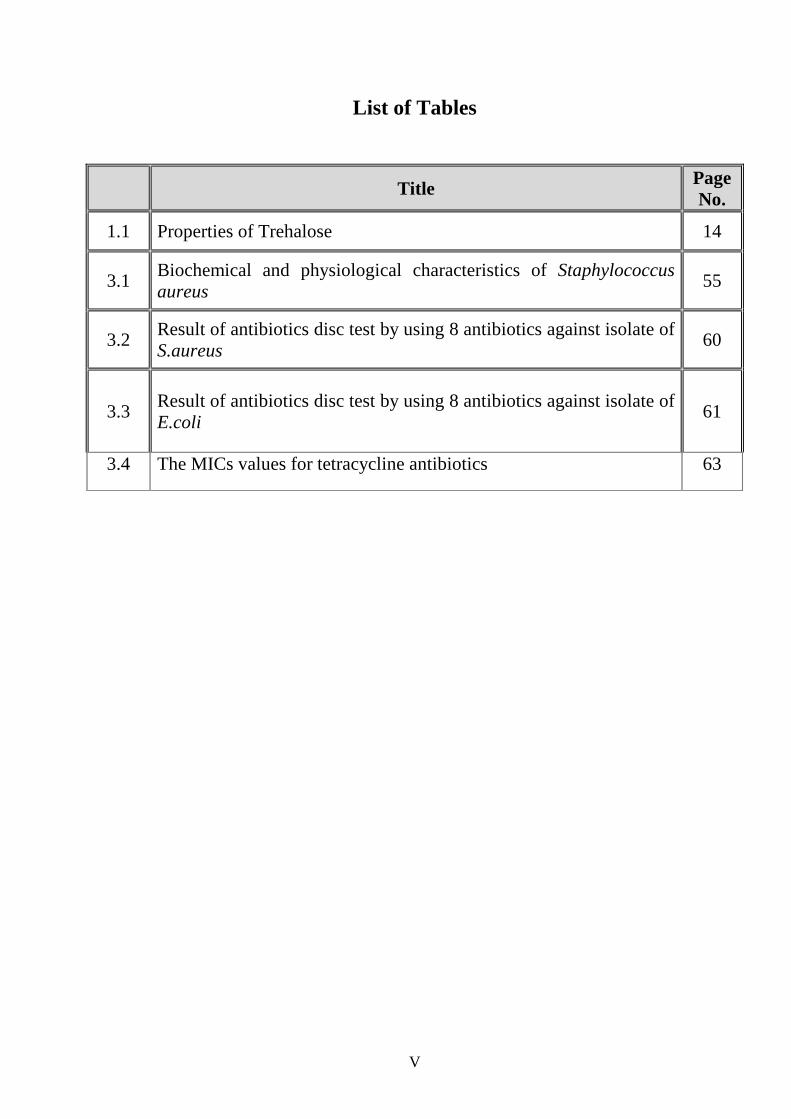
List of Tables
Title Page No.
1.1 Properties of Trehalose 14
3.1 Biochemical and physiological characteristics of Staphylococcus aureus
55
3.2 Result of antibiotics disc test by using 8 antibiotics against isolate of S.aureus
60
3.3 Result of antibiotics disc test by using 8 antibiotics against isolate of E.coli
61
3.4 The MICs values for tetracycline antibiotics 63
VI
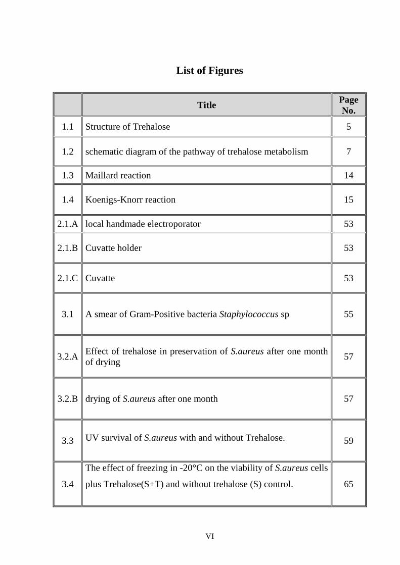
List of Figures
Title Page No.
1.1 Structure of Trehalose 5
1.2 schematic diagram of the pathway of trehalose metabolism 7
1.3 Maillard reaction 14
1.4 Koenigs-Knorr reaction 15
2.1.A local handmade electroporator 53
2.1.B Cuvatte holder 53
2.1.C Cuvatte 53
3.1 A smear of Gram-Positive bacteria Staphylococcus sp 55
3.2.A Effect of trehalose in preservation of S.aureus after one month of drying
57
3.2.B drying of S.aureus after one month 57
3.3 UV survival of S.aureus with and without Trehalose.
59
3.4
The effect of freezing in -20°C on the viability of S.aureus cells
plus Trehalose(S+T) and without trehalose (S) control.
65
VII
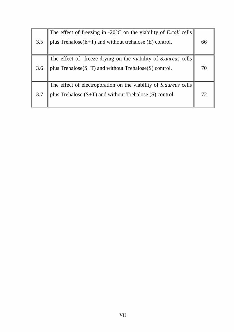
3.5
The effect of freezing in -20°C on the viability of E.coli cells
plus Trehalose(E+T) and without trehalose (E) control.
66
3.6
The effect of freeze-drying on the viability of S.aureus cells
plus Trehalose(S+T) and without Trehalose(S) control.
70
3.7
The effect of electroporation on the viability of S.aureus cells
plus Trehalose (S+T) and without Trehalose (S) control.
72
x
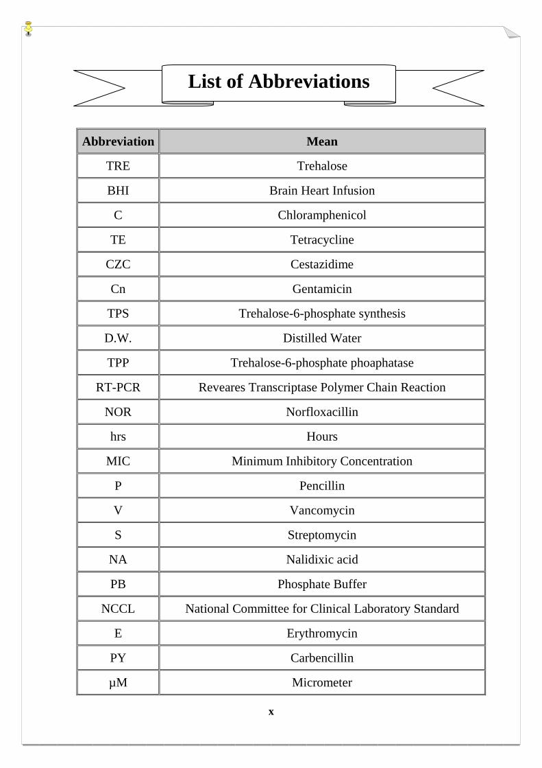
Abbreviation Mean
TRE Trehalose
BHI Brain Heart Infusion
C Chloramphenicol
TE Tetracycline
CZC Cestazidime
Cn Gentamicin
TPS Trehalose-6-phosphate synthesis
D.W. Distilled Water
TPP Trehalose-6-phosphate phoaphatase
RT-PCR Reveares Transcriptase Polymer Chain Reaction
NOR Norfloxacillin
hrs Hours
MIC Minimum Inhibitory Concentration
P Pencillin
V Vancomycin
S Streptomycin
NA Nalidixic acid
PB Phosphate Buffer
NCCL National Committee for Clinical Laboratory Standard
E Erythromycin
PY Carbencillin
µM Micrometer
List of Abbreviations
xi
AK Amikacin
mm Millimeter
HXK Hexokinase
NMR nuclear magnetic resonance
RACE PCR rapid amplification of 5′ and 3′ cDNA ends

ر�� ا���اق�� وزارة ا������ ا����� وا���� ا�����
��� ا������� ���� ا���م
ا��-رات ا���,د�� ا�*ھ��� و أ'���&�� �������ز %�$ �ا��#"��ات ا��� ��
ن� . ا�,
رسالة
مقدمة الى كلية العلوم جامعة النهرين
التقانة االحيائية وهي جزء من متطلبات نيل درجة ماجستير علوم في
ن قبلم
�34م 2��� ���1 رؤوف
جامعة النهرينبكلوريوس تقانة احيائية
٢٠٠٣
شعبان 1427

أب ٢٠٠٦
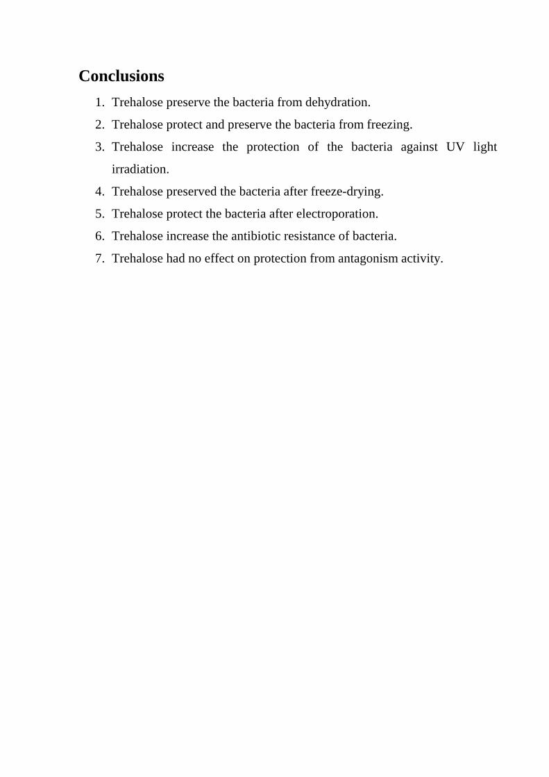
Conclusions
1. Trehalose preserve the bacteria from dehydration.
2. Trehalose protect and preserve the bacteria from freezing.
3. Trehalose increase the protection of the bacteria against UV light
irradiation.
4. Trehalose preserved the bacteria after freeze-drying.
5. Trehalose protect the bacteria after electroporation.
6. Trehalose increase the antibiotic resistance of bacteria.
7. Trehalose had no effect on protection from antagonism activity.

Acknowledgments
Praise to Allah the lord of the universe and creation, the merciful and
kindness, and blessing upon Mohammed prophet of Allah and upon his family
and companions.
First I thanks and appreciate my supervisors Dr. Ayad Mohammed Al-
Aubaidy for great support throughout my studies.
I thank my family for their supports and assistance for all things.
It’s my pleasure to thank my friends, Oday Adnan, Hassan Abdulhadi ,
Solaf Jawhar , Sura Ali , Mayassa Fadhil, Hiba Abdulkader , Muntaha
Abdulrazaq , Rana Munther , Mr. Ali Al-Sammak, Miss Shayma’a , Miss
Oroba , Miss Tania and Mr.Yasin for their assistance .
Special thank to special person who helped and supported me a lot, thanks
Farah.
I also thank all staff and employers of biotechnology department of
Al Nahrain University who assist me.
Committee Certification
We, the examining committee, certify that we have read this thesis and examined the student in its contents and that, according to our opinion, is accepted as a thesis for the degree of Master of Science in Biotechnology.
Signature: Name:
Scientific Degree: Date:
(Chairman)
Signature: Signature: Name: Name: Scientific Degree: Scientific Degree: Date: Date: (Member) (Member) Signature: Signature: Name: Name: Scientific Degree: Scientific Degree: Date: Date: (Member/Supervisor) (Member/Supervisor) I hereby certify upon the decision of the examining committee
Signature:
Name: Dr.Laith Abdul Al-Aziz Al-Ani.
Scientific Degree: Assistant Professor
Title: Dean of College of Science
Date:
Supervisor Certification
I certify that this was prepared under my supervision in Al-Nahrain University-College of Science as a partial fulfillment of degree of master of Science in Biotechnology.
Signature: Signature: Supervisor: Supervisor: Date: Date: In review of the available recommendation I forward this thesis for debate by examining committee. Signature:
Name:
Chairman of Biotechnology Department
Date:

Dedication
To my eyes, my Father and Mother
To my Dearest friend, my Brother
To my soul, the sweet angel my Sister
Chapter Three Results and Discussion
١
1.1 Introduction
α−α-Trehalose, a naturally occurring, non reducing disaccharide
consist of two glucose units, is commonly found in bacteria ( Koch and
Koch, 1998 ), yeasts (Zahringer et al., 1998; Fernandez et al., 1998), algae,
insects, plants (Ring and Danks 1998), and animals or human organs (Schick
et al., 1991).
Trehalose is known to help certain animals and plants and microorganisms
to survive desiccation, high osmolarity, and damage by both freezing and
heat. It is used to preserve biological materials (Schick et al., 1991 and
Kizawa et al., 1995) and as a stabilizer for unstable proteins, including
dehydrated and frozen enzymes, diagnostic reagents, pharmaceuticals, and
cosmetics (Yoshida et al., 1995).
When bacterial cells are freeze dried with 100 mM trehalose as the
lyoprotectant (lyophilized protectant), the viability of microorganisms
improves four fold and eight fold. The improved viability may be the result
of the ability of Trehalose to lower the temperature of the dry membrane
phase transition and to maintain general protein structures in a dry state
(Leslie et al., 1995).
These findings suggest that Trehalose added to the growth medium and to
organelle membranes.
Due to its particular physical features, Trehalose is able to protect the
integrity of the cell against a variety of environmental injuries and
nutritional limitations. In addition, data available on several species of
bacteria and yeast suggest specific functions for Trehalose in these
organisms. Some bacteria can use exogenous Trehalose as the sole source of
Chapter Three Results and Discussion
٢
carbon and energy as well as synthesize enormous amounts of the
disaccharide as compatible solute .
This ability to accumulate trehalose is the result of an elaborate genetic
system, which is regulated by osmolarity. Some mycobacteria contain
sterified trehalose as a structural component of the cell wall, whereas yeast
cells are largely unable to grow on trehalose as carbon source. In these lower
eukaryotes, trehalose appears to play a dual function: as a reserve
compound, mainly stored in vegetative resting cells and reproductive
structures, and as a stress metabolite. Recent findings also point to important
biotechnological applications for Trehalose.
It has been shown that trehalose can protect proteins and cellular membranes
from inactivation or denaturation caused by a variety of stress conditions,
including desiccation, dehydration, heat, cold, and oxidation. Finally, in
Mycobacteria and Corynebacteria, Trehalose is an integral component of
various glycolipids that are important cell wall structures (Richard et al.,
2002).
1.2 Aim of the study
1. Identifying the bacterial isolate Staphylococcus sp.
2. Study the effect of Trehalose on S.aureus and E.coli in preservation in
dry state at room temperature.
3. Study the effect of Trehalose on protection of bacteria against UV
light.
4. Study the effect of Trehalose on protection of bacteria after
lyophilization.

Chapter Three Results and Discussion
٣
5. Study the effect of Trehalose on protection of bacteria against
freezing.
6. Study the effect of Trehalose on protection of bacteria against
antibiotics.
7. Study the effect of Trehalose on protection of bacteria against
antagonism activity.
Chapter Three Results and Discussion
٤
1.3: Trehalose
Trehalose is a non reducing disaccharide in which the two glucose
units are linked in an, -1, 1-glycosidic linkage (Qiaofang Chen and Haddad
2004).
In 1974, the current view on the role of trehalose was that it served as a
storehouse of glucose for energy and/or for synthesis of cellular components
(Elbein, 1974).
Since that time, our knowledge on the various functions of this simple
disaccharide has greatly expanded; it is now clear that trehalose is much
more than simply a storage compound. Certainly it’s function in that
capacity in some organisms, but in others it has a structural or transport role
(Takayama and Armstrong, 1976), whereas in still others it may be involved
in signaling or regulation, or functions to protect membranes and proteins
against the adverse effects of stresses, such as heat, cold, desiccation, and
anoxia (Crowe et al., 1984).
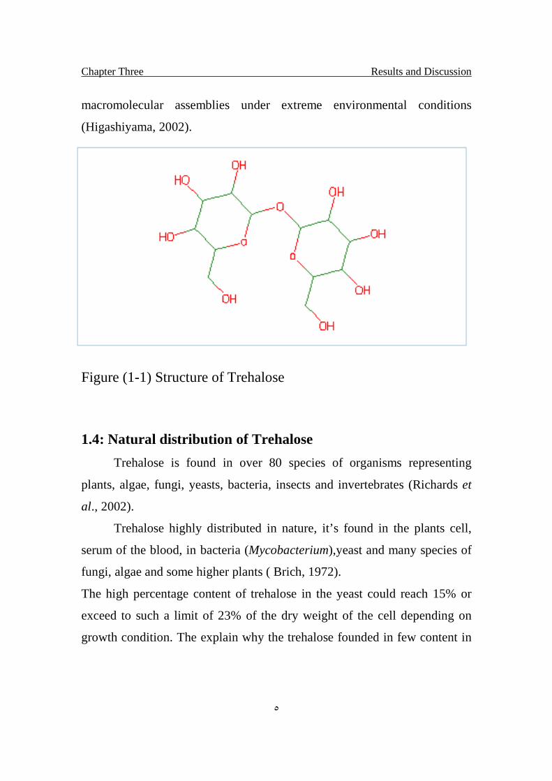
As shown in Fig.1-1 the reducing end of a glucosyl residue is
connected with the other, trehalose has no reducing power. Trehalose is
widely distributed in nature. It is known to be one of the sources of energy in
most living organisms and can be found in many organisms, including
bacteria, fungi, insects, plants, and invertebrates. Mushrooms contain up to
10–25 % trehalose by dry weight. Furthermore, trehalose protects organisms
against various stresses, such as dryness, freezing, and osmopressure. In the
case of resurrection plants, which can live in a dry state, when the water
dries up, the plants dry up too. However, they can successfully revive when
placed in water. The anhydrobitic organisms are able to tolerate the lack of
water owing to their ability to synthesize large quantities of trehalose, and
the trehalose plays a key role in stabilizing membranes and other
Chapter Three Results and Discussion
٥
macromolecular assemblies under extreme environmental conditions
(Higashiyama, 2002).
Figure (1-1) Structure of Trehalose
1.4: Natural distribution of Trehalose
Trehalose is found in over 80 species of organisms representing
plants, algae, fungi, yeasts, bacteria, insects and invertebrates (Richards et
al., 2002).
Trehalose highly distributed in nature, it’s found in the plants cell,
serum of the blood, in bacteria (Mycobacterium),yeast and many species of
fungi, algae and some higher plants ( Brich, 1972).
The high percentage content of trehalose in the yeast could reach 15% or
exceed to such a limit of 23% of the dry weight of the cell depending on
growth condition. The explain why the trehalose founded in few content in

Chapter Three Results and Discussion
٦
the bread (1.2-1.5g/Kg) from the dry weight, in the honey (0.1-2.3g/100g)
( Elbein , 1974).
Also the trehalose found in many species of Insect, Invertebrates including
Nematoda, Trehalose is the main sugar in blood of many Insects in a
concentration around (1-2%) in all the development stages of the Insect but
in different concentrations. In flying Insect trehalose use as energy source
and flying continuity. Also could be play as anti freezing in some Insects.
Trehalose found in high concentrations (7%) in materials named (Manna)
used by bidwen peoples in north of Iraq desert as coffee sweeting, The
Manna could be one of secretions of some Insects (Elbin, 1974).
Trehalose is also found in a number of different bacteria, including
Streptomyces hygroscopicus and other of Streptomyces spp. (Martin et al.,
1986).Different mycobacteria including Mycobacterium smegmatis and
Mycobacterium tuberculosis (Elbein and Mitchell, 1973) and
Corynebacteria (Shimakata and Minatagawa, 2000).
In Mycobacterium and Corynebacteria, this disaccharide plays a structural
role as a cell wall component, but it may also serve other functions in these
organisms. It is also present in Escherichia coli (Kaasen et al., 1994) and a
number of other bacteria, such as Rhizobium spp. Sulfdolobus
acidocaldarius, Pimelobacter spp. R48 (Nishimoto et al., 1995 and Maruta
et al., 1996 ). Arthrobacter sp. Q36 and so on. In many of these organisms,
the function of trehalose is still not clear. Several of the organisms listed
appear to have rather unusual biosynthetic pathways for synthesizing
trehalose (Elbein, 2003).
Chapter Three Results and Discussion
٧
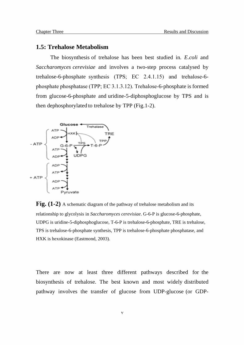
1.5: Trehalose Metabolism
The biosynthesis of trehalose has been best studied in. E.coli and
Saccharomyces cerevisiae and involves a two-step process catalysed by
trehalose-6-phosphate synthesis (TPS; EC 2.4.1.15) and trehalose-6-
phosphate phosphatase (TPP; EC 3.1.3.12). Trehalose-6-phosphate is formed
from glucose-6-phosphate and uridine-5-diphosphoglucose by TPS and is
then dephosphorylated to trehalose by TPP (Fig.1-2).
Fig. (1-2) A schematic diagram of the pathway of trehalose metabolism and its
relationship to glycolysis in Saccharomyces cerevisiae. G-6-P is glucose-6-phosphate,
UDPG is uridine-5-diphosphoglucose, T-6-P is trehalose-6-phosphate, TRE is trehalose,
TPS is trehalose-6-phosphate synthesis, TPP is trehalose-6-phosphate phosphatase, and
HXK is hexokinase (Eastmond, 2003).
There are now at least three different pathways described for the
biosynthesis of trehalose. The best known and most widely distributed
pathway involves the transfer of glucose from UDP-glucose (or GDP-
Chapter Three Results and Discussion
٨
glucose in some cases) to glucose 6-phosphate to form trehalose-6-phosphate
and UDP. This reaction is catalyzed by the trehalose-P synthesis (TPS, or
OtsA in E.coli. Organisms that use this pathway usually also have a
trehalose-P phosphatase (TPP, or OtsB in E. coli) that converts the trehalose-
P to free trehalose. A second pathway that has been reported in a few
unusual bacteria involves the intramolecular rearrangement of maltose
(glucosyl- 1, 4-glucopyranoside) to convert the 1, 4-linkage to the 1, 1-bond
of trehalose. This reaction is catalyzed by the enzyme called trehalose
synthesis and gives rise to free trehalose as the initial product. A third
pathway involves several different enzymes, the first of which rearranges the
glucose at the reducing end of a glycogen chain to convert the 1,4-linkage
to an , 1,1-bond. A second enzyme then releases the trehalose disaccharide
from the reducing end of the glycogen molecule. Finally, in mushrooms
there is a trehalose phosphorylase that catalyzes the phosphorolysis of
trehalose to produce glucose-1-phosphate and glucose. This reaction is
reversible in vitro and could theoretically give rise to trehalose from glucose-
1-P and glucose. Another important enzyme in trehalose metabolism is
trehalase (T), which may be involved in energy metabolism and also have a
regulatory role in controlling the levels of trehalose in cells. This enzyme
may be important in lowering trehalose concentrations once the stress is
alleviated. Recent studies in yeast indicate that the enzymes involved in
trehalose synthesis (TPS, TPP) exist together in a complex that is highly
regulated at the activity level as well as at the genetic level (Elbein, 2003).
In brewer's yeast, the biosynthesis of trehalose is catalysed by enzymes that
facilitate the reaction of uridine diphosphate-D-glucose with D-glucose 6-
phosphate, resulting in uridine diphosphate and a.a-trehalose 6-phosphate.
The phosphate is enzymatically removed leaving a trehalose molecule.
Chapter Three Results and Discussion
٩
Several other organisms produce trehalose by similar mechanisms.
Degradation of trehalose is accomplished by a highly specific enzyme,
trehalase. Trehalase has been identified in many organisms shown to contain
trehalose but is not found in mammals (Richards et al., 2002).
Maltose and trehalose assimilating pathways in Lactococcus lactis.
The maltose phosphorylase (MP) and beta-phosphoglucomutase (b-PGM) of
L. lactic were characterized and shown to constitute the major maltose-
degrading pathway in this bacterium. Furthermore, MP and b-PGM were
shown to be present in many other strains of LAB belonging to the low G+C
content LAB of the clostridial sub-branch of Gram-positive bacteria. The
MP-encoding gene, Mal P, was localized in an operon distinctive from that
of the gene encoding b-PGM, Pgm B. In addition, Mal R, encoding the
maltose operon regulator Mal R, was localized downstream of Mal P. The
presence of Mal R was shown to be crucial for the synthesis of an ATP-
dependent maltose translocation system. However, MP and b-PGM activity
were not affected by a disruption of the Mal R-encoding gene. Instead,
synthesis of b-PGM has been shown to be exposed to carbon catabolite
repression, which was also shown to be the case for MP. The Pgm B is
located in the putative trehalose operon including the genes presumed to
code for the trehalose-specific components of the phosphotransferase system
transporting trehalose into the cells. Furthermore, directly upstream of Pgm
B, Trep P was localized. This gene was shown to encode a novel
phosphorylase, trehalose 6-phosphate phosphorylase Trep p, catalysing the
reversible Pi-dependent phosphorolysis of trehalose r6-phosphate to beta-
glucose 1-phosphate and glucose 6-phosphate. The Trep p was biochemical
characterized and shown to be present in a few other species, mainly
Enterococcus faecalis, of low G+C content LAB. The role of b-PGM in
Chapter Three Results and Discussion
١٠
trehalose metabolism and in polysaccharide synthesis was assessed by
disrupting its encoding gene. B-PGM was shown to be crucial for trehalose
assimilation in L. lactic, while the b-PGM-deficient strain continued to grow
with a tenfold decreased growth rate on maltose, compared to the wild-type
strain. The b-PGM-deficient strain showed an enhanced production of
polysaccharide, composed of alpha-1, 4-linked glucose units when cultivated
on maltose. It was suggested that this polysaccharide was a result from
another metabolic pathway, resembling the maltodextrin system in E.coli.
(Andersson, 2002).
Axenically grown Arabidopsis thaliana plants were analyzed for the
occurrence of trehalose. Using gas chromatography–mass spectrometry
(GC–MS) analysis, trehalose was unambiguously identified in extracts from
Arabidopsis spp. inflorescences. In a variety of organisms, the synthesis of
trehalose is catalysed by trehalose-6-phosphate synthesis (TPS) and
trehalose-6-phosphate phosphatase (TPP). Based on EST (expressed
sequence tag) sequences, three full-length Arabidopsis cDNAs whose
predicted protein sequences show extensive homologies to known TPS and
TPP proteins were amplified by rapid amplification of 5′ and 3′ cDNA ends
(RACE–PCR). The expression of the corresponding genes, Attpsa, Attpsb
and Attpsc, and of the previously described TPS gene, Attps1, was analysed
by quantitative Reverse Transcriptase (RT–PCR). All of the genes were
expressed in the rosette leaves, stems and flowers of Arabidopsis plants and,
to a lower extent, in the roots. To study the role of the Arabidopsis genes, the
Attpsa and Attps1 cDNAs were expressed in Saccharomyces cerevisiae
mutants deficient in trehalose synthesis. In contrast to Attps1, expression of
Attpsca and Attpsc in the Tps 1 mutant lacking TPS activity did not
complement trehalose formation after heat shock or growth on glucose. In
Chapter Three Results and Discussion
١١
addition, no TPP function could be identified for Attpsa and Attpsc in
complementation studies with the S. cerevisiae Tps 2 mutant lacking TPP
activity. The results indicate that while Attps1 is involved in the formation of
trehalose in Arabidopsis, some of the Arabidopsis genes with homologies to
known TPS/TPP genes encode proteins lacking catalytic activity in trehalose
synthesis (Vogel et al., 2001).
1.6: Physiological roles of Trehalose in bacteria and yeasts:
The disaccharide trehalose is widely distributed in nature and can be
found in many organisms, including bacteria, fungi, plants, invertebrates and
mammals. Due to its particular physical features, trehalose is able to protect
the integrity of the cell against a variety of environmental stress and
nutritional limitations. In addition, data available on several species of
bacteria and yeast suggest specific functions for trehalose in these
organisms. Bacteria can use exogenous trehalose as the sole source of
carbon and energy as well as synthesize enormous amounts of the
disaccharide as compatible solute .Cells of many organisms accumulate
certain small organic molecules-called compatible and counteracting solutes,
compensatory solutes, or chemical chaperones--in response to certain
physical stresses. These solutes include certain carbohydrates, amino acids,
methylamine and methylsulphonium zwitterions, and urea. In osmotic
dehydrating stress, these solutes serve as cellular osmolytes. Unlike common
salt ions and urea (which inhibit proteins), some organic osmolytes are
compatible, This ability to accumulate trehalose is the result of an elaborate
genetic system, which is regulated by osmolarity. Some Mycobacterium
contains sterified trehalose as a structural component of the cell wall,
Chapter Three Results and Discussion
١٢
whereas yeast cells are largely unable to grow on trehalose as carbon source.
In these lower eukaryotes, trehalose appears to play a dual function: as a
reserve compound, mainly stored in vegetative resting cells and reproductive
structures, and as a stress metabolite. Recent findings also point to important
biotechnological applications for trehalose (Arguelles, 2000).
1.7: Physical-chemical properties:
Trehalose (, -Trehalose) is a disaccharide formed by a 1, 1 linkage of
two D-glucose molecules. It is a non-reducing sugar that is not easily
hydrolyzed by acid, and the glycosidic bond is not cleaved by the enzyme a-
glucosidase. The molecular formula and weight are C12H22O11 and 342.31,
respectively. When purified it is usually found in a dihydrate form, which is
the typical commercial product.
Trehalose can impart some beneficial properties to food products. Compared
to most sugars, trehalose is more stable to wide ranges of pH and heat and it
does not easily interact with proteinaceous molecules.
Trehalose has a low hygroscopic profile which is a main advantage
compared to other sugars. It appears that trehalose could be of benefit
compared with other sugars in dry blending operations in which low
hygroscopicity is desired. The water content of trehalose dehydrate remains
stable (9.54%) up to a relative humidity of approximately 92%. (Richards et
al., 2002).
Physical properties that make trehalose unique are its high degree of
optical rotation ([a] 2 D + 178°) and its melting behavior. Trehalose first
melts at 97 ° C. Additional heat drives off the water of crystallization until
Chapter Three Results and Discussion
١٣
the material re-solidifies at 130 ° C, and then the anhydrous trehalose melts
at 203 ° C. The combination of the molecular structure and the physical-
chemical properties of trehalose results in a very stable disaccharide.
Trehalose has a solubility and osmotic profile similar to maltose.
Above 80 ° C Trehalose becomes more soluble in water relative to other
sugars.
Trehalose possesses several unique properties, including high
hydrophilicity, chemical stability, non hygroscopic glass formation and no
internal hydrogen bond formation. The combination of these features
explains the principal role of trehalose as a stress metabolite (Richards et al.,
2002).
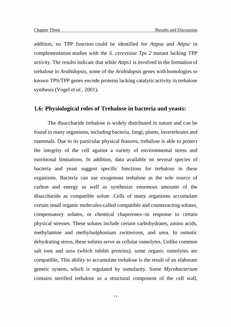
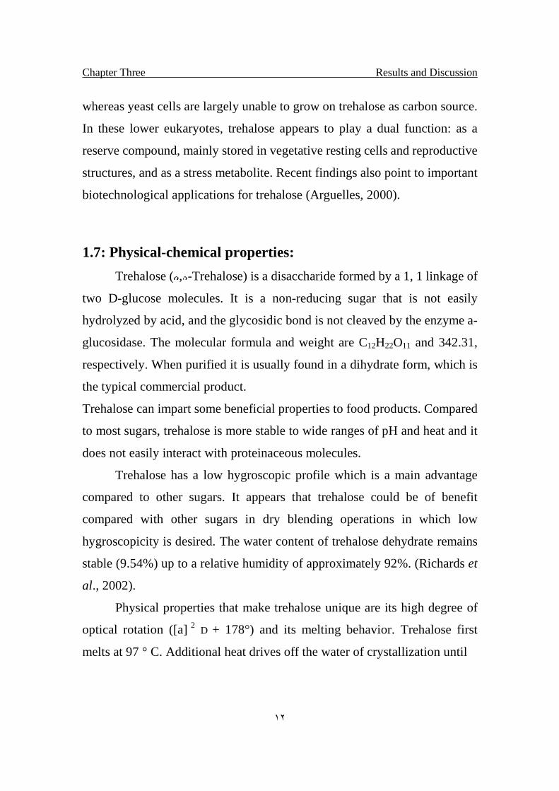
The properties of trehalose are shown in (table 1.1); its relative sweetness is
45 % of sucrose. Trehalose has high thermostability and a wide pH-stability
range. Therefore, it is one of the most stable saccharides. When 4 %
trehalose solutions with (3.5 - 10) pH were heated at 100 °C for 24 hrs, no
degradation of trehalose was observed in any case. Because of no reducing
sugar, this saccharide does not show Maillard reaction with amino
compounds such as amino acids or proteins.
Chapter Three Results and Discussion
١٤
Figure (1-3) Maillard reaction is a type of non-enzymatic browning which involves the
reaction of simple sugars (carbonyl groups) and amino acids (free amino groups). They
begin to occur at lower temperatures and at higher dilutions than caramelization.
Its particular physical features make it an extremely attractive substance for
industrial applications. Furthermore, this saccharide shows good sweetness
like sucrose, and in the food industry, this saccharide is used as a sweetener
(Higashiyama, 2002).
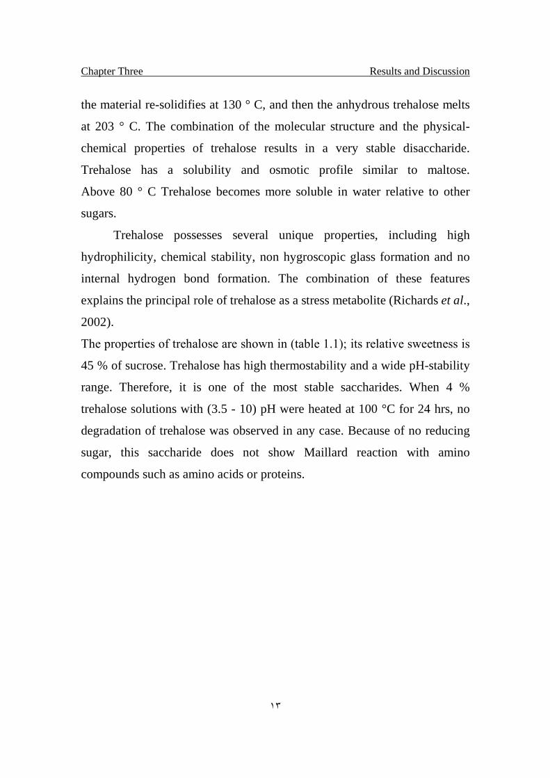
Table (1- 1) Properties of Trehalose (Higashiyama, 2002).
Melting point dihydrate 97.0 °C
Anhydride 210.5 °C
Heat of fusion dihydrate 57.8 kJ mol–1
Anhydride 53.4 kJ mol–1
Solubility 68.9 g/100 g H2O at 20 °C
Relative sweetness 45 % of sucrose
Digestibility digested and absorbed by the small intestine
pH stability of solution >99 % (pH 3.5–10, at 100 °C for 24 h)
Heat stability of solution >99 % (at 120 °C for 90 min)
Chapter Three Results and Discussion
١٥
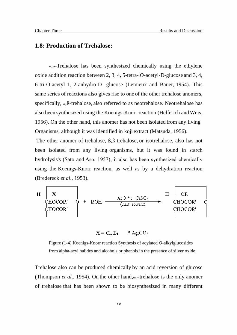
1.8: Production of Trehalose:
, -Trehalose has been synthesized chemically using the ethylene
oxide addition reaction between 2, 3, 4, 5-tetra- O-acetyl-D-glucose and 3, 4,
6-tri-O-acetyl-1, 2-anhydro-D- glucose (Lemieux and Bauer, 1954). This
same series of reactions also gives rise to one of the other trehalose anomers,
specifically, ,ß-trehalose, also referred to as neotrehalose. Neotrehalose has
also been synthesized using the Koenigs-Knorr reaction (Helferich and Weis,
1956). On the other hand, this anomer has not been isolated from any living
Organisms, although it was identified in koji extract (Matsuda, 1956).
The other anomer of trehalose, ß,ß-trehalose, or isotrehalose, also has not
been isolated from any living organisms, but it was found in starch
hydrolysis's (Sato and Aso, 1957); it also has been synthesized chemically
using the Koenigs-Knorr reaction, as well as by a dehydration reaction
(Bredereck et al., 1953).
Figure (1-4) Koenigs-Knorr reaction Synthesis of acylated O-alkylglucosides
from alpha-acyl halides and alcohols or phenols in the presence of silver oxide.
Trehalose also can be produced chemically by an acid reversion of glucose
(Thompson et al., 1954). On the other hand,-trehalose is the only anomer
of trehalose that has been shown to be biosynthesized in many different
Chapter Three Results and Discussion
١٦
types of organisms, although its usefulness was recognized, trehalose was
not produced on an industrial scale until 1994 (Elbein, 2003).
The conventional method for production, for example, extraction from yeast,
had too low yield and too high cost to be used. In order to implement
industrial production of trehalose, they have researched new enzyme
systems and have succeeded in isolating a novel enzyme system from a
bacterial strain belonging to the genus Arthrobacter sp. Q36 that was
obtained from soil. The system has been found to consist of two novel
enzymes: malto-oligosyltrehalose synthesis (or MTSase) and malto-
oligosyltrehalose trehalohydorolase (or MTHase). In the first step, MTSase
catalyzes the intramolecular transglycosylation of glucose residue at the
reducing end of malto-oligosaccharide from -1, 4 bonds to -1, 1 bond, then
malto-oligosyltrehalose is produced. This contains trehalose residue at the
end of the saccharide chain. Next, trehalose is liberated from malto-
oligosyltrehalose by MTHase. This pathway needs no high-energy sugar
derivative such as sugar phosphate or sugar nucleotide. And those enzymes
can repeatedly act on -1, 4 glucan to produce trehalose up to 80 % yield
(Higashiyama, 2002).
1.9: History of human consumption:
In general, the fate of ingested or parenterally administered trehalose
corresponds to that of glucose since trehalose is rapidly hydrolyzed to
glucose by the enzyme trehalase. Trehalase is found in humans and most
animals at the brush border of the intestinal mucosa, as well as in the kidney,
liver, and blood plasma (Hore & Messer, 1968; Demelier et al., 1975; Labat-
Chapter Three Results and Discussion
١٧
Robert, 1982; Niederst & Dauça, 1985; Eze, 1989; Riby et al., 1990;
Yoshida, 1993). Trehalase activity has been found in the small intestine of
humans, mice, rats, guinea-pigs, rabbits, pigs, and baboons (Hietanen, 1973;
Ruppin et al., 1974; Maestracci, 1976; Garland, 1989). No trehalase activity
is found in the small intestine of cats (Hore & Messer, 1968; Hietanen, 1973;
Garland, 1989).
A very few individuals have trehalase deficiency, which may be hereditary
or acquired. However, in Greenland, the prevalence of trehalase deficiency
has been reported to be 8%, which is considerably higher than that seen
elsewhere (Dahlqvist, 1974; Gudmand-Høyer et al., 1988). The incidence of
trehalase deficiency is lower than that of lactase deficiency, which in the
United Kingdom is 3.2–6% (Gudmand-Høyer & Skovbjerg, 1996).
When trehalose is ingested by such individuals, it is either incompletely
digested or undigested, and a small fraction (approximately 0.5%) may be
absorbed by passive diffusion, as shown for other disaccharides (Van Elburg
et al., 1995).
The absorbed trehalose may then be metabolized to glucose in the liver or
kidney or be excreted unchanged in the urine (Demelier et al., 1975).
Unabsorbed trehalose is likely to be fermented by the intestinal microflora to
short-chain fatty acids such as acetate, propionate, and butyrate (Abbott,
2002).
Modem food sources may contain substantial quantities of trehalose. Some
of these include honey (0.1 - 1.9%), mirin (1.3 - 2.2%), Sherries (< 10 - 391
mg/1), brewer's yeast (0.01 -5.0%) and baker's yeast (15 - 20%), and
therefore most items made using yeast. Commercially grown mushrooms
may contain 8-17 % (w/w) trehalose. It also occurs in lobsters (2.5 mg/100
Chapter Three Results and Discussion
١٨
ml blood), crab (1.5 mg/100 ml blood) and prawns (0.5% dry weight).
Trehalose is not presently a significant part of the modem diet but has been a
consistent part of the human diet for thousands of years (Richards et al,
2002).
Trehalose does not produce as great a sensation of sweetness as dose
sucrose. Trehalose is believed to have only one glucose molecule occupying
the binding site on the sweet taste receptor. Trehalose in aqueous solution (at
concentrations from about 10-34% anhydrous trehalose) has a sweetness of
about 40-45% relative to that of sucrose. The concentration at which a
solution of trehalose is perceived as sweet, is about two-times higher than
that of sucrose and the sweetness persists longer than sucrose (Richards et
al., 2002).
1.10: Interaction of Trehalose with Phospholipid Bilayer:
Nature has developed numerous strategies for the long-term survival
of organisms. Among the most intriguing are the biological mechanisms that
preserve living organisms exposed to damaging conditions like extreme cold,
dryness, or heat, or the absence of oxygen (Feofilova, 2003). This
phenomenon, named cryptobiosis, involves a reversible suspension of the
metabolism and an effective isolation from the environmental changes
(Keilin, 1959; Clegg, 2001). Cryptobiosis is widespread in the animal and
plant kingdoms, and occurs for example in tardigrades, nematodes, cysts of
crustaceans, yeasts, bacteria, fungi, mosses, pollens seeds, and even in entire
higher plants (Crowe et al., 1992; Guppy and Withers 1999; Feofilova,
2003).
Chapter Three Results and Discussion
١٩
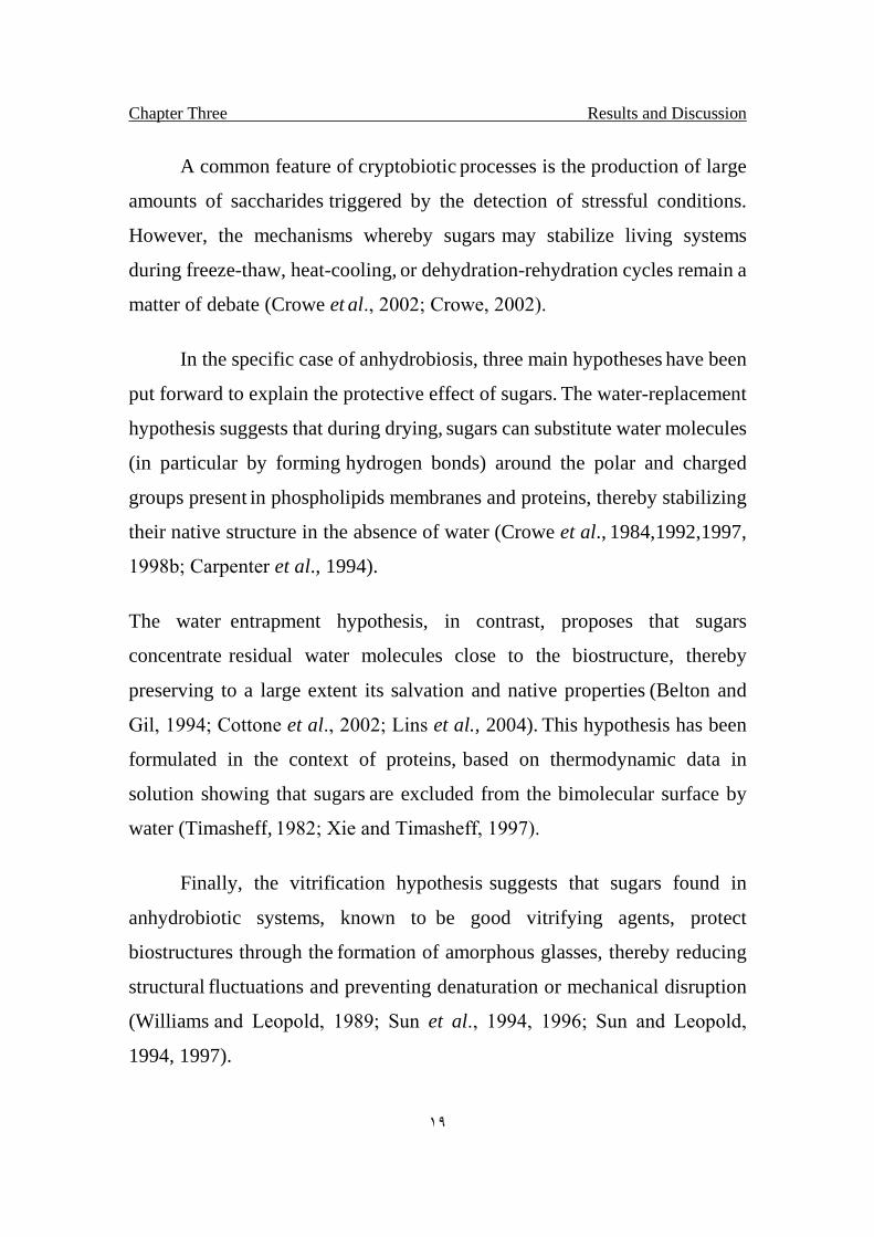
A common feature of cryptobiotic processes is the production of large
amounts of saccharides triggered by the detection of stressful conditions.
However, the mechanisms whereby sugars may stabilize living systems
during freeze-thaw, heat-cooling, or dehydration-rehydration cycles remain a
matter of debate (Crowe et al., 2002; Crowe, 2002).
In the specific case of anhydrobiosis, three main hypotheses have been
put forward to explain the protective effect of sugars. The water-replacement
hypothesis suggests that during drying, sugars can substitute water molecules
(in particular by forming hydrogen bonds) around the polar and charged
groups present in phospholipids membranes and proteins, thereby stabilizing
their native structure in the absence of water (Crowe et al., 1984,1992,1997,
1998b; Carpenter et al., 1994).
The water entrapment hypothesis, in contrast, proposes that sugars
concentrate residual water molecules close to the biostructure, thereby
preserving to a large extent its salvation and native properties (Belton and
Gil, 1994; Cottone et al., 2002; Lins et al., 2004). This hypothesis has been
formulated in the context of proteins, based on thermodynamic data in
solution showing that sugars are excluded from the bimolecular surface by
water (Timasheff, 1982; Xie and Timasheff, 1997).
Finally, the vitrification hypothesis suggests that sugars found in
anhydrobiotic systems, known to be good vitrifying agents, protect
biostructures through the formation of amorphous glasses, thereby reducing
structural fluctuations and preventing denaturation or mechanical disruption
(Williams and Leopold, 1989; Sun et al., 1994, 1996; Sun and Leopold,
1994, 1997).
Chapter Three Results and Discussion
٢٠
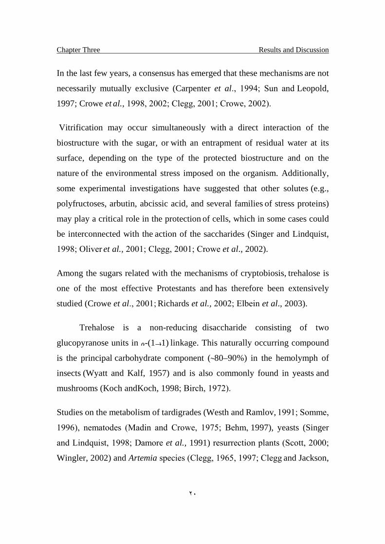
In the last few years, a consensus has emerged that these mechanisms are not
necessarily mutually exclusive (Carpenter et al., 1994; Sun and Leopold,
1997; Crowe et al., 1998, 2002; Clegg, 2001; Crowe, 2002).
Vitrification may occur simultaneously with a direct interaction of the
biostructure with the sugar, or with an entrapment of residual water at its
surface, depending on the type of the protected biostructure and on the
nature of the environmental stress imposed on the organism. Additionally,
some experimental investigations have suggested that other solutes (e.g.,
polyfructoses, arbutin, abcissic acid, and several families of stress proteins)
may play a critical role in the protection of cells, which in some cases could
be interconnected with the action of the saccharides (Singer and Lindquist,
1998; Oliver et al., 2001; Clegg, 2001; Crowe et al., 2002).
Among the sugars related with the mechanisms of cryptobiosis, trehalose is
one of the most effective Protestants and has therefore been extensively
studied (Crowe et al., 2001; Richards et al., 2002; Elbein et al., 2003).
Trehalose is a non-reducing disaccharide consisting of two
glucopyranose units in -(1 1) linkage. This naturally occurring compound
is the principal carbohydrate component (80–90%) in the hemolymph of
insects (Wyatt and Kalf, 1957) and is also commonly found in yeasts and
mushrooms (Koch andKoch, 1998; Birch, 1972).
Studies on the metabolism of tardigrades (Westh and Ramlov, 1991; Somme,
1996), nematodes (Madin and Crowe, 1975; Behm, 1997), yeasts (Singer
and Lindquist, 1998; Damore et al., 1991) resurrection plants (Scott, 2000;
Wingler, 2002) and Artemia species (Clegg, 1965, 1997; Clegg and Jackson,
Chapter Three Results and Discussion
٢١
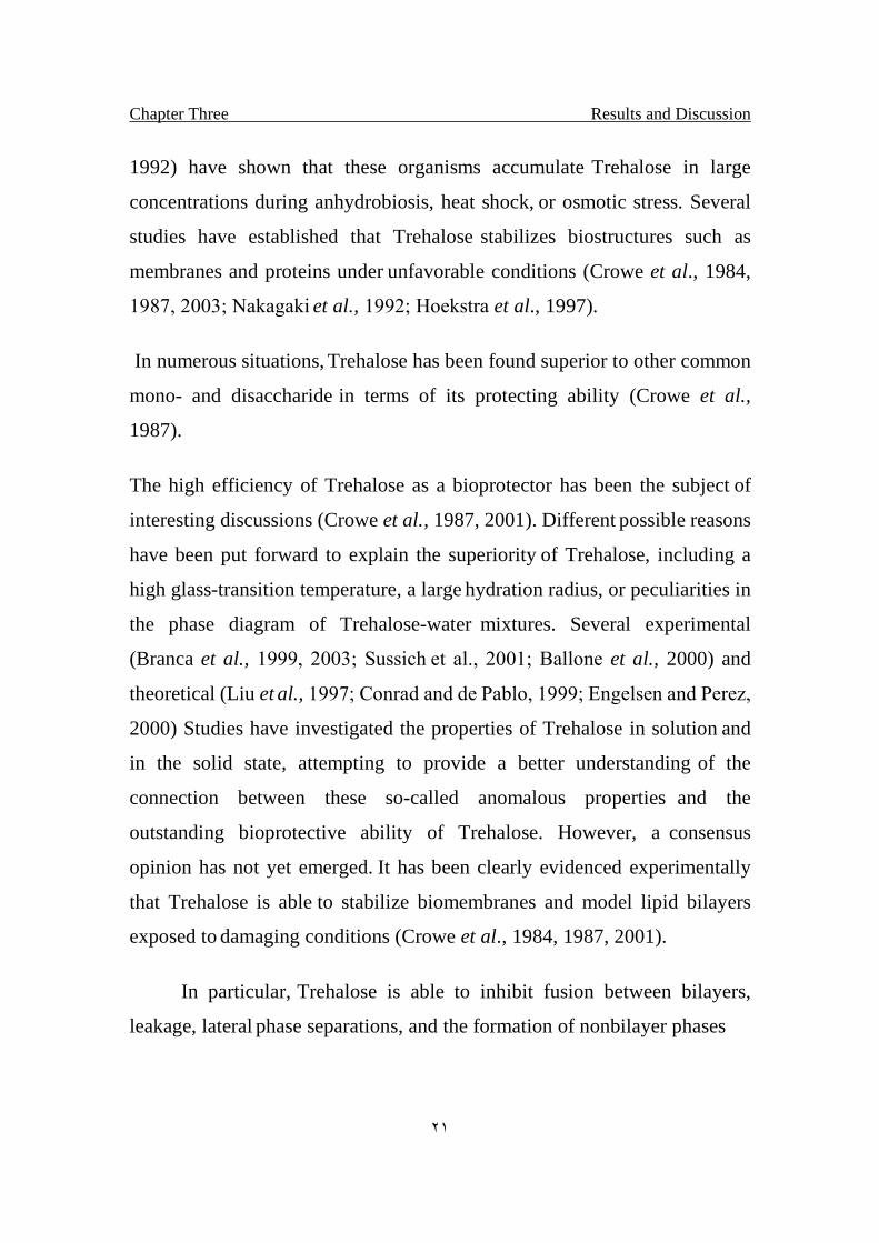
1992) have shown that these organisms accumulate Trehalose in large
concentrations during anhydrobiosis, heat shock, or osmotic stress. Several
studies have established that Trehalose stabilizes biostructures such as
membranes and proteins under unfavorable conditions (Crowe et al., 1984,
1987, 2003; Nakagaki et al., 1992; Hoekstra et al., 1997).
In numerous situations, Trehalose has been found superior to other common
mono- and disaccharide in terms of its protecting ability (Crowe et al.,
1987).
The high efficiency of Trehalose as a bioprotector has been the subject of
interesting discussions (Crowe et al., 1987, 2001). Different possible reasons
have been put forward to explain the superiority of Trehalose, including a
high glass-transition temperature, a large hydration radius, or peculiarities in
the phase diagram of Trehalose-water mixtures. Several experimental
(Branca et al., 1999, 2003; Sussich et al., 2001; Ballone et al., 2000) and
theoretical (Liu et al., 1997; Conrad and de Pablo, 1999; Engelsen and Perez,
2000) Studies have investigated the properties of Trehalose in solution and
in the solid state, attempting to provide a better understanding of the
connection between these so-called anomalous properties and the
outstanding bioprotective ability of Trehalose. However, a consensus
opinion has not yet emerged. It has been clearly evidenced experimentally
that Trehalose is able to stabilize biomembranes and model lipid bilayers
exposed to damaging conditions (Crowe et al., 1984, 1987, 2001).
In particular, Trehalose is able to inhibit fusion between bilayers,
leakage, lateral phase separations, and the formation of nonbilayer phases
Chapter Three Results and Discussion
٢٢
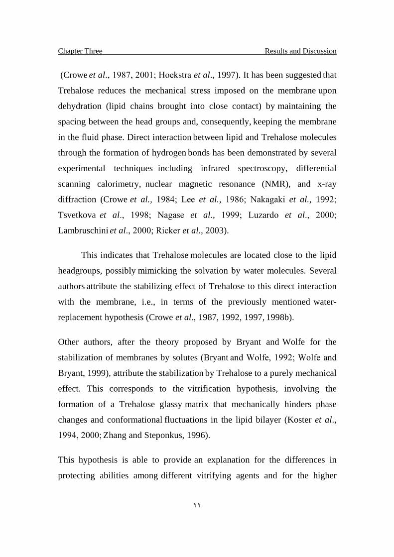
(Crowe et al., 1987, 2001; Hoekstra et al., 1997). It has been suggested that
Trehalose reduces the mechanical stress imposed on the membrane upon
dehydration (lipid chains brought into close contact) by maintaining the
spacing between the head groups and, consequently, keeping the membrane
in the fluid phase. Direct interaction between lipid and Trehalose molecules
through the formation of hydrogen bonds has been demonstrated by several
experimental techniques including infrared spectroscopy, differential
scanning calorimetry, nuclear magnetic resonance (NMR), and x-ray
diffraction (Crowe et al., 1984; Lee et al., 1986; Nakagaki et al., 1992;
Tsvetkova et al., 1998; Nagase et al., 1999; Luzardo et al., 2000;
Lambruschini et al., 2000; Ricker et al., 2003).
This indicates that Trehalose molecules are located close to the lipid
headgroups, possibly mimicking the solvation by water molecules. Several
authors attribute the stabilizing effect of Trehalose to this direct interaction
with the membrane, i.e., in terms of the previously mentioned water-
replacement hypothesis (Crowe et al., 1987, 1992, 1997, 1998b).
Other authors, after the theory proposed by Bryant and Wolfe for the
stabilization of membranes by solutes (Bryant and Wolfe, 1992; Wolfe and
Bryant, 1999), attribute the stabilization by Trehalose to a purely mechanical
effect. This corresponds to the vitrification hypothesis, involving the
formation of a Trehalose glassy matrix that mechanically hinders phase
changes and conformational fluctuations in the lipid bilayer (Koster et al.,
1994, 2000; Zhang and Steponkus, 1996).
This hypothesis is able to provide an explanation for the differences in
protecting abilities among different vitrifying agents and for the higher
Chapter Three Results and Discussion
٢٣
efficiency of Trehalose as a stabilizer. The theory does not exclude the
possibility of specific hydrogen bonds between lipid and sugar molecules.
However, such interactions are not viewed as a determinant factor in the
stabilization process (Koster et al., 2000; Wolfe and Bryant, 1999).
In the few last decades, numerous experimental studies have addressed the
problem of Trehalose-membrane interactions. However, only two theoretical
investigations relying on modeling methods (energy minimization protocols)
have been reported (Chandrasekhar and Gaber, 1988; Rudolph et al., 1990).
These two investigations showed that, in the absence of Trehalose is able to
form energetically stable conformations bridging a number of lipid
molecules (Chandrasekhar and Gaber, 1988). However, an extension of the
method to sucrose and glucose did not succeed in reproducing the
experimental order for the stabilizing efficiency of these sugars (Rudolph et
al., 1990).
Although Trehalose-protein interactions have been studied by atomistic
simulations (Cottone et al., 2001, 2002; Lins et al., 2003), no simulations of
Trehalose-membrane systems have been reported to date. Therefore a
detailed picture for the molecular mechanism responsible for membrane
stabilization is still lacking (Pereira et al., 2004).
1.11: Functions and Applications of trehalose
Trehalose levels may vary greatly in certain cells depending on the
stage of growth, the nutritional state of the organism or cell, and the
environmental conditions prevailing at the time of measurement. In insects,
Chapter Three Results and Discussion
٢٤
trehalose is a major sugar in the hemolymph and thorax muscles and is
consumed during flight (Becker et al., 1996).
Trehalose is also an important component in fungal spores, where trehalose
hydrolysis is a major event during early germination and presumably serves
as a source of carbon for synthesis and glucose for energy (Rosseau et al.,
1972; Thevelein, 1984).
1.11.1: As a stabilizer and protectant of proteins and
membranes (Protection from dehydration).
The particular properties of Trehalose have given rise to a surprisingly
wide range of applications of this disaccharide in technology including the
stabilization of proteins, membranes, liposome, and vaccines (Crowe et al.,
2001).The hypothermic storage of mammalian cells and organs (Crowe et
al., 2003; Fukuse et al., 1999; Eroglu et al., 2002), and its use in cosmetics
(Norcia, 2000) and food products (Richards et al., 2002).
Trehalose also appears to be efficient in the treatment of dry eye syndrome,
for which it is currently being tested in human clinical trials (Matsuo, 2001;
Matsuo et al., 2002).
Although water is obviously necessary for life, some organisms are able to
survive almost complete dehydration, for instance, even when 99% of their
water content is removed. This includes common organisms such as plants,
yeast cells, and fungal spores, but also microscopic animals such as
nematodes, rotifers, and the cysts of brine shrimp (Leopold, 1986).
Chapter Three Results and Discussion
٢٥
Some of these dried organisms may remain in this state, known as
anhydrobiosis, for decades under unfavorable conditions until water
becomes available. When that happens, these organisms swell and resume
the active state. Recent studies on these organisms have demonstrated
mechanisms that allow them to survive dehydration; understanding some of
these mechanisms have enabled researchers to develop new methods for the
preservation of biological materials that are normally sensitive to drying
(Crowe et al., 1992).
Anhydrobiotic organisms generally contain high concentrations of trehalose
(and sometimes other disaccharides and oligosaccharides). Thus it was
shown that when the nematode Aphelenchus avenae was slowly dehydrated,
it converted as much as 20% of its dry weight into trehalose (Madin and
Crowe, 1975).
The ability of this organism and others to survive in the absence of water has
shown a strong correlation with the synthesis of trehalose (Madin and
Crowe, 1975). Log-phase cultures of yeast have low concentrations of
trehalose and are quite susceptible to dehydration, but as they enter the
stationary phase of growth the levels of trehalose increase, along with their
ability to survive dehydration (Gadd et al., 1987).
This ability to survive in the presence of trehalose is independent of the
growth phase of the cells because log-phase cells subjected to heat shock
rapidly synthesize trehalose and also acquire the ability to survive
dehydration (Hottinger et al., 1987). These results are also applicable to a
variety of other organisms ranging from brine shrimp (Leopold, 1986) to the
resurrection plant (Zentella et al., 1999).
Chapter Three Results and Discussion
٢٦
This use of trehalose to enable cells to survive dehydration (and other
stresses) may be an ancient adaptation because even Archaebacteria have
been found to accumulate trehalose in response to stress (Nicolaus et al.,
1988). Interestingly, in plants the disaccharide sucrose plays a similar role to
that of trehalose in yeast and nematodes (Anandarajah and Mckersie, 1990).
According to Leopold (1986), trehalose is preferred over sucrose by most
organisms because it has fewer tendencies to form crystals than dose
sucrose.
The two primary stresses that are proposed to destabilize lipid bilayers
during dehydration are fusion and lipid phase transitions. Studies by laser
light scattering or other techniques demonstrate that trehalose and other
sugars inhibit fusion between the vesicles during drying, but the inhibition of
fusion alone is not sufficient to preserve the dry vesicles. Thus trehalose is
also necessary to prevent phase transitions (Crowe and Crowe, 1990).
The evidence suggests that trehalose depresses the phase transition
temperature of the dry lipids, which maintains them in the liquid crystalline
phase in the absence of water (Crowe and Crowe, 1988). A large body of
evidence indicates that the stabilizing effect of trehalose is due to its
structure and stereochemistry. X-ray diffraction studies show that trehalose
fits well between the polar head groups with multiple sites of interaction and
suggests that the strong stabilizing effects of trehalose are related to its
stereochemistry, which provides the most favorable fit with the polar head
groups (Rudolph et al., 1990).
Chapter Three Results and Discussion
٢٧
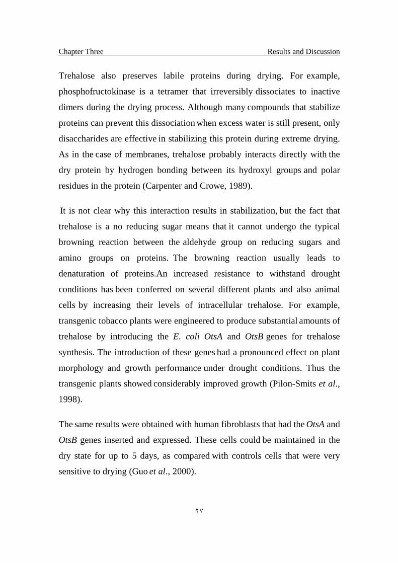
Trehalose also preserves labile proteins during drying. For example,
phosphofructokinase is a tetramer that irreversibly dissociates to inactive
dimers during the drying process. Although many compounds that stabilize
proteins can prevent this dissociation when excess water is still present, only
disaccharides are effective in stabilizing this protein during extreme drying.
As in the case of membranes, trehalose probably interacts directly with the
dry protein by hydrogen bonding between its hydroxyl groups and polar
residues in the protein (Carpenter and Crowe, 1989).
It is not clear why this interaction results in stabilization, but the fact that
trehalose is a no reducing sugar means that it cannot undergo the typical
browning reaction between the aldehyde group on reducing sugars and
amino groups on proteins. The browning reaction usually leads to
denaturation of proteins.An increased resistance to withstand drought
conditions has been conferred on several different plants and also animal
cells by increasing their levels of intracellular trehalose. For example,
transgenic tobacco plants were engineered to produce substantial amounts of
trehalose by introducing the E. coli OtsA and OtsB genes for trehalose
synthesis. The introduction of these genes had a pronounced effect on plant
morphology and growth performance under drought conditions. Thus the
transgenic plants showed considerably improved growth (Pilon-Smits et al.,
1998).
The same results were obtained with human fibroblasts that had the OtsA and
OtsB genes inserted and expressed. These cells could be maintained in the
dry state for up to 5 days, as compared with controls cells that were very
sensitive to drying (Guo et al., 2000).
Chapter Three Results and Discussion
٢٨
1.11.2: Protection against heat.
Trehalose has been described to act as the best stabilizer of structure
and function of several macromolecules. Although other sugars also stabilize
macromolecules, none of them are as effective as trehalose. The
extraordinary effect of trehalose has been attributed to several of its
properties such as making hydrogen bonds with membranes or the ability to
modify the salvation layer of proteins. However, the explanations always
result in a question: Why is trehalose more effective than other sugars? Here,
we show that trehalose has a larger hydrated volume than other related
sugars. According to our results, trehalose occupies at least 2.5 times larger
volume than sucrose, maltose, glucose, and fructose. We correlate this
property with the ability to protect the structure and function of enzymes
against thermal inactivation. When the concentrations of all sugars were
corrected by the percentage of the occupied volume, they presented the same
effectiveness. results suggest that because of this larger hydrated volume,
trehalose can substitute more water molecules in the solution, and this
property is very close to its effectiveness. Finally, these data drive us to
conclude that the higher size exclusion effect is responsible for the
difference in efficiency of protection against thermal inactivation of
enzymes .
Yeast cells have had to develop mechanisms in order to protect themselves
from chemical and physical agents of the environment to which they are
exposed. One of these physical agents is thermal variation. Some yeast cells
are known to accumulate high concentrations of trehalose when submitted to
heat shock (Elbein, 2003).
Chapter Three Results and Discussion
٢٩
In yeast, stimuli that trigger the heat shock response also cause the
accumulation of trehalose. In fact at least two subunits of the trehalose-6-P
synthesis complex of S. cerevesiae are actively synthesized during heat
shock (Bell et al., 1992), and physiological concentrations of trehalose (up to
0.5 M) were found to protect enzymes of yeast and other organisms from
heat inactivation in vitro. Trehalose also reduced the heat-induced formation
of protein aggregates. Trehalose was as good or better as a protein stabilizer
than any of a number of comparable solutes, including polyols, sugars, and
amino acids (DeVirgilio et al., 1994).
Yeast mutants were prepared that were defective in genes coding for key
enzymes in trehalose metabolism (TPS1, TPS2), and these mutants showed
an inability to accumulate trehalose on mild heat shock and were much less
resistant to heating than was the wild-type organism. These various studies
strongly implicate trehalose as playing a key role in thermo tolerance and
also indicate that the enzymes that synthesize trehalose are induced in
response to the stress in order to increase the levels of trehalose. An
important in vivo and in vitro study showed that trehalose protects cells from
heat by stabilizing proteins at high temperatures. Using two different
temperature-sensitive reporter proteins, these investigators showed that
enzymes are better able to retain activity during heat shock in cells that are
producing trehalose (Singer and Lindquist, 1998).
These studies showed an additional and important role of trehalose, that is,
the ability to suppress aggregation of proteins that have already been
denatured. Based on these studies, these researchers also explained why
trehalose must be degraded rapidly after the heat shock has ended, that is, if
the unfolded luciferase, one of their reporter proteins, is removed from or
Chapter Three Results and Discussion
٣٠
diluted out of the trehalose, and it can be refolded by molecular chaperones.
On the other hand, if the trehalose concentration remains high, it interferes
with the refolding process, and the protein is not renatured by the chaperone
(Singer and Lindquist, 1998). Thus, it may be important to have active
trehalase present once the heat stress is alleviated.
1.11.3: Protection from damage by oxygen radicals:
Another role for trehalose is in protecting cells against oxygen
radicals. Exposure of S. cerevesiae to a mild heat shock or to a proteosome
inhibitor induced trehalose accumulation and also markedly increased the
viability of cells on exposure to a free radical–generating system
(H2O2/iron). However, when the cells were returned to the normal growth
temperature, both the trehalose content and resistance to oxygen stress
decreased rapidly and returned to the wild-type level. A mutant cell line
defective in trehalose synthesis was much more sensitive to oxygen killing
than was the wild type, but adding trehalose to the medium enhanced the
resistance of these cells to H2O2. The effect of oxygen radicals on these cells
was to damage amino acids in cellular proteins and the presence of high
concentrations of trehalose in the cells prevented this damage. The trehalose-
defective mutant showed a much higher content of damaged proteins even
after only a brief exposure to oxygen stress. The suggestion is that trehalose
acts as a free radical scavenger. In these studies, mannitol and galactose also
protected but less so than trehalose, whereas sucrose was ineffective. This
lack of protection by sucrose may have to do with its inability to quench
oxygen radicals or be taken up by cells (Banaroudj et al., 2001).
Chapter Three Results and Discussion
٣١
As indicated earlier in this review, trehalose and sucrose are both no
reducing disaccharides that may have considerable similarity in synthesis
and function. Both are stored in the cytosol of cells, and both may be present
in their respective cells in high concentrations depending on various
environmental conditions. Another commonality is that when either of these
oligosaccharides are present in high concentration, the cells become quite
resistant to a variety of stress conditions, including heat, dehydration, oxygen
stress, and so on (Hincha et al., 2002).
In plants, oligosaccharides of the raffinose series (Gal1-6Sucrose and
higher) can accumulate in cells to a level of 15% of the dry weight, and these
plants may have considerable stress resistance (Hincha et al., 2002).
This striking correlation cannot be ignored and needs further investigation to
determine whether raffinose and stachyose and other sucrose
oligosaccharides are in fact produced as a protection against different stress
conditions. The same may be true of the trehalose oligosaccharides that have
been isolated from the cytosol of M. smegmatis. Although their
concentrations in these cells were fairly low, that could be because those
cells were not stressed. It will be important to examine the levels of these
various trehalose analogs after cells have been exposed to stress. (Ohta et al.,
2002).
Chapter Three Results and Discussion
٣٢
1.11.4: Protection from cold.
Nature has developed numerous strategies for the long-term survival
of organisms. Among the most intriguing are the biological mechanisms that
preserve living organisms exposed to damaging conditions like extreme
cold, dryness, or heat, or the absence of oxygen (Feofilova, 2003).
This phenomenon, named cryptobiosis, involves a reversible suspension of
the metabolism and an effective isolation from the environmental changes
(Keilin, 1959; Clegg, 2001). Cryptobiosis is widespread in the animal and
plant kingdoms, and occurs for example in tardigrades, nematodes, cysts of
crustaceans, yeasts, bacteria, fungi, mosses, pollens, seeds, and even in
entire higher plants (Clegg, 2001; Crowe et al., 1992; Guppy and Withers,
1999; Feofilova, 2003).
A common feature of cryptobiotic processes is the production of large
amounts of saccharides triggered by the detection of stressful conditions
(Crowe et al., 2002; Crowe, 2002). However, the mechanisms whereby
sugars may stabilize living systems during freeze-thaw, heat- cooling, or
dehydration-rehydration cycles remain a matter of debate. Studies had
shown that a combination of the bio-antioxidant catalase and the membrane
stabilizer trehalose in the conventional freezing mixture affords better
cryoprotection to hematopoietic cells as judged by clonogenic assays
(Sasnoor, 2003).
A mutant strain of E. coli that was unable to produce trehalose died much
faster that did the wild type at 4°C. However, transformation of this mutant
with OtsA/otsB genes restored the ability to synthesize trehalose and also cell
viability in the cold (Kandror et al., 2002).

Chapter Three Results and Discussion
٣٣
Additional studies showed that downshifting cells from 37°C to 16°C
caused an eightfold increase in trehalose levels and a marked increase in
mRNA levels for OtsA and OtsB. The authors speculate that because a
number of proteins denature and precipitate in the cold where the
hydrophobic effects are relatively weak, it is possible that trehalose also
prevents the denaturation and aggregation of specific proteins at cold
temperatures. Trehalose may also stabilize cell membranes whose fluidity
decreases during temperature downshift. Thus, exogenous trehalose has been
shown to protect a variety of organisms against freezing, with maximum
protection seen when trehalose is present on both sides of the membrane
(Kandror et al., 2002).
1.11.5: As a sensing compound and/or growth regulator:
Although tobacco plants transformed with the genes for enzymes of
trehalose synthesis (TPS or TPP) do exhibit a slight increase in drought
tolerance (still somewhat controversial), the expression of the microbiol
genes for trehalose synthesis causes severe growth defects, such as dwarfism
and aberrant root development (Vogel et al., 2001).
These findings have led these researchers to postulate that trehalose or
related metabolites might function as regulators of plant growth and
development. This effect could be similar to the effect of trehalose-6-P on
hexokinase and glycolysis in yeast, or it could be due to an effect on other
metabolic pathways. Interestingly, similar growth defects were observed
with transformed rice plants even though these plants did not accumulate
large amounts of trehalose (Muller et al., 1999).
Chapter Three Results and Discussion
٣٤
The authors provide three possible explanations as follows: (1) the
pleiotropic phenotype may be due to a disturbance of normal plant
metabolism, such as exhaustion of UDP-glucose; (2) even the small amounts
of trehalose or trehalose-P might be toxic to plants; or (3) trehalose
metabolism may be a signal in sugar sensing and partitioning of assimilates.
1.11.6: As a structural component of the bacterial cell wall:
In mycobacterium and corynebacteria, trehalose is the basic
component of a number of cell wall glycolipids (Lederer, 1976).
The best known and most widely studied of these trehalose lipids is cord
factor, a cell wall lipid of M. tuberculosis that contains the unusual fatty acid
mycolic acid esterified to the 6-hydroxyl group of each glucose to give
trehalose-dimycolate. This lipid is considered to be one of the major toxic
components of the cell wall and is also largely responsible for the low
permeability of the mycobacterium cell wall, which confers considerable
drug resistance to these organisms (Brennan and Nikaido, 1995).
Trehalose-monomycolate is the proposed precursor to trehalose-dimycolate
(cord factor), but it also appears to serve as the donor of mycolic acid
residues to the cell wall arabinogalactan to form the mycolyl-
arabinogalactan-peptidoglycan complex (Chatterjee, 1997).
A mycolyl transferase was isolated, and this enzyme was shown to catalyze
the transfer and exchange of mycolic acid from trehalose-monomycolate to
free trehalose to produce both mono- and dimycolyl-trehalose (Belisle et al.,
1997).
Chapter Three Results and Discussion
٣٥
Whether this enzyme or a similar transferase is involved in transferring
mycolic acid residues to cell wall polymers remains to be determined.
Corynebacteria and nocardia also contain trehalose glycolipids that have
fatty acids that are related to but not identical with the mycolic acids, and
these fatty acids are referred to as corynomycolic or nocardomycolic acids
(Lederer, 1976). The function of these lipids, besides their obvious structural
role, is not known. There are other antigenic glycolipids in the mycobacterial
cell wall that also have trehalose as the base. For example there are a variety
of acylated-trehaloses that have three major types of fatty acids attached to
the 2 and 3 hydroxyl groups of the same glucose. These fatty acids are: n-
C16–19 saturated fatty acids, C21–25 -methyl branched fatty acids, and C24–28 -
methyl branched, ß-hydroxy fatty acids (Besra et al., 1992).
M. tuberculosis and other mycobacteria also have trehalose lipids that
contain sulfate, such as 2,3,6,6'-tetra-acyl-2-sulfate trehalose (sulfatide I)
(Alugupalli et al., 1995), or other types of fatty acids, such as phthienoic
acids (Daffe et al., 1988).
This great variation in the types of fatty acids found in these organisms and
as cell wall components suggests a probable function, but so far specific
functions have not been demonstrated. Finally, mycobacterium, such as
Mycobacterium kansasii, is characterized by the presence of seven species-
specific neutral lipooligosaccharide antigens. These oligosaccharide antigens
have a common tetraglucose core which is distinguished by an, -trehalose
substituent to which are linked such various sugars as xylose, 3-O-
methylrhamnose, fucose, and a novel N-acyl aminosugar. The exact structure
of these compounds has not been determined. Analogous but specific
Chapter Three Results and Discussion
٣٦
lipooligosaccharides typify a host of other typical Mycobacterium (Hunter et
al., 1983).
1.11.7: Protection against freeze-drying (lyophilization)
Freeze-drying is often used for preservation and storage of biological
samples; however, it has some undesirable side effects, such as denaturation
of sensitive proteins and decreased viability for many cell types. To prevent
or reduce these adverse effects, protective substances such as skim milk,
sucrose, glycerol, and dimethyl sulfoxide are commonly added to samples
before freezing or freeze-drying. While the addition of solutes is known to
increase the number of viable cells in a freeze-dried sample, viability
remains below that of the initial culture, and the physical mechanism of their
protective action remains to be established. Previous work has shown that no
reducing disaccharides such as sucrose and trehalose can protect liposome,
isolated biological membranes, and some intact cells from the adverse
effects of freezing and drying. Liposome dried and rehydrated without the
addition of a disaccharide suffer imbibitional damage and leak their contents
to the surrounding media, while those dried with a disaccharide retain their
contents. Vesicles of isolated lobster sarcoplasmic reticulum dried without a
disaccharide suffer fusion and a total loss of Ca21 transport activity. Vesicles
from the same sarcoplasmic reticulum preparation dried with a disaccharide
exhibit no adverse effects. Work with intact pollen from the cattail Typha
latifolia has shown that sucrose plays a vital role in pollen’s ability to
tolerate drying and storage (Richard et al., 2002).
Chapter Three Results and Discussion
٣٧
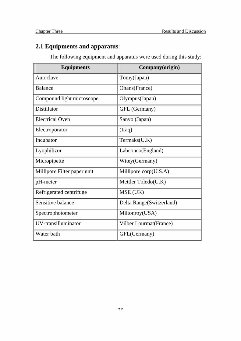
2.1 Equipments and apparatus:
The following equipment and apparatus were used during this study:
Equipments Company(origin)
Autoclave Tomy(Japan)
Balance Ohans(France)
Compound light microscope Olympus(Japan)
Distillator GFL (Germany)
Electrical Oven Sanyo (Japan)
Electroporator (Iraq)
Incubator Termaks(U.K)
Lyophilizor Labconco(England)
Micropipette Witey(Germany)
Millipore Filter paper unit Millipore corp(U.S.A)
pH-meter Mettler Toledo(U.K)
Refrigerated centrifuge MSE (UK)
Sensitive balance Delta Range(Switzerland)
Spectrophotometer Miltonroy(USA)
UV-transilluminator Vilber Lourmat(France)
Water bath GFL(Germany)
Chapter Three Results and Discussion
٣٨
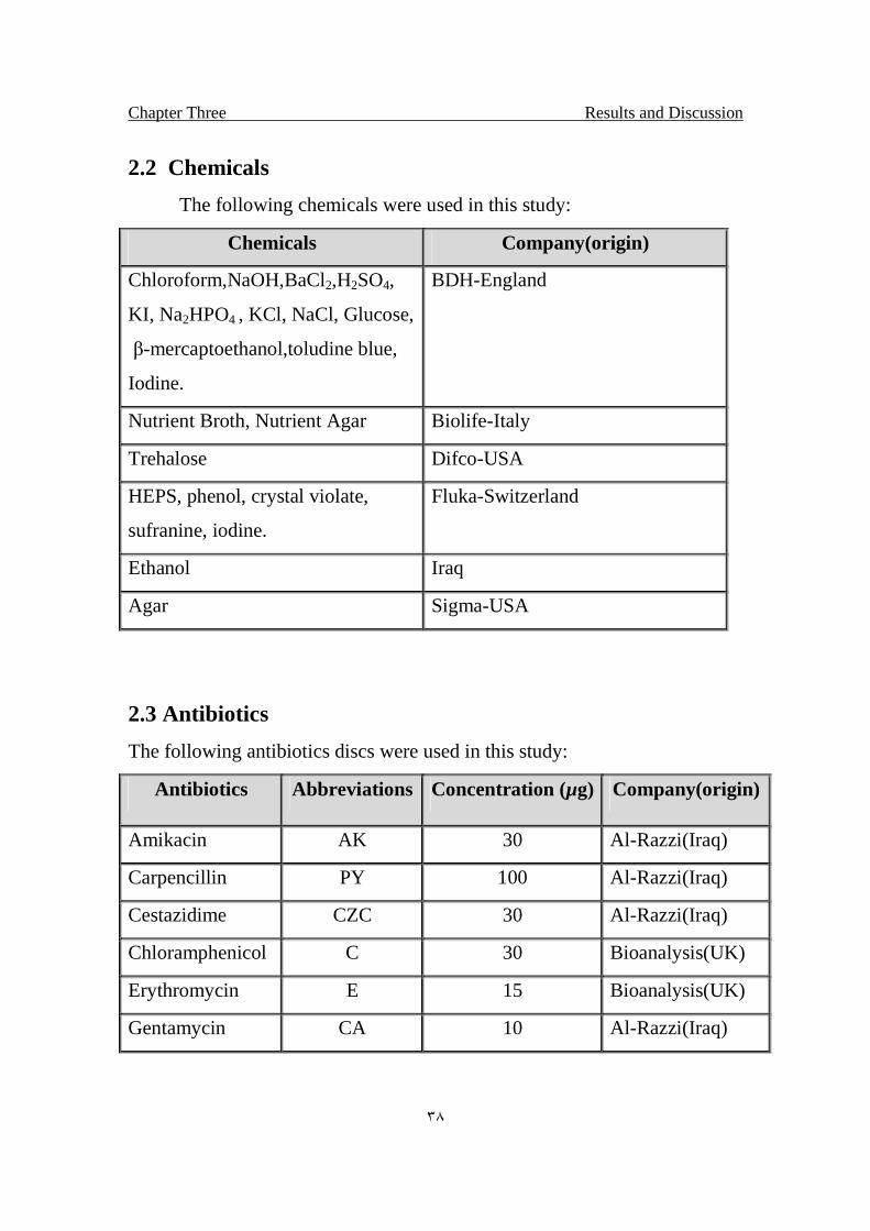
2.2 Chemicals
The following chemicals were used in this study:
Chemicals Company(origin)
Chloroform,NaOH,BaCl2,H2SO4,
KI, Na2HPO4 , KCl, NaCl, Glucose,
β-mercaptoethanol,toludine blue,
Iodine.
BDH-England
Nutrient Broth, Nutrient Agar Biolife-Italy
Trehalose Difco-USA
HEPS, phenol, crystal violate,
sufranine, iodine.
Fluka-Switzerland
Ethanol Iraq
Agar Sigma-USA
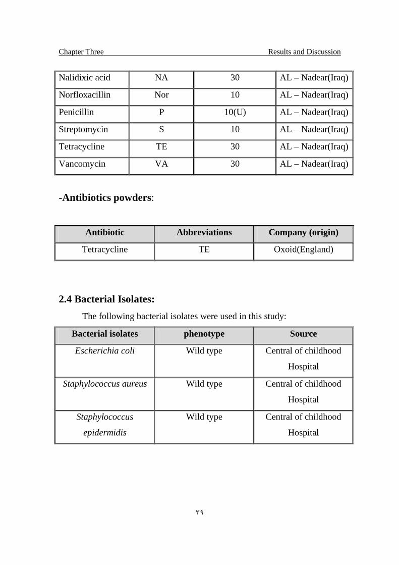
2.3 Antibiotics
The following antibiotics discs were used in this study:
Antibiotics Abbreviations Concentration (µg) Company(origin)
Amikacin AK 30 Al-Razzi(Iraq)
Carpencillin PY 100 Al-Razzi(Iraq)
Cestazidime CZC 30 Al-Razzi(Iraq)
Chloramphenicol C 30 Bioanalysis(UK)
Erythromycin E 15 Bioanalysis(UK)
Gentamycin CA 10 Al-Razzi(Iraq)
Chapter Three Results and Discussion
٣٩
Nalidixic acid NA 30 AL – Nadear(Iraq)
Norfloxacillin Nor 10 AL – Nadear(Iraq)
Penicillin P 10(U) AL – Nadear(Iraq)
Streptomycin S 10 AL – Nadear(Iraq)
Tetracycline TE 30 AL – Nadear(Iraq)
Vancomycin VA 30 AL – Nadear(Iraq)
-Antibiotics powders:
Antibiotic Abbreviations Company (origin)
Tetracycline TE Oxoid(England)
2.4 Bacterial Isolates:
The following bacterial isolates were used in this study:
Bacterial isolates phenotype Source
Escherichia coli Wild type Central of childhood
Hospital
Staphylococcus aureus Wild type Central of childhood
Hospital
Staphylococcus
epidermidis
Wild type Central of childhood
Hospital
Chapter Three Results and Discussion
٤٠
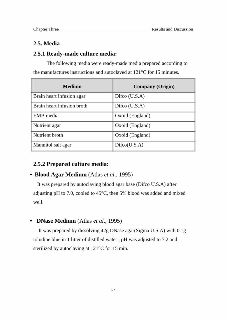
2.5. Media
2.5.1 Ready-made culture media:
The following media were ready-made media prepared according to
the manufactures instructions and autoclaved at 121°C for 15 minutes.
Medium Company (Origin)
Brain heart infusion agar Difco (U.S.A)
Brain heart infusion broth Difco (U.S.A)
EMB media Oxoid (England)
Nutrient agar Oxoid (England)
Nutrient broth Oxoid (England)
Mannitol salt agar Difco(U.S.A)
2.5.2 Prepared culture media:
• Blood Agar Medium (Atlas et al., 1995)
It was prepared by autoclaving blood agar base (Difco U.S.A) after
adjusting pH to 7.0, cooled to 45°C, then 5% blood was added and mixed
well.
• DNase Medium (Atlas et al., 1995)
It was prepared by dissolving 42g DNase agar(Sigma U.S.A) with 0.1g
toludine blue in 1 litter of distilled water , pH was adjusted to 7.2 and
sterilized by autoclaving at 121°C for 15 min.
Chapter Three Results and Discussion
٤١
2.6 Reagent and Stains:
• Hydrogen Peroxide Reagent (Atlas et al., 1995)
This reagent used for catalase test at 3% concentration.
• Oxidase Reagent (Atlas et al.,1995)
This reagent was composed of 1% of tetra methyl-p-
phenylenediamine dihydrochloride (Freshly prepared solution).
• Crystal violate stain (Atlas et al., 1995)
This stain was prepared by dissolving 2g of crystal violate in 20ml of
95% ethanol and the final volume was completed to 100ml with distilled
water and filtered before use.
• Safranine Counter Stain (Atlas et al., 1995)
This stain was prepared by dissolving 0.25g of safranine O in
10ml of 95% ethanol and the final volume was completed to 110ml
with distilled water allow to stand several days and filtered before
used.
• Lugol's Iodine Reagent (Atlas et al., 1995)
It was prepared by mixing 2g of potassium iodide with 1g of
iodine in 300 ml of distilled water.
Chapter Three Results and Discussion
٤٢
2.7 Buffers and solution:
• Electroporation buffer (Internet # 1):
HEPES (pH 7.0) 20 mM
NaCl 137 mM
KCl 5 mM
Na2HPO4 0.7 mM
Glucose 6 mM
ß-mercaptoethanol 0.1 mM
Dissolved in distilled water, Sterilized by Filtration
• Phosphate Buffer ph 7.0 (Atlas et al., 1995)
Na2HPO4 9.52 g/L
NaH2PO4 6.00 g/L
Sterilized by autoclaving at 121 °C for 15 min.
• Antibiotic solution (Maniatis et al., 1982).
Tetracycline
It was prepared as stock solution of 12.5 mg/ml of tetracycline
hydrochloride in solution of ethanol/ water (50% V/V). Sterilized by
filtration and stored in aliquots at -20°C in the dark.
Methods
2.8 Collection of Samples
Skin sample (pus) were collected from wound infection of patient in central
childhood hospital, during the period from 10/04 to 11/04, one sample was
collected and diagnosed, and after that the isolate was used for this study.
Chapter Three Results and Discussion
٤٣
2.9 Maintenance of bacterial isolate:-
Maintenance of bacterial isolate was performed according to (Atlas et al.,
1995), as the following:
2-9-1 short-term Storage:
Colonies of bacteria were maintained for periods of few weeks on the
surface of agar medium (Nutrient agar). The plates were tightly warped with
parafilm and stored at 4°C.
2-9-2 Medium-term Storage:
Bacterial isolates were maintained in stab cultures for long periods
(few months). Such medium were prepared in screw-capped vials containing
5-8ml of nutrient agar medium and stored at 4°C.
2-9-3 long-Term Storage:
Ten ml of 15-20% glycerol were added to screw tubes containing
nutrient broth. After autoclaving, inoculate with bacteria and incubate at
37°C for 24 hr. aerobically then kept in freezer, bacteria can be stored for
many years in this medium without significant loss of viability (Contreras et
al., 1991).
2.10 Inoculum's preparation:
Cells of bacterial isolates grown in nutrient broth until mid
exponential phase (O.D600nm = 1), were pelleted from 10ml by
centrifugation at 6000 rpm for 10 min. washed and resuspended with normal
saline, 1% (v/v) This prepared inoculums were then used in our experiment.
Chapter Three Results and Discussion
٤٤
2.11 Measurement of Bacterial Growth:
Growth of bacterial was monitored by MacFarland tube No. 5
turbidity standard (which prepared by adding 0.6 ml of 0.048 M BaCl2
[1.175% w/v BaCl2 2H2O] to 99.5 ml of 0.36 N H2SO4), which is equivalent
to bacterial concentration for inoculums 1.5 x 10⁸ organisms/ml.
2.12 Identification of bacterial isolates (Holt et al., 1994; Atlas
et al., 1995)
2.12.1 Morphological and Cultural Characteristics.
Morphology of colonies were studied on brain heart infusion agar
(Collee et al., 1996).
Color, shape, size and edge of colonies were recorded after 24 hrs. Of
incubation at 37°C.
• Gram's Stain (Atlas et al., 1995)
A single colony was transferred by a loop to a clean slide. The smear
was stained with crystal violet, treated with iodine, decolorized with 95%
alcohol, and counterstained with safranine, then examined by light
microscope.
Chapter Three Results and Discussion
٤٥
2.12.2 Biochemical and Physiological Characteristics.
2.12.2.1 Catalase Production test (Maza et al., 1997)
This test was performed by adding (2-3) drops of hydrogen peroxide
(H2O2) (3%) on a single colony grown on BHI agar plate. Appearance of
bubbles was regarded as positive result.
2.12.2.2 Oxidase Production test (Harely and Prescott, 1996)
Filter paper was saturated with the substrate (tetramethyle-p-
phneylene-diamine-dihydrochloride), colony of bacterial isolate to be test
was rubbed on the filter paper with a sterile wooden applicator stick. An
immediate color change to deep blue indicates a positive result.
2.12.2.3 Growth at 45°C.
Test tubes containing BHI broth were inoculated with overnight
culture of bacterial isolate, incubated at 45 °C for 24 hrs. Bacterial growth
(turbidity) was regarded as positive result.
2.12.2.4 Coagulase Production Test.
Plasma diluted 1:10 in normal saline (0.85%NaCl) was prepared and
placed in small tube. The bacterial isolate under test was inoculated into the
tube, by adding 0.l ml of overnight culture. The tube was incubate at 37°C
and examined for coagulation after 1, 3 and 6 hrs.
The tubes that showed negative results were left at room temperature
overnight and reexamined. The conversion of the plasma into a soft gel was
observed by tilting the tube to a horizontal position.
Chapter Three Results and Discussion
٤٦
2.12.2.5 DNase Production Test (Collee et al., 1996)
DNase agar plate containing toluidine blue (100mg/ml) indicator, was
inoculated with the bacterial culture, and then incubated at 37°C for 24-48
hrs.
Development of a rose pink halo surrounding the areas of bacterial growth
indicated the hydrolysis of DNA.
2.12.2.6 Hemolysis Patterns on Blood Agar (Atlas et al., 1995)
A single colony of overnight growth culture was streaked on blood
agar. The type of hemolysis produced by the growing colonies was observed
and recorded.
2.12.2.7 Aerobic Mannitol Fermentation Test (Atlas et al.,
1995)
The isolated bacterium was streaked on the plate of mannitol salt agar
and the plate was incubated at 37°C overnight. The change in color from
deep pink to yellow was a positive indication of mannitol fermentation.
2.12.2.8 Tolerance to NaCI (7-9%) (Atlas et al., 1995)
A loop full of an overnight culture of bacterial isolate was inoculated
into brain heart infusion broth supplemented with 7-9% NaCI, incubated at
37°C for 24hrs. Bacterial growth was regarded as positive result.
Chapter Three Results and Discussion
٤٧
2.13 Effect of Trehalose on bacterial isolate preserved in the
dry state at room temperature:
A novel procedure was used to evaluate the effect of Trehalose on the
S. aureus and E.coli isolates. These were used viable dried bacteria produced
by natural drying in the presence of Trehalose.
A novel method was provided for preserving live bacteria by
subjected an aqueous system containing the growing bacteria to drying
without special equipment, in the presence of trehalose, a dried compound
for preservation of aerobic bacteria in a viable state is provided. The dried
composition consists essentially of dried via aerobic bacteria and an
appropriate growth medium. The bacteria and growth medium were initially
placed in an aqueous solution of 100mM, and dried in room temperature.
Bacterial isolate with the following phenotypic characteristics were
each tested for their ability to be preserved and reconstituted using trehalose
as preservation agent: E.coli, S.aureus. The two bacterial isolates listed
above were each individually inoculated into standard culture media in wells
of a 96-well microtiter dish. They were grown overnight at 37oC. In the
following day an equal volume of preservation media, with 10 mM of
divalent cation (CaCl2) containing a trehalose was added to the growing
cultures giving a final concentration of trehalose 100mM. The cultures were
gently rocked over a 96 hour period and the contents allowed drying. The
dish was then covered and placed at room temperature. After one week the
individual cultures were rehydrated with sterile water, pre-warmed to
37oC.added to nutrient broth media to test for viability. After 1 month,
Chapter Three Results and Discussion
٤٨
individual cultures were rehydrated with sterile water, pre-warmed to
37oC.added to nutrient broth media to test for viability.
2.14 Trehalose protection against UV light
Culture of S.aureus grown in Brain Heart medium with 100mM
trehalose and without trehalose(control) until the mid exponential phase,
were pelleted from 5ml by centrifugation at 4000 rpm for 10 min., washed
twice and resuspended in the same volume of phosphate buffer (pH=7.0).
The UV source was UV-Transiluminator-Cross Linker [FLX-20-M, Vibler
Lourmat, France].
The tray size for irradiation was approximately 15x25 cm. Then the samples
were exposed on Petri dishes, multi-well plates, membrane, etc, to direct
irradiation from(for 15 watts) 254nm bulbs. A UV photoelectric cell detects
actual intensity. The dose rate of UV irradiation was 2-5 J/m2/s.The
experiments in this study were preformed under red light. The distance
between UV source and irradiated suspension was 11cm.
Three ml samples of the bacterial suspension in phosphate buffer contain
Trehalose and the control was irradiating in sterile Petri dish for the
following doses: (0, 10, 20, 30, 40, and 50) J/m2.
Two samples of each treatment were taken and as follows : 0.1 ml sample
diluted properly and spread on BHA, then incubated at 37ºC for 24 hr. to
determine the total viable count(survival fraction).
Chapter Three Results and Discussion
٤٩
2.15 Antibiotic sensitivity Test (Baron and Finegold, 1994):
Disk Diffusion Test:
Two test tubes contain 10 ml of nutrient broth medium one with
100mM of trehalose and the other without(control) inoculated with the
bacterial isolate, S.aureus, E.coli the cultures were incubated at 37°C to mid
log phase (O.D600nm a bout 1) giving 1x10⁸ bacterium/ml. 0.1 ml of
inoculated broth transferred to BHA. A sterile cotton swab was used to
streak the inoculums on the plate surface in 3 different planes (by rotating
the plate approximately 60 each time to obtain an even distribution of the
inoculums). The inoculated plates were then placed at room temperature for
10 minutes to allow absorption of excess moisture. With sterile forceps the
selected antibiotic disks were placed on the Inoculated plates and incubated
the plates at 37°C for 18 hrs. in an inverted position.
After incubation, the diameter of inhibition zone were noted and measured
by a ruler in mm, results were determined according to the National
Committee for laboratory standards (NCCLS, 1986).
Minimum Inhibitory Concentration (MIC)
Twenty test tubes containing nutrient broth were prepared. Ten test
tubes have been added 100mM trehalose and the other ten test tubes
treatments left without trehalose (control), to each have been added
quantities of antibiotics, serially diluted from 0.4µg/ml to 100µg/ml. The last
tubes were free of antibiotics and serves as a growth control. Twenty tubes
were inoculated with a calibrated suspension of the S.aureus, following by
incubation at 37°C for 18 hrs. At the end of the incubation period, the tubes
Chapter Three Results and Discussion
٥٠
were visually examined for turbidity. Cloudiness indicates that bacterial
growth has not been inhibited by the concentration of antibiotic contained in
medium.
2.16 Effect of Trehalose on Antibacterial activity:
Antibacterial Agents Test: Inhibitory effect was determined by the agar
diffusion method described by (Nathan et al., 1978).
Using the bacterial inoculation technique. Commercially available 60 mm.
petri dishes containing BHA were inoculated with S.aureus isolate with
100mM trehalose and without trehalose as control by using sterile swabs.
Two evenly spaced holes 3 mm. in diameter were made in the agar of each
plate with sterile cork borer. To identify the intrinsic antibacterial activity of
the diluents, wells were filled with S.epidermidis. An equal volume (200ml)
of each agent was expressed into each well (Two replicate plates were
prepared for each agent). Test plates were then incubated at 37°C for 24
hours and zones of inhibition were measured with millimeters. A clear
inhibition zone indicates that the agent had retained its antibacterial activity.
2.17 Effect of Trehalose against freezing
The two tests isolate S.aureus and E.coli were cultured in nutrient
broth medium for 18 hrs. After the incubation period a 100 mM Trehalose
was then added to the culture, while the control left without any addition.
Chapter Three Results and Discussion
٥١
All the tubes were then sealed and stored in -20ºC for 40 days; a
sample were then lifted each 10 days, recultured on nutrient agar medium to
determine its viability.
2.18 Effect of Trehalose after freeze drying (Lyophilized
Bacteria)
Two flasks containing 50 ml of nutrient broth culture medium were
prepared, and inoculated with S.aureus 18 hrs. at 37°C.
A 100 mM trehalose was added to the culture while the control left without
any addition.
The two flask then transfer to sterilize lyophilizes flask and then lyophilized.
After the lyophilizing the flasks sealed and stored for 4 months, each month
sample were then diluted and cultured in Brain Heart Agar to determine the
viable cells after incubating for 18 hrs. at 37°C.
2.19 Effect of Trehalose on Electroporated bacteria
Two tubes containing 10 ml of nutrient broth culture medium were
prepared, and inoculated with S.aureus 18 hrs. at 37°C
100 mM trehalose was added to the culture while the control treatment left
without any addition.
The cluttered were centrifuged (3000rpm for 10 min.) and wash twice with
electroporation buffer, then transferred to a cuvette. Gently to avoided any
bubbles formation in the gap between the plate electrodes. The cuvette
inserted into cuvatte holder of the local handmade electroporator fig.(2-1) ,
which the plastic nose of the cuvatte fits into slit of the cuvatte holder. The
cuvatte holder was pushed into the electroporator until it clicks into position.
The voltage were selected and switched on. After electroporation the cuvatte

Chapter Three Results and Discussion
٥٢
was removed from the cuvatte holder. The bacterial solution was then
transferred into BHI broth media to determine the viable cells.
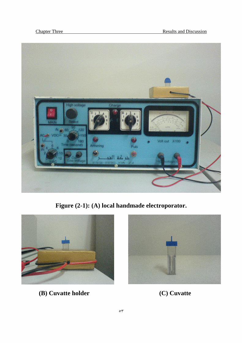
Chapter Three Results and Discussion
٥٣
Figure (2-1): (A) local handmade electroporator.
(B) Cuvatte holder (C) Cuvatte
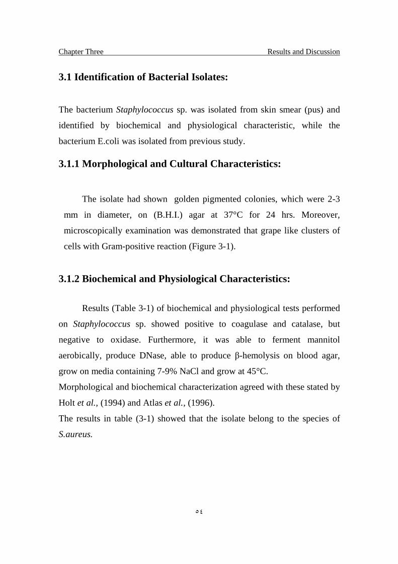
Chapter Three Results and Discussion
٥٤
3.1 Identification of Bacterial Isolates:
The bacterium Staphylococcus sp. was isolated from skin smear (pus) and
identified by biochemical and physiological characteristic, while the
bacterium E.coli was isolated from previous study.
3.1.1 Morphological and Cultural Characteristics:
The isolate had shown golden pigmented colonies, which were 2-3
mm in diameter, on (B.H.I.) agar at 37°C for 24 hrs. Moreover,
microscopically examination was demonstrated that grape like clusters of
cells with Gram-positive reaction (Figure 3-1).
3.1.2 Biochemical and Physiological Characteristics:
Results (Table 3-1) of biochemical and physiological tests performed
on Staphylococcus sp. showed positive to coagulase and catalase, but
negative to oxidase. Furthermore, it was able to ferment mannitol
aerobically, produce DNase, able to produce β-hemolysis on blood agar,
grow on media containing 7-9% NaCl and grow at 45°C.
Morphological and biochemical characterization agreed with these stated by
Holt et al., (1994) and Atlas et al., (1996).
The results in table (3-1) showed that the isolate belong to the species of
S.aureus.
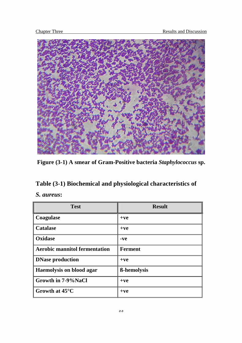
Chapter Three Results and Discussion
٥٥
Figure (3-1) A smear of Gram-Positive bacteria Staphylococcus sp.
Table (3-1) Biochemical and physiological characteristics of
S. aureus:
Test Result
Coagulase +ve
Catalase +ve
Oxidase -ve
Aerobic mannitol fermentation Ferment
DNase production +ve
Haemolysis on blood agar ß-hemolysis
Growth in 7-9%NaCl +ve
Growth at 45°C +ve

Chapter Three Results and Discussion
٥٦
3.2 Effect of Trehalose on drying (dehydrations):
The results were shown in fig (3-2) .Isolate of S.aureus had a higher
level of viability after one month when preserved in the presence of
Trehalose than control. The two primary stresses that were proposed to
destabilize lipid bilayers during dehydration are fusion and lipid phase
transitions. Studies by laser light scattering or other techniques demonstrate
that Trehalose and other sugars inhibit fusion between the vesicles during
drying, but the inhibition of fusion alone is not sufficient to preserve the dry
vesicles. Thus Trehalose is also necessary to prevent phase transitions
(Crowe and Crowe, 1990).
Trehalose depresses the phase transition temperature of the dry lipids, which
maintains them in the liquid crystalline phase in the absence of water (Crowe
and Crowe, 1988).
While the result of E.coli isolate shown no effect of Trehalose preservation
in dry state. The disaccharide Trehalose acts as a carbon source in E.coli. At
high osmolarity of the growth medium, however, they can also degrade
Trehalose as the sole source of carbon under both high- and low-osmolarity
growth conditions. The modes of Trehalose utilization are different under
conditions and have to be well regulated (Boos et al., 1990)
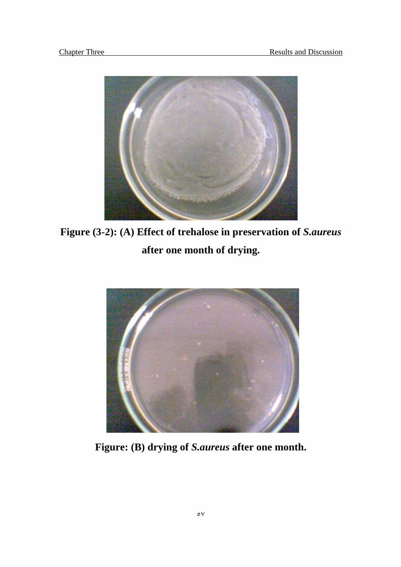
Chapter Three Results and Discussion
٥٧
Figure (3-2): (A) Effect of trehalose in preservation of S.aureus
after one month of drying.
Figure: (B) drying of S.aureus after one month.
Chapter Three Results and Discussion
٥٨
3.3 UV Survival Curve of Staphylococcus aureus with and
without addition of Trehalose:
The survival curve of S.aureus after exposed to different doses of UV
irradiation is shown in figure (3-3). The result indicated that S.aureus lost it's
viability exponentially while addition of Trehalose make the bacterial isolate
more resistance to UV. The survival curve of S.aureus without addition of
Trehalose (control) showed increases of lethal percentage exponentially with
the increase of UV doses and the highest lethal percentage or the less
survival fraction is l.lxl014 when the bacteria exposed to 40 J/m2 of UV
irradiation. But the survival curve of S.aureus. With the addition of
Trehalose showed more resistance to the UV (The most serious damage to
cells exposed to radiation is attributed mostly to effects on the structure of
cellular DNA. We found that trehalose protects DNA from irradiation, In the
presence of Trehalose, DNA can be protected from about 4 times higher
doses of irradiation, The protective effect increases with the amount of the
sugar , Yoshinaga, 1997).
Other study of lethal effect of UV light on S.aureus showed that this
bacterium was sensitive (Al-Bakri and Umrann, 1994; Al-Zubaei, 2001).
Results indicated that S.aureus without addition of Trehalose was
sensitive to mutation induced by UV, while the addition of Trehalose, the
bacteria become more resist to UV irradiation(Yoshinaga, 1997).
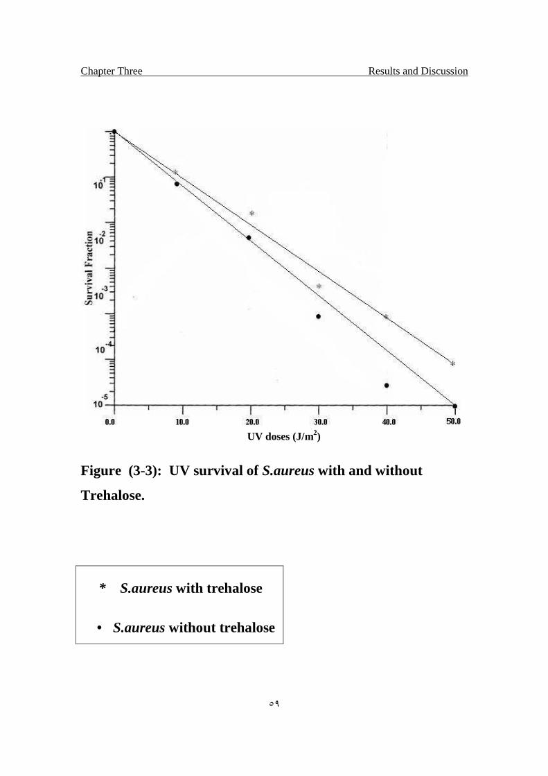
Chapter Three Results and Discussion
٥٩
UV doses (J/m2)
Figure (3-3): UV survival of S.aureus with and without
Trehalose.
* S.aureus with trehalose
• S.aureus without trehalose
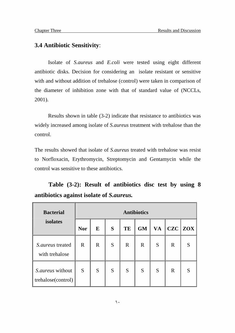
Chapter Three Results and Discussion
٦٠
3.4 Antibiotic Sensitivity:
Isolate of S.aureus and E.coli were tested using eight different
antibiotic disks. Decision for considering an isolate resistant or sensitive
with and without addition of trehalose (control) were taken in comparison of
the diameter of inhibition zone with that of standard value of (NCCLs,
2001).
Results shown in table (3-2) indicate that resistance to antibiotics was
widely increased among isolate of S.aureus treatment with trehalose than the
control.
The results showed that isolate of S.aureus treated with trehalose was resist
to Norfloxacin, Erythromycin, Streptomycin and Gentamycin while the
control was sensitive to these antibiotics.
Table (3-2): Result of antibiotics disc test by using 8
antibiotics against isolate of S.aureus.
Bacterial
isolates
Antibiotics
Nor E S TE GM VA CZC ZOX
S.aureus treated
with trehalose
R R S R R S R S
S.aureus without
trehalose(control)
S S S S S S R S
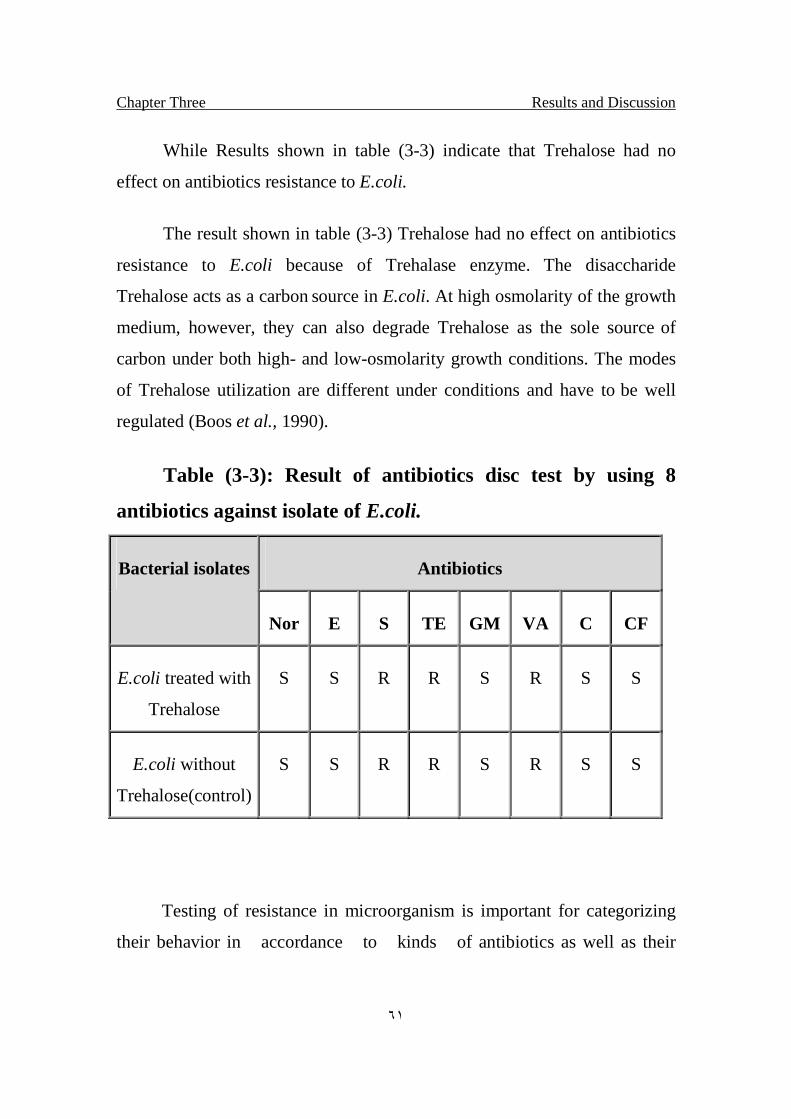
Chapter Three Results and Discussion
٦١
While Results shown in table (3-3) indicate that Trehalose had no
effect on antibiotics resistance to E.coli.
The result shown in table (3-3) Trehalose had no effect on antibiotics
resistance to E.coli because of Trehalase enzyme. The disaccharide
Trehalose acts as a carbon source in E.coli. At high osmolarity of the growth
medium, however, they can also degrade Trehalose as the sole source of
carbon under both high- and low-osmolarity growth conditions. The modes
of Trehalose utilization are different under conditions and have to be well
regulated (Boos et al., 1990).
Table (3-3): Result of antibiotics disc test by using 8
antibiotics against isolate of E.coli.
Bacterial isolates Antibiotics
Nor E S TE GM VA C CF
E.coli treated with
Trehalose
S S R R S R S S
E.coli without
Trehalose(control)
S S R R S R S S
Testing of resistance in microorganism is important for categorizing
their behavior in accordance to kinds of antibiotics as well as their
Chapter Three Results and Discussion
٦٢
medical applications concerning their effectiveness in treatment of diseases
and distribution of resistance among isolates for designing pattern of
resistance, serve as a picture for following transfer of genetic elements
among species and hence spread of resistance (Baron et al., 1994).
For screening purposes, test is required for separation of huge
numbers of isolates. Accordingly, disc method is useful to do so. The test is
fast and simple, however it suffer from lowerness of accuracy as long as
zone of inhibition widely effected by medium composition and interference
of some ions with spread of antibiotics through the medium (Baron et al.,
1994).
For research studies, Minimum Inhibitory Concentrations (MICs) is
useful to specify minimum concentration of antibiotic showing inhibition of
growth. Such concentration could also be useful for medical purposes.
Accordingly, for testing antibiotic sensitivity, the two methods were used
wherever it is needed.
3.5 Minimal Inhibitory Concentration (MIC) Test:
Isolates were selected due to multiple antibiotic resistances, thereby;
these isolates were selected in order to evaluate bacterial susceptibility to
antibiotic as in the table (3-4). The breakpoints were applied following
(NCCLs, 1991), recommendations, when resistance level were calculated,
" MIC " in both the intermediate and resistant ranges (as defined by the
(NCCLs, 1991) were considered as non susceptible in this study. MIC was
defined as the lowest drug concentration in microgram per milliter that
inhibited the visible growth of bacteria (Kinoshita et al., 1997).
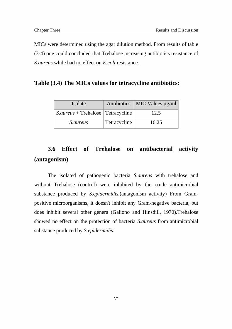
Chapter Three Results and Discussion
٦٣
MICs were determined using the agar dilution method. From results of table
(3-4) one could concluded that Trehalose increasing antibiotics resistance of
S.aureus while had no effect on E.coli resistance.
Table (3.4) The MICs values for tetracycline antibiotics:
Isolate Antibiotics MIC Values µg/ml
S.aureus + Trehalose Tetracycline 12.5
S.aureus Tetracycline 16.25
3.6 Effect of Trehalose on antibacterial activity
(antagonism)
The isolated of pathogenic bacteria S.aureus with trehalose and
without Trehalose (control) were inhibited by the crude antimicrobial
substance produced by S.epidermidis.(antagonism activity) From Gram-
positive microorganisms, it doesn't inhibit any Gram-negative bacteria, but
does inhibit several other genera (Galiono and Hinsdill, 1970).Trehalose
showed no effect on the protection of bacteria S.aureus from antimicrobial
substance produced by S.epidermidis.
Chapter Three Results and Discussion
٦٤
3.7 Store of bacterial isolates from freezing temperature in the
presence and absence of Trehalose:
Two isolates of bacteria S.aureus, E.coli were stored by using the
freezing technique and the results appears by the diagrams bellow for storing
of the bacterial isolates in -20ºC in addition of 100mM Trehalose and
without(control).There was increasing capability of viability of bacterial
isolate S.aureus, when added 100mM of Trehalose before freezing compared
with the control. Figure (3-4) showed that the bacterial isolate S.aureus
keeps its viability after 4 weeks of freeze preservation while the control loss
more of it’s viability after preservation for the same time. In preservation of
E.coli with Trehalose, showed the same effect in preservation against freeze
than the control as shown in figure (3-5). The plasma membrane is a key
component of the cell and must be maintained during freezing conditions if
the cell is to be kept alive. It has been demonstrated with artificial
membranes, such as unilamellar vesicles, that damage measured by
intermixing and fusion can be reduced by a series of cryoprotectants, with
trehalose and sucrose being more protective than glycerol. Thus, these sugars
probably play a key role in preventing deleterious alteration to the membrane
during reduced-water states (Anchordoguy et al., 1987).
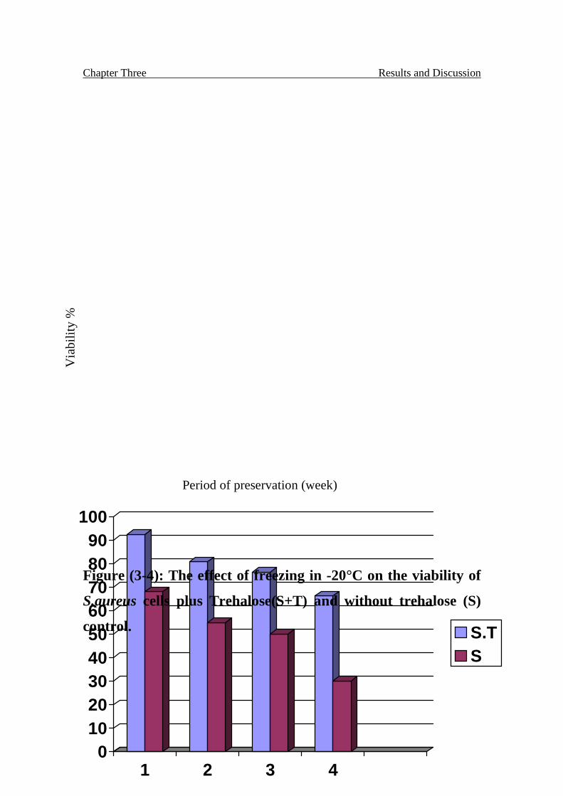
Chapter Three Results and Discussion
٦٥
0102030405060708090
100
1 2 3 4
S.TS
Period of preservation (week)
Figure (3-4): The effect of freezing in -20°C on the viability of
S.aureus cells plus Trehalose(S+T) and without trehalose (S)
control.
Via
bilit
y %
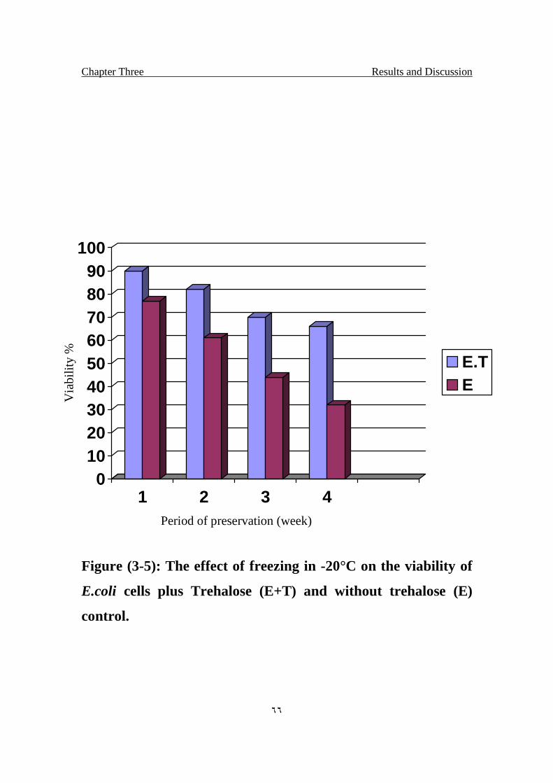
Chapter Three Results and Discussion
٦٦
0102030405060708090
100
1 2 3 4
E.TE
Period of preservation (week)
Figure (3-5): The effect of freezing in -20°C on the viability of
E.coli cells plus Trehalose (E+T) and without trehalose (E)
control.
Via
bilit
y %

Chapter Three Results and Discussion
٦٧
3.8 Protection of bacterial isolate from freeze-drying
(lyophilization):
S.aureus was lyophilized with Trehalose and without Trehalose
(control). The experiment was proved that Trehalose keeps the bacteria more
viable after lyophilization and stored in room temperature after 4 months of
storing higher decrease was observed on viability of bacteria preserved
without Trehalose, while the bacteria that preserved with Trehalose keep
more of it's viability after the same time fig. (3-6).
The diagram shows the effect of Trehalose for preservation of bacteria after
lyophilization and stored in room temperature.
Molecular simulations of model, hydrated phospholipids bilayers
(e.g., dipalmitoylphosphatidylcholine) are performed over wide ranges of
temperatures with and without protectants. The area per head group
increases with temperature from 58 to 77 Å2. Other properties such as
hydration number, alkyl tail order-parameter, diffusion coefficients and
radial distribution functions exhibit a clear dependence on temperature. The
simulated area per head group of the bilayer is not affected by the presence
of disaccharides, suggesting that the overall structure of the bilayer remains
undisturbed. The results of simulations reveal that the interaction of
disaccharide molecules with the bilayer occurs primarily at the surface of the
bilayer, and it is governed by the formation of multiple hydrogen bonds to
specific groups of the lipid. Disaccharide molecules are observed to adopt
specific conformations to fit onto the surface topology of the bilayer, often
interacting with up to three different lipids simultaneously. At high
disaccharide concentrations, the results of simulations indicate that
disaccharides can serve as an effective replacement for water under
Chapter Three Results and Discussion
٦٨
anhydrous conditions, which helps explain their effectiveness as
lyophilization agents for liposome and cells (MTSM).
The protection of biological molecules during freezing and freeze-
drying (lyophilization) is a subject of considerable practical importance,
particularly in the pharmaceutical industry. Much work has been conducted
on the use of a wide variety of compounds as cryoprotectants for these types
of processes. They have been found to protect proteins during freezing and
drying stresses. They have also been shown to prevent damage to cells
during freezing and drying. Trehalose has been found to be a highly
effective. Several structural and thermodynamic arguments have been put
forward to explain the mechanisms of action of molecules such as Trehalose
(MTSM).
Damage to biological systems resulting from freeze-drying can be attributed
to two primary causes: changes in the physical state of membrane lipids and
changes in the structure of sensitive proteins. Removal of hydrogen-bonded
water from the head group region of phospholipid bilayers increases the
head group packing (Crowe et al., 1985), and forces the acyl chains together,
increasing the probability of Van Der Waals interactions. As a result, the
lipid may undergo a transition from liquid crystalline to gel phase (Crowe et
al., 1988).
Upon rehydration, dry membranes, which are in gel phase at room
temperature, undergo a transition from gel to liquid crystal phase. As the
membranes pass through this phase transition there are regions with packing
defects, making the membranes leaky (Byler and Susi 1985).
Chapter Three Results and Discussion
٦٩
Adding a disaccharide such as trehalose before drying lowers the transition
temperature (Tm) of the dry membranes by replacing the water between the
lipid head groups, preventing the phase transition and its accompanying
leakage upon rehydration (Crowe et al., 1985, Crowe et al., 1989).
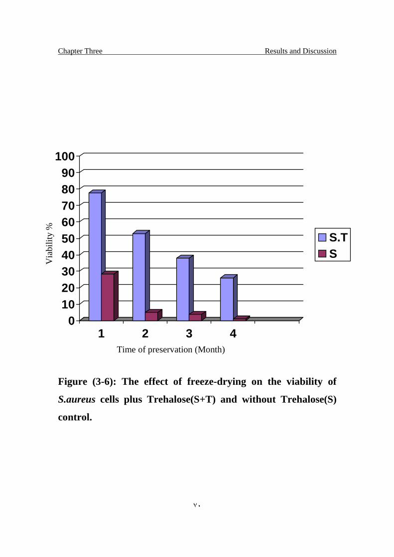
Chapter Three Results and Discussion
٧٠
0102030405060708090
100
1 2 3 4
S.TS
Time of preservation (Month)
Figure (3-6): The effect of freeze-drying on the viability of
S.aureus cells plus Trehalose(S+T) and without Trehalose(S)
control.
Via
bilit
y %
Chapter Three Results and Discussion
٧١
3.9 Effect of Trehalose on Electroporated bacteria
Trehalose showed an effect on protection of bacteria after
electroporation. Result showed in fig. (3-7) indicate Trehalose increase the
resistance of the bacteria against electroporation.
Electroporation is widely used for introduction of DNA and other
foreign molecules into eukaryotic cells. However, conditions yielding the
greatest molecule uptake and gene expression can result in low cell survival.
In this study, we assessed the efficiency of Trehalose for enhancing cell
viability after excessive Electroporation. This disaccharide was chosen
because of its capability of stabilizing cell membranes under various
stressful conditions, such as dehydration and freezing However,
electroporation is very damaging to the cell membranes. (Mussauer et al.,
1999).
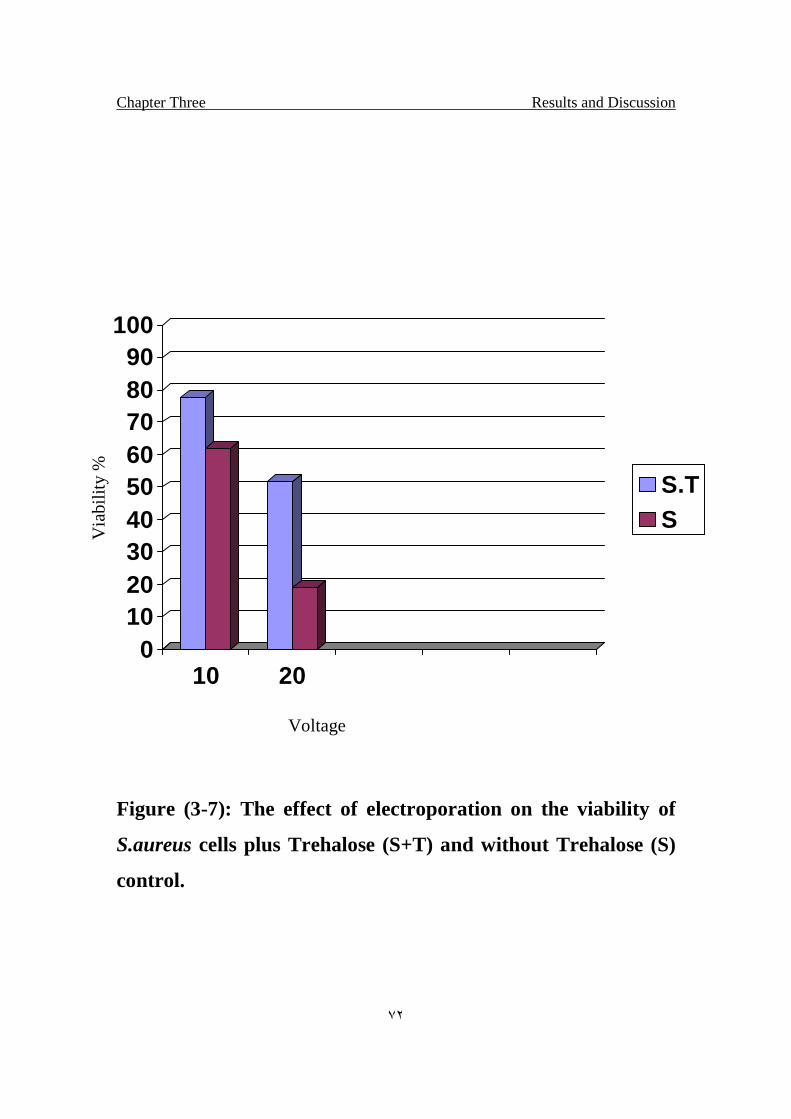
Chapter Three Results and Discussion
٧٢
0102030405060708090
100
10 20
S.TS
Figure (3-7): The effect of electroporation on the viability of
S.aureus cells plus Trehalose (S+T) and without Trehalose (S)
control.
Via
bilit
y %
Voltage

نسان من علق )1(اقـرأ باسم ربك الذي خلق خلق اإلعلم ) ٤(الذي علم بالقلم )٣(اقـرأ وربك األكرم )2(
نسان ما مل يـعلم )٥(اإل
صدق اهللا العلي العظيم )٥-١( سورة العلق من أية
Recommendations
1. The study of the effect of in vivo injected Trehalose and its role in the
protection of living cells.
2. Increase the production of Trehalose from microorganisms produced
it.
3. Study the effect of Trehalose in protection of bacteria from curing
agents.
٧٥
Abbott P.T. (1999) Low trehalase activity is associated with abdominal
symptoms caused by edible mushrooms. Scand. J. Gastroenterol., 34,
898–903.
Al-Bakri G.H.; and Umran M.A. (1994). Mutagenesis of noval
halotolerant bacteria (micrococcus sp.) using ultraviolet light and N-
methyl-N-nitrosoguanidine. Iraq. J. Microbial 6:55-65.
Anandarajah K. and Mckersie B.D. (1990) Manipulating the
desiccation tolerance and vigor of dry somatic embryos of Medicago
sativa L with sucrose, heat shock and abscissic acid. Plant Cell Rep., 9,
451–455.
Anchordoguy T. J.; Rudolph A.S.; Carpenter J.F.; Crowe J.H.(1995).
Modes of interaction of cryoprotectants with membrane phospholipids
during freezing. Cryobiology 24:324-331.
Andersson U.(2002). Beta-glucose 1-phosphate interconverting enzymes
in Lactococcus lactis: physiological role and regulation in maltose,
trehalose and glucose metabolism, Center for Chemistry and Chemical
Engineering, Hall K:C.
Argüelles J.C. (2000). Physiological roles of trehalose in bacteria and
yeasts: a comparative analysis 174: 217 – 224.
Atlas R.M. (1995). Principles of microbiology. Mosby-year Book, Inc.,
USA.P:547 and 668.
٧٦
Ballone P.; Marchi M.; Branca C.; and Magazu S. (2000) . Structural
and vibrational properties of trehalose: a density functional study. J.
Phys. Chem. B. 104:6313–6317.
Banaroudj, N., Lee, D.H., and Goldberg, A.L. (2001) Trehalose
accumulation during cellular stress protects cells and cellular proteins
from damage by oxygen radicals. J. Biol. Chem., 276, 24261–24267.
Behm C. A. (1997). The role of trehalose in the physiology of
nematodes. Int. J. Parasitol. 27:215–229.
Becker A.; Schloeder P.; Steele J.E.; and Wegener G. (1996) The
regulation of trehalose metabolism in insects. Experientia, 52, 433–439.
Belisle, J.T., Vissa, V.D., Sievert, T., Takayama, K., Brennan, P.J.,
and Bersa, G.S. (1997) Role of the major antigen of Mycobacterium
tuberculosis in cell wall biogenesis. Science, 276, 1420–1422.
Belton P. S.; and Gil A. H. (1994). IR and Raman spectroscopic studies
of the interaction of trehalose with hen egg white lysozyme. Biopolymers.
34:957–961.
Besra, G.S., Bolton, R.C., McNeil, M.R., Ridell, M., Simpson, K.E.,
Glushka, J., van Halbeek, H., Brennan, P.J., and Minnikin, D.E.
(1992) Structural elucidation of a novel family of acyltrehaloses from
Mycobacterium tuberculosis. Biochemistry, 31, 9832–9837.
Birch, G. G. (1972). Mushroom sugar. In Health and Food. G. G. Birch,
L. F. Green, and I. G. Plaskett, editors. Elsevier Science, New York. 49–
53.
٧٧
Boos W.; Ehmann U.; Forki W.; Klein M.; Rimmele M.; and Postma
P. (1990) Trehalose transport and metabolism in Escherichia coli. J.
Bacteriol., 172, 3450–3461.
Branca C.; Magazu S.; Maisano G.; and Migliardo P.(1999).
Anomalous cryoprotective effectiveness of trehalose: Raman scattering
evidences. J. Chem. Phys. 111:281–287.
Branca C.; Magazu S.; Maisano, G.; Bennington, S. M.; and Fak B.
(2003). Vibrational studies on disaccharide/H2O systems by inelastic
neutron scattering, Raman, and IR spectroscopy. J. Phys. Chem. B.
107:1444–1451.
Bredereck, H.; Hoschele, G.; and Ruck, K. (1953) Synthesis of non-
reducing disaccharides. Chem. Ber., 86:1277–128.
Brennan, P.J. and Nikaido, H. (1995) The envelope of mycobacteria.
Annu. Rev. Biochem., 64, 29–63.
Bryant, G.; and Wolfe, J.(1992). Interfacial forces in cryobiology and
anhydrobiology. Cryo-Letters. 13:23–36.
Byler, D. M.; and Susi H. (1986). Examination of the secondary
structure of proteins by deconvolved FTIR spectra. Biopolymers 25:469–
487.
Carpenter J.F. and Crowe J.F. (1989) An infrared spectroscopic study
of the interactions of carbohydrates with dried proteins. Biochemistry, 28,
3916–3922
Carpenter, J. F.; Prestrelski, S. J.; Anchordoguy, T. J.; and
Arakawa, T. (1994). Interactions of stabilizers with proteins during
٧٨
freezing and drying. In Formulation and Delivery of Proteins and
Peptides. J. L. Cleland and R. Langer, editors. American Chemical
Society, Washington, DC. 134–147.
Cerda, J.J.; Preiser, H.; and Crane, R.K. (1972) Brush border enzymes
and malabsorption: Elevated disaccharides in chronic pancreatic
insufficiency with diabetes mellitus. Gastroenterology, 62: 841.
Chandrasekhar, I.; and Gaber, B. P. (1988). Stabilization of the bio-
membrane by small molecules: interaction of trehalose with the
phospholipids bilayer. J. Biomol. Struct. Dyn. 5:1163–1171.
Chatterjee, D. (1997) The mycobacterial cell wall: structure,
biosynthesis and sites of drug action. Curr. Opin. Chem. Biol., 1, 578–
588.
Clegg, J. S. (1965). The origin of trehalose and its significance during
formation of encysted dormant embryos of Artemia salina. Comp.
Biochem. Physiol. 14:135–143.
Clegg, J. S. (1997). Embryos of Artemia franciscana survive four years
of continuous anoxia: the case for complete metabolic rate depression. J.
Exp. Biol. 200:467–475.
Clegg, J. S. (2001). Cryptobiosis—a peculiar state of biological
organization. Comp. Biochem. Physiol. 128B:613–624.
Clegg, J. S.; and Jackson, S. A. (1992). Aerobic heat-shock activates
trehalose synthesis in embryos of Artemia franciscana. FEBS Lett.
303:45–47.
٧٩
Collee, J.C.; Fraser, A.G.; Marminon, B.P. and Simmons, A. (1996).
Staphylococcus: cluster-forming Gram-positive cocci. In practical
medical microbiology 14th edition. Churchill livigstone. UK. P: 245.
Conrad, P. B.; and de Pablo, J. J. (1999). Computer simulation of the
cryoprotectant disaccharide alpha, alpha-trehalose in aqueous solution. J.
Phys. Chem. A. 103:4049–4055.
Cottone, G.; Ciccotti, G.; and Cordone, L. (2002). Protein-trehalose-
water structures in trehalose coated carboxy-myoglobin. J. Chem. Phys.
117:9862–9866.
Cottone, G.; Cordone, L.; and Ciccotti, G. (2001). Molecular dynamics
simulation of carboxy-myoglobin embedded in a trehalose-water matrix.
Biophys. J. 80:931–938.
Crowe J. H.; Carpenter J. F.;and Crowe L. M . (1989a). The role of
verification in anhydrobiosis. Annual Review of Physiology 60: 73-103.
Crowe, J. H.; Carpenter, J. F.; and Crowe, L. M. (1998b). The role of
vitrification in anhydrobiosis. Annu. Rev. Physiol. 60:73–103.
Crowe, J. H.; Clegg, J. S.; and Crowe, L. M. (1998). Anhydrobiosis:
the water replacement hypothesis. In The Properties of Water in Foods
(ISO-POW 6). D. S. Reid, editor. Chapman and Hall, New York. 440–
455.
Crowe, J. H.; Crowe, L. M.; and Chapman, D. (1984). Preservation of
membranes in anhydrobiotic organisms: the role of trehalose. Science.
223:701–703.
٨٠
Crowe, J. H.; Crowe, L. M.; Carpenter, J. F.; and Wistrom, C. A.
(1987). Stabilization of dry phospholipid bilayers and proteins by sugars.
Biochem. J. 242:1–10.
Crowe, J. H.; Crowe, L. M.; Carpenter, J. F.; Prestrelski, S.;
Hoekstra, F. A.; Araújo, P. de; and Panek, A. D. (1997).
Anhydrobiosis: cellular adaptation to extreme dehydration. In Handbook
of Physiology, Vol. 2. W. H. Dantzler, editor. Oxford University Press,
Oxford, UK. 1445–1477.
Crowe, J. H.; Crowe, L. M.; Oliver, A. E.; Tsvetkova, N.; Wolkers,
W.; and Tablin, F. (2001). The trehalose myth revisited: introduction to
a symposium on stabilization of cells in the dry state. Cryobiology.
43:89–105.
Crowe, J. H.; F. Hoekstra, A.; and Crowe, L. M. (1992).
Anhydrobiosis. Annu. Rev. Physiol. 54:579–599.
Crowe, J. H.; Oliver, A. E.; and Tablin, F. (2002). Is there a single
biochemical adaptation to anhydrobiosis? Integr. Comp. Biol. 42:497–
503.
Crowe, J. H.; Tablin, F.; Wolkers, W. F.; Gousset, K.; Tsvetkova, N.
M.; and Ricker, J. (2003). Stabilization of membranes in human
platelets freeze-dried with trehalose. Chem. Phys. Lipids. 122:41–52.
Crowe, J.; Crowe, L.; and Chapman, D. (1984) Preservation of
membranes in anhydrobiotic organisms. The role of trehalose. Science,
223:209–217.
٨١
Crowe, J.H.; and Crowe, L.M. (1990) Lyotropic effects of water on
phospholipids. In: F. Franks (ed.), Water science reviews. Cambridge
University Press, Cambridge, England, 1–23.
Crowe, L. M. (2002). Lessons from nature: the role of sugars in
anhydrobiosis. Comp. Biochem. Physiol. 131A:505–513.
Crowe, L. M.; J. H. Crowe, A.; Rudolph, C. Womersley; and L.
Appel. (1985). Preservation of freeze-dried liposomes by trehalose. Arch.
Biochem. Biophys. 242:240-247.
Crowe, L.M.; and Crowe, J.H. (1988) Trehalose and dry dipalmitoyl
phosphatidylcholine revisited. Biochim. Biophys. Acta, 946:193–201.
Daffe, M., Lacave, C., Laneelle, M.-A., Gillois, M., and Laneele, G.
(1988) Polyphthienoyl trehalose, glycolipids specific for virulent strains
of the tubercle bacillus. Eur. J. Biochem., 172, 579–584.
Dahlqvist, A. (1974) Enzyme deficiency and malabsorption of
carbohydrates. In: Sipple, H.L & McNutt, K.W., eds, Sugars in Nutrition,
New York: Academic Press, 187–214.
Damore, T. G.; R. Crumplen, G.; and Stewart, G. G. (1991). The
involvement of trehalose in yeast stress tolerance. J. Ind. Microbiol.
7:191–195.
Demelier, J.-F.; Labat, J. and Courtois, J.-E. (1975) Influence of
parenteral injection of trehalose on mammals with distinct trehalose
equipment. C.R. Acad. Sc. (Paris) Serie D, 280: 669–672.
٨٢
De Virgilio, C., Hottinger, T., Dominiguez, J., Boller, T., and
Wiemken, A. (1994) The role of trehalose synthesis for the acquisition of
thermotolerance in yeast. Eur. J. Biochem., 219, 179–186.
Eastmond,P.J.; Yi Li , Graham,I.A.(2003) Is trehalose-6-phosphate a
regulator of sugar metabolism in plants? Journal of Experimental Botany,
54:533-537.
Elbein, A.D. (1974) The metabolism of , -trehalose. Adv. Carbohyd.
Chem. Biochem., 30:227–256.
Elbein, A.D.; and Mitchell, M . (1973) Levels of glycogen and trehalose
in Mycobacterium smegmatis, and the purification and properties of the
glycogen synthase. J. Bacteriol., 113:863–873.
Elbein,A.D.; Pan ,Y.T.; Pastuszak,I ; Carroll,D.( 2003). New insights
on trehalose: a multifunctional molecule, 13:17-27.
Engelsen, S. B.; and Perez, S. (2000). Unique similarity of the
asymmetric trehalose solid-state hydration and the diluted aqueous-
solution hydration. J. Phys. Chem. B. 104:9301–9311.
Eroglu, A.; Toner, M.; and Toth, T. L. (2002). Beneficial effect of
microinjected trehalose on the cryosurvival of human oocytes. Fertil.
Steril. 77:152–158.
Eze, L.C. (1989) Plasma trehalase activity and diabetes mellitus.
Biochem. Gen., 27:487–495.
Felix,C.F.; Moreira, C. C.; Oliveira, M. S.; Sola-Penna, M. ; Meyer-
Fernandes, J. R. ; Scofano, H. M.; Ferreira-Pereira, A. (1999).
Protection against thermal denaturation by trehalose on the plasma
٨٣
membrane H+-ATPase from yeast Synergetic effect between trehalose
and phospholipid environmentEur. J. Biochem. 266:660-664
Feofilova, E. P. (2003). Deceleration of vital activity as a universal
biochemical mechanism ensuring adaptation of microorganisms to stress
factors: a review. Appl. Biochem. Microbiol. 39:1–18.
Fernandez J.; Sato T.; Franco A.; Vicentre-Soler J.; Cansado J.;
Gacto M. (1998). Enhancement of neutral trehalose activity by oxidative
stress in the fission yeast Schizosaccharmyces pombe. Fungal Genet Biol
25:79-86.
Fukuse, T.; Hirata, T.; Nakamura, T.; Ueda, M.; Kawashima, M.;
Hitomi, S.; and Wada, H. (1999). Role of saccharides on lung
preservation. Transplantation. 68:110–117.
Gadd G.M.; Chalmers K. and Reed R.H. (1987) The role of trehalose
in dehydration resistance of Saccharomyces cerevesiae. FEMS Microbiol.
Lett., 48, 249–254.
Garland, G. (1989) Brush border membrane sucrase–isomaltase,
maltase–glucoamylase and trehalase in mammals. Comp. Biochem.
Physiol., 94B:1–11.
Gudmand-Høyer, E. and Skovbjerg, H. (1996) Disaccharide digestion
and maldigestion. Scand. J. Gastroenterol., 31 : 111–121.
Gudmand-Høyer, E.; Fenger, H.J.; Skovbjerg, H.; Kernhansen, P.
and Rørbaek Madsen, P. (1988) Trehalase deficiency in Greenland.
Scand. J. Gastroenterol., 23:775–778.
٨٤
Guo, N., Puhlev, I., Brown, D.R., Mansbridge, J., and Levine, F.
(2000) Trehalose expression confers desiccation tolerance on human
cells. Nature Biotechnol., 18, 168–171.
Guppy, M.; and Withers, P. (1999). Metabolic depression in animals:
physiological perspectives and biochemical generalizations. Biol. Rev.
74:1–40.
Harely, P.J. and Prescott, M.L. (1996). Laboratory exercises in
microbiology. Mc grow-Hill, USA.
Helferich, B.; and Weis, K. (1956) The synthesis of glucosides and of
nonreducing disaccharides. Chem. Ber., 89:314–321.
Hietanen, E. (1973) Interspecific variation in the levels of intestinal
alkaline phosphatase, adenosine triphosphatase and disaccharidases.
Comp. Biochem. Physiol., 46A:359–369.
Higashiyama,T.(2002). Novel functions and applications of trehalos.
Pure Appl. Chem., 74: 1263–1269.
Hincha, D.K., Zuther, E., Hellwege, E.M., and Heyer, A.G. (2002)
Specific effects of fructo- and gluco-oligosaccharides in the preservation
of liposomes during drying. Glycobiology, 12, 103–110.
Hoekstra, F. A.; Wolkers, W. F.; Buitink, J.; Golovina, E. A.; Crowe,
J. H.; and Crowe, L. M. (1997). Membrane stabilization in the dry state.
Comp. Biochem. Physiol. 117A:335–341.
Holt, J.G. (1994). Bergey´s manual of determinative bacteriology. 9.ed.
Philadelphia: Williams & Wilkins,. 787.
٨٥
Hore, P. and Messer, M. (1968) Studies on disaccharidase activities of
the small intestine of the domestic cat and other carnivorous mammals.
Comp. Biochem. Physiol., 24:717–725.
Hottinger T.; Boller T. and Wiemken, A. (1987) Rapid changes of heat
and desiccation tolerance correlated with changes of trehalose content in
Saccharomyces cerevesiae. FEBS Lett., 220, 113–115.
Hunenberger,P.H.(2004). Interaction of the Disaccharide Trehalose
With a Phospholipid Bilayer: A Molecular Dynamics Study Laboratory
of Physical Chemistry, ETH-Honggerberg, HCI G233, CH 8093 Zurich,
Switzerland.
Hunter, S.W., Murphy, R.C., Clay, K., Goren, M.B., and Brennan,
P.J. (1983) Trehalose-containing lipooligosaccharides. J. Biol. Chem.,
258, 10481–10487.
Kaasen, I.; McDougall, J.; and Strom, A.R. (1994) Analysis of the
otsBA operon for osmoregulatory trehalose synthesis in Escherichia coli.
Gene, 145:9–15.
Kandror, O., DeLeon, A., and Goldberg, A.L. (2002) Trehalose
synthesis is induced upon exposure of Escherichia coli to cold and is
essential for viability at low temperatures. Proc. Natl. Acad. Sci. USA,
99, 9727–9732.
Keilin, D. (1959). The problem of anabiosis or latent life: history and
current concept. Proc. R. Soc. Lond. B Biol. Sci. 150:149–191.
Kinoshita J.; Yoshida, K.; and Egawa, A. (1997) Valuing
Environmental Benefits of Farmland at the Urban Fringe by the Double-
٨٦
Bounded Dichotomous Choice CVM, Journal of Rural Economics, 69:43-
51.
Kizawa H.; Miyazaki J.; Yokoda A.; Kanegae Y.; Miyagawa K.;
Sugiyama Y. (1995). Trehalose production by a strain of Micrococcus
varians. Biosci Biochem 59:1522-1527.
Koch, E. M.; and Koch, F. C.(1998). The presence of trehalose in yeast.
Science. 61:570–572.(Midline).
Koster, K. L.; Lei, Y. P.; Anderson, M.; Martin, S.; and Bryant, G.
(2000). Effects of vitrified and nonvitrified sugars on phosphatidylcholine
fluid-to-gel phase transitions. Biophys. J. 78:1932–1946.
Koster, K. L.; Webb, M. S.; and Bryant, G. (1994). Interactions
between soluble sugars and POPC (1-palmitoyl-2-
oleoylphosphatidylcholine) during dehydration: vitrification of sugars
alters the phase-behavior of the phospholipid. Biochim. Biophys. Acta.
1193:143–150.
Labat-Robert, J. (1982) Trehalases. In: Lee, C.K. & Lindley, M.G., eds,
Developments in Food Carbohydrate, Vol. 3, Disaccharidases, London:
Applied Science Publishers. 84–106.
Lambruschini, C.; Relini, N.; Ridi, A.; Cordone, L.; and Gliozzi, A.
(2000). Trehalose interacts with phospholipid polar heads in Langmuir
monolayers. Langmuir. 16:5467–5470.
Lederer, E. (1976) Cord factor and related trehalose esters. Chem. Phys.
Lipids, 16, 91–106.
٨٧
Lee, C. W. B.; Waugh, J. S.; and Griffin, R. G. (1986). Solid-state
NMR-study of trehalose/1,2-dipalmitoyl-sn-phosphatidylcholine
interactions. Biochemistry. 25:3737–3742.
Lemieux, R.U.; and Bauer, H.F. (1954) A chemical synthesis of D-
trehalose. Can. J. Chem., 32:340–344.
Leopold A.C. (1986) Membranes, metabolism, and dry organisms.
Cornell University Press, Ithaca, NY. , 1–377.
Leslie, S. B.; Crowe, J. H.; Rowe, L. M. (1995).Trehalose and Sucrose
Protect Both Membranes and Proteins in Intact Bacteria during Drying
Applied and environmental microbiology. 61:3592–3597.
Lins, R. D.; Pereira, C. S.; and Hünenberger, P. H. (2004). Trehalose-
protein interaction in aqueous solution. Proteins. 55:177–186.
Liu, Q.; Schmidt, R. K.; Teo, B.; Karplus, P. A.; and Brady, J. W.
(1997). Molecular dynamics studies of the hydration of alpha, alpha-
trehalose. J. Am. Chem. Soc. 119:7851–7862.
Luzardo, M. D.; Amalfa, F.; Nunez, A. M.; Diaz, S.; de Lopez, A. C.
B.; and Disalvo, E. A. (2000). Effect of trehalose and sucrose on the
hydration and dipole potential of lipid bilayers. Biophys. J. 78:2452–
2458.
Madin, K. A. C.; and Crowe, J. (1975). Anhydrobiosis in nematodes:
carbohydrate and lipid metabolism during drying. J. Exp. Zool. 193:335–
342.
Maestracci, D. (1976) Enzymic solubilization of the human intestinal
brush border membrane enzymes. Biochim. Biophy. Acta, 433, 469–481.
٨٨
Maniatis, T.; Fritch, E.F. and Sambrook, J. (1982). Molecular cloning.
A laboratory manual. Cold Spring Harbour. NewYourk.
Martin, M.C.; Diaz, L.A.; Manzanal, M.B.; and Hardi sson, C. (1986)
Role of trehalose in the spores of Streptomyces. FEMS Micro. Lett., 35:
49–54.
Maruta, K.; Hattori, K.; Nakada, T.; Kubota, M.; Su gimoto, T.; and
Kurimoto, M. (1996a) Cloning and sequencing of trehalose biosynthesis
genes from Rhizobium sp. M-11. Biosci. Biotechnol. Biochem., 60:717–
720.
Maruta, K.; Hattori, K.; Nakada, T.; Kubota, M.; Su gimoto, T.; and
Kurimoto, M . (1996b) Cloning and sequencing of trehalose biosynthesis
genes from Arthrobacter sp. Q36. Biochim. Biophys. Acta, 1289:10–13.
Maruta, K.; Mitsuzumi, H.; Nakada, T.; Kubota, M.; Chaen, H.;
Fukuda, S.; Sugimoto, T.; and Kurimoto, M. (1996c) Cloning and
sequencing of a cluster of genes encoding novel enzymes of trehalose
biosynthesis from thermophilic archaebacterium Sulfolobus
acidocaldarius. Biochim. Biophys. Acta, 1291:177–181.
Matsuda, K. (1956) Studies on the unfermentable disaccharides in koji
extract. IV. Isolation of , - and ,ß-trehalose. Tohoku J. Agric. Res., 6:
271–277.
Matsuo, T. (2001). Trehalose protects corneal epithelial cells from death
by drying. Br. J. Ophthalmol. 85:610–612.
٨٩
Matsuo, T.; Tsuchida, Y.; and Morimoto, N. (2002). Trehalose eye
drops in the treatment of dry eye syndrome. Ophthalmology. 109:2024–
2029.
Maza, L.M; Pezzlo, M.T. and Baron, E.J. (1997). Color atlas of
diagnostic microbiology Mosby-Year Book, Inc.USA.
Muller, J., Wiemken, A., and Aeschbacher, R. (1999) Trehalose
metabolism in sugar sensing and plant development. Plant Sci., 147, 37–
47.
Mussauer, H.; Sukhorukov V. L.; Hasse A.; and Zimmermann U..
(1999). Resistivity of red blood cells against high-intensity, short-
duration electric field pulses induced by chelating agents. J. Membr. Biol.
170:121–133
Nagase, H.; Ueda, H.; and Nakagaki, M. (1999). Relationship between
hydrophobic index of saccharide and gel-liquid crystal transition
temperature of the L-alpha-dipalmitoyl phosphatidylcholine
(DPPC)/saccharide/water system. Chem. Pharm. Bull. 47:607–610.
Nagle, J. F.; and Tristram-Nagle, S. (2000). Structure of lipid bilayers.
Biochim. Biophys. Acta. 1469:159–195.
Nakagaki, M.; Nagase, H.; and Ueda, H. (1992). Stabilization of the
lamellar structure of phosphatidylcholine by complex-formation with
trehalose. J. Membr. Sci. 73:173–180.
٩٠
Nathan, P.; Law, E.J.; Murphy, D.F. (1978). A laboratory method for
the selection of topical antimicrobial agents to treat infected burn
wounds. Burns 4:177–178.
Nicolaus B.; Gambacorta A.; Basso A.L.; Riccio R.; DeRosa M. and
Grant W.D. (1988) Trehalose in Archaebacteria systems. Appl.
Microbiol., 10, 215–217.
Niederst, C. and Dauça, M. (1985) [Activities of renal hydrolases
during pre- and postnatal development of mice.] Can. J. Physiol.
Pharmacol., 63: 731–734.
Nishimoto, T.; Nakano, M.; Bnakada, T.; Chaen, H.; Fukuda, S.;
Sugimoto, T.; Kurimoto, M.; and Tsujisaka, Y. (1995) Purification and
properties of a novel enzyme, trehalose synthase, from Pimelobacter sp.
R48. Biosci. Biotechnol. Biochem., 60:640–644.
Norcia, M. A. (2000). Compositions and methods for wound
management. Off. Gaz. Pat. Office. 1232:424–448.
Ohta, M., Pan, Y.T., Laine, R.A., and Elbein, A.D. (2002) Trehalose-
based oligosaccharides isolated from the cytoplasm of Mycobacterium
smegmatis. Eur. J. Biochem., 269, 1–8.
Oliver, A. E.; Leprince, O.; Wolkers, W. F.; Hincha, D. K.; Heyer, A.
G.; and Crowe, J. H. (2001). Non-disaccharide-based mechanisms of
protection during drying. Cryobiology. 43:151–167.
Pereira , C. S.; Lins , R. D.; Chandrasekhar , I.; Freitas, L. C. G. ;
Hünenberger,P.H.(2004).Interaction of the Disaccharide Trehalose with
٩١
a Phospholipid Bilayer: A Molecular Dynamics Study Biophysical
Journal 86:2273-2285.
Pilon-Smits, E.A.H., Terry, N., Sears, T., Kim, H., Zayed, A., Hwang,
S., van Dun, K., Voogd, E., Verwoerd, T.C., Krutwagen, R.W.H.H.,
and Goddijn, O.J.M. (1998) Trehalose-producing transgenic tobacco
plants show improved growth performance under drought stress. J. Plant
Physiol., 152, 525–532.
Qiaofang Chen; and Haddad,G.G.(2004). Role of trehalose phosphate
synthase and trehalose during hypoxia: from flies to mammals Journal of
Experimental Biology 207:3125-3129.
Riby, J.; Sunshine, S. and; Kretchmer, N. (1990) Renal trehalase:
Function and development. Comp. Biochem. Physiol., 95A:95–99.
Richards A.B.; Krakowka S.; Dexter L. B.; Schmid H.; Wolterbeek
A.P.; Waalkens-Berendsen D.H.; Shigoyuki A.; and Kurimoto M.
(2002a). Trehalose: areview of properties, history of use and human
tolerance, and results of multiple safety studies. Food and Chemical
Toxicology 40:871-898.
Richards, A. B.; Krakowka, S.; Dexter, L. B.; Schmid, H.;
Wolterbeek, A. P. M.; Waalkens-Berendsen, D. H.; Shigoyuki, A.;
and Kurimoto, M. (2002b). Trehalose: a review of properties, history of
use and human tolerance, and results of multiple safety studies. Food
Chem. Toxicol. 40:871–898.
Ricker, J. V.; Tsvetkova, N. M.; Wolkers, W. F.; Leidy, C., Tablin,
F.; Longo, M.; and Crowe, J. H. (2003). Trehalose maintains phase
separation in an air-dried binary lipid mixture. Biophys. J. 84:3045–3051.
٩٢
Ring R. A.; Danks H.V. (1998). The role of trehalose in cold hardiness
and desiccation Crg. Letter 19:257-282.
Rosseau P.; Halvorson H.O.; Bulla L.A. and Jullan, G. (1972)
Germination and outgrowth of single spores of Saccharomyces cerevesiae
viewed by scanning electron and phase contrast microscopy. J. Bacteriol.,
109, 1232–1238.
Rudolph, B. R.; Chandrasekhar, I., Gaber, B. P., and Nagumo, M.
(1990). Molecular modeling of saccharide-lipid Interactions. Chem. Phys.
Lipids. 53:243–261.
Ruppin, H.; Domschke, W.; Domschke, S. and Classen, M. (1974)
[Intestinal disaccharidase activities in juvenile diabetes mellitus.] Klin.
Wschr., 52:568–570.
Sasnoor, L.M; Kale, V.P.; Limaye, L.S. (2003). Supplementation of
Conventional Freezing Medium with a Combination of Catalase and
Trehalose Results in Better Protection of Surface Molecules and
Functionality of Hematopoietic Cells. 12: 553-564.
Sato, A.; and Aso, K. (1957) Kojibiose, 2-O--D-glucopyranosyl-D-
glucose. Nature, 180: 984–985.
Schick I.; Fleckenstein J.; Weber H.; Kulbp K.D. (1991). Coenzyme-
independent enzymetic synthesis of alpha,alpha trehalose. Biochem Enz.
Stuttgart 128-129.
Scott, P. (2000). Resurrection plants and the secrets of the leaf. Ann. Bot.
85:159–166.
٩٣
Shimakata, T.; and Minatagawa, Y. (2000) Essential role of trehalose
in the synthesis and subsequent metabolism of corynomycolic acid in
Corynebacterium matruchotii. Arch. Biochem. Biophys., 380:331–338.
Singer, M. A.; and Lindquist, S. (1998). Thermotolerance in
Saccharomyces cerevisiae: the yin and yang of trehalose. Trends
Biotechnol. 16:460–468.
Sola-Penna M.; Meyer-Fernandes J. R.(1998).Stabilization against
thermal inactivation promoted by sugars on enzyme structure and
function: why is trehalose more effective than other sugars? 1;360(1):10-
4
Somme, L. (1996). Anhydrobiosis and cold tolerance in tardigrades. Eur.
J. Entomol. 93:349–357.
Sun, W. Q.; and Leopold, A. C. (1994). Glassy state and seed storage
stability: a viability equation analysis. Ann. Bot. 74:601–604.
Sun, W. Q.; and Leopold, A. C. (1997). Cytoplasmic vitrification acid
survival of anhydrobiotic organisms. Comp. Biochem. Physiol.
117A:327–333.
Sun, W. Q.; Irving, T. C.; and Leopold, A. C. (1994). The role of
sugar, vitrification and membrane phase-transition in seed desiccation
tolerance. Physiol. Plantarum. 90:621–628.
Sun, W. Q.; Leopold, A. C.; Crowe, L. M.; and Crowe, J. H. (1996).
Stability of dry liposomes in sugar glasses. Biophys. J. 70:1769–1776.
٩٤
Sussich, F.; Skopec, C.; Brady, J.; and Cesaro, A. (2001). Reversible
dehydration of trehalose and anhydrobiosis: from solution state to an
exotic crystal? Carbohydr. Res. 334:165–176.
Takahashi, H.; Ohmae, H.; and Hatta, I. (1997). Trehalose-induced
destabilization of interdigitated gel phase in
dihexadecylphosphatidylcholine. Biophys. J. 73:3030–3038.
Takayama, K.; and Armstrong, E.L. (1976) Isolation, characterization
and function of 6-mycolyl-6'acetyltrehalose in the H37Rv strain of
Mycobacterium tuberculosis. Biochemistry, 15:441–446.
Thevelein J.M. (1984) Regulation of trehalose metabolism in fungi.
Microbiol. Rev., 48, 42–59.
Thompson, A.; Anno, K.; Wolfrom, M.L.; and Inatome, M. (1954)
Acid reversion products from D-glucose. J. Am. Chem. Soc., 76:1309–
1311.
Timasheff, S. N. (1982). The control of protein stability and association
by weak-interactions with water. In Biophysics of Water. F. Franks and
S. Mathias, editors. Wiley, New York. 70–72.
Tsvetkova, N. M.; Phillips, B. L.; Crowe, L. M., Crowe, J. H.; and
Risbud, S. H. (1998). Effect of sugars on headgroup mobility in freeze-
dried dipalmitoylphosphatidylcholine bilayers: solid-state P-31 NMR and
FTIR studies. Biophys. J. 75:2947–2955.
Van Elburg, R.M.; Uil, J.J.; Kokke, F.T.M.; Mulder, A.M.; Van de
Broek, W. G. M.; Mulder, C.J.J. and Heymans, H. S. A. (1995)
Repeatability of the sugar-absorption test, using lactulose and mannitol,
٩٥
for measuring intestinal permeability for sugars. J. Pediatr.
Gastroenterol. Nutr., 20:184–188.
Vogel, G.; Fiehn, O.; Louis Jean - Richard- dit- Bressel, Boller, T. ;
Wiemken, A.; Aeschbacher ,R. A. ;Wingler, A. (2001).metabolism in
Arabidopsis: occurrence of trehalose and molecular cloning and
characterization of trehalose-6-phosphate synthase homologues Journal
of Experimental Botany, 52: 1817-1826
Westh, P.; and Ramlov, H. (1991). Trehalose accumulation in the
tardigrade adorybiotus-coronifer during anhydrobiosis. J. Exp. Zool.
258:303–311.
Williams, R. J.; and Leopold, A. C. (1989). The glassy state in corn
embryos. Plant Physiol. 89:977–981.
Wingler, A. (2002). The function of trehalose biosynthesis in plants.
Phytochemistry. 60:437–440.
Wolfe, J.; and Bryant, G. (1999). Freezing, drying and or vitrification of
membrane-solute-water systems. Cryobiology. 39:103–129.
Wyatt, G. R.; and Kalf, G. F. (1957). The chemistry of isect
hemolymph. 2. Trehalose and other carbohydrates. J. Gen. Physiol.
40:833–847.
Xie, G. F.; and Timasheff,S. N. (1997). The thermodynamic mechanism
of protein stabilization by trehalose. Biophys. Chem. 64:25–43.
Yashida M.; Nakamura N.; Herikeshi K. (1995). Production and
application of maltose phosphorylase and trehalose phospherase by strain
of Plesiomonus , Oye toshito kagaka 42:19-25.
٩٦
Yoshida, K. (1993) Serum trehalase activity in patients with rheumatoid
arthritis. Clin. Chim. Acta, 215:123–124.
Yoshinaga K.; Yoshioka H.; Kurosaki H.; Hirasawa M.; Uritani M.;
Hasegawa K..(1997). Protection by trehalose of DNA from radiation
damage. Biosci Biotechnol Biochem.;61(1):160-1.
Zahringer H.; Holzer H.; Nwaka S. (1998). Stability of neutral
trehalose during heat stress in Saccharomyceses cerevisiae in dependent
on the activiyy of the catalytic subunits of cAMP- dependant protein
kinase TPK1 and TPK2 eur. J. Biochem. 255:544-551.
Zentella R.; Mascorro-Gallardo J.O.; Van Dijck P.; Folch-Mallol J.;
Bonini B.; VanVaeck C.; Gaxiola R.; Covarrubias A.A.; Nieto-Sotelo
J.; Thevelein J.M. and Iturriaga, G. (1999) A Selaginella lepidophylla
trehalose-6-phosphate synthase complements growth and stress-tolerance
defects in a yeast tps1 mutant. Plant Physiol., 119, 1473–1482.
Zhang, J. and; Steponkus, P. L. (1996). Proposed mechanism for
depression of the liquid-crystalline-to-gel phase transition temperature of
phospholipids in dehydrated sugar-phospholipid mixtures. Cryobiology.
33:625–626.

ا�����
� ا������رات ا�ھ���� �ا�������� �������ن ���� ھ��ه ا��را�������وا'&��%ى �ا#���ھ�� "!���د ا����
. �و&/�4/*� ا�����#��( �وا�,�2.3 �����%(� ا����1ن وا��� �0/� ا����دا �.� -,�"*� ا��+*%(
���7 �7 ا��%(*���ز ��را�= "�>�%ات ھ�ا ا����ع �.� ��'ر 7��١٠٠ ب����.� ا����%(� �7 56 ا�
.ا��3% �� #,@ ا����%(� �7 ا�!�ا�? ا���!�ده
��7 ا�2,��ف �ا������C D4ن ا��%(*����ز ��= ا���1ره �.�� #,�@ ��C%(�� ا�����رات ا�ھ��� *%تظأ
ةا�J�� %K�3 �!��. �و�H�6 ا����%(� ا�4�1C %I6= ��1رH �ا'����د( �0*% �� در�= #%اره ا�G%� ةو���
.)%��C �� %�<L" 5&@ اي) N� 7�# ��1���3 ا��� ا�,�C =.��!7 ا��%(*���ز ���ن ا���.
%وف ظ��ا������D4 ا����L>�% ا�O����P ا����3% ا��%(*�����ز ���� #����= &���ن ا����%(��� ���� وأظ*��%ت
��1رJ� =H ا�K�3%ه % ٦٧ ��C ��4�1C ��3H �H�6%(� ا����رات ا�ھ��� ا��J�C ٤ا'��2Hد و���ه #�ا��
=�3�Cا، %٣٠ �H�6 ا����1ن �)%��C �� ����C D4��� %.٣٢وا�K�3%ة% �٦٦
=�Wوف ا���%�X ��� ��# (��%ھ� ا��3% �.���%(� ا��2,�ه �ا�� =)��Pره ا��Y �ا� �Z)ا D4���وا�0رت ا�
=�45� %�Gا�)=�%Gاره ا�%# =�ا��4�1C =��3H [��# %*�0= ا����%(�� ا��!��.�= ���C%(*����ز ٤و����ه ) در
٢٧ %%K�3٢ ةو ا�.%
�,�2��3= و�6�^ ا����%(��� ا�����1I= ا� �.���%(��� ا��[�!= ����ق �ا�L�" ��)��P>�%ات*�% ا��%(*�����ز ظا�
��4�C%*6.
) N� 7�# ��+ ^7 ذ���� O��1�*% اي �Hع ���L>�%ات ا���P(�= ��7 ا�,!����= ا����Zدد(= ��� #��7 �.�� ا�
*% ا��%(*�وز "�>�%ات واPX= �� ا�O�!C 7�� =�)��P ا����Zدات ا���P(�= وذ��^ ��7 &�5ل ز(��ده ظا
ا��!��.��� #���] اظ*��%ت ا������D4 ان ��C%(��� ا������رات ا�ھ�����،ه ا�����Zدات���3H= ا���1و���= ا"���2ه ھ��
,����Z�.� : Tetracycline, Gentamycinدات �����و�1ا-�������C � �P%(*������ز
Erythromycin Norfloxacin, 7����# �����ة%K����3�����1 ا�C %�4L���" اي %���*+) N������3 �*���� و����#
.��P(��.�%(*���ز �� ��1و�� ��C%(� ا����1ن �.��Zدات ا�